Citation: Martínez-Barrios, E.; Cesar, S.; Cruzalegui, J.; Hernandez, C.; Arbelo, E.; Fiol, V.; Brugada, J.; Brugada, R.; Campuzano, O.; Sarquella-Brugada, G. Clinical Genetics of Inherited Arrhythmogenic Disease in the Pediatric Population. Biomedicines 2022, 10, 106. https://doi.org/ 10.3390/biomedicines10010106 Academic Editors: Francesco Massari and Pietro Scicchitano Received: 3 December 2021 Accepted: 31 December 2021 Published: 5 January 2022 Publisher’s Note: MDPI stays neutral with regard to jurisdictional claims in published maps and institutional affil- iations. Copyright: © 2022 by the authors. Licensee MDPI, Basel, Switzerland. This article is an open access article distributed under the terms and conditions of the Creative Commons Attribution (CC BY) license (https:// creativecommons.org/licenses/by/ 4.0/). biomedicines Review Clinical Genetics of Inherited Arrhythmogenic Disease in the Pediatric Population Estefanía Martínez-Barrios 1 , Sergi Cesar 1 , José Cruzalegui 1 , Clara Hernandez 1 , Elena Arbelo 2,3 , Victoria Fiol 1 , Josep Brugada 1,2,3 , Ramon Brugada 2,4,5,6 , Oscar Campuzano 2,4,5, * ,† and Georgia Sarquella-Brugada 1,4, * ,† 1 Arrhythmias Unit, Hospital Sant Joan de Déu, University of Barcelona, 08007 Barcelona, Spain; [email protected] (E.M.-B.); [email protected] (S.C.); [email protected] (J.C.); [email protected] (C.H.); victoria.fi[email protected] (V.F.); [email protected] (J.B.) 2 Centro de Investigación Biomédica en Red, Enfermedades Cardiovasculares (CIBERCV), 28029 Madrid, Spain; [email protected] (E.A.); [email protected] (R.B.) 3 Arrhythmias Unit, Hospital Clinic, University of Barcelona-IDIBAPS, 08036 Barcelona, Spain 4 Medical Science Department, School of Medicine, University of Girona, 17004 Girona, Spain 5 Cardiovascular Genetics Center, University of Girona-IDIBGI, 17190 Girona, Spain 6 Cardiology Service, Hospital Josep Trueta, University of Girona, 17007 Girona, Spain * Correspondence: [email protected] (O.C.); [email protected] (G.S.-B.) † These authors contributed equally to this work. Abstract: Sudden death is a rare event in the pediatric population but with a social shock due to its presentation as the first symptom in previously healthy children. Comprehensive autopsy in pediatric cases identify an inconclusive cause in 40–50% of cases. In such cases, a diagnosis of sudden arrhythmic death syndrome is suggested as the main potential cause of death. Molecular autopsy identifies nearly 30% of cases under 16 years of age carrying a pathogenic/potentially pathogenic alteration in genes associated with any inherited arrhythmogenic disease. In the last few years, despite the increasing rate of post-mortem genetic diagnosis, many families still remain without a conclusive genetic cause of the unexpected death. Current challenges in genetic diagnosis are the establishment of a correct genotype–phenotype association between genes and inherited arrhythmogenic disease, as well as the classification of variants of uncertain significance. In this review, we provide an update on the state of the art in the genetic diagnosis of inherited arrhythmogenic disease in the pediatric population. We focus on emerging publications on gene curation for genotype–phenotype associations, cases of genetic overlap and advances in the classification of variants of uncertain significance. Our goal is to facilitate the translation of genetic diagnosis to the clinical area, helping risk stratification, treatment and the genetic counselling of families. Keywords: Brugada syndrome; catecholaminergic polymorphic ventricular tachycardia; chan- nelopathies; long QT syndrome; short QT syndrome 1. Introduction While sudden cardiac death (SCD) is a rare event in pediatrics, it has a significant social impact, since it often presents as the first symptom in previously healthy children. The reported incidence rate of SCD in children and young adults is estimated to be between 1.3 and 1.7 per 100,000 persons-year [1,2], with twice as many cases in males than females. SCD is almost 10 times higher in young adults aged 31–35 years, while very low in children aged 6–10 years of age [3]. In addition to age and sex, other demographic factors such as ethnicity, exercise habits and geographic location impact on the incidence and survival rate of SCD, but the causes remain to be established [4,5]. During the first year of life, unexpected deaths remain unexplained after a comprehensive autopsy accounts for the highest infant mortality rate and is usually labelled as sudden infant death syndrome (SIDS) [6]. Biomedicines 2022, 10, 106. https://doi.org/10.3390/biomedicines10010106 https://www.mdpi.com/journal/biomedicines

Welcome message from author
This document is posted to help you gain knowledge. Please leave a comment to let me know what you think about it! Share it to your friends and learn new things together.
Transcript
�����������������
Citation: Martínez-Barrios, E.; Cesar,
S.; Cruzalegui, J.; Hernandez, C.;
Arbelo, E.; Fiol, V.; Brugada, J.;
Brugada, R.; Campuzano, O.;
Sarquella-Brugada, G. Clinical
Genetics of Inherited
Arrhythmogenic Disease in the
Pediatric Population. Biomedicines
2022, 10, 106. https://doi.org/
10.3390/biomedicines10010106
Academic Editors: Francesco Massari
and Pietro Scicchitano
Received: 3 December 2021
Accepted: 31 December 2021
Published: 5 January 2022
Publisher’s Note: MDPI stays neutral
with regard to jurisdictional claims in
published maps and institutional affil-
iations.
Copyright: © 2022 by the authors.
Licensee MDPI, Basel, Switzerland.
This article is an open access article
distributed under the terms and
conditions of the Creative Commons
Attribution (CC BY) license (https://
creativecommons.org/licenses/by/
4.0/).
biomedicines
Review
Clinical Genetics of Inherited Arrhythmogenic Disease in thePediatric PopulationEstefanía Martínez-Barrios 1, Sergi Cesar 1, José Cruzalegui 1, Clara Hernandez 1, Elena Arbelo 2,3, Victoria Fiol 1,Josep Brugada 1,2,3, Ramon Brugada 2,4,5,6, Oscar Campuzano 2,4,5,*,† and Georgia Sarquella-Brugada 1,4,*,†
1 Arrhythmias Unit, Hospital Sant Joan de Déu, University of Barcelona, 08007 Barcelona, Spain;[email protected] (E.M.-B.); [email protected] (S.C.); [email protected] (J.C.);[email protected] (C.H.); [email protected] (V.F.); [email protected] (J.B.)
2 Centro de Investigación Biomédica en Red, Enfermedades Cardiovasculares (CIBERCV), 28029 Madrid, Spain;[email protected] (E.A.); [email protected] (R.B.)
3 Arrhythmias Unit, Hospital Clinic, University of Barcelona-IDIBAPS, 08036 Barcelona, Spain4 Medical Science Department, School of Medicine, University of Girona, 17004 Girona, Spain5 Cardiovascular Genetics Center, University of Girona-IDIBGI, 17190 Girona, Spain6 Cardiology Service, Hospital Josep Trueta, University of Girona, 17007 Girona, Spain* Correspondence: [email protected] (O.C.); [email protected] (G.S.-B.)† These authors contributed equally to this work.
Abstract: Sudden death is a rare event in the pediatric population but with a social shock due toits presentation as the first symptom in previously healthy children. Comprehensive autopsy inpediatric cases identify an inconclusive cause in 40–50% of cases. In such cases, a diagnosis of suddenarrhythmic death syndrome is suggested as the main potential cause of death. Molecular autopsyidentifies nearly 30% of cases under 16 years of age carrying a pathogenic/potentially pathogenicalteration in genes associated with any inherited arrhythmogenic disease. In the last few years, despitethe increasing rate of post-mortem genetic diagnosis, many families still remain without a conclusivegenetic cause of the unexpected death. Current challenges in genetic diagnosis are the establishmentof a correct genotype–phenotype association between genes and inherited arrhythmogenic disease,as well as the classification of variants of uncertain significance. In this review, we provide anupdate on the state of the art in the genetic diagnosis of inherited arrhythmogenic disease in thepediatric population. We focus on emerging publications on gene curation for genotype–phenotypeassociations, cases of genetic overlap and advances in the classification of variants of uncertainsignificance. Our goal is to facilitate the translation of genetic diagnosis to the clinical area, helpingrisk stratification, treatment and the genetic counselling of families.
Keywords: Brugada syndrome; catecholaminergic polymorphic ventricular tachycardia; chan-nelopathies; long QT syndrome; short QT syndrome
1. Introduction
While sudden cardiac death (SCD) is a rare event in pediatrics, it has a significantsocial impact, since it often presents as the first symptom in previously healthy children.The reported incidence rate of SCD in children and young adults is estimated to be between1.3 and 1.7 per 100,000 persons-year [1,2], with twice as many cases in males than females.SCD is almost 10 times higher in young adults aged 31–35 years, while very low in childrenaged 6–10 years of age [3]. In addition to age and sex, other demographic factors such asethnicity, exercise habits and geographic location impact on the incidence and survivalrate of SCD, but the causes remain to be established [4,5]. During the first year of life,unexpected deaths remain unexplained after a comprehensive autopsy accounts for thehighest infant mortality rate and is usually labelled as sudden infant death syndrome(SIDS) [6].
Biomedicines 2022, 10, 106. https://doi.org/10.3390/biomedicines10010106 https://www.mdpi.com/journal/biomedicines
Biomedicines 2022, 10, 106 2 of 28
The most prevalent etiologies identified by medico-legal autopsy in children andyoung adults are cardiomyopathies, accounting for about 30% of SCD [7]. Nevertheless, anegative autopsy result is the most common finding in pediatrics, accounting for 40–50%of cases in the population under 16 years old. This outcome suggests a diagnosis of suddenarrhythmic death syndrome (SADS), the leading cause of SCD in children [8]. In SADS, theregular heart rhythm is abruptly replaced by a lethal ventricular arrhythmia, in the contextof a structurally normal heart [9], the so-called inherited arrhythmia syndromes (IASs) orcardiac channelopathies.
Thanks to recent technological advances in the field of genetics, a considerable numberof potentially IASs-causing genes have been identified although not comprehensivelycharacterized. Current panels used in genetic testing of IASs range from the most commongenes recommended by the clinical guidelines to the less common genes described in a fewfamilies, in large part not definitively associated with any IASs. Those that can be tested ina fast and cost-effective approach in clinical practice remain a current matter of argument.Therefore, studying a larger number of genes represents new challenges, such as limitationsin establishing valid associations between genes and phenotypes. To address this problem,in 2013 the National Institute of Health (NIH) encouraged the development of ClinGen(Clinical Genome Resource https://clinicalgenome.org/ accessed on 3 December 2021),an international consortium of geneticists, genomic scientists and experts in the clinicalfield, which has established an evidence-based gene curation approach to establish gene–disease associations. Recently, evaluations of IAS-associated genes according to ClinGen’sapproach have been published [10–13]. A further major challenge is due to the largenumber of variants of uncertain significance (VUS) yielded in next-generation sequencing(NGS) studies. The American College of Medical Genetics and Genomics/Association forMolecular Pathology (ACMG/AMP) guidelines are the current gold standard for classifyinggenetic variants. However, these guidelines were designed mainly for the classification ofgenetic variants in recessive or dominant pathologies with complete penetrance. IASs arecharacterized as diseases with an autosomal dominant inheritance pattern (AD), incompletepenetrance and variable expressivity, thus some criteria of the ACMG/AMP guidelinespresent limitations for their classification. Together with its strict criteria, this leads tohigh rates of VUS in the genetic diagnosis of IASs [14–17], making it necessary to workwith adjusted ACMG/AMP criteria for each pathology and gene. Some researchers inthe field of primary arrhythmias have aimed to perform a quantitative implementation ofthe ACMG/AMP guidelines for IAS’s genetic testing, with success in reducing the VUSrate [18].
We provide a state of the art overview of the genetic diagnosis of IASs in the pediatricpopulation and its translation into clinical practice, based on international expert guidelines,recent advances in evidence-based genetic curation, and IAS-focused variant classification.We will emphasize genotype–phenotype and variant–phenotype correlation. Our goal is tofacilitate translation of the genetic diagnosis to the clinical area, helping in risk stratification,treatment and the genetic counseling of families.
2. Inherited Arrhythmia Syndromes
Cardiac channelopathies are caused by defects in the genes encoding sodium (Na+),potassium (K+) and calcium (Ca2+) ion channels or their associated proteins. Defects inthese proteins impair the generation and transmission of the action potential (AP) predispos-ing the patient to fatal arrhythmias [19]. The arrhythmias are usually of the type ventriculartachycardia (VT) or rapid ventricular fibrillation (VF), and generally polymorphic [20]. De-spite the fact that each channelopathy usually has a characteristic electrocardiogram (ECG)profile, an associated clinical phenotype and specific genes involved, these factors can oftenbe shared by two or more syndromes, leading to difficulties in establishing a differentialdiagnosis. In addition, some of these syndromes are not associated with baseline ECGabnormalities, which make them difficult to diagnose, with the added distress that SCD orresuscitated cardiac arrest is the initial symptom in most cases [21]. Therefore, an accurate
Biomedicines 2022, 10, 106 3 of 28
genetic diagnosis can help to predict the prognosis and management of patients and theirfamilies. IASs are predominantly monogenic syndromes with an AD inheritance pattern,characterized by incomplete penetrance and variable expressivity [22]. Phenotypes withan autosomal recessive (AR) and X-linked inheritance pattern occur, but they are a minor-ity [23]. Four major IASs can be typically observed in pediatrics: long QT syndrome (LQTS),short QT syndrome (SQTS), Brugada syndrome (BrS) and catecholaminergic polymorphicventricular tachycardia (CPVT) [24].
3. Long QT Syndrome
LQTS is the most common of the IASs and the main contributor for SCD in youngpeople under 20 years of age. LQTS affects approximately 1 per 2000–2500 persons-year [25]with a slight predominance of females [26]. Moreover, it is the major arrhythmogenicsyndrome, responsible for infants’ deaths, especially in the first days of life [27]. It ischaracterized by QT interval prolongation and T-wave abnormalities in ECG, polymorphicventricular tachycardia (PVT) in torsade de pointes (TdP), VF and syncope or SCD [28]. LQTScan be hereditary, congenital or acquired, usually associated with drugs and electrolyteimbalance (hypokalemia) (http://www.torsades.org accessed on 3 December 2021).
3.1. Genetics
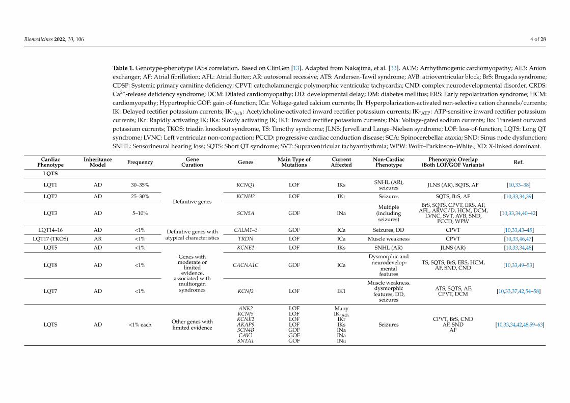
Originally characterized as an AD inheritance syndrome, called Romano–Ward syn-drome (LQT1–6 and LQT9–13), today 17 LQTS-associated genes are known (AKAP9, ANK2,CACNA1C, CALM1, CALM2, CAV3, KCNE1, KCNE2, KCNH2, KCNJ2, KCNJ5, KCNQ1,SCN1B, SCN4B, SCN5A, SNTA1 and TRDN) (Table 1) [29,30]. Most manifest with an ADinheritance pattern, except for the Jervell and Lange–Nielsen syndromes (JLNS) [31] andthe recently characterized triadin knock out syndrome (LQT17) [32], which are inherited inan AR manner. In the most recent evaluation by an expert consensus, only three of thesegenes were considered to have adequate evidence to be classified as definitive for typicalLQTS (KCNQ1, KCNH2, SCN5A) and four genes for LQTS with atypical features (CALM1,CALM2, CALM3, TRDN). The remaining 10 genes, despite being associated with LQTS,were considered to require further evidence for their classification as causals [10].
3.2. Definitive Genes for LQTS
Pathogenic variants in 3 genes (KCNQ1, KCNH2 and SCN5A) are responsible forapproximately 90% of all diagnosed cases of LQTS [98]. With LQT1 (KCNQ1) account-ing for 30–35% of cases, LQT2 (KCNH2) between 25–30% and LQT3 (SCN5A) around5–10% [99,100]. The age of onset of clinical manifestations in each type is variable. LQT1affect predominantly the pediatric population between 5–15 years of age, whereas LQT2and LQT3 occur during puberty or later [34,101]. Even though severe causes of neonatal oreven fetal manifestation have been reported, a similar mechanism for arrhythmogenesis isfound in types 1 and 2. Both syndromes are caused by loss-of-function pathogenic variantsin genes encoding for K+ channels. Disruption of these channels causes a repolarizationdelay with an increase in the AP (phase 3), leading to the QT interval prolongation. Incomparison, in LQT3, this phenotype is caused by gain-of-function pathogenic variantsin the SCN5A gene, which codes for a Na+ channel [102]. Adrenergic stimuli have animportant role as a trigger of symptoms in LQTS types 1 and 2. Physical exercise is themain trigger of SCD in LQT1. A strong sudden startle, loud noise or emotional stress aretriggers of LQT2. Moreover, LQT3 presents with malignant arrhythmias at rest or duringsleep [34].
Biomedicines 2022, 10, 106 4 of 28
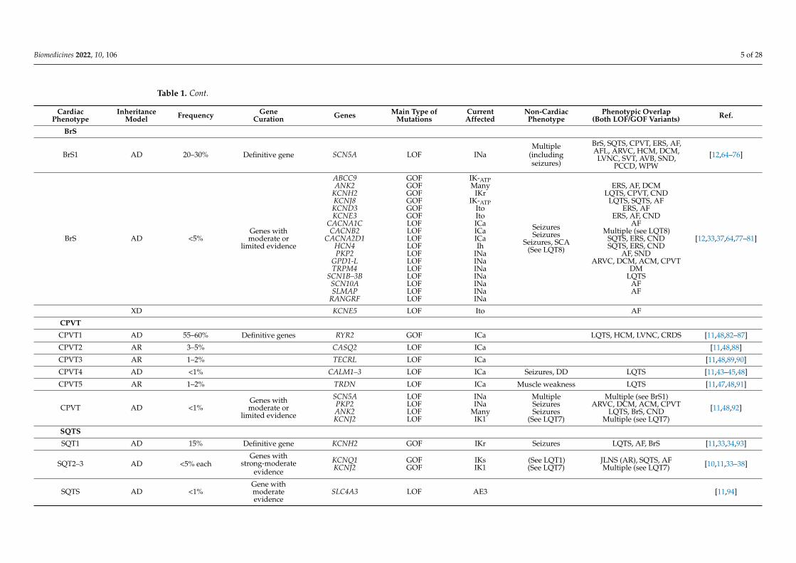
Table 1. Genotype-phenotype IASs correlation. Based on ClinGen [13]. Adapted from Nakajima, et al. [33]. ACM: Arrhythmogenic cardiomyopathy; AE3: Anionexchanger; AF: Atrial fibrillation; AFL: Atrial flutter; AR: autosomal recessive; ATS: Andersen-Tawil syndrome; AVB: atrioventricular block; BrS: Brugada syndrome;CDSP: Systemic primary carnitine deficiency; CPVT: catecholaminergic polymorphic ventricular tachycardia; CND: complex neurodevelopmental disorder; CRDS:Ca2+-release deficiency syndrome; DCM: Dilated cardiomyopathy; DD: developmental delay; DM: diabetes mellitus; ERS: Early repolarization syndrome; HCM:cardiomyopathy; Hypertrophic GOF: gain-of-function; ICa: Voltage-gated calcium currents; Ih: Hyperpolarization-activated non-selective cation channels/currents;IK: Delayed rectifier potassium currents; IK-Ach: Acetylcholine-activated inward rectifier potassium currents; IK-ATP: ATP-sensitive inward rectifier potassiumcurrents; IKr: Rapidly activating IK; IKs: Slowly activating IK; IK1: Inward rectifier potassium currents; INa: Voltage-gated sodium currents; Ito: Transient outwardpotassium currents; TKOS: triadin knockout syndrome, TS: Timothy syndrome; JLNS: Jervell and Lange–Nielsen syndrome; LOF: loss-of-function; LQTS: Long QTsyndrome; LVNC: Left ventricular non-compaction; PCCD: progressive cardiac conduction disease; SCA: Spinocerebellar ataxia; SND: Sinus node dysfunction;SNHL: Sensorineural hearing loss; SQTS: Short QT syndrome; SVT: Supraventricular tachyarrhythmia; WPW: Wolff–Parkinson–White.; XD: X-linked dominant.
CardiacPhenotype
InheritanceModel Frequency Gene
Curation Genes Main Type ofMutations
CurrentAffected
Non-CardiacPhenotype
Phenotypic Overlap(Both LOF/GOF Variants) Ref.
LQTS
LQT1 AD 30–35%
Definitive genes
KCNQ1 LOF IKs SNHL (AR),seizures JLNS (AR), SQTS, AF [10,33–38]
LQT2 AD 25–30% KCNH2 LOF IKr Seizures SQTS, BrS, AF [10,33,34,39]
LQT3 AD 5–10% SCN5A GOF INaMultiple
(includingseizures)
BrS, SQTS, CPVT, ERS, AF,AFL, ARVC/D, HCM, DCM,
LVNC, SVT, AVB, SND,PCCD, WPW
[10,33,34,40–42]
LQT14–16 AD <1% Definitive genes withatypical characteristics
CALM1–3 GOF ICa Seizures, DD CPVT [10,33,43–45]
LQT17 (TKOS) AR <1% TRDN LOF ICa Muscle weakness CPVT [10,33,46,47]
LQT5 AD <1%
Genes withmoderate or
limitedevidence,
associated withmultiorgansyndromes
KCNE1 LOF IKs SNHL (AR) JLNS (AR) [10,33,34,48]
LQT8 AD <1% CACNA1C GOF ICa
Dysmorphic andneurodevelop-
mentalfeatures
TS, SQTS, BrS, ERS, HCM,AF, SND, CND [10,33,49–53]
LQT7 AD <1% KCNJ2 LOF IK1
Muscle weakness,dysmorphicfeatures, DD,
seizures
ATS, SQTS, AF,CPVT, DCM [10,33,37,42,54–58]
LQTS AD <1% each Other genes withlimited evidence
ANK2KCNJ5KCNE2AKAP9SCN4BCAV3
SNTA1
LOFLOFLOFLOFGOFGOFGOF
ManyIK-Ach
IKrIKsINaINaINa
SeizuresCPVT, BrS, CND
AF, SNDAF
[10,33,34,42,48,59–63]
Biomedicines 2022, 10, 106 5 of 28
Table 1. Cont.
CardiacPhenotype
InheritanceModel Frequency Gene
Curation Genes Main Type ofMutations
CurrentAffected
Non-CardiacPhenotype
Phenotypic Overlap(Both LOF/GOF Variants) Ref.
BrS
BrS1 AD 20–30% Definitive gene SCN5A LOF INaMultiple
(includingseizures)
BrS, SQTS, CPVT, ERS, AF,AFL, ARVC, HCM, DCM,
LVNC, SVT, AVB, SND,PCCD, WPW
[12,64–76]
BrS AD <5%Genes withmoderate or
limited evidence
ABCC9ANK2
KCNH2KCNJ8KCND3KCNE3
CACNA1CCACNB2
CACNA2D1HCN4PKP2
GPD1-LTRPM4
SCN1B–3BSCN10ASLMAP
RANGRF
GOFGOFGOFGOFGOFGOFLOFLOFLOFLOFLOFLOFLOFLOFLOFLOFLOF
IK-ATPMany
IKrIK-ATP
ItoItoICaICaICaIh
INaINaINaINaINaINaINa
SeizuresSeizures
Seizures, SCA(See LQT8)
ERS, AF, DCMLQTS, CPVT, CND
LQTS, SQTS, AFERS, AF
ERS, AF, CNDAF
Multiple (see LQT8)SQTS, ERS, CNDSQTS, ERS, CND
AF, SNDARVC, DCM, ACM, CPVT
DMLQTS
AFAF
[12,33,37,64,77–81]
XD KCNE5 LOF Ito AF
CPVT
CPVT1 AD 55–60% Definitive genes RYR2 GOF ICa LQTS, HCM, LVNC, CRDS [11,48,82–87]
CPVT2 AR 3–5% CASQ2 LOF ICa [11,48,88]
CPVT3 AR 1–2% TECRL LOF ICa [11,48,89,90]
CPVT4 AD <1% CALM1–3 LOF ICa Seizures, DD LQTS [11,43–45,48]
CPVT5 AR 1–2% TRDN LOF ICa Muscle weakness LQTS [11,47,48,91]
CPVT AD <1%Genes withmoderate or
limited evidence
SCN5APKP2ANK2KCNJ2
LOFLOFLOFLOF
INaINa
ManyIK1
MultipleSeizuresSeizures
(See LQT7)
Multiple (see BrS1)ARVC, DCM, ACM, CPVT
LQTS, BrS, CNDMultiple (see LQT7)
[11,48,92]
SQTS
SQT1 AD 15% Definitive gene KCNH2 GOF IKr Seizures LQTS, AF, BrS [11,33,34,93]
SQT2–3 AD <5% eachGenes with
strong-moderateevidence
KCNQ1KCNJ2
GOFGOF
IKsIK1
(See LQT1)(See LQT7)
JLNS (AR), SQTS, AFMultiple (see LQT7) [10,11,33–38]
SQTS AD <1%Gene withmoderateevidence
SLC4A3 LOF AE3 [11,94]
Biomedicines 2022, 10, 106 6 of 28
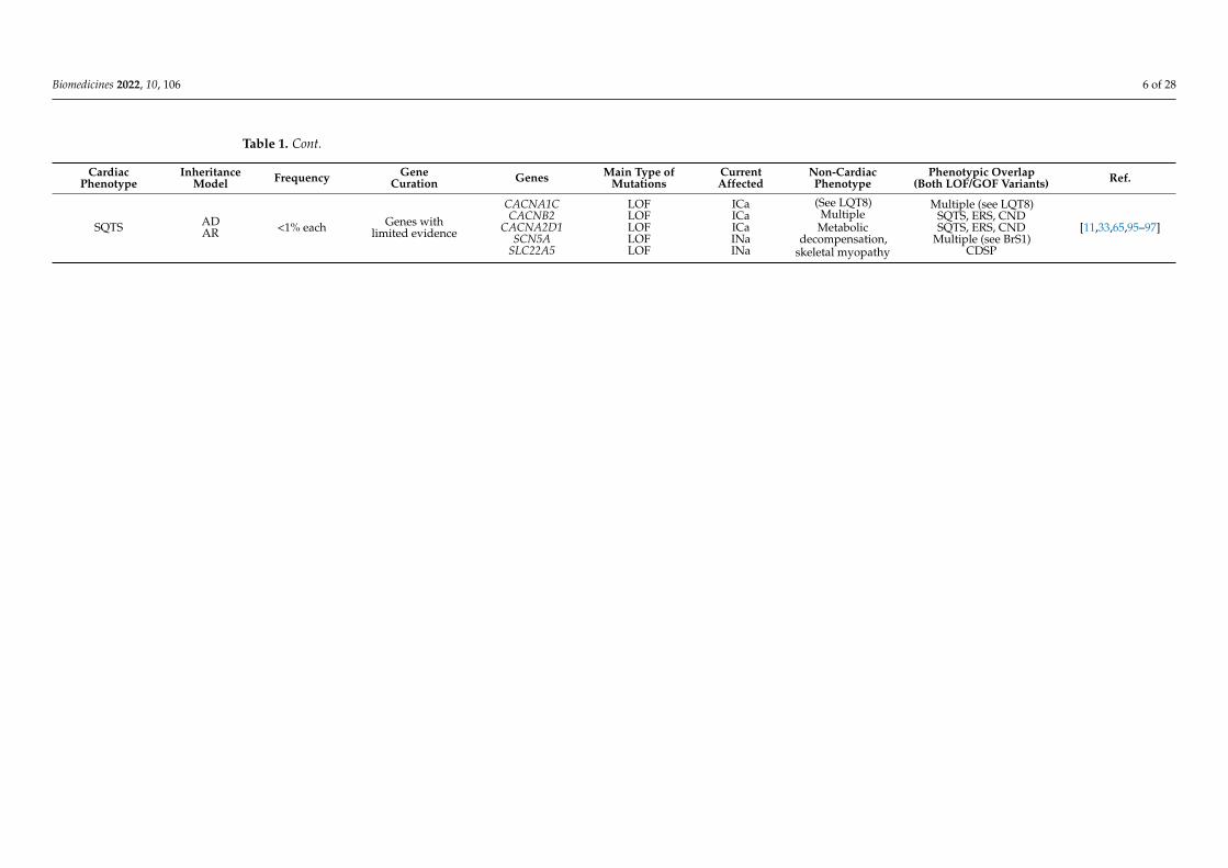
Table 1. Cont.
CardiacPhenotype
InheritanceModel Frequency Gene
Curation Genes Main Type ofMutations
CurrentAffected
Non-CardiacPhenotype
Phenotypic Overlap(Both LOF/GOF Variants) Ref.
SQTS ADAR <1% each Genes with
limited evidence
CACNA1CCACNB2
CACNA2D1SCN5A
SLC22A5
LOFLOFLOFLOFLOF
ICaICaICaINaINa
(See LQT8)Multiple
Metabolicdecompensation,
skeletal myopathy
Multiple (see LQT8)SQTS, ERS, CNDSQTS, ERS, CND
Multiple (see BrS1)CDSP
[11,33,65,95–97]
Biomedicines 2022, 10, 106 7 of 28
3.3. Definitive Genes for LQTS with Atypical Characteristics
Loss-of-function pathogenic variants in the CALM1, CALM2, CALM3 and TRDN genescause LQTS types 14 to 17, respectively. The CALM, CALM2 and CALM3 genes codefor calmodulin, and the TRDN gene for triadin, proteins involved in calcium-dependentprocesses and ion channel regulation [43,46]. LQT14–16 present atypical features, includingseizures and neurodevelopmental delay [103] with symptoms manifesting in infants andyoung children between the ages of 0–5 years old and a high mortality rate [104]. LQT17 ortriadin knockout syndrome (TKOS) also has poor prognosis, and exercise-induced SCDoccurs in children aged 0–3 years old. Almost all patients are symptomatic by the age of10 years old. The observation of negative T waves in the precordial leads is characteristicof LQT17 [47,105].
3.4. Genes with Moderate or Limited Evidence for LQTS
According to the recent evaluation by Adler et al., there is insufficient evidence toclassify 10 of the 17 LQTS-related genes (CACNA1C, AKAP9, ANK2, CAV3, KCNE1, KCNE2,KCNJ2, KCNJ5, SCN4 and SNTA1) as LQTS-causing genes [31983240]. Each of these typesrepresents less than 1% of LQTS. Some of them are associated with phenotypic syndromesand specific ECG features (Table 1). These include Andersen–Tawil syndrome (LQT7,KCNJ2) in which skeletal developmental abnormalities are observed [54]; Timothy syn-drome (LQT8, CACNA1C) with characteristic neurological, facial and limb features [106]and JLNS syndrome, associated with sensorineural deafness (KCNQ1-KCNE1 genes) [107].Although the association of pathogenic variants in these genes with multiorganic syn-dromes is clear, the level of evidence for the specific cardiac phenotype is not so clear, andfurther studies are needed [10].
3.5. Genetic Modifiers and Acquired LQTS
GWAS studies have identified polymorphisms associated with increased risk of trig-gering LQTS [108,109]. Common variants in the NOS1AP (Nitric Oxide Synthase 1 AdaptorProtein) gene confer an increased risk of SCD in patients with LQT1 [110]. Likewise, somevariants have a protective effect, as is the case of p.H558R (SCN5A), which reduces thepathogenic effect of other pathogenic variants, producing a less severe phenotype [22].The common variants p.D85N (KCNE1) and T8A-MiRP1 (KCNE2) in Caucasians [111,112]and p.S1103Y (SCN5A) in African Americans [113], confer risk in the presence of othertriggers such as drugs, a phenotype known as acquired LQTS (aLQTS). These variantsare insufficient by themselves to cause LQTS in the absence of other interval-prolongingfactors [10,114]. However, they are of major importance due to the high frequency ofaLQTS [115].
3.6. Diagnosis
According to the 2015 ESC Guideline, LQTS is diagnosed when the patient presentsone of the following criteria: a corrected QT (QTc) ≥480 ms (repeated 12-lead ECGs), aLQTS risk score >3, or when a pathogenic alteration in one of the LQTS-causing genesis identified [116]. The LQTS risk score combines the altered ECG parameters with thepatient’s clinical and family history and is based on the clinical score proposed by Schwartzin 1993 [117]. Regardless, in the presence of unexplained syncope a QTc ≥460 ms issufficient to diagnose LQTS [116] [26318695]. LQTS presents cardiac and extracardiacphenotypic features, as well as ECG characteristics that allow its classification, although25% of individuals with positive genetics show a normal baseline ECG [118].
3.7. Risk Stratification
Multiple factors are known to raise the likelihood of SCD in patients with LQTS. Thepresence of a QTc >500 ms is the strongest of these predictors [119,120]. In children, thisparameter can be modified by the patient’s age, sex and genotype, with a critical transitionperiod between 12–14 years of age [121]. Patients who have suffered syncope during
Biomedicines 2022, 10, 106 8 of 28
childhood have an increased risk of recurrent episodes, which can be reduced through theuse of beta-blockers (BB) and/or implantable cardiac defibrillators (ICD) [122,123]. Amongthe types, LQT3 has a worse prognosis and the first presentation is usually SCD [119,124].Likewise, LQT14–16 types have been associated with a very severe phenotype in infantsand poor response to available therapies [103]. Women with LQT2 have an increased riskof SCD in the first 6 months postpartum, suggesting a potential hormonal effect [125].Pathogenic variants type and location, as well as other additive genetic factors may increasethe risk of SCD. For example, female adults with LQT2 run a higher risk of SCD than males.However, when missense pathogenic variants in the KCNH2 gene are in the pore loopregions, males are at a higher risk of SCD than their female counterparts [126]. Indeed,pathogenic variants in these regions have been found to be associated with QT intervalprolongation and the development of TdP during fever, suggesting that fever may be apotential trigger of arrhythmias in patients with LQT2 [127].
3.8. Genetic Counselling
LQTS has incomplete penetrance and variable expressivity, even in the same family.The penetrance is estimated to be about 40%, a range that can vary depending on genotype,pathogenic variants type and location, age and sex, among other things [22,128]. Theidentification of a pathogenic variant in one of the LQTS-associated genes (Table 1) isimportant to establish a differential diagnosis of patients and their relatives. In LQTS,treatment and risk stratification differ depending on the gene causing the disease, andcan be variant-specific [129]. According to guideline recommendations, only genes withdefinitive evidence for LQTS (KCNQ1, KCNH2 and SCN5A) should be routinely used inthe evaluation of patients and their families. In patients with clinical findings consistentwith the phenotypic expression demonstrated in LQTS with atypical features, relatedgenes (CALM1, CALM2, CALM3 and TRDN) should also be tested [10]. Genetic testing canidentify an LQTS-causing alteration in 70–80% of cases [99]. The proportion of LQTS causedby de novo pathogenic variants is difficult to estimate, but is expected to be low [130].Between 5–9% of familial cases of LQTS have two or more pathogenic variants (biallelicor digenic), which have been associated with a more severe phenotype [131,132]. Thecoexistence of two or more pathogenic variants could explain the variable expressivityobserved in some families [133]. The copy number variants (CNV) detection rate amongLQTS families is not very clear, but is estimated to be between 2–11% [134–138]. Populationscreening by ECG has been promoted to identify individuals at risk of LQTS, which has beensuccessful in reducing SCD rates among patient family members [139] neonates [27,140]and athletes [141,142].
3.9. Management and Treatment
In children with LQTS the first approach is to avoid genotype-specific triggers, such ascompetitive sports, especially swimming in LQT1, and exposure to loud noise in childrenwith LQT2 as well as the avoidance of the QT interval prolonging drugs in all carriers ofLQTS-associated variants (http://crediblemeds.org/ accessed on 3 December 2021) [116].Long-acting BB (nadolol) are recommended in all types, including asymptomatic geneticcarriers [143,144], as their use decreases the risk of SCD [145]. Specific treatment of LQT3with mexiletine and/or flecainide has proven to be highly effective. In LQT1 the use of BB isvery effective, and some authors suggest that it is not necessary to place an ICD in patientsat low risk of SCD (asymptomatic prepubertal girls and adults >20 years with normalECG) [146]. Left-cardiac sympathetic denervation is indicated in patients with LQT1 orwhen BB therapy is contraindicated or badly tolerated [147]. ICDs are used in patientsat high risk of SCD despite previous therapies, in those who have previously presentedsyncope while taking BB, and effective LCSD (left cardiac sympathetic denervation) hasbeen performed [148]. Despite its efficacy, ICD has a high economic cost and can presentnumerous complications. Its use in the pediatric population should be assessed on acase-by-case basis by specialists [149].
Biomedicines 2022, 10, 106 9 of 28
4. Brugada Syndrome
BrS in children and young adults is rare, and its incidence rate and clinical implicationsremain unclear. In the general population, its prevalence is estimated to be between 1 in2000–5000 person-years [150]. The syndrome has a higher prevalence in Southeast Asiancountries [151], and is more frequent among males than females [152]. It is characterizedby a right bundle branch block, a very sharp T wave and spontaneous or drug-induced ST-segment elevation (J point) in the right precordial leads (V1–V3), known as ‘type-1’ BrS ECGpattern [64]. Clinical manifestations may appear between the ages of 2 months and 77 yearsold, but the mean age of presentation is 40 years old [153]. Symptoms present at rest, duringsleep or febrile episodes, including nocturnal agonal respirations, palpitations, seizures, andpolymorphic ventricular tachycardia (PVT) or VF. Most individuals remain asymptomatic,although SCD occurs in 17–42% of the cases and can be the initial presentation [154].
4.1. Genetics
Genetically described as a Mendelian syndrome with an autosomal dominant inheri-tance pattern and incomplete penetrance [155], recent evidence suggests that BrS may be anoligogenic disease, involving several genetic factors [156]. However, the lack of conclusivedata on these genetic alterations leads it to remain classified as a monogenic syndrome [157].To date, more than 20 genes have been associated with BrS (ABCC9, ANK2, CACNA1C,CACNA2D1, CACNB2, GPD1-L, HCN4, KCND3, KCNE3, KCNE5, KCNH2, KCNJ8, PKP2,RANGRF, SCN10A, SCN1B, SCN5A, SCN2B, SLMAP and TRPM4), reappraisal of thesegenes by Hosseini et al., established that only SCN5A had definitive evidence of being acausal gene [12] and its genetic analysis is the only one recommended by current guide-lines [23,158].
4.2. Definitive Gene for BrS
Loss-of-function pathogenic variants in the SCN5A gene account for approximately30% of genetically positive BrS cases. This gene encodes the alpha subunit of the cardiacsodium channel Nav1.5, responsible for phase 0 of the AP. Inactivation of the channelleads to a delay in ventricular polarization, resulting in the development of ventriculartachycardia and fibrillation (VT/VF) [150].
4.3. BrS2–12 and Other Susceptibility Genes with Limited Evidence
Pathogenic variants associated with BrS2–12, genes (GPD1-L, CACNA1C, CACNB2,SCN1B, KCNE3, SCN3B, HCN4, KCND3, KCNJ8, CACNA2D1 and MOG1) together representless than 5% of all diagnosed cases [159]. Over the past few years, other genes havebeen suggested as possible causes of BrS (ABCC9, ANK2, FGF12, HEY2, KCND2, KCNH2,KCNE5, LRRC10, SEMA3A, PKP2, RANGRF, SCN10A, SCN2B, SLMAP and TRPM4), butno comprehensive clinical and cellular studies have confirmed this association [157]. Allof them follow an AD inheritance pattern, except for the KCNE5 gene, which follows anX-linked dominant pattern [160,161].
4.4. Diagnosis
According to the 2015 ESC guidelines, BrS is diagnosed in patients with a ‘type 1’ECG pattern, ST-segment elevation ≥2 mm (J-point) in one or more of the right precordialleads (V1–V3) [116]. This ECG pattern may occur spontaneously or be unmasked by aprovocation test with a class Ic drug (sodium channel blockers such as ajmaline, flecainide,procainamide, or pilsicainide), in this case additional clinical criteria are required fordiagnosis [64]. Fever is a trigger for ventricular arrhythmias in patients with BrS and mayunmask the characteristic ECG pattern, especially in children under 5 years of age [162].A 12-lead ECG is recommended during febrile episodes in children with a family historyof BrS and in all children with febrile seizures [163]. Initially presumed not to have anystructural abnormalities, postmortem histological studies and endomyocardial biopsieshave shown changes at the tissular and molecular level in patients with BrS. These changes
Biomedicines 2022, 10, 106 10 of 28
include localized electroanatomical and structural abnormalities in the right ventricularoutflow tract (RVOT), fibrosis, fatty infiltration, increased epicardial collagen, and decreasedexpression of Connexin 43 at right ventricular gap junctions [164–166].
4.5. Risk Stratification
Despite advances in risk stratification of IAS, in BrS it remains challenging. The mostimportant risk marker is the presentation of a previous arrhythmogenic event (AE, AF,syncope, or SCD), which increases the likelihood of SCD in both young and adults [167,168].The age of onset is a notable prognostic marker. For instance, although BrS is uncommon inchildren, they present with a more severe form of the disease [169]. Gender is an importantrisk factor, with males being up to 5–8 times more affected than females, presenting a moresevere phenotype, earlier symptom debut and a higher number of events [152,170]. Thesegender differences have not been observed in children under 12 years old [167]. Familyhistory of SCD and the presence of pathogenic variants in SCN5A could also be predictorsof high risk in adults and adolescents, [167] even though their role in risk stratification isstill controversial [171,172]. Other risk factors observed are spontaneous variation of the‘type 1’ ECG pattern and fragmentation of the QRS complex [171,173], nevertheless furtherstudies are required to confirm this association.
4.6. Management and Treatment
In children with BrS or a family history, BrS-inducing drugs should be avoided(http://www.brugadadrugs.org accessed on 3 December 2021). Appropriate treatment ofany fever with antipyretic drugs should be provided [167]. ICD implantation is the onlytreatment that reduces the risk of SCD in BrS. It is indicated in all patients resuscitatedfrom arrhythmic syncope, those with documented VT or VF or with a spontaneous ‘type 1’ECG pattern [116,149]. In patients with electrical storms, administration of isoproterenolis recommended [174,175]. Quinidine is recommended for patients who refuse ICD im-plantation or in those who, despite having an ICD, still have a high risk of SCD [176,177].Catheter ablation is useful in high-risk patients with a history of electrical storms or re-peated appropriate ICD shocks [178]. ICD implantation in asymptomatic individuals witha ‘type 1’ ECG pattern for primary prevention, including children, remains controversialand is a challenge for specialists who have to handle it on a case-by-case basis [149,177].
4.7. Genetic Counseling
BrS penetrance is highly variable in the different published studies. It is estimatedto range from 12.5% to 50% [22,179]. About 70–80% of families with BrS do not have agenetic diagnosis. However, although the results of genetic screening do not currentlyinfluence prognosis or treatment, genetic testing should be performed in all first-degreerelatives if the index case tested positive [99,116]. Moreover, the ECG pattern of BrS‘type 1’ is uncommon in children and genetic testing may help with their diagnosis [21]. InBrS families counseling should also include an ECG, because negative-genotype positive-phenotype cases are not uncommon [180]. The proportion of cases caused by de novopathogenic variants is estimated at 1% and the number of cases with a CNV variant isapproximately 1.3–2.9% [138,181]. ECG screening using a provocation test for BrS detectionis controversial [182,183], but should be performed when an abnormal ECG is found.ECG screening has benefits in the prevention of SCD in neonates [140] and the youngpopulation [184].
5. Short QT Syndrome
SQTS is an extremely rare inherited disease associated with SCD. To date, less than200 cases have been reported worldwide [93]. The estimated prevalence varies between0.18–2.9%, with a higher incidence in males than females [25]. The incidence rate can evenbe lower (0.02–0.10%) if more restrictive values are considered for its diagnosis [185,186].While prevalence of the syndrome in children and adolescents is low (about 0.05%), early
Biomedicines 2022, 10, 106 11 of 28
detection is important, as it is potentially lethal for all age groups. It is characterizedby a short QT interval on the ECG (<330 ms), with an asymmetric and peaked T wave.Symptoms occur mostly in men between the ages of 14 and 40 and may be favored byhormonal causes [187]. Cardiac events usually occur in adrenergic situations (noise orexercise), although it can also occur at rest. Clinical presentation includes ventricularrepolarization abnormalities (AF and VT) and syncope. The probability of presenting SCDas the first symptom increases with age, reaching 41% at the age of 40 [188]. Currently,approximately 40% of cases remain asymptomatic [189].
5.1. Genetics
At present, nine genes have been associated with SQTS (CACNA1C, CACNA2D1,CACNB2, KCNH2, KCNJ2 and KCNQ1, SLC22A5, SLC4A3 and SCN5A) [190]. Evaluation ofthese genes, by Walsh et al., showed that only the KCNH2 gene had definitive evidence forSQTS causality. Three other genes (KCNQ1, KCNJ2, SLC4A3) presented strong to moderateevidence. Causality of the other SQTS-associated genes remains still in dispute [11]. Thesedata are consistent with the findings published by Campuzano et al., who found thatall variants with a conclusive pathogenic role in SQTS clustered in three genes (KCNQ1,KCNH2 and KCNJ2). In that study, the SLC4A3 gene was excluded, since carriers were in agray zone of SQTS diagnosis (with a QTc ≤370 ms) [190].
5.2. SQT1 Definitive Gene: KCNH2
The p.T618I and p.N588K pathogenic variants in the KCNH2 gene are the most frequentassociated with SQTS, accounting for 85% of SQT1 and 55% of all genetically identifiedcases of SQTS [189]. Gain-of-function pathogenic variants in KCNH2 lead to prolonged K+
channel activation and accelerated cardiac repolarization with shorter refractory periods,potentially triggering life-threatening supraventricular and ventricular arrhythmias [189].
5.3. Genes with Strong or Moderate Evidence for SQTS
SQT2 and SQT3 are driven by gain-of-function pathogenic variants in genes encodingfor K+ channels (KCNQ1 and KCNJ2, respectively). The mechanism of arrhythmogenicityis similar to that presented by KCNH2. The SLC4A3 gene, recently associated with SQTS,presents an uncommon mechanism for the development of malignant arrhythmia. SLC4A3encodes the plasma membrane anion exchange protein 3 (AE3) and acts by mediating partof the Cl−/HCO3− exchange in cardiac myocytes. Loss-of-function pathogenic variants inthe SLC4A3 gene would cause an increase in pHi and a decrease in [Cl−]i, shortening theduration of the AP [94].
5.4. Diagnosis
According to the 2015 ESC guidelines, SQTS is diagnosed by the presence of aQTc ≤340 ms, or ≤360 ms when one the following clinical criteria occur: the detection of aknown pathogenic alteration, a family history of SQTS, a family history of SCD before theage of 40 years, or reanimated cardiac arrest with a structurally normal heart [116].
5.5. Risk Stratification
On account of the limited number of patients with SQTS and the phenotypic variabilityof the syndrome, risk stratification currently represents a challenge. To date, the onlypredictor of SCD found in patients with SQTS is a history of cardiac arrest [93]. Genotype-phenotype correlation studies have found that SQTS1 manifests at an older age and patientshave a shorter QTc than other patients with SQTS. Nevertheless, no association of thisreduction with an increased risk of SCD has been found [191].
5.6. Management and Treatment
ICD implantation is recommended for all patients with SQTS, especially for patientswho have survived an aborted cardiac arrest or have presented spontaneous sustained
Biomedicines 2022, 10, 106 12 of 28
VT [186,192]. QT interval prolonging drugs (quinidine and sotalol) should be consideredfor all patients at risk of SQTS in both, asymptomatic and symptomatic patients who don’thave an ICD, especially young children [186].
5.7. Genetic Counselling
Due to the low number of cases, the penetrance of SQTS is difficult to estimate.However, pathogenic variants with a penetrance of 100% have been reported [189]. Thediagnostic yield of genetic testing in SQTS is low (<25%) [186]. Current guidelines recom-mend analysis of five genes: KCNH2, KCNQ1, KCNJ2, KCNJ2, CACNA1C and CACNB2 inthe diagnosis of SQTS [116], with the KCNH2 gene as the most cost-effective option [193].Familial genetic analysis is recommended, both to clarify the pathogenic role of newlyidentified variants and to identify family members at risk for SCD. To date, there is nopublished data on CNV analysis on patients with SQTS. De novo variants in the KCNQ1gene have been associated with a particular in utero phenotype with clinical diagnosisof AF with concomitant bradycardia and short QT interval [194,195]. Some researcherssupport the screening of SQTS in the pediatric population, given its high lethality and thebenefits of early diagnosis in the prevention of SCD. These studies have shown that thediagnostic criteria for QTc should be adjusted in each population based on factors includingsex and age, to avoid false positives [196–199].
6. Catecholaminergic Polymorphic Ventricular Tachycardia
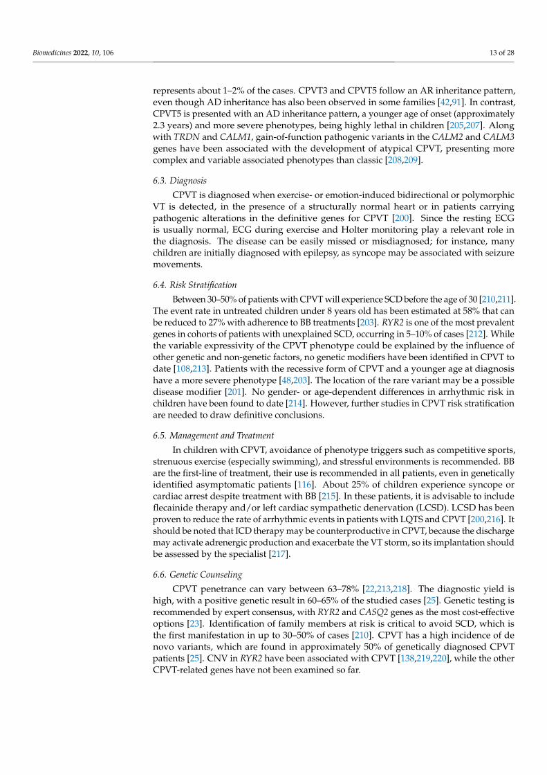
The prevalence of CPVT is estimated to be 1 per 10,000 population. However, thereal prevalence is uncertain, as it might be underestimated due to its high lethality at ayoung age and difficulty in diagnosis [25,200]. CPVT is characterized by a bidirectionalpolymorphic VT, triggered by an adrenergic stimulus mainly during exertion, extremestress or emotion that can lead to syncope and SCD. Syncopal episodes are increasinglyreported during “awake rest” possibly due to anxiety, stress, or other psychological stimuliunrelated to exertion [201]. The age of onset can range from infancy to the age of 30,although it is more common in children aged 7–10 years old [202]. By the age of 10, about35% of patients are symptomatic, increasing to 72% by the age of 21 [203]. A younger ageof debut is often accompanied by more severe phenotypes and increased risk of SCD [42].
6.1. Genetics
According to the recent evaluation by Walsh et al., seven genes were classified ascausing of CPVT with definite to moderate evidence. Four of them present an AD inheri-tance pattern (RYR2, CALM1, CALM2, CALM3) and three AR inheritance (CASQ2, TRDN,TECRL). Three genes (KCNJ2, PKP2, SCN5A) were reported for phenotypes that were notrepresentative of CPVT, while the reported variants in the ANK2 gene were considered toocommon in the population to be disease-causing (Table 1) [11].
6.2. Definitive Genes for CPVT
CPVT1 is the most prevalent variant, accounting for more than 60% of all geneticallydiagnosed cases of CPVT [204]. CPVT1 with a high incidence in children around 10 yearsof age [205]. Thus, it is caused by pathogenic variants in the RYR2 gene, which encodes forryanodine receptor 2, responsible for calcium regulation in the cardiomyocyte. A majorityof pathogenic variants in the RYR2 gene are gain-of-function, which promote an increasedCa2+ release from the sarcoplasmic reticulum of cardiomyocytes into the cytoplasm leadingto late after-depolarizations [48]. The remaining types of CPVT represent about 10% ofcases. Most are caused by loss-of-function pathogenic variants in genes encoding proteinsinvolved in the storage and release of Ca2+ in the sarcoplasmic reticulum [206]. CPVT2is caused by homozygous or compound heterozygous pathogenic variants in the CASQ2gene, following an AR inheritance model. CPVT2 accounts for about 3–5% of cases [203],affecting mainly children around the age of 7 years [205]. Pathogenic variants in TRDN,CALM1 and TECRL are responsible for CPVT3, CPVT4, and CPVT5, respectively. Each
Biomedicines 2022, 10, 106 13 of 28
represents about 1–2% of the cases. CPVT3 and CPVT5 follow an AR inheritance pattern,even though AD inheritance has also been observed in some families [42,91]. In contrast,CPVT5 is presented with an AD inheritance pattern, a younger age of onset (approximately2.3 years) and more severe phenotypes, being highly lethal in children [205,207]. Alongwith TRDN and CALM1, gain-of-function pathogenic variants in the CALM2 and CALM3genes have been associated with the development of atypical CPVT, presenting morecomplex and variable associated phenotypes than classic [208,209].
6.3. Diagnosis
CPVT is diagnosed when exercise- or emotion-induced bidirectional or polymorphicVT is detected, in the presence of a structurally normal heart or in patients carryingpathogenic alterations in the definitive genes for CPVT [200]. Since the resting ECGis usually normal, ECG during exercise and Holter monitoring play a relevant role inthe diagnosis. The disease can be easily missed or misdiagnosed; for instance, manychildren are initially diagnosed with epilepsy, as syncope may be associated with seizuremovements.
6.4. Risk Stratification
Between 30–50% of patients with CPVT will experience SCD before the age of 30 [210,211].The event rate in untreated children under 8 years old has been estimated at 58% that canbe reduced to 27% with adherence to BB treatments [203]. RYR2 is one of the most prevalentgenes in cohorts of patients with unexplained SCD, occurring in 5–10% of cases [212]. Whilethe variable expressivity of the CPVT phenotype could be explained by the influence ofother genetic and non-genetic factors, no genetic modifiers have been identified in CPVT todate [108,213]. Patients with the recessive form of CPVT and a younger age at diagnosishave a more severe phenotype [48,203]. The location of the rare variant may be a possibledisease modifier [201]. No gender- or age-dependent differences in arrhythmic risk inchildren have been found to date [214]. However, further studies in CPVT risk stratificationare needed to draw definitive conclusions.
6.5. Management and Treatment
In children with CPVT, avoidance of phenotype triggers such as competitive sports,strenuous exercise (especially swimming), and stressful environments is recommended. BBare the first-line of treatment, their use is recommended in all patients, even in geneticallyidentified asymptomatic patients [116]. About 25% of children experience syncope orcardiac arrest despite treatment with BB [215]. In these patients, it is advisable to includeflecainide therapy and/or left cardiac sympathetic denervation (LCSD). LCSD has beenproven to reduce the rate of arrhythmic events in patients with LQTS and CPVT [200,216]. Itshould be noted that ICD therapy may be counterproductive in CPVT, because the dischargemay activate adrenergic production and exacerbate the VT storm, so its implantation shouldbe assessed by the specialist [217].
6.6. Genetic Counseling
CPVT penetrance can vary between 63–78% [22,213,218]. The diagnostic yield ishigh, with a positive genetic result in 60–65% of the studied cases [25]. Genetic testing isrecommended by expert consensus, with RYR2 and CASQ2 genes as the most cost-effectiveoptions [23]. Identification of family members at risk is critical to avoid SCD, which isthe first manifestation in up to 30–50% of cases [210]. CPVT has a high incidence of denovo variants, which are found in approximately 50% of genetically diagnosed CPVTpatients [25]. CNV in RYR2 have been associated with CPVT [138,219,220], while the otherCPVT-related genes have not been examined so far.
Biomedicines 2022, 10, 106 14 of 28
7. Genetic Overlap
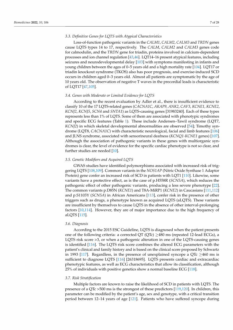
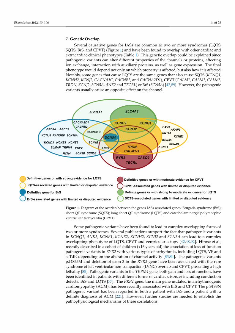
Several causative genes for IASs are common to two or more syndromes (LQTS,SQTS, BrS, and CPVT) (Figure 1) and have been found to overlap with other cardiac andextracardiac clinical phenotypes (Table 1). This genetic overlap could be explained sincepathogenic variants can alter different properties of the channels or proteins, affectingion exchange, interaction with auxiliary proteins, as well as gene expression. The finalphenotype would depend not only on which property is affected, but also how it is affected.Notably, some genes that cause LQTS are the same genes that also cause SQTS (KCNQ1,KCNH2, KCNJ2, CACNA1C, CACNB2, and CACNA2D1), CPVT (CALM1, CALM2, CALM3,TRDN, KCNJ2, SCN5A, ANK2 and TECRL) or BrS (SCN5A) [42,89]. However, the pathogenicvariants usually cause an opposite effect on the channel.
Biomedicines 2022, 10, x FOR PEER REVIEW 15 of 30
Figure 1. Diagram of the overlap between the genes IASs-associated genes: Brugada syndrome
(BrS); short QT syndrome (SQTS); long short QT syndrome (LQTS) and catecholaminergic polymor-
phic ventricular tachycardia (CPVT).
Some pathogenic variants have been found to lead to complex overlapping forms of
two or more syndromes. Several publications support the fact that pathogenic variants in
KCNQ1, ANK2, KCNE1, KCNE2, KCNH2, KCNJ2 and SCN5A can lead to a complex over-
lapping phenotype of LQTS, CPVT and ventricular ectopy [42,48,92]. Hirose et al., recently
described in a cohort of children (<16 years old) the association of loss-of-function patho-
genic variants in RYR2 with various types of arrhythmia, including LQTS, VF and scTdP,
depending on the alteration of channel activity [83,84]. The pathogenic variants p.I4855M
and deletion of exon 3 in the RYR2 gene have been associated with the rare syndrome of
left ventricular non-compaction (LVNC) overlap and CPVT, presenting a high lethality
[85]. Pathogenic variants in the TRPM4 gene, both gain and loss of function, have been
identified in patients with different forms of cardiac disorder including conduction de-
fects, BrS and LQTS [77]. The PKP2 gene, the main gene mutated in arrhythmogenic car-
diomyopathy (ACM), has been recently associated with BrS and CPVT. The p.S183N path-
ogenic variant has been reported in both a patient with BrS and a patient with a definite
diagnosis of ACM [221]. However, further studies are needed to establish the pathophys-
iological mechanisms of these correlations.
7.1. SCN5A Clinical Overlap
Pathogenic variants affecting SCN5A have been found in all major IAS, as well as in
other associated cardiac phenotypes (Figure 2). Gain-of-function pathogenic variants in
SCN5A (LQT3), have been associated with other arrhythmias including multifocal ectopic
Purkinje-related premature contractions [222,223] and atypical CPVT-like phenotype
[224]. For instance, p.T1857I and p.I141V variants, have been associated with tach-
yarrhythmias and exercise-induced polymorphic ventricular arrhythmia [224,225]. How-
ever, the correlation between SCN5A pathogenic variants and CPVT remains debated.
Meanwhile, loss-of-function pathogenic variants in SCN5A (usually associated with BrS)
have also been implicated in certain phenotypes including isolated cardiac conduction
Figure 1. Diagram of the overlap between the genes IASs-associated genes: Brugada syndrome (BrS);short QT syndrome (SQTS); long short QT syndrome (LQTS) and catecholaminergic polymorphicventricular tachycardia (CPVT).
Some pathogenic variants have been found to lead to complex overlapping forms oftwo or more syndromes. Several publications support the fact that pathogenic variantsin KCNQ1, ANK2, KCNE1, KCNE2, KCNH2, KCNJ2 and SCN5A can lead to a complexoverlapping phenotype of LQTS, CPVT and ventricular ectopy [42,48,92]. Hirose et al.,recently described in a cohort of children (<16 years old) the association of loss-of-functionpathogenic variants in RYR2 with various types of arrhythmia, including LQTS, VF andscTdP, depending on the alteration of channel activity [83,84]. The pathogenic variantsp.I4855M and deletion of exon 3 in the RYR2 gene have been associated with the raresyndrome of left ventricular non-compaction (LVNC) overlap and CPVT, presenting a highlethality [85]. Pathogenic variants in the TRPM4 gene, both gain and loss of function, havebeen identified in patients with different forms of cardiac disorder including conductiondefects, BrS and LQTS [77]. The PKP2 gene, the main gene mutated in arrhythmogeniccardiomyopathy (ACM), has been recently associated with BrS and CPVT. The p.S183Npathogenic variant has been reported in both a patient with BrS and a patient with adefinite diagnosis of ACM [221]. However, further studies are needed to establish thepathophysiological mechanisms of these correlations.
Biomedicines 2022, 10, 106 15 of 28
7.1. SCN5A Clinical Overlap
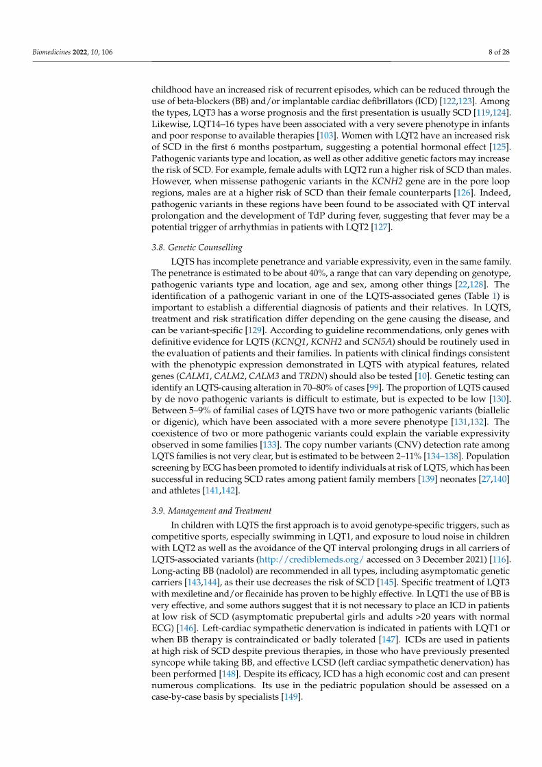
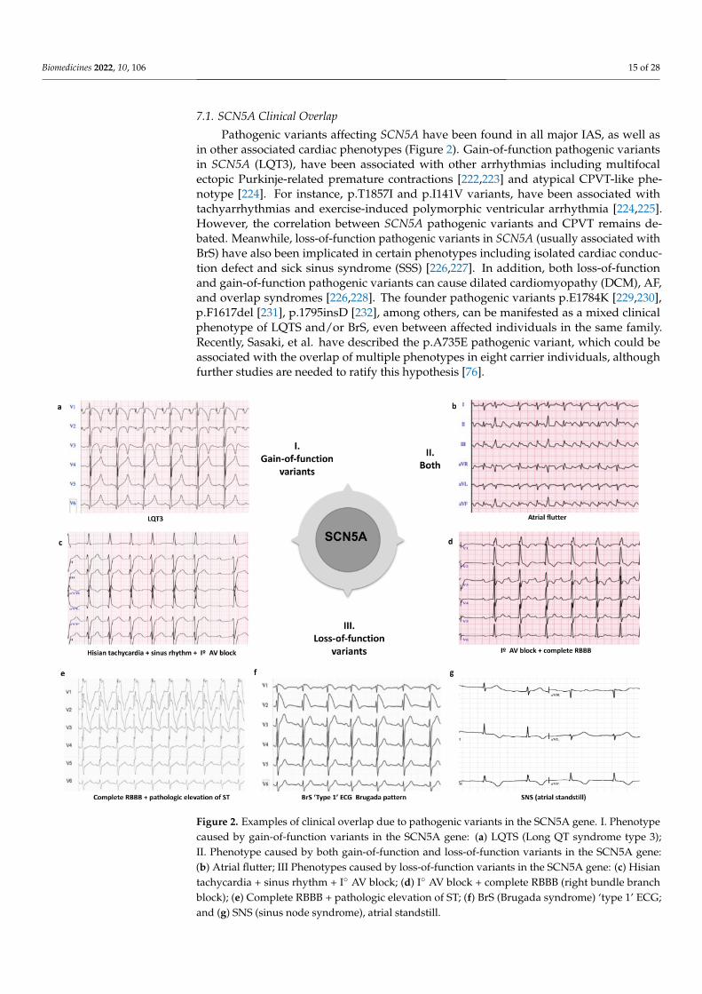
Pathogenic variants affecting SCN5A have been found in all major IAS, as well asin other associated cardiac phenotypes (Figure 2). Gain-of-function pathogenic variantsin SCN5A (LQT3), have been associated with other arrhythmias including multifocalectopic Purkinje-related premature contractions [222,223] and atypical CPVT-like phe-notype [224]. For instance, p.T1857I and p.I141V variants, have been associated withtachyarrhythmias and exercise-induced polymorphic ventricular arrhythmia [224,225].However, the correlation between SCN5A pathogenic variants and CPVT remains de-bated. Meanwhile, loss-of-function pathogenic variants in SCN5A (usually associated withBrS) have also been implicated in certain phenotypes including isolated cardiac conduc-tion defect and sick sinus syndrome (SSS) [226,227]. In addition, both loss-of-functionand gain-of-function pathogenic variants can cause dilated cardiomyopathy (DCM), AF,and overlap syndromes [226,228]. The founder pathogenic variants p.E1784K [229,230],p.F1617del [231], p.1795insD [232], among others, can be manifested as a mixed clinicalphenotype of LQTS and/or BrS, even between affected individuals in the same family.Recently, Sasaki, et al. have described the p.A735E pathogenic variant, which could beassociated with the overlap of multiple phenotypes in eight carrier individuals, althoughfurther studies are needed to ratify this hypothesis [76].
Figure 2. Examples of clinical overlap due to pathogenic variants in the SCN5A gene. I. Phenotypecaused by gain-of-function variants in the SCN5A gene: (a) LQTS (Long QT syndrome type 3);II. Phenotype caused by both gain-of-function and loss-of-function variants in the SCN5A gene:(b) Atrial flutter; III Phenotypes caused by loss-of-function variants in the SCN5A gene: (c) Hisiantachycardia + sinus rhythm + I◦ AV block; (d) I◦ AV block + complete RBBB (right bundle branchblock); (e) Complete RBBB + pathologic elevation of ST; (f) BrS (Brugada syndrome) ‘type 1’ ECG;and (g) SNS (sinus node syndrome), atrial standstill.
Biomedicines 2022, 10, 106 16 of 28
7.2. Genetic Overlap of Arrhythmogenic Phenotypes and Epilepsy
Recently, evidence for genetic overlap between IASs and epilepsy has been reported.Sudden unexpected death in epilepsy (SUDEP) share many features with SADS in theyoung and may have a similar genetic contribution [233,234]. In a systematic review, Anwarand Chahal, et al., found that 11% of the most frequent pathogenic variants identified bymolecular autopsy in SUDEP were found in genes related to cardiac channelopathies [235].
7.3. Non-Genetic Phenotype Overlapping
Multiple phenotypic overlaps between IASs have been described, in which no un-derlying genetic cause has been observed ERS and BrS [66,236], BrS and CA [237], ACMand BrS [238]. The overlap of ACM and BrS is controversial, since their diagnostic criteriaexclude the coexistence of both syndromes in the same individual. This overlap wouldnot be fully explained by a genetic overlap. The ECG pattern of BrS (drug-induced) wasobserved both in patients with ACM and pathogenic variants in the SCN5A gene [239] aswell as in patients without SCN5A pathogenic variants [240,241]. Therefore, it is possiblethat genetic (coding or non-coding variants) and non-genetic (demographic variables orexogenous factors) modifiers could be involved in this variability, all of them contributingadditively to the expression of the phenotype. An alternative explanation for phenotypicoverlap could be misdiagnosis due to the use of drugs in ECG testing. Notably, it has beenshown that flecainide can induce ST-segment elevation in ECG in patients with LQTS3, andlead to misdiagnosis in some cases [242].
8. How to Deal with the Variants of Uncertain Significance in Inherited ArrhythmiaSyndromes (IASs)
The classification of VUS remains a current challenge in genetic field. When functionaland segregation studies are not feasible due to the rarity and exclusivity of some variants,population allele frequency (https://gnomad.broadinstitute.org/ accessed on 3 Decem-ber 2021) and familial segregation are a fundamental tool for variant classification [132].Continued reclassification of rare variants based on the ACMG-AMP criteria has led to anincreasing understanding of the potential impact of a variant on a disease; for example,it has been shown that rare variants previously described as pathogenic may have toohigh a population frequency to be responsible for IASs, and that a large number of rarevariants may be benign. On the other hand, as mentioned, the ACMG/AMP guidelinespresent limitations for their application in the classification of rare variants in IASs and theircriteria need to be adjusted. To date, few validations have been performed with adjustedcriteria. The reasons are probably due to the difficulty of establishing valid and homoge-neous criteria that can be generally applied to such heterogeneous diseases such as IAS.Assigning erroneous classifications to variants carries great danger, both for false positives(assigning pathogenic causality to variants that are not) that can have severe consequences,for example leading to the implantation of an unnecessary ICD or, on the contrary, leavingas VUS variants those that are truly causative of the disease. Nevertheless, working withcriteria adjusted to each disease could be a useful approach to achieve greater success inthe classification of genetic variants in IASs and decrease the number of cases without aconclusive genetic diagnosis. In the recent validation of variants in LQTS and BrS by Walshet al. they developed a quantitative implementation (disease-specific) of the ACMG-AMPguidelines following the ClinGen recommendations [243]. These refinements consistedof defining population frequency thresholds for rare variants taking into account bothdisease prevalence and estimated penetrance, together with the maximum allelic contribu-tion. They further used data from case-control studies to identify genetic regions highlyenriched in rare variants in IASs cohorts compared to the control population, together withthe information derived from functional studies. These data proved to be effective, andimplementation of these criteria led to a significant reduction in the proportion of caseswith VUS in both syndromes [18].

Biomedicines 2022, 10, 106 17 of 28
9. Conclusions
Nowadays, the diagnosis, management and risk stratification of SADS in the pediatricpopulation is still a challenge for clinicians. Genetic diagnosis plays an important role in thedifferential diagnosis of SADS. In addition to directing the clinical management, treatmentand risk stratification of patients in most cases, it allows risk stratification and preventionof their relatives, who may remain asymptomatic. International guidelines recommendgenetic analysis in families with IASs, testing only the main causal genes associated witheach syndrome [244], always in the context of pretest and posttest genetic counseling. For agenetic result of VUS, it is important to emphasize that it does not necessarily imply loweror higher risk for any carrier patient. It means that there is currently insufficient evidence tosupport or rule out the pathogenic role of the variant in the phenotype. Therefore, clinicaltranslation of VUS should be undertaken with caution and should not be excluded orused in clinical decision-making until follow-up testing is completed, and its clinical roleclarified [15]. Continuous refinements in clinical and genetic tools have improved diagnosisin families. Nevertheless, more than 50% of families remain without a conclusive geneticresult, with the concern that unexpected death is often the first manifestation of the disease.Owing to recent evaluations of IASs-associated genes according to the evidence-basedapproach proposed by ClinGen, we now have a better genotype–phenotype correlationof the main causative genes. However, it is necessary to continue this curation process toclarify the association of minority genes with these diseases. Additionally, adjustment of theACMG-AMP criteria considering the inheritance model, population frequency, prevalenceand penetrance of the IASs, among other factors, will allow a more accurate classificationof these rare variants before applying knowledge to clinical practice in a personalizedapproach.
Author Contributions: O.C., G.S.-B., E.A., J.B. and R.B. developed the concept. E.M.-B., S.C., J.C.,C.H., E.A. and V.F. acquired, pre-processed, and analyzed the data. O.C., E.M.-B. and G.S.-B. preparedthe manuscript. O.C., G.S.-B., J.B. and R.B. supervised the study. All authors have read and agreed tothe published version of the manuscript.
Funding: This work was supported by Obra Social “La Caixa Foundation” (LCF/PR/GN16/50290001and LCF/PR/GN19/50320002). CIBERCV is an initiative of the ISCIII, Spanish Ministry of Econ-omy and Competitiveness. Funders had no role in study design, data collection, data analysis,interpretation, or writing of the report.
Institutional Review Board Statement: Not applicable.
Informed Consent Statement: Not applicable.
Data Availability Statement: Not applicable.
Conflicts of Interest: The authors declare no conflict of interest.
References1. Ha, F.J.; Han, H.-C.; Sanders, P.; Fendel, K.; Teh, A.W.; Kalman, J.M.; O’Donnell, D.; Leong, T.; Farouque, O.; Lim, H.S. Sudden
Cardiac Death in the Young. Circ. Cardiovasc. Qual. Outcomes 2020, 13, e006470. [CrossRef] [PubMed]2. Couper, K.; Putt, O.; Field, R.; Poole, K.; Bradlow, W.; Clarke, A.; Perkins, G.D.; Royle, P.; Yeung, J.; Taylor-Phillips, S. Incidence of
sudden cardiac death in the young: A systematic review. BMJ Open 2020, 10, e040815. [CrossRef] [PubMed]3. Bagnall, R.D.; Singer, E.S.; Tfelt-Hansen, J. Sudden Cardiac Death in the Young. Heart Lung Circ. 2020, 29, 498–504. [CrossRef]4. Hayashi, M.; Shimizu, W.; Albert, C.M. The Spectrum of Epidemiology Underlying Sudden Cardiac Death. Circ. Res. 2015, 116,
1887–1906. [CrossRef] [PubMed]5. Meyer, L.; Stubbs, B.; Fahrenbruch, C.; Maeda, C.; Harmon, K.; Eisenberg, M.; Drezner, J. Incidence, causes, and survival trends
from cardiovascular-related sudden cardiac arrest in children and young adults 0 to 35 years of age: A 30-year review. Circulation2012, 126, 1363–1372. [CrossRef]
6. Goldberg, N.; Rodriguez-Prado, Y.; Tillery, R.; Chua, C. Sudden Infant Death Syndrome: A Review. Pediatr. Ann. 2018, 47,e118–e123. [CrossRef]
7. Morentin, B.; Suárez-Mier, M.P.; Monzó, A.; Molina, P.; Lucena, J.S. Sports-related sudden cardiac death due to myocardialdiseases on a population from 1–35 years: A multicentre forensic study in Spain. Forensic Sci. Res. 2019, 4, 257–266. [CrossRef][PubMed]
Biomedicines 2022, 10, 106 18 of 28
8. Bagnall, R.D.; Weintraub, R.G.; Ingles, J.; Duflou, J.; Yeates, L.; Lam, L.; Davis, A.M.; Thompson, T.; Connell, V.; Wallace, J.; et al. AProspective Study of Sudden Cardiac Death among Children and Young Adults. N. Engl. J. Med. 2016, 374, 2441–2452. [CrossRef]
9. Tsuda, T.; Fitzgerald, K.K.; Templer, J. Sudden cardiac death in children and young adults without structural heart disease: Acomprehensive review. Rev. Cardiovasc. Med. 2020, 21, 205–216. [CrossRef]
10. Adler, A.; Novelli, V.; Amin, A.S.; Abiusi, E.; Care, M.; Nannenberg, E.A.; Feilotter, H.; Amenta, S.; Mazza, D.; Bikker, H.; et al. AnInternational, Multicentered, Evidence-Based Reappraisal of Genes Reported to Cause Congenital Long QT Syndrome. Circulation2020, 141, 418–428. [CrossRef]
11. Walsh, R.; Adler, A.; Amin, A.S.; Abiusi, E.; Care, M.; Bikker, H.; Amenta, S.; Feilotter, H.; Nannenberg, E.A.; Mazzarotto, F.; et al.Evaluation of gene validity for CPVT and short QT syndrome in sudden arrhythmic death. Eur. Heart J. 2021, ehab687. [CrossRef][PubMed]
12. Hosseini, S.M.; Kim, R.; Udupa, S.; Costain, G.; Jobling, R.; Liston, E.; Jamal, S.M.; Szybowska, M.; Morel, C.F.; Bowdin, S.; et al.Reappraisal of reported genes for sudden arrhythmic death: Evidence-based evaluation of gene validity for brugada syndrome.Circulation 2018, 138, 1195–1205. [CrossRef] [PubMed]
13. Rehm, H.L.; Berg, J.S.; Brooks, L.D.; Bustamante, C.D.; Evans, J.P.; Landrum, M.J.; Ledbetter, D.H.; Maglott, D.R.; Martin, C.L.;Nussbaum, R.L.; et al. ClinGen—The Clinical Genome Resource. N. Engl. J. Med. 2015, 372, 2235–2242. [CrossRef] [PubMed]
14. Denham, N.C.; Pearman, C.M.; Ding, W.Y.; Waktare, J.; Gupta, D.; Snowdon, R.; Hall, M.; Cooper, R.; Modi, S.; Todd, D.; et al.Systematic re-evaluation of SCN5A variants associated with Brugada syndrome. J. Cardiovasc. Electrophysiol. 2019, 30, 118–127.[CrossRef]
15. Campuzano, O.; Sarquella-Brugada, G.; Fernandez-Falgueras, A.; Coll, M.; Iglesias, A.; Ferrer-Costa, C.; Cesar, S.; Arbelo, E.;García-Álvarez, A.; Jordà, P.; et al. Reanalysis and reclassification of rare genetic variants associated with inherited arrhythmogenicsyndromes. EBioMedicine 2020, 54, 102732. [CrossRef]
16. Grassi, S.; Campuzano, O.; Coll, M.; Brión, M.; Arena, V.; Iglesias, A.; Carracedo, A.; Brugada, R.; Oliva, A. Genetic variants ofuncertain significance: How to match scientific rigour and standard of proof in sudden cardiac death? Leg. Med. 2020, 45, 101712.[CrossRef]
17. Campuzano, O.; Sanchez-Molero, O.; Fernandez, A.; Mademont-Soler, I.; Coll, M.; Perez-Serra, A.; Mates, J.; del Olmo, B.; Pico, F.;Nogue-Navarro, L.; et al. Sudden Arrhythmic Death During Exercise: A Post-Mortem Genetic Analysis. Sports Med. 2017, 47,2101–2115. [CrossRef]
18. Walsh, R.; Lahrouchi, N.; Tadros, R.; Kyndt, F.; Glinge, C.; Postema, P.G.; Amin, A.S.; Nannenberg, E.A.; Ware, J.S.; Whiffin, N.;et al. Enhancing rare variant interpretation in inherited arrhythmias through quantitative analysis of consortium disease cohortsand population controls. Genet. Med. 2021, 23, 47–58. [CrossRef]
19. Kline, J.; Costantini, O. Inherited Cardiac Arrhythmias and Channelopathies. Med. Clin. N. Am. 2019, 103, 809–820. [CrossRef]20. Tse, G.; Chan, Y.W.F.; Keung, W.; Yan, B.P. Electrophysiological mechanisms of long and short QT syndromes. IJC Heart Vasc.
2017, 14, 8–13. [CrossRef]21. Campuzano, O.; Sarquella-Brugada, G.; Cesar, S.; Iglesias, A.; Arbelo, E.; Brugada, J.; Brugada, R. Genetics of inherited arrhythmias
in pediatrics. Curr. Opin. Pediatr. 2015, 27, 665–674. [CrossRef] [PubMed]22. Coll, M.; Pérez-Serra, A.; Mates, J.; Del Olmo, B.; Puigmulé, M.; Fernandez-Falgueras, A.; Iglesias, A.; Picó, F.; Lopez, L.; Brugada,
R.; et al. Incomplete Penetrance and Variable Expressivity: Hallmarks in Channelopathies Associated with Sudden Cardiac Death.Biology 2017, 7, 3. [CrossRef] [PubMed]
23. Ackerman, M.J.; Priori, S.G.; Willems, S.; Berul, C.; Brugada, R.; Calkins, H.; Camm, A.J.; Ellinor, P.T.; Gollob, M.; Hamilton,R.; et al. HRS/EHRA expert consensus statement on the state of genetic testing for the channelopathies and cardiomyopathies:This document was developed as a partnership between the Heart Rhythm Society (HRS) and the European Heart RhythmAssociation (EHRA). Heart Rhythm 2011, 8, 1308–1339. [CrossRef] [PubMed]
24. Heying, R.; Albert, D.C.; Voges, I.; Sendzikaite, S.; Sarquella-Brugada, G.; Pluchinotta, F.; Brzezinska-Rajszys, G.; Stein, J.I.;Milanesi, O. Association for European Paediatric and Congenital Cardiology recommendations for basic training in paediatricand congenital cardiology 2020. Cardiol. Young 2020, 30, 1572–1587. [CrossRef]
25. Offerhaus, J.A.; Bezzina, C.R.; Wilde, A.A.M. Epidemiology of inherited arrhythmias. Nat. Rev. Cardiol. 2020, 17, 205–215.[CrossRef]
26. Schwartz, P.J.; Stramba-Badiale, M.; Crotti, L.; Pedrazzini, M.; Besana, A.; Bosi, G.; Gabbarini, F.; Goulene, K.; Insolia, R.;Mannarino, S.; et al. Prevalence of the congenital long-qt syndrome. Circulation 2009, 120, 1761–1767. [CrossRef] [PubMed]
27. Sarquella-Brugada, G.; García-Algar, O.; Zambrano, M.D.; Fernández-Falgueres, A.; Sailer, S.; Cesar, S.; Sebastiani, G.; Martí-Almor, J.; Aurensanz, E.; Cruzalegui, J.C.; et al. Early Identification of Prolonged QT Interval for Prevention of Sudden InfantDeath. Front. Pediatr. 2021, 9, 704580. [CrossRef]
28. Goldenberg, I.; Moss, A.J. Long QT Syndrome. J. Am. Coll. Cardiol. 2008, 51, 2291–2300. [CrossRef]29. Waddell-Smith, K.E.; Skinner, J.R. Update on the Diagnosis and Management of Familial Long QT Syndrome. Heart Lung Circ.
2016, 25, 769–776. [CrossRef]30. Skinner, J.R.; Winbo, A.; Abrams, D.; Vohra, J.; Wilde, A.A. Channelopathies That Lead to Sudden Cardiac Death: Clinical and
Genetic Aspects. Heart Lung Circ. 2019, 28, 22–30. [CrossRef]31. Krishnan, M.N.; Pavithran, K. Jervell and Lange-Nielsen Syndrome. Available online: https://pubmed.ncbi.nlm.nih.gov/307259
85/ (accessed on 31 October 2021).
Biomedicines 2022, 10, 106 19 of 28
32. Sarquella-Brugada, G.; Fernandez-Falgueras, A.; Cesar, S.; Arbelo, E.; Jordà, P.; García-Álvarez, A.; Cruzalegui, J.C.; Merchan,E.F.; Fiol, V.; Brugada, J.; et al. Pediatric Malignant Arrhythmias Caused by Rare Homozygous Genetic Variants in TRDN: AComprehensive Interpretation. Front. Pediatr. 2021, 8, 754. [CrossRef] [PubMed]
33. Nakajima, T.; Tamura, S.; Kurabayashi, M.; Kaneko, Y. Towards Mutation-Specific Precision Medicine in Atypical ClinicalPhenotypes of Inherited Arrhythmia Syndromes. Int. J. Mol. Sci. 2021, 22, 3930. [CrossRef]
34. Wallace, E.; Howard, L.; Liu, M.; O’Brien, T.; Ward, D.; Shen, S.; Prendiville, T. Long QT Syndrome: Genetics and FuturePerspective. Pediatr. Cardiol. 2019, 40, 1419–1430. [CrossRef]
35. Neyroud, N.; Tesson, F.; Denjoy, I.; Leibovici, M.; Donger, C.; Barhanin, J.; Fauré, S.; Gary, F.; Coumel, P.; Petit, C.; et al. A novelmutation in the potassium channel gene KVLQT1 causes the Jervell and Lange-Nielsen cardioauditory syndrome. Nat. Genet.1997, 15, 186–189. [CrossRef]
36. Bellocq, C.; Van Ginneken, A.C.G.; Bezzina, C.R.; Alders, M.; Escande, D.; Mannens, M.M.A.M.; Baró, I.; Wilde, A.A.M. Mutationin the KCNQ1 gene leading to the short QT-interval syndrome. Circulation 2004, 109, 2394–2397. [CrossRef]
37. Feghaly, J.; Zakka, P.; London, B.; MacRae, C.A.; Refaat, M.M. Genetics of Atrial Fibrillation. J. Am. Heart Assoc. 2018, 7, e009884.[CrossRef] [PubMed]
38. González, A.; Aurlien, D.; Haugaa, K.H.; Taubøll, E. Epilepsy in patients with long QT syndrome type 1: A Norwegian family.Epilepsy Behav. Case Rep. 2018, 10, 118–121. [CrossRef] [PubMed]
39. Zamorano-León, J.J.; Yañez, R.; Jaime, G.; Rodriguez-Sierra, P.; Calatrava-Ledrado, L.; Alvarez-Granada, R.R.; Mateos-Cáceres, P.J.;MacAya, C.; López-Farré, A.J. KCNH2 gene mutation: A potential link between epilepsy and long QT-2 syndrome. J. Neurogenet.2012, 26, 382–386. [CrossRef]
40. McNair, W.P.; Ku, L.; Taylor, M.R.G.; Fain, P.R.; Dao, D.; Wolfel, E.; Mestroni, L. SCN5A mutation associated with dilatedcardiomyopathy, conduction disorder, and arrhythmia. Circulation 2004, 110, 2163–2167. [CrossRef]
41. García-Cisneros, S.; Sánchez-Alemán, M.; Conde-Glez, C.J.; Lara-Zaragoza, S.J.; Herrera-Ortiz, A.; Plett-Torres, T.; Olamendi-Portugal, M. Performance of ELISA and Western blot to detect antibodies against HSV-2 using dried blood spots. J. Infect. PublicHealth 2019, 12, 224–228. [CrossRef]
42. Kallas, D.; Lamba, A.; Roston, T.M.; Arslanova, A.; Franciosi, S.; Tibbits, G.F.; Sanatani, S. Pediatric Catecholaminergic Polymor-phic Ventricular Tachycardia: A Translational Perspective for the Clinician-Scientist. Int. J. Mol. Sci. 2021, 22, 9293. [CrossRef][PubMed]
43. Badone, B.; Ronchi, C.; Kotta, M.-C.; Sala, L.; Ghidoni, A.; Crotti, L.; Zaza, A. Calmodulinopathy: Functional Effects of CALMMutations and Their Relationship With Clinical Phenotypes. Front. Cardiovasc. Med. 2018, 5, 5. [CrossRef] [PubMed]
44. Makita, N.; Yagihara, N.; Crotti, L.; Johnson, C.N.; Beckmann, B.M.; Roh, M.S.; Shigemizu, D.; Lichtner, P.; Ishikawa, T.; Aiba, T.;et al. Novel calmodulin mutations associated with congenital arrhythmia susceptibility. Circ. Cardiovasc. Genet. 2014, 7, 466–474.[CrossRef] [PubMed]
45. Reed, G.J.; Boczek, N.J.; Etheridge, S.P.; Ackerman, M.J. CALM3 mutation associated with long QT syndrome. Hear. Rhythm 2015,12, 419–422. [CrossRef]
46. Rabbani, B.; Khorgami, M.; Dalili, M.; Zamani, N.; Mahdieh, N.; Gollob, M.H. Novel cases of pediatric sudden cardiac deathsecondary to TRDN mutations presenting as long QT syndrome at rest and catecholaminergic polymorphic ventricular tachycardiaduring exercise: The TRDN arrhythmia syndrome. Am. J. Med. Genet. Part A 2021, 185, 3433–3445. [CrossRef] [PubMed]
47. Altmann, H.M.; Tester, D.J.; Will, M.L.; Middha, S.; Evans, J.M.; Eckloff, B.W.; Ackerman, M.J. Homozygous/compoundheterozygous triadin mutations associated with autosomal-recessive long-QT syndrome and pediatric sudden cardiac arrest:Elucidation of the triadin knockout syndrome. Circulation 2015, 131, 2051–2060. [CrossRef] [PubMed]
48. Song, J.; Luo, Y.; Jiang, Y.; He, J. Advances in the Molecular Genetics of Catecholaminergic Polymorphic Ventricular Tachycardia.Front. Pharmacol. 2021, 12, 2148. [CrossRef]
49. Gakenheimer-Smith, L.; Meyers, L.; Lundahl, D.; Menon, S.C.; Bunch, T.J.; Sawyer, B.L.; Tristani-Firouzi, M.; Etheridge, S.P.Expanding the phenotype of CACNA1C mutation disorders. Mol. Genet. Genom. Med. 2021, 9, e1673. [CrossRef]
50. Endres, D.; Decher, N.; Röhr, I.; Vowinkel, K.; Domschke, K.; Komlosi, K.; Tzschach, A.; Gläser, B.; Schiele, M.A.; Runge, K.; et al.New CaV1.2 channelopathy with high-functioning autism, affective disorder, severe dental enamel defects, a short QT interval,and a novel cacna1c loss-of-function mutation. Int. J. Mol. Sci. 2020, 21, 8611. [CrossRef]
51. Di Mauro, V.; Ceriotti, P.; Lodola, F.; Salvarani, N.; Modica, J.; Bang, M.L.; Mazzanti, A.; Napolitano, C.; Priori, S.G.; Catalucci, D.Peptide-Based Targeting of the L-Type Calcium Channel Corrects the Loss-of-Function Phenotype of Two Novel Mutations of theCACNA1 Gene Associated With Brugada Syndrome. Front. Physiol. 2021, 11, 1741. [CrossRef]
52. Liu, X.; Shen, Y.; Xie, J.; Bao, H.; Cao, Q.; Wan, R.; Xu, X.; Zhou, H.; Huang, L.; Xu, Z.; et al. A mutation in the CACNA1C geneleads to early repolarization syndrome with incomplete penetrance: A Chinese family study. PLoS ONE 2017, 12, e0177532.[CrossRef]
53. Splawski, I.; Timothy, K.W.; Sharpe, L.M.; Decher, N.; Kumar, P.; Bloise, R.; Napolitano, C.; Schwartz, P.J.; Joseph, R.M.; Condouris,K.; et al. CaV1.2 calcium channel dysfunction causes a multisystem disorder including arrhythmia and autism. Cell 2004, 119,19–31. [CrossRef] [PubMed]
54. Mazzanti, A.; Guz, D.; Trancuccio, A.; Pagan, E.; Kukavica, D.; Chargeishvili, T.; Olivetti, N.; Biernacka, E.K.; Sacilotto, L.;Sarquella-Brugada, G.; et al. Natural History and Risk Stratification in Andersen-Tawil Syndrome Type 1. J. Am. Coll. Cardiol.2020, 75, 1772–1784. [CrossRef]
Biomedicines 2022, 10, 106 20 of 28
55. Kalscheur, M.M.; Vaidyanathan, R.; Orland, K.M.; Abozeid, S.; Fabry, N.; Maginot, K.R.; January, C.T.; Makielski, J.C.; Eck-hardt, L.L. KCNJ2 mutation causes an adrenergic-dependent rectification abnormality with calcium sensitivity and ventriculararrhythmia. Heart Rhythm 2014, 11, 885–894. [CrossRef] [PubMed]
56. Kimura, H.; Zhou, J.; Kawamura, M.; Itoh, H.; Mizusawa, Y.; Ding, W.G.; Wu, J.; Ohno, S.; Makiyama, T.; Miyamoto, A.; et al.Phenotype variability in patients carrying KCNJ2 mutations. Circ. Cardiovasc. Genet. 2012, 5, 344–353. [CrossRef]
57. Rezazadeh, S.; Guo, J.; Duff, H.J.; Ferrier, R.A.; Gerull, B. Reversible Dilated Cardiomyopathy Caused by a High Burden ofVentricular Arrhythmias in Andersen-Tawil Syndrome. Can. J. Cardiol. 2016, 32, 1576.e15–1576.e18. [CrossRef]
58. Priori, S.G.; Pandit, S.V.; Rivolta, I.; Berenfeld, O.; Ronchetti, E.; Dhamoon, A.; Napolitano, C.; Anumonwo, J.; Di Barletta, M.R.;Gudapakkam, S.; et al. A novel form of short QT syndrome (SQT3) is caused by a mutation in the KCNJ2 gene. Circ. Res. 2005, 96,800–807. [CrossRef]
59. Mohler, P.J.; Splawski, I.; Napolitano, C.; Bottelli, G.; Sharpe, L.; Timothy, K.; Priori, S.G.; Keating, M.T.; Bennett, V. A cardiacarrhythmia syndrome caused by loss of ankyrin-B function. Proc. Natl. Acad. Sci. USA 2004, 101, 9137–9142. [CrossRef] [PubMed]
60. Mohler, P.J.; Rivolta, I.; Napolitano, C.; LeMaillet, G.; Lambert, S.; Priori, S.G.; Bennett, V. Nav1.5 E1053K mutation causingBrugada syndrome blocks binding to ankyrin-G and expression of Nav1.5 on the of cardiomyocytes. Proc. Natl. Acad. Sci. USA2004, 101, 17533–17538. [CrossRef]
61. Giudicessi, J.R.; Ackerman, M.J. Established Loss-of-Function Variants in ANK2 -Encoded Ankyrin-B Rarely Cause a concerningCardiac Phenotype in Humans. Circ. Genom. Precis. Med. 2020, 13, 80–82. [CrossRef]
62. Yang, R.; Walder-Christensen, K.K.; Kim, N.; Wu, D.; Lorenzo, D.N.; Badea, A.; Jiang, Y.H.; Yin, H.H.; Wetsel, W.C.; Bennett, V.ANK2 autism mutation targeting giant ankyrin-B promotes axon branching and ectopic connectivity. Proc. Natl. Acad. Sci. USA2019, 116, 15262–15271. [CrossRef] [PubMed]
63. Yamada, N.; Asano, Y.; Fujita, M.; Yamazaki, S.; Inanobe, A.; Matsuura, N.; Kobayashi, H.; Ohno, S.; Ebana, Y.; Tsukamoto, O.;et al. Mutant KCNJ3 and KCNJ5 Potassium Channels as Novel Molecular Targets in Bradyarrhythmias and Atrial Fibrillation.Circulation 2019, 139, 2157–2169. [CrossRef] [PubMed]
64. Antzelevitch, C.; Yan, G.X.; Ackerman, M.J.; Borggrefe, M.; Corrado, D.; Guo, J.; Gussak, I.; Hasdemir, C.; Horie, M.; Huikuri,H.; et al. J-Wave syndromes expert consensus conference report: Emerging concepts and gaps in knowledge. Europace 2017, 19,665–694. [CrossRef]
65. D’Imperio, S.; Monasky, M.M.; Micaglio, E.; Ciconte, G.; Anastasia, L.; Pappone, C. Brugada Syndrome: Warning of a SystemicCondition? Front. Cardiovasc. Med. 2021, 8, 1386. [CrossRef]
66. Boukens, B.J.; Potse, M.; Coronel, R. Fibrosis and conduction abnormalities as basis for overlap of brugada syndrome and earlyrepolarization syndrome. Int. J. Mol. Sci. 2021, 22, 1570. [CrossRef]
67. Moncayo-Arlandi, J.; Brugada, R. Unmasking the molecular link between arrhythmogenic cardiomyopathy and Brugadasyndrome. Nat. Rev. Cardiol. 2017, 14, 744–756. [CrossRef] [PubMed]
68. Erdogan, O. Coexistence of Wolff-Parkinson-White and Brugada ECG. Turk Kardiyol. Dern. Ars. 2018, 46, 433–434. [CrossRef]69. Hothi, S.S.; Ara, F.; Timperley, J. p.Y1449C SCN5A mutation associated with overlap disorder comprising conduction disease,
Brugada syndrome, and atrial flutter. J. Cardiovasc. Electrophysiol. 2015, 26, 93–97. [CrossRef] [PubMed]70. Vlachos, K.; Mascia, G.; Martin, C.A.; Bazoukis, G.; Frontera, A.; Cheniti, G.; Letsas, K.P.; Efremidis, M.; Georgopoulos, S.;
Gkalapis, C.; et al. Atrial fibrillation in Brugada syndrome: Current perspectives. J. Cardiovasc. Electrophysiol. 2020, 31, 975–984.[CrossRef]
71. Kewcharoen, J.; Rattanawong, P.; Kanitsoraphan, C.; Mekritthikrai, R.; Prasitlumkum, N.; Putthapiban, P.; Mekraksakit, P.;Pattison, R.J.; Vutthikraivit, W. Atrial fibrillation and risk of major arrhythmic events in Brugada syndrome: A meta-analysis.Ann. Noninvasive Electrocardiol. 2019, 24, e12676. [CrossRef]
72. Abdelghani, M.; Chapra, A.; Asaad, N.; Hayat, S. Epilepsy and Brugada Syndrome: Association or Uncommon Presentation?Heart Views 2020, 21, 114. [CrossRef] [PubMed]
73. Parisi, P.; Oliva, A.; Coll Vidal, M.; Partemi, S.; Campuzano, O.; Iglesias, A.; Pisani, D.; Pascali, V.L.; Paolino, M.C.; Villa, M.P.; et al.Coexistence of epilepsy and Brugada syndrome in a family with SCN5A mutation. Epilepsy Res. 2013, 105, 415–418. [CrossRef]
74. Sandorfi, G.; Clemens, B.; Csanadi, Z. Electrical storm in the brain and in the heart: Epilepsy and Brugada syndrome. Mayo Clin.Proc. 2013, 88, 1167–1173. [CrossRef] [PubMed]
75. Camacho Velásquez, J.L.; Rivero Sanz, E.; Velazquez Benito, A.; Mauri Llerda, J.A. Epilepsia y síndrome de Brugada. Neurologia2017, 32, 58–60. [CrossRef]
76. Sasaki, T.; Ikeda, K.; Nakajima, T.; Kawabata-Iwakawa, R.; Iizuka, T.; Dharmawan, T.; Tamura, S.; Niwamae, N.; Tange, S.;Nishiyama, M.; et al. Multiple arrhythmic and cardiomyopathic phenotypes associated with an SCN5A A735E mutation. J.Electrocardiol. 2021, 65, 122–127. [CrossRef]
77. Amarouch, M.Y.; El Hilaly, J. Inherited Cardiac Arrhythmia Syndromes: Focus on Molecular Mechanisms Underlying TRPM4Channelopathies. Cardiovasc. Ther. 2020, 2020, 1–10. [CrossRef]
78. Bienengraeber, M.; Olson, T.M.; Selivanov, V.A.; Kathmann, E.C.; O’Cochlain, F.; Gao, F.; Karger, A.B.; Ballew, J.D.; Hodgson,D.M.; Zingman, L.V.; et al. ABCC9 mutations identified in human dilated cardiomyopathy disrupt catalytic KATP channel gating.Nat. Genet. 2004, 36, 382–387. [CrossRef] [PubMed]
Biomedicines 2022, 10, 106 21 of 28
79. Gerull, B.; Heuser, A.; Wichter, T.; Paul, M.; Basson, C.T.; McDermott, D.A.; Lerman, B.B.; Markowitz, S.M.; Ellinor, P.T.; MacRae,C.A.; et al. Mutations in the desmosomal protein plakophilin-2 are common in arrhythmogenic right ventricular cardiomyopathy.Nat. Genet. 2004, 36, 1162–1164. [CrossRef]
80. Elliott, P.; O’Mahony, C.; Syrris, P.; Evans, A.; Sorensen, C.R.; Sheppard, M.N.; Carr-White, G.; Pantazis, A.; McKenna, W.J.Prevalence of desmosomal protein gene mutations in patients with dilated cardiomyopathy. Circ. Cardiovasc. Genet. 2010, 3,314–322. [CrossRef]
81. James, C.A.; Jongbloed, J.D.H.; Hershberger, R.E.; Morales, A.; Judge, D.P.; Syrris, P.; Pilichou, K.; Domingo, A.M.; Murray, B.;Cadrin-Tourigny, J.; et al. International Evidence Based Reappraisal of Genes Associated with Arrhythmogenic Right VentricularCardiomyopathy Using the Clinical Genome Resource Framework. Circ. Genom. Precis. Med. 2021, 14, 273–284. [CrossRef]
82. Bottillo, I.; D’Angelantonio, D.; Caputo, V.; Paiardini, A.; Lipari, M.; De Bernardo, C.; Giannarelli, D.; Pizzuti, A.; Majore, S.;Castori, M.; et al. Molecular analysis of sarcomeric and non-sarcomeric genes in patients with hypertrophic cardiomyopathy.Gene 2016, 577, 227–235. [CrossRef]
83. Taniguchi, Y.; Miyazaki, A.; Sakaguchi, H.; Hayama, Y.; Ebishima, N.; Negishi, J.; Noritake, K.; Miyamoto, Y.; Shimizu, W.; Aiba,T.; et al. Prominent QTc prolongation in a patient with a rare variant in the cardiac ryanodine receptor gene. Heart Vessels 2017, 32,229–233. [CrossRef]
84. Hirose, S.; Murayama, T.; Tetsuo, N.; Hoshiai, M.; Kise, H.; Yoshinaga, M.; Aoki, H.; Fukuyama, M.; Wuriyanghai, Y.; Wada, Y.;et al. Loss-of-function mutations in cardiac ryanodine receptor channel cause various types of arrhythmias including long QTsyndrome. Europace 2021, euab250. [CrossRef]
85. Roston, T.M.; Guo, W.; Krahn, A.D.; Wang, R.; Van Petegem, F.; Sanatani, S.; Chen, S.R.W.; Lehman, A. A novel RYR2 loss-of-function mutation (I4855M) is associated with left ventricular non-compaction and atypical catecholaminergic polymorphicventricular tachycardia. J. Electrocardiol. 2017, 50, 227–233. [CrossRef]
86. Roston, T.M.; Wei, J.; Guo, W.; Li, Y.; Zhong, X.; Wang, R.; Estillore, J.P.; Peltenburg, P.J.; Noguer, F.R.I.; Till, J.; et al. Clinicaland Functional Characterization of Ryanodine Receptor 2 Variants Implicated in Calcium-Release Deficiency Syndrome. JAMACardiol. 2021, e214458. [CrossRef] [PubMed]
87. Li, Y.; Wei, J.; Guo, W.; Sun, B.; Estillore, J.P.; Wang, R.; Yoruk, A.; Roston, T.M.; Sanatani, S.; Wilde, A.A.M.; et al. HumanRyR2 (Ryanodine Receptor 2) Loss-of-Function Mutations: Clinical Phenotypes and In Vitro Characterization. Circ. Arrhythm.Electrophysiol. 2021, 14, 874–885. [CrossRef]
88. Lahat, H.; Pras, E.; Olender, T.; Avidan, N.; Ben-Asher, E.; Man, O.; Levy-Nissenbaum, E.; Khoury, A.; Lorber, A.; Goldman, B.;et al. A missense mutation in a highly conserved region of CASQ2 is associated with autosomal recessive catecholamine-inducedpolymorphic ventricular tachycardia in Bedouin families from Israel. Am. J. Hum. Genet. 2001, 69, 1378–1384. [CrossRef][PubMed]
89. Devalla, H.D.; Gélinas, R.; Aburawi, E.H.; Beqqali, A.; Goyette, P.; Freund, C.; Chaix, M.; Tadros, R.; Jiang, H.; Le Béchec, A.; et al.TECRL, a new life-threatening inherited arrhythmia gene associated with overlapping clinical features of both LQTS and CPVT.EMBO Mol. Med. 2016, 8, 1390–1408. [CrossRef] [PubMed]
90. Jalloul, Y.; Refaat, M.M. Novel variants in TECRL cause catecholaminergic polymorphic ventricular tachycardia. J. Cardiovasc.Electrophysiol. 2020, 31, 1536–1538. [CrossRef] [PubMed]
91. Roux-buisson, N.; Cacheux, M.; Fourest-lieuvin, A.; Fauconnier, J.; Brocard, J.; Denjoy, I.; Durand, P.; Guicheney, P.; Kyndt, F.;Leenhardt, A.; et al. Absence of triadin, a protein of the calcium release complex, is responsible for cardiac arrhythmia withsudden death in human. Hum. Mol. Genet. 2012, 21, 2759–2767. [CrossRef]
92. Imberti, J.F.; Underwood, K.; Mazzanti, A.; Priori, S.G. Clinical Challenges in Catecholaminergic Polymorphic VentricularTachycardia. Heart Lung Circ. 2016, 25, 777–783. [CrossRef] [PubMed]
93. Campuzano, O.; Sarquella-Brugada, G.; Cesar, S.; Arbelo, E.; Brugada, J.; Brugada, R. Recent Advances in Short QT Syndrome.Front. Cardiovasc. Med. 2018, 5. [CrossRef] [PubMed]
94. Thorsen, K.; Dam, V.S.; Kjaer-Sorensen, K.; Pedersen, L.N.; Skeberdis, V.A.; Jurevicius, J.; Treinys, R.; Petersen, I.M.B.S.; Nielsen,M.S.; Oxvig, C.; et al. Loss-of-activity-mutation in the cardiac chloride-bicarbonate exchanger AE3 causes short QT syndrome.Nat. Commun. 2017, 8. [CrossRef] [PubMed]
95. Roussel, J.; Labarthe, F.; Thireau, J.; Ferro, F.; Farah, C.; Roy, J.; Horiuchi, M.; Tardieu, M.; Lefort, B.; François Benoist, J.; et al.Carnitine deficiency induces a short QT syndrome. Heart Rhythm 2016, 13, 165–174. [CrossRef] [PubMed]
96. Gélinas, R.; Leach, E.; Horvath, G.; Laksman, Z. Molecular Autopsy Implicates Primary Carnitine Deficiency in SuddenUnexplained Death and Reversible Short QT Syndrome. Can. J. Cardiol. 2019, 35, 1256.e1–1256.e2. [CrossRef] [PubMed]
97. Kessi, M.; Chen, B.; Peng, J.; Yan, F.; Yang, L.; Yin, F. Calcium channelopathies and intellectual disability: A systematic review.Orphanet J. Rare Dis. 2021, 16. [CrossRef]
98. Schwartz, P.J.; Ackerman, M.J.; Antzelevitch, C.; Bezzina, C.R.; Borggrefe, M.; Cuneo, B.F.; Wilde, A.A.M. Inherited cardiacarrhythmias. Nat. Rev. Dis. Prim. 2020, 6, 1–22. [CrossRef]
99. Mizusawa, Y. Recent advances in genetic testing and counseling for inherited arrhythmias. J. Arrhythm. 2016, 32, 389–397.[CrossRef]
100. Tester, D.J.; Will, M.L.; Haglund, C.M.; Ackerman, M.J. Compendium of cardiac channel mutations in 541 consecutive unrelatedpatients referred for long QT syndrome genetic testing. Heart Rhythm 2005, 2, 507–517. [CrossRef]
Biomedicines 2022, 10, 106 22 of 28
101. Schwartz, P.J.; Priori, S.G.; Spazzolini, C.; Moss, A.J.; Michael Vincent, G.; Napolitano, C.; Denjoy, I.; Guicheney, P.; Breithardt,G.; Keating, M.T.; et al. Genotype-phenotype correlation in the long-QT syndrome: Gene-specific triggers for life-threateningarrhythmias. Circulation 2001, 103, 89–95. [CrossRef] [PubMed]
102. Bohnen, M.S.; Peng, G.; Robey, S.H.; Terrenoire, C.; Iyer, V.; Sampson, K.J.; Kass, R.S. Molecular pathophysiology of congenitallong QT syndrome. Physiol. Rev. 2017, 97, 89–134. [CrossRef]
103. Crotti, L.; Johnson, C.N.; Graf, E.; De Ferrari, G.M.; Cuneo, B.F.; Ovadia, M.; Papagiannis, J.; Feldkamp, M.D.; Rathi, S.G.; Kunic,J.D.; et al. Calmodulin mutations associated with recurrent cardiac arrest in infants. Circulation 2013, 127, 1009–1017. [CrossRef]
104. Crotti, L.; Johnson, C.N.; Graf, E.; De Ferrari, G.M.; Cuneo, B.F.; Ovadia, M.; Papagiannis, J.; Feldkamp, M.D.; Rathi, S.G.; Kunic,J.D.; et al. Calmodulin mutations and life-threatening cardiac arrhythmias: Insights from the International CalmodulinopathyRegistry. Eur. Heart J. 2019, 40, 2964–2975. [CrossRef]
105. Clemens, D.J.; Gray, B.; Bagnall, R.D.; Tester, D.J.; Dotzler, S.M.; Giudicessi, J.R.; Matthews, E.; Semsarian, C.; Behr, E.R.; Ackerman,M.J. Triadin Knockout Syndrome Is Absent in a Multi-Center Molecular Autopsy Cohort of Sudden Infant Death Syndrome andSudden Unexplained Death in the Young and Is Extremely Rare in the General Population. Circ. Genom. Precis. Med. 2020, 13,52–58. [CrossRef]
106. Korkosh, V.S.; Kiselev, A.M.; Mikhaylov, E.N.; Kostareva, A.A.; Zhorov, B.S. Atomic mechanisms of Timothy syndrome-associatedmutations in calcium channel Cav1.2. Front. Physiol. 2019, 10. [CrossRef] [PubMed]
107. Matsuda, S.; Ohnuki, Y.; Okami, M.; Ochiai, E.; Yamada, S.; Takahashi, K.; Osawa, M.; Okami, K.; Iida, M.; Mochizuki, H. Jervelland Lange-Nielsen syndrome with novel KCNQ1 and additional gene mutations. Hum. Genome Var. 2020, 7. [CrossRef]
108. Lahrouchi, N.; Tadros, R.; Crotti, L.; Mizusawa, Y.; Postema, P.G.; Beekman, L.; Walsh, R.; Hasegawa, K.; Barc, J.; Ernsting, M.; et al.Transethnic genome-wide association study provides insights in the genetic architecture and heritability of long QT syndrome.Circulation 2020, 142, 324–338. [CrossRef]
109. Napolitano, C.; Mazzanti, A.; Priori, S.G. Genetic risk stratification in cardiac arrhythmias. Curr. Opin. Cardiol. 2018, 33, 298–303.[CrossRef] [PubMed]
110. Ronchi, C.; Bernardi, J.; Mura, M.; Stefanello, M.; Badone, B.; Rocchetti, M.; Crotti, L.; Brink, P.; Schwartz, P.J.; Gnecchi, M.; et al.NOS1AP polymorphisms reduce NOS1 activity and interact with prolonged repolarization in arrhythmogenesis. Cardiovasc. Res.2021, 117, 472–483. [CrossRef] [PubMed]
111. Kääb, S.; Crawford, D.C.; Sinner, M.F.; Behr, E.R.; Kannankeril, P.J.; Wilde, A.A.M.; Bezzina, C.R.; Schulze-Bahr, E.; Guicheney,P.; Bishopric, N.H.; et al. A large candidate gene survey identifies the KCNE1D85N polymorphism as a possible modulator ofdrug-induced torsades de pointes. Circ. Cardiovasc. Genet. 2012, 5, 91–99. [CrossRef]
112. Sesti, F.; Abbott, G.W.; Wei, J.; Murray, K.T.; Saksena, S.; Schwartz, P.J.; Priori, S.G.; Roden, D.M.; George, A.L.; Goldstein, S.A.N. Acommon polymorphism associated with antibiotic-induced cardiac arrhythmia. Proc. Natl. Acad. Sci. USA 2000, 97, 10613–10618.[CrossRef]
113. Cheng, J.; Tester, D.J.; Tan, B.H.; Valdivia, C.R.; Kroboth, S.; Ye, B.; January, C.T.; Ackerman, M.J.; Makielski, J.C. The commonAfrican American polymorphism SCN5A-S1103Y interacts with mutation SCN5A-R680h to increase late Na current. Physiol.Genom. 2011, 43, 461–466. [CrossRef] [PubMed]
114. Roberts, J.D.; Krahn, A.D.; Ackerman, M.J.; Rohatgi, R.K.; Moss, A.J.; Nazer, B.; Tadros, R.; Gerull, B.; Sanatani, S.; Wijeyeratne,Y.D.; et al. Loss-of-Function KCNE2 Variants: True Monogenic Culprits of Long-QT Syndrome or Proarrhythmic VariantsRequiring Secondary Provocation? Circ. Arrhythm. Electrophysiol. 2017, 10. [CrossRef]
115. El-Sherif, N.; Turitto, G.; Boutjdir, M. Acquired long QT syndrome and torsade de pointes. Pacing Clin. Electrophysiol. 2018, 41,414–421. [CrossRef]
116. Priori, S.G.; Blomström-Lundqvist, C.; Mazzanti, A.; Bloma, N.; Borggrefe, M.; Camm, J.; Elliott, P.M.; Fitzsimons, D.; Hatala,R.; Hindricks, G.; et al. 2015 ESC Guidelines for the management of patients with ventricular arrhythmias and the preventionof sudden cardiac death: The Task Force for the Management of Patients with Ventricular Arrhythmias and the Prevention ofSudden Cardiac Death of the Europe. Europace 2015, 17, 1601–1687. [CrossRef] [PubMed]
117. Schwartz, P.J.; Moss, A.J.; Vincent, G.M.; Crampton, R.S. Diagnostic criteria for the long QT syndrome. An update. Circulation1993, 88, 782–784. [CrossRef]
118. Goldenberg, I.; Horr, S.; Moss, A.J.; Lopes, C.M.; Barsheshet, A.; McNitt, S.; Zareba, W.; Andrews, M.L.; Robinson, J.L.; Locati,E.H.; et al. Risk for life-threatening cardiac events in patients with genotype-confirmed long-QT syndrome and normal-rangecorrected QT intervals. J. Am. Coll. Cardiol. 2011, 57, 51–59. [CrossRef] [PubMed]
119. Priori, S.G.; Schwartz, P.J.; Napolitano, C.; Bloise, R.; Ronchetti, E.; Grillo, M.; Vicentini, A.; Spazzolini, C.; Nastoli, J.; Bottelli, G.;et al. Risk Stratification in the Long-QT Syndrome. N. Engl. J. Med. 2003, 348, 1866–1874. [CrossRef]
120. Mazzanti, A.; Maragna, R.; Vacanti, G.; Monteforte, N.; Bloise, R.; Marino, M.; Braghieri, L.; Gambelli, P.; Memmi, M.; Pagan, E.;et al. Interplay Between Genetic Substrate, QTc Duration, and Arrhythmia Risk in Patients With Long QT Syndrome. J. Am. Coll.Cardiol. 2018, 71, 1663–1671. [CrossRef]
121. Vink, A.S.; Clur, S.A.B.; Geskus, R.B.; Blank, A.C.; De Kezel, C.C.A.; Yoshinaga, M.; Hofman, N.; Wilde, A.A.M.; Blom, N.A.Effect of Age and Sex on the QTc Interval in Children and Adolescents with Type 1 and 2 Long-QT Syndrome. Circ. Arrhythm.Electrophysiol. 2017, 10. [CrossRef]
122. Jons, C.; Moss, A.J.; Goldenberg, I.; Liu, J.; McNitt, S.; Zareba, W.; Qi, M.; Robinson, J.L. Risk of Fatal Arrhythmic Events in LongQT Syndrome Patients After Syncope. J. Am. Coll. Cardiol. 2010, 55, 783–788. [CrossRef]
Biomedicines 2022, 10, 106 23 of 28
123. Sharma, N.; Cortez, D.; Disori, K.; Imundo, J.R.; Beck, M. A Review of Long QT Syndrome: Everything a Hospitalist ShouldKnow. Hosp. Pediatr. 2020, 10, 369–375. [CrossRef]
124. Moore, J.P.; Gallotti, R.G.; Shannon, K.M.; Bos, J.M.; Sadeghi, E.; Strasburger, J.F.; Wakai, R.T.; Horigome, H.; Clur, S.A.; Hill, A.C.;et al. Genotype Predicts Outcomes in Fetuses and Neonates With Severe Congenital Long QT Syndrome. JACC Clin. Electrophysiol.2020, 6, 1561–1570. [CrossRef]
125. Seth, R.; Moss, A.J.; McNitt, S.; Zareba, W.; Andrews, M.L.; Qi, M.; Robinson, J.L.; Goldenberg, I.; Ackerman, M.J.; Benhorin, J.;et al. Long QT Syndrome and Pregnancy. J. Am. Coll. Cardiol. 2007, 49, 1092–1098. [CrossRef]
126. Shimizu, W.; Makimoto, H.; Yamagata, K.; Kamakura, T.; Wada, M.; Miyamoto, K.; Inoue-Yamada, Y.; Okamura, H.; Ishibashi, K.;Noda, T.; et al. Association of Genetic and Clinical Aspects of Congenital Long QT Syndrome with Life-Threatening Arrhythmiasin Japanese Patients. JAMA Cardiol. 2019, 4, 246–254. [CrossRef]
127. Amin, A.S.; Herfst, L.J.; Delisle, B.P.; Klemens, C.A.; Rook, M.B.; Bezzina, C.R.; Underkofler, H.A.S.; Holzem, K.M.; Ruijter, J.M.;Tan, H.L.; et al. Fever-induced QTc prolongation and ventricular arrhythmias in individuals with type 2 congenital long QTsyndrome. J. Clin. Investig. 2008, 118, 2552–2561. [CrossRef] [PubMed]
128. Giudicessi, J.R.; Ackerman, M.J. Determinants of incomplete penetrance and variable expressivity in heritable cardiac arrhythmiasyndromes. Transl. Res. 2013, 161, 1–14. [CrossRef] [PubMed]
129. Crotti, L.; Spazzolini, C.; Schwartz, P.J.; Shimizu, W.; Denjoy, I.; Schulze-Bahr, E.; Zaklyazminskaya, E.V.; Swan, H.; Ackerman,M.J.; Moss, A.J.; et al. The common long-QT syndrome mutation KCNQ1/A341V causes unusually severe clinical manifestationsin patients with different ethnic backgrounds: Toward a mutation-specific risk stratification. Circulation 2007, 116, 2366–2375.[CrossRef] [PubMed]
130. Priest, J.R.; Gawad, C.; Kahlig, K.M.; Yuf, J.K.; O’Hara, T.; Boyle, P.M.; Rajamani, S.; Clark, M.J.; Garcia, S.T.K.; Ceresnak, S.; et al.Early somatic mosaicism is a rare cause of long-QT syndrome. Proc. Natl. Acad. Sci. USA 2016, 113, 11555–11560. [CrossRef]
131. Napolitano, C.; Novelli, V.; Francis, M.D.; Priori, S.G. Genetic modulators of the phenotype in the long QT syndrome: State of theart and clinical impact. Curr. Opin. Genet. Dev. 2015, 33, 17–24. [CrossRef]
132. Riuró, H.; Campuzano, O.; Berne, P.; Arbelo, E.; Iglesias, A.; Pérez-Serra, A.; Coll-Vidal, M.; Partemi, S.; Mademont-Soler, I.; Picó,F.; et al. Genetic analysis, in silico prediction, and family segregation in long QT syndrome. Eur. J. Hum. Genet. 2015, 23, 79–85.[CrossRef]
133. Marín, P.N.; Jiménez-Jáimez, J.; Tinaquero, D.; Alfayate, S.; Utrilla, R.G.; Del Rey, M.D.M.R.V.; Perin, F.; Sarquella-Brugada, G.;Monserrat, L.; Brugada, J.; et al. Digenic Heterozigosity in SCN5A and CACNA1C Explains the Variable Expressivity of the LongQT Phenotype in a Spanish Family. Rev. Española Cardiol. (Engl. Ed.) 2019, 72, 324–332. [CrossRef]
134. Eddy, C.A.; MacCormick, J.M.; Chung, S.K.; Crawford, J.R.; Love, D.R.; Rees, M.I.; Skinner, J.R.; Shelling, A.N. Identificationof large gene deletions and duplications in KCNQ1 and KCNH2 in patients with long QT syndrome. Heart Rhythm 2008, 5,1275–1281. [CrossRef]
135. Barc, J.; Briec, F.; Schmitt, S.; Kyndt, F.; Le Cunff, M.; Baron, E.; Vieyres, C.; Sacher, F.; Redon, R.; Le Caignec, C.; et al. Screeningfor copy number variation in genes associated with the long QT syndrome: Clinical relevance. J. Am. Coll. Cardiol. 2011, 57, 40–47.[CrossRef] [PubMed]
136. Stattin, E.L.; Boström, I.M.; Winbo, A.; Cederquist, K.; Jonasson, J.; Jonsson, B.A.; Diamant, U.B.; Jensen, S.M.; Rydberg, A.;Norberg, A. Founder mutations characterise the mutation panorama in 200 Swedish index cases referred for Long QT syndromegenetic testing. BMC Cardiovasc. Disord. 2012, 12, 95. [CrossRef]
137. Campuzano, O.; Sarquella-Brugada, G.; Mademont-Soler, I.; Allegue, C.; Cesar, S.; Ferrer-Costa, C.; Coll, M.; Mates, J.; Iglesias, A.;Brugada, J.; et al. Identification of genetic alterations, as causative genetic defects in long QT syndrome, using next generationsequencing technology. PLoS ONE 2014, 9, e114894. [CrossRef]
138. Mates, J.; Mademont-Soler, I.; Del Olmo, B.; Ferrer-Costa, C.; Coll, M.; Pérez-Serra, A.; Picó, F.; Allegue, C.; Fernandez-Falgueras,A.; Álvarez, P.; et al. Role of copy number variants in sudden cardiac death and related diseases: Genetic analysis and translationinto clinical practice. Eur. J. Hum. Genet. 2018, 26, 1014–1025. [CrossRef] [PubMed]
139. Earle, N.; Crawford, J.; Smith, W.; Hayes, I.; Shelling, A.; Hood, M.; Stiles, M.; Maxwell, F.; Heaven, D.; Love, D.R.; et al.Community detection of long QT syndrome with a clinical registry: An alternative to ECG screening programs? Heart Rhythm2013, 10, 233–238. [CrossRef] [PubMed]
140. Sarquella-Brugada, G.; Cesar, S.; Zambrano, M.D.; Fernandez-Falgueras, A.; Fiol, V.; Iglesias, A.; Torres, F.; Garcia-Algar, O.;Arbelo, E.; Brugada, J.; et al. Electrocardiographic Assessment and Genetic Analysis in Neonates: A Current Topic of Discussion.Curr. Cardiol. Rev. 2018, 15, 30–37. [CrossRef]
141. Gomez, A.T.; Prutkin, J.M.; Rao, A.L. Evaluation and Management of Athletes With Long QT Syndrome: An Evolved Paradigm.Sports Health 2016, 8, 527–535. [CrossRef]
142. Ahluwalia, N.; Raju, H. Assessment of the QT Interval in Athletes: Red Flags and Pitfalls. Curr. Treat. Options Cardiovasc. Med.2018, 20, 1–12. [CrossRef] [PubMed]
143. Wilde, A.A.M.; Moss, A.J.; Kaufman, E.S.; Shimizu, W.; Peterson, D.R.; Benhorin, J.; Lopes, C.; Towbin, J.A.; Spazzolini, C.; Crotti,L.; et al. Clinical Aspects of Type 3 Long-QT Syndrome: An International Multicenter Study. Circulation 2016, 134, 872–882.[CrossRef]
144. Yang, Y.; Ly, T.T.; Li, S.Y.; Zhang, P. Sodium channel blockers in the management of long QT syndrome types 3 and 2: A systemreview and meta-analysis. J. Cardiovasc. Electrophysiol. 2021, 32, 3057–3067. [CrossRef] [PubMed]
Biomedicines 2022, 10, 106 24 of 28
145. Ackerman, M.J.; Priori, S.G.; Dubin, A.M.; Kowey, P.; Linker, N.J.; Slotwiner, D.; Triedman, J.; Van Hare, G.F.; Gold, M.R.Beta-blocker therapy for long QT syndrome and catecholaminergic polymorphic ventricular tachycardia: Are all beta-blockersequivalent? Heart Rhythm 2017, 14, e41–e44. [CrossRef]
146. Waddell-Smith, K.E.; Earle, N.; Skinner, J.R. Must every child with long QT syndrome take a beta blocker? Arch. Dis. Child. 2015,100, 279–282. [CrossRef] [PubMed]
147. Niaz, T.; Bos, J.M.; Sorensen, K.B.; Moir, C.; Ackerman, M.J. Left Cardiac Sympathetic Denervation Monotherapy in Patients withCongenital Long QT Syndrome. Circ. Arrhythm. Electrophysiol. 2020, 13, e008830. [CrossRef]
148. Schwartz, P.J.; Spazzolini, C.; Priori, S.G.; Crotti, L.; Vicentini, A.; Landolina, M.; Gasparini, M.; Wilde, A.A.M.; Knops, R.E.;Denjoy, I.; et al. Who are the long-QT syndrome patients who receive an implantable cardioverter-defibrillator and what happensto them?: Data from the European Long-QT syndrome implantable cardioverter-defibrillator (LQTS ICD) registry. Circulation2010, 122, 1272–1282. [CrossRef]
149. Shah, M.J.; Silka, M.J.; Silva, J.N.A.; Balaji, S.; Beach, C.M.; Benjamin, M.N.; Berul, C.I.; Cannon, B.; Cecchin, F.; Cohen, M.I.; et al.2021 PACES Expert Consensus Statement on the Indications and Management of Cardiovascular Implantable Electronic Devicesin Pediatric Patients. Heart Rhythm 2021, 18, 1888–1924. [CrossRef] [PubMed]
150. Brugada, J.; Campuzano, O.; Arbelo, E.; Sarquella-Brugada, G.; Brugada, R. Present Status of Brugada Syndrome: JACCState-of-the-Art Review. J. Am. Coll. Cardiol. 2018, 72, 1046–1059. [CrossRef]
151. Mizusawa, Y.; Wilde, A.A.M. Brugada syndrome. Circ. Arrhythm. Electrophysiol. 2012, 5, 606–616. [CrossRef]152. Benito, B.; Sarkozy, A.; Mont, L.; Henkens, S.; Berruezo, A.; Tamborero, D.; Arzamendi, D.; Berne, P.; Brugada, R.; Brugada, P.;
et al. Gender Differences in Clinical Manifestations of Brugada Syndrome. J. Am. Coll. Cardiol. 2008, 52, 1567–1573. [CrossRef]153. Milman, A.; Andorin, A.; Gourraud, J.B.; Sacher, F.; Mabo, P.; Kim, S.H.; Maeda, S.; Takahashi, Y.; Kamakura, T.; Aiba, T.; et al. Age
of First Arrhythmic Event in Brugada Syndrome: Data from the SABRUS (Survey on Arrhythmic Events in Brugada Syndrome)in 678 Patients. Circ. Arrhythm. Electrophysiol. 2017, 10, e005222. [CrossRef]
154. Raharjo, S.B.; Maulana, R.; Maghfirah, I.; Alzahra, F.; Putrinarita, A.D.; Hanafy, D.A.; Yuniadi, Y. SCN5A gene mutations andthe risk of ventricular fibrillation and syncope in brugada syndrome patients: A meta-analysis. J. Arrhythm. 2018, 34, 473–477.[CrossRef]
155. Brugada, P.; Brugada, J. Right bundle branch block, persistent ST segment elevation and sudden cardiac death: A distinct clinicaland electrocardiographic syndrome. A multicenter report. J. Am. Coll. Cardiol. 1992, 20, 1391–1396. [CrossRef]
156. Monasky, M.M.; Micaglio, E.; Ciconte, G.; Pappone, C. Brugada syndrome: Oligogenic or mendelian disease? Int. J. Mol. Sci. 2020,21, 1687. [CrossRef] [PubMed]
157. Campuzano, O.; Sarquella-Brugada, G.; Cesar, S.; Arbelo, E.; Brugada, J.; Brugada, R. Update on genetic basis of Brugadasyndrome: Monogenic, polygenic or oligogenic? Int. J. Mol. Sci. 2020, 21, 7155. [CrossRef]
158. Musunuru, K.; Hershberger, R.E.; Day, S.M.; Klinedinst, N.J.; Landstrom, A.P.; Parikh, V.N.; Prakash, S.; Semsarian, C.; Sturm,A.C. Genetic testing for inherited cardiovascular diseases: A scientific statement from the american heart association. Circ. Genom.Precis. Med. 2020, 13, 373–385. [CrossRef] [PubMed]
159. Crotti, L.; Marcou, C.A.; Tester, D.J.; Castelletti, S.; Giudicessi, J.R.; Torchio, M.; Medeiros-Domingo, A.; Simone, S.; Will, M.L.;Dagradi, F.; et al. Spectrum and prevalence of mutations involving BrS1-Through BrS12-susceptibility genes in a cohort ofunrelated patients referred for brugada syndrome genetic testing: Implications for genetic testing. J. Am. Coll. Cardiol. 2012, 60,1410–1418. [CrossRef]
160. Abbott, G.W. KCNE4 and KCNE5: K(+) channel regulation and cardiac arrhythmogenesis. Gene 2016, 593, 249–260. [CrossRef][PubMed]
161. Ackerman, M.J.; Priori, S.G.; Willems, S.; Berul, C.; Brugada, R.; Calkins, H.; Camm, A.J.; Ellinor, P.T.; Gollob, M.; Hamilton,R.; et al. HRS/EHRA expert consensus statement on the state of genetic testing for the channelopathies and cardiomyopathies:This document was developed as a partnership between the Heart Rhythm Society (HRS) and the European Heart RhythmAssociation (EHRA). Europace 2011, 13, 1077–1109. [CrossRef]
162. Michowitz, Y.; Milman, A.; Sarquella-Brugada, G.; Andorin, A.; Champagne, J.; Postema, P.G.; Casado-Arroyo, R.; Leshem, E.;Juang, J.J.; Giustetto, C.; et al. Fever-related arrhythmic events in the multicenter Survey on Arrhythmic Events in BrugadaSyndrome. Heart Rhythm 2018, 15, 1394–1401. [CrossRef]
163. Chen, X.; Zhao, H.; Sun, L.; Zhu, W.; Zhang, F. Electrocardiogram Characteristics and Arrhythmic Events during Fever in Patientswith Fever-Induced Brugada Syndrome. Cardiology 2020, 145, 130–135. [CrossRef]
164. Coronel, R.; Casini, S.; Koopmann, T.T.; Wilms-Schopman, F.J.G.; Verkerk, A.O.; De Groot, J.R.; Bhuiyan, Z.; Bezzina, C.R.;Veldkamp, M.W.; Linnenbank, A.C.; et al. Right ventricular fibrosis and conduction delay in a patient with clinical signs ofBrugada syndrome: A combined electrophysiological, genetic, histopathologic, and computational study. Circulation 2005, 112,2769–2777. [CrossRef]
165. Nademanee, K.; Raju, H.; De Noronha, S.V.; Papadakis, M.; Robinson, L.; Rothery, S.; Makita, N.; Kowase, S.; Boonmee, N.;Vitayakritsirikul, V.; et al. Fibrosis, connexin-43, and conduction abnormalities in the Brugada syndrome. J. Am. Coll. Cardiol.2015, 66, 1976–1986. [CrossRef] [PubMed]
166. Pieroni, M.; Notarstefano, P.; Oliva, A.; Campuzano, O.; Santangeli, P.; Coll, M.; Nesti, M.; Carnevali, A.; Fraticelli, A.; Iglesias, A.;et al. Electroanatomic and Pathologic Right Ventricular Outflow Tract Abnormalities in Patients With Brugada Syndrome. J. Am.Coll. Cardiol. 2018, 72, 2747–2757. [CrossRef] [PubMed]
Biomedicines 2022, 10, 106 25 of 28
167. Michowitz, Y.; Milman, A.; Andorin, A.; Sarquella-Brugada, G.; Gonzalez Corcia, M.C.; Gourraud, J.B.; Conte, G.; Sacher, F.; Juang,J.J.M.; Kim, S.H.; et al. Characterization and Management of Arrhythmic Events in Young Patients With Brugada Syndrome. J.Am. Coll. Cardiol. 2019, 73, 1756–1765. [CrossRef] [PubMed]
168. Gourraud, J.-B.; Barc, J.; Thollet, A.; Le Marec, H.; Probst, V. Brugada syndrome: Diagnosis, risk stratification and management.Arch. Cardiovasc. Dis. 2017, 110, 188–195. [CrossRef]
169. Gonzalez Corcia, M.C.; Sieira, J.; Sarkozy, A.; De Asmundis, C.; Chierchia, G.B.; Hernandez Ojeda, J.; Pappaert, G.; Brugada, P.Brugada syndrome in the young: An assessment of risk factors predicting future events. Europace 2017, 19, 1864–1873. [CrossRef]
170. Milman, A.; Gourraud, J.B.; Andorin, A.; Postema, P.G.; Sacher, F.; Mabo, P.; Conte, G.; Giustetto, C.; Sarquella-Brugada, G.;Hochstadt, A.; et al. Gender differences in patients with Brugada syndrome and arrhythmic events: Data from a survey onarrhythmic events in 678 patients. Heart Rhythm 2018, 15, 1457–1465. [CrossRef]
171. Priori, S.G.; Gasparini, M.; Napolitano, C.; Della Bella, P.; Ottonelli, A.G.; Sassone, B.; Giordano, U.; Pappone, C.; Mascioli, G.;Rossetti, G.; et al. Risk Stratification in Brugada Syndrome: Results of the PRELUDE (PRogrammed ELectrical stimUlationpreDictive valuE) Registry. J. Am. Coll. Cardiol. 2012, 59, 37–45. [CrossRef]
172. Probst, V.; Veltmann, C.; Eckardt, L.; Meregalli, P.G.; Gaita, F.; Tan, H.L.; Babuty, D.; Sacher, F.; Giustetto, C.; Schulze-Bahr, E.;et al. Long-term prognosis of patients diagnosed with brugada syndrome: Results from the finger brugada syndrome registry.Circulation 2010, 121, 635–643. [CrossRef] [PubMed]
173. Morita, H.; Kusano, K.F.; Miura, D.; Nagase, S.; Nakamura, K.; Morita, S.T.; Ohe, T.; Zipes, D.P.; Wu, J. Fragmented QRS as amarker of conduction abnormality and a predictor of prognosis of Brugada syndrome. Circulation 2008, 118, 1697–1704. [CrossRef][PubMed]
174. Argenziano, M.; Antzelevitch, C. Recent advances in the treatment of Brugada syndrome. Expert Rev. Cardiovasc. Ther. 2018, 16,387–404. [CrossRef]
175. Jongman, J.K.; Jepkes-Bruin, N.; Ramdat Misier, A.R.; Beukema, W.P.; Delnoy, P.P.H.M.; Oude Luttikhuis, H.; Dambrink, J.H.E.;Hoorntje, J.C.A.; Elvan, A. Electrical storms in Brugada syndrome successfully treated with isoproterenol infusion and quinidineorally. Neth. Heart J. 2007, 15, 151–154. [CrossRef] [PubMed]
176. Gonzalez Corcia, M.C.; Sieira, J.; Pappaert, G.; de Asmundis, C.; Chierchia, G.B.; La Meir, M.; Sarkozy, A.; Brugada, P. ImplantableCardioverter-Defibrillators in Children and Adolescents With Brugada Syndrome. J. Am. Coll. Cardiol. 2018, 71, 148–157.[CrossRef]
177. Hernandez-Ojeda, J.; Arbelo, E.; Borras, R.; Berne, P.; Tolosana, J.M.; Gomez-Juanatey, A.; Berruezo, A.; Campuzano, O.; Sarquella-Brugada, G.; Mont, L.; et al. Patients With Brugada Syndrome and Implanted Cardioverter-Defibrillators: Long-Term Follow-Up.J. Am. Coll. Cardiol. 2017, 70, 1991–2002. [CrossRef]
178. Rizzo, A.; de Asmundis, C.; Brugada, P.; La Meir, M.; Chierchia, G.B. Ablation for the treatment of Brugada syndrome: Currentstatus and future prospects. Expert Rev. Med. Devices 2020, 17, 123–130. [CrossRef]
179. Priori, S.G.; Napolitano, C.; Gasparini, M.; Pappone, C.; Della Bella, P.; Brignole, M.; Giordano, U.; Giovannini, T.; Menozzi,C.; Bloise, R.; et al. Clinical and genetic heterogeneity of right bundle branch block and ST-segment elevation syndrome: Aprospective evaluation of 52 families. Circulation 2000, 102, 2509–2515. [CrossRef]
180. Probst, V.; Wilde, A.A.M.; Barc, J.; Sacher, F.; Babuty, D.; Mabo, P.; Mansourati, J.; Le Scouarnec, S.; Kyndt, F.; Le Caignec, C.; et al.SCN5A Mutations and the role of genetic background in the pathophysiology of brugada syndrome. Circ. Cardiovasc. Genet. 2009,2, 552–557. [CrossRef]
181. Mates, J.; Mademont-Soler, I.; Fernandez-Falgueras, A.; Sarquella-Brugada, G.; Cesar, S.; Arbelo, E.; García-Álvarez, A.; Jordà, P.;Toro, R.; Coll, M.; et al. Sudden Cardiac Death and Copy Number Variants: What Do We Know after 10 Years of Genetic Analysis?Forensic Sci. Int. Genet. 2020, 47, 102281. [CrossRef]
182. Gonzalez Corcia, M.C.; Brugada, P. Family Screening for Brugada Syndrome in Asymptomatic Young Patients. Is it Better not toKnow? Pediatr. Cardiol. 2017, 38, 1313–1314. [CrossRef]
183. Therasse, D.; Sacher, F.; Petit, B.; Babuty, D.; Mabo, P.; Martins, R.; Jesel, L.; Maury, P.; Pasquie, J.L.; Mansourati, J.; et al.Sodium-channel blocker challenge in the familial screening of Brugada syndrome: Safety and predictors of positivity. HeartRhythm 2017, 14, 1442–1448. [CrossRef] [PubMed]
184. Rizzo, A.; Borio, G.; Sieira, J.; Van Dooren, S.; Overeinder, I.; Bala, G.; Pappaert, G.; Maj, R.; Osório, T.G.; Terasawa, M.; et al.Ajmaline Testing and the Brugada Syndrome. Am. J. Cardiol. 2020, 135, 91–98. [CrossRef] [PubMed]
185. Guerrier, K.; Kwiatkowski, D.; Czosek, R.J.; Spar, D.S.; Anderson, J.B.; Knilans, T.K. Short QT Interval Prevalence and ClinicalOutcomes in a Pediatric Population. Circ. Arrhythm. Electrophysiol. 2015, 8, 1460–1464. [CrossRef] [PubMed]
186. Bjerregaard, P. Diagnosis and management of short QT syndrome. Heart Rhythm 2018, 15, 1261–1267. [CrossRef]187. El-Battrawy, I.; Besler, J.; Liebe, V.; Schimpf, R.; Tülümen, E.; Rudic, B.; Lang, S.; Wolpert, C.; Zhou, X.; Akin, I.; et al. Long-term
follow-up of patients with short QT syndrome: Clinical profile and outcome. J. Am. Heart Assoc. 2018, 7, e010073. [CrossRef][PubMed]
188. Mazzanti, A.; Kanthan, A.; Monteforte, N.; Memmi, M.; Bloise, R.; Novelli, V.; Miceli, C.; O’Rourke, S.; Borio, G.; Zienciuk-Krajka,A.; et al. Novel insight into the natural history of short QT syndrome. J. Am. Coll. Cardiol. 2014, 63, 1300–1308. [CrossRef]
189. Hu, D.; Li, Y.; Zhang, J.; Pfeiffer, R.; Gollob, M.H.; Healey, J.; Harrell, D.T.; Makita, N.; Abe, H.; Sun, Y.; et al. The PhenotypicSpectrum of a Mutation Hotspot Responsible for the Short QT Syndrome. JACC Clin. Electrophysiol. 2017, 3, 727–743. [CrossRef]
Biomedicines 2022, 10, 106 26 of 28
190. Campuzano, O.; Fernandez-Falgueras, A.; Lemus, X.; Sarquella-Brugada, G.; Cesar, S.; Coll, M.; Mates, J.; Arbelo, E.; Jordà, P.;Perez-Serra, A.; et al. Short QT syndrome: A comprehensive genetic interpretation and clinical translation of rare variants. J. Clin.Med. 2019, 8, 1035. [CrossRef]
191. Harrell, D.T.; Ashihara, T.; Ishikawa, T.; Tominaga, I.; Mazzanti, A.; Takahashi, K.; Oginosawa, Y.; Abe, H.; Maemura, K.;Sumitomo, N.; et al. Genotype-dependent differences in age of manifestation and arrhythmia complications in short QTsyndrome. Int. J. Cardiol. 2015, 190, 393–402. [CrossRef]
192. Dewi, I.P.; Dharmadjati, B.B. Short QT syndrome: The current evidences of diagnosis and management. J. Arrhythm. 2020, 36,962–966. [CrossRef] [PubMed]
193. Chinese Expert Consensus Statement on the Diagnosis and Management of Patients with Inherited Primary ArrhythmiaSyndromes. Available online: https://pubmed.ncbi.nlm.nih.gov/25876716/ (accessed on 1 November 2021).
194. Hong, K.; Piper, D.R.; Diaz-Valdecantos, A.; Brugada, J.; Oliva, A.; Burashnikov, E.; Santos-De-Soto, J.; Grueso-Montero, J.;Diaz-Enfante, E.; Brugada, P.; et al. De novo KCNQ1 mutation responsible for atrial fibrillation and short QT syndrome in utero.Cardiovasc. Res. 2005, 68, 433–440. [CrossRef]
195. Sarquella-Brugada, G.; Campuzano, O.; Iglesias, A.; Grueso, J.; Bradley, D.J.; Kerst, G.; Shmorhun, D.; Brugada, J.; Brugada, R.Short QT and atrial fibrillation: A KCNQ1 mutation-specific disease. Late follow-up in three unrelated children. HeartRhythmCase Rep. 2015, 1, 193–197. [CrossRef]
196. Providência, R.; Karim, N.; Srinivasan, N.; Honarbakhsh, S.; Ferreira, M.J.V.; Gonçalves, L.; Marijon, E.; Lambiase, P.D. Impact ofQTc formulae in the prevalence of short corrected QT interval and impact on probability and diagnosis of short QT syndrome.Heart 2018, 104, 502–508. [CrossRef]
197. Hazeki, D.; Ninomiya, Y.; Ueno, K.; Yoshinaga, M. Tentative screening criteria for short QT interval in children and adolescents.Circ. J. 2018, 82, 2627–2633. [CrossRef]
198. Fan, X.; Yang, G.; Kowitx, J.; Duru, F.; Saguner, A.M.; Akin, I.; Zhou, X.; El-Battrawy, I. Preclinical short QT syndrome models:Studying the phenotype and drug-screening. Europace 2021, euab214. [CrossRef] [PubMed]
199. Suzuki, H.; Horie, M.; Ozawa, J.; Sumitomo, N.; Ohno, S.; Hoshino, K.; Ehara, E.; Takahashi, K.; Maeda, Y.; Yoshinaga, M.; et al.Novel electrocardiographic criteria for short QT syndrome in children and adolescents. Europace 2021, 23, 2029–2038. [CrossRef]
200. Priori, S.G.; Wilde, A.A.; Horie, M.; Cho, Y.; Behr, E.R.; Berul, C.; Blom, N.; Brugada, J.; Chiang, C.E.; Huikuri, H.; et al.HRS/EHRA/APHRS expert consensus statement on the diagnosis and management of patients with inherited primary arrhyth-mia syndromes: Document endorsed by HRS, EHRA, and APHRS in May 2013 and by ACCF, AHA, PACES, and AEPC in June2013. Heart Rhythm 2013, 10, 1932–1963. [CrossRef] [PubMed]
201. Roston, T.M.; Yuchi, Z.; Kannankeril, P.J.; Hathaway, J.; Vinocur, J.M.; Etheridge, S.P.; Potts, J.E.; Maginot, K.R.; Salerno, J.C.;Cohen, M.I.; et al. The clinical and genetic spectrum of catecholaminergic polymorphic ventricular tachycardia: Findings from aninternational multicentre registry. Europace 2018, 20, 541–547. [CrossRef]
202. Priori, S.G.; Napolitano, C.; Memmi, M.; Colombi, B.; Drago, F.; Gasparini, M.; DeSimone, L.; Coltorti, F.; Bloise, R.; Keegan, R.;et al. Clinical and molecular characterization of patients with catecholaminergic polymorphic ventricular tachycardia. Circulation2002, 106, 69–74. [CrossRef] [PubMed]
203. Hayashi, M.; Denjoy, I.; Extramiana, F.; Maltret, A.; Buisson, N.R.; Lupoglazoff, J.M.; Klug, D.; Hayashi, M.; Takatsuki, S.; Villain,E.; et al. Incidence and risk factors of arrhythmic events in catecholaminergic polymorphic ventricular tachycardia. Circulation2009, 119, 2426–2434. [CrossRef]
204. Priori, S.G.; Napolitano, C.; Tiso, N.; Memmi, M.; Vignati, G.; Bloise, R.; Sorrentino, V.; Danieli, G.A. Mutataions in the cardiacryanodine receptor gene (hRyR2) underlie catecholaminergic polymorphic ventricular tachycardia. Circulation 2001, 103, 196–200.[CrossRef]
205. Pérez-Riera, A.R.; Barbosa-Barros, R.; de Rezende Barbosa, M.P.C.; Daminello-Raimundo, R.; de Lucca, A.A.; de Abreu, L.C.Catecholaminergic polymorphic ventricular tachycardia, an update. Ann. Noninvasive Electrocardiol. 2018, 23, e12512. [CrossRef][PubMed]
206. Gaburjakova, M.; Bal, N.C.; Gaburjakova, J.; Periasamy, M. Functional interaction between calsequestrin and ryanodine receptorin the heart. Cell. Mol. Life Sci. 2013, 70, 2935–2945. [CrossRef]
207. Webster, G.; Aburawi, E.H.; Chaix, M.A.; Chandler, S.; Foo, R.; Islam, A.K.M.M.; A E Kammeraad, J.; Rioux, J.D.; Al-Gazali, L.;Sayeed, Z.; et al. Life-threatening arrhythmias with autosomal recessive TECRL variants. Europace 2021, 23, 781–788. [CrossRef]
208. Nyegaard, M.; Overgaard, M.T.; Sondergaard, M.T.; Vranas, M.; Behr, E.R.; Hildebrandt, L.L.; Lund, J.; Hedley, P.L.; Camm, A.J.;Wettrell, G.; et al. Mutations in calmodulin cause ventricular tachycardia and sudden cardiac death. Am. J. Hum. Genet. 2012, 91,703–712. [CrossRef] [PubMed]
209. Gomez-Hurtado, N.; Boczek, N.J.; Kryshtal, D.O.; Johnson, C.N.; Sun, J.; Nitu, F.R.; Cornea, R.L.; Chazin, W.J.; Calvert, M.L.;Tester, D.J.; et al. Novel CPVT-Associated Calmodulin Mutation in CALM3 (CALM3-A103V) Activates Arrhythmogenic CaWaves and Sparks. Circ. Arrhythm. Electrophysiol. 2016, 9, 9. [CrossRef] [PubMed]
210. Broendberg, A.K.; Nielsen, J.C.; Bjerre, J.; Pedersen, L.N.; Kristensen, J.; Henriksen, F.L.; Bundgaard, H.; Jensen, H.K. Nationwideexperience of catecholaminergic polymorphic ventricular tachycardia caused by RyR2 mutations. Heart 2017, 103, 901–909.[CrossRef]
Biomedicines 2022, 10, 106 27 of 28
211. Laitinen, P.J.; Brown, K.M.; Piippo, K.; Swan, H.; Devaney, J.M.; Brahmbhatt, B.; Donarum, E.A.; Marino, M.; Tiso, N.; Viitasalo,M.; et al. Mutations of the cardiac ryanodine receptor (RyR2) gene in familial polymorphic ventricular tachycardia. Circulation2001, 103, 485–490. [CrossRef]
212. Neubauer, J.; Lecca, M.R.; Russo, G.; Bartsch, C.; Medeiros-Domingo, A.; Berger, W.; Haas, C. Exome analysis in 34 suddenunexplained death (SUD) victims mainly identified variants in channelopathy-associated genes. Int. J. Legal Med. 2018, 132,1057–1065. [CrossRef]
213. Van Der Werf, C.; Nederend, I.; Hofman, N.; Van Geloven, N.; Ebink, C.; Frohn-Mulder, I.M.E.; Alings, A.M.W.; Bosker, H.A.;Bracke, F.A.; Van Dan Heuvel, F.; et al. Familial evaluation in catecholaminergic polymorphic ventricular tachycardia diseasepenetrance and expression in cardiac ryanodine receptor mutation-carrying relatives. Circ. Arrhythm. Electrophysiol. 2012, 5,748–756. [CrossRef]
214. Kallas, D.; Roston, T.M.; Franciosi, S.; Brett, L.; Lieve, K.V.; Kwok, S.-Y.; Kannankeril, P.J.; Krahn, A.D.; LaPage, M.J.; Etheridge, S.;et al. Evaluation of age at symptom onset, proband status, and sex as predictors of disease severity in pediatric catecholaminergicpolymorphic ventricular tachycardia. Heart Rhythm 2021, 18, 1825–1832. [CrossRef] [PubMed]
215. Roston, T.M.; Vinocur, J.M.; Maginot, K.R.; Mohammed, S.; Salerno, J.C.; Etheridge, S.P.; Cohen, M.; Hamilton, R.M.; Pflaumer, A.;Kanter, R.J.; et al. Catecholaminergic Polymorphic Ventricular Tachycardia in Children: Analysis of Therapeutic Strategies andOutcomes from an International Multicenter Registry. Circ. Arrhythm. Electrophysiol. 2015, 8, 633–642. [CrossRef] [PubMed]
216. McNamara, C.; Cullen, P.; Rackauskas, M.; Kelly, R.; O’Sullivan, K.E.; Galvin, J.; Eaton, D. Left cardiac sympathetic denervation:Case series and technical report. Ir. J. Med. Sci. 2017, 186, 607–613. [CrossRef] [PubMed]
217. Roston, T.M.; Jones, K.; Hawkins, N.M.; Bos, J.M.; Schwartz, P.J.; Perry, F.; Ackerman, M.J.; Laksman, Z.W.; Kaul, P.; Lieve, K.V.;et al. Implantable cardioverter-defibrillator use in catecholaminergic polymorphic ventricular tachycardia: A systematic review.Heart Rhythm 2018, 15, 1791–1799. [CrossRef]
218. Postma, A.V.; Denjoy, I.; Kamblock, J.; Alders, M.; Lupoglazoff, J.M.; Vaksmann, G.; Dubosq-Bidot, L.; Sebillon, P.; Mannens,M.M.A.M.; Guicheney, P.; et al. Catecholaminergic polymorphic ventricular tachycardia: RYR2 mutations, bradycardia, andfollow up of the patients. J. Med. Genet. 2005, 42, 863–870. [CrossRef]
219. Campbell, M.; Czosek, R.J.; Hinton, R.B.; Miller, E.M. Exon 3 deletion of ryanodine receptor causes left ventricular noncompaction,worsening catecholaminergic polymorphic ventricular tachycardia, and sudden cardiac arrest. Am. J. Med. Genet. Part A 2015,167, 2197–2200. [CrossRef]
220. Bhuiyan, Z.A.; Van Den Berg, M.P.; Van Tintelen, J.P.; Bink-Boelkens, M.T.E.; Wiesfeld, A.C.P.; Alders, M.; Postma, A.V.; VanLangen, I.; Mannens, M.M.A.M.; Wilde, A.A.M. Expanding spectrum of human RYR2-related disease: New electrocardiographic,structural, and genetic features. Circulation 2007, 116, 1569–1576. [CrossRef]
221. Persampieri, S.; Pilato, C.A.; Sommariva, E.; Maione, A.S.; Stadiotti, I.; Ranalletta, A.; Torchio, M.; Dello Russo, A.; Basso,C.; Pompilio, G.; et al. Clinical and Molecular Data Define a Diagnosis of Arrhythmogenic Cardiomyopathy in a Carrier of aBrugada-Syndrome-Associated PKP2 Mutation. Genes (Basel) 2020, 11, 571. [CrossRef]
222. Laurent, G.; Saal, S.; Amarouch, M.Y.; Béziau, D.M.; Marsman, R.F.; Faivre, L.; Barc, J.; Dina, C.; Bertaux, G.; Barthez, O.; et al.Multifocal Ectopic Purkinje-Related Premature Contractions: A New SCN5A-Related Cardiac Channelopathy. J. Am. Coll. Cardiol.2012, 60, 144–156. [CrossRef]
223. Amin, A.S. SCN5a overlap syndromes-This episode: Long QT syndrome type 3 meets multifocal ectopic Purkinje-relatedpremature contractions. Heart Rhythm 2020, 17, 1777–1778. [CrossRef] [PubMed]
224. Swan, H.; Amarouch, M.Y.; Leinonen, J.; Marjamaa, A.; Kucera, J.P.; Laitinen-Forsblom, P.J.; Lahtinen, A.M.; Palotie, A.;Kontula, K.; Toivonen, L.; et al. Gain-of-function mutation of the SCN5A gene causes exercise-induced polymorphic ventriculararrhythmias. Circ. Cardiovasc. Genet. 2014, 7, 771–781. [CrossRef]
225. Ghovanloo, M.R.; Atallah, J.; Escudero, C.A.; Ruben, P.C. Biophysical Characterization of a Novel SCN5A Mutation Associatedwith an Atypical Phenotype of Atrial and Ventricular Arrhythmias and Sudden Death. Front. Physiol. 2020, 11, 11. [CrossRef][PubMed]
226. Wilde, A.A.; Amin, A.S. Clinical Spectrum of SCN5A Mutations: Long QT Syndrome, Brugada Syndrome, and Cardiomyopathy.JACC. Clin. Electrophysiol. 2018, 4, 569–579. [CrossRef]
227. Hayashi, H.; Sumiyoshi, M.; Nakazato, Y.; Daida, H. Brugada syndrome and sinus node dysfunction. J. Arrhythm. 2018, 34,216–221. [CrossRef]
228. Shan, L.; Makita, N.; Xing, Y.; Watanabe, S.; Futatani, T.; Ye, F.; Saito, K.; Ibuki, K.; Watanabe, K.; Hirono, K.; et al. SCN5A variantsin Japanese patients with left ventricular noncompaction and arrhythmia. Mol. Genet. Metab. 2008, 93, 468–474. [CrossRef]
229. Veltmann, C.; Barajas-Martinez, H.; Wolpert, C.; Borggrefe, M.; Schimpf, R.; Pfeiffer, R.; Cáceres, G.; Burashnikov, E.; Antzelevitch,C.; Hu, D. Further Insights in the Most Common SCN5A Mutation Causing Overlapping Phenotype of Long QT Syndrome,Brugada Syndrome, and Conduction Defect. J. Am. Heart Assoc. 2016, 5, 5. [CrossRef]
230. Sandhu, A.; Borne, R.T.; Mam, C.; Bunch, T.J.; Aleong, R.G. Double jeopardy: Long QT3 and Brugada syndromes. Clin. Case Rep.2017, 5, 1315–1319. [CrossRef]
231. Bekke, R.M.; Isaacs, A.; Barysenka, A.; Hoos, M.B.; Jongbloed, J.D.; Hoorntje, J.C.; Patelski, A.S.; Enden, A.T.H.-V.D.; Wijngaard,A.V.D.; Stoll, M.; et al. Heritability in a SCN5A-mutation founder population with increased female susceptibility to non-nocturnalventricular tachyarrhythmia and sudden cardiac death. Heart Rhythm 2017, 14, 1873–1881. [CrossRef]
Biomedicines 2022, 10, 106 28 of 28
232. Bezzina, C.; Veldkamp, M.W.; Van Den Berg, M.P.; Postma, A.V.; Rook, M.B.; Viersma, J.W.; Van Langen, I.M.; Tan-Sindhunata, G.;Bink-Boelkens, M.T.E.; Van Der Hout, A.H.; et al. A single Na+ channel mutation causing both long-QT and Brugada syndromes.Circ. Res. 1999, 85, 1206–1213. [CrossRef]
233. Coll, M.; Striano, P.; Ferrer-Costa, C.; Campuzano, O.; Matés, J.; Del Olmo, B.; Iglesias, A.; Pérez-Serra, A.; Mademont, I.; Picó, F.;et al. Targeted next-generation sequencing provides novel clues for associated epilepsy and cardiac conduction disorder/SUDEP.PLoS ONE 2017, 12, e0189618. [CrossRef]
234. Coll, M.; Oliva, A.; Grassi, S.; Brugada, R.; Campuzano, O. Update on the Genetic Basis of Sudden Unexpected Death in Epilepsy.Int. J. Mol. Sci. 2019, 20, 1979. [CrossRef]
235. Chahal, C.A.A.; Salloum, M.N.; Alahdab, F.; Gottwald, J.A.; Tester, D.J.; Anwer, L.A.; So, E.L.; Murad, M.H.; Louis, E.K.S.;Ackerman, M.J.; et al. Systematic Review of the Genetics of Sudden Unexpected Death in Epilepsy: Potential Overlap WithSudden Cardiac Death and Arrhythmia-Related Genes. J. Am. Heart Assoc. 2020, 9, e012264. [CrossRef] [PubMed]
236. Kim, S.H.; Nam, G.B.; Yun, S.C.; Choi, H.O.; Choi, K.J.; Joung, B.; Pak, H.N.; Lee, M.H.; Kim, S.S.; Park, S.J.; et al. Variants ofBrugada Syndrome and Early Repolarization Syndrome: An Expanded Concept of J-Wave Syndrome. Pacing Clin. Electrophysiol.2017, 40, 162–174. [CrossRef]
237. Scheirlynck, E.; Chivulescu, M.; Lie, Ø.H.; Scheirlynck, E.; Chivulescu, M.; Øyvind, L.; Motoc, A.; Koulalis, J.; de Asmundis, C.;Sieira, J.; et al. Worse Prognosis in Brugada Syndrome Patients with Arrhythmogenic Cardiomyopathy Features. JACC Clin.Electrophysiol. 2020, 6, 1353–1363. [CrossRef]
238. Rabah, H.; Rabah, A. Arrhythmogenic Right Ventricular Dysplasia and Brugada Syndrome Overlap. Cureus 2021, 13, 13.[CrossRef] [PubMed]
239. Peters, S.; Trümmel, M.; Denecke, S.; Koehler, B. Results of ajmaline testing in patients with arrhythmogenic right ventriculardysplasia-cardiomyopathy. Int. J. Cardiol. 2004, 95, 207–210. [CrossRef] [PubMed]
240. Frustaci, A.; Priori, S.G.; Pieroni, M.; Chimenti, C.; Napolitano, C.; Rivolta, I.; Sanna, T.; Bellocci, F.; Russo, M.A. Cardiachistological substrate in patients with clinical phenotype of Brugada syndrome. Circulation 2005, 112, 3680–3687. [CrossRef]
241. Hoogendijk, M.G. Diagnostic Dilemmas: Overlapping Features of Brugada Syndrome and Arrhythmogenic Right VentricularCardiomyopathy. Front. Physiol. 2012, 3, 144. [CrossRef]
242. Priori, S.G.; Napolitano, C.; Schwartz, P.J.; Bloise, R.; Crotti, L.; Ronchetti, E. The elusive link between LQT3 and brugadasyndrome: The role of flecainide challenge. Circulation 2000, 102, 945–947. [CrossRef]
243. Abou Tayoun, A.N.; Pesaran, T.; DiStefano, M.T.; Oza, A.; Rehm, H.L.; Biesecker, L.G.; Harrison, S.M. Recommendations forinterpreting the loss of function PVS1 ACMG/AMP variant criterion. Hum. Mutat. 2018, 39, 1517–1524. [CrossRef] [PubMed]
244. Richards, S.; Aziz, N.; Bale, S.; Bick, D.; Das, S.; Gastier-Foster, J.; Grody, W.W.; Hegde, M.; Lyon, E.; Spector, E.; et al. Standardsand guidelines for the interpretation of sequence variants: A joint consensus recommendation of the American College of MedicalGenetics and Genomics and the Association for Molecular Pathology. Genet. Med. 2015, 17, 405–424. [CrossRef] [PubMed]
Related Documents