Citation: Sufaru, I.-G.; Lazar, L.; Sincar, D.-C.; Martu, M.-A.; Pasarin, L.; Luca, E.-O.; Stefanescu, A.; Froicu, E.-M.; Solomon, S.-M. Clinical Effects of Locally Delivered Lactobacillus reuteri as Adjunctive Therapy in Patients with Periodontitis: A Split-Mouth Study. Appl. Sci. 2022, 12, 2470. https://doi.org/10.3390/ app12052470 Academic Editor: Gaetano Isola Received: 9 February 2022 Accepted: 24 February 2022 Published: 26 February 2022 Publisher’s Note: MDPI stays neutral with regard to jurisdictional claims in published maps and institutional affil- iations. Copyright: © 2022 by the authors. Licensee MDPI, Basel, Switzerland. This article is an open access article distributed under the terms and conditions of the Creative Commons Attribution (CC BY) license (https:// creativecommons.org/licenses/by/ 4.0/). applied sciences Article Clinical Effects of Locally Delivered Lactobacillus reuteri as Adjunctive Therapy in Patients with Periodontitis: A Split-Mouth Study Irina-Georgeta Sufaru 1, * ,† , Luminita Lazar 2,† , Dorina-Cerasella Sincar 3,† , Maria-Alexandra Martu 1, * ,† , Liliana Pasarin 1,† , Elena-Odette Luca 4,† , Ada Stefanescu 5,† , Eliza-Maria Froicu 6,† and Sorina-Mihaela Solomon 1,† 1 Department of Periodontology, Grigore T. Popa University of Medicine and Pharmacy Iasi, Universitatii Street 16, 700115 Iasi, Romania; liliana.pasarin@umfiasi.ro (L.P.); sorina.solomon@umfiasi.ro (S.-M.S.) 2 Department of Periodontology, Faculty of Dental Medicine, George Emil Palade University of Medicine and Pharmacy Targu Mures, Ghe. Marinescu Street 38, 540139 Targu Mures, Romania; [email protected] 3 Department of Periodontology, Dunarea de Jos University of Medicine and Pharmacy Galati, Domneasca Street 47, 800008 Galati, Romania; [email protected] 4 Department of Implantology, Mobile Restorations and Technology, Grigore T. Popa University of Medicine and Pharmacy Iasi, Universitatii Street 16, 700115 Iasi, Romania; elena-odette.luca@umfiasi.ro 5 Department of Dental Prosthetics, Dunarea de Jos University of Medicine and Pharmacy Galati, Domneasca Street 47, 800008 Galati, Romania; [email protected] 6 Department of Oncology, Grigore T. Popa University of Medicine and Pharmacy Iasi, Universitatii Street 16, 700115 Iasi, Romania; [email protected]fiasi.ro * Correspondence: ursarescu.irina@umfiasi.ro (I.-G.S.); maria-alexandra.martu@umfiasi.ro (M.-A.M.) † These authors contributed equally to this work. Abstract: Different methods and products have been investigated as measures of adjunctive therapy to scaling and root planing (SRP). Probiotic use has gained interest for this particular application, especially Lactobacillus spp. This split-mouth interventional prospective study aimed to evaluate the clinical effects of L. reuteri DSM 17938 with local application in periodontal pockets of severe peri- odontitis patients. The study was conducted on 40 subjects with stage 3–4 periodontitis who, based on a split-mouth model, followed SRP and SRP + L. reuteri solution in five weekly sessions. Probing depth (PD), clinical attachment loss (CAL), and bleeding on probing (BOP) were assessed at baseline and at three months after probiotic treatment completion. Both SRP and SRP + L. reuteri generated significant improvements of all three clinical parameters, but the changes were significantly better for SRP + L. reuteri treated sites. Therefore, we can conclude that adjunctive therapy with L. reuteri DSM 17938 could represent an interesting treatment option, particularly for severe periodontitis cases. Keywords: periodontitis; adjunctive therapy; probiotics; Lactobacillus reuteri 1. Introduction Periodontal disease represents an infectious disease, extremely widespread world- wide [1]. The main etiological factor is given by the periodontopathogenic microorganisms, among which we mention Porphyromonas gingivalis, Treponema denticola, and Tannerella forsythia, organized in the bacterial biofilm [2]. The human body will react to bacterial aggression by activating non-specific and specific immune systems, generating the in- flammatory reaction [3]. Of course, this interaction may involve grafting or a series of local and/or systemic risk factors that may influence the quantity or quality of the supra- and subgingival bacterial plaque, or the immune response capacity of the host, or even both components [4]. Following this exacerbated inflammatory response, the first clinical changes appear in the superficial periodontal tissues (gingival tissues), a pathological Appl. Sci. 2022, 12, 2470. https://doi.org/10.3390/app12052470 https://www.mdpi.com/journal/applsci

Welcome message from author
This document is posted to help you gain knowledge. Please leave a comment to let me know what you think about it! Share it to your friends and learn new things together.
Transcript
�����������������
Citation: Sufaru, I.-G.; Lazar, L.;
Sincar, D.-C.; Martu, M.-A.; Pasarin,
L.; Luca, E.-O.; Stefanescu, A.; Froicu,
E.-M.; Solomon, S.-M. Clinical Effects
of Locally Delivered Lactobacillus
reuteri as Adjunctive Therapy in
Patients with Periodontitis: A
Split-Mouth Study. Appl. Sci. 2022, 12,
2470. https://doi.org/10.3390/
app12052470
Academic Editor: Gaetano Isola
Received: 9 February 2022
Accepted: 24 February 2022
Published: 26 February 2022
Publisher’s Note: MDPI stays neutral
with regard to jurisdictional claims in
published maps and institutional affil-
iations.
Copyright: © 2022 by the authors.
Licensee MDPI, Basel, Switzerland.
This article is an open access article
distributed under the terms and
conditions of the Creative Commons
Attribution (CC BY) license (https://
creativecommons.org/licenses/by/
4.0/).
applied sciences
Article
Clinical Effects of Locally Delivered Lactobacillus reuteri asAdjunctive Therapy in Patients with Periodontitis:A Split-Mouth StudyIrina-Georgeta Sufaru 1,*,†, Luminita Lazar 2,† , Dorina-Cerasella Sincar 3,†, Maria-Alexandra Martu 1,*,† ,Liliana Pasarin 1,†, Elena-Odette Luca 4,†, Ada Stefanescu 5,†, Eliza-Maria Froicu 6,†
and Sorina-Mihaela Solomon 1,†
1 Department of Periodontology, Grigore T. Popa University of Medicine and Pharmacy Iasi,Universitatii Street 16, 700115 Iasi, Romania; [email protected] (L.P.);[email protected] (S.-M.S.)
2 Department of Periodontology, Faculty of Dental Medicine, George Emil Palade University of Medicine andPharmacy Targu Mures, Ghe. Marinescu Street 38, 540139 Targu Mures, Romania; [email protected]
3 Department of Periodontology, Dunarea de Jos University of Medicine and Pharmacy Galati,Domneasca Street 47, 800008 Galati, Romania; [email protected]
4 Department of Implantology, Mobile Restorations and Technology, Grigore T. Popa University of Medicineand Pharmacy Iasi, Universitatii Street 16, 700115 Iasi, Romania; [email protected]
5 Department of Dental Prosthetics, Dunarea de Jos University of Medicine and Pharmacy Galati,Domneasca Street 47, 800008 Galati, Romania; [email protected]
6 Department of Oncology, Grigore T. Popa University of Medicine and Pharmacy Iasi, Universitatii Street 16,700115 Iasi, Romania; [email protected]
* Correspondence: [email protected] (I.-G.S.); [email protected] (M.-A.M.)† These authors contributed equally to this work.
Abstract: Different methods and products have been investigated as measures of adjunctive therapyto scaling and root planing (SRP). Probiotic use has gained interest for this particular application,especially Lactobacillus spp. This split-mouth interventional prospective study aimed to evaluate theclinical effects of L. reuteri DSM 17938 with local application in periodontal pockets of severe peri-odontitis patients. The study was conducted on 40 subjects with stage 3–4 periodontitis who, basedon a split-mouth model, followed SRP and SRP + L. reuteri solution in five weekly sessions. Probingdepth (PD), clinical attachment loss (CAL), and bleeding on probing (BOP) were assessed at baselineand at three months after probiotic treatment completion. Both SRP and SRP + L. reuteri generatedsignificant improvements of all three clinical parameters, but the changes were significantly better forSRP + L. reuteri treated sites. Therefore, we can conclude that adjunctive therapy with L. reuteri DSM17938 could represent an interesting treatment option, particularly for severe periodontitis cases.
Keywords: periodontitis; adjunctive therapy; probiotics; Lactobacillus reuteri
1. Introduction
Periodontal disease represents an infectious disease, extremely widespread world-wide [1]. The main etiological factor is given by the periodontopathogenic microorganisms,among which we mention Porphyromonas gingivalis, Treponema denticola, and Tannerellaforsythia, organized in the bacterial biofilm [2]. The human body will react to bacterialaggression by activating non-specific and specific immune systems, generating the in-flammatory reaction [3]. Of course, this interaction may involve grafting or a series oflocal and/or systemic risk factors that may influence the quantity or quality of the supra-and subgingival bacterial plaque, or the immune response capacity of the host, or evenboth components [4]. Following this exacerbated inflammatory response, the first clinicalchanges appear in the superficial periodontal tissues (gingival tissues), a pathological
Appl. Sci. 2022, 12, 2470. https://doi.org/10.3390/app12052470 https://www.mdpi.com/journal/applsci
Appl. Sci. 2022, 12, 2470 2 of 10
phenomenon included in the group of gingivitis. If left untreated, inflammatory and bacte-rial products can affect the supporting periodontal tissues, such as periodontal ligaments,cementum, or alveolar bone, with gingivitis evolving into periodontitis, a disease that canhave as a negative endpoint teeth loss [5].
Etiological periodontal therapy addresses precisely these factors that lead to the onsetand evolution of destructive periodontal phenomena. The gold standard in periodontaltherapy remains scaling and root planing (SRP), a non-surgical debridement therapy thataims to disorganize the bacterial biofilm and create biologically acceptable surfaces thatallow the formation of a new periodontal attachment and prevent bacterial adhesion [6].Over time, various forms of adjunctive therapy have been associated with SRP in order toimprove the results and response of the human body to standard therapy. These forms oftreatment include topically or systemically administered antibiotic/antiseptic therapy, orphotoactivation therapy, among many other methods [7,8].
Particular attention was paid to the use of probiotics as an additional means in peri-odontal etiological therapy. Probiotics are viable microorganisms that, when administeredin adequate amounts, provide a health benefit to the human body. The use of probiotics hasbeen shown to be effective in controlling intestinal diseases and appears to act by resistingcolonization and/or modulating the immune system [9]. Regarding the use of probioticsfor the additional therapy of various diseases of the oral cavity, the strains of Lactobacillus,Streptococcus, and Bifidobacterium are the most frequently investigated [10].
A number of mechanisms of action have been proposed for probiotics, phenom-ena which include: (a) Direct interactions within the oral bacterial plaque, disruption ofplaque biofilm formation by competition for binding sites on host tissues and for nutrients;probiotic species are also thought to produce a number of antimicrobial compounds (or-ganic acids, hydrogen peroxide, peptides, bacteriocins, and anti-adhesion molecules) [11];(b) Modulation of both innate and adaptive immune function, with an alteration of cytokineproduction and subsequent effects on general immunity [11]. Moreover, some probioticspecies may improve mucin production and barrier function, regulate host defense peptides,as well as promote angiogenesis and wound healing [12].
The most common bacterial strains used as probiotics include the Lactobacillus species,and the genus reuteri appears to generate the most significant results and is the strongestof all [13]. L. reuteri ATCC 55730 was originally isolated from breast milk and may bepresent in humans on the lining of the gastric body and antrum, duodenum, and ileum [14].L. reuteri ATCC 55730 was found to exhibit a potentially transferable resistance trait fortetracycline and lincomycin. Therefore, it was replaced with L. reuteri DSM 17938, a strainwithout undesired resistance [15].
Given the presented benefits of probiotics, starting from the hypothesis that their localadministration in periodontal pockets would generate beneficial effects, we conducted aninterventional study aimed at evaluating periodontal clinical parameters before and aftersupplementing SRP with L. reuteri DSM 17938 applied in periodontal pockets in patientswith stages 3–4 periodontitis.
2. Materials and Methods
The study was performed on a group of 40 patients. The criteria for inclusion inthe study were patients with periodontitis stages 3–4, with at least 20 teeth present onthe arches and the study excluded smoking patients, patients with systemic diseases thatcould interfere with periodontal status, patients who have followed by antibiotic/antiseptictherapy or periodontal therapy in the last 3 months prior to the start of the study, andpregnant or lactating patients.
The study methodology was explained to each patient and signed informed consentwas obtained. The methodology of the study complied with the rules set out in the HelsinkiDeclaration and approved by the “Grigore T. Popa” UMPh Ethics Committee/30 July 2020.
Each patient underwent a rigorous clinical examination to determine the followingperiodontal parameters: probing depth (PD), clinical periodontal attachment loss (CAL),
Appl. Sci. 2022, 12, 2470 3 of 10
and bleeding on probing index (BOP). The measurements were performed by two experi-enced and calibrated examiners, with an agreement of 99.85%, using the periodontal probe(Williams Color-Coded Probe PQW, Hu Friedy Mfg. Co., LLC, Chicago, IL, USA).
In order to eliminate the inter-individual variability, a split-mouth design was used forthe therapeutic approach. Full-mouth ultrasonic scaling (Woodpecker UDS-A-LED, GuilinWoodpecker Medical Instruments Co., Ltd., Guangxi, China) and root planing (Graceycurettes, Hu Friedy Mfg. Co., LLC, Chicago, IL, USA) were performed in one session foreach subject.
In addition, local application of L. reuteri to periodontal pockets of two quadrantsper patient was performed. The L. reuteri treatment was randomly assigned to either theright or the left halves of the dentition. The applied solution comprised 108 living CFUL. reuteri strain DSM 17938 per 0.2 mL (Protectis®, BioGaia, Stockholm, Sweden), witha mean volume of 0.96 ± 0.3 mL per subject. For the application, each site was isolatedwith cotton rolls, gently dried and L. reuteri solution was instilled in the pocket with ablunt syringe. The area was kept isolated for 10 min. The procedure was performed inthe same session with SRP and repeated at 7, 14, 21, and 28 days, respectively. Forty-fourpatients were initially enrolled in the study but 4 of them (9.09%) could not follow all thetreatment sessions and were, therefore, excluded. The clinical measurements were reprisedat 3 months after the treatment completion (4 months from baseline).
Oral hygiene instructions were given in terms of toothbrushing and dental flossingand the patients were required to avoid the use of antiseptics, antibiotics, anti-inflammatorydrugs or any other probiotics.
All the data were registered for each subject and statistically analyzed (Microsoft Excel2021 and Wizard 2 for Mac, Evan Miller®). A power analysis was performed for CAL asa parameter, estimating an 8% reduction, with a 90% power and alpha 0.05, generatinga result of 32 subjects dimension group; we also estimated a 10% rate of abandon. AShapiro–Wilk test was conducted as normality test. Normally distributed values werecompared with the paired t-test and for non-normally distributed values we used theMann–Whitney U test.
3. Results
Forty patients followed the present study, of which 19 subjects were male (47.5%) and21 subjects were female (52.5%). The mean age was 48.65 ± 6.62 years old.
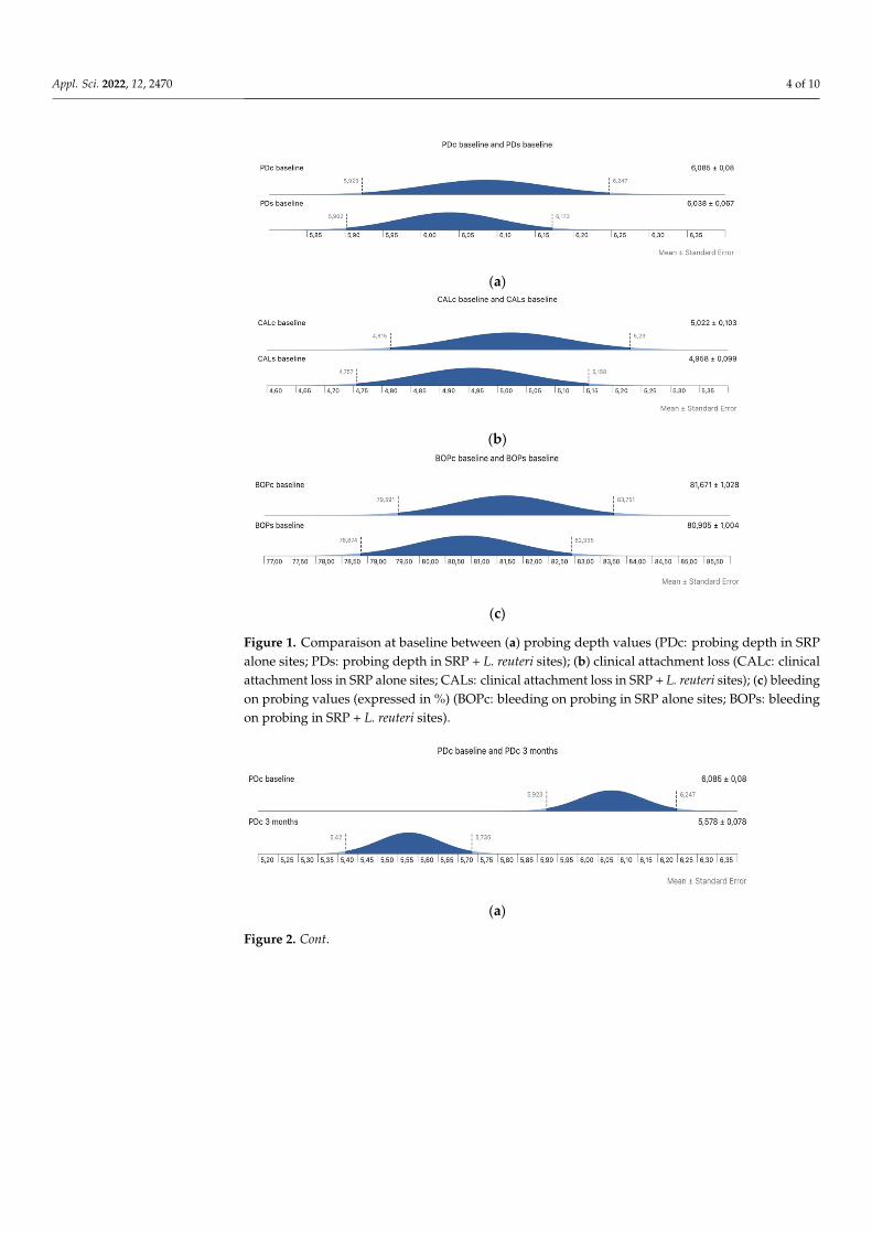
No significant differences were observed between study sites groups at baselineregarding probing depth, clinical attachment loss, or bleeding on probing (p = 0.650,p = 0.650 and p = 0.595, respectively) (Figure 1a–c).
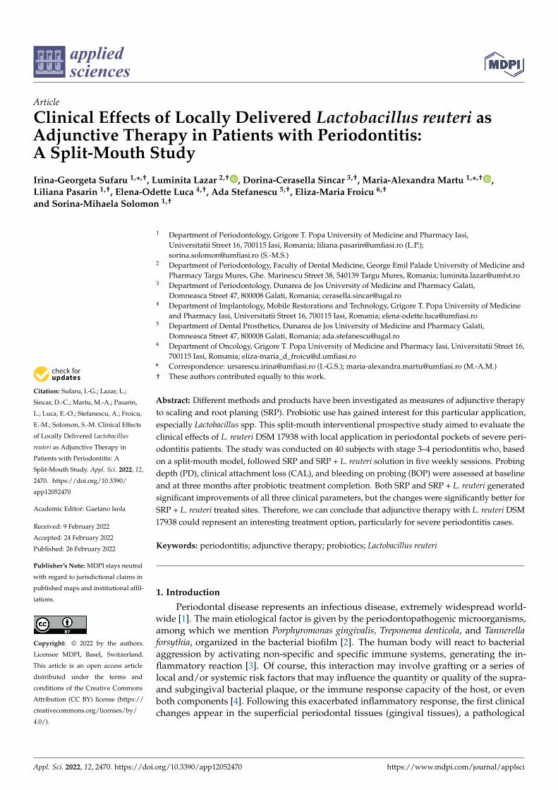
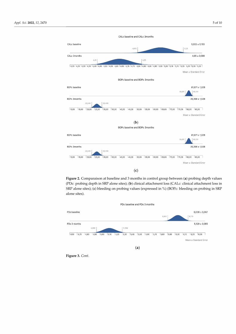
In the control group of sites who followed only SRP, we observed a significant reduc-tion of all three examined parameters after three months (6.09 ± 0.51 mm to 5.58 ± 0.49 mmfor PD; 5.02 ± 0.65 mm to 4.65 ± 0.62 mm for CAL; 81.67 ± 6.5 to 26.40 ± 9.54 for BOP)(p < 0.001 for all three parameters in the same study group) (Figure 2a–c).
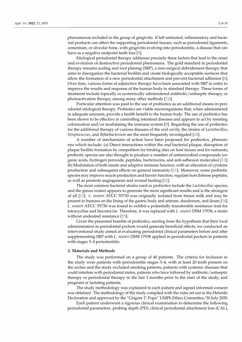
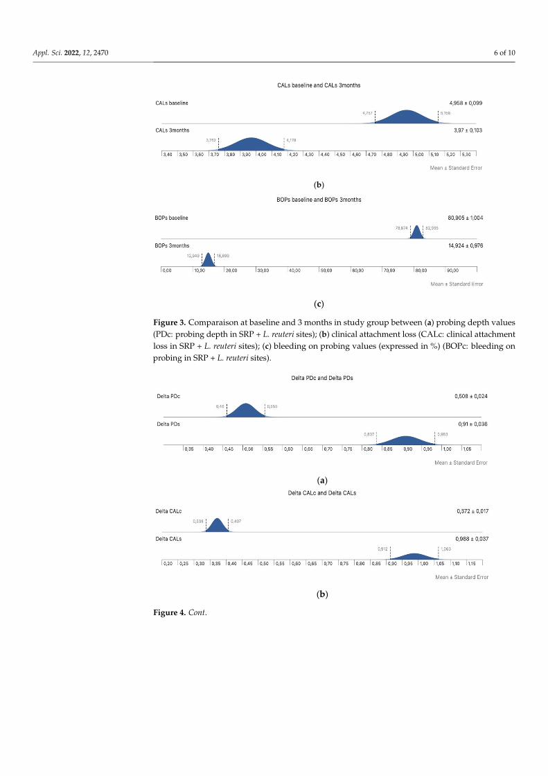
Significant differences were also observed for the study group who followed supple-mentary therapy with L. reuteri (reductions from 6.04 ± 0.42 mm to 5.13 ± 0.54 mm for PD;from 4.96 ± 0.63 mm to 3.97 ± 0.65 mm for CAL; from 80.90 ± 6.35 to 14.92 ± 6.17 for BOP;p < 0.001 for all three parameters) (Figure 3a–c).
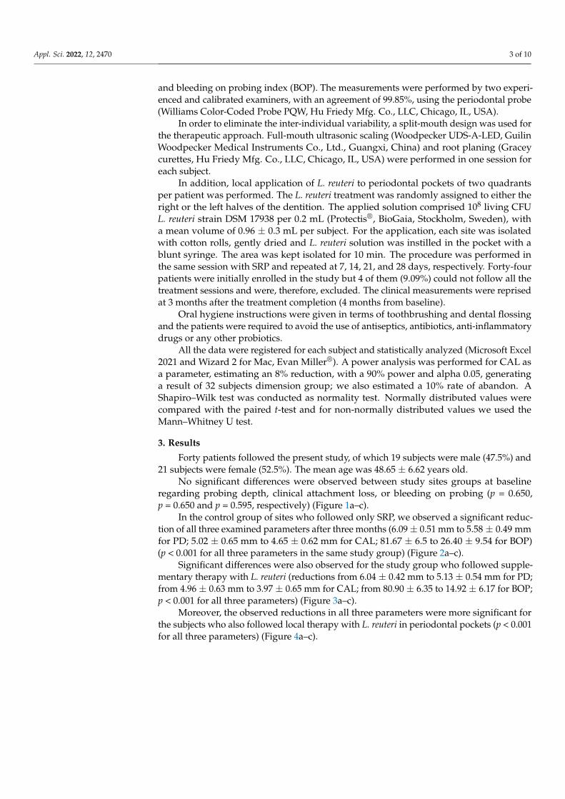
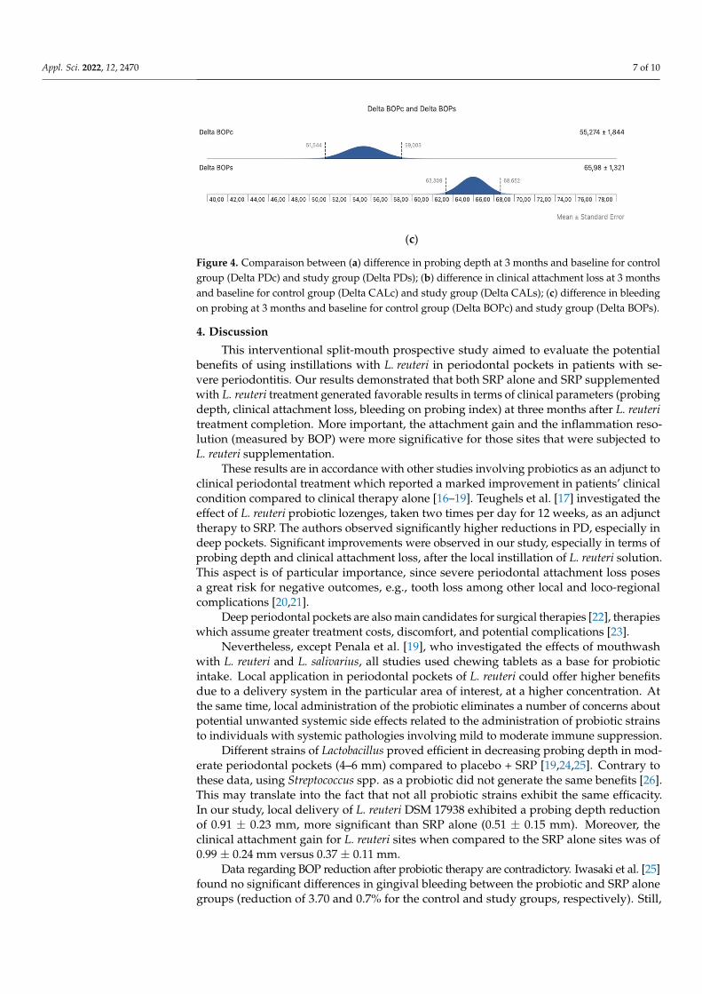
Moreover, the observed reductions in all three parameters were more significant forthe subjects who also followed local therapy with L. reuteri in periodontal pockets (p < 0.001for all three parameters) (Figure 4a–c).
Appl. Sci. 2022, 12, 2470 4 of 10Appl. Sci. 2022, 12, 2470 4 of 10
(a)
(b)
(c)
Figure 1. Comparaison at baseline between (a) probing depth values (PDc: probing depth in SRP alone sites; PDs: probing depth in SRP + L. reuteri sites); (b) clinical attachment loss (CALc: clinical attachment loss in SRP alone sites; CALs: clinical attachment loss in SRP + L. reuteri sites); (c) bleeding on probing values (expressed in %) (BOPc: bleeding on probing in SRP alone sites; BOPs: bleeding on probing in SRP + L. reuteri sites).
In the control group of sites who followed only SRP, we observed a significant reduction of all three examined parameters after three months (6.09 ± 0.51mm to 5.58 ± 0.49mm for PD; 5.02 ± 0.65mm to 4.65 ± 0.62mm for CAL; 81.67 ± 6.5 to 26.40 ± 9.54 for BOP) (p < 0.001 for all three parameters in the same study group) (Figure 2a–c).
Figure 1. Comparaison at baseline between (a) probing depth values (PDc: probing depth in SRPalone sites; PDs: probing depth in SRP + L. reuteri sites); (b) clinical attachment loss (CALc: clinicalattachment loss in SRP alone sites; CALs: clinical attachment loss in SRP + L. reuteri sites); (c) bleedingon probing values (expressed in %) (BOPc: bleeding on probing in SRP alone sites; BOPs: bleedingon probing in SRP + L. reuteri sites).
Appl. Sci. 2022, 12, 2470 4 of 10
(a)
(b)
(c)
Figure 1. Comparaison at baseline between (a) probing depth values (PDc: probing depth in SRP
alone sites; PDs: probing depth in SRP + L. reuteri sites); (b) clinical attachment loss (CALc: clinical
attachment loss in SRP alone sites; CALs: clinical attachment loss in SRP + L. reuteri sites); (c)
bleeding on probing values (expressed in %) (BOPc: bleeding on probing in SRP alone sites; BOPs:
bleeding on probing in SRP + L. reuteri sites).
In the control group of sites who followed only SRP, we observed a significant re‐
duction of all three examined parameters after three months (6.09 ± 0.51mm to 5.58 ±
0.49mm for PD; 5.02 ± 0.65mm to 4.65 ± 0.62mm for CAL; 81.67 ± 6.5 to 26.40 ± 9.54 for
BOP) (p < 0.001 for all three parameters in the same study group) (Figure 2a–c).
(a)
Figure 2. Cont.
Appl. Sci. 2022, 12, 2470 5 of 10Appl. Sci. 2022, 12, 2470 5 of 10
(b)
(c)
Figure 2. Comparaison at baseline and 3 months in control group between (a) probing depth val‐
ues (PDc: probing depth in SRP alone sites); (b) clinical attachment loss (CALc: clinical attachment
loss in SRP alone sites); (c) bleeding on probing values (expressed in %) (BOPc: bleeding on prob‐
ing in SRP alone sites).
(a)
(b)
Figure 2. Comparaison at baseline and 3 months in control group between (a) probing depth values(PDc: probing depth in SRP alone sites); (b) clinical attachment loss (CALc: clinical attachment loss inSRP alone sites); (c) bleeding on probing values (expressed in %) (BOPc: bleeding on probing in SRPalone sites).
Appl. Sci. 2022, 12, 2470 5 of 10
(b)
(c)
Figure 2. Comparaison at baseline and 3 months in control group between (a) probing depth val‐
ues (PDc: probing depth in SRP alone sites); (b) clinical attachment loss (CALc: clinical attachment
loss in SRP alone sites); (c) bleeding on probing values (expressed in %) (BOPc: bleeding on prob‐
ing in SRP alone sites).
probing depth in SRP alone sites); (b) clinical attachment loss (CALc: clinical attachment loss in
SRP alone sites); (c) bleeding on probing values (expressed in %) (BOPc: bleeding on probing in
SRP alone sites).
probing depth in SRP alone sites); (b) clinical attachment loss (CALc: clinical attachment loss in
SRP alone sites); (c) bleeding on probing values (expressed in %) (BOPc: bleeding on probing in
SRP alone sites).
(a)
Figure 3. Cont.
Appl. Sci. 2022, 12, 2470 6 of 10Appl. Sci. 2022, 12, 2470 6 of 10
(b)
(c)
Figure 3. Comparaison at baseline and 3 months in study group between (a) probing depth values
(PDc: probing depth in SRP + L. reuteri sites); (b) clinical attachment loss (CALc: clinical attach‐
ment loss in SRP + L. reuteri sites); (c) bleeding on probing values (expressed in %) (BOPc: bleeding
on probing in SRP + L. reuteri sites).
Moreover, the observed reductions in all three parameters were more significant for
the subjects who also followed local therapy with L. reuteri in periodontal pockets (p <
0.001 for all three parameters) (Figure 4a–c).
(a)
(b)
Figure 3. Comparaison at baseline and 3 months in study group between (a) probing depth values(PDc: probing depth in SRP + L. reuteri sites); (b) clinical attachment loss (CALc: clinical attachmentloss in SRP + L. reuteri sites); (c) bleeding on probing values (expressed in %) (BOPc: bleeding onprobing in SRP + L. reuteri sites).
Appl. Sci. 2022, 12, 2470 6 of 10
(b)
(c)
Figure 3. Comparaison at baseline and 3 months in study group between (a) probing depth values
(PDc: probing depth in SRP + L. reuteri sites); (b) clinical attachment loss (CALc: clinical attach‐
ment loss in SRP + L. reuteri sites); (c) bleeding on probing values (expressed in %) (BOPc: bleeding
on probing in SRP + L. reuteri sites).
Moreover, the observed reductions in all three parameters were more significant for
the subjects who also followed local therapy with L. reuteri in periodontal pockets (p <
0.001 for all three parameters) (Figure 4a–c).
(a)
(b)
Figure 4. Cont.
Appl. Sci. 2022, 12, 2470 7 of 10Appl. Sci. 2022, 12, 2470 7 of 10
(c)
Figure 4. Comparaison between (a) difference in probing depth at 3 months and baseline for con‐
trol group (Delta PDc) and study group (Delta PDs); (b) difference in clinical attachment loss at 3
months and baseline for control group (Delta CALc) and study group (Delta CALs); (c) difference
in bleeding on probing at 3 months and baseline for control group (Delta BOPc) and study group
(Delta BOPs).
4. Discussion
This interventional split‐mouth prospective study aimed to evaluate the potential
benefits of using instillations with L. reuteri in periodontal pockets in patients with severe
periodontitis. Our results demonstrated that both SRP alone and SRP supplemented with
L. reuteri treatment generated favorable results in terms of clinical parameters (probing
depth, clinical attachment loss, bleeding on probing index) at three months after L. reuteri
treatment completion. More important, the attachment gain and the inflammation resolu‐
tion (measured by BOP) were more significative for those sites that were subjected to L.
reuteri supplementation.
These results are in accordance with other studies involving probiotics as an adjunct
to clinical periodontal treatment which reported a marked improvement in patients’ clin‐
ical condition compared to clinical therapy alone [16–19]. Teughels et al. [17] investigated
the effect of L. reuteri probiotic lozenges, taken two times per day for 12 weeks, as an ad‐
junct therapy to SRP. The authors observed significantly higher reductions in PD, espe‐
cially in deep pockets. Significant improvements were observed in our study, especially
in terms of probing depth and clinical attachment loss, after the local instillation of L. reu‐
teri solution. This aspect is of particular importance, since severe periodontal attachment
loss poses a great risk for negative outcomes, e.g., tooth loss among other local and loco‐
regional complications [20,21].
Deep periodontal pockets are also main candidates for surgical therapies [22], thera‐
pies which assume greater treatment costs, discomfort, and potential complications [23].
Nevertheless, except Penala et al. [19], who investigated the effects of mouthwash
with L. reuteri and L. salivarius, all studies used chewing tablets as a base for probiotic
intake. Local application in periodontal pockets of L. reuteri could offer higher benefits
due to a delivery system in the particular area of interest, at a higher concentration. At the
same time, local administration of the probiotic eliminates a number of concerns about
potential unwanted systemic side effects related to the administration of probiotic strains
to individuals with systemic pathologies involving mild to moderate immune suppres‐
sion.
Different strains of Lactobacillus proved efficient in decreasing probing depth in mod‐
erate periodontal pockets (4–6 mm) compared to placebo + SRP [19,24,25]. Contrary to
these data, using Streptococcus spp. as a probiotic did not generate the same benefits [26].
This may translate into the fact that not all probiotic strains exhibit the same efficacity. In
our study, local delivery of L. reuteri DSM 17938 exhibited a probing depth reduction of
0.91 ± 0.23mm, more significant than SRP alone (0.51 ± 0.15mm). Moreover, the clinical
attachment gain for L. reuteri sites when compared to the SRP alone sites was of 0.99 ±
0.24mm versus 0.37 ± 0.11mm.
Figure 4. Comparaison between (a) difference in probing depth at 3 months and baseline for controlgroup (Delta PDc) and study group (Delta PDs); (b) difference in clinical attachment loss at 3 monthsand baseline for control group (Delta CALc) and study group (Delta CALs); (c) difference in bleedingon probing at 3 months and baseline for control group (Delta BOPc) and study group (Delta BOPs).
4. Discussion
This interventional split-mouth prospective study aimed to evaluate the potentialbenefits of using instillations with L. reuteri in periodontal pockets in patients with se-vere periodontitis. Our results demonstrated that both SRP alone and SRP supplementedwith L. reuteri treatment generated favorable results in terms of clinical parameters (probingdepth, clinical attachment loss, bleeding on probing index) at three months after L. reuteritreatment completion. More important, the attachment gain and the inflammation reso-lution (measured by BOP) were more significative for those sites that were subjected toL. reuteri supplementation.
These results are in accordance with other studies involving probiotics as an adjunct toclinical periodontal treatment which reported a marked improvement in patients’ clinicalcondition compared to clinical therapy alone [16–19]. Teughels et al. [17] investigated theeffect of L. reuteri probiotic lozenges, taken two times per day for 12 weeks, as an adjuncttherapy to SRP. The authors observed significantly higher reductions in PD, especially indeep pockets. Significant improvements were observed in our study, especially in terms ofprobing depth and clinical attachment loss, after the local instillation of L. reuteri solution.This aspect is of particular importance, since severe periodontal attachment loss posesa great risk for negative outcomes, e.g., tooth loss among other local and loco-regionalcomplications [20,21].
Deep periodontal pockets are also main candidates for surgical therapies [22], therapieswhich assume greater treatment costs, discomfort, and potential complications [23].
Nevertheless, except Penala et al. [19], who investigated the effects of mouthwashwith L. reuteri and L. salivarius, all studies used chewing tablets as a base for probioticintake. Local application in periodontal pockets of L. reuteri could offer higher benefitsdue to a delivery system in the particular area of interest, at a higher concentration. Atthe same time, local administration of the probiotic eliminates a number of concerns aboutpotential unwanted systemic side effects related to the administration of probiotic strainsto individuals with systemic pathologies involving mild to moderate immune suppression.
Different strains of Lactobacillus proved efficient in decreasing probing depth in mod-erate periodontal pockets (4–6 mm) compared to placebo + SRP [19,24,25]. Contrary tothese data, using Streptococcus spp. as a probiotic did not generate the same benefits [26].This may translate into the fact that not all probiotic strains exhibit the same efficacity.In our study, local delivery of L. reuteri DSM 17938 exhibited a probing depth reductionof 0.91 ± 0.23 mm, more significant than SRP alone (0.51 ± 0.15 mm). Moreover, theclinical attachment gain for L. reuteri sites when compared to the SRP alone sites was of0.99 ± 0.24 mm versus 0.37 ± 0.11 mm.
Data regarding BOP reduction after probiotic therapy are contradictory. Iwasaki et al. [25]found no significant differences in gingival bleeding between the probiotic and SRP alonegroups (reduction of 3.70 and 0.7% for the control and study groups, respectively). Still,
Appl. Sci. 2022, 12, 2470 8 of 10
their subjects followed periodontal maintenance sessions throughout the study period, afact that could interfere with the clear impact of probiotics. In our study, BOP reductionin L. reuteri sites after five sessions of local delivery was of 65.98% versus 55.27% incontrol sites, with no proper maintenance program. Tekce et al. [24] and Ince et al. [27]measured BOP one year after consuming L. reuteri pills for three weeks without additionalSRP and obtained a reduction of 69.6% and 50.95% in the control group and 77.85% and77.3% in the study group, respectively. Teughels et al. [17] observed a reduced BOP inthe study group but did not obtain statistically significant differences in the three-monthassessment. Various anti-inflammatory effects have been reported for L. reuteri, includingthe reduction in MMP-8 expression [28] and an increase in TIMP-1, which is a modulatorof MMP activity [27]. Moreover, L. reuteri proved to be associated with a decrease inproinflammatory cytokines, such as TNF-α, IL-1β, or IL-17 [29]. It also forms reuterin,which induces oxidative stress [27]. This aspect is particularly important if we think of theimportant influence of periodontal inflammation on the systemic status and the relationshipbetween periodontitis and cardiovascular diseases, osteoporosis, and other conditions andoutcomes [30–33].
In terms of microbiological results, one study reported that probiotic pills, eitheralone or in combination with SRP, decreased the number of A. actinomycetemcomitans,P. gingivalis, and Prevotella intermedia. Tekce et al. [24] reported greater reductions inperiodontal pathogens on days 21, 90, and 180, but did not show any differences betweengroups on day 360, suggesting that the beneficial effects of probiotics are temporary.
Our study is a pilot one, with certain limitations. First of all, the number of participantsis not large enough to extrapolate our results in the general population. Furthermore, ourresearch focused on clinical aspects only, with the main purpose to expand our interestsin microbiological and immunological investigations related to the use of L. reuteri asadjunctive periodontal therapy. Larger study periods of time are also required to clarify thelong-term potential benefit of probiotics. We strongly believe that probiotics could play animportant role in the management of particularly severe or resilient cases of periodontitis.However, of course, further studies are required in order to establish a clear work protocol,in terms of particular strains, dosage, number of sessions, and product use. We considerthat research should be also conducted in order to obtain a locally delivered product with acontrolled release system.
5. Conclusions
Local delivery of L. reuteri DSM 17938 associated to conventional non-surgical therapydemonstrated significant improvements of periodontal attachment and a reduction ofgingival bleeding in patients with stage 3–4 periodontitis. Further investigations arerequired to obtain a clear protocol of probiotics as an alternative adjunctive therapy.
Author Contributions: Conceptualization, I.-G.S. and S.-M.S.; methodology, I.-G.S., L.L., D.-C.S.,L.P., A.S. and S.-M.S.; software, I.-G.S.; validation, I.-G.S., M.-A.M., E.-O.L. and S.-M.S.; formalanalysis, I.-G.S., M.-A.M. and E.-M.F.; investigation, I.-G.S., E.-O.L., L.L., D.-C.S., L.P. and M.-A.M.;resources, L.L., D.-C.S., L.P., A.S., E.-M.F. and S.-M.S.; data curation, I.-G.S., L.L., D.-C.S., L.P. andM.-A.M.; writing—original draft preparation, I.-G.S. and M.-A.M.; writing—review and editing,L.P., E.-O.L., A.S., E.-M.F. and S.-M.S.; visualization, S.-M.S.; supervision, I.-G.S., L.L. and S.-M.S.;project administration, S.-M.S.; funding acquisition, L.L., D.-C.S., L.P., A.S., E.-M.F. and S.-M.S.All authors share equal contribution. All authors have read and agreed to the published versionof the manuscript.
Funding: This research received no external funding.
Institutional Review Board Statement: The study was conducted according to the guidelines of theDeclaration of Helsinki and approved by the “Grigore T. Popa” UMPh Ethics Committee/30 July 2020.
Informed Consent Statement: Informed consent was obtained from all subjects involved in the study.
Appl. Sci. 2022, 12, 2470 9 of 10
Data Availability Statement: The data used to support the findings of this study are available fromthe corresponding author upon reasonable request.
Conflicts of Interest: The authors declare no conflict of interest.
References1. Nazir, M.A. Prevalence of periodontal disease, its association with systemic diseases and prevention. Int. J. Health Sci. 2017,
11, 72–80.2. Fragkioudakis, I.; Riggio, M.P.; Apatzidou, D.A. Understanding the microbial components of periodontal disease and periodontal
treatment-induced microbiological shifts. J. Med. Microbiol. 2021, 70, 001247. [CrossRef] [PubMed]3. Nascimento, G.G.; Leite, F.R.M.; Scheutz, F.; López, R. Periodontitis: From infection to inflammation. Curr. Oral Health Rep. 2017,
4, 301–308. [CrossRef]4. Teodorescu, A.C.; Martu, I.; Teslaru, S.; Kappenberg-Nitescu, D.C.; Goriuc, A.; Luchian, I.; Martu, M.A.; Solomon, S.M.; Martu, S.
Assessment of salivary levels of RANKL and OPG in aggressive versus chronic periodontitis. J. Immunol. Res. 2019, 2019, 6195258.[CrossRef]
5. Slots, J. Periodontitis: Facts, fallacies and the future. Periodontol. 2000 2017, 75, 7–23. [CrossRef]6. Slots, J. Primer on etiology and treatment of progressive/severe periodontitis: A systemic health perspective. Periodontol. 2000
2020, 83, 272–276. [CrossRef]7. Alassy, H.; Pizarek, J.A.; Kormas, I.; Pedercinic, A.; Wolff, L.F. Antimicrobial adjuncts in the management of periodontal and
peri-implant diseases and conditions: A narrative review. Front. Oral Maxillofac. Med. 2021, 3, 16. [CrossRef]8. Mocanu, R.C.; Martu, M.A.; Luchian, I.; Sufaru, I.G.; Maftei, G.A.; Ioanid, N.; Martu, S.; Tatarciuc, M. Microbiologic profiles of
patients with dental prosthetic treatment and periodontitis before and after photoactivation therapy—Randomized clinical trial.Microorganisms 2021, 9, 713. [CrossRef]
9. Allaker, R.P.; Stephen, A.S. Use of probiotics and oral health. Curr. Oral Health Rep. 2017, 4, 309–318. [CrossRef]10. Martin-Cabezas, R.; Davideau, J.L.; Tenenbaum, H.; Huck, O. Clinical efficacy of probiotics as an adjunctive therapy to non-
surgical periodontal treatment of chronic periodontitis: A systematic review and meta-analysis. J. Clin. Periodontol. 2016, 43,520–530. [CrossRef]
11. Meurman, J.H. Probiotics: Do they have a role in oral medicine and dentistry? Eur. J. Oral Sci. 2005, 113, 188–196. [CrossRef]12. Devine, D.A.; Marsh, P.D.; Meade, J. Modulation of host responses by oral commensal bacteria. J. Oral Microbiol. 2015, 7, 26941.
[CrossRef] [PubMed]13. Alok, A.; Singh, I.D.; Singh, S.; Kishore, M.; Jha, P.C.; Iqubal, M.A. Probiotics: A new era of biotherapy. Adv. Biomed. Res. 2017, 6,
31. [CrossRef]14. Valeur, N.; Engel, P.; Carbajal, N.; Connolly, E.; Ladefoged, K. Colonization and immunomodulation by Lactobacillus reuteri ATCC
55730 in the human gastrointestinal tract. Appl. Environ. Microbiol. 2004, 70, 1176–1181. [CrossRef] [PubMed]15. Rosander, A.; Connolly, E.; Roos, S. Removal of antibiotic resistance gene-carrying plasmids from Lactobacillus reuteri ATCC
55730 and characterization of the resulting daughter strain, L. reuteri DSM 17938. Appl. Environ. Microbiol. 2008, 74, 6032–6040.[CrossRef]
16. Hallström, H.; Lindgren, S.; Yucel-Lindberg, T.; Dahlén, G.; Renvert, S.; Twetman, S. Effect of probiotic lozenges on inflammatoryreactions and oral biofilm during experimental gingivitis. Acta Odontol. Scand. 2013, 71, 828–833. [CrossRef] [PubMed]
17. Teughels, W.; Durukan, A.; Ozcelik, O.; Pauwels, M.; Quirynen, M.; Haytac, M.C. Clinical and microbiological effects ofLactobacillus reuteri probiotics in the treatment of chronic periodontitis: A randomized placebo-controlled study. J. Clin. Periodontol.2013, 40, 1025–1035. [CrossRef] [PubMed]
18. Vicario, M.; Santos, A.; Violant, D.; Nart, J.; Giner, L. Clinical changes in periodontal subjects with the probiotic Lactobacillus reuteriProdentis: A preliminary randomized clinical trial. Acta Odontol. Scand. 2013, 71, 813–819. [CrossRef]
19. Penala, S.; Kalakonda, B.; Pathakota, K.R.; Jayakumar, A.; Koppolu, P.; Lakshmi, B.V.; Pandey, R.; Mishra, A. Efficacy of local useof probiotics as an adjunct to scaling and root planing in chronic periodontitis and halitosis: A randomized controlled trial. J. Res.Pharm. Pract. 2016, 5, 86–93. [CrossRef]
20. Dumitrescu, D.; Fanuta, B.; Stepan, A.E.; Fronie, A.I.; Dumitrescu, C.I.; Martu, M.C.; Surlin, P.; Surlin, V.; Popescu, M. Silent sinussyndrome—Report of a case. Rom. J. Morphol. Embryol. 2015, 56, 229–237.
21. Kim, J.H.; Kim, S.J.; Choi, J.I.; Lee, J.Y. Periodontal attachment loss of extracted teeth for periodontal reasons. J. Korean Acad.Periodontol. 2006, 36, 1049594. [CrossRef]
22. Roshna, T. Generalized aggressive periodontitis and its treatment options: Case reports and review of literature. Case Rep. Med.2012, 2012, 535321. [CrossRef]
23. Maftei, G.A.; Martu, C.M.; Popa, C.; Geletu, G.; Danila, V.; Jelihovschi, I.; Foia, L. The biomechanical properties of suture materialsand their relationship to bacterial adherence. Mat. Plast. 2019, 56, 980–985. [CrossRef]
24. Tekce, M.; Ince, G.; Gursoy, H.; Dirikan Ipci, S.; Cakar, G.; Kadir, T.; Ylmaz, S. Clinical and microbiological effects of probioticlozenges in the treatment of chronic periodontitis: A 1-year follow-up study. J. Clin. Periodontol. 2015, 42, 363–372. [CrossRef][PubMed]
Appl. Sci. 2022, 12, 2470 10 of 10
25. Iwasaki, K.; Maeda, K.; Hidaka, K.; Nemoto, K.; Hirose, Y.; Deguchi, S. Daily intake of heat-killed Lactobacillus plantarum L-137decreases the probing Depth in patients undergoing supportive periodontal therapy. Oral Health Prev. Dent. 2016, 14, 207–214.
26. Laleman, I.; Yilmaz, E.; Ozcelik, O.; Haytac, C.; Pauwels, M.; Herrero, E.R.; Slomka, V.; Quirynen, M.; Alkaya, B.; Teughels, W.The effect of a Streptococci containing probiotic in periodontal therapy: A randomized controlled trial. J. Clin. Periodontol. 2015,42, 1032–1041. [CrossRef]
27. Ince, G.; Gürsoy, H.; Ipçi, S.D.; Cakar, G.; Emekli-Alturfan, E.; Yılmaz, S. Clinical and biochemical evaluation of lozengescontaining Lactobacillus reuteri as an adjunct to non-surgical periodontal therapy in chronic periodontitis. J. Periodontol. 2015, 86,746–754. [CrossRef]
28. Lee, J.K.; Kim, S.J.; Ko, S.H.; Ouwehand, A.C.; Ma, D.S. Modulation of the host response by probiotic Lactobacillus brevis CD2 inexperimental gingivitis. Oral Dis. 2015, 21, 705–712. [CrossRef]
29. Szkaradkiewicz, A.K.; Stopa, J.; Karpinski, T.M. Effect of oral administration involving a probiotic strain of Lactobacillus reuteri onproinflammatory cytokine response in patients with chronic periodontitis. Arch. Immunol. Ther. Exp. 2014, 62, 495–500. [CrossRef][PubMed]
30. Ursarescu, I.G.; Martu-Stefanache, M.A.; Solomon, S.M.; Pasarin, L.; Boatca, R.M.; Caruntu, I.D.; Martu, S. The assessment of IL-6and RANKL in the association between chronic periodontitis and osteoporosis. Rev. Chem. 2016, 67, 386–389.
31. Anton, D.M.; Martu, M.A.; Maris, M.; Maftei, G.A.; Sufaru, I.G.; Tatarciuc, D.; Luchian, I.; Ioanid, N.; Martu, S. Study on theeffects of melatonin on glycemic control and periodontal parameters in patients with type II diabetes mellitus and periodontaldisease. Medicina 2021, 57, 140. [CrossRef] [PubMed]
32. De Sire, A.; Invernizzi, M.; Ferrillo, M.; Gimigliano, F.; Baricich, A.; Cisari, C.; De Marchi, F.; Foglio Bonda, P.L.; Mazzini, L.;Migliario, M. Functional status and oral health in patients with amyotrophic lateral sclerosis: A cross-sectional study. NeuroRehabil. 2021, 48, 49–57. [CrossRef] [PubMed]
33. Ferrillo, M.; Migliario, M.; Roccuzzo, A.; Molinero-Mourelle, P.; Falcicchio, G.; Umano, G.R.; Pezzotti, F.; Foglio Bonda, P.L.;Calafiore, D.; de Sire, A. Periodontal disease and vitamin D deficiency in pregnant women: Which correlation with preterm andlow-weight birth? J. Clin. Med. 2021, 10, 4578. [CrossRef] [PubMed]
Related Documents