470 www.e-enm.org Endocrinol Metab 2014;29:470-478 http://dx.doi.org/10.3803/EnM.2014.29.4.470 pISSN 2093-596X · eISSN 2093-5978 Original Article Clinical Characteristics, Management, and Outcome of 22 Cases of Primary Hypophysitis Sun Mi Park 1, * , Ji Cheol Bae 2, * , Ji Young Joung 1 , Yoon Young Cho 1 , Tae Hun Kim 3 , Sang-Man Jin 1 , Sunghwan Suh 4 , Kyu Yeon Hur 1 , Kwang-Won Kim 5 1 Division of Endocrinology and Metabolism, Department of Internal Medicine, Samsung Medical Center, Sungkyunkwan University School of Medicine, Seoul; 2 Division of Endocrinology and Metabolism, Department of Internal Medicine, Samsung Changwon Hospital, Sungkyunkwan University School of Medicine, Changwon; 3 Division of Endocrinology and Metabolism, Department of Internal Medicine, Andong Sungso hospital, Andong; 4 Division of Endocrinology and Metabolism, Department of Internal Medicine, Dong-A Medical Center, Dong-A University College of Medicine, Busan; 5 Division of Endocrinology and Metabolism, Department of Internal Medicine, Gachon University Gil Medical Center, Gachon University of Medicine and Science, Incheon, Korea Background: Primary hypophysitis causes varying degrees of endocrine dysfunction and mass effect. The natural course and best treatment have not been well established. Methods: Medical records of 22 patients who had been diagnosed with primary hypophysitis between January 2001 and March 2013 were retrospectively reviewed. Based on the anatomical location, we classified the cases as adenohypophysitis (AH), infun- dibuloneurohypophysitis (INH), and panhypophysitis (PH). Clinical presentation, endocrine function, pathologic findings, mag- netic resonance imaging findings, and treatment courses were reviewed. Results: Among 22 patients with primary hypophysitis, 81.8% (18/22) had involvement of the posterior pituitary lobe. Two pa- tients of the AH (2/3, 66.6%) and three patients of the PH (3/10, 30%) groups initially underwent surgical mass reduction. Five patients, including three of the PH (3/10, 33.3%) group and one from each of the AH (1/3, 33.3%) and INH (1/9, 11.1%) groups, initially received high-dose glucocorticoid treatment. Nearly all of the patients treated with surgery or high-dose steroid treatment (9/11, 82%) required continuous hormone replacement during the follow-up period. Twelve patients received no treatment for mass reduction due to the absence of acute symptoms and signs related to a compressive mass effect. Most of them (11/12, 92%) did not show disease progression, and three patients recovered partially from hormone deficiency. Conclusion: Deficits of the posterior pituitary were the most common features in our cases of primary hypophysitis. Pituitary en- docrine defects responded less favorably to glucocorticoid treatment and surgery. In the absence of symptoms related to mass ef- fect and with the mild defect of endocrine function, it may not require treatment to reduce mass except hormone replacement. Keywords: Pituitary; Hypophysitis; Diabetes insipidus; Hypopituitarism; Steroids Received: 25 December 2013, Revised: 17 April 2014, Accepted: 29 April 2014 Corresponding author: Kwang-Won Kim Division of Endocrinology and Metabolism, Department of Internal Medicine, Gachon University Gil Medical Center, Gachon University of Medicine and Science, 21 Namdong-daero 774beon-gil, Namdong-gu, Incheon 405-760, Korea Tel: +82-32-460-8309, Fax: +82-32-469-4320, E-mail: [email protected] *These authors contributed equally to this work. Copyright © 2014 Korean Endocrine Society This is an Open Access article distributed under the terms of the Creative Com- mons Attribution Non-Commercial License (http://creativecommons.org/ licenses/by-nc/3.0/) which permits unrestricted non-commercial use, distribu- tion, and reproduction in any medium, provided the original work is properly cited.

Welcome message from author
This document is posted to help you gain knowledge. Please leave a comment to let me know what you think about it! Share it to your friends and learn new things together.
Transcript

470 www.e-enm.org
Endocrinol Metab 2014;29:470-478http://dx.doi.org/10.3803/EnM.2014.29.4.470pISSN 2093-596X · eISSN 2093-5978
OriginalArticle
Clinical Characteristics, Management, and Outcome of 22 Cases of Primary HypophysitisSun Mi Park1,*, Ji Cheol Bae2,*, Ji Young Joung1, Yoon Young Cho1, Tae Hun Kim3, Sang-Man Jin1, Sunghwan Suh4, Kyu Yeon Hur1, Kwang-Won Kim5
1Division of Endocrinology and Metabolism, Department of Internal Medicine, Samsung Medical Center, Sungkyunkwan University School of Medicine, Seoul; 2Division of Endocrinology and Metabolism, Department of Internal Medicine, Samsung Changwon Hospital, Sungkyunkwan University School of Medicine, Changwon; 3Division of Endocrinology and Metabolism, Department of Internal Medicine, Andong Sungso hospital, Andong; 4Division of Endocrinology and Metabolism, Department of Internal Medicine, Dong-A Medical Center, Dong-A University College of Medicine, Busan; 5Division of Endocrinology and Metabolism, Department of Internal Medicine, Gachon University Gil Medical Center, Gachon University of Medicine and Science, Incheon, Korea
Background: Primary hypophysitis causes varying degrees of endocrine dysfunction and mass effect. The natural course and best treatment have not been well established.Methods: Medical records of 22 patients who had been diagnosed with primary hypophysitis between January 2001 and March 2013 were retrospectively reviewed. Based on the anatomical location, we classified the cases as adenohypophysitis (AH), infun-dibuloneurohypophysitis (INH), and panhypophysitis (PH). Clinical presentation, endocrine function, pathologic findings, mag-netic resonance imaging findings, and treatment courses were reviewed. Results: Among 22 patients with primary hypophysitis, 81.8% (18/22) had involvement of the posterior pituitary lobe. Two pa-tients of the AH (2/3, 66.6%) and three patients of the PH (3/10, 30%) groups initially underwent surgical mass reduction. Five patients, including three of the PH (3/10, 33.3%) group and one from each of the AH (1/3, 33.3%) and INH (1/9, 11.1%) groups, initially received high-dose glucocorticoid treatment. Nearly all of the patients treated with surgery or high-dose steroid treatment (9/11, 82%) required continuous hormone replacement during the follow-up period. Twelve patients received no treatment for mass reduction due to the absence of acute symptoms and signs related to a compressive mass effect. Most of them (11/12, 92%) did not show disease progression, and three patients recovered partially from hormone deficiency.Conclusion: Deficits of the posterior pituitary were the most common features in our cases of primary hypophysitis. Pituitary en-docrine defects responded less favorably to glucocorticoid treatment and surgery. In the absence of symptoms related to mass ef-fect and with the mild defect of endocrine function, it may not require treatment to reduce mass except hormone replacement.
Keywords: Pituitary; Hypophysitis; Diabetes insipidus; Hypopituitarism; Steroids
Received: 25 December 2013, Revised: 17 April 2014, Accepted: 29 April 2014Corresponding author: Kwang-Won KimDivision of Endocrinology and Metabolism, Department of Internal Medicine, Gachon University Gil Medical Center, Gachon University of Medicine and Science, 21 Namdong-daero 774beon-gil, Namdong-gu, Incheon 405-760, KoreaTel: +82-32-460-8309, Fax: +82-32-469-4320, E-mail: [email protected]*These authors contributed equally to this work.
Copyright © 2014 Korean Endocrine SocietyThis is an Open Access article distributed under the terms of the Creative Com-mons Attribution Non-Commercial License (http://creativecommons.org/licenses/by-nc/3.0/) which permits unrestricted non-commercial use, distribu-tion, and reproduction in any medium, provided the original work is properly cited.

22 Cases of Primary Hypophysis
Copyright © 2014 Korean Endocrine Society www.e-enm.org 471
Endocrinol Metab 2014;29:470-478http://dx.doi.org/10.3803/EnM.2014.29.4.470pISSN 2093-596X · eISSN 2093-5978
INTRODUCTION
Primary hypophysitis is a chronic inflammatory disease of the pituitary gland that is not secondary to infection, systemic in-flammatory disorders, or tumorous conditions. Primary hy-pophysitis is usually classified as lymphocytic, granuloma-tous, or xanthomatous hypophysitis based on the histologic findings. Based on the anatomical location, it is also classified into adenohypophysitis (AH), infundibuloneurohypophysitis (INH), or panhypophysitis (PH) [1]. Diffuse infiltration of the pituitary gland by inflammatory cells results in varying de-grees of endocrine dysfunction due to partial or complete defi-ciency of pituitary hormones. Particularly, involvement of the neurohypophysis and infundibular stem can cause diabetes in-sipidus [2,3]. Although the recent increasing reported cases have improved our understanding of primary hypophysitis, the natural history and best treatment have not been well estab-lished [4,5]. The authors describe a single-center experience of clinical features, endocrinological findings, and treatment courses of 22 primary hypophysitis patients.
METHODS
The study subjects were 22 patients who had been diagnosed with primary hypophysitis at Samsung Medical Center, Korea between January 2001 and March 2013. They were composed of 17 women and 5 men with a mean age of 48 years (range, 21 to 67) at the time of diagnosis. Among these patients, 11 were histologically proven cases, and the others were suspect-ed cases diagnosed by magnetic resonance imaging (MRI) findings with clinical features. Diagnosis based on MRI find-ings included known features suggestive of hypophysitis, in-cluding symmetric enlargement of the pituitary gland, a thick-ened but rarely displaced stalk, a usually intact sellar floor, homogeneity of the pituitary mass, and its intense enhance-ment after gadolinium. In patients with clinical symptoms and signs of diabetes insipidus, MRI findings suggestive of INH, including the loss of precontrast T1 hyperintensity in the neu-rohypophysis, swelling of the posterior pituitary, and thicken-ing of the pituitary stalk were also considered sufficient for di-agnosis of hypophysitis. Combined anterior pituitary stimula-tion testing and MRI scans of the sella area were performed in all patients at the time of diagnosis. Based on the anatomical location, we classified these cases into AH, INH, or PH (Table 1) [1]. Clinical presentation, en-docrine function, pathologic findings, MRI findings, and treat-
ment courses were reviewed retrospectively from electronic medical records. This study was approved by the Institutional Review Board of Samsung Medical Center.
RESULTS
Clinical presentation The most common clinical symptoms of this study group were polyuria and polydipsia (18/22, 81.8%), followed by headache (6/22, 27.2%), general weakness (5/22, 22%), and amenorrhea (3/22, 13%). Visual disturbance, such as diplopia and visual field defect, was noted in two patients (9%). Based on the ana-tomical location classification, the most common symptoms in AH were headache and amenorrhea (2/3, 66%) (Table 2). In INH, general weakness was the only symptom except for poly-uria and polydipsia. Only one case was associated with a recent pregnancy, and her initial symptoms of headache and visual disturbance appeared during the third trimester. Two patients had coexisting autoimmune disease, one with Hashimoto’s thy-roiditis and another with limited Wegener’s granulomatosis.
Table 1. Classification of 22 Patients with Primary Hypophy-sitis Based on the Anatomical location
Variable No. of patients
Adenohypophysitis (n=3)
Histology shows infiltrated A, but N was not recorded. No symptoms and signs (including image) of N involvement
3
Clinics and imaging show A involvement, but no symptoms of diabetes insipidus and no signs (including image) of N involvement
0
Infundibuloneurohypophysitis (n=9)
Histology shows infiltrated N, but A was not recorded. No symptoms and signs (including image) of A involvement
2
Clinics and imaging show N involvement, but no symptoms or signs (including image) of A involvement
7
Panhypophysitis (n=10)
Histology shows infiltrated A, but N was not recorded. With symptoms or signs (including image) of N involvement
6
Clinics and imaging show A involvement. With symptoms or signs (including image) of N involvement
4
A, anterior hypophysis; N, infundibuloneurohypophysis.

Park SM, et al.
472 www.e-enm.org Copyright © 2014 Korean Endocrine Society
Endocrinological assessment Among 22 patients with primary hypophysitis, 81.8% (18/22) had involvement of the posterior pituitary lobe resulting in di-abetes insipidus. Exclusive involvement of the infundibular stem or the posterior lobe was found in 40.9% (9/22) of cases (Table 2). The most frequently observed defect in the anterior pituitary was corticotrophs (8/22, 36.3%) and thyrotrophs (8/22, 36.3%), followed by gonadotrophs (7/22, 31.8%). Im-paired function of somatotrophs was seen in five of 22 cases (22.7%). Prolactin levels were increased in 22.7% (5/22) of cases. In patients with AH, deficiency in adrenocorticotropic hormone (ACTH), thyroid stimulating hormone (TSH), and luteinizing hormone (LH)/follicle stimulating hormone (FSH) was seen in 66.6% (2/3), and prolactin levels were also in-creased in 66.6% (2/3) of cases. All patients with INH and PH had dysfunction in secretion of antidiuretic hormone. In pa-tients with PH, deficiency of ACTH and TSH was noted equal in 60% (6/10), followed by LH/FSH deficiency (5/10, 50%)
and growth hormone (GH) deficiency (4/10, 40%) (Table 2).
Findings on MRIIn the AH and PH (n=13) cases, symmetric enlargement of the entire pituitary gland with homogeneous enhancement af-ter gadolinium was seen in seven patients. Among these 13 patients, thickening of the pituitary stalk was seen in eight pa-tients (61.5%), and suprasellar extension was seen in six pa-tients (46.1%). In one patient, the finding was seen as promi-nent cystic area on MRI. In the INH (n=9) cases, all patients showed thickening of the pituitary stalk and loss of T1 hyper-intensity in the neurohypophysis on MRI. One of these pa-tients also demonstrated suprasellar extension. One case showed swelling of the posterior pituitary gland. Among all patients (n=22), an abnormally thickened infundibular stalk was found in 17 patients, and loss of T1 hyperintensity in the posterior lobe was seen in 18 patients.
Table 2. Clinical Features, Pathologic Findings, and Treatments of 22 Patients with Primary Hypophysitis Based on the Anatomical Location
Variable Adenohypophysitis (n=3) Infundibuloneurohypophysitis (n=9) Panhypophysitis (n=10)
Mean age, yr 41.7 (29–59) 49.9 (21–64) 48.5 (23–67)
Male 0 1 (11.1) 4 (40)
Symptom
Headache 2 (66) 0 4 (40)
Visual disturbance 1 (33) 0 1 (10)
Amenorrhea 2 (66) 0 1 (10)
Endocrine defect
Adrenal axis 2 (66) 0 6 (60)
Thyroidal axis 2 (66) 0 6 (60)
Gonadal axis 2 (66) 0 5 (50)
Growth hormone 1 (33) 0 4 (40)
Hyperprolactinemia 2 (66) 1 (11.1) 2 (20)
Diabetes insipidus 0 9 (100) 9 (90)
Histologic finding
Lymphocytic hypophysitis 3 2 3
Granulomatous hypophysitis 0 0 2
Xanthomatous hypophysitis 0 0 1
Initial treatment
Surgical decompression 2 (66) 0 3 (30)
High-dose steroid 1 (33) 1 (11.1) 3 (30)
Symptomatic treatment 0 8 (88.8) 4 (40)
Values are expressed as mean (range) or number (%).
22 Cases of Primary Hypophysis
Copyright © 2014 Korean Endocrine Society www.e-enm.org 473
Pathological examinationA histological diagnosis was available in 11 patients (50%), including six patients who underwent surgical treatment for mass reduction and five patients who underwent biopsy for di-agnosis through a transsphenoidal approach. Among these pa-tients, hypophysitis was diagnosed as lymphocytic in eight pa-tients, granulomatous in two patients, and xanthomatous in one patient (Table 2). The histologic diagnosis in the patient with a prominent cystic lesion on MRI was xanthomatous hy-pophysitis. Patients with lymphocytic hypophysitis showed
varied anatomical involvement: three patients had deficits in the anterior pituitary, two patients in the posterior pituitary, and three patients in both pituitary lobes. Patients with xantho-mathous hypophysitis and granulomatous hypophysitis had deficits in both the anterior and posterior pituitary (Table 2).
Treatment and patient outcomesTwo patients of the AH and three patients of the PH groups initially underwent surgical mass reduction for decompression through a transsphenoidal approach (Table 2). Five patients,
Table 3. Treatment Courses with Follow-Up Endocrine and Image Outcomes in 22 Patients
Case No.
Classifi-cation Diagnosis Initial treatment Alternative
treatmentEndocrine defect Follow-up
mass statusFollow-up,
moPretreatment Posttreatment
1 PH Biopsy Conservative care - ACTH, LH/ FSH, DI
DI No change 43
2 AH Postoperative TSA with mass reduction - ACTH, TSH, LH/FSH
ACTH, TSH, LH, FSH DI
Mass reduction 138
3 INH Clinical Conservative care - DI DI No change 32
4 INH Biopsy Conservative care - DI DI No change 124
5 INH Clinical Conservative care - DI DI No change 120
6 PH Clinical Conservative care - ACTH, TSH, LH/FSH, DI
LH/FSH, DI No change 111
7 PH Postoperative TSA with mass reduction - GH TSH, DI Mass reduction 61
8 PH Postoperative TSA with mass reduction - DI DI Mass reduction 92
9 PH Clinical Conservative care - ACTH, TSH, DI ACTH, TSH, DI No change 65
10 INH Clinical Conservative care - DI DI No change 36
11 INH Clinical Conservative care - DI DI No change 37
12 INH Clinical Conservative care - DI DI No change 51
13 INH Clinical Conservative care - DI DI No change 53
14 PH Clinical, then postoperative
High dose steroid TSA with mass reduction
ACTH, LH/ FSH, GH, DI
ACTH, LH/ FSH, GH, DI
Mass reduction 44
15 AH Biopsy High dose steroid - ACTH, TSH, LH/FSH, GH
ACTH, TSH, LH/FSH
Mass reduction 46
16 INH Clinical Conservative care High dose steroid DI DI Loss to follow-up 14
17 PH Postoperative TSA with mass reduction High dose steroid ACTH, TSH, LH/FSH, DI
ACTH, TSH, LH/FSH, DI
Mass reduction 26
18 PH Clinical High dose steroid - ACTH, GH, DI DI Mass reduction 15
19 AH Postoperative TSA with mass reduction - ACTH - Mass reduction 50
20 PH Clinical Conservative care - TSH, GH, DI TSH, GH, DI No change 63
21 INH Biopsy High dose steroid - DI DI No change 19
22 PH Clinical High dose steroid - ACTH, TSH, LH/FSH,
GH, DI
- Mass reduction 7
PH, panhypophysitis; ACTH, adrenocorticotropic hormone; LH, luteinizing hormone; FSH, follicle stimulating hormone; DI, diabetes insipidus; AH, adenohypophysitis; TSA, transsphenoidal approach; TSH, thyroid stimulating hormone; INH, infundibuloneurohypophysitis; GH, growth hormone.

Park SM, et al.
474 www.e-enm.org Copyright © 2014 Korean Endocrine Society
including three from the PH and one from each of the AH and INH groups, initially received high-dose glucocorticoid treat-ment. Eight patients with INH and four patients with PH re-ceived no treatment for mass reduction due to the absence of acute symptoms and signs related to mass effect such as head-ache, visual field defect, or optic chiasm compression. Fourteen patients (63.6%) underwent further anterior pitu-itary stimulation testing at least once during the follow-up pe-riod. Eight other patients with INH followed only basal pitu-itary hormone. In cases of surgical or high-dose steroid treat-ment, MRI scans were repeated within 3 months of treatment. Follow-up MRI scans were then performed regularly every 1 or 2 years in all patients (except those lost to follow-up) dur-ing the remaining follow-up period. Mean duration of follow-up was 57 months (range, 7 to 138; median, 48). All of the patients who initially underwent surgical treat-ment and four of five patients who initially received high-dose glucocorticoids showed mass reduction on follow-up MRI im-ages (Table 3). One patient in the high-dose glucocorticoid treatment group demonstrated no response to treatment; how-ever, this patient has not experienced disease progression dur-
ing 19 months of follow-up without alternative treatment, as seen in most patients (11 of 12) in the non-treatment group. During the follow-up period, two (one in each treatment group) of 10 patients who responded to initial surgical or high-dose glucocorticoid treatment presented with a recurring pituitary mass, and one patient in the non-treatment group had an in-crease in pituitary mass size. These three patients required a second alternative treatment to reduce the pituitary mass (Ta-bles 3, 4). Among the patients who underwent surgical resec-tion, recurring pituitary mass was seen in one patient with xan-thomatous hypophysitis, developed visual disturbance due to suprasellar extension 10 months after surgery. This patient un-derwent adjunctive high-dose glucocorticoid treatment (meth-ylprednisolone 500 mg intravenous for 3 days) and has shown good response in mass reduction without disease progression during 15 months of follow-up (Fig. 1) [6]. One patient treated with high-dose glucocorticoids (1 mg/kg dose of prednisolone) showed good response in headache and mass reduction; how-ever, she was unable to reduce her prednisolone below 15 mg due to headache. Unfortunately, during long-term treatment with low-dose prednisolone, she gained 30 kg, developed dia-
Table 4. Pretreatment and Posttreatment Endocrine Assessment of the Patients Based on the Treatment
Variable TSA (n=6) Steroid (n=7) No treatment (n=12)
As initial treatment 5 5 12
As alternative treatment 1 2 -
Follow-up period, mo 68.5 (26–138) 24.4 (7–46) 62.4 (32–124)
Response to high-dose glucocorticoids - 6 -
Required long term hormone replacement 5 6 12
Pretreatment endocrine defect
Adrenal axis 4 4 3
Thyroidal axis 2 3 3
Gonadal axis 3 4 2
Growth hormone 1 3 1
Hyperprolactinemia 4 1 1
Diabetes insipidus 3 6 12
Posttreatment endocrine defect
Adrenal axis 3 3 1
Thyroidal axis 3 3 1
Gonadal axis 3 3 1
Growth hormone 1 1 0
Hyperprolactinemia 0 1 0
Diabetes insipidus 6 6 12
Values are expressed as mean (range).

22 Cases of Primary Hypophysis
Copyright © 2014 Korean Endocrine Society www.e-enm.org 475
betes, and experienced an increase in the size of the pituitary mass. Twelve months after initial steroid treatment, she under-went hypophysectomy, and lymphocytic hypophysitis was his-tologically confirmed. Her headaches resolved without disease progression during 19 months after surgery. She then died from cerebral infarction. The panhypopituitarism in these two patients did not improve at all despite treatment. Among pa-tients without surgical or high-dose steroid treatment, one pa-tient with INH received high-dose glucocorticoids (1 mg/kg dose of prednisolone) due to progression in mass size during 19 months of follow-up (Fig. 1). Hormone deficiency re-mained unchanged and required desmopressin despite the high-dose steroid treatment. A follow-up MRI could not be performed, as the patient was lost to follow-up. Seven patients received high-dose glucocorticoids initially or alternatively, and five patients showed mass reduction. Among these five patients, four patients had symptoms for less than 3 months before steroid treatment. One patient who didn’t respond to high-dose steroids had symptoms for 5 months. Nearly all of the patients treated with surgery or high-dose steroids (9/11, 82%) required continuous hormone replace-ment during the follow-up period. Only two patients have shown complete improvement after mass reduction without need for hormone replacement (Table 4). One patient had PH due to granulomatous hypophysitis associated with Wegener’s granulomatosis, and another patient had AH due to lympho-cytic hypophysitis. Pituitary mass reduction was achieved by high-dose steroid treatment and surgery, respectively. Among patients without surgical or high-dose steroid treatment, two
patients showed spontaneous partial recovery in hormone de-ficiency (Table 3). Also, two patients treated with high-dose steroids recovered partially from hormone deficiency. All pa-tients treated with high-dose steroids had weight gain, and one patient developed avascular necrosis of the femoral head. Three of six patients who received tumor removal surgery de-veloped additional hypopituitarism, such as diabetes insipidus or hypothyroidism, after surgery (Table 4).
DISCUSSION
The clinical presentations of primary hypophysitis are variable including symptoms related to sellar compression, hypopitu-itarism, and diabetes insipidus. The presentation depends on whether the immune system affects the anterior lobe, posterior lobe, or both [7]. In a review analyzing 379 cases of primary lymphocytic hypophysitis based on 370 articles published as case reports or small case series [1], 89.7% (340/379) had symptoms from partial or complete deficiency of the anterior pituitary hormones, and 35.3% (134/379) developed diabetes insipidus. PH presenting with both symptoms was seen in 25.1% (95/379) and INH was seen only in 10.2% (39/379). In our study, among 22 patients with primary hypophysitis, 59.1% (13/22) had symptoms due to anterior pituitary hor-mone deficiency. Also, 81.8% (18/22) had diabetes insipidus, and 40.9% (9/22) had this symptom exclusively. Compared with the previous review [1,8], it is interesting that symptoms due to posterior pituitary hormone deficiency were most com-mon in our study group. Some primary hypophysitis cases
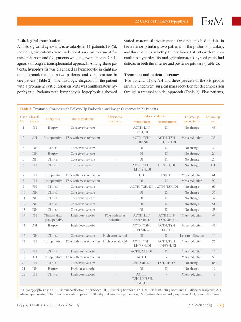
Adenohypophysitis(n=3)
(n=2)
TSA with tumorremoval(n=5)
High dosesteroid(n=2)
(n=1)
Panhypophysitis(n=10)
(n=3)
High dosesteroid(n=5)
TSA with tumorremoval(n=1)
(n=3) (n=4)
Infundibuloneurohypophysitis(n=9)
(n=8)
Conservativecare
(n=12)
Alternative treatment
Initial treatment
(n=1)
n=1a n=1b n=1c
Fig. 1. Summary of treatment courses of 22 patients with primary hypophysitis. TSA, transsphenoidal approach. aDue to regrowth of mass; bDue to steroid related side effect and progress with steroid tapering; cDue to progression in mass size.

Park SM, et al.
476 www.e-enm.org Copyright © 2014 Korean Endocrine Society
may still go undiagnosed because of their indolent, subclinical course [2,9]. In particular, acute symptoms related to mass ef-fect were rare in INH [1,7]. Thus, cases of primary hypophysi-tis with only polydipsia and polyuria may not be published as case reports and might result in underestimation of INH case number [9]. The most common deficit of the anterior pituitary hormones was ACTH followed by TSH, GH, and gonadotro-pins, similar to previous review [1,7,8]. The natural history of primary hypophysitis is variable and unpredictable [5,10,11]. Typically, it follows a progressive course in which the pituitary initially becomes inflamed, edem-atous, and enlarged, and then the patient develops symptoms secondary to mass effects. With destruction of pituicytes, the parenchyma is replaced by fibrosis resulting in pituitary atro-phy leading to hypopituitarism [1]. In some cases, the disease course was aggressive and rapidly progressed with neurologic deficits [1,5,7,12,13]. Meanwhile, partial or full recovery of pi-tuitary function as well as resolution of pituitary masses in the absence of any intervention has also been well documented in several case reports and reviews [14-20]. In our study, 12 pa-tients had no surgical or steroid treatment because of the ab-sence of acute symptoms and signs related to the mass effect. In these patients, no one has shown complete spontaneous re-covery of pituitary function and mass; however, two patients have shown spontaneous partial recovery of pituitary function without mass reduction, and nine patients remained unchanged in pituitary function and mass size without disease progression during follow-up. Only one patient progressed in mass size without further changes in hormone deficiency. The treatment of primary hypophysitis was only symptom-atic in considerable cases, including reducing the size of the pituitary mass or replacing the deficient endocrine function [1,8]. Surgery, in addition to providing a histological diagno-sis, was very effective in achieving decompression of the sel-lar mass and promptly resolving symptoms related to mass ef-fect. Glucocorticoids were also effective for reducing the size of the pituitary mass or the thickened stalk because of their well-known lymphocytolytic properties [21-23]. There were some reports showing recovery of anterior and posterior pitu-itary function [21,24,25] as well as mass reduction [21] after glucocorticoid treatment. In our study, all of the patients who underwent surgical treatment and five of seven patients who received high-dose glucocorticoids showed mass reduction. However, complete recovery of pituitary function without need for hormone replacement was seen only in two patients (18%), suggesting that pituitary endocrine defect was less likely to re-
spond to both glucocorticoid and surgical treatment despite apparent response to treatment via mass reduction. Pituitary endocrine defects are due to diffuse lymphocytic infiltration resulting in cell destruction, rather than compression of nor-mal parenchyma by a pituitary mass [1]. Thus, as seen in our study, patients presenting with hypopituitarism or diabetes in-sipidus rarely benefit from surgical decompression, and re-sponse to glucocorticoid treatment varies with disease stage. In particular, it is likely that fibrous stages of hypophysitis will be unresponsive to glucocorticoids [1]. In the present study, the majority of hypophysitis cases with-out acute symptoms related to mass effect did not show disease progression during follow-up in the absence of any interven-tion. Although surgical treatment was very effective for achiev-ing decompression of pituitary masses, it rarely improved pitu-itary endocrine deficiency. Surgery also carries a risk of com-plications such as bleeding, cerebrospinal fluid leaks, and dia-betes insipidus [4,8,18]. Glucocorticoids were effective for re-ducing the size of the pituitary mass. In addition, although the occurrence of spontaneous recovery can be a confounder, some cases (three of seven) have shown complete or partial improve-ment of hormonal status in response to glucocorticoids. How-ever, complications related to the use of glucocorticoids, such as diabetes, weight gain, and avascular necrosis of bone, devel-oped. Appropriate management remains still controversial due to variability in the natural history of primary hypophysitis [4,5]. However, current literature suggests that surgery should be performed only in the presence of serious and progressive deficits of visual fields, visual acuity, or ocular movements, and not responsive to medical treatment [4,26]. Thus, if a diag-nosis of primary hypophysitis is suspected clinically and there are no urgent visual symptoms, the use of glucocorticoids are preferable to surgery. Additionally, considering our experienc-es, it seems reasonable to suggest that there is no need for other treatment except for hormone replacement in the absence of symptoms related to mass effect. In conclusion, the deficit of the posterior pituitary was the most common feature in our cases of primary hypophysitis. It seems reasonable to use glucocorticoids as the first-line treat-ment for primary hypophysitis; however, pituitary endocrine defects responded less favorably to glucocorticoid treatment and surgery despite mass reduction. In the absence of symp-toms related to mass effect and with mild defects in endocrine function, it may not require treatment to reduce mass except hormone replacement.

22 Cases of Primary Hypophysis
Copyright © 2014 Korean Endocrine Society www.e-enm.org 477
CONFLICTS OF INTEREST
No potential conflict of interest relevant to this article was re-ported.
REFERENCES
1. Caturegli P, Newschaffer C, Olivi A, Pomper MG, Burger PC, Rose NR. Autoimmune hypophysitis. Endocr Rev 2005; 26:599-614.
2. Abe T. Lymphocytic infundibulo-neurohypophysitis and infundibulo-panhypophysitis regarded as lymphocytic hy-pophysitis variant. Brain Tumor Pathol 2008;25:59-66.
3. Kojima H, Nojima T, Nagashima K, Ono Y, Kudo M, Ishi-kura M. Diabetes insipidus caused by lymphocytic infun-dibuloneurohypophysitis. Arch Pathol Lab Med 1989;113: 1399-401.
4. Leung GK, Lopes MB, Thorner MO, Vance ML, Laws ER Jr. Primary hypophysitis: a single-center experience in 16 cases. J Neurosurg 2004;101:262-71.
5. Cosman F, Post KD, Holub DA, Wardlaw SL. Lymphocyt-ic hypophysitis: report of 3 new cases and review of the literature. Medicine (Baltimore) 1989;68:240-56.
6. Joung JY, Jeong H, Cho YY, Huh K, Suh YL, Kim KW, Bae JC. Steroid responsive xanthomatous hypophysitis as-sociated with autoimmune thyroiditis: a case report. Endo-crinol Metab (Seoul) 2013;28:65-9.
7. Rivera JA. Lymphocytic hypophysitis: disease spectrum and approach to diagnosis and therapy. Pituitary 2006;9:35-45.
8. Gutenberg A, Hans V, Puchner MJ, Kreutzer J, Brück W, Caturegli P, Buchfelder M. Primary hypophysitis: clinical-pathological correlations. Eur J Endocrinol 2006;155:101-7.
9. Imura H, Nakao K, Shimatsu A, Ogawa Y, Sando T, Fujisa-wa I, Yamabe H. Lymphocytic infundibuloneurohypophy-sitis as a cause of central diabetes insipidus. N Engl J Med 1993;329:683-9.
10. Tanaka S, Tatsumi KI, Kimura M, Takano T, Murakami Y, Takao T, Hashimoto K, Kato Y, Amino N. Detection of au-toantibodies against the pituitary-specific proteins in pa-tients with lymphocytic hypophysitis. Eur J Endocrinol 2002;147:767-75.
11. Nishiki M, Murakami Y, Ozawa Y, Kato Y. Serum antibod-ies to human pituitary membrane antigens in patients with autoimmune lymphocytic hypophysitis and infundibulo-neurohypophysitis. Clin Endocrinol (Oxf) 2001;54:327-33.
12. Lury KM. Inflammatory and infectious processes involv-
ing the pituitary gland. Top Magn Reson Imaging 2005;16: 301-6.
13. Bellastella A, Bizzarro A, Coronella C, Bellastella G, Sinisi AA, De Bellis A. Lymphocytic hypophysitis: a rare or un-derestimated disease? Eur J Endocrinol 2003;149:363-76.
14. Ishihara T, Hino M, Kurahachi H, Kobayashi H, Kajikawa M, Moridera K, Ikekubo K, Hattori N. Long-term clinical course of two cases of lymphocytic adenohypophysitis. En-docr J 1996;43:433-40.
15. Castle D, de Villiers JC, Melvill R. Lymphocytic adenohy-pophysitis. Report of a case with demonstration of sponta-neous tumour regression and a review of the literature. Br J Neurosurg 1988;2:401-5.
16. Leiba S, Schindel B, Weinstein R, Lidor I, Friedman S, Matz S. Spontaneous postpartum regression of pituitary mass with return of function. JAMA 1986;255:230-2.
17. Zeller JR, Cerletty JM, Rabinovitch RA, Daniels D. Spon-taneous regression of a postpartum pituitary mass demon-strated by computed tomography. Arch Intern Med 1982; 142:373-4.
18. Hashimoto K, Takao T, Makino S. Lymphocytic adenohy-pophysitis and lymphocytic infundibuloneurohypophysitis. Endocr J 1997;44:1-10.
19. Bitton RN, Slavin M, Decker RE, Zito J, Schneider BS. The course of lymphocytic hypophysitis. Surg Neurol 1991; 36: 40-3.
20. McGrail KM, Beyerl BD, Black PM, Klibanski A, Zervas NT. Lymphocytic adenohypophysitis of pregnancy with complete recovery. Neurosurgery 1987;20:791-3.
21. Kristof RA, Van Roost D, Klingmuller D, Springer W, Schramm J. Lymphocytic hypophysitis: non-invasive diag-nosis and treatment by high dose methylprednisolone pulse therapy? J Neurol Neurosurg Psychiatry 1999;67:398-402.
22. Nussbaum CE, Okawara SH, Jacobs LS. Lymphocytic hy-pophysitis with involvement of the cavernous sinus and hypothalamus. Neurosurgery 1991;28:440-4.
23. Feigenbaum SL, Martin MC, Wilson CB, Jaffe RB. Lym-phocytic adenohypophysitis: a pituitary mass lesion occur-ring in pregnancy. Proposal for medical treatment. Am J Obstet Gynecol 1991;164(6 Pt 1):1549-55.
24. Yamagami K, Yoshioka K, Sakai H, Fukumoto M, Yamaki-ta T, Hosoi M, Ishii T, Sato T, Tanaka S, Fujii S. Treatment of lymphocytic hypophysitis by high-dose methylpredniso-lone pulse therapy. Intern Med 2003;42:168-73.
25. Beressi N, Cohen R, Beressi JP, Dumas JL, Legrand M, Iba-Zizen MT, Modigliani E. Pseudotumoral lymphocytic

Park SM, et al.
478 www.e-enm.org Copyright © 2014 Korean Endocrine Society
hypophysitis successfully treated by corticosteroid alone: first case report. Neurosurgery 1994;35:505-8.
26. Cheung CC, Ezzat S, Smyth HS, Asa SL. The spectrum and
significance of primary hypophysitis. J Clin Endocrinol Metab 2001;86:1048-53.
Related Documents