Clinical autopsy (CA). DNR or CPR ? Ramon P. Pujol Farriols. MD, PhD INTERNAL MEDICINE ESIM-Sardinia June 2014

Welcome message from author
This document is posted to help you gain knowledge. Please leave a comment to let me know what you think about it! Share it to your friends and learn new things together.
Transcript

Clinical autopsy (CA).DNR or CPR ?
Ramon P. Pujol Farriols. MD, PhDINTERNAL MEDICINEESIM-SardiniaJune 2014

Richard Cabot 1868 -1939
Etherdome-MGH
MGH
• 1st CPC 1924• A study of 3,000 autopsies
JAMA 1912;59:2295-
The of CPC
• Diabetes, Typhoid > 90% concordance• Cirrhosis, Endocarditis, Bronchopneumonia,
Acute Nephritis < 50 %

1994 A visit to the ‘temple’

DNR or CPR ?
1. Why is CA in crisis ?
2. What can we miss out on?
3. How to redirect the situation ?
4. Is it only a problem for clinicians?
5. Which is the opinion of Y.I ?

Traditional strengths of CA
• Research
• Mortality statistics
• Quality control
• Information to relatives
• Medical education

Educational issues
• Pathophysiology• Clinico-pathological correlation• Anatomy• Developing visual skills• Forensic pathology• Reliability of death certificates• Mortality statistics• Public health• Assessment of new drugs or techniques
Burton JL. The autopsy in modern undergraduate medical education Med Educ 2003;37:1073

0
10
20
30
40
50
60
69 71 73 75 77 79 81 83 85 87 89 91 93 95
Year
%
Chicago
Nueva York
mean US
Evolution of CA in North American Hospitals

0
5
10
15
20
25
30
35
40
45
1989 1990 1991 1992 1993 1994 1995 1996 1997 1998 1999 2000 2001 2002 2003
Year
%
Grenoble
Clinic-BCN
HUBellvitge
Evolution of CA in European Hospitals

• No availability on week-ends 17,5 %
• Lack of relative’s permission 15,9 %
• Difficulties to approach relatives 13,3 %
• Refusal of Pathology department 11,9 %
• Delayed report from the Pathology dpt. 10,9 %
• Inconclusive data from the CA 10,4 %
• Clinical diagnoses are definitive and CA
is unnecessary 10,2 %
• Doctor on duty not aware about the case 5,1 %
• Fear of being sued 2,7 %
• Excessive bureaucracy 2,1 %
Why this decrease in CA ?
Survey done with 102 doctors at the Hospital de Bellvitge, june 1997

Accuracy of clinical diagnoses
• Britton, 1974 43% 383
• Fowler, 1977 36% 1000
• Sandritter, 1980 58% 1096
• Cameron, 1981 39% 1152
• Pounder, 1983 33% 100
• Scottolini, 1983 31% 100
• Goldman, 1983 22% 300
• Landefeld, 1988 23% 230
• Sarode, 1993 32% 1000
• Veress, 1994 39% 3042
• Szende, 1994 43% 2000
• Nichols, 1998 45% 176
• Zarbo, 1999 40% 2479
• Fusco Fares, 2011 56% 409
• Tejerina, 2012 18,5% 834
• Kuijpers, 2014 23,5% 460
% of CA showing unexpected major findings contributing to death Ner of CA

Meta-analysis 1980-2004
• 13,930 CA
• Discrepancies 30-63%
• 1/3 wrong certificates
Roulson et al. Histopathology 2005;47:551

Less CA = More discrepancy
Current situation in the majority of hospitals
Shojania KG. Et al. Changes in rates of autopsy-detected diagnostic errors over time. JAMA 2003;289:2849

What can we miss out on?

82 y. female
• Past medical history:
– HBP, DM-2, DLP
– IHD. Stent in DA, 4/2007
– Paroxismal AF
– Stroke, 5/2007
– HCV
• Main complaint: shortness of breath, cough, ankle edema over one week.
A/07/00100

• Physical ex.: jugular distention & reflux, bilateral edema.
• Lab tests:
– Hb 10,4 VCM 83, creatinine 174 mmol, D-dimer2.176 µg/L. pH 7,47 pCO2 35, pO2 62, CO3H 26,4, satO2 93%.
• EKG: SR at 80 pm. RBBB.
82 y. female

24/07/2007

18/04/2007

• CT: RHF; no signs of PE in main vessels, doubts in a segmentary branch. Bilateral pleural effusion.
• Pulmonary V/Q scan: low probability of PE.
• Cardiac US: EF: 60%, RV (46 mm), PAP 92 mmHg(4/2007 de 35 mmHg). Cava vein low mobility, no pericardial effusion
• Tx. heparin
• 48 h. later: bruising, anemia, jaundice, lowplatelet count, LDH . Shock.
82 y. female

Multiple pulmonary emboli of papilar adenocarcinoma
X 40 X 100
Cytokeratin 7 stain
Subacute pulmonar hypertensionfollowing multiple neoplastic PE from genital tract.

Presentation of case
• Male, 77 y.
• Severe epilepsy since 10 y.-old
• Post-op. pulmonary embolism, 50 y.-old
• Chronic bone pain
• Small-cell lymphoma, 9 m. before. Clinical improvement on Chlorambucil

• Bilateral pleural effusion
• Generalized edema
• Xanthelasma
• EKG: low diffuse QRS voltage
• Lab tests
– Acute phase reactants
– Hypoalbuminemia
– Hypoxemia
Presentation of casePresentation of case

• Pleural fluid, exudate, inflammatory, cultures neg.
• Pericardial fluid, similar
• Pneumothorax & pericardial tamponade
• Died after 4 weeks
Presentation of casePresentation of case

Figure 1. Chest X-ray

Figure 2. CT-scan

CT-scan Prior to admission. December 2009

CT-scan Prior to admission. December 2009
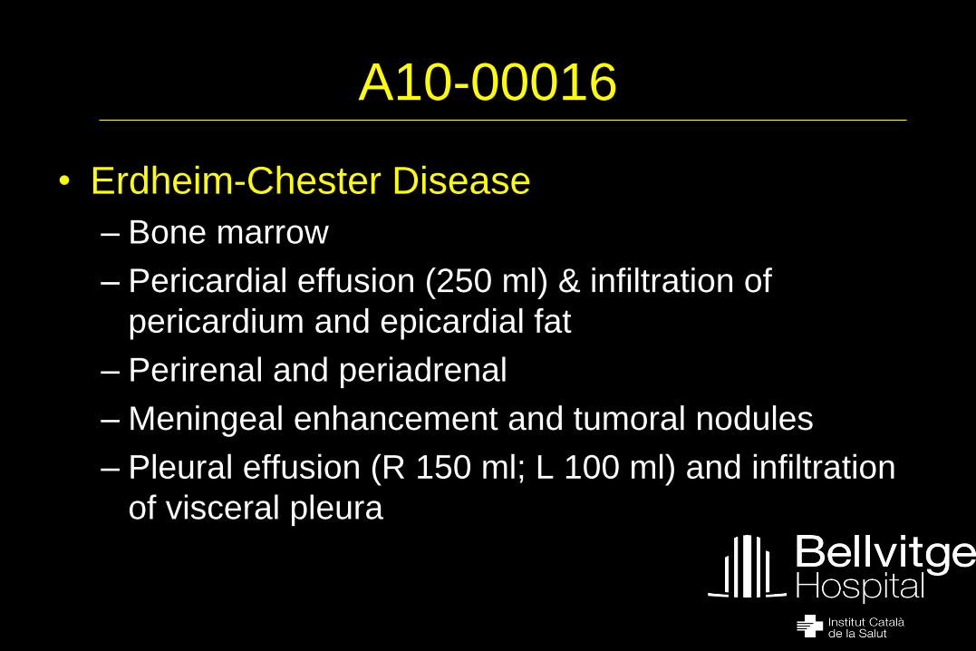
A10-00016
• Erdheim-Chester Disease
– Bone marrow
– Pericardial effusion (250 ml) & infiltration of
pericardium and epicardial fat
– Perirenal and periadrenal
– Meningeal enhancement and tumoral nodules
– Pleural effusion (R 150 ml; L 100 ml) and infiltration
of visceral pleura

Erdheim-Chester Disease
• First report in 1930 by Chester
• Non Langerhans form of histiocytosis
• Over 300 cases reported so farClassification of Histiocytosis syndromes
1. Langerhans-cell Histiocytosis
(prev. Histiocytosis X) Eosinophilic granuloma
Hand-Schüller-Christian disease
Letterer-Siwe disease
2. Non-Langerhans-cell Histiocytosis Hemophagocytic lymphohistiocytosis
Rosai-Dorfman disease
Reticulohistyocitosis
Erdheim Chester disease
3. Malignant Histiocytic disorders • Acute Monocytic leukemia
• Histyocitic lymphoma
• Malignant histiocytosis

Retroperitoneal
• Rarely symptomatic
• Dysuria, abdominal pain,
enlarged kidneys
• CT-scan showing
retroperitoneal and/or
pelvic infiltration

Pericardium & large vessels
• Pericardium
• Myocardium
• Heart valves
• Coronary arteries
• Aorta & aortic branches
• Cava and pulmonary veins
• Systemic hypertension
“coated aorta”Stiff pericardium

Lesson of Anatomy of Dr. Willem Van der Meer- Pieter Van Miereveld, 1617
Redirect the situation!

Deaths from 1994 to 1999
• Internal Medicine
• Critical Care Unit
• Emergency department

Objectives PhD, M. Vadillo
• Population data
• Factors related with the practice of CA
• Analysis of clinico-pathological discrepancy (“blind” clinical diagnoses)
• Aetiologies on discrepancy cases
• Specific analysis on people > 65 a.

Factors related with the practice of CA
Autopsy No autopsy p n=266 (8,9%) n=2718 (91,1%)
AGE (SD) 62.8 (16.6) 68.2 (16,5) <0,001
Mean stay (SD) 13.0 (16,1) 10,2 ( 6,5) <0,02
Men 170 (63,9) 1609 (59,2) NSprevious admission (%) 106 (39.8) 1059 (39,0) NS
Service (%) <0,01
MIV 98 (36,8) 716 (26,3)MIR 104 (39,1) 667 (24,5)URG 64 (24,1) 1335 (49,1)
Initial clinical diagnosis (%) ---Cardiovascular 70 (26,3) 1177 (43,3)Cancer 82 (30,8) 538 (19,8)Respiratory 29 (10,9) 340 (12,5)

Global Discrepancy through time
24,7 25,6
7,5
15,8 15,5
32,1
35,9
21,1
32,5
17,2
46,951,3
37,5
23,7 25,9
0
10
20
30
40
50
60
year 1 year 2 year 3 year 4 year 5
Perc
enta
ge o
f dis
cre
pancy
Global (p = 0,001)
Immediate (p = 0,029 )
Fundamental (p = 0,08 )

Factors related with discrepancy.Logistic Regression
• variables considered:– Age
– Length of stay (days)
– Gender
– Previous admissions
– Service
– Autopsy diagnosis
• Age was the only factor related with higher likelihood of discrepancy RR 1.04; IC 95%; 1.01-1.06

Prevalent Discrepancy Diagnoses
0
2
4
6
8
10
12
14
Pneumonía
PE Ca. Hematol
MI
Septic shock
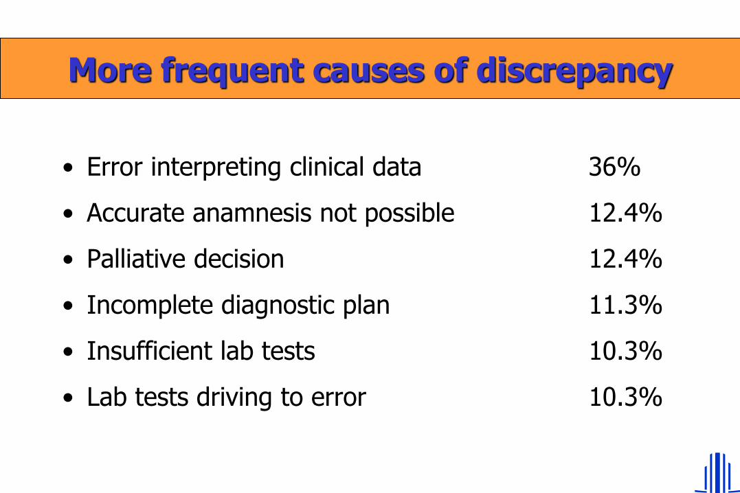
More frequent causes of discrepancy
• Error interpreting clinical data 36%
• Accurate anamnesis not possible 12.4%
• Palliative decision 12.4%
• Incomplete diagnostic plan 11.3%
• Insufficient lab tests 10.3%
• Lab tests driving to error 10.3%


Enrique Simonet. The autopsy-1890
Only a problem for clinicians?

Arch Pathol Lab Med 1998;122:650-655

Resultados, TD M. Aranda
Muestra S- PAAF E-PAAF S-Autop E-autop
Total 80.9 66.7 87.2 44.4
Sangre 34 84.4 61.7 55.6
Hígado 21.3 97.8 31.9 86.7
Bazo 25.3 100 46.8 80
LID 59.6 80 68.1 82.2
La PAAF detecta menos veces patógenos, pero cuando lo hace tiene mas valor.

How clinicians can improve their interest in CA ?
• Realising that pathologists are also interested in it
• Obtaining quick reports on their autopsied patients
• Working together with pathologists on definitive diagnoses
• Training young doctors (residents, students) on CA request
• Including CA rate as one of the service incentives
• Fostering meetings between clinicians and pathologists
• Promoting research projects in this field


Recent papers
• Aline Fusco Fares et al. Discrepancias clínico-patológicas y hallazgos cardiovasculares en 409 autopsias consecutivas. Arq Bras Cardiol 2011;97:449-453
infa
rto m
esen... IA
M
disecc
cion A
o.TEP
Discrepancias
8464 64 62
%

Recent papers
The value of autopsies in the era of high-tech medicine: discrepant findings persist.Chantal C H J Kuijpers, Judith Fronczek, Frank R W van de Goot, Hans W M Niessen, Paul J van Diest, Mehdi Jiwa
Journal of Clinical Pathology 2014; 67:512-
Clinical diagnoses and autopsy findings: discrepancies in critically ill patients.Tejerina E, Esteban A, Fernández-Segoviano P, María Rodríguez-Barbero J, Gordo F, Frutos-Vivar F, Aramburu J, Algaba A, Gonzalo Salcedo García O, Lorente JA.
Crit Care Med 2012; 40:842-

“ It may be more appropriate to savethe autopsy rather than pronounce it DNR“
James E. DalenEditor

www.bellvitgehospital.cat
Related Documents


