Instructions for use Title Clinical and immunological features of pemphigus relapse Author(s) Ujiie, I; Ujiie, H.; Iwata, H.; Shimizu, H. Citation British journal of dermatology, 180(6), 1498-1505 https://doi.org/10.1111/bjd.17591 Issue Date 2019-06 Doc URL http://hdl.handle.net/2115/78255 Rights This is the peer reviewed version of the following article: I Ujiie, H Ujiie, H Iwata, and H Shimizu. (2019) Clinical and immunological features of pemphigus relapse. British Journal of Dermatology, 180(6): 1498-1505., which has been published in final form at https://doi.org/10.1111/bjd.17591. This article may be used for non-commercial purposes in accordance with Wiley Terms and Conditions for Use of Self-Archived Versions. Type article (author version) File Information BJD manuscript_Ujiie I.pdf Hokkaido University Collection of Scholarly and Academic Papers : HUSCAP

Welcome message from author
This document is posted to help you gain knowledge. Please leave a comment to let me know what you think about it! Share it to your friends and learn new things together.
Transcript

Instructions for use
Title Clinical and immunological features of pemphigus relapse
Author(s) Ujiie, I; Ujiie, H.; Iwata, H.; Shimizu, H.
Citation British journal of dermatology, 180(6), 1498-1505https://doi.org/10.1111/bjd.17591
Issue Date 2019-06
Doc URL http://hdl.handle.net/2115/78255
RightsThis is the peer reviewed version of the following article: I Ujiie, H Ujiie, H Iwata, and H Shimizu. (2019) Clinical andimmunological features of pemphigus relapse. British Journal of Dermatology, 180(6): 1498-1505., which has beenpublished in final form at https://doi.org/10.1111/bjd.17591. This article may be used for non-commercial purposes inaccordance with Wiley Terms and Conditions for Use of Self-Archived Versions.
Type article (author version)
File Information BJD manuscript_Ujiie I.pdf
Hokkaido University Collection of Scholarly and Academic Papers : HUSCAP

1
Article type: Original article 1
2
Title: Clinical and immunological features of pemphigus relapse 3
4
I Ujiie, H Ujiie, H Iwata, and H Shimizu 5
6
Department of Dermatology, Hokkaido University Graduate School of Medicine, Sapporo, 7
Japan 8
9
Corresponding author: 10
Hideyuki Ujiie 11
N15 W7, Kita-ku, Sapporo 060-8638, Japan 12
Telephone: +81-11-706-7387 13
Fax: +81-11-706-7820 14
Email: [email protected] 15
16
Running head: Clinical and immunological features of pemphigus relapse 17
18
Funding sources: None 19
20
IRB approval status: Reviewed and approved by the local ethics committee and the 21
Institutional Review Board of Hokkaido University (approval #15-025) 22
23
Conflicts of Interest: No conflicts of interest to declare. 24
25
26
27
28

2
Manuscript word count: 2959 words 29
Abstract word count: 193 words 30
References: 40 31
Figures: 3 32
Tables: 6 33
34
35
What’s already known about this topic? 36
Pemphigus frequently recurs through the disease course. 37
38
What does this study add? 39
Most patients of mucocutaneous pemphigus vulgaris with relapse were taking less than 40
1mg/kg of initial prednisolone and pemphigus recurred when tapered to around 0.1mg/kg of 41
prednisolone. 42

3
Abstract 43
Background: More than half of pemphigus patients experience relapse during the disease 44
course. However, the risk factors and clinical and immunological characteristics of relapse 45
remain largely unclear. 46
47
Objective: To elucidate risk factors and clinical features of pemphigus relapse 48
49
Methods: Retrospective review of the clinical records of 42 pemphigus cases in a single 50
center. 51
52
Results: 61.9% of cases experienced relapse, usually when oral prednisolone was tapered 53
to around 0.1mg/kg. In mucocutaneous pemphigus vulgaris (mcPV), the initial doses of 54
prednisolone were significantly lower in cases with relapse (0.78 ± 0.24 mg/kg) than without 55
relapse (1.01 ± 0.01 mg/kg). At relapse, mcPV shifted to mucosal dominant PV (mPV) 56
(40%), pemphigus foliaceus (PF) (20%) or others (20%). In contrast, the relapsing mPV and 57
PF had the same clinical phenotypes as the initial phenotypes. Patients with both anti-Dsg1 58
and anti-Dsg3 antibodies at onset had recurrence with anti-Dsg3 antibodies alone (40%), 59
with both anti-Dsg1 and Dsg3 antibodies (30%) or with anti-Dsg1 antibody alone (20%), or 60
were subthreshold (10%). 61
62
Conclusion: mcPV shows transitions in clinical phenotype and autoantibody profile at 63
relapse. At least 1mg/kg/day of prednisolone, especially for mcPV cases, and prudent 64
tapering around 0.1mg/kg may lead to better outcomes. 65

4
Introduction 66
Pemphigus is an autoimmune blistering disease characterized by circulating autoantibodies 67
to desmosomal molecules of cell-cell adhesion in the epidermis and/or mucous membrane, 68
followed by blistering or erosion. The clinical phenotypes of pemphigus are defined by the 69
clinical manifestations and the anti-desmoglein (Dsg) antibody profile.1 It is mainly classified 70
into two major types: pemphigus vulgaris (PV) and pemphigus foliaceus (PF). Patients with 71
PV present with erosions and blisters of the skin and/or mucosa, with the histological feature 72
of suprabasal acantholysis. These are caused by anti-Dsg3 autoantibodies or anti-Dsg1 and 73
anti-Dsg3 autoantibodies. In contrast, PF is characterized by scaly and crusted erosions of 74
the skin with subcorneal acantholysis caused by anti-Dsg1 antibodies.2 75
The mainstream therapies for pemphigus are corticosteroids and 76
immunosuppressive agents.3–5 The goal of treatment is to achieve the absence of new 77
lesions with minimal or no therapy. However, many patients experience several relapses and 78
it is often difficult for them to achieve remission. The risk factors and clinical/immunological 79
characteristics of relapse remain largely unclear. 80
To find indicators of optimal initial treatment and to predict relapse, we retrospectively 81
investigated clinical findings such as the age of onset, the clinical phenotypes at onset and 82
relapse, the initial dose of prednisolone (PSL), the disease severity at onset, the time course 83
of anti-Dsg antibody titers and the clinical outcomes precisely in a single center. The novel 84
findings in this study may provide valuable information for pemphigus management. 85
5
METHODS 86
Patients 87
We retrospectively examined patients with pemphigus attending the Department of 88
Dermatology at Hokkaido University Hospital between 2001 and 2017. Forty two patients 89
with at least a 9-month-period of observation and for whom the following information was 90
available from clinical records were selected: age at onset, gender, date of onset of skin 91
and/or mucosal lesions and initiation of systemic PSL, disease severity at onset, period of 92
administration of initial dose of PSL, date of achievement of PSL 10mg, doses of PSL and 93
titers of anti-Dsg1 and/or -Dsg3 antibodies at onset, at the first relapse and at the latest 94
physician office visit, and with or without adjuvant treatment. The patients without relapse 95
were enrolled only by the achievement of tapering PSL to 10mg or less during observation 96
period. Anti-Dsg antibody titers at onset indicated the latest titers before the start of PSL 97
treatment. This study was approved by the local ethics committee and the Institutional 98
Review Board of Hokkaido University. 99
100
Diagnosis, definition of clinical phenotypes and clinical outcomes 101
The clinical diagnosis was made based on the clinical features, histopathology, direct 102
immunofluorescence test and serological tests. The serological tests included indirect 103
immunofluorescence and/or ELISA or chemiluminescent enzyme immunoassay (CLEIA).3 104
Patients with mucosal dominant PV (mPV) predominantly had oral erosions with limited skin 105
involvement, defined as no more than 5 or 6 scattered or isolated erosions or blisters no 106
larger than 5 cm in diameter. Patients with mucocutaneous PV (mcPV) presented oral 107
involvement in addition to more than 6 erosions or blisters on the skin larger than 5 cm in 108
diameter.1 PF patients had cutaneous lesions with anti-Dsg1 antibodies but no mucosal 109
lesions or anti-Dsg3 antibodies. 110
Disease severity was evaluated based on PDAI (pemphigus disease area index) with 111
reference to the clinical records or clinical pictures at onset.6 Disease severity during the 112
clinical course was arbitrarily evaluated on a scale of 0-3 with reference to the clinical 113
6
records. Cutaneous severity was graded according to the body surface area (BSA) 114
involvement of erythema, vesicles, bullae or erosions as follows: 0, no lesions; 1, up to 5% 115
BSA involvement; 2, up to 15% BSA involvement; 3, ≥ 15% BSA or extensive involvement. 116
Oral severity was graded according to the oral surface area (OSA) involvement of vesicles, 117
bullae, or erosions as follows: 0, no lesions; 1, up to 5% OSA involvement; 2, up to 30% 118
OSA involvement; 3, ≥ 30% OSA or extensive involvement. 119
The definitions of therapeutic response were based on the consensus statement for 120
pemphigus.7 Briefly, relapse was defined as the appearance of ≥ 3 new lesions/month that 121
did not heal spontaneously within one week or the extension of established lesions in 122
patients who had achieved disease control. The remission mentioned in this study was 123
defined as including complete and partial remission, as stated in the consensus statement 124
for pemphigus. 7 That is, patients who were taking PSL at ≤ 10 mg/day and/or minimal 125
adjuvant therapy with the absence of new or established lesions, or the presence of transient 126
new lesions that healed spontaneously within one week. 127
128
Statistical analysis 129
GraphPad Prism ver. 6.00 for Windows (GraphPad Software, San Diego CA) was used for 130
statistical analyses. The Mann-Whitney rank sum test was used to determine differences in 131
various clinical features between patients with relapse versus without relapse or differences 132
in anti-Dsg antibody at relapse between clinical phenotypes. The Dunn’s multiple 133
comparisons test was performed to determine differences in clinical phenotypes. The 134
Wilcoxon matched-pairs signed-rank test was used to determine the differences in the anti-135
Dsg antibody titers at onset versus at relapse. The Fisher’s exact probability test was used 136
to determine differences in relapse rate. P < 0.05 was considered statistically significant. 137
138

7
RESULTS 139
Clinical features and anti-Dsg antibody profile at pemphigus onset 140
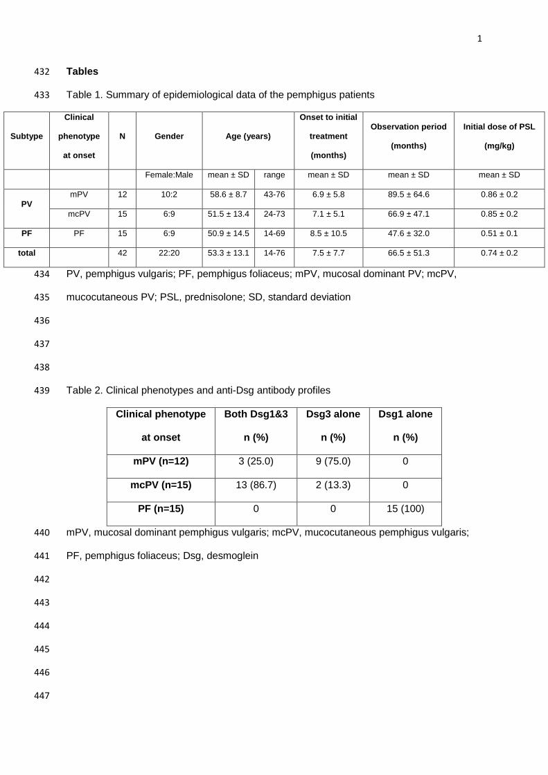
Of the 42 patients, 27 (64.3%) were diagnosed with PV and 15 (35.7%) were diagnosed with 141
PF. The PV patients consisted of 12 mPV (28.6%) and 15 mcPV (35.7%). The patients’ age 142
at onset, the period between onset and the initiation of oral PSL were not statistically 143
different between clinical phenotypes. The female:male ratios for mPV, mcPV and PF were 144
10:2, 6:9 and 6:9, respectively. The gender ratios in previous reports were various,8–16 and 145
the female:male ratio of mPV in our study was remarkably high. The observation periods of 146
mPV, mcPV and PF were 89.5 ± 64.6, 66.9 ± 47.1 and 47.6 ± 32.0 months, respectively. 147
Initial doses of PSL were higher for mPV (0.86 ± 0.2 mg/kg) and mcPV (0.85 ± 0.2 mg/kg) 148
than for PF (0.51 ± 0.1 mg/kg) (Table 1). 149
Next, we examined the autoantibody profiles at onset. For mPV, 3 of 12 cases (25%) 150
had both anti-Dsg1 and anti-Dsg3 antibodies and 9 of 12 cases (75%) had anti-Dsg3 151
antibodies alone at onset. In contrast, in mcPV, 13 of 15 cases (86.7%) had both anti-Dsg1 152
and anti-Dsg3 antibodies at onset and 2 of 15 cases (13.3%) had anti-Dsg3 antibodies alone 153
at onset. All 16 PF patients had anti-Dsg1 antibodies alone (Table 2). It is noteworthy that 2 154
of 11 cases (18.2%) with anti-Dsg3 antibodies alone presented as mcPV. As expected, anti-155
Dsg1 antibody titers were significantly higher in mcPV (161.8 ± 230.8 index value) and PF 156
(327.8 ± 385.2 index value) than in mPV (13.8 ± 23.0 index value). Interestingly, anti-Dsg3 157
antibody titers were similar in mPV (329.0 ± 288.7 index value) and mcPV (392.1 ± 372.3 158
index value). These results confirm that anti-Dsg1 antibodies and anti-Dsg3 antibodies 159
strongly correlate with the development of cutaneous lesions and mucosal lesions, 160
respectively. 161
162
A possible risk factor for pemphigus relapse 163
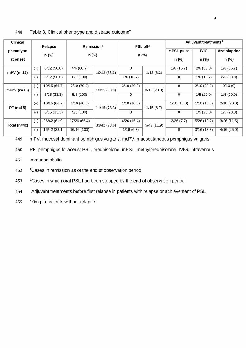
The relapse rates of mPV, mcPV and PF were 50%, 66.7% and 66.7%, respectively (Table 164
3). We compared clinical indexes of patients with relapse versus without relapse. Notably, in 165
mcPV, the initial doses of PSL were significantly lower in cases with relapse (0.78 ± 0.24 166

8
mg/kg) than without relapse (1.01 ± 0.01 mg/kg) (Figure 1a). The duration of initial dose of 167
PSL or the cumulative dose of PSL to 10mg did not significantly differ between patients with 168
relapse versus without relapse (Figure 1b, c). This indicates that the pace of reduction did 169
not differ between patients with relapse and patients without relapse. Adjuvant therapies 170
were used in a few cases with relapse before first relapse or without relapse before the 171
reduction to PSL 10mg (Table3). The relapse rate was similar in patients with versus without 172
adjuvant therapies, although statistical analysis could not be performed due to the small 173
number of patients with adjuvant therapy. There were no significant differences in the period 174
between onset and the administration of oral PSL (data not shown) and in the age or anti-175
Dsg antibody titers at onset between patients with relapse versus without relapse (Figure 1d-176
f). The disease severity at onset did not significantly differ between patients with relapse 177
versus without relapse (Figure 1g). Thus, a lower initial dose of PSL is a possible risk factor 178
for mcPV relapse. 179
180
Clinical findings at first relapse 181
Next, we analyzed the clinical data at first relapse. The durations between the initiation of 182
PSL and first relapse in mPV, mcPV and PF were 28.7 ± 11.6, 19.1 ± 9.4 and 15.9 ± 12.8 183
months, respectively. Importantly, the doses of PSL (mg/kg) at first relapse were similar 184
among mPV (0.11 ± 0.09), mcPV (0.12 ± 0.07) and PF (0.14 ± 0.09) (Figure 2a). It is worth 185
noting that most of the patients experienced relapse when taking around 0.1mg/kg of PSL 186
irrespective of clinical phenotype. Patients who relapsed at PSL >10mg/day numbered one 187
each for mPV, mcPV and PF. Anti-Dsg1 antibody titers at first relapse were higher in PF 188
(115.0 ± 104.8 index value) than in mcPV (36.5 ± 37.8 index value), but the difference was 189
not statistically significant (Figure 2b). Anti-Dsg3 antibody titers at first relapse were similar 190
between mPV (181.2 ± 326.9) and mcPV (109.6 ± 176.5) (Figure 2c). In Japan, the method 191
for measuring anti-Dsg antibodies shifted from ELISA to chemiluminescent enzyme 192
immunoassay (CLEIA) in 2013. To appropriately compare anti-Dsg antibody titers at onset to 193
those at first relapse, we extracted the cases in which the titers were examined by ELISA 194
9
alone or CLEIA alone in the 2 timepoints. In PV and PF cases, anti-Dsg1 antibody titers 195
were higher at onset (216.3 ± 336.7 index value) than at first relapse (58.2 ± 65.6 index 196
value) (Figure 2d). Similarly, in PV, anti-Dsg3 antibody titers were significantly higher at 197
onset (273.5 ± 250.9 index value) than those at first relapse (33.7 ± 34.4 index value) 198
(Figure 2e). Most of the patients experienced a relapse when the antibody titers were less 199
than those at onset. The average ratio of anti-Dsg1 and Dsg3 antibody titers at relapse to 200
those at onset were 0.81 and 0.3, respectively (Figure 2f). 201
202
Transitions of clinical and immunological phenotypes at first relapse 203
At first relapse, clinical phenotype and autoantibody profile were changed from initial 204
presentation in some patients. Although mPV and PF patients developed the same clinical 205
phenotypes as the initial phenotypes at first relapse, mcPV patients (n=10) shifted to mPV 206
(40%), mcPV (20%), PF (20%) or others (20%) (Table 4). In mcPV patients, there were two 207
patients who had only cutaneous lesions with both anti-Dsg1 and anti-Dsg3 antibodies at 208
first relapse. Although they could be possibly classified as cutaneous PV or PF, we classified 209
them as “others” because of unverified histopathology of those lesions at relapse. Of note, 210
no patients with mucosal lesions alone at onset developed cutaneous lesions at relapse, and 211
vice versa. For example, mPV cases did not shift to mcPV or PF. Next, we analyzed the time 212
course of autoantibody titers in cases with relapse. Patients with both anti-Dsg1 and anti-213
Dsg3 antibodies at onset (n=10) showed anti-Dsg3 antibodies alone (40%), both anti-Dsg1 214
and anti-Dsg3 antibodies (30%) or anti-Dsg1 autoantibodies alone (20%), or were 215
subthreshold (10%) at first relapse. In contrast, patients with anti-Dsg3 antibodies alone at 216
onset (n=6) had anti-Dsg3 antibodies alone (83.3%) or were subthreshold (16.7%) at first 217
relapse. Patients with anti-Dsg1 antibodies alone at onset (n=10) had anti-Dsg1 antibodies 218
alone (80%) or were subthreshold (20%) at first relapse (Table 4 and 5). 219
In this study, clinical phenotype shifted from mcPV to PF in four of the cases by the 220
end of the observation period. In 3 of those cases, anti-Dsg1 and anti-Dsg3 antibodies were 221
both positive at onset and only anti-Dsg1 antibodies were detected in the PF phase. One of 222

10
the cases initially presented as mcPV with anti-Dsg3 antibodies alone, relapsed as mPV with 223
slightly elevated anti-Dsg3 antibody titers and finally developed into PF with anti-Dsg1 224
antibodies alone. In contrast, none of the cases shifted from PF to PV. The time course of 225
disease severity and autoantibody titers in representative cases with clinical transition from 226
mcPV to mPV and from mcPV to PF are shown in Figure 3. 227
228
Clinical outcomes and anti-Dsg antibody titers at the end of observation period 229
The remission rates of mPV, mcPV and PF were 83.3%, 80% and 73.3%, respectively. 230
When the patients who achieved remission before relapse were included, the remission 231
rates for mPV, mcPV and PF were 91.7%, 93.3% and 93.3%, respectively. Of the patients 232
with at least one relapse, the remission rates of mPV, mcPV and PF were 66.7%, 70% and 233
60%, respectively. The patients who were finally off PSL accounted for 8.3%, 20% and 6.7% 234
of the mPV, mcPV and PF cases, respectively (Table 3). Anti-Dsg antibodies became 235
negative in most cases during the disease course. Anti-Dsg3 was more prone to be negative 236
in mcPV (80%) than in mPV (41.7%) (Table 6). 237
11
DISCUSSION 238
Nowadays, systemic corticosteroids are the mainstay treatment for pemphigus. In Japan, 239
1mg/kg of PSL is recommended for moderate to severe cases.3 At our institute, most cases 240
are started with PSL at 1mg/kg for PV and at 0.5mg/kg for PF. Notably, less than 1mg/kg of 241
initial PSL in mcPV cases were associated with relapse. mcPV is reported to be less 242
responsive to treatment than mPV and PF.17 A report on 155 patients with pemphigus 243
showed the initial dose of corticosteroids to have no significant effect on the prognosis.16 244
However, all of the patients in the study were treated with systemic corticosteroids at more 245
than 1mg/kg. It also has been reported that dosages higher than 1mg/kg have no advantage 246
over dosages of 1mg/kg in terms of the time to disease control 13 and that stratifying the 247
initial dose of PSL according to PV disease severity at presentation is important.18 From 248
these findings, we consider that a sufficient dose of PSL (1mg/kg) is important for the initial 249
treatment, especially for mcPV cases. 250
Interestingly, there was no significant difference in relapse rate between PF and PV, 251
even though the initial dose of PSL for PF was roughly half that for PV. PF is considered to 252
have a better prognosis than PV.10 Therefore, the recommended dose of PSL might differ 253
according to the clinical phenotype. It is intriguing that the dose of PSL at first relapse is 254
roughly same around 0.1mg/kg regardless of clinical phenotype. 255
PV disease severity correlates with anti-Dsg antibody titer.1,17,19–22 It is noteworthy 256
that anti-Dsg1 and anti-Dsg3 antibody titers at onset did not significantly correlate with 257
relapse in this study. Because of the substantial differences of anti-Dsg antibody titers 258
among individuals, these autoantibodies can be used to monitor the disease course within a 259
given individual but not to compare disease severity between patients.23 Anti-Dsg antibody 260
titers were lower at first relapse than at onset in this study. In most cases, it took some time 261
after onset until anti-Dsg antibodies were analyzed. Therefore, the primary autoantibody 262
titers, which were evaluated before the initiation of systemic PSL, might have been higher 263
than those at true onset. Thus, we should be careful to manage when autoantibody titers 264
appear to increase even if those are lower than the levels at onset. 265
12
This study has shown cases with a transition of anti-Dsg antibody profile associated 266
with changes of clinical phenotype at first relapse. Several reports have described the 267
transition of clinical phenotype between PV and PF.24–32 The transition from PF to PV is less 268
common than that from PV to PF. In cases with a clinical transition from PV to PF, the 269
change of antibody profile showed two patterns: a shift from anti-Dsg3 antibodies alone to 270
anti-Dsg1 antibodies alone,24,28,29 or a shift from both anti-Dsg1 and anti-Dsg3 antibodies to 271
anti-Dsg1 antibodies alone.24,25,27 In contrast, cases with a clinical transition from PF to PV 272
showed the change of antibody profile from anti-Dsg1 antibodies alone to both anti-Dsg1 273
and anti-Dsg3 or to anti-Dsg3 antibodies alone.26,31 On the other hand, a case with a clinical 274
transition from mPV to mcPV had only anti-Dsg3 antibodies at the mPV phase with the late 275
development of anti-Dsg1 antibodies.33 These cases were explained by the ‘epitope 276
spreading’ hypothesis.34 However, the epitope spreading phenomenon is regarded as being 277
rarely seen in PV and PF,35 and the mechanism remains controversial. 278
Clinical relapse is commonly seen in pemphigus. The relapse rates have ranged 279
between 13% and 82%.12–16,36–38 We found that none of the clinical factors, such as age, 280
clinical phenotype or disease severity at onset had an impact on the occurrence of relapse. 281
The study of 134 patients with pemphigus indicated that those with mucosal involvement and 282
younger age (< 61 years) at presentation were more likely to achieve complete remission off 283
therapy.38 In contrast, other reports have suggested that young age at diagnosis (< 40 284
years), mucosal involvement at diagnosis or higher anti-Dsg1 or anti-Dsg3 antibody titers 285
related to higher likelihood of recurrence.8,12,16,36,39 Thus, there are no factors that 286
consistently relate to clinical outcomes. 287
Complete or partial remission rates have been reported as ranging 50% and 100%. 288
The patients with off therapy varies from 1.4 to 75% and with minimal therapy varies from 13 289
to 94.4%.12,14–16,18,36,38,40 The variability between the reports may be due to differences in 290
disease severity, follow-up period or relapse definition. In our study, the remission rate was 291
65% in PV and PF cases with relapse and of whom 15.4% could be off PSL. The remission 292
13
rate could be higher with prolonged observation period, and the incidence of relapse may not 293
necessarily lead to poor outcome. 294
In conclusion, at least 1mg/kg dose of PSL is important for preventing relapse, 295
especially for mcPV. In addition, when the dose of PSL is tapered to roughly 0.1mg/kg, we 296
should carefully monitor for elevated anti-Dsg antibody titers and for clinical appearance, 297
which can differ from that at onset. 298
299
14
References 300
1 Amagai M, Tsunoda K, Zillikens D, et al. The clinical phenotype of pemphigus is 301
defined by the anti-desmoglein autoantibody profile. J Am Acad Dermatol 1999; 302
40:167–70. 303
2 Amagai M, Komai A, Hashimoto T, et al. Usefulness of enzyme-linked immunosorbent 304
assay using recombinant desmogleins 1 and 3 for serodiagnosis of pemphigus. Br J 305
Dermatol 1999; 140:351–7. 306
3 Amagai M, Tanikawa A, Shimizu T, et al. Japanese guidelines for the management of 307
pemphigus. J Dermatol 2014; 41:471–86. 308
4 Harman KE, Brown D, Exton LS, et al. British Association of Dermatologists’ 309
guidelines for the management of pemphigus vulgaris 2017. Br J Dermatol 2017; 310
177:1170–201. 311
5 Murrell DF, Peña S, Joly P, et al. Diagnosis and Management of Pemphigus: 312
recommendations by an International Panel of Experts. J Am Acad Dermatol 2018. 313
doi:10.1016/j.jaad.2018.02.021. 314
6 Rosenbach M, Murrell DF, Bystryn JC, et al. Reliability and convergent validity of two 315
outcome instruments for pemphigus. J Invest Dermatol 2009; 129:2404–10. 316
7 Murrell DF, Dick S, Ahmed AR, et al. Consensus statement on definitions of disease, 317
end points, and therapeutic response for pemphigus. J Am Acad Dermatol 2008; 318
58:1043–6. 319
8 Herrero-Gonzälez JE, Iranzo P, Benítez D, et al. Correlation of immunological profile 320
with phenotype and disease outcome in pemphigus. Acta Derm Venereol 2010; 321
90:401–5. 322
15
9 Ishii N, Maeyama Y, Karashima T, et al. A clinical study of patients with pemphigus 323
vulgaris and pemphigus foliaceous: An 11-year retrospective study (1996-2006). Clin 324
Exp Dermatol 2008; 33:641–3. 325
10 Kridin K, Zelber-Sagi S, Bergman R. Pemphigus Vulgaris and Pemphigus Foliaceus: 326
Differences in Epidemiology and Mortality. Acta Derm Venereol 2017; :0. 327
11 Shah AA, Seiffert-Sinha K, Sirois D, et al. Development of a Disease Registry for 328
Autoimmune Bullous Diseases: Initial Analysis of The Pemphigus Vulgaris Subset. 329
Acta Derm Venereol 2015; 95:86–90. 330
12 Khaled A, Taazayet S Ben, Ben Alaya N, et al. The course and prognosis of 331
pemphigus in 47 Tunisian patients. J Eur Acad Dermatology Venereol 2013; 27:81–5. 332
13 Ramassamy S, Agrawal P, Sathishkumar D, et al. Clinical , immunological profile and 333
follow up of patients with pemphigus : A study from. Indian J Dermatol Venereol 334
Leprol 2018; :1–6. 335
14 Svecova D. Pemphigus vulgaris : a clinical study of 44 cases over a 20-year period. 336
Int J Dermatol 2015; 54:1138–44. 337
15 Bai YX, Zhang LM, Xiao T, Chen HD. A 6-year treatment experience for pemphigus: 338
Retrospective study of 69 Chinese patients. Dermatol Ther 2016; 29:84–7. 339
16 Mimouni D, Bar H, Gdalevich M, et al. Pemphigus, analysis of 155 patients. J Eur 340
Acad Dermatology Venereol 2010; 24:947–52. 341
17 Cozzani E, Di Zenzo G, Riva S, et al. Are clinical phenotype and autoantibody profile 342
always concordant in pemphigus? A study in a cohort of pemphigus patients. Eur J 343
Dermatology 2013; 23:40–8. 344
18 Lyakhovitsky A, Baum S, Scope A, et al. The impact of stratifying initial dose of 345
corticosteroids by severity of pemphigus vulgaris on long-term disease severity. Int J 346
Dermatol 2011; 50:1014–9. 347

16
19 Daneshpazhooh M, Chams-davatchi C, Khamesipour A, et al. Desmoglein 1 and 3 348
enzyme-linked immunosorbent assay in Iranian patients with pemphigus vulgaris: 349
Correlation with phenotype, severity, and disease activity. J Eur Acad Dermatology 350
Venereol 2007; 21:1319–24. 351
20 Sharma VK, Prasad HRY, Khandpur S, Kumar A. Evaluation of desmoglein enzyme-352
linked immunosorbent assay (ELISA) in Indian patients with pemphigus vulgaris. Int J 353
Dermatol 2006; 45:518–22. 354
21 Harman KE, Seed PT, Gratian MJ, et al. The severity of cutaneous and oral 355
pemphigus is related to desmoglein 1 and 3 antibody levels. Br J Dermatol 2001; 356
144:775–80. 357
22 Cheng SW, Kobayashi M, Kinoshita-Kuroda K, et al. Monitoring disease activity in 358
pemphigus with enzyme-linked immunosorbent assay using recombinant desmogleins 359
1 and 3. Br J Dermatol 2002; 147:261–5. 360
23 Fujio Y, Kojima K, Hashiguchi M, et al. Validation of chemiluminescent enzyme 361
immunoassay in detection of autoantibodies in pemphigus and pemphigoid. J 362
Dermatol Sci 2017; 85:208–15. 363
24 Espana A, Koga H, Suarez-Fernandez R, et al. Antibodies to the amino-terminal 364
domain of desmoglein 1 are retained during transition from pemphigus vulgaris to 365
pemphigus foliaceus. Eur J Dermatol 2014; 24:174–9. 366
25 Toth, GG, Pas, HH, Jonkman M. Transition of pemphigus vulgaris into pemphigus 367
foliaceus confirmed by antidesmoglein ELISA profile. Int J Dermatol 2002; 41:525–7. 368
26 Komai A, Amagai M, Ishii K, et al. The clinical transition between pemphigus foliaceus 369
and pemphigus vulgaris correlates well with the changes in autoantibody profile 370
assessed by an enzyme-linked immunosorbent assay. Br J Dermatol 2001; 371
144:1177–82. 372

17
27 Harman KE, Gratian MJ, Shirlaw PJ, et al. The transition of pemphigus vulgaris into 373
pemphigus foliaceus: A reflection of changing desmoglein 1 and 3 autoantibody levels 374
in pemphigus vulgaris. Br J Dermatol 2002; 146:684–7. 375
28 Ito T, Moriuchi R, Kikuchi K, Shimizu S. Rapid transition from pemphigus vulgaris to 376
pemphigus foliaceus. J. Eur. Acad. Dermatol. Venereol. 2016; 30:455–7. 377
29 Tsuji Y, Kawashima T, Yokota K, et al. Clinical and Serological Transition From 378
Pemphigus Vulgaris to Pemphigus Foliaceus Demonstrated by Desmoglein ELISA 379
System. Arch Dermatol 2002; 138:95–6. 380
30 Ng PPL, Thng STG. Three cases of transition from pemphigus vulgaris to pemphigus 381
foliaceus confirmed by desmoglein ELISA. Dermatology 2005; 210:319–21. 382
31 Awazawa R, Yamamoto Y, Gushi M, et al. Case of pemphigus foliaceus that shifted 383
into pemphigus vulgaris after adrenal tumor resection. J Dermatol 2007; 34:549–55. 384
32 Ishii K, Amagai M, Ohata Y, et al. Development of pemphigus vulgaris in a patient 385
with pemphigus foliaceus: antidesmoglein antibody profile shift confirmed by enzyme-386
linked immunosorbent assay. J Am Acad Dermatol 2000; 42:859–61. 387
33 Miyagawa S, Amagai M, Iida T, et al. Late development of antidesmoglein 1 388
antibodies in pemphigus vulgaris: Correlation with disease progression. Br J Dermatol 389
1999; 141:1084–7. 390
34 Chan LS, Vanderlugt CJ, Hashimoto T, et al. Epitope spreading: Lessons from 391
autoimmune skin diseases. J Invest Dermatol 1998; 110:103–9. 392
35 Ohyama B, Nishifuji K, Chan PT, et al. Epitope Spreading Is Rarely Found in 393
Pemphigus Vulgaris by Large-Scale Longitudinal Study Using Desmoglein 2–Based 394
Swapped Molecules. J Invest Dermatol 2012; 132:1158–68. 395

18
36 Daneshpazhooh M, Sedigh VZ, Balighi K, et al. Immunologic prediction of relapse in 396
patients with pemphigus vulgaris (PV) in clinical remission. J Am Dermatology 2016; 397
74:1160–5. 398
37 Shahidi-Dadras M, Karami A, Toosy P, Shafiyan A. Pulse versus oral 399
methylprednisolone therapy in pemphigus vulgaris. Arch Iran Med 2007; 10:1–6. 400
38 Almugairen N, Hospital V, Bedane C, et al. Assessment of the rate of long-term 401
complete remission off therapy in patients with pemphigus treated with different 402
regimens including medium- and high-dose corticosteroids. J Am Acad Dermatol 403
2013; 69:583–8. 404
39 Saleh MA. A prospective study comparing patients with early and late relapsing 405
pemphigus treated with rituximab. J Am Acad Dermatol 2018; :1–7. 406
40 Herbst A, Bystryn JC. Patterns of remission in pemphigus vulgaris. J Am Acad 407
Dermatol 2000; 42:422–7. 408
409

19
Figure legend 410
Figure 1. The clinical findings for each clinical phenotype with or without relapse. (a) Initial 411
dose of PSL. (b) Duration of initial dose of PSL. (c) Cumulative dose of PSL to 10mg. (d) 412
Age at onset. Anti-Dsg1 (e) and anti-Dsg3 (f) antibody titers at onset. (g) Disease severity at 413
onset. The bar indicates the median for each value. * P < 0.05. 414
415
Figure 2. Dose of PSL and titers of antibody at first relapse. (a) Dose of PSL at first relapse. 416
Anti-Dsg1 (b) and anti-Dsg3 (c) antibody titers at first relapse. The titers of anti-Dsg1 (d) and 417
anti-Dsg3 (e) at onset and first relapse. (f) The ratio of antibody titers at relapse to those at 418
onset. The bar indicates the median for each value. *P < 0.05, ** P < 0.01. NS, not significant. 419
420
Figure 3. The transition of clinical and anti-Dsg antibody profiles through the disease course. 421
The patients with mcPV at onset shifted to mPV (a) or PF (b). The cutoff values of anti-Dsg 422
antibodies changed in April 2014 at our facility. 423
424
Table Legend 425
Table 1. Summary of epidemiological data of the pemphigus patients 426
Table 2. Clinical phenotypes and anti-Dsg antibody profiles 427
Table 3. Clinical phenotype and disease outcome 428
Table 4. Transition of clinical phenotype at first relapse 429
Table 5. Transition of anti-Dsg autoantibody profiles at first relapse 430
Table 6. Rate of negative anti-Dsg antibody at the end of observation period 431
1
Tables 432
Table 1. Summary of epidemiological data of the pemphigus patients 433
Subtype
Clinical
phenotype
at onset
N Gender Age (years)
Onset to initial
treatment
(months)
Observation period
(months)
Initial dose of PSL
(mg/kg)
Female:Male mean ± SD range mean ± SD mean ± SD mean ± SD
PV mPV 12 10:2 58.6 ± 8.7 43-76 6.9 ± 5.8 89.5 ± 64.6 0.86 ± 0.2
mcPV 15 6:9 51.5 ± 13.4 24-73 7.1 ± 5.1 66.9 ± 47.1 0.85 ± 0.2
PF PF 15 6:9 50.9 ± 14.5 14-69 8.5 ± 10.5 47.6 ± 32.0 0.51 ± 0.1
total 42 22:20 53.3 ± 13.1 14-76 7.5 ± 7.7 66.5 ± 51.3 0.74 ± 0.2
PV, pemphigus vulgaris; PF, pemphigus foliaceus; mPV, mucosal dominant PV; mcPV, 434
mucocutaneous PV; PSL, prednisolone; SD, standard deviation 435
436
437
438
Table 2. Clinical phenotypes and anti-Dsg antibody profiles 439
Clinical phenotype
at onset
Both Dsg1&3
n (%)
Dsg3 alone
n (%)
Dsg1 alone
n (%)
mPV (n=12) 3 (25.0) 9 (75.0) 0
mcPV (n=15) 13 (86.7) 2 (13.3) 0
PF (n=15) 0 0 15 (100)
mPV, mucosal dominant pemphigus vulgaris; mcPV, mucocutaneous pemphigus vulgaris; 440
PF, pemphigus foliaceus; Dsg, desmoglein 441
442
443
444
445
446
447
2
Table 3. Clinical phenotype and disease outcome” 448
Clinical
phenotype
at onset
Relapse
n (%)
Remission1
n (%)
PSL off2
n (%)
Adjuvant treatments3
mPSL pulse
n (%)
IVIG
n (%)
Azathioprine
n (%)
mPV (n=12) (+) 6/12 (50.0) 4/6 (66.7)
10/12 (83.3) 0
1/12 (8.3) 1/6 (16.7) 2/6 (33.3) 1/6 (16.7)
(-) 6/12 (50.0) 6/6 (100) 1/6 (16.7) 0 1/6 (16.7) 2/6 (33.3)
mcPV (n=15) (+) 10/15 (66.7) 7/10 (70.0)
12/15 (80.0) 3/10 (30.0)
3/15 (20.0) 0 2/10 (20.0) 0/10 (0)
(-) 5/15 (33.3) 5/5 (100) 0 0 1/5 (20.0) 1/5 (20.0)
PF (n=15) (+) 10/15 (66.7) 6/10 (60.0)
11/15 (73.3) 1/10 (10.0)
1/15 (6.7) 1/10 (10.0) 1/10 (10.0) 2/10 (20.0)
(-) 5/15 (33.3) 5/5 (100) 0 0 1/5 (20.0) 1/5 (20.0)
Total (n=42) (+) 26/42 (61.9) 17/26 (65.4)
33/42 (78.6) 4/26 (15.4)
5/42 (11.9) 2/26 (7.7) 5/26 (19.2) 3/26 (11.5)
(-) 16/42 (38.1) 16/16 (100) 1/16 (6.3) 0 3/16 (18.8) 4/16 (25.0)
mPV, mucosal dominant pemphigus vulgaris; mcPV, mucocutaneous pemphigus vulgaris; 449
PF, pemphigus foliaceus; PSL, prednisolone; mPSL, methylprednisolone; IVIG, intravenous 450
immunoglobulin 451
1Cases in remission as of the end of observation period 452
2Cases in which oral PSL had been stopped by the end of observation period 453
3Adjuvant treatments before first relapse in patients with relapse or achievement of PSL 454
10mg in patients without relapse 455

3
Table 4. Transition of clinical phenotype at first relapse 456
Onset First relapse n (%)
mPV (n=6) mPV 6 (100)
mcPV (n=10)
mPV 4 (40.0)
mcPV 2 (20.0)
PF 2 (20.0)
Others1 2 (20.0)
PF (n=10) PF 10 (100)
mPV, mucosal dominant pemphigus vulgaris; mcPV, mucocutaneous pemphigus vulgaris; 457
PF, pemphigus foliaceus 458
1Others: only cutaneous lesions with both anti-Dsg1 and anti-Dsg3 antibodies. 459
460
461
462
Table 5. Transition of anti-Dsg autoantibody profiles at first relapse 463
Onset First relapse n (%)
Dsg3 (n=6) Dsg3 5 (83.3)
Subthreshold 1 (16.7)
Dsg1/3 (n=10)
Dsg3 4 (40.0)
Dsg1/3 3 (30.0)
Dsg1 2 (20.0)
Subthreshold 1 (10.0)
Dsg1 (n=10) Dsg1 8 (80.0)
Subthreshold 2 (20.0)
Dsg, desmoglein 464
465
466

4
Table 6. Rate of negative anti-Dsg antibody at the end of observation period 467
Clinical phenotype
at onset Anti-Dsg antibody
Positive at onset
n (%)
Turned negative
n (%)
mPV (n=12) Dsg1 3/12 (23.1) 3/3 (100)
Dsg3 12/12 (100) 5/12 (41.7)
mcPV (n=15) Dsg1 13/15 (86.7) 11/13 (84.6)
Dsg3 15/15 (100) 12/15 (80.0)
PF (n=15) Dsg1 15/15 (100) 9/15 (60.0)
Dsg3 0 -
mPV, mucosal dominant pemphigus vulgaris; mcPV, mucocutaneous pemphigus vulgaris; 468
PF, pemphigus foliaceus; Dsg, desmoglein 469
470


Related Documents


![Manifestations buccales du pemphigus paranéoplasique · 2013-02-08 · différencier le pemphigus paranéoplasique du pemphigus vulgaire [41, 63, 75, 83, 93, 100]. Par contre, la](https://static.cupdf.com/doc/110x72/5f49b405f3d6f653f74e2428/manifestations-buccales-du-pemphigus-paranoplasique-2013-02-08-diffrencier.jpg)


