CLINICAL AND VACCINE IMMUNOLOGY, Oct. 2008, p. 1580–1589 Vol. 15, No. 10 1556-6811/08/$08.000 doi:10.1128/CVI.00173-08 Copyright © 2008, American Society for Microbiology. All Rights Reserved. Clinical and Immunologic Features of an Atypical Intracranial Mycobacterium avium Complex (MAC) Infection Compared with Those of Pulmonary MAC Infections Mouhannad Sadek, 2 Feng Yun Yue, 1 Erika Yue Lee, 1 Gabor Gyenes, 1 R. Brad Jones, 1 Victor Hoffstein, 2 David G. Munoz, 2 Ignatius Fong, 2 and Mario Ostrowski 1,2 * Clinical Sciences Division and Department of Immunology, University of Toronto, Toronto, Ontario, Canada, 1 and St. Michael’s Hospital, University of Toronto, Toronto, Ontario, Canada 2 Received 15 May 2008/Returned for modification 4 June 2008/Accepted 18 July 2008 Members of the Mycobacterium avium complex (MAC) may cause chronic pulmonary infections in otherwise healthy elderly persons but rarely invade parts of the body outside of the lungs in immunocompetent hosts. We present a case of an isolated intracranial MAC infection in an apparently immunocompetent individual and review previous reports. We studied the T-cell and monocyte responses in healthy volunteers, individuals with a pulmonary MAC infection, and one individual with an isolated intracranial MAC infection. Genomic DNA from the individual with the brain MAC infection was studied for gamma interferon (IFN-) receptor mutations. Individuals with localized pulmonary MAC infections showed increased activation of monocytes and enhanced monocyte and T-cell tumor necrosis factor alpha (TNF-) production in response to lipopoly- saccharide and MAC antigens but defects in T-cell IFN- secretion. The individual with an intracranial MAC infection showed a lack of monocyte activation and deficiencies in both monocyte and T-cell TNF- production and monocyte interleukin-12 (IL-12) production but had preserved T-cell IFN- production. Mutations or deletions in the IFN- receptor were not detected in the individual with the intracranial MAC infection. Our data suggest that distinct immune defects characterize two different manifestations of MAC infection. A relative defect in IFN- production in response to MAC may predispose an individual to localized but partially controlled lung disease, whereas defects leading to reduced IL-12 and TNF- production may allow the dissemination of MAC. Further studies delineating the potential role of TNF- in limiting the spread of MAC outside the lung are warranted. Members of the Mycobacterium avium complex (MAC) are ubiquitous nontuberculous mycobacteria that seldom cause disease in immunocompetent hosts. In human immunodefi- ciency virus (HIV)-infected individuals who have severe CD4 T-cell depletion, MAC causes disseminated infections. How- ever, MAC can also cause chronic pulmonary infections in elderly persons with apparently normal immune function (34). Characteristically, some older nonsmoking women, generally those with bronchiectasis, may develop right middle lobe infil- trates and a nonproductive cough, termed “Lady Windermere syndrome.” The coordinated function of T cells and monocytes is generally required to resolve mycobacterial infections (10). Few case reports have demonstrated that isolated intracranial MAC infections in the absence of an underlying medical con- dition account for immunosuppression (7, 8, 32, 40). However, immune defects or genetic predispositions were not adequately described in those reports. Immune defects have previously been reported in elderly persons with pulmonary MAC infec- tions. These have specifically included a level of higher pro- duction of interleukin-10 (IL-10) but lower concentrations of gamma interferon (IFN-), IL-12, and tumor necrosis factor alpha (TNF-) in peripheral blood mononuclear cells (PBMCs) in response to heat-killed MAC (17, 35, 36, 41). Granuloma formation, which is essential for mycobacterial control, consists of a complex interplay between activated - T-cell-receptor-expressing cells that have been primed to re- spond to mycobacterial antigens and macrophages that have engulfed organisms. Their overall effect is to contain the bacilli and prevent further spread. Signals which aid macrophages in granuloma formation and the clearance of mycobacterial in- fections are complex and are incompletely understood. These include IFN- and TNF-. TNF- is secreted by macrophages and T cells and is able to induce mycobacteriostatic and my- cobactericidal activity in both cultured mouse and human mac- rophages (3, 14). Recently, TNF- has been demonstrated to recruit T cells to the granuloma site, thus maintaining the granuloma (12). IFN- is a macrophage-activating factor se- creted by T-helper cells that leads to multiple cellular pathways within the macrophage, including the production of superox- ide, TNF-, IL-12, IL-1, and IL-6 (4). IL-12 is thought to potentiate IFN- secretion by T cells via a positive-feedback mechanism and thus contributes to the control of mycobacte- rial infections (2, 15). Defects in either IFN- or TNF- sig- naling are associated with poorly defined granuloma formation and the escape of the mycobacteria (37). Granulomas also induce intense inflammatory responses in the presence of per- sisting mycobacteria, which can lead to tissue damage and clinical manifestations of mycobacterial disease. In this regard, IL-10, a counterregulatory cytokine, prevents T-cell prolifera- * Corresponding author. Mailing address: Clinical Sciences Divi- sion, Rm. 6271, 1 King’s College Circle, University of Toronto, To- ronto, Ontario M5S 1A8, Canada. Phone: (416) 946-5805. Fax: (416) 978-8765. E-mail: [email protected]. Published ahead of print on 13 August 2008. 1580 on February 26, 2016 by guest http://cvi.asm.org/ Downloaded from

Welcome message from author
This document is posted to help you gain knowledge. Please leave a comment to let me know what you think about it! Share it to your friends and learn new things together.
Transcript

CLINICAL AND VACCINE IMMUNOLOGY, Oct. 2008, p. 1580–1589 Vol. 15, No. 101556-6811/08/$08.00�0 doi:10.1128/CVI.00173-08Copyright © 2008, American Society for Microbiology. All Rights Reserved.
Clinical and Immunologic Features of an Atypical IntracranialMycobacterium avium Complex (MAC) Infection Compared with
Those of Pulmonary MAC Infections�
Mouhannad Sadek,2 Feng Yun Yue,1 Erika Yue Lee,1 Gabor Gyenes,1 R. Brad Jones,1Victor Hoffstein,2 David G. Munoz,2 Ignatius Fong,2 and Mario Ostrowski1,2*
Clinical Sciences Division and Department of Immunology, University of Toronto, Toronto, Ontario, Canada,1 andSt. Michael’s Hospital, University of Toronto, Toronto, Ontario, Canada2
Received 15 May 2008/Returned for modification 4 June 2008/Accepted 18 July 2008
Members of the Mycobacterium avium complex (MAC) may cause chronic pulmonary infections in otherwisehealthy elderly persons but rarely invade parts of the body outside of the lungs in immunocompetent hosts. Wepresent a case of an isolated intracranial MAC infection in an apparently immunocompetent individual andreview previous reports. We studied the T-cell and monocyte responses in healthy volunteers, individuals witha pulmonary MAC infection, and one individual with an isolated intracranial MAC infection. Genomic DNAfrom the individual with the brain MAC infection was studied for gamma interferon (IFN-�) receptormutations. Individuals with localized pulmonary MAC infections showed increased activation of monocytesand enhanced monocyte and T-cell tumor necrosis factor alpha (TNF-�) production in response to lipopoly-saccharide and MAC antigens but defects in T-cell IFN-� secretion. The individual with an intracranial MACinfection showed a lack of monocyte activation and deficiencies in both monocyte and T-cell TNF-� productionand monocyte interleukin-12 (IL-12) production but had preserved T-cell IFN-� production. Mutations ordeletions in the IFN-� receptor were not detected in the individual with the intracranial MAC infection. Ourdata suggest that distinct immune defects characterize two different manifestations of MAC infection. Arelative defect in IFN-� production in response to MAC may predispose an individual to localized but partiallycontrolled lung disease, whereas defects leading to reduced IL-12 and TNF-� production may allow thedissemination of MAC. Further studies delineating the potential role of TNF-� in limiting the spread of MACoutside the lung are warranted.
Members of the Mycobacterium avium complex (MAC) areubiquitous nontuberculous mycobacteria that seldom causedisease in immunocompetent hosts. In human immunodefi-ciency virus (HIV)-infected individuals who have severe CD4T-cell depletion, MAC causes disseminated infections. How-ever, MAC can also cause chronic pulmonary infections inelderly persons with apparently normal immune function (34).Characteristically, some older nonsmoking women, generallythose with bronchiectasis, may develop right middle lobe infil-trates and a nonproductive cough, termed “Lady Windermeresyndrome.” The coordinated function of T cells and monocytesis generally required to resolve mycobacterial infections (10).Few case reports have demonstrated that isolated intracranialMAC infections in the absence of an underlying medical con-dition account for immunosuppression (7, 8, 32, 40). However,immune defects or genetic predispositions were not adequatelydescribed in those reports. Immune defects have previouslybeen reported in elderly persons with pulmonary MAC infec-tions. These have specifically included a level of higher pro-duction of interleukin-10 (IL-10) but lower concentrations ofgamma interferon (IFN-�), IL-12, and tumor necrosis factor
alpha (TNF-�) in peripheral blood mononuclear cells (PBMCs)in response to heat-killed MAC (17, 35, 36, 41).
Granuloma formation, which is essential for mycobacterialcontrol, consists of a complex interplay between activated ��-T-cell-receptor-expressing cells that have been primed to re-spond to mycobacterial antigens and macrophages that haveengulfed organisms. Their overall effect is to contain the bacilliand prevent further spread. Signals which aid macrophages ingranuloma formation and the clearance of mycobacterial in-fections are complex and are incompletely understood. Theseinclude IFN-� and TNF-�. TNF-� is secreted by macrophagesand T cells and is able to induce mycobacteriostatic and my-cobactericidal activity in both cultured mouse and human mac-rophages (3, 14). Recently, TNF-� has been demonstrated torecruit T cells to the granuloma site, thus maintaining thegranuloma (12). IFN-� is a macrophage-activating factor se-creted by T-helper cells that leads to multiple cellular pathwayswithin the macrophage, including the production of superox-ide, TNF-�, IL-12, IL-1, and IL-6 (4). IL-12 is thought topotentiate IFN-� secretion by T cells via a positive-feedbackmechanism and thus contributes to the control of mycobacte-rial infections (2, 15). Defects in either IFN-� or TNF-� sig-naling are associated with poorly defined granuloma formationand the escape of the mycobacteria (37). Granulomas alsoinduce intense inflammatory responses in the presence of per-sisting mycobacteria, which can lead to tissue damage andclinical manifestations of mycobacterial disease. In this regard,IL-10, a counterregulatory cytokine, prevents T-cell prolifera-
* Corresponding author. Mailing address: Clinical Sciences Divi-sion, Rm. 6271, 1 King’s College Circle, University of Toronto, To-ronto, Ontario M5S 1A8, Canada. Phone: (416) 946-5805. Fax: (416)978-8765. E-mail: [email protected].
� Published ahead of print on 13 August 2008.
1580
on February 26, 2016 by guest
http://cvi.asm.org/
Dow
nloaded from

tion and inhibits T-cell responses (19), thus alleviating excessinflammation. It is also, however, responsible for the inductionof anergy and suppression of the immune response to myco-bacteria (5), likely by inhibition of TNF-� expression (9).
The aims of this study were to (i) examine in depth theimmune response of an index patient with an intracranialMAC infection who had an apparently normal immune func-tion and (ii) compare the cytokine profiles of the index caseto those of individuals with pulmonary MAC infections andhealthy controls. Our findings have elucidated potential im-mune mechanisms important against atypical mycobacterialorganisms in humans.
MATERIALS AND METHODS
Study population. Written informed consent was obtained from the partici-pants in accordance with the guidelines for the conduct of clinical research at theUniversity of Toronto and St. Michael’s Hospital, and the protocol was approvedby the review board of St. Michael’s Hospital.
(i) Patients. Blood was obtained from four patients (age range, 60 to 63 years)with a diagnosis of pulmonary or intracranial disease due to MAC. A diagnosiswas made on the basis of the guidelines recommended by the American ThoracicSociety (18). In all cases of pulmonary MAC, sputum culture results were pos-itive, chest radiographs were abnormal, the disease was confined to the lung, andthe patients were symptomatic. They were excluded if they had (i) evidence ofimmunological compromise (e.g., HIV infection) or (ii) if they were takingimmunosuppressive medication.
(ii) Healthy control subjects. Blood was obtained from four healthy controlsubjects (age range, 28 to 46 years). Volunteers were excluded if they had ahistory of respiratory illness or were taking any immunosuppressive medication.
Isolation of PBMCs. PBMCs were isolated from blood at the time of diagnosisby differential centrifugation over Ficoll-Paque (Amersham Phamacia Biotech),and the samples were stored in a nitrogen freezer (�150°C) for future use.
Antigens used. MAC antigen was produced as an aqueous solution of asonicated heat-killed culture of an M. avium intracellulare isolate derived from apatient from the same geographical area as the study subjects and was obtainedfrom the provincial public health laboratory (Ontario Public Health Laborato-ries). Various dilutions of MAC antigen (1/10, 1/50, 1/100, 1/200, 1/500, and1/1,000) were tested in PBMC cultures, and the levels of production of cytokinesfrom monocytes was found to be the highest at the 1/500 dilution, which was setas our standard. Lipopolysaccharide (LPS; Sigma, Oakville, Ontario, Canada)was used at 1 �g/ml to stimulate monocytes, and anti-CD3 (BD Biosciences) wasused at 0.1 �g/ml to stimulate T cells.
Flow cytometry and intracellular cytokine determination. To assess the mono-cytes ex vivo, freshly isolated PBMCs were stained with CD14 ex vivo and run ona FACSCalibur flow cytometer. Monocytes were gated according to their for-ward and side scatters and assessed for CD14 expression. In addition, thePBMCs were cultured in the presence of medium or antigen (LPS or MACantigen) for 6 h and then assessed for CD14 expression by flow cytometry.The procedure for intracellular staining of cytokines in T cells and monocytesfrom PBMC samples was performed according to the protocols of BD Bio-sciences (San Diego, CA). Briefly, PBMCs were incubated with one of fourantigens, (i) medium alone, (ii) MAC antigen, (iii) LPS, and (iv) anti-CD3,for 6 h in the presence of 10 �g/ml brefeldin A and 1 �g/ml anti-CD49d andanti-CD28 antibodies for costimulation (BD Biosciences). Samples werestained with a panel of conjugated antibodies after they were permeabilized(BD Biosciences). The following antibodies were used: IFN-�–fluoresceinisothiocyanate, IL-10–phycoerthyrin, CD3-peridinin chlorophyll protein com-plex, CD14-peridinin chlorophyll protein complex, and TNF-�–antigen-pre-senting cells. The cells were then washed and resuspended in 1% paraform-adehyde–phosphate-buffered saline and were then analyzed on the followingday on a FACSCalibur flow cytometer (BD Biosciences). Data were acquired bythe use of CellQuest software (BD Biosciences) and were analyzed with theFloJo program (Tree Star, San Carlos, CA). A total of 100,000 events wereacquired per sample.
Measurement of extracellular cytokine concentration. PBMCs (1 � 106) werecultured in triplicate in 48-well plates in RPMI 1640 supplemented with 10%heat-inactivated fetal bovine serum, 2 mM L-glutamine, 25 mM HEPES, andantibiotics (Invitrogen Life Tech, Carlsbad, CA) under the following conditions:(i) with medium alone, (ii) with MAC antigen, (iii) with LPS, or (iv) with
antibodies to CD3 and CD28 (0.1 �g/ml and 1 �g/ml, respectively; BD Bio-sciences) at 37°C under a 5% CO2 atmosphere for 5 days. The supernatants werecollected and stored at �20°C. The concentrations of IL-12 (after 24 h) andIFN-� (after 5 days) in the supernatants were measured by enzyme-linked im-munosorbent assay, according to the manufacturer’s instructions (R&D Systems,Minneapolis, MN).
Sequencing of IFN-�R1 and IFN-�R2 genes. DNA was extracted from PBMCsfrom the index patient (Puregene kit; Qiagen). For the detection of partial IFN-�receptor 1 (IFN-�R1) deficiency, exon 3 and exon 6 of the IFN-�R1 gene wereamplified by PCR and sequenced as described previously (26). For the detectionof mutations in IFN-�R2, exon 3 of IFN-�R2 was amplified and sequenced asdescribed previously (26).
Statistical analysis. Data were compared by Student’s t test (two tailed) withSPSS software (Chicago, IL).
RESULTS
Case report and previous literature. A 63-year-old man pre-sented with a history of 4 months of headaches and word-finding difficulties but no constitutional symptoms. His medicalhistory was significant for a history of pulmonary sarcoidosis, ofwhich he had no respiratory symptoms at the time of presen-tation. He was not on any oral medications, had not recentlytraveled, and had no infectious exposures. The physical exam-ination was unremarkable. His total white cell count was 9,000cells/mm3, and his lymphocyte count was 1,170 cells/mm3 (CD4count, 220 cells/mm3; CD8 count, 157 cells/mm3). A test forHIV and the Mantoux skin test were negative.
A contrast head computed tomography revealed a ring en-hancing lesion in the left frontal lobe (Fig. 1a). A left frontalcraniotomy was performed and revealed purulent-looking ne-crotic tissue. Microscopic examination revealed multiple fociof spindle cell pseudotumor formation (Fig. 1b). Ziehl-Neelsenstaining demonstrated a large number of acid-fast bacilli (Fig.1c). There was no evidence of an intrapulmonary MAC infec-tion either clinically or radiologically. He was started on cla-rithromycin and ethambutol. After approximately 1 year oftreatment, he showed resolution of all symptoms, and imagingdemonstrated abscess resolution with residual scarring.
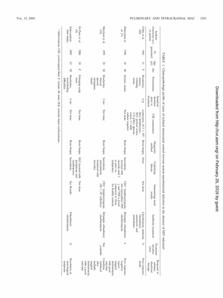
The clinical characteristics of previously reported cases andour case of isolated intracranial MAC infection in the absenceof HIV infection are depicted in Table 1. Of note is that threeof five cases had a history of sarcoidosis. Three of the fourpreviously reported cases had a history of intermittent steroidtreatment, and the current case had a history of local inhaledsteroid use but had not used inhaled steroids during at least the6 months prior to presentation. At least three of the five caseshad preexisting CD4 lymphocytopenia. Treatment includedethambutol and a macrolide for most cases, and the outcomeswere variable, with poor responses reported in two of the fivecases. Detailed immune studies were reported for only onecase, who showed a reduced ability to produce TNF-� andIFN-� in response to mitogen stimulation.
Study population. In order to identify the immune defectsthat might be associated with the extrapulmonary dissemina-tion of MAC, as observed in our index patient, we studiedthree individuals with localized pulmonary MAC infection andfour healthy volunteers.
Monocyte activation pattern. The activation of CD14 mono-cytes has previously been shown to be associated with down-regulation of the CD14 receptor, which produces a CD14dim
phenotype. CD14dim monocytes are thought to be major pro-
VOL. 15, 2008 PULMONARY AND INTRACRANIAL MAC 1581
on February 26, 2016 by guest
http://cvi.asm.org/
Dow
nloaded from

ducers of cytokines, and the levels of CD14dim monocytes havepreviously been shown to be elevated in septic patients (13). Inaddition, the percentage of CD14dim (activated) monocytes hasbeen shown to increase after stimulation with sepsis-specificantigens, such as LPS, staphylococcal enterotoxin B, pepti-doglycan, and even TNF-� (38). Ex vivo PBMCs were isolated,and the percentage of CD14dim monocytes was measured as amarker of baseline inflammatory responses. Patients with lo-calized pulmonary MAC infections had a higher percentage ofCD14dim monocytes circulating in their peripheral blood thanthe controls and the individual with a brain MAC infection,even after stimulation with MAC antigen in culture (Table 2).LPS and MAC antigens activated monocytes in all subjectswith pulmonary MAC and all controls, as assessed by CD14downregulation, but this effect was less pronounced in ourindex patient with an intracranial MAC infection (Table 2).
Monocyte responses. Monocytes were examined for theirability to produce cytokines in response to LPS (a Toll-likereceptor 4 [TLR4] agonist) and to MAC antigens (a TLR2 andTLR4 agonist) (Fig. 2a and b). Monocytes from the individualswith pulmonary MAC infections produced significantly moreTNF-� and significantly less IL-10 in response to LPS or MACantigen than the healthy controls (for LPS, 86.9 � 14.5%TNF-� and 9.3 � 5.6% IL-10 versus 37.6 � 15.3% TNF-� and39.6 � 1.9% IL-10, respectively [P 0.01]; for MAC antigen,75.3 � 15.5% TNF-� and 8.2 � 4.6% IL-10 versus 40.1 �14.0% TNF-� and 24.5 � 4.2% IL-10, respectively [P 0.05]).
Monocytes from our patient with intracranial MAC infec-tion showed levels of TNF-� expression similar to those for thehealthy controls in response to LPS but reduced levels ofTNF-� production in response to MAC antigen. In addition,monocytes from this individual produced small amounts of
FIG. 1. A contrast-enhanced head computed tomography of index patient revealed a ring enhancing lesion in the left frontal lobe (a).Microscopic examination of the brain biopsy specimen revealed multiple foci of spindle cell pseudotumor formation, consistent with a poorlydeveloped granulomatous response (b). Ziehl-Neelsen stains of the biopsy specimen demonstrate a large number of acid-fast bacilli (c).
1582 SADEK ET AL. CLIN. VACCINE IMMUNOL.
on February 26, 2016 by guest
http://cvi.asm.org/
Dow
nloaded from
TA
BL
E1.
Clinicopathologic
profileof
casesof
isolatedintracranialcentralnervous
systemm
ycobacterialinfectionin
theabsence
ofH
IVinfection
a
Authors
(referenceno.)
orpatient
Yr
presentedA
ge(yr)
SexPresentation
Symptom
durationprior
totreatm
ent
CSF
examination
Diagnosticm
ethodU
nderlyingdisease
Imm
unologystudy
resultsA
ntibiotictreatm
entT
reatment
duration(m
o)
Response
toantim
yc-obacterial
therapy
Uldry
etal.
(40)1985
31F
Headaches,nausea,vom
iting,ataxia
4yr
Leukocytes,45.3
�10
6/liter;lym
phocytes,80%
;proteinconcn,
2.76g/liter;glucose
concn,3.1m
M;
culturenegative
Brain
biopsyN
oneN
otdone
Ciprofloxacin,am
ikacin,etham
butol,ansam
ycin,andclofazim
ine
11Poor
response,death
Dickerm
anet
al.(7)1996
38M
Seizure,ataxiaN
otdone
Brain
biopsySarcoidosis
(treatedw
ithsteroids
until3yr
priorto
symptom
s)
CD
4�
lymphocytopenia,
lowextracellular
INF
-�
andT
NF
-�levels
compared
tothose
forhealthy
controls
Rifam
pin,ethambutol,
clarithromycin
6Poor
response;requiredrepeatsurgicaldebridem
ent,w
ithgood
outcome
Morrison
etal.
(32)1999
38M
Headaches,slurredspeech,nystagm
us,ataxia
2m
oN
otdone
Brain
biopsySarcoidosis
(intermittent
treatment
with
steroids)
CD
4�
lymphocytopenia
(90�
106
cells/liter)R
ifampin,etham
butol,clarithrom
ycinN
otavailableC
onditionstabilizedafterm
ultiplesurgicaldebridem
ent,w
itha
goodoutcom
eD
iPatreet
al.(8)
200050
FE
nlargingscalp
mass
Not
doneB
rainbiopsy
SLE
(treatedw
ithprednisone
andazathioprine)
Not
done
Indexpatient
(thisstudy)
200563
MH
eadaches,w
ord-findingdifficulties
4m
oN
otdone
Brain
biopsySarcoidosis
SeeR
esultsE
thambutol,
clarithromycin
11R
esolutionof
neurologicalsym
ptoms
aA
bbreviations:CSF
,cerebrospinalfluid;F,fem
ale;M,m
ale;SLE
,systemic
lupuserythem
atosus.
VOL. 15, 2008 PULMONARY AND INTRACRANIAL MAC 1583
on February 26, 2016 by guest
http://cvi.asm.org/
Dow
nloaded from

IL-10 to LPS and MAC compared to the amounts produced bythe monocytes from the healthy controls.
We also measured the relative contributions of cytokineexpression within the CD14bright and CD14dim fractions in eachsample and found that TNF-� was equally distributed in bothfractions, whereas the majority of IL-10 produced in thehealthy controls was found in the CD14dim subset (data not
shown) under both LPS and MAC antigen stimulation condi-tions.
Overall, monocytes isolated from the individuals with pul-monary MAC infections produced more TNF-� than thehealthy controls and the patient with intracranial MAC infec-tion and less IL-10 than the healthy controls, whether it was inresponse to LPS or MAC antigen. Monocytes from the indexpatient with intracranial MAC infection appeared to have arelative defect in TNF-� production in response to MAC an-tigen, even compared to that for the controls, and this defectwas not associated with any increased production of IL-10. Inaddition, the CD14dim monocytes from the healthy controlsproduced significant amounts of IL-10 compared to theamounts produced by the patients with pulmonary and intra-cranial MAC infections.
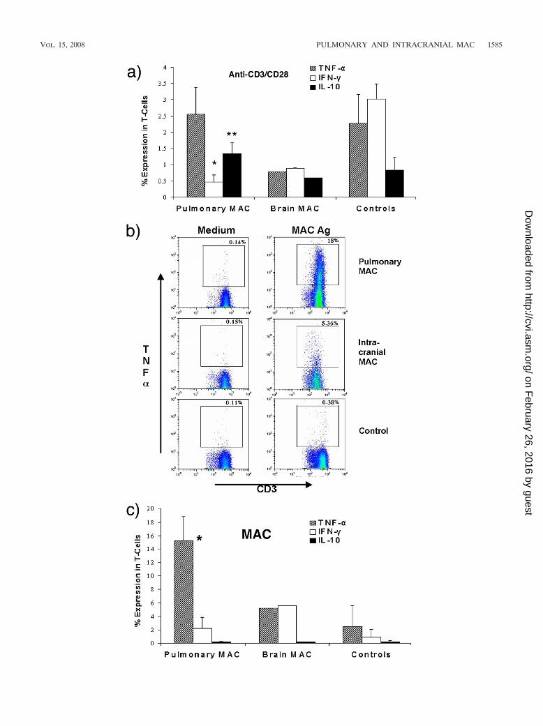
T-cell response to CD3/CD28. In order to test for generaldefects in T-cell function, the cytokine response to incubationwith CD3/CD28 costimulation was measured (Fig. 3a). T cellsfrom individuals with pulmonary MAC infection showed sig-
FIG. 2. Production of TNF-� and IL-10 by antigen-stimulated CD14 monocytes. PBMCs from patients with pulmonary MAC infections (n 3), an intracranial MAC infection (n 1), and healthy controls (n 4) were incubated ex vivo with LPS (a) or heat-killed MAC antigen (b) for6 h in the presence of brefeldin A. The cells were then harvested, stained for CD14, and then permeabilized and stained for cytokines. Intracellularcytokine expression within CD14 gated cells was subsequently measured by flow cytometry, as described in the text. Mean values � standardsdeviations are shown. *, P 0.05 for patients with pulmonary MAC infections compared with the results for the healthy controls; **, P 0.01for patients with pulmonary MAC infections compared with the results for the healthy controls.
TABLE 2. CD14dim profiles ex vivo and after stimulation
Infection group
% CD14dim monocytes
Ex vivo andunstimulated
After LPSstimulation
After MAC antigenstimulation
Pulmonary MAC 55.1 � 7.8a 71.1 � 12.5 83.1 � 5.1a,b
Brain MAC 20.1 44.1 46Controls 25.8 � 5.8 69.9 � 9.5b 62.0 � 10.8b
a P 0.01 for patients with lung MAC infection compared with the results forthe healthy controls.
b P 0.01 compared with the results obtained in the unstimulated condition.
1584 SADEK ET AL. CLIN. VACCINE IMMUNOL.
on February 26, 2016 by guest
http://cvi.asm.org/
Dow
nloaded from
VOL. 15, 2008 PULMONARY AND INTRACRANIAL MAC 1585
on February 26, 2016 by guest
http://cvi.asm.org/
Dow
nloaded from

nificantly reduced IFN-� responses compared to those for thehealthy controls but showed TNF-� responses similar to thosefor the healthy controls (for TNF-�, 2.6 � 0.8% and 2.3 �0.9%, respectively; for IFN-�, 0.5 � 0.2% and 3.0 � 0.5%,respectively [P 0.01]). T cells from our patient with intra-cranial MAC infection showed reduced IFN-� and TNF-�responses compared to those of T cells from the healthy con-trols. The IL-10 responses were comparable between theMAC-infected individuals and the healthy controls.
Thus, compared to the healthy controls, T cells from indi-viduals with localized MAC infections display a reduced ca-pacity to produce IFN-� in response to T-cell stimulation viathe CD3 receptor, and T cells from the patient with the intra-cranial MAC infection showed a reduced capacity to produceboth TNF-� and IFN-� when they were stimulated with CD3.
T-cell responses to MAC antigen. In order to test for specificT-cell dysfunction in response to MAC infection, the responseto incubation with isolated MAC antigen was measured. Theresults of a representative experiment are shown in Fig. 3b, andsummary data are depicted in Fig. 3c. T cells from individualswith localized pulmonary MAC infections showed significantlyenhanced TNF-� production compared to that for the healthycontrols (15.2 � 3.6% and 2.5 � 3.1%, respectively [P 0.01])and a more modest but not significant (P 0.14) increase inthe level of IFN-� production compared to that for the healthycontrols. The individual with brain MAC infection showedTNF-� responses to MAC similar to those for the healthycontrols, but the TNF-� levels were at least threefold lowerthan those seen in the patients with pulmonary MAC infection,that is, in the context of an infection. Conversely, the IFN-�responses to MAC in this individual were greater than those ofthe healthy controls and the patients with pulmonary MACinfections (Fig. 3b). There was no evidence of enhanced IL-10production in response to MAC antigen in any individual.
PBMC extracellular cytokine production in response toMAC. It has previously been reported that individuals withpulmonary MAC infection may have T cells capable of intra-cellular IFN-� production but have an inability to secrete them(35). In order to test for a relative deficiency in the ability tosecrete cytokines in response to MAC antigen, PMBCs wereincubated with MAC antigen and an enzyme-linked immu-nosorbent assay was performed to measure IFN-� and IL-12levels (Fig. 4a and b, respectively). For IFN-�, PBMCs fromthe individual with brain MAC infections produced largeamounts in response to MAC antigen, whereas the group withlocalized pulmonary MAC did not show any enhanced IFN-�secretion compared to that for the healthy controls. Con-versely, the index patient with brain MAC infection had adeficiency in IL-12 production in response to MAC antigenand LPS compared to the responses of the healthy controls,
whereas individuals with pulmonary MAC infections producedlarger amounts of IL-12 than the healthy controls.
Sequencing of exons 3 and 6 of IFN-�R1 and exon 3 ofIFN-�R2. Given that the index patient secreted high levels ofIFN-� in response to MAC antigen, we hypothesized that adefect in the receptors for IFN-�, i.e., IFN-�R1 and IFN-�R2,could explain defective immune control of mycobacteria. Par-tial IFN-�R1 and IFN-�R2 deficiency has been associated withlate-onset mycobacterial infections (23). The most frequentlydescribed mutations have included nucleotide substitutions atpositions 187 (C to T) and 260 (T to C) in exon 3 or deletionsin exon 6 (position 818) for IFN-�R2 deficiency and mutationsat position 114 (C to T) of exon 3 for IFN-�R1 deficiency (23,29, 30). Sequencing of exons 3 and 6 of IFN-�R1 and exon 3 ofIFN-�R2 in the index patient revealed none of these previouslydescribed mutations or deletions.
DISCUSSION
Isolated intracranial MAC infections are rare in the absenceof obvious immunosuppression, such as HIV infection (20).Evaluation of our case with those reported previously revealeda number of features associated with intracranial MAC infec-tions. These include the presence of sarcoidosis, intermittentsteroid use, and CD4 lymphocytopenia. We noted a strikingassociation between the reported cases of intracranial MACand sarcoidosis. Whether some cases of sarcoidosis representoccult MAC pulmonary infections prior to the development ofintracranial infection will require further study.
We contrasted the immunologic features of our case withthose of a more common manifestation of MAC infection, thatis, localized pulmonary MAC infection. Adequate monocyte/macrophage function is critical for the containment of myco-bacterial infections. Patients with localized pulmonary MACinfections have a higher percentage of CD14dim monocytes,reflecting a greater degree of monocyte activation in vivo. Thissuggests an activated immune response at the baseline, whichmay be due to the chronic infectious process. We did not seethis baseline level of activation in healthy controls, and the lackof monocyte activation in our index patient with an intracranialMAC infection suggests a defect in monocyte activation in thepresence of MAC antigen. Despite the presence of ongoingmonocyte activation in patients with localized pulmonaryMAC infections, these individuals are unable to clear the in-fection from their lungs.
We show that localized pulmonary MAC infections areassociated with the production of high levels of TNF-� inresponse to MAC but low levels of production of IFN-�relative to those in healthy controls. There have been con-flicting reports about the pathophysiology of localized pul-
FIG. 3. Production of TNF-�, IFN-�, and IL-10 by antigen-stimulated T cells. T cells from patients with pulmonary MAC infections (n 3),an intracranial MAC infection (n 1), and healthy controls (n 4) were incubated with antibodies to CD3 and CD28 (a) or heat-killed MACantigen (b and c) for 6 h in the presence of brefeldin A. The cells were harvested and stained, and cytokine expression on gated CD3-positive cellswas subsequently measured by flow cytometry, as described in the text. Representative data from the index patient, a healthy volunteer, and anindividual with a localized pulmonary MAC infection are shown in panel b. In panel b, the values above the boxes indicate the percentage of CD3staining cells expressing TNF-� In panels a and c, mean values � standards deviation are shown. *, P 0.01 for patients with pulmonary MACinfections compared with the results for the healthy controls; **, P 0.05 for patients with pulmonary MAC infections compared with the resultsfor the healthy controls.
1586 SADEK ET AL. CLIN. VACCINE IMMUNOL.
on February 26, 2016 by guest
http://cvi.asm.org/
Dow
nloaded from

monary MAC infections. Previous reports support structuralabnormalities of the thorax that predispose elderly patientsto such infections (27, 28). However, more recent data havedocumented IFN-� deficiency as an underlying etiology (17,36). Other reports have documented a defect in IFN-� se-cretion as the main factor (35, 41). IFN-� is important inantimycobacterial host defense via macrophage activationand granuloma formation. A defect in the synthesis and,possibly, secretion of IFN-� predisposes individuals to per-sistent nontuberculous mycobacterial infections and may ex-plain the poor response to treatment and frequent relapsesin these patients (25, 34). Our data also show that in re-sponse to MAC antigens, patients with pulmonary MACinfections displayed a relative IFN-� deficiency. Comparisonof the intracellular levels to the extracellular levels of IFN-�in patients with pulmonary MAC infections also suggeststhat a secretory defect leading to relatively low levels ofextracellular IFN-� may also be a contributing factor. Stud-ies have shown clinical improvement with the administrationof IFN-� in patients with pulmonary MAC infections (21),and this supports a secretory dysfunction rather than a re-ceptor abnormality. The mechanism of this IFN-� secretion
defect has yet to be characterized. The presence however, ofa potent TNF-� response suggests that TNF-� may be im-portant in limiting the spread of a localized infection as wellas contributing to overall macrophage activation.
Our patient with an intracranial MAC infection showed aselective TNF-� and IL-12 defect of T cells and monocytes inresponse to MAC antigen but high levels of IFN-�. This cyto-kine profile is suggestive of a partial IFN-� receptor deficiencystate. The lack of IL-12 production in response to LPS or MACantigen, despite the induction of high levels of IFN-�, is inkeeping with a failure of signaling through the IFN-� receptor,since IFN-� primes monocytes/macrophages to produce IL-12(39). Previous work with patients with IFN-� receptor defi-ciency showed that such mutations lead to the loss of augmen-tation of TNF-� production by PBMCs (24), with the resultbeing disseminated MAC infections (11, 22). We did not, how-ever, detect the commonly described mutations in the IFN-�R1 and IFN-�R2 genes in this individual. Further studiesexamining mutations elsewhere in these genes or in othergenes such as stat-5 (downstream from IFN-� signaling) orIL-12 will likely be required to further elucidate a genetic basisfor the index patient’s immune abnormalities. Of note is a
FIG. 4. Individuals with localized lung MAC infections may have an IFN-� secretion defect in response to MAC antigen, and the patient withan intracranial MAC infection displays IL-12 deficiency in response to MAC antigen and LPS. The extracellular production of IFN-� (a) and IL-12(b) by MAC antigen-stimulated PBMCs was measured. PBMCs were isolated from patients with pulmonary MAC infections, a patient with anintracranial MAC infection, and healthy controls and were cultured with heat-killed MAC antigen and/or LPS. The cytokine concentrations withinthe supernatants were measured by enzyme-linked immunosorbent assay. Mean values � standard deviations are shown.
VOL. 15, 2008 PULMONARY AND INTRACRANIAL MAC 1587
on February 26, 2016 by guest
http://cvi.asm.org/
Dow
nloaded from
recent report which also could not find any evidence of partialdeficiencies of IFN-�R1 and IFN-�R2 in association with lo-calized nontuberculous mycobacterial lung disease (26).
TNF-� deficiency leads to susceptibility to infections by M.tuberculosis and the observed lack of granuloma formation (1),the latter of which was reflected in our index patient. The lackof TNF-� induction also suggests that this cytokine plays acentral role in macrophage activation and the prevention of thespread of MAC to other tissues in humans. In this regard,previous reports have indicated that humans with tuberculosiswere treated with TNF-�-blocking agents, such as etanerceptor infliximab (16). It is thus quite possible that the increasinguse of such agents will lead to more cases involving extrapul-monary MAC infections.
Boussiotos et al. previously showed that in patients withMycobacterium tuberculosis infections, the IL-10 productioninduced by M. tuberculosis induces a relative immune suppres-sion that prevents control of the organism (5). We did not findsupport for enhanced IL-10 expression as a mechanism formycobacterial persistence either in patients with localized pul-monary MAC or in the patient with the intracranial MACinfection. Of interest, however, was that in the healthy con-trols, activated monocytes produced large amounts of IL-10,suggesting that monocytes may have a regulatory function inthe absence of an active infectious process.
The association of sarcoidosis with our case and those re-ported previously is interesting and requires further study. Theimmune profile of our case differed from that usually describedin patients with sarcoidosis; that is, the enhanced production ofT cells producing IFN-� and proinflammatory cytokines, suchas TNF-�, is generally reported in patients with sarcoidosis(33). Recently, Carlisle et al. (6) demonstrated that 12/30 in-dividuals with sarcoidosis had detectable IFN-� responses byenzyme-linked immunospot assay to two or more mycobacte-rial antigens in their PBMCs, even though they were negativefor purified protein derivative. It was unclear whether theseindividuals indeed had persistent mycobacterial infectionswhich were responsible for their symptoms. However, suchfindings warrant further studies to determine whether a subsetof cases of sarcoidosis are due to persistent mycobacteria orinsufficient immune responses to the organism and if suchpersistence gives the potential for dissemination to other or-gans. In addition, a recent report showed that in patients withsarcoidosis, there is an amplification of T regulatory cells in thelungs, which is associated with a local proliferative defect of Tcells in response to antigen (31). Whether such a defect pre-disposes individuals to invasive mycobacterial infections willalso require further study.
In summary, we highlight the immunologic features of twoforms of MAC infection. We find that individuals with themore common, localized pulmonary MAC infections have arelative deficiency of IFN-� production that leads to a localizedinability to eradicate the mycobacteria. In our case of an in-tracranial MAC infection, we find defects in both TNF-� andIL-12 production. Further studies are warranted to investigatethe role of defects in the production of these cytokines inhumans in the development of MAC-related diseases.
ACKNOWLEDGMENTS
We thank our patients for contributing their time to this study.
Financial support for this study was obtained from the CanadianInstitutes of Health Research (CIHR) and the Ontario HIV Treat-ment Network (OHTN). M.O. is a career scientist for the CIHR andOHTN. M.O. receives unrestricted research grants from Sanofi-Pas-teur and Pfizer.
REFERENCES
1. Algood, H. M., P. L. Lin, D. Yankura, A. Jones, J. Chan, and J. L. Flynn.2004. TNF influences chemokine expression of macrophages in vitro andthat of CD11b� cells in vivo during Mycobacterium tuberculosis infection.J. Immunol. 172:6846–6857.
2. Altare, F., A. Durandy, D. Lammas, J. F. Emile, S. Lamhamedi, F. Le Deist,P. Drysdale, E. Jouanguy, R. Doffinger, F. Bernaudin, O. Jeppsson, J. A.Gollob, E. Meinl, A. W. Segal, A. Fischer, D. Kumararatne, and J. L.Casanova. 1998. Impairment of mycobacterial immunity in human interleu-kin-12 receptor deficiency. Science 280:1432–1435.
3. Bean, A. G., D. R. Roach, H. Briscoe, M. P. France, H. Korner, J. D.Sedgwick, and W. J. Britton. 1999. Structural deficiencies in granulomaformation in TNF gene-targeted mice underlie the heightened susceptibilityto aerosol Mycobacterium tuberculosis infection, which is not compensatedfor by lymphotoxin. J. Immunol. 162:3504–3511.
4. Boehm, U., T. Klamp, M. Groot, and J. C. Howard. 1997. Cellular responsesto interferon-gamma. Annu. Rev. Immunol. 15:749–795.
5. Boussiotis, V. A., E. Y. Tsai, E. J. Yunis, S. Thim, J. C. Delgado, C. C.Dascher, A. Berezovskaya, D. Rousset, J. M. Reynes, and A. E. Goldfeld.2000. IL-10-producing T cells suppress immune responses in anergic tuber-culosis patients. J. Clin. Investig. 105:1317–1325.
6. Carlisle, J., W. Evans, R. Hajizadeh, M. Nadaf, B. Shepherd, R. D. Ott, K.Richter, and W. Drake. 2007. Multiple Mycobacterium antigens induce in-terferon-gamma production from sarcoidosis peripheral blood mononuclearcells. Clin. Exp. Immunol. 150:460–468.
7. Dickerman, R. D., Q. E. Stevens, R. Rak, S. E. Dorman, S. M. Holland, andT. T. Nguyen. 2003. Isolated intracranial infection with Mycobacteriumavium complex. J. Neurosurg. Sci. 47:101–105.
8. Di Patre, P. L., W. Radziszewski, N. A. Martin, A. Brooks, and H. V. Vinters.2000. A meningioma-mimicking tumor caused by Mycobacterium aviumcomplex in an immunocompromised patient. Am. J. Surg. Pathol. 24:136–139.
9. Donnelly, R. P., S. L. Freeman, and M. P. Hayes. 1995. Inhibition of IL-10expression by IFN-gamma up-regulates transcription of TNF-alpha in hu-man monocytes. J. Immunol. 155:1420–1427.
10. Dorman, S. E., and S. M. Holland. 1998. Mutation in the signal-transducingchain of the interferon-gamma receptor and susceptibility to mycobacterialinfection. J. Clin. Investig. 101:2364–2369.
11. Dorman, S. E., C. Picard, D. Lammas, K. Heyne, J. T. van Dissel, R. Baretto,S. D. Rosenzweig, M. Newport, M. Levin, J. Roesler, D. Kumararatne, J. L.Casanova, and S. M. Holland. 2004. Clinical features of dominant andrecessive interferon gamma receptor 1 deficiencies. Lancet 364:2113–2121.
12. Egen, J. G., A. G. Rothfuchs, C. G. Feng, N. Winter, A. Sher, and R. N.Germain. 2008. Macrophage and T cell dynamics during the developmentand disintegration of mycobacterial granulomas. Immunity 28:271–284.
13. Fingerle, G., A. Pforte, B. Passlick, M. Blumenstein, M. Strobel, and H. W.Ziegler-Heitbrock. 1993. The novel subset of CD14�/CD16� blood mono-cytes is expanded in sepsis patients. Blood 82:3170–3176.
14. Flynn, J. L., M. M. Goldstein, J. Chan, K. J. Triebold, K. Pfeffer, C. J.Lowenstein, R. Schreiber, T. W. Mak, and B. R. Bloom. 1995. Tumor ne-crosis factor-alpha is required in the protective immune response againstMycobacterium tuberculosis in mice. Immunity 2:561–572.
15. Frucht, D. M., and S. M. Holland. 1996. Defective monocyte costimulationfor IFN-gamma production in familial disseminated Mycobacterium aviumcomplex infection: abnormal IL-12 regulation. J. Immunol. 157:411–416.
16. Gomez-Reino, J. J., L. Carmona, and M. Angel Descalzo. 2007. Risk oftuberculosis in patients treated with tumor necrosis factor antagonists due toincomplete prevention of reactivation of latent infection. Arthritis Rheum.57:756–761.
17. Greinert, U., M. Schlaak, S. Rusch-Gerdes, H. D. Flad, and M. Ernst. 2000.Low in vitro production of interferon-gamma and tumor necrosis factor-alpha in HIV-seronegative patients with pulmonary disease caused by non-tuberculous mycobacteria. J. Clin. Immunol. 20:445–452.
18. Griffith, D. E., T. Aksamit, B. A. Brown-Elliott, A. Catanzaro, C. Daley, F.Gordin, S. M. Holland, R. Horsburgh, G. Huitt, M. F. Iademarco, M.Iseman, K. Olivier, S. Ruoss, C. F. von Reyn, R. J. Wallace, Jr., and K.Winthrop. 2007. An official ATS/IDSA statement: diagnosis, treatment, andprevention of nontuberculous mycobacterial diseases. Am. J. Respir. Crit.Care Med. 175:367–416.
19. Groux, H., M. Bigler, J. E. de Vries, and M. G. Roncarolo. 1996. Interleu-kin-10 induces a long-term antigen-specific anergic state in human CD4� Tcells. J. Exp. Med. 184:19–29.
20. Gyure, K. A., R. A. Prayson, M. L. Estes, and G. S. Hall. 1995. SymptomaticMycobacterium avium complex infection of the central nervous system. A
1588 SADEK ET AL. CLIN. VACCINE IMMUNOL.
on February 26, 2016 by guest
http://cvi.asm.org/
Dow
nloaded from

case report and review of the literature. Arch. Pathol. Lab. Med. 119:836–839.
21. Hallstrand, T. S., H. D. Ochs, Q. Zhu, and W. C. Liles. 2004. InhaledIFN-gamma for persistent nontuberculous mycobacterial pulmonary diseasedue to functional IFN-gamma deficiency. Eur. Respir. J. 24:367–370.
22. Han, J. Y., S. D. Rosenzweig, J. A. Church, S. M. Holland, and L. A. Ross.2004. Variable presentation of disseminated nontuberculous mycobacterialinfections in a family with an interferon-gamma receptor mutation. Clin.Infect. Dis. 39:868–870.
23. Haverkamp, M. H., J. T. van Dissel, and S. M. Holland. 2006. Human hostgenetic factors in nontuberculous mycobacterial infection: lessons from sin-gle gene disorders affecting innate and adaptive immunity and lessons frommolecular defects in interferon-gamma-dependent signaling. Microbes In-fect. 8:1157–1166.
24. Holland, S. M., S. E. Dorman, A. Kwon, I. F. Pitha-Rowe, D. M. Frucht,S. M. Gerstberger, G. J. Noel, P. Vesterhus, M. R. Brown, and T. A. Fleisher.1998. Abnormal regulation of interferon-gamma, interleukin-12, and tumornecrosis factor-alpha in human interferon-gamma receptor 1 deficiency.J. Infect. Dis. 178:1095–1104.
25. Huang, J. H., P. N. Kao, V. Adi, and S. J. Ruoss. 1999. Mycobacteriumavium-intracellulare pulmonary infection in HIV-negative patients withoutpreexisting lung disease: diagnostic and management limitations. Chest 115:1033–1040.
26. Hwang, J. H., W. J. Koh, E. J. Kim, E. H. Kang, G. Y. Suh, M. P. Chung, H.Kim, and O. J. Kwon. 2006. Partial interferon-gamma receptor deficiencyand non-tuberculous mycobacterial lung disease. Tuberculosis (Edinburgh)86:382–385.
27. Iseman, M. D. 1996. That’s no lady. Chest 109:1411.28. Iseman, M. D., D. L. Buschman, and L. M. Ackerson. 1991. Pectus excava-
tum and scoliosis. Thoracic anomalies associated with pulmonary diseasecaused by Mycobacterium avium complex. Am. Rev. Respir. Dis. 144:914–916.
29. Jouanguy, E., S. Lamhamedi-Cherradi, F. Altare, M. C. Fondaneche, D.Tuerlinckx, S. Blanche, J. F. Emile, J. L. Gaillard, R. Schreiber, M. Levin,A. Fischer, C. Hivroz, and J. L. Casanova. 1997. Partial interferon-gammareceptor 1 deficiency in a child with tuberculoid bacillus Calmette-Guerininfection and a sibling with clinical tuberculosis. J. Clin. Investig. 100:2658–2664.
30. Jouanguy, E., S. Lamhamedi-Cherradi, D. Lammas, S. E. Dorman, M. C.Fondaneche, S. Dupuis, R. Doffinger, F. Altare, J. Girdlestone, J. F. Emile,
H. Ducoulombier, D. Edgar, J. Clarke, V. A. Oxelius, M. Brai, V. Novelli, K.Heyne, A. Fischer, S. M. Holland, D. S. Kumararatne, R. D. Schreiber, andJ. L. Casanova. 1999. A human IFNGR1 small deletion hotspot associatedwith dominant susceptibility to mycobacterial infection. Nat. Genet. 21:370–378.
31. Miyara, M., Z. Amoura, C. Parizot, C. Badoual, K. Dorgham, S. Trad, M.Kambouchner, D. Valeyre, C. Chapelon-Abric, P. Debre, J. C. Piette, and G.Gorochov. 2006. The immune paradox of sarcoidosis and regulatory T cells.J. Exp. Med. 203:359–370.
32. Morrison, A., K. A. Gyure, J. Stone, K. Wong, P. McEvoy, K. Koeller, and H.Mena. 1999. Mycobacterial spindle cell pseudotumor of the brain: a casereport and review of the literature. Am. J. Surg. Pathol. 23:1294–1299.
33. Newman, L. S., C. S. Rose, and L. A. Maier. 1997. Sarcoidosis. N. Engl.J. Med. 336:1224–1234.
34. Prince, D. S., D. D. Peterson, R. M. Steiner, J. E. Gottlieb, R. Scott, H. L.Israel, W. G. Figueroa, and J. E. Fish. 1989. Infection with Mycobacteriumavium complex in patients without predisposing conditions. N. Engl. J. Med.321:863–868.
35. Safdar, A., D. Armstrong, and H. W. Murray. 2003. A novel defect ininterferon-gamma secretion in patients with refractory nontuberculous pul-monary mycobacteriosis. Ann. Intern. Med. 138:521.
36. Safdar, A., D. A. White, D. Stover, D. Armstrong, and H. W. Murray. 2002.Profound interferon gamma deficiency in patients with chronic pulmonarynontuberculous mycobacteriosis. Am. J. Med. 113:756–759.
37. Saunders, B. M., and W. J. Britton. 2007. Life and death in the granuloma:immunopathology of tuberculosis. Immunol. Cell Biol. 85:103–111.
38. Skinner, N. A., C. M. MacIsaac, J. A. Hamilton, and K. Visvanathan. 2005.Regulation of Toll-like receptor (TLR)2 and TLR4 on CD14dimCD16�
monocytes in response to sepsis-related antigens. Clin. Exp. Immunol. 141:270–278.
39. Trinchieri, G., and F. Gerosa. 1996. Immunoregulation by interleukin-12.J. Leukoc. Biol. 59:505–511.
40. Uldry, P. A., J. Bogousslavsky, F. Regli, J. P. Chave, and V. Beer. 1992.Chronic Mycobacterium avium complex infection of the central nervoussystem in a nonimmunosuppressed woman. Eur. Neurol. 32:285–288.
41. Vankayalapati, R., B. Wizel, B. Samten, D. E. Griffith, H. Shams, M. R.Galland, C. F. Von Reyn, W. M. Girard, R. J. Wallace, Jr., and P. F. Barnes.2001. Cytokine profiles in immunocompetent persons infected with Myco-bacterium avium complex. J. Infect. Dis. 183:478–484.
VOL. 15, 2008 PULMONARY AND INTRACRANIAL MAC 1589
on February 26, 2016 by guest
http://cvi.asm.org/
Dow
nloaded from
Related Documents