The International Journal of Periodontics & Restorative Dentistry © 2011 BY QUINTESSENCE PUBLISHING CO, INC. PRINTING OF THIS DOCUMENT IS RESTRICTED TO PERSONAL USE ONLY.. NO PART OF MAY BE REPRODUCED OR TRANSMITTED IN ANY FORM WITHOUT WRITTEN PERMISSION FROM THE PUBLISHER.

Welcome message from author
This document is posted to help you gain knowledge. Please leave a comment to let me know what you think about it! Share it to your friends and learn new things together.
Transcript

The International Journal of Periodontics & Restorative Dentistry
© 2011 BY QUINTESSENCE PUBLISHING CO, INC. PRINTING OF THIS DOCUMENT IS RESTRICTED TO PERSONAL USE ONLY.. NO PART OF MAY BE REPRODUCED OR TRANSMITTED IN ANY FORM WITHOUT WRITTEN PERMISSION FROM THE PUBLISHER.
Volume 31, Number 5, 2011
535
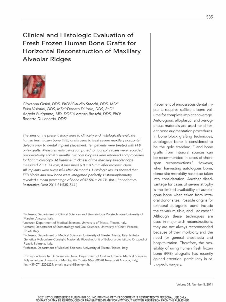
Clinical and Histologic Evaluation of Fresh Frozen Human Bone Grafts for Horizontal Reconstruction of Maxillary Alveolar Ridges
Giovanna Orsini, DDS, PhD1/Claudio Stacchi, DDS, MSc2 Erika Visintini, DDS, MSc2/Donato Di Iorio, DDS, PhD3 Angelo Putignano, MD, DDS1/Lorenzo Breschi, DDS, PhD4 Roberto Di Lenarda, DDS5
Placement of endosseous dental im-plants requires sufficient bone vol-ume for complete implant coverage. Autologous, alloplastic, and xenog-enous materials are used for differ-ent bone augmentation procedures. In bone block grafting techniques, autologous bone is considered to be the gold standard,1,2 and bone grafts from intraoral sources can be recommended in cases of short-span reconstructions.3 However, when harvesting autologous bone, donor site morbidity has to be taken into consideration. Another disad-vantage for cases of severe atrophy is the limited availability of autolo-gous bone when taken from intra-oral donor sites. Possible origins for extraoral autogenic bone include the calvarium, tibia, and iliac crest.4,5 Although these techniques are used in major arch reconstructions, they are not always recommended because of their morbidity and the need for general anesthesia and hospitalization. Therefore, the pos-sibility of using human fresh frozen bone (FFB) allografts has recently gained attention, particularly in or-thopedic surgery.
The aims of the present study were to clinically and histologically evaluate human fresh frozen bone (FFB) grafts used to treat severe maxillary horizontal defects prior to dental implant placement. Ten patients were treated with FFB onlay grafts. Measurements using computed tomography scans were recorded preoperatively and at 5 months. Six core biopsies were retrieved and processed for light microscopy. At baseline, thickness of the maxillary alveolar ridge measured 2.3 ± 0.4 mm; it measured 6.8 ± 0.5 mm after reconstruction. All implants were successful after 24 months. Histologic results showed that FFB blocks and new bone were integrated perfectly. Histomorphometry revealed a mean percentage of bone of 57.5% ± 24.7%. (Int J Periodontics Restorative Dent 2011;31:535–544.)
1 Professor, Department of Clinical Sciences and Stomatology, Polytechnique University of Marche, Ancona, Italy.
2 Lecturer, Department of Medical Sciences, University of Trieste, Trieste, Italy. 3 Lecturer, Department of Stomatology and Oral Sciences, University of Chieti-Pescara, Chieti, Italy.
4 Professor, Department of Medical Sciences, University of Trieste, Trieste, Italy; Istituto Genetica Molecolare–Consiglio Nazionale Ricerche, Unit of Bologna c/o Istituto Ortopedici Rizzoli, Bologna, Italy.
5 Professor, Department of Medical Sciences, University of Trieste, Trieste, Italy. Correspondence to: Dr Giovanna Orsini, Department of Oral and Clinical Medical Sciences, Polytechnique University of Marche, Via Tronto 10/a, 60020 Torrette di Ancona, Italy; fax: +39 071 2206221; email: [email protected].
© 2011 BY QUINTESSENCE PUBLISHING CO, INC. PRINTING OF THIS DOCUMENT IS RESTRICTED TO PERSONAL USE ONLY.. NO PART OF MAY BE REPRODUCED OR TRANSMITTED IN ANY FORM WITHOUT WRITTEN PERMISSION FROM THE PUBLISHER.
The International Journal of Periodontics & Restorative Dentistry
536
FFB grafting has been success-fully used to treat bone loss in revi-sion total hip arthroplasties.6–8 Femoral heads or iliac crests from bone banks are most often used for this technique, according to the standards of the Musculoskeletal Council of the American Association of Tissue Banks and the European Association of Musculo Skeletal Transplantation.9,10 Immediately af-ter removal, the bone grafts are stored at –80°C for at least 6 months, and if no contraindications arise, the FFB is then suitable for implanta-tion.11 The successful use of FFB in orthopedic surgery has paved the
way to introduce this procedure in oral surgery and regenerative appli-cations, not only to augment bone in maxillary sinus procedures,12 but also for alveolar ridge reconstruction in cases of horizontal insufficient bone volume before dental implant placement. Onlay bone grafting techniques have been used in situa-tions with normal or acceptable maxillomandibular relationships. The onlay integration implicates a series of biologic events critical for long-term success, some of which take place at the bone-onlay interface, mainly in the first period of the heal-ing process.13
The aims of the present study were to report clinical, histologic, and histomorphometric results of horizontal augmentation procedures using human FFB allografts in cases of severe maxillary ridge defects.
Method and materials
Ten patients (five men, five women; age range, 19 to 69 years; mean age, 49.0 ± 14.0 years) requiring implant-supported maxillary re-habilitations participated in this study (Table 1). The protocol was approved by the Ethics Committee
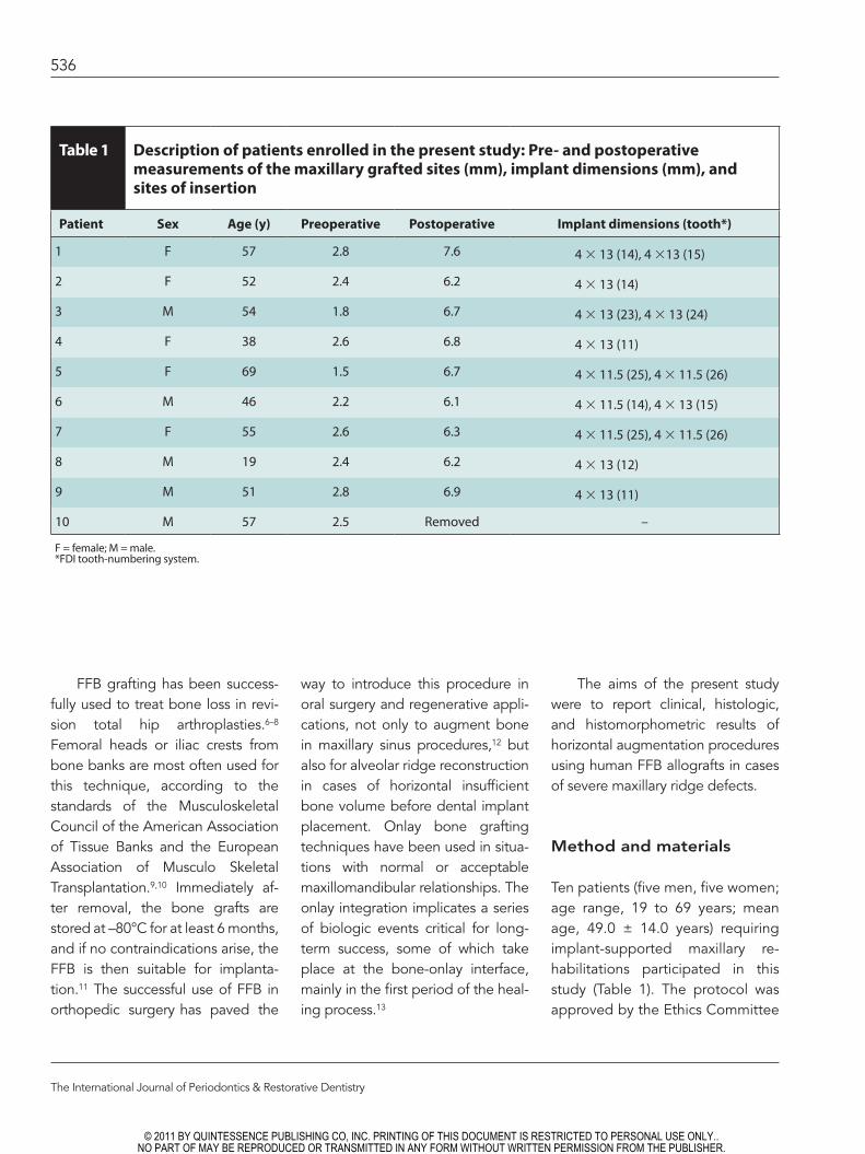
Table 1 Description of patients enrolled in the present study: Pre- and postoperative measurements of the maxillary grafted sites (mm), implant dimensions (mm), and sites of insertion
Patient Sex Age (y) Preoperative Postoperative Implant dimensions (tooth*)
1 F 57 2.8 7.6 4 × 13 (14), 4 ×13 (15)
2 F 52 2.4 6.2 4 × 13 (14)
3 M 54 1.8 6.7 4 × 13 (23), 4 × 13 (24)
4 F 38 2.6 6.8 4 × 13 (11)
5 F 69 1.5 6.7 4 × 11.5 (25), 4 × 11.5 (26)
6 M 46 2.2 6.1 4 × 11.5 (14), 4 × 13 (15)
7 F 55 2.6 6.3 4 × 11.5 (25), 4 × 11.5 (26)
8 M 19 2.4 6.2 4 × 13 (12)
9 M 51 2.8 6.9 4 × 13 (11)
10 M 57 2.5 Removed –
F = female; M = male.*FDI tooth-numbering system.
© 2011 BY QUINTESSENCE PUBLISHING CO, INC. PRINTING OF THIS DOCUMENT IS RESTRICTED TO PERSONAL USE ONLY.. NO PART OF MAY BE REPRODUCED OR TRANSMITTED IN ANY FORM WITHOUT WRITTEN PERMISSION FROM THE PUBLISHER.
Volume 31, Number 5, 2011
537
of the University of Trieste, Trieste, Italy, and all patients signed a writ-ten informed consent form. Inclu-sion criteria were maxillary partial edentulism, to be treated with the insertion of one or more implants, associated with severe horizontal ridge defects (Cawood and How-ell Class IV14 with residual bone thickness less than 3 mm). General exclusion criteria were acute myo-cardial infarction within the past 6 months, uncontrolled coagulation disorders, uncontrolled metabolic diseases, patients treated with ra-diotherapy to the head or neck region within the past 24 months,
patients treated with bisphospho-nates, patients with psychologic or psychiatric problems, heavy smok-ers, and alcohol or drug abusers. Local exclusion criteria were oral infections and uncontrolled peri-odontal disease. At the initial visit, all patients underwent a clinical and occlusal examination, pros-thetic evaluation with diagnostic waxing, periapical and panoramic radiographs, and computed to-mography (CT) scanning to study the planned implant sites as well as to evaluate the morphology of the alveolar ridge.
Surgical Protocol
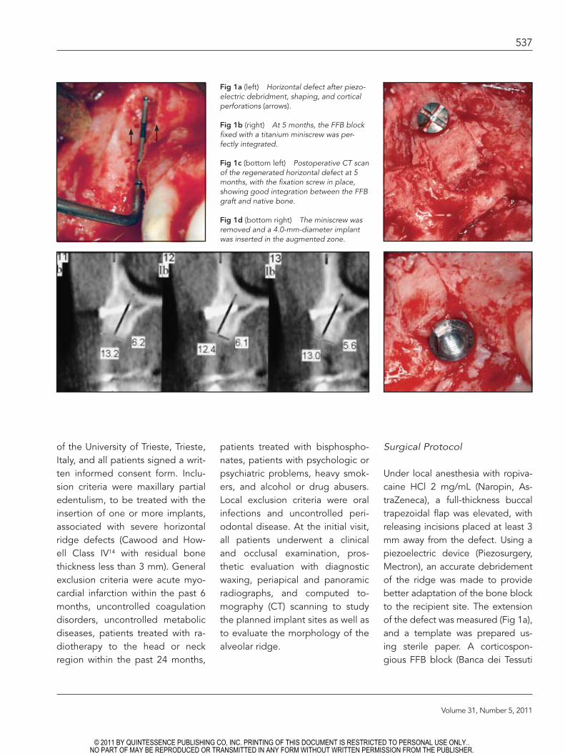
Under local anesthesia with ropiva-caine HCl 2 mg/mL (Naropin, As-traZeneca), a full-thickness buccal trapezoidal flap was elevated, with releasing incisions placed at least 3 mm away from the defect. Using a piezoelectric device (Piezosurgery, Mectron), an accurate debridement of the ridge was made to provide better adaptation of the bone block to the recipient site. The extension of the defect was measured (Fig 1a), and a template was prepared us-ing sterile paper. A corticospon-gious FFB block (Banca dei Tessuti
Fig 1a (left) Horizontal defect after piezo-electric debridment, shaping, and cortical perforations (arrows).
Fig 1b (right) At 5 months, the FFB block fixed with a titanium miniscrew was per-fectly integrated.
Fig 1c (bottom left) Postoperative CT scan of the regenerated horizontal defect at 5 months, with the fixation screw in place, showing good integration between the FFB graft and native bone.
Fig 1d (bottom right) The miniscrew was removed and a 4.0-mm-diameter implant was inserted in the augmented zone.
© 2011 BY QUINTESSENCE PUBLISHING CO, INC. PRINTING OF THIS DOCUMENT IS RESTRICTED TO PERSONAL USE ONLY.. NO PART OF MAY BE REPRODUCED OR TRANSMITTED IN ANY FORM WITHOUT WRITTEN PERMISSION FROM THE PUBLISHER.

The International Journal of Periodontics & Restorative Dentistry
538
della Regione Veneto) was cut with the piezoelectric scalpel and mod-eled using the template, tested on the recipient site, and corrected until satisfactory adaptation was reached. To facilitate blood supply to the graft from the spongious na-tive bone, the cortical aspect of the defect was perforated with a piezo-electric insert. Soft FFB chips were placed on the recipient site to com-plete the adaptation, and the block was fixed with titanium miniscrews SQ 17 (Nuova Geass) (Figs 1b and 2a). The flap was released with hori-
zontal periosteal incisions, and the augmented zone was covered pas-sively and sutured with multiple hor-izontal mattress and single sutures. Amoxicillin/clavulanate potassium (875 + 125 mg) tablets (Augmentin, GlaxoSmithKline; one tablet twice a day) and ibuprofen (Brufen, Abbott Laboratories; 600 mg twice a day) were prescribed for 1 week. Sutures were removed 10 days after surgery.
After 5 months, a second CT scan was performed (Figs 1c, 2b, and 2c); the dimensions of the graft were evaluated, and by means of
a surgical template, titanium den-tal implants were placed using a staged approach (Osseospeed, Astra Tech and Osseogrip, Plan-1Health) (Figs 1d and 2d). Six bone cores (one from each of the first six patients enrolled) were harvest-ed from the alveolar crest using a 3 × 10-mm diameter trephine un-der cold sterile saline solution ir-rigation during implant surgery. The retrieved bone cores were processed using light microscopy. Stage-two surgery was carried out after an additional 5 months.
Fig 2a An atrophic maxilla treated for horizontal augmentation with a buccal FFB block.
Figs 2b and 2c CT scan at (left) baseline and (right) after 5 months, with the onlay in place, before implant placement. At this time, the augmented thickness of the alveolar bone was recognizable.
Fig 2d After implant placement, the buc-cal walls of the implants were in contact with the FFB graft and the FFB bone chips.
© 2011 BY QUINTESSENCE PUBLISHING CO, INC. PRINTING OF THIS DOCUMENT IS RESTRICTED TO PERSONAL USE ONLY.. NO PART OF MAY BE REPRODUCED OR TRANSMITTED IN ANY FORM WITHOUT WRITTEN PERMISSION FROM THE PUBLISHER.

Volume 31, Number 5, 2011
539
Onlay graft preparation
FFB allograft preparation was per-formed in accordance with the fol-lowing bone banking procedures.12 All donors gave informed consent and were screened by a question-naire in relation to their medical, social, and sexual histories and subsequently were interviewed by a medical doctor. Then, a thor-ough physical and routine blood examination were performed. Im-mediately after resection, the do-nor bone was stored at –80°C for at least 6 months, following the pro-tocol of Egli et al.15 Microbiologic and serologic tests were performed to minimize the risk for transmis-sion of disease. Tests for hepatitis B and C, human immunodeficiency virus, human T-lymphotropic virus, cytomegalovirus, toxoplasma, and syphilis were performed.12 In cases of active disease or increased titres, the donor bone was excluded from implantation. The donor bone graft was either rejected or approved for donation by the bone bank coordi-nator after 6 months of storage.
Specimen processing for light microscopy
The maxillary onlay graft biopsies were fixed immediately in 10% for-malin, dehydrated in an ascending series of alcohol rinses, and embed-ded in a London resin (LR White Resin, London Resin). After polym-erization, specimens were sectioned with a high-precision diamond disk and ground down to approximately
40 µm with a specially designed grinding machine (Micromet, Remet). Slides were stained with acid fuch-sin and toluidine blue or with acid fuchsin and a mixture of methylene blue and Azzurro II (Merck). The slides were observed under a light microscope (Leitz Laborlux, Leica Microsystems) connected to a high-resolution video camera (3CCD JVC KY-F55B, JVC) and interfaced to a monitor and personal computer (Intel Pentium III 1200 MMX, Intel). This optical system was associated with a digitizing pad (Matrix Vision) and a histometry software package with image-capturing capabilities (Image-Pro Plus 4.5, Media Cyber-netics; Immagini & Computer).
Statistical analysis
Baseline and posttreatment mea-surements and data on newly formed bone were presented as means ± standard deviation and were analyzed using a computer-ized statistical package (Primer 4.02, McGraw Hill).
© 2011 BY QUINTESSENCE PUBLISHING CO, INC. PRINTING OF THIS DOCUMENT IS RESTRICTED TO PERSONAL USE ONLY.. NO PART OF MAY BE REPRODUCED OR TRANSMITTED IN ANY FORM WITHOUT WRITTEN PERMISSION FROM THE PUBLISHER.
The International Journal of Periodontics & Restorative Dentistry
540
Results
Clinical observations
No dropouts were observed dur-ing the entire observation period. Nine of 10 patients showed suc-cessful horizontal reconstruction; in 1 patient, exposure of the graft occurred after 1 month and the graft was removed. No additional postoperative complications were present during augmentation pro-cedures or implant surgeries.
At baseline, CT scan measure-ments of the planned implant sites showed edentulous ridge thick-
nesses ranging from 1.5 to 2.8 mm, with a mean of 2.3 ± 0.4 mm. CT scan measurements performed af-ter onlay integration and before implant placement revealed thick-ness differences from 6.2 to 7.6 mm (mean, 6.8 ± 0.5 mm; mean bone thickness gain, 4.6 ± 0.5 mm). Four-teen implants (4.0 mm in diameter) were placed, and after 5 months of healing, at clinical and radiographic examinations, all implants appeared osseointegrated. Table 1 shows the treated patient characteristics, pre- and postoperative measurements of maxillary grafted sites, implant dimensions, and insertion sites. All
patients received provisional fixed acrylic resin prostheses after abut-ment connection and underwent definitive prosthetic rehabilita-tion with cemented metal-ceramic crowns. At 24 months, all implants were successful.
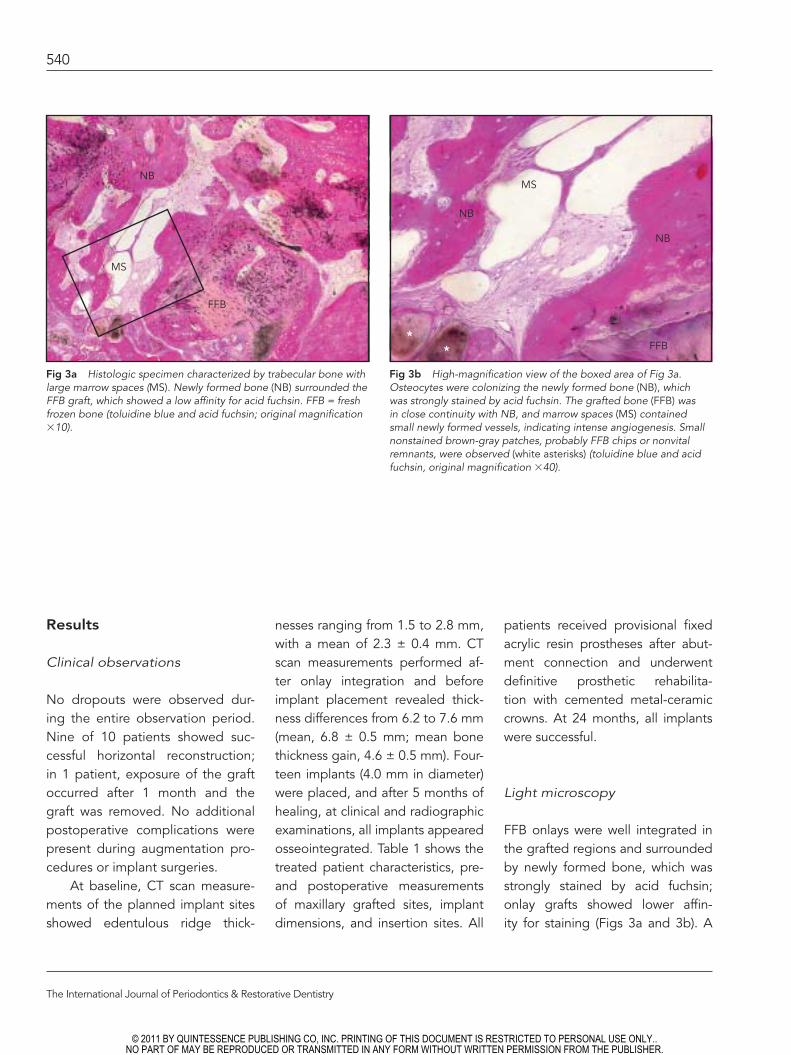
Light microscopy
FFB onlays were well integrated in the grafted regions and surrounded by newly formed bone, which was strongly stained by acid fuchsin; onlay grafts showed lower affin-ity for staining (Figs 3a and 3b). A
Fig 3a Histologic specimen characterized by trabecular bone with large marrow spaces (MS). Newly formed bone (NB) surrounded the FFB graft, which showed a low affinity for acid fuchsin. FFB = fresh frozen bone (toluidine blue and acid fuchsin; original magnification ×10).
Fig 3b High-magnification view of the boxed area of Fig 3a. Osteocytes were colonizing the newly formed bone (NB), which was strongly stained by acid fuchsin. The grafted bone (FFB) was in close continuity with NB, and marrow spaces (MS) contained small newly formed vessels, indicating intense angiogenesis. Small nonstained brown-gray patches, probably FFB chips or nonvital remnants, were observed (white asterisks) (toluidine blue and acid fuchsin, original magnification ×40).
NB
MS
FFB
NB
MS
NB
FFB*
*
© 2011 BY QUINTESSENCE PUBLISHING CO, INC. PRINTING OF THIS DOCUMENT IS RESTRICTED TO PERSONAL USE ONLY.. NO PART OF MAY BE REPRODUCED OR TRANSMITTED IN ANY FORM WITHOUT WRITTEN PERMISSION FROM THE PUBLISHER.
Volume 31, Number 5, 2011
541
few nonstained brown-gray patch-es were observed sporadically, possibly ascribed as FFB chips or nonvital FFB remnants, and were well incorporated and surrounded by new bone (Fig 3b). The newly formed osseous tissue presented features of mainly trabecular bone with large marrow spaces (Fig 3b). Numerous osteocytes were trapped within the mineralized matrix; exten-
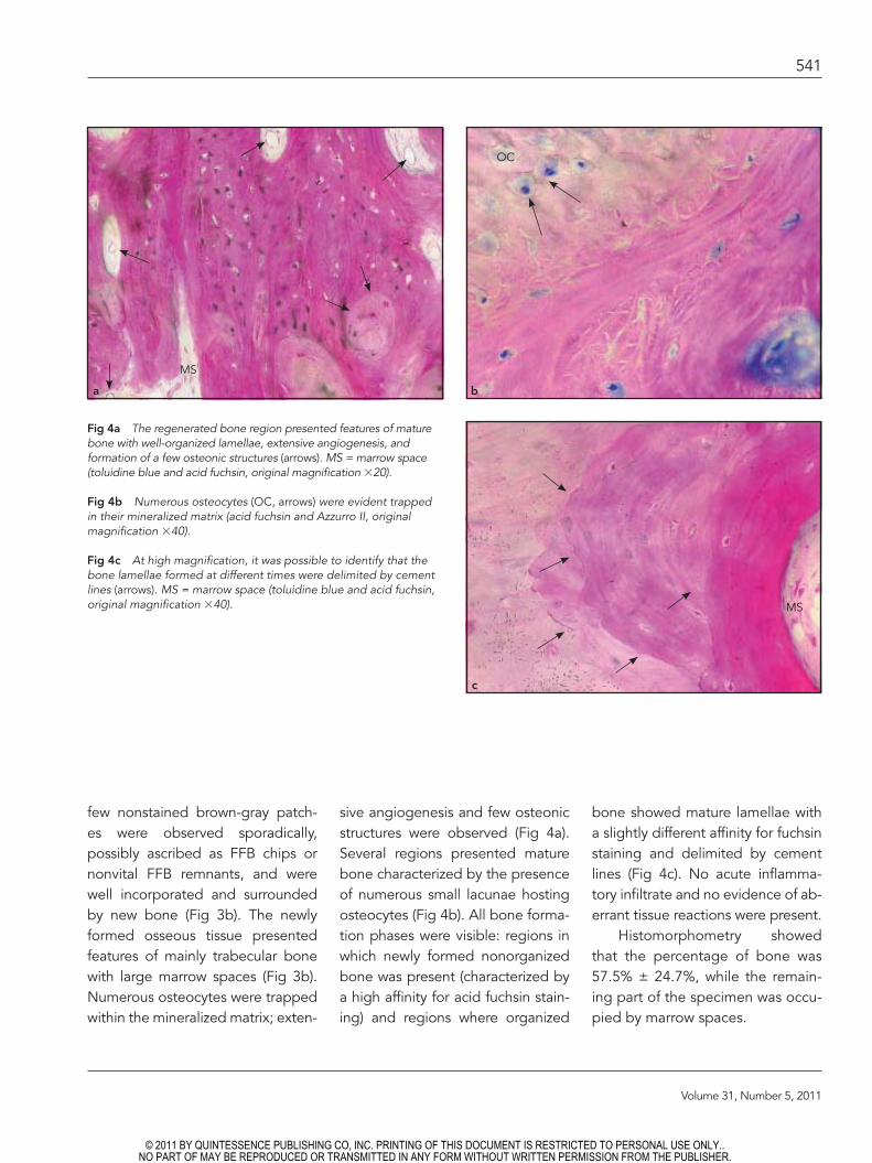
sive angiogenesis and few osteonic structures were observed (Fig 4a). Several regions presented mature bone characterized by the presence of numerous small lacunae hosting osteocytes (Fig 4b). All bone forma-tion phases were visible: regions in which newly formed nonorganized bone was present (characterized by a high affinity for acid fuchsin stain-ing) and regions where organized
bone showed mature lamellae with a slightly different affinity for fuchsin staining and delimited by cement lines (Fig 4c). No acute inflamma-tory infiltrate and no evidence of ab-errant tissue reactions were present.
Histomorphometry showed that the percentage of bone was 57.5% ± 24.7%, while the remain-ing part of the specimen was occu-pied by marrow spaces.
Fig 4a The regenerated bone region presented features of mature bone with well-organized lamellae, extensive angiogenesis, and formation of a few osteonic structures (arrows). MS = marrow space (toluidine blue and acid fuchsin, original magnification ×20).
Fig 4b Numerous osteocytes (OC, arrows) were evident trapped in their mineralized matrix (acid fuchsin and Azzurro II, original magnification ×40).
Fig 4c At high magnification, it was possible to identify that the bone lamellae formed at different times were delimited by cement lines (arrows). MS = marrow space (toluidine blue and acid fuchsin, original magnification ×40).
MS
OC
MS
a b
c
© 2011 BY QUINTESSENCE PUBLISHING CO, INC. PRINTING OF THIS DOCUMENT IS RESTRICTED TO PERSONAL USE ONLY.. NO PART OF MAY BE REPRODUCED OR TRANSMITTED IN ANY FORM WITHOUT WRITTEN PERMISSION FROM THE PUBLISHER.

The International Journal of Periodontics & Restorative Dentistry
542
Discussion
The present study shows that FFB block, if used alone as an onlay graft, promotes bone formation for horizontal maxillary reconstruction, promoting bone regeneration and implant osseointegration. The greater amount of high-quality bone after ridge augmentation al-lows the clinician to place implants in sufficient bone volume and in the proper position. Indeed, FFB onlays prepare the alveolar ridge with adequate width of bone, thus both facial and lingual/palatal im-plant surfaces can be osseointe-grated circumferentially.
Revascularization is the key factor for successful incorporation and remodeling of the bone graft.16
Close adaptation of the graft to the recipient site, together with firm stabilization of the block, are pivotal to obtaining rapid integra-tion of the graft.16–19 Furthermore, the bone healing potential following piezoelectric osseous surgery seems to be more favorable compared with traditional bone surgery performed using burs or saws.20 Optimal inte-gration of FFB onlay grafts in host sites may depend on the fact that the banked FFB blocks, following FFB preparation standards, con-tained living cells with growth ca-pacity.11,21 In fact, previous studies reported that the vital cells con-tained in human allografts, such as FFB, positively influence osteo-conduction because they stimulate the release of chemotactic factors, which contribute to increased vas-cularization, followed by resorption
of the grafted bone by osteoclasts and formation of new woven bone by osteoblasts.7,22
An initial concern about the possibility of introducing infec-tious diseases with the donor bone and inducing unfavorable immune responses was present. However, recent studies have shown that the method of FFB processing and storing respects high standards for screening and collecting pro-cedures; thus, it is safe and useful for osseous reconstructive surger-ies.23,24 Moreover, cryopreservation of FFB using dimethyl sulfoxide and glycerol may preserve up to 80% of viable cells by removing water during the freezing process, and osteoblast-related cells can be grown from FFB and have been morphologically indistinguishable from those grown from freshly har-vested trabecular bone.21,25–27
A major possible negative con-sequence of the presence of viable cells is acute allograft rejection re-sponses.26 However, no acute al-lograft rejection responses after impaction grafting were seen in the present clinical study. After 24 months of follow-up, no infective complications were noted. In fact, using clinical and radiographic eval-uations, good osseointegration of the restored dental implants was re-corded. Although postoperative ra-diographic examinations at the time of implant-abutment connection and at 24 months showed no rele-vant change of bone height around the implants, a potential limitation of this study is that patients were not submitted to a third CT scan at this
© 2011 BY QUINTESSENCE PUBLISHING CO, INC. PRINTING OF THIS DOCUMENT IS RESTRICTED TO PERSONAL USE ONLY.. NO PART OF MAY BE REPRODUCED OR TRANSMITTED IN ANY FORM WITHOUT WRITTEN PERMISSION FROM THE PUBLISHER.

Volume 31, Number 5, 2011
543
latter time period. Thus, it was not possible to discern exactly whether, and of which account, resorption of the grafted FFB was present. Pub-lished data on resorption rates and speed related to autologous aug-mentation material vary. Most studies describe reduction of approximately 30% after 1 year.28 Resorption usually stagnates after 1 year,29 and long-term evaluations show favorable data on clinical outcomes of im-plants placed in the reconstructed areas.30–32 Calvarium has been sug-gested as a reliable source of autog-enous bone to limit resorption of the grafts because of its dense corti-cal bone.4,33 However, the drawback of postoperative morbidity is always present, even if it has been reported to be less painful than harvesting bone from the iliac crest.4 Therefore, the absence of postoperative com-plications, the human origin, the re-ported safety of the FFB graft, and its biocompatibility and osteocon-ductive properties render this mate-rial a possible good alternative for reconstruction of the alveolar ridge. Qualitative and quantitative evalua-tions by means of light microscopy and histomorphometry demonstrat-ed that the regenerated region shows features similar to pre-existing osseous tissue, and FFB acts as an osteoconductive conduit on which host bone is laid down, presenting all the phases of bone formation starting from highly woven fuchsin-stained osseous tissue surrounding the residual FFB and leading to more mature trabecular bone with numerous osteocytes at the periph-ery of the graft.
The results of this study could increase scientific knowledge in understanding the events occur-ring after FFB implantation and have confirmed pre-existing litera-ture in which regenerative proce-dures were performed using either autogenous bone or different al-lografts. Indeed, both clinical and biologic responses to FFB onlay grafts were favorable and allowed implant placement in regions pre-viously reported as having poor bone quantity.13,30–33 Indeed, al-though recent scientific literature revealed accepted biocompatibility of FFB grafts by means of several in vitro studies11,15,21,25 and a clinical report in orthopedic surgery,34 very few clinical studies have been per-formed on oral regenerative pro-cedures,35–39 and, to the authors’ knowledge, there is only one in vivo study supported by histologic data that has proven the utility and safety of human FFB for maxillary sinus augmentation procedures.12 The fact that FFB grafts were per-fectly incorporated in the recipient site and formed a well-structured and consolidated osseous tissue in which dental implants were in-serted successfully warrant future studies on performing immediate loading protocols, since the sur-vival rates of immediately loaded implants in grafted sites are consis-tent with those of implants placed in native nonreconstructed bone.4
Moreover, even if it has been re-ported that horizontal reconstruc-tions are generally more stable than vertical augmentations,32 to test the stability of FFB over time,
future prospective studies will be directed toward the resorption rate analysis of this biomaterial.
Conclusions
This in vivo study represents the first attempt to reveal a case series of clinical, histologic, and histomor-phometric data on FFB apposition grafts used for horizontal maxillary reconstruction in humans. Despite the limited number of patients, it may represent an important start for forthcoming clinical trials and additional long-term analyses. The use of FFB in oral and maxil-lofacial surgery could become very important, especially in the recon-struction of severe atrophies and important posttraumatic defects where large amounts of bone graft are needed and intraoral and extra-oral sources can be difficult to find.
References
1. Nyström E, Ahlqvist J, Kahnberg KE, Rosenquist JB. Autogenous onlay bone grafts fixed with screw implants for the treatment of severely resorbed maxillae. Radiographic evaluation of preoperative bone dimensions, postoperative bone loss, and changes in soft-tissue profile. Int J Oral Maxillofac Surg 1996;25:351–359.
2. Schwartz-Arad D, Levin L. Intraoral autog-enous block onlay bone grafting for exten-sive reconstruction of atrophic maxillary alveolar ridges. J Periodontol 2005;76: 636–641.
3. Misch CM. The harvest of ramus bone in conjunction with third molar removal for onlay grafting before placement of dental implants. Int J Oral Maxillofac Surg 1999; 57:1376–1379.
© 2011 BY QUINTESSENCE PUBLISHING CO, INC. PRINTING OF THIS DOCUMENT IS RESTRICTED TO PERSONAL USE ONLY.. NO PART OF MAY BE REPRODUCED OR TRANSMITTED IN ANY FORM WITHOUT WRITTEN PERMISSION FROM THE PUBLISHER.

The International Journal of Periodontics & Restorative Dentistry
544
4. Chiapasco M, Gatti C, Gatti F. Immedi-ate loading of dental implants placed in severely resorbed edentulous mandibles reconstructed with autogenous calvarial grafts. Clin Oral Implants Res 2007;18: 13–20.
5. Schwartz-Arad D, Dori S. Intraoral autog-enous onlay block bone grafting for im-plant dentistry [in Hebrew]. Refuat Hapeh Vehashinayim 2002;19:35–39.
6. van Biezen FC, ten Have BL, Verhaar JA. Impaction bone-grafting of severely defective femora in revision total hip sur-gery: 21 hips followed for 41-85 months. Acta Orthop Scand 2000;71:135–142.
7. Buma P, Donk S, Slooff TJ, Schreurs W. Bone graft incorporation after reconstruc-tion of bony defects with impacted mor-selized bone graft. Histology of animals and patients. Ortop Traumatol Rehabil 2001;3:41–47.
8. van der Donk S, Buma P, Slooff TJ, Gar-deniers JW, Schreurs BW. Incorporation of morselized bone grafts: A study of 24 acetabular biopsy specimens. Clin Or-thop Relat Res 2002;(396):131–141.
9. American Association of Tissue Banks. Standards for Tissue Banking. American Association of Tissue Banks, 1996. www.aatb.org. Accessed 9 July 2009.
10. European Association of Musculo Skel-etal Transplantation. Common standard for musculo-skeletal tissue banking. Eu-ropean Association of Musculo Skeletal Transplantation, 1997. www.eamst.org. Accessed 9 July 2009.
11. Heyligers IC, Klein-Nulend J. Detection of living cells in non-processed but deep-frozen bone allografts. Cell Tissue Bank 2005;6:25–31.
12. Stacchi C, Orsini G, Di Iorio D, Breschi L, Di Lenarda R. Clinical, histologic, and his-tomorphometric analyses of regenerated bone in maxillary sinus augmentation us-ing fresh frozen human bone allografts. J Periodontol 2008;79:1789–1796.
13. Brunski JB, Puleo DA, Nanci A. Bioma-terials and biomechanics of oral and maxillofacial implants: Current status and future developments. Int J Oral Maxillo-fac Implants 2000;15:15–46.
14. Cawood JI, Howell RA. A classification of the edentulous jaws. Int J Oral Maxillofac Surg 1988;17:232–236.
15. Egli RJ, Sckell A, Fraitzl CR, et al. Cryo-preservation with dimethyl sulfoxide sustains partially the biological function of osteochondral tissue. Bone 2003;33: 352–361.
16. Gordh M, Alberius P. Some basic factors essential to autogeneic nonvascularized onlay bone grafting to the craniofacial skeleton. Scand J Plast Reconstr Surg Hand Surg 1999;33:129–146.
17. Gordh M, Alberius P, Lindberg L, Johnell O. Bone graft incorporation after cortical perforations of the host bed. Otolaryngol Head Neck Surg 1997;117:664–670.
18. Bays RA. Rigid stabilization system for maxillary osteotomies. J Oral Maxillofac Surg 1985;43:60–63.
19. Johnson EE, Urist MR, Finerman GA. Resistant nonunions and partial or com-plete segmental defects of long bones. Treatment with implants of a composite of human bone morphogenetic protein (BMP) and autolyzed, antigen-extracted, allogeneic (AAA) bone. Clin Orthop Relat Res 1992;(277):229–237.
20. Vercellotti T, Nevins ML, Kim DM, et al. Osseous response following resective surgery with Piezosurgery. Int J Periodon-tics Restorative Dent 2005;25:543–549.
21. Simpson D, Kakarala G, Hampson K, Steele N, Ashton B. Viable cells survive in fresh frozen human bone allografts. Acta Orthop 2007;78:26–30.
22. Mejdahl S, Hansen CA, Skjødt H, Reimann I. Human bone bank allografts stimulate bone resorption and inhibit proliferation in cultures of human osteoblast-like cells. Acta Orthop Scand 1998;69:63–68.
23. Simonds RJ, Holmberg SD, Hurwitz RL, et al. Transmission of human immunodefi-ciency virus type 1 from a seronegative or-gan and tissue donor. N Engl J Med 1992; 326:726–732.
24. Conrad EU, Gretch DR, Obermeyer KR, et al. Transmission of the hepatitis-C virus by tissue transplantation. J Bone Joint Surg Am 1995;77:214–224.
25. Egli RJ, Wingenfeld C, Hölzle M, et al. His-topathology of cryopreserved bone allo- and isografts: Pretreatment with dimethyl sulfoxide. J Invest Surg 2006;19:87–96.
26. Aho AJ, Eskola J, Ekfors T, Manner I, Kouri T, Hollmen T. Immune responses and clinical outcome of massive human osteoarticular allografts. Clin Orthop Relat Res 1998;(346):196–206.
27. Wingenfeld C, Egli RJ, Hempfing A, Ganz R, Leunig M. Cryopreservation of osteochondral allografts: Dimethyl sulf-oxide promotes angiogenesis and im-mune tolerance in mice. J Bone Joint Surg Am 2002;84-A:1420–1429 [erratum 2002;84-A:1855].
28. Van der Meij AJ, Baart JA, Prahl-Ander-sen B, Valk J, Kostense PJ, Tuinzing DB. Computed tomography in evaluation of early secondary bone grafting. Int J Oral Maxillofac Surg 1994;23:132–136.
29. Reinert S, König S, Bremerich A, Eufinger H, Krimmel M. Stability of bone grafting and placement of implants in the severe-ly atrophic maxilla. Br J Oral Maxillofac Surg 2003;41:249–255.
30. Clayman L. Implant reconstruction of the bone-grafted maxilla: Review of the liter-ature and presentation of 8 cases. J Oral Maxillofac Surg 2006;64:674–682.
31. von Arx T, Buser D. Horizontal ridge augmentation using autogenous block grafts and the guided bone regeneration technique with collagen membranes: A clinical study with 42 patients. Clin Oral Implants Res 2006;17:359–366.
32. Orsini G, Bianchi AE, Vinci R, Piattelli A. Histologic evaluation of autogenous calvarial bone in maxillary onlay bone grafts: A report of two cases. Int J Oral Maxillofac Implants 2003;18:594–598.
33. Aghaloo TL, Moy PK. Which hard tissue augmentation techniques are the most successful in furnishing bony support for implant placement? Int J Oral Maxillofac Implants 2007;22(suppl):49–70 [erratum 2008;23;56].
34. van Haaren EH, Heyligers IC, Alexander FG, Wuisman PI. High rate of failure of im-paction grafting in large acetabular defects. J Bone Joint Surg Br 2007;89:296–300.
35. Perrott DH, Smith RA, Kaban LB. The use of fresh frozen allogeneic bone for maxil-lary and mandibular reconstruction. Int J Oral Maxillofac Surg 1992;21:260–265.
36. Rochanawutanon S, Suddhasthira T, Pai-ruchvej V, Vajaradul Y. Long term follow-up of reconstruction with allogeneic mandibular bone crib packed with autog-enous particulate cancellous bone mar-row. Cell Tissue Bank 2002;3:183–197.
37. Viscioni A, Franco M, Paolin A, et al. Effec-tiveness of fresh frozen and cryopreserved homologue iliac crest grafts used in sinus lifting: A comparative study [epub ahead of print 6 July 2010]. Cell Tissue Bank.
38. Keith JD Jr, Petrungaro P, Leonetti JA, et al. Clinical and histologic evaluation of a mineralized block allograft: Results from the developmental period (2001-2004). Int J Periodontics Restorative Dent 2006; 26:321–327.
39. Keith JD Jr. Localized ridge augmentation with a block allograft followed by second-ary implant placement: A case report. Int J Periodontics Restorative Dent 2004; 24:11–17.
© 2011 BY QUINTESSENCE PUBLISHING CO, INC. PRINTING OF THIS DOCUMENT IS RESTRICTED TO PERSONAL USE ONLY.. NO PART OF MAY BE REPRODUCED OR TRANSMITTED IN ANY FORM WITHOUT WRITTEN PERMISSION FROM THE PUBLISHER.
Related Documents