Clinical Anatomy of the Lower Limb Gareth Davies

Welcome message from author
This document is posted to help you gain knowledge. Please leave a comment to let me know what you think about it! Share it to your friends and learn new things together.
Transcript

Clinical Anatomy of the Lower Limb
Gareth Davies

Joint Classification (Structure) • Fibrous
– Synostosis (Cranial Sutures) – Gomphoses (Dentoalveolar syndesmosis) – Syndesmosis (Distal Tibiofibular joint)
• Cartilaginous – Primary/Synchondrosis (costochondral, physis) – Secondary/ Symphysis (Pubic symphysis)
• Synovial – Plane/Gliding (Prox. Tibio fibular, most Intertarsal joints,
Tarsometatarsal, Sacroiliac, Patellofemoral) – Hinge (Talocrural, Interphalangeal, Knee*) – Ellipsoid (Metatarsophalangeal) – Ball & Socket (Hip, Talonavicular) – Pivot (Prox. Radio-ulnar, median atlanto-axial) – Saddle (1st Metacarpophalangeal, sternoclavicular)

Joint Classification (Function)
• Synarthrosis – Permits very little or no movement
• Amphiarthrosis – Slight movement
• Diarthrosis – Freely moveable
• Uniaxial • Biaxial • Multiaxial
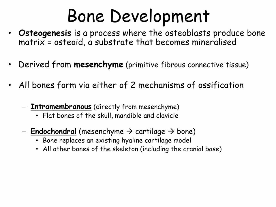
Bone Development • Osteogenesis is a process where the osteoblasts produce bone
matrix = osteoid, a substrate that becomes mineralised
• Derived from mesenchyme (primitive fibrous connective tissue)
• All bones form via either of 2 mechanisms of ossification – Intramembranous (directly from mesenchyme)
• Flat bones of the skull, mandible and clavicle
– Endochondral (mesenchyme cartilage bone) • Bone replaces an existing hyaline cartilage model • All other bones of the skeleton (including the cranial base)
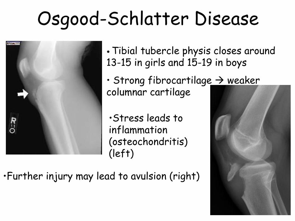
Osgood-Schlatter Disease
• Tibial tubercle physis closes around 13-15 in girls and 15-19 in boys
• Strong fibrocartilage weaker columnar cartilage
•Further injury may lead to avulsion (right)
•Stress leads to inflammation (osteochondritis) (left)
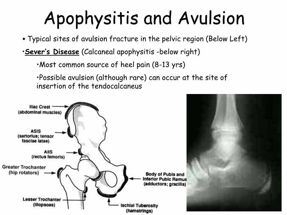
Apophysitis and Avulsion • Typical sites of avulsion fracture in the pelvic region (Below Left)
•Sever’s Disease (Calcaneal apophysitis -below right)
•Most common source of heel pain (8-13 yrs)
•Possible avulsion (although rare) can occur at the site of insertion of the tendocalcaneus
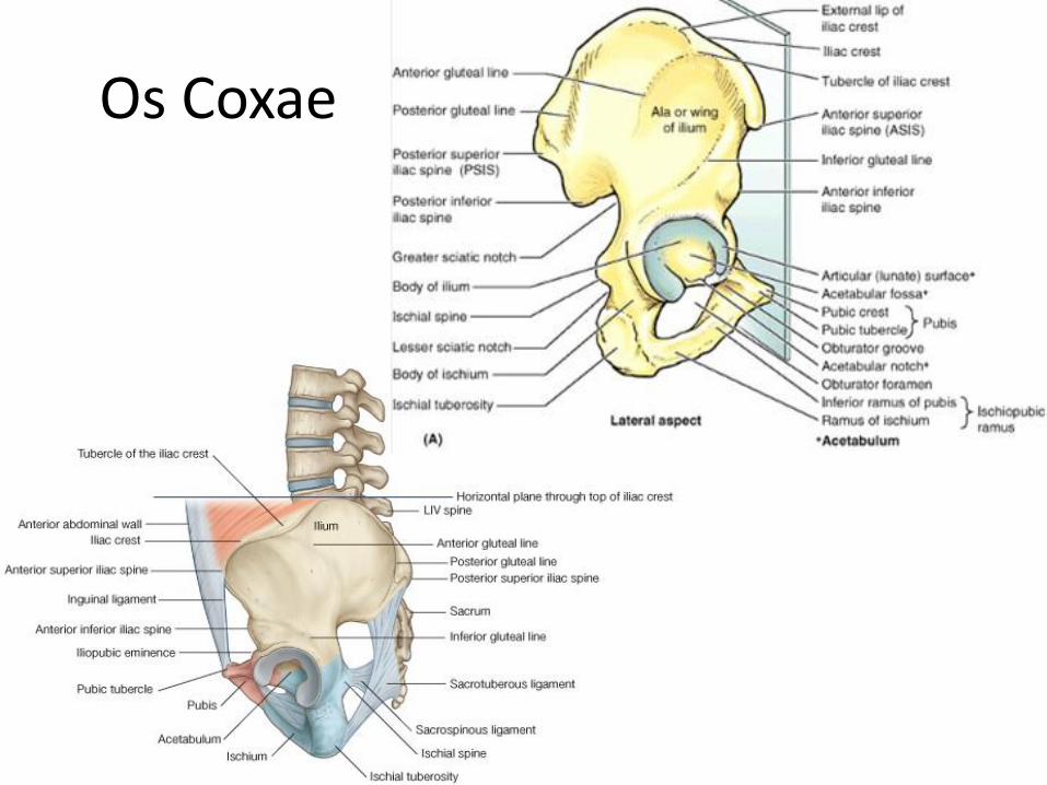
Os Coxae

Fractures of the Femoral Neck
•Common sites of fracture in the elderly
•Reduced bone density
•Decreased angle of inclination
• Common Sites
•Subcapital
•Cervical
•Basal
•Pertrochanteric
See - Moore, Dalley & Agur p. 659
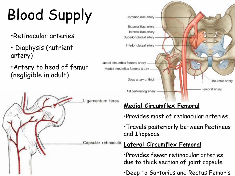
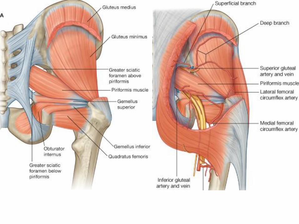
Blood Supply •Retinacular arteries
• Diaphysis (nutrient artery)
•Artery to head of femur (negligible in adult)
Medial Circumflex Femoral
•Provides most of retinacular arteries
•Travels posteriorly between Pectineus and Iliopsoas
Lateral Circumflex Femoral
•Provides fewer retinacular arteries due to thick section of joint capsule
•Deep to Sartorius and Rectus Femoris

Avascular Necrosis of Femoral Head
•(a) Pertrochanteric fracture does not inhibit retinacular blood supply - avascular necrosis does not occur
•(b) Subcapital fracture cuts off most of the retinacular supply to the femoral head – avascular necrosis is common
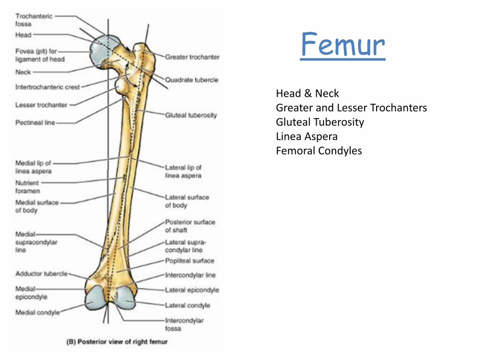
Femur
Head & Neck Greater and Lesser Trochanters Gluteal Tuberosity Linea Aspera Femoral Condyles

Anterior – Hip Flexion, Knee Extension Femoral Nerve (L2-L4) Medial – Hip Adduction Obturator Nerve (L2-L4)

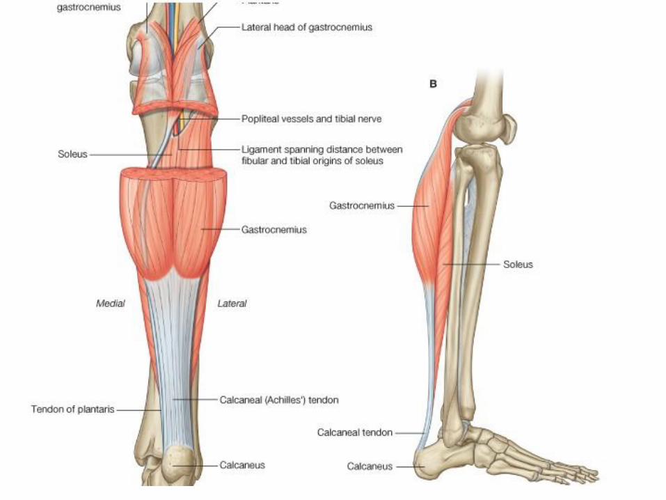
• Posterior Compartment
– Hip Extension
– Knee Flexion
– Sciatic Nerve (L4-S3)
• By Tibial division except for Short Head of BF (Common Fibular Division)

Distal Femoral Fracture
•Fracture immediately superior to the femoral condyles can be dangerous and difficult to treat
•Gastrocnemius draws the bone posteriorly
• The sharp proximal edge of this distal fragment may rupture the popliteal artery which lies directly posterior to it.
See - Moore, Dalley & Agur p. 527

Periarticular Genicular Anastamosis
Lateral Circumflex Femoral
•Descending branch
Femoral •Descending Genicular
•Musculoarterial branch •Saphenous branch
Popliteal •Superior Lateral Genicular •Superior Medial Genicular •Inferior Lateral Genicular •Inferior Medial Genicular •Middle Genicular
Anterior Tibial
•Recurrent branch

The Knee Joint Complex
• Modified Hinge, synovial joint
• Comprised of three articulations – Two femorotibial articulations (lateral & medial) – Femoropatellar articulation (plane, synovial)
• Joint stability due to
– Surrounding muscles & their tendons – Ligaments connecting femur & tibia

Structure of the Knee Joint
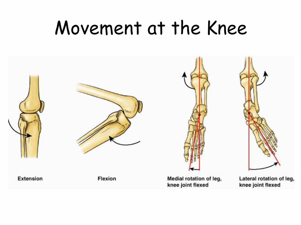
Movement at the Knee

“Unhappy Triad”
• Ruptured ACL
• Ruptured TCL (MCL)
• Torn medial meniscus
See - Moore, Dalley & Agur p. 662

ACL - Mechanism of injury
• Noncontact mechanism — The typical mechanism for a noncontact ACL injury involves a running or jumping athlete who suddenly decelerates and changes direction (eg, cutting) or pivots in a way that involves rotation or lateral bending (ie, valgus stress) of the knee.
• Contact mechanism — Contact-related ACL injuries usually occur from a direct blow causing hyperextension or valgus stress to the knee.

ACL Rupture • Related to hyperextension or medial rotation of the femoral condyles on the tibial plateau
See - Moore, Dalley & Agur p. 663
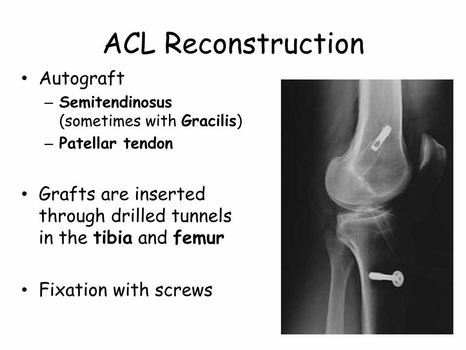
ACL Reconstruction • Autograft
– Semitendinosus (sometimes with Gracilis)
– Patellar tendon
• Grafts are inserted through drilled tunnels in the tibia and femur
• Fixation with screws

Posterior Compartment of Leg Plantar Flexion Inversion Tibial Nerve Posterior Tibial artery Fibular Artery
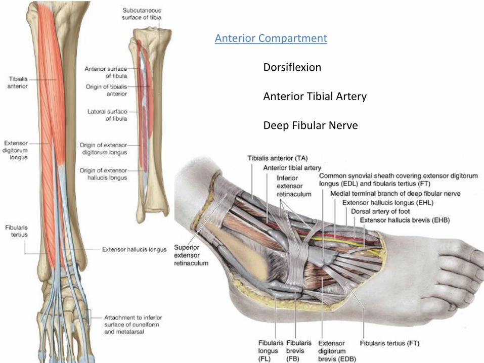
Anterior Compartment Dorsiflexion Anterior Tibial Artery Deep Fibular Nerve

•Lateral Compartment •Eversion •Plantar Flexion •Superficial Fibular Nerve

Common Fibular Nerve • Common fibular nerve is
vulnerable to injury as it winds around the head of the fibula
• Foot drop and inversion result
• Loss of sensation over the anterior and lateral aspects of the leg and foot
See - Moore, Dalley & Agur p. 605

Ankle Sprains •Lateral ligament sprains are much more common due to the weaker ligament in comparison the medial (deltoid) ligament
•The anterior talofibular ligament is vulnerable with forced inversion of the foot during plantar flexion
See - Moore, Dalley & Agur p. 665


Great Saphenous Vein
•Varicose Veins •dilation of the vessel accompanied by valvular insufficiency •Blood allowed to flow inferiorly
•Saphenous Vein Grafts •Often used for CABG surgery 1. Readily accessible 2. Distance between tributaries and
perforating veins allow usable lengths to be harvested
3. High percentage of muscular & elastic fibres
See - Moore, Dalley & Agur p. 540

References
•Drake, R., Vogl, W., & Mitchell, A. (2009). Gray’s Anatomy for Students. London, UK: Churchill Livingstone. •Ellis, H. (2006). Clinical Anatomy, Eleventh Edition. Carlton, Australia: Blackwell Publishing •Moore, K.L., Dalley, A.F., & Agur, A.M.R. (2009). Clinically Oriented Anatomy, Sixth Edition. Baltimore, USA: Lippincott Williams & Wilkins. •Snell, R.S. (2007). Clinical Anatomy by Systems. Baltimore, USA: Lippincott Williams & Wilkins.
Related Documents