Accepted Manuscript Title: Clarithromycin modulates immune responses in experimental peritonitis Authors: Stefanos Atmatzidis, Ioannis Koutelidakis, Grigorios Chatzimavroudis, Konstantinos Louis, Aikaterini Pistiki, Konstantinos Roditis, Konstantinos Atmatzidis, Evangelos J. Giamarellos-Bourboulis PII: S0924-8579(11)00038-0 DOI: doi:10.1016/j.ijantimicag.2010.11.037 Reference: ANTAGE 3530 To appear in: International Journal of Antimicrobial Agents Received date: 16-7-2010 Revised date: 18-11-2010 Accepted date: 18-11-2010 Please cite this article as: Atmatzidis S, Koutelidakis I, Chatzimavroudis G, Louis K, Pistiki A, Roditis K, Atmatzidis K, Giamarellos-Bourboulis EJ, Clarithromycin modulates immune responses in experimental peritonitis, International Journal of Antimicrobial Agents (2010), doi:10.1016/j.ijantimicag.2010.11.037 This is a PDF file of an unedited manuscript that has been accepted for publication. As a service to our customers we are providing this early version of the manuscript. The manuscript will undergo copyediting, typesetting, and review of the resulting proof before it is published in its final form. Please note that during the production process errors may be discovered which could affect the content, and all legal disclaimers that apply to the journal pertain.

Welcome message from author
This document is posted to help you gain knowledge. Please leave a comment to let me know what you think about it! Share it to your friends and learn new things together.
Transcript
Accepted Manuscript
Title: Clarithromycin modulates immune responses inexperimental peritonitis
Authors: Stefanos Atmatzidis, Ioannis Koutelidakis, GrigoriosChatzimavroudis, Konstantinos Louis, Aikaterini Pistiki,Konstantinos Roditis, Konstantinos Atmatzidis, Evangelos J.Giamarellos-Bourboulis
PII: S0924-8579(11)00038-0DOI: doi:10.1016/j.ijantimicag.2010.11.037Reference: ANTAGE 3530
To appear in: International Journal of Antimicrobial Agents
Received date: 16-7-2010Revised date: 18-11-2010Accepted date: 18-11-2010
Please cite this article as: Atmatzidis S, Koutelidakis I, Chatzimavroudis G, LouisK, Pistiki A, Roditis K, Atmatzidis K, Giamarellos-Bourboulis EJ, Clarithromycinmodulates immune responses in experimental peritonitis, International Journal ofAntimicrobial Agents (2010), doi:10.1016/j.ijantimicag.2010.11.037
This is a PDF file of an unedited manuscript that has been accepted for publication.As a service to our customers we are providing this early version of the manuscript.The manuscript will undergo copyediting, typesetting, and review of the resulting proofbefore it is published in its final form. Please note that during the production processerrors may be discovered which could affect the content, and all legal disclaimers thatapply to the journal pertain.
Page 1 of 26
Accep
ted
Man
uscr
ipt
Clarithromycin modulates immune responses in experimental
peritonitis
Stefanos Atmatzidis a, Ioannis Koutelidakis a, Grigorios Chatzimavroudis a,
Konstantinos Louis b, Aikaterini Pistiki b, Konstantinos Roditis b, Konstantinos
Atmatzidis a, Evangelos J. Giamarellos-Bourboulis b,*
a 2nd Department of Surgery, University of Thessaloniki, Medical School, Greece
b 4th Department of Internal Medicine, University of Athens, Medical School, Greece
ARTICLE INFO
Article history:
Received 16 July 2010
Accepted 18 November 2010
Keywords:
Clarithromycin
Apoptosis
Peritonitis
TNF
* Corresponding author. Present address: 4th Department of Internal Medicine,
ATTIKON University Hospital, 12462 Athens, Greece. Tel.: +30 210 58 31 994.
E-mail address: [email protected] (E.J. Giamarellos-Bourboulis).
Edited manuscript
Page 2 of 26
Accep
ted
Man
uscr
ipt
2
ABSTRACT
Based on clinical data revealing a promising immunomodulatory effect of
clarithromycin in sepsis due to ventilator-associated pneumonia, the efficacy of
clarithromycin in experimental peritonitis and sepsis was assessed with particular
emphasis on immune function. Cecal puncture and ligation was performed in rabbits
assigned to the following groups: Group A, controls (n = 12); Group B, intravenous
clarithromycin treatment (n = 15); Group C, piperacillin/tazobactam (TZP) treatment
(n = 10); and Group D, clarithromycin + TZP combination treatment (n = 12). Blood
was sampled at serial time intervals and peripheral blood mononuclear cells
(PBMCs) were isolated. Apoptosis of lymphocytes and monocytes was measured by
flow cytometric analysis. PBMCs were stimulated with lipopolysaccharide (LPS) and
Pam3Cys for the release of tumour necrosis factor-alpha (TNF). Tissue bacterial
growth was quantitatively measured after death or sacrifice. Survival in Group D after
10 days was prolonged compared with the other groups. Early apoptosis of
lymphocytes in Group B was lower compared with Group D at 2 h and compared with
Group C at 4 h. Early apoptosis of monocytes in Group B was lower compared with
Group C at 24 h. Following stimulation of PBMCs with LPS, release of TNF was
decreased in Group B compared with Groups A and D at 2 h. Bacterial growth in
tissues of Groups C and D was decreased compared with Group A. It is concluded
that clarithromycin modulates the function of the immune response in experimental
peritonitis by decreasing the rate of early apoptosis of lymphocytes and monocytes
and by decreasing the ex vivo release of TNF by blood monocytes.
Page 3 of 26
Accep
ted
Man
uscr
ipt
3
1. Introduction
Severe sepsis and septic shock are among the leading causes of death. It is
estimated that almost 3 million cases occur annually in Northern America and
Europe, 35–50% of whom die [1]. This enormous death rate has led physicians to
realise that single antibiotics cannot alter mortality and that other factors apart from
the microbes themselves impact on patient outcome. One of the most important
factors appears to be the pathogen–host interaction. The host’s septic reaction is
initiated when well conserved microbial structures known as pathogen-associated
molecular patterns (PAMPs) bind to receptors embedded either on the cell
membrane or inside the cell cytoplasm of cells of the innate immune system, namely
blood monocytes and tissue macrophages. These receptors are known as pattern
recognition receptors (PRRs). Toll-like receptors (TLRs) are among the best studied
PRRs. Eleven TLRs have been recognised. Monomers of the cell wall peptidoglycan
of Gram-positive cocci bind to TLR2, and lipopolysaccharide (LPS) of the outer
membrane of Gram-negative bacteria binds to TLR4. The interaction of TLRs with
PAMPs leads to activation of a series of intracellular pathways ending with the
production of pro-inflammatory cytokines such as tumour necrosis factor-alpha
(TNF), interleukin (IL)-1, IL-6 and IL-8. These pro-inflammatory mediators
orchestrate the septic reaction of the host leading to severe sepsis and multiple
organ dysfunction syndrome (MODS) [2].
Increase in current knowledge on the pathogenesis of sepsis created the concept of
developing therapeutic agents that can modulate the microbial–host interaction.
Several agents have been tested in clinical trials. Antibodies targeting LPS and
TNF, soluble receptors targeting ΤΝF and IL-1, recombinant human activated
Page 4 of 26
Accep
ted
Man
uscr
ipt
4
protein C, continuous infusion of insulin, and hydrocortisone replacement have all
been administered with contradictory results [3]. One recently described approach is
intravenous (i.v.) administration of clarithromycin, which was initially tested in a
model of acute pyelonephritis and sepsis in rabbits. This model represents a
monomicrobial type of experimental sepsis. Gram-negative bacteria, either
susceptible or multidrug-resistant to antimicrobials, were applied as challenge.
Intravenous treatment with clarithromycin was started either in parallel with bacterial
challenge or when signs of sepsis appeared. Co-administration of clarithromycin and
antimicrobials prolonged survival and attenuated the systemic inflammatory response
[4–8]. However, the effect of clarithromycin on the function of monocytes was not
studied.
Based on the above experimental results, a double-blind randomised clinical trial was
designed of patients with sepsis developed in the field of ventilator-associated
pneumonia (VAP). Two hundred patients were allocated to i.v. placebo or
clarithromycin for 3 consecutive days through a central catheter. Antimicrobial
therapy was administered according to standard guidelines. Results revealed that
clarithromycin treatment was associated with a significant reduction in the risk of
death by septic shock and MODS as well as significant earlier resolution of VAP.
However, it should be underscored that VAP is a monomicrobial infection [9].
Acute intra-abdominal infection (IAI) is recognised as the third most common cause
of sepsis [10]. It is polymicrobial in origin and generates a high bacterial load that
disseminates very quickly via the rapidly absorbing peritoneum into the entire body.
Polymicrobial infections create more complex microbial–host interactions compared

Page 5 of 26
Accep
ted
Man
uscr
ipt
5
with monomicrobial infections, making the efficacy of immunomodulatory treatment
questionable [11].
In the present study, clarithromycin was administered intravenously in an animal
model of intra-abdominal sepsis with high bacterial load. The immune function of the
host was closely monitored in order to determine the effect of clarithromycin.
2. Animals and methods
2.1. Animals
A total of 59 male White New Zealand rabbits with a mean standard deviation
(S.D.) weight of 3.19 ± 0.30 kg were studied. The study received a permit from the
Veterinary Directorate of the Prefecture of Thessaloniki according to Greek
legislation in conformance with the 160/1991 Council Directive of the European
Union. Animals were housed in single metal cages and had access to tap water and
standard balanced rabbit chow ad libitum. Room temperature ranged between 18 C
and 22 C, relative humidity between 55% and 65% and the light/dark cycle was
06:00h/18:00h.
2.2. Model of peritonitis
Animals were initially sedated by intramuscular (i.m.) injection of 25 mg/kg ketamine
and 5 mg/kg xylazine, and anaesthesia was maintained by i.m. administration of 15
mg/kg xylazine at 30-min intervals. The peritoneal cavity was entered through an
upper midline abdominal incision and the intestines were displaced to the left. The

Page 6 of 26
Accep
ted
Man
uscr
ipt
6
caecum was recognised and was ligated with a 3.0 suture. Three punctures were
performed just above the suture with a 3.0 sterile needle. The bowel content was
then gently squeezed to allow passage of 1 mL of liquid content of the bowel into the
peritoneal cavity. The peritoneal cavity and the abdominal wall were then closed in
layers. Ten rabbits were subject to only opening and closure of the peritoneal cavity
and were studied as sham-operated animals.
The remaining animals were randomly assigned into four study groups as follows.
Group A (n = 12): controls, i.e. infected animals administered 30 mL of normal
saline i.v. 0.5 h post operatively; the infusion time was 30 min. Animals were
also administered 2 mL bolus infusion of normal saline 2 h post operatively.
Group B (n = 15): infected animals administered 80 mg/kg clarithromycin 0.5 h
post operatively as described previously [4]; the infusion time was 30 min.
Animals were also administered 2 mL bolus infusion of normal saline 2 h post
operatively. Clarithromycin was provided as pyrogen-free amorphous powder
(Abbott, Chicago, IL) and was reconstituted with 10 mL of 5% glucose and
added to NaCl 0.9% at a final volume of 30 mL.
Group C (n = 10): infected animals administered 30 mL of normal saline i.v.
0.5 hours post operatively; the infusion time was 30 min. Animals were also
administered piperacillin/tazobactam (TZP) as a 400 mg/kg 2 mL bolus
infusion in normal saline 2 h post operatively. The dose of TZP was selected
in accordance with previous studies [12].
Group D (n = 12): infected animals administered both clarithromycin and TZP
in the regimens described above for Groups B and C.

Page 7 of 26
Accep
ted
Man
uscr
ipt
7
Single doses of agents were administered in an attempt to discern better the
changes in the immune responses over time in each study group.
A volume of 3 mL of blood was sampled from the vein of the left ear of each animal
under aseptic conditions before the operation and at 2, 4, 24 and 48 h. Blood was
collected in heparin-coated tubes for flow cytometry and stimulation assays.
Survival of animals was recorded every 12 h for a total follow-up period of 10 days.
Autopsy was performed following death; animals remaining alive after 10 days of
follow-up were sacrificed by i.v. administration of sodium thiopental. Under sterile
conditions, segments from the right kidney, liver, spleen and lower lobe of the right
lung were taken and placed in separate sterile plastic containers for quantitative
cultures and biopsy.
2.3. Cell apoptosis and cell stimulation
Peripheral blood mononuclear cells (PBMCs) were isolated following gradient
centrifugation of heparinised whole blood over Ficoll (Biochrom, Berlin, Germany).
Following three consecutive washings in ice-cold phosphate-buffered saline (pH 7.2)
(Biochrom), PBMCs were counted in a Neubauer chamber after trypan blue
exclusion of dead cells. One-half of the PBMCs were stained with the protein
ANNEXIN-V conjugated to the fluorochrome fluorescein isothiocyanate (FITC)
(emission 525 nm) (Immunotech, Marseille, France) and with propidium iodine (PI)
(emission 575 nm) (Immunotech). Cells were analysed after running through an
EPICS XL/MSL flow cytometer (Beckman Coulter Co., Miami, FL) with separate

Page 8 of 26
Accep
ted
Man
uscr
ipt
8
gating for lymphocyte and monocyte cells based on their characteristic forward
scattering and side scattering. Cells staining positive for FITC-conjugated ANNEXIN-
V and negative for PI were considered early apoptotic.
The remaining one-half of the PBMCs were distributed into the wells of a 96-well
plate at a final volume of 0.2 mL/well with RMPI 1640 enriched with 10% fetal bovine
serum (FBS) (Biochrom), 2 mM glutamine and 10 mM pyruvate at a density of 2
106 PBMCs/mL. Cells were stimulated without or with 10 ng/mL LPS of Escherichia
coli O55:B5, which is a TLR4 ligand (Sigma Chemical Co., St Louis, MO), or without
or with 5 g/mL Pam3Cys-SKKK (EMC Microcollections GmbH, Tübingen,
Germany), which a TLR2 ligand. The plates were incubated for 24 h at 37 C in 5%
CO2. Following incubation, plates were centrifuged and the supernatants were
collected and stored at –70 C until assayed for TNF. All stimulation assays were
performed in duplicate.
2.4. Bioassay for measurement of TNF
TNF was measured by bioassay on an L929 fibrosarcoma cell line as described
previously [4–8]. Briefly, confluent cells were thoroughly washed with Hank’s solution
and were harvested with 0.25% trypsin/0.02% ethylenediamine tetra-acetic acid
(EDTA) (Biochrom). Cells were centrifuged, re-suspended in RMPI 1640
supplemented with 10% FBS and 2 mM glutamine, and were distributed into a 96-
well cell culture plate at a density of 1 105 cells/well. The final volume of fluid in
each well was 0.05 mL. Following incubation for 2 h at 37 C at 5% CO2, 0.06 mL of
supernatant or standard dilutions of known concentrations of human TNF (Sigma
Page 9 of 26
Accep
ted
Man
uscr
ipt
9
Chemical Co.) (range 5.75–375.00 pg/mL) were added into each well followed by
0.05 mL of a 0.3 mg/mL dilution of cycloheximide (Sigma Chemical Co.) to inhibit de
novo protein biosynthesis. Following overnight incubation, the supernatant of each
well was discarded by aspiration and 0.1 mL of a 0.5 mg/mL methylene blue solution
in methanol 99% was added. After 10 min the dye was removed and the wells were
thoroughly washed three times with 0.9% NaCl. Wells were left to dry and remnants
of the dye in each well was solublised by addition of 0.1 mL of 50% glacial acetic acid
(Merck, Darmstadt, Germany). The optical density in each well was read at 495 nm
(OD495) using a Hitachi spectrophotometer (Hitachi, Tokyo, Japan) against blank
wells and control wells without added serum. Concentrations of TNF were
estimated by reduction in the OD495 of control wells by unknown samples applying a
standard curve generated by standard concentrations. All determinations were
performed in quadruplicate. The lower limit of detection was 11.5 pg/mL. The
interday coefficient of variation of the assay was 13.75%.
2.5. Tissue cultures
Tissue segments were weighed and homogenised. One 0.1 mL aliquot was diluted
1:10 in sterile NaCl four consecutive times. Another aliquot of 0.1 mL of each dilution
was plated onto MacConkey agar and was incubated at 35 C for a total period of 3
days. Plates were incubated at 35 C and the number of viable colonies was counted
in each dilution and was multiplied by the appropriate dilution factor. Identification of
bacteria was performed by the API20E and API20NE systems (bioMérieux, Paris,
France). The lower limit of detection was 30 colony-forming units/g. Bacterial cells
were expressed by their log10 value.
Page 10 of 26
Accep
ted
Man
uscr
ipt
10
2.6. Statistical analysis
The Kolmogorov–Smirnov test proved that the sample values of early apoptosis of
lymphocytes and monocytes at all measured times did not deviate from the normal
distribution, allowing for the application of parametric procedures. Specifically in the
applied one-way analysis of variance (ANOVA) procedures the main question to be
answered was whether the clarithromycin group differed significantly from the other
experimental groups and at what times. This issue was addressed with post-hoc
Dunnett’s tests using the clarithromycin group (Group B) as the control category for
comparisons. For each series of measurements, the pharmacokinetic value of the
area under the concentration–time curve (AUC) for the first 4 h was calculated using
the trapezoid rule. These AUCs were also compared using the above statistical
procedures.
To define whether clarithromycin changed the ability of PBMCs to release TNF after
stimulation with LPS or Pam3Cys, the ratios of released TNF after stimulation to the
same concentrations without stimulation were calculated for every time of sampling.
These ratios did not follow the normal distribution, therefore before subjecting them
to the same ANOVA procedure as described above the values were log-transformed.
For the same reason, the four experimental groups were described through their
geometric means and 95% confidence intervals (CIs).
3. Results
None of the sham-operated animals died. Eleven animals died in Group A (controls)
(survival 8.3%), 14 animals died in Group B (treatment with clarithromycin) (survival

Page 11 of 26
Accep
ted
Man
uscr
ipt
11
6.7%), 9 died animals in group C (treatment with TZP) (survival 10.0%) and 5
animals died in group D (treatment with both agents) (survival 58.3%) (P < 0.0001 by
Pearson’s 2 test) (Fig. 1). Survival in Group D was prolonged compared with Group
A (log-rank, 3.95; P = 0.047). Survival in Groups B and C was not prolonged
compared with Group A.
The study of the influence of treatment with clarithromycin on the immune response
of the host involved a study on (a) the effect of cell apoptosis and (b) the innate
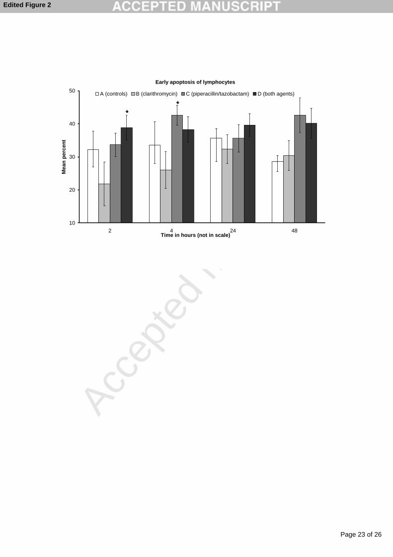
immune response. Mean standard error (S.E.) early apoptosis of lymphocytes in
sham-operated animals at 2, 4, 24 and 48 h was 42.89 6.58%, 45.81 3.60%,
34.92 4.03% and 37.94 9.19%, respectively. The comparative rate of early
apoptosis of lymphocytes during follow-up is shown in Fig. 2. As shown in Fig. 2, the
rate of early apoptosis of lymphocytes in the clarithromycin group (B) was lower over
almost the whole range of time measurements. Thus, overall apoptosis expressed
through the areas under the corresponding curves are significantly lower. The
corresponding mean ± S.D. values of the 4-h AUCs for the four groups were: Group
A, 74.5 ± 31.7; Group B, 48.3 ± 34.5; Group C, 77.8 ± 12.3; and Group D, 81.3 ±
32.8. It is interesting to note that the mean values of the three experimental Groups
A, C and D are very close together but, as the Dunnett's post-hoc test showed, were
significantly greater than for the clarithromycin group (B). In addition, the Dunnett’s
post-hoc test showed statistically significant differences of the clarithromycin
treatment group (B) in comparison with Group D at 2 h and in comparison with Group
C at 4 h post operatively.
Page 12 of 26
Accep
ted
Man
uscr
ipt
12
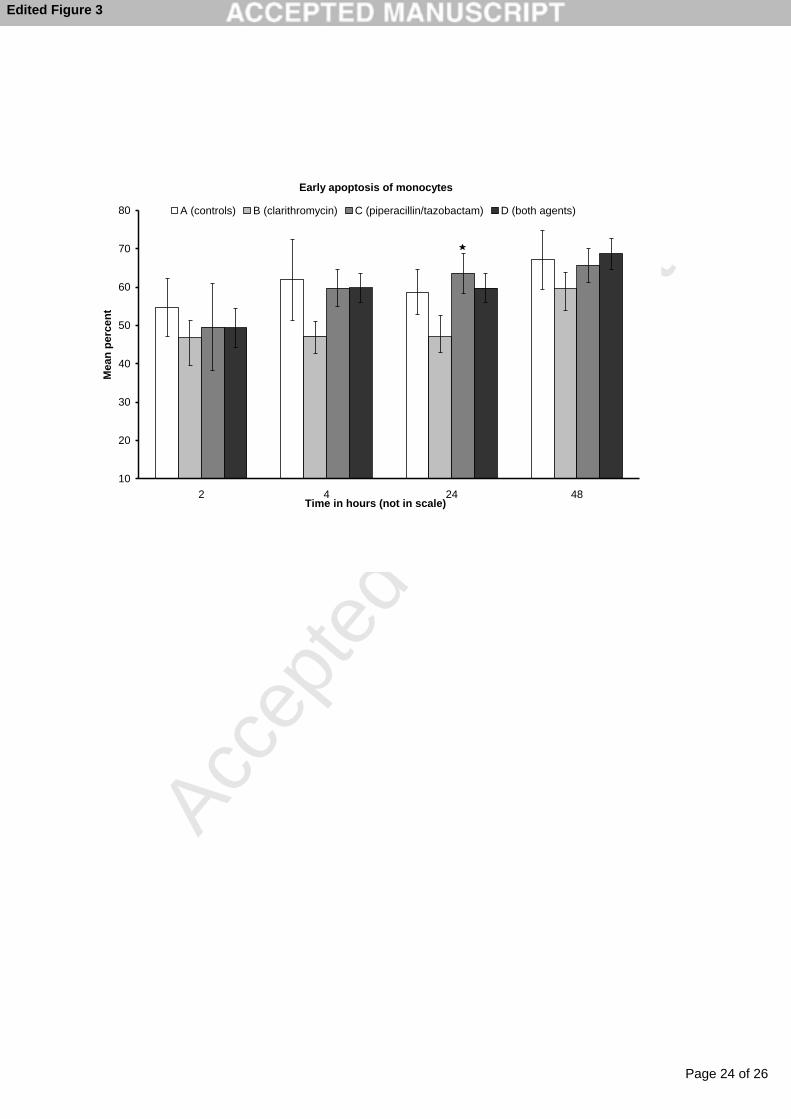
Mean S.E. early apoptosis of monocytes of sham-operated animals at 2, 4, 24 and
48 h was 59.59 7.03%, 61.69 4.14%, 60.28 5.93% and 62.45 2.42%,
respectively. The comparative rate of early apoptosis of monocytes during follow-up
is shown in Fig. 3. The same pattern as with lymphocytes is observed for monocytes,
only in this case although the values for the clarithromycin group (B) are lower than
for the three other groups, the differences do not achieve statistical significance
except in the case of the Dunnett’s post-hoc comparison with Group C (TZP) at 24 h.
PBMCs isolated at baseline and post operatively were assayed for ex vivo production
of TNF as an index of the innate immune function of rabbits. The geometric means
and their 95% CIs of the ratios of the concentrations of TNF after stimulation with
LPS to the same concentrations without stimulation at the 2-h measurement for the
four experimental groups are shown in Fig. 4. As the Dunnett's post-hoc test has
shown for the 2-h measurements, the clarithromycin group (B) had the lowest values,
which are significantly lower than the control group (A) and the combination group
(D). No differences were found between Group B and the other groups at any other
time of sampling (data not shown).
Comparison of the geometric means and their 95% CIs of the ratios of the
concentrations of TNF after stimulation with Pam3Cys to the same concentrations
without stimulation for the four experimental groups failed to disclose any differences
at any time of sampling (data not shown).
Bacterial species isolated from tissues were E. coli, Enterobacter cloacae and
Enterococcus spp. Their counts are shown in Fig. 5. Bacterial growth in tissues of
Page 13 of 26
Accep
ted
Man
uscr
ipt
13
animals in Groups C and D was significantly lower than growth in tissues of Group A.
Those of Group B did not differ from Group A.
4. Discussion
The pathogenesis of sepsis is considered to follow two phases. In the first phase,
hyperactivation of the innate immune system takes place. Notably, a burst of pro-
inflammatory cytokines is produced when PAMPs of the invading pathogens bind to
PRRs of cells of the innate immune system. Produced cytokines effect the systemic
inflammatory response. In the second phase, hypoactivation of the innate immune
system predominates. In this latter stage, blood monocytes are not able to produce
an adequate amount of cytokines once activated by PAMPs. Moreover, apoptosis of
lymphocytes supervenes. This second stage is also described as a stage of
immunoparalysis of the septic host when organ failure and MODS arrive. This
distinction of phases is most important for the type of administered
immunointervention, since some agents such as those targeting TNF act as an anti-
inflammatory, and others such as interferon-gamma (IFN) are pro-inflammatory;
however, it is very difficult to discern at which phase each patient lies [2].
Recent data from the Hellenic Sepsis Study Group showed that both the innate and
adaptive immune responses differ according to the underlying type of infection,
particularly the kinetics of how a systemic inflammatory response transits into severe
sepsis and/or septic shock. This appears to be of particular relevance for IAIs. More
precisely, the rate of apoptosis of lymphocytes and the rate of anergy of monocytes
in sepsis developing from IAI are greater compared with sepsis developing from
other types of infections [13]. As a consequence, when immunointervention is
Page 14 of 26
Accep
ted
Man
uscr
ipt
14
considered for abdominal sepsis, these unique characteristics of pathogenesis
should be taken into consideration.
Clarithromycin has been shown to be a successful immunomodulator in sepsis.
When co-administered with antibiotics, survival was prolonged in an animal model of
acute pyelonephritis [4–8]. In the clinical setting, clarithromycin decreased relative
risk of death from septic shock and MODS among patients with VAP [9].
In the present study, clarithromycin was administered in an animal model of
polymicrobial sepsis. This model is similar to everyday clinical practice owing to its
great lethality as well as the dissemination of a variety of Gram-positive and Gram-
negative pathogens to deep organs. Clarithromycin or TZP also could not alter the
physical course. However, when clarithromycin and TZP were co-administered,
survival was prolonged. This study focused on the effect of clarithromycin in the
function of the innate and adaptive immune responses. These are intensively
followed-up over consecutive time intervals, an approach never previously described
for experimental models of sepsis.
The reported findings suggest that clarithromycin may act in at least two different
sites: modulation of the immune response and enhanced clearance of pathogens.
Modulation of the immune response by clarithromycin is shown both in the rate of
apoptosis of lymphocytes and monocytes and in the innate immune responses. The
latter are measured through ex vivo stimulation of TNF production by PBMCs. Early
apoptosis of lymphocytes and monocytes increase within the first 2 h post induction
of peritonitis in infected controls. Following treatment with clarithromycin, release of

Page 15 of 26
Accep
ted
Man
uscr
ipt
15
TNF after ex vivo stimulation with a TLR4 agonist from blood monocytes decreased.
We assume that this observed effect of clarithromycin may be of importance for the
management of the septic host. Induction of apoptosis of lymphocytes and
inadequate release of cytokines from monocytes are characteristics of the second
hypoinflammatory phase of sepsis [2]. The results show that clarithromycin may well
interfere with this phase.
It should be underscored that the effects of clarithromycin in the immune system
were predominantly shown when it was given alone and not in combination with TZP.
A clear explanation for this finding does not exist. One probable explanation lies in a
pharmacokinetic interaction between the agents.
Apart from the assumed effect of clarithromycin in the stage of immunoparalysis, part
of its effect may be mediated through enhancement of clearance of pathogens, since
a trend was observed for tissue bacterial growth of clarithromycin-treated rabbits to
be lower compared with non-treated animals. Isolated bacteria from tissues, namely
members of the Enterobacteriaceae and enterococci, are not part of the antimicrobial
spectrum of macrolides. Although the present study does not provide any data on
blood neutrophils, it has been reported that macrolides may enhance phagocytosis of
bacteria by neutrophils [14]. The studied effect of clarithromycin on the function of
blood monocytes may also favour improved clearance of offending pathogens.
Two main limitations of this study should be addressed. First, single doses of
clarithromycin and TZP were administered. However, this allowed the
immunomodulatory effect of clarithromycin to be disclosed clearly within the first 2 h
Page 16 of 26
Accep
ted
Man
uscr
ipt
16
post administration. Such an effect could not be demonstrated later when
clarithromycin levels were expected to decrease in the systemic circulation. Second,
tissue cultures were performed on MacConkey agar. This growth medium mainly
favours isolation of Enterobacteriaceae, enterococci and certain species of
staphylococci. As a consequence, the effect of treatment on other pathogens such as
anaerobes cannot be estimated.
The results presented here show that clarithromycin administered in an animal model
of peritonitis modulates the function of the immune response of the host. This was
mediated through a decreased rate of early apoptosis of lymphocytes and monocytes
and by decreased ex vivo release of TNF by blood monocytes. These experimental
results, in light of the beneficial results of i.v. administration in patients with VAP [9],
create a novel perspective for a role of clarithromycin in the management of the
septic host.
Funding
None.
Competing interests
EJG-B has received independent educational grants from Abbott Hellas SA, Wyeth
Hellas SA and Sanofi-Aventis (Greece) and from BRAHMS GmbH (Germany).
Ethical approval

Page 17 of 26
Accep
ted
Man
uscr
ipt
17
This study received a permit from the Veterinary Directorate of the Prefecture of
Thessaloniki according to Greek legislation in conformance with the 160/1991
Council Directive of the European Union.
Page 18 of 26
Accep
ted
Man
uscr
ipt
18
References
[1] Dellinger RP, Levy MM, Carlet JM, Bion J, Parker MM, Jaeschke R, et al.
Surviving Sepsis Campaign: international guidelines for management of severe
sepsis and septic shock: 2008. Crit Care Med 2008;36:296–327.
[2] Rittisch D, Flierl MA, Ward PA. Harmful molecular mechanisms in sepsis. Nature
Immunol 2008;8:776–86.
[3] Vincent JL, Sun Q, Dubois MJ. Clinical trials of immunomodulatory therapies in
severe sepsis and septic shock. Clin Infect Dis 2003;34:1084–93.
[4] Giamarellos-Bourboulis EJ, Adamis T, Laoutaris G, Sabracos L, Koussoulas V,
Mouktaroudi M, et al. Immunomodulatory clarithromycin treatment of experimental
sepsis and acute pyelonephritis caused by multidrug-resistant Pseudomonas
aeruginosa. Antimicrob Agents Chemother 2004;48:93–9.
[5] Giamarellos-Bourboulis EJ, Baziaka F, Antonopoulou A, Koutoukas P, Kousoulas
V, Sabracos L, et al. Clarithromycin co-administered with amikacin attenuates
systemic inflammation in experimental sepsis by Escherichia coli. Int J Antimicrob
Agents 2005;25:168–72.
[6] Giamarellos-Bourboulis EJ, Adamis T, Sabracos L, Raftogiannis M, Baziaka T,
Tsaganos T, et al. Clarithromycin: immunomodulatory therapy of experimental
sepsis and acute pyelonephritis by Escherichia coli. Scand J Infect Dis
2005;37:48–54.
[7] Giamarellos-Bourboulis EJ, Antonopoulou A, Raftogiannis M, Koutoukas P,
Tsaganos T, Tziortzioti V, et al. Clarithromycin is an effective immunomodulator
when administered late in experimental pyelonephritis by multidrug-resistant
Pseudomonas aeruginosa. BMC Infect Dis 2006;6:31.
Page 19 of 26
Accep
ted
Man
uscr
ipt
19
[8] Giamarellos-Bourboulis EJ, Tziortzioti V, Koutoukas P, Baziaka F, Antonopoulou
A, Adamis T, et al. Clarithromycin is an effective immunomodulator in
experimental pyelonephritis caused by pan-resistant Klebsiella pneumoniae. J
Antimicrob Chemother 2006;57:937–44.
[9] Giamarellos-Bourboulis EJ, Pechère JC, Routsi C, Plachouras D, Kollias S,
Raftogiannis M, et al. Effect of clarithromycin in patients with sepsis and
ventilator-associated pneumonia. Clin Infect Dis 2008;46:1157–64.
[10] Hodgin KE, Moss M. The epidemiology of sepsis. Curr Pharm Des
2008;14:1833–9.
[11] Wang H, Ma S. The cytokine storm and factors determining the sequence and
severity of organ dysfunction in multiple organ dysfunction syndrome. Am J
Emerg Med 2008;26:711–5.
[12] Li C, Nikolau DP, Lister PD, Quintiliani R, Nightngale CH. Pharmacodynamic
study of -lactams alone and in combination with -lactamase inhibitors against
Pseudomonas aeruginosa possessing an inducible -lactamase. J Antimicrob
Chemother 2004;53:297–304.
[13] Gogos C, Kotsaki A, Pelekanou A, Giannikopoulos G, Vaki I, Maravitsa P, et
al. Early alterations of the innate and adaptive immune statuses in sepsis
according to the type of underlying infection. Crit Care 2010;14:R96.
[14] Schultz MJ. Macrolide activities beyond their antimicrobial effects: macrolides
in diffuse panbronchiolitis and cystic fibrosis. J Antimicrob Chemother
2004;54:21–8.
Page 20 of 26
Accep
ted
Man
uscr
ipt
20
Fig. 1. Comparative survival of rabbits subject to experimental peritonitis: Group A,
controls (n = 12); Group B, clarithromycin treatment (n = 15); Group C,
piperacillin/tazobactam (TZP) treatment (n = 10); Group D, combination treatment
with clarithromycin and TZP (n = 12). None of the sham-operated animals died.
Fig. 2. Comparative early apoptosis of lymphocytes during consecutive follow-up of
rabbits subject to experimental peritonitis. Error bars are the standard errors of the
means. * Denotes statistically significant difference at a P-value of 0.05 of the
experimental group compared with the clarithromycin group (B) by post-hoc
Dunnett’s test.
Fig. 3. Comparative early apoptosis of monocytes during consecutive follow-up of
rabbits subject to experimental peritonitis. Error bars are the standard errors of the
means. * Denotes statistically significant difference at a P-value of 0.05 of the
experimental group compared with the clarithromycin group (B) by post-hoc
Dunnett’s test.
Fig. 4. Comparative ratio of 2-h concentrations of tumour necrosis factor-alpha
(TNF) in supernatants of peripheral blood mononuclear cells of rabbits subject to
experimental peritonitis following stimulation with 10 ng/mL lipopolysaccharide (LPS)
to the same concentration without stimulation. Error bars are the 95% confidence
interval of the geometric means. * Denotes statistically significant difference at a P-
value of 0.05 of the experimental group compared with the clarithromycin group (B)
by post-hoc Dunnett’s test.
Page 21 of 26
Accep
ted
Man
uscr
ipt
21
Fig. 5. Tissue bacterial growth in liver, spleen, right kidney and lower right lung lobe
of rabbits subject to experimental peritonitis after death or sacrifice: Group A, controls
(n = 12); Group B, clarithromycin treatment (n = 15); group C, piperacillin/tazobactam
(TZP) treatment (n = 10); and Group D, combination treatment with clarithromycin
and TZP (n = 12). * Denotes statistically significant difference compared with the
respective tissue growth of Group A. No bacterial growth was found in tissues of
sham-operated animals. CFU, colony-forming units.

Page 22 of 26
Accep
ted
Man
uscr
ipt
Edited Figure 1
Page 23 of 26
Accep
ted
Man
uscr
ipt
Early apoptosis of lymphocytes
10
20
30
40
50
2 4 24 48Time in hours (not in scale)
Me
an
pe
rce
nt
A (controls) B (clarithromycin) C (piperacillin/tazobactam) D (both agents)
Edited Figure 2
Page 24 of 26
Accep
ted
Man
uscr
ipt
Early apoptosis of monocytes
10
20
30
40
50
60
70
80
2 4 24 48Time in hours (not in scale)
Mean
perc
en
t
A (controls) B (clarithromycin) C (piperacillin/tazobactam) D (both agents)
Edited Figure 3

Page 25 of 26
Accep
ted
Man
uscr
ipt
-0.6
0
0.6
1.2
1.8
A (controls) B (clarithromycin) C
(piperacillin/tazobactam)
D (both agents)
Ge
om
etr
ic m
ea
n
Edited Figure 4

Page 26 of 26
Accep
ted
Man
uscr
ipt
** * *
* **
*
Edited Figure 5
Related Documents