2047 INTRODUCTION Silicate- and/or silica-depositing organisms are found throughout the prokaryotic and eukaryotic world (reviewed in Müller, 2003; Ehrlich, 2010; Ehrlich et al., 2010a). Whereas prokaryotic organisms require a supersaturated silica environment to induce precipitation of amorphous silica during the exponential growth phase (Inagaki et al., 2003), eukaryotic organisms are able to accumulate silicate to concentrations that allow its polycondensation within their cell(s). Plants have the capacity to take up silicic acid in the range of 0.1 to 0.6 mmol l –1 from the soil in their rhizosphere; they then translocate this monomeric ortho-silicic acid to their branches and deposit it as amorphous silica or poly(silicate), also termed biosilica (Morse, 1999; Müller et al., 2008e), in their cell walls (Ma, 2003) or in cell vacuoles (Neumann, 2003). Diatoms (unicellular algae) accumulate silicic acid from the aqueous milieu via specific transporters at concentrations of up to 340 mmol l –1 (Hildebrand and Wetherbee, 2003) and deposit it in special vesicles from where it is channeled onto distinct proteins (Perry, 2003), e.g. silaffins (Kröger and Sumper, 2000), and also onto chitin (Brunner et al., 2009). Among the metazoans, the siliceous sponges (Porifera: Demospongiae and Hexactinellida) are the only taxa that build their skeleton (spiculae) of amorphous silica (Morse, 1999; Uriz, 2006; Müller et al., 2007d; Ehrlich et al., 2010b). Their silicification deposition–polycondensation pathway is distinguished from others by their ability to take up and accumulate silicic acid from a very silicon-poor aqueous environment (5 mol l –1 ) (Maldonado et al., 2005) via a special transporter (Schröder et al., 2004) in vacuoles (silicasomes) (Schröder et al., 2007), and to subsequently polycondensate silicic acid enzymatically to biosilica (Morse, 1999) or poly(silicate) (Müller et al., 2008e). A further characteristic of sponge spicule formation is that only a few genes control the formation of this inorganic matrix, poly(silicate). The major genes involved are the silicateins, a family of genes that encode the poly(silicate)-forming enzyme (Shimizu et al., 1998; Cha et al., 1999; Krasko et al., 2000; Müller et al., 2007b), and the silintaphins. In the center of the spicules within axial canals, axial filaments exist both in Hexactinellida (Müller et al., 2008c) and in Demospongiae (Shimizu et al., 1998); the main component of the axial filaments is the silicateins. In addition, silintaphin-1 has been identified, which functions as a silicatein interactor and stabilizes silica nanoparticles under formation of silica nano- and micro-fibers and/or rods (Wiens et al., 2009). Furthermore, silintaphin-2 likewise interacts with silicatein and might serve as a final member of a signal transduction chain that controls spiculogenesis (Wiens et al., 2011). Post- transcriptional modifications of silicateins, primarily via phosphorylation, likely also affect the shape, size and form of axial filaments (Müller et al., 2005; Müller et al., 2007a). The spicules of sponges display an amazing diversity of forms (Boury-Esnault and Rützler, 1997; Uriz et al., 2003; Uriz, 2006; Ehrlich, 2011). This remarkable and striking polymorphism of spicules, which is genetically controlled, is used in taxonomy as one of the major species characteristics of sponges (Hooper, 1997). The Journal of Experimental Biology 214, 2047-2056 © 2011. Published by The Company of Biologists Ltd doi:10.1242/jeb.056275 RESEARCH ARTICLE Circumferential spicule growth by pericellular silica deposition in the hexactinellid sponge Monorhaphis chuni Xiaohong Wang 1,2 , Matthias Wiens 1 , Heinz C. Schröder 1 , Klaus P. Jochum 3 , Ute Schloßmacher 1 , Hermann Götz 4 , Heinz Duschner 4 and Werner E. G. Müller 1, * 1 ERC Advanced Grant Research Group at the Institute for Physiological Chemistry, University Medical Center of the Johannes Gutenberg University Mainz, Duesbergweg 6, D-55128 Mainz, Germany, 2 National Research Center for Geoanalysis, 26 Baiwanzhuang Dajie, Beijing CHN-100037, China, 3 Max-Planck Institute for Chemistry, Postbox 3060, D-55020 Mainz, Germany and 4 Institute of Applied Structure- and Microanalysis, University Medical Center of the Johannes Gutenberg-University, Obere Zahlbacherstr. 63, Geb 911, D-55131 Mainz, Germany *Author for correspondence ([email protected]) Accepted 15 March 2011 SUMMARY The giant basal spicule of the hexactinellid sponge Monorhaphis chuni represents the longest natural siliceous structure on Earth. This spicule is composed of concentrically arranged lamellae that are approximately 10 m thick. In the present study, we investigated the formation of outer lamellae on a cellular level using microscopic and spectroscopic techniques. It is shown that the formation of an outermost lamella begins with the association of cell clusters with the surface of the thickening and/or growing spicule. The cells release silica for controlled formation of a lamella. The pericellular (silica) material fuses to a delimited and textured layer of silica with depressions approximately 20–30 m in diameter. The newly formed layer initially displays 40 m wide, well-structured banded ribbons and only attains its plain surface in a final step. The chemical composition in the depressions was studied using energy dispersive X-ray spectroscopy and by staining with Texas Red. The data suggest that those depressions are the nests for the silica-forming cells and that silica formation starts with a direct association of silica- forming cells with the outer surface of the spicule, where they remain and initiate the development of the next lamellae. Key words: spicule, Monorhaphis chuni, Hexactinellida, sponge, pericellular silica formation. THE JOURNAL OF EXPERIMENTAL BIOLOGY

Welcome message from author
This document is posted to help you gain knowledge. Please leave a comment to let me know what you think about it! Share it to your friends and learn new things together.
Transcript
2047
INTRODUCTIONSilicate- and/or silica-depositing organisms are found throughout theprokaryotic and eukaryotic world (reviewed in Müller, 2003; Ehrlich,2010; Ehrlich et al., 2010a). Whereas prokaryotic organisms requirea supersaturated silica environment to induce precipitation ofamorphous silica during the exponential growth phase (Inagaki et al.,2003), eukaryotic organisms are able to accumulate silicate toconcentrations that allow its polycondensation within their cell(s).Plants have the capacity to take up silicic acid in the range of 0.1 to0.6mmoll–1 from the soil in their rhizosphere; they then translocatethis monomeric ortho-silicic acid to their branches and deposit it asamorphous silica or poly(silicate), also termed biosilica (Morse, 1999;Müller et al., 2008e), in their cell walls (Ma, 2003) or in cell vacuoles(Neumann, 2003). Diatoms (unicellular algae) accumulate silicic acidfrom the aqueous milieu via specific transporters at concentrationsof up to 340mmoll–1 (Hildebrand and Wetherbee, 2003) and depositit in special vesicles from where it is channeled onto distinct proteins(Perry, 2003), e.g. silaffins (Kröger and Sumper, 2000), and also ontochitin (Brunner et al., 2009).
Among the metazoans, the siliceous sponges (Porifera:Demospongiae and Hexactinellida) are the only taxa that build theirskeleton (spiculae) of amorphous silica (Morse, 1999; Uriz, 2006;Müller et al., 2007d; Ehrlich et al., 2010b). Their silicificationdeposition–polycondensation pathway is distinguished from othersby their ability to take up and accumulate silicic acid from a verysilicon-poor aqueous environment (5moll–1) (Maldonado et al.,
2005) via a special transporter (Schröder et al., 2004) in vacuoles(silicasomes) (Schröder et al., 2007), and to subsequentlypolycondensate silicic acid enzymatically to biosilica (Morse, 1999)or poly(silicate) (Müller et al., 2008e). A further characteristic ofsponge spicule formation is that only a few genes control theformation of this inorganic matrix, poly(silicate). The major genesinvolved are the silicateins, a family of genes that encode thepoly(silicate)-forming enzyme (Shimizu et al., 1998; Cha et al.,1999; Krasko et al., 2000; Müller et al., 2007b), and the silintaphins.In the center of the spicules within axial canals, axial filaments existboth in Hexactinellida (Müller et al., 2008c) and in Demospongiae(Shimizu et al., 1998); the main component of the axial filamentsis the silicateins. In addition, silintaphin-1 has been identified, whichfunctions as a silicatein interactor and stabilizes silica nanoparticlesunder formation of silica nano- and micro-fibers and/or rods (Wienset al., 2009). Furthermore, silintaphin-2 likewise interacts withsilicatein and might serve as a final member of a signal transductionchain that controls spiculogenesis (Wiens et al., 2011). Post-transcriptional modifications of silicateins, primarily viaphosphorylation, likely also affect the shape, size and form of axialfilaments (Müller et al., 2005; Müller et al., 2007a).
The spicules of sponges display an amazing diversity of forms(Boury-Esnault and Rützler, 1997; Uriz et al., 2003; Uriz, 2006;Ehrlich, 2011). This remarkable and striking polymorphism ofspicules, which is genetically controlled, is used in taxonomy asone of the major species characteristics of sponges (Hooper, 1997).
The Journal of Experimental Biology 214, 2047-2056© 2011. Published by The Company of Biologists Ltddoi:10.1242/jeb.056275
RESEARCH ARTICLE
Circumferential spicule growth by pericellular silica deposition in the hexactinellidsponge Monorhaphis chuni
Xiaohong Wang1,2, Matthias Wiens1, Heinz C. Schröder1, Klaus P. Jochum3, Ute Schloßmacher1, Hermann Götz4, Heinz Duschner4 and Werner E. G. Müller1,*
1ERC Advanced Grant Research Group at the Institute for Physiological Chemistry, University Medical Center of the JohannesGutenberg University Mainz, Duesbergweg 6, D-55128 Mainz, Germany, 2National Research Center for Geoanalysis,
26 Baiwanzhuang Dajie, Beijing CHN-100037, China, 3Max-Planck Institute for Chemistry, Postbox 3060, D-55020 Mainz, Germany and 4Institute of Applied Structure- and Microanalysis, University Medical Center of the Johannes Gutenberg-University,
Obere Zahlbacherstr. 63, Geb 911, D-55131 Mainz, Germany*Author for correspondence ([email protected])
Accepted 15 March 2011
SUMMARYThe giant basal spicule of the hexactinellid sponge Monorhaphis chuni represents the longest natural siliceous structure onEarth. This spicule is composed of concentrically arranged lamellae that are approximately 10m thick. In the present study, weinvestigated the formation of outer lamellae on a cellular level using microscopic and spectroscopic techniques. It is shown thatthe formation of an outermost lamella begins with the association of cell clusters with the surface of the thickening and/orgrowing spicule. The cells release silica for controlled formation of a lamella. The pericellular (silica) material fuses to a delimitedand textured layer of silica with depressions approximately 20–30m in diameter. The newly formed layer initially displays 40mwide, well-structured banded ribbons and only attains its plain surface in a final step. The chemical composition in thedepressions was studied using energy dispersive X-ray spectroscopy and by staining with Texas Red. The data suggest thatthose depressions are the nests for the silica-forming cells and that silica formation starts with a direct association of silica-forming cells with the outer surface of the spicule, where they remain and initiate the development of the next lamellae.
Key words: spicule, Monorhaphis chuni, Hexactinellida, sponge, pericellular silica formation.
THE JOURNAL OF EXPERIMENTAL BIOLOGY

2048
The formation of siliceous spicules starts intracellularly in vesicleswith the formation of an axial filament composed of silicatein,around which the first layer of poly(silicate) is deposited (Mugnaioliet al., 2009; Müller et al., 2005). The resulting immature rods areextruded from the spicule-forming cells (sclerocytes) into theextracellular space, the mesohyl, where they grow in length andwidth. The longitudinal growth of the spicules is promoted byelongation of the axial filament through the addition of silicateinmolecule units, which exist in the mesohyl, to the tip(s) of thespicules. In hexactinellids, the axial canal of (most) spicules remainsopen at the tips (Schulze, 1904; Müller et al., 2007c), whereas indemosponges the axial canal of the mature spicules is usually sealedby silica (Minchin, 1909). However, even some demosponge speciespossess mature spicules with open axial canals, e.g. the dichotriaenesof Erylus mammillaris (W.E.G.M., personal observation). Thelateral growth of the spicules, i.e. the thickening, proceeds indemosponges (Schröder et al., 2006) and hexactinellids (Wang etal., 2007; Müller et al., 2007e; Müller et al., 2009b) by appositionallayering of silica cylinders, guided by galectin and collagen. Therequired building elements for poly(silicate) formation in the lateraldirection (lamellae formation) are the initially intracellularly locatedsilicatein(s) and the substrate monomeric/oligomeric silicate releasedfrom the cells (Müller et al., 2005; Schröder et al., 2006; Schröderet al., 2007). It has been postulated that during appositional growththe silicate substrate is released from the sclerocytes and used bysilicatein to form the poly(silicate) lamellae (Schröder et al., 2007).
It should be mentioned that biosilica polycondensation in vitrois driven enzymatically (Cha et al., 1999; Krasko et al., 2000; Mülleret al., 2008c) under physiological natural concentrations of silica(<1mmoll–1; pH ~7) whereas higher concentrations (≥4.5mmoll–1
Na-silicate) are needed for polycondensation in the absence of theenzyme (Ehrlich et al., 2008; Ehrlich et al., 2010c). However, untilnow, no detailed experimental data or observations describing themorphogenetic events controlling the appositional layering of thesilica cylinders along the axial biosilica rod have been presented.Likewise unknown are the processes that allow the formation ofspines and tubercles existing on the central rods of the spicules, andcharacteristic rays originating from them, like in the spiculesclassed according to major architectural types, e.g. the monaxons,tetraxons or triaxons (Butler, 1961). Some progress has been maderecently to describe the bio-sintering process, e.g. fusion of distinctbiosilica layers within spicules from demosponges and fusion ofdifferent spicules in hexactinellids to form combined interlockedframeworks (Müller et al., 2008d; Müller et al., 2009a).
It is difficult to elucidate the processes culminating in thecharacteristic morphology of the extracellularly located spicules indemosponges because spicule formation is a rapid process. In thedemosponge Ephydatia fluviatilis, the spicules grow at a speed of5mh–1 (Weissenfels, 1989). The short annual growth phase duringwhich the spicules are formed makes studies of the concerted actionsof the spicule-forming cells even more difficult. One technique thathas been used in the past is following the development of the spiculesin the three-dimensional cell culture system (primmorphs) (Mülleret al., 2005). Using this approach, researchers inferred that theappositional growth involves an organic cylinder composed ofgalectin, into which biosilica is extruded (Schröder et al., 2006).However, it was not yet possible to study the major structural andfunctional components governing the coordinated movement andassembly of the sclerocytes, or the synthesis of the spicules in timeand space. Only through the use of the giant basal spicule (GBS)from the hexactinellid giant sponge Monorhaphis chuni was a partialdescription of the molecular, cellular and functional processes made
possible (reviewed in Wang et al., 2009a). According to isotoperatio measurements, the specimens of this deep-sea sponge speciescan reach ages far older than 1000years (K.P.J., unpublished data),during which they form those giant spicules that can reach 3m inlength and 12mm in diameter (Levi et al., 1989; Wang et al., 2009a).Monorhaphis chuni specimens grow with their cylindrical, round-to oval-shaped body around the middle to upper part of the GBSand anchor with the basal, i.e. oldest, part of the GBS to the seafloor(Fig.1A,E).
In the present study, we analyzed the surfaces of the lamellarlyorganized GBS in those regions that are in life covered by tissueusing optical and electron microscopic techniques coupled withspectroscopic analyses. These regions are involved in thickening ofthe spicules (Wang et al., 2009a). Our analyses show for the firsttime the involvement of the sclerocytes in the genesis of the biosilicalamellae; previously it was assumed that silicatein and its substratewere extruded into an organic cylinder surrounding the growingspicule (Schröder et al., 2006). In the present study, we did notinvestigate the other relatively large spicule type in M. chuni, thetauactins, as they are even more intimately surrounded by the spongetissue than the GBS (Müller et al., 2007a), a fact that could evokeartifacts. A detailed description of the GBS, depicted in Fig.1, isgiven in the Discussion.
The data presented here indicate that small clusters of sclerocytesexist on the surfaces of the spicules and promote the deposition ofpoly(silicate) onto the existing lamella. After completion of onelamella, the cells remain on the surface and are then possiblyavailable for a further round of lamella formation. We term thecomplete process of lamellar construction in the GBS ‘circumferentialpericellular spicule formation’. The fact that silicatein depositsbiosilica on cell surfaces has been demonstrated previously inEscherichia coli that were transfected with silicatein (Müller et al.,2008a). In silicate-containing culture medium, those geneticallymodified bacteria were shown to synthesize a biosilica coat.
MATERIALS AND METHODSSponge and GBSs
Specimens of the hexactinellid Monorhaphis chuni Schulze 1904(Porifera: Hexactinellida: Amphidiscosida: Monorhaphididae), thespecies that forms the GBSs, were collected in the South ChineseSea from a depth of 1110m; the longest GBS found in 2008measured 2.7m in length and 12mm in diameter (Fig.2A). Thespicules had been sealed in plastic bags and were stored at 4°C (coldroom in the Institute of Oceanology, Chinese Academy of Sciences,Qingdao, China) since the date of collection. The spicules had notbeen freed from the (partial) organic coat by mechanical cleaningor detergents (sodium dodecyl sulphate) (Müller et al., 2007c; Wanget al., 2008). Among those spicules, one sample that measured 2.5min length and 7mm (maximum) in diameter was selected for theanalyses summarized here.
Digital light microscopyThe analyses were performed with a VHX-600 Digital Microscopefrom KEYENCE (Neu-Isenburg, Germany), equipped either witha VH-Z25 zoom lens (25� to 175� magnification) or a VH-Z-100long-distance high-performance zoom lens (up to 1000�magnification) (Wang et al., 2009b).
Microtomography analysisThe microtomography (-CT) analyses were performed as describedpreviously (Wang et al., 2008) using a Desktop Cone-Beam -CTScanner (lCT40, SCANCO Medical AG, Brüttisellen, Switzerland).
X. Wang and others
THE JOURNAL OF EXPERIMENTAL BIOLOGY

2049Lamella formation in spicules
EDX spectroscopyEnergy dispersive X-ray spectroscopy (EDX) was performed withan EDAX Genesis EDX System attached to a scanning electronmicroscope (Nova 600 Nanolab; FEI, Eindhoven, The Netherlands)operating at 10kV with a collection time of 30s. Areas ofapproximately 20m2 were analyzed by EDX as described by Wanget al. (Wang and Müller, 2009; Wang et al., 2009b) and by Wienset al. (Wiens et al., 2010).
Labeling procedure with Texas RedTexas Red sulfonyl chloride (Molecular Probes, Eugene, OR, USA)was used for conjugation with macromolecules present on thesurface of the GBS. The conjugation procedure was performed atpH9.0 as described previously (Titus et al., 1982; Panchuk-Voloshina et al., 1999). After washing with PBS, the samples wereinspected with a KEYENCE BZ-8000 Epi-fluorescence Microscopeusing an S-Plan-Fluor 20� lens. The filter setting ‘ex 560±40–em630±60nm’ was used to detect the signals. In a control series, thespicules were pre-treated with 20gml–1 Proteinase K (Sigma-Aldrich, Taufkirchen, Germany) at 37°C for 2h in 10mmoll–1 Tris-HCl buffer (pH8.0; 100mmoll–1 ethylenediaminetetraacetic acid,50mmoll–1 NaCl, 0.5% sodium dodecyl sulphate). Experimental
samples, which were not treated with Proteinase K, were incubatedwith the same buffer and under the same incubation conditions andwere subsequently coupled to Texas Red (Sigma-Aldrich).
Determination of the hardness of the silica layersMicro-hardness determination of the silica layers was performedby application of the nanoindentation method with a NanoTest 550indentation system (Micro Materials Ltd, Wrexham, UK) asdescribed previously (Li and Bhushan, 2002). This equipment wasconnected to a sharp Berkovich diamond indenter with a tip radiusof 200nm. Prior to analysis, the spicule was sliced into ~3mm thicksections that were subsequently polished (Müller et al., 2007c). Thespicule of M. chuni used for this analysis had a diameter of 7mm.Sample slices were embedded in epoxy resin. Finally, thedetermination of the hardness was performed applying a surfaceapproach velocity of 5nms–1; the depth limit was 1500nm.
LA-ICP-MS analysisLaser ablation inductively coupled plasma mass spectrometry (LA-ICP-MS) analysis was performed with a sector-fieldThermoFinnigan Element 2 mass spectrometer equipped with a NewWave UP 213 laser ablation system (Max Planck Institute for
Fig.1. Schematic representation of a giant basal spicule (GBS), formed by Monorhaphis chuni, and its pericellular circumferential growth pattern. (A)Thecylindrical/oval body (bo) of M. chuni, interspersed with many atrial openings (at), surrounds the GBS. (B)Pericellular circumferential lamella formation. (B-a–B-c) It is proposed that, during appositional growth of the GBS, sclerocytes (sc) cluster together and attach to the surface of the existing lamellae witha smooth/plain surface (ps). The sclerocytes start to extrude silicic acid that is converted extracellularly by silicatein to a poly(silicate) coating (B-d). With theprogression of the pericellular silicification, a new outer lamella with depressions (de) is formed. Those depressions are explained as imprints caused by thepoly(silicate)-forming sclerocytes (B-e). Subsequently, the existing depressions fuse (B-f) under formation of a textured lamella (tl). (B-f)The circumferentialpericellular lamella formation is terminated after completion of a plain surface. (B-g)In the later stage, the sclerocytes detach from the completed plainsurface (B-h). (C)‘Immobilization’ of the clusters of sclerocytes by a fibrous network (fn) embedded in depressions. (D-a,D-b) The coating of the GBS andthe related spicules (sp) in M. chuni, the comitalia, with a fibrous network (fn) have already been sketched by Schulze (Schulze, 1904). The formation shownin D-a is a more progressed stage of lamella formation than that shown in D-b. (E)The same author (Schulze 1904) already described that youngspecimens of M. chuni grow around one GBS. (E-a)This drawing represents a younger specimen than that shown in E-b. At the top of the specimens,spicules, including the GBS, protrude from the body of the specimens that are not covered by tissue. The transition zones (tz) between the body and thenaked spicules are marked; there the formation of the lamellae starts.
THE JOURNAL OF EXPERIMENTAL BIOLOGY
2050
Chemistry, Mainz, Germany) (Müller et al., 2008b). Spot sizes of50m were selected on sections of the GBS. The internal standardelement was Si, as described previously (Müller et al., 2008b).Calibration was performed with NIST SRM 612 glass (StandardReference Materials Program NIST, Gaithersburg, MD, USA), usingthe values published in the GeoReM database (http://georem.mpch-mainz.gwdg.de). The detection limits ranged between 0.3 and10ngg–1. Five parallel series of experiments were performed; themean values (±s.d.) and the respective statistical significances weredetermined as described previously (Sachs, 1984).
RESULTSGBSs and their surface silicification pattern
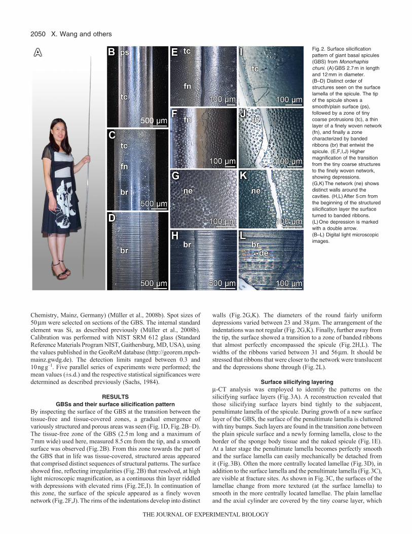
By inspecting the surface of the GBS at the transition between thetissue-free and tissue-covered zones, a gradual emergence ofvariously structured and porous areas was seen (Fig.1D, Fig.2B–D).The tissue-free zone of the GBS (2.5m long and a maximum of7mm wide) used here, measured 8.5cm from the tip, and a smoothsurface was observed (Fig.2B). From this zone towards the part ofthe GBS that in life was tissue-covered, structured areas appearedthat comprised distinct sequences of structural patterns. The surfaceshowed fine, reflecting irregularities (Fig.2B) that resolved, at highlight microscopic magnification, as a continuous thin layer riddledwith depressions with elevated rims (Fig.2E,I). In continuation ofthis zone, the surface of the spicule appeared as a finely wovennetwork (Fig.2F,J). The rims of the indentations develop into distinct
walls (Fig.2G,K). The diameters of the round fairly uniformdepressions varied between 23 and 38m. The arrangement of theindentations was not regular (Fig.2G,K). Finally, further away fromthe tip, the surface showed a transition to a zone of banded ribbonsthat almost perfectly encompassed the spicule (Fig.2H,L). Thewidths of the ribbons varied between 31 and 56m. It should bestressed that ribbons that were closer to the network were translucentand the depressions shone through (Fig.2L).
Surface silicifying layering-CT analysis was employed to identify the patterns on thesilicifying surface layers (Fig.3A). A reconstruction revealed thatthose silicifying surface layers bind tightly to the subjacent,penultimate lamella of the spicule. During growth of a new surfacelayer of the GBS, the surface of the penultimate lamella is clutteredwith tiny bumps. Such layers are found in the transition zone betweenthe plain spicule surface and a newly forming lamella, close to theborder of the sponge body tissue and the naked spicule (Fig.1E).At a later stage the penultimate lamella becomes perfectly smoothand the surface lamella can easily mechanically be detached fromit (Fig.3B). Often the more centrally located lamellae (Fig.3D), inaddition to the surface lamella and the penultimate lamella (Fig.3C),are visible at fracture sites. As shown in Fig.3C, the surfaces of thelamellae change from more textured (at the surface lamella) tosmooth in the more centrally located lamellae. The plain lamellaeand the axial cylinder are covered by the tiny coarse layer, which
X. Wang and others
Fig.2. Surface silicificationpattern of giant basal spicules(GBS) from Monorhaphischuni. (A)GBS 2.7m in lengthand 12mm in diameter. (B–D) Distinct order ofstructures seen on the surfacelamella of the spicule. The tipof the spicule shows asmooth/plain surface (ps),followed by a zone of tinycoarse protrusions (tc), a thinlayer of a finely woven network(fn), and finally a zonecharacterized by bandedribbons (br) that entwist thespicule. (E,F,I,J) Highermagnification of the transitionfrom the tiny coarse structuresto the finely woven network,showing depressions.(G,K)The network (ne) showsdistinct walls around thecavities. (H,L)After 5cm fromthe beginning of the structuredsilicification layer the surfaceturned to banded ribbons.(L)One depression is markedwith a double arrow. (B–L) Digital light microscopicimages.
THE JOURNAL OF EXPERIMENTAL BIOLOGY

2051Lamella formation in spicules
also includes the fine network around which the banded ribbons arecirculating (Fig.3E,F). At a higher magnification (Fig.3F), thenetwork around the plain rod and the banded ribbons are illustratedin a tilted view. This image also shows that the widths of the ribbons,31 to 56m, are slightly larger than the diameters of the depressions(23–38m).
Element distribution resolved by EDX spectroscopy in thesurface layer
In order to identify the chemical composition of the lamellarsurface region, scanning electron microscopy (SEM) images weretaken at different depths from the surface and, in parallel, thecorresponding EDX spectra were recorded (Fig.4). For thisanalysis, the spicule sample was moderately bent to inducecracking of the outer layers, as described by Müller et al. (Mülleret al., 2008d) (Fig.4A,B). The SEM images were obtained fromdifferent depths below the surface. All corresponding EDXanalyses that were taken from more centrally located lamellae,approximately 30 layers from the surface (Fig.4C-a),approximately 20 layers from the surface (Fig.4D-a), or from thepenultimate lamella (Fig.4E-a) gave almost identical EDX spectrawith dominant peaks for Si and O, reflecting the biosilica matrix(Fig.4C-b–E-b). Only the EDX spectrum (Fig.4F-b) obtainedfrom the surface of the banded ribbon layer (Fig.4F-a) gave amore diverse distribution. Besides Si and O, indicating thepresence of silica (SiO2), surface layers are composed of tracesof organic material, as assessed from the presence of the peaksfor Na, Mg, S, Cl, K and especially C. We have previously
described the banded ribbon structure as an organic layer (Wanget al., 2008).
Element analysis of the depressions within the ribbonsThe spicule surfaces in the central part of the sponge body displaya pronounced banded ribbon-like structure (Fig.5A). It is strikingthat the ribbons have two appearances: non-perforated andperforated. Approximately 10 non-perforated ribbons are followedby approximately 20 perforated ribbons (Fig.5A,B). The widths ofthe ribbons (31–56m) are slightly larger than the diameters of thedepressions (23–38m; Fig.5C,D). Optical microscopic analysesshowed that the depressions are filled with ball-like structures(~10m). However, when these depressions were inspected by SEM(Fig.5F), no structures within the holes could be resolved. Thisfinding suggests that the ball-like structures were destroyed by theelectron beam used for SEM analysis (10kV). In order to obtaininsight into the chemical difference between the smooth/plainregions within the ribbons and their depressions (Fig.5E-a), EDXanalyses were performed in parallel. The EDX spectra revealed thatthe smooth/plain regions only gave peaks for Si and O (Fig.5E-b),reflecting silica, whereas the depression regions compriseddominantly peaks for elements characteristic for organic matter, e.g.C, Na, Mg, S, Cl and K (Fig.5F-b). Two other biogenic elements,P and N, are not marked in Fig.5F-b, as their signals cannot beunequivocally assigned. It is very likely that the N signal is shieldedby the large C signal, whereas the (probable) P signal, at 2.0 keV,is very small. We hypothesize that those depressions might be filledwith cells, or fragments of them.
Fig.3. Distinct construction of the surface silicificating layer visualized by (A,B)-CT analysis or (C–G) optical microscopy. (A)The-CT reconstruction of aregion comprising a growing surface layer (sl) localized closer to the transition zone from the plain spicule surface to newly forming lamella. The surface ofthe penultimate lamella (pul) has a knobbed surface. (B)Further down the GBS, the surface lamella is less tightly attached to the penultimate lamella, verylikely because of its plain surface. (C,D)Frequently, the penultimate lamella, here with tiny coarse structures (tc), and the lamellae underneath, here with thesmooth/plain surface (ps), can be distinguished. The surface is formed by the banded ribbons layer (br). (D)The same region as that shown in C shown in atilted view. (E–G) Often a lamella with the plain surface is surrounded by a tiny coarse layer that is combined with the network (ne). The width of one ribbonand the diameter of one depression are marked (double-headed arrow; de).
THE JOURNAL OF EXPERIMENTAL BIOLOGY

2052
Staining the content matter in the depressions for organicmatter
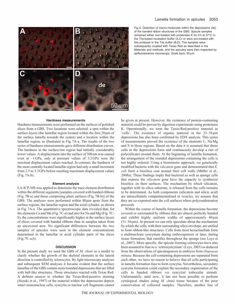
In an attempt to verify that organic matter potentially exists in thedepressions, the spicules were stained with Texas Red. As shownin Fig.6A,C, bright signals were seen within the depressions. Inorder to check whether the dye is indeed conjugated with aproteinaceous material, the spicule samples were pre-incubated withproteinase K. After that treatment, no significant signals could bedetected (Fig.6B,D).
X. Wang and others
Fig.4. Element analyses of different sites within the surface lamellar region.(A,B)Cross fracture through the spicule displaying the banded ribbon layer(br) and the sub-surface lamellar (la) architecture. SEM analyses.(B)Higher magnification of the image shown in A. (C–F) In parallel series,SEM images were taken from different sites of the surface (series a) andcorresponding EDX analyses were performed to determine the elementcomposition (series b). (C)Determination at the sub-surface region,approximately 30 layers from the surface. (D)SEM/EDX analysis in thelamellar zone, approximately 20 layers from the surface. (E)Surface,penultimate lamella region and (F) SEM/EDX analysis from the surface ofthe banded ribbons (br) zone. The areas that were selected for EDXanalysis are marked (square) in the corresponding SEM image.
Fig.5. Element analyses within the banded ribbon (br) layers. The surfacesof the samples were inspected by optical microscopy (A,B) to demonstratethe ordered sequence of ribbon structures (br) of non-perforated ribbons(np) [not provided with visible depression] and perforated ribbons (p) [withdepressions (de)]. (C,D)At higher magnification, ball-like structures werevisible within the depressions. The widths of the ribbons are marked withdouble-headed arrows. EDX analysis of the plain surface of a bandedribbon (E) and the depression existing within the band (F). Parallel studiesby SEM inspection (series a) and EDX spectroscopy (series b) wereperformed.
THE JOURNAL OF EXPERIMENTAL BIOLOGY
2053Lamella formation in spicules
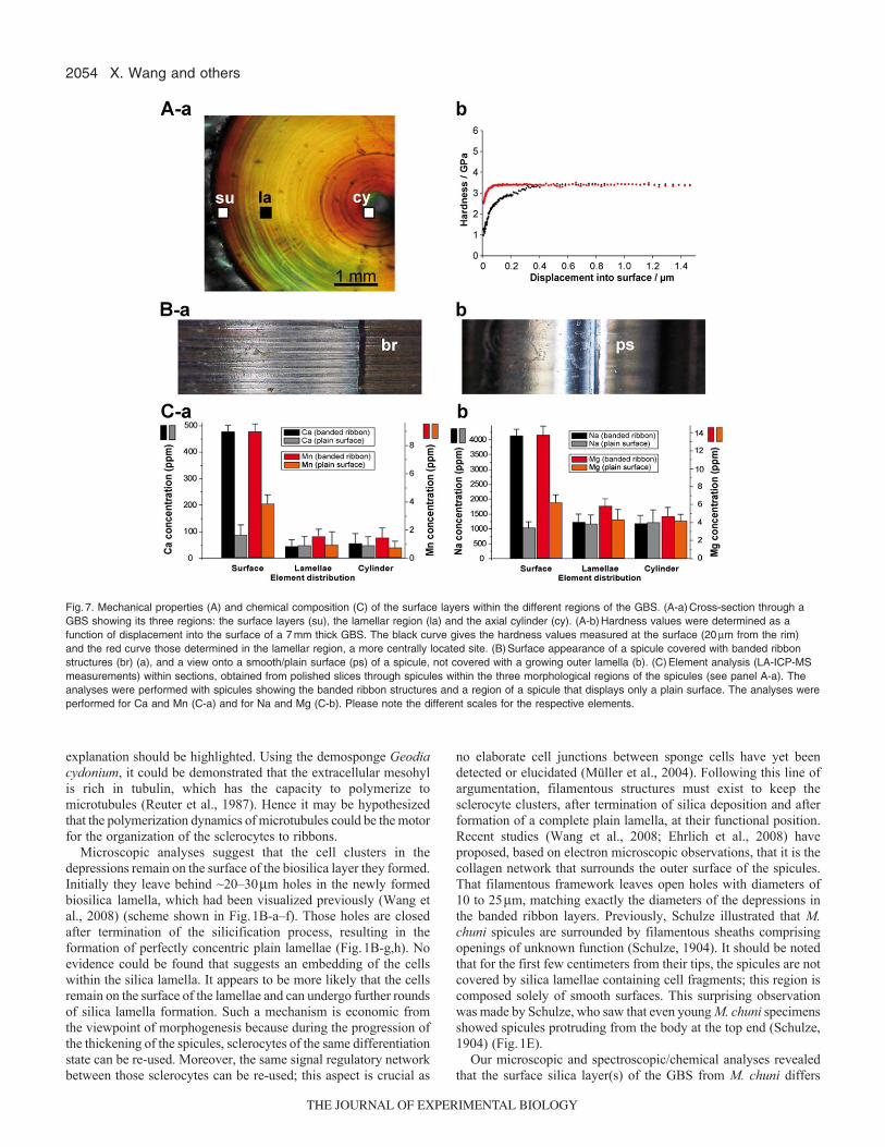
Hardness measurementsHardness measurements were performed on the surfaces of polishedslices from a GBS. Two locations were selected: a spot within thesurface layers (the lamellar region located within the first 20m ofthe surface lamella towards the center) and a location within thelamellar region, as illustrated in Fig.7A-a. The results of the twoseries of hardness measurements gave different distribution curves.The hardness in the surface/rim region had initially considerablylower values. A displacement into the surface of 300nm was causedeven at ~1GPa; only at pressure values of 3.2GPa were themaximal displacement values reached. In contrast, the hardness ofthe more centrally located lamellar region had only a small incrementfrom 2.5 to 3.3GPa before reaching maximum displacement values(Fig.7A-b).
Element analysisLA-ICP-MS was applied to determine the trace element distributionwithin the different segments [samples covered with banded ribbons(Fig.7B-a) and those comprising plain surfaces (Fig.7B-b)] of theGBS. The analyses were performed within 80m spots from thesurface regions, the lamellar region and the axial cylinder, as shownin Fig.7A-a. The quantitative spectroscopic data revealed that, forthe elements Ca and Mn (Fig.7C-a) and also for Na and Mg (Fig.7C-b), the concentrations were significantly higher in the surface layersof slices covered with banded ribbons than in samples taken froman uncovered area. No significant differences between the twosamples of spicules were seen in the element concentrationsdetermined in the lamellar or axial cylinder parts of the slices(Fig.7C-a,b).
DISCUSSIONIn the present study we used the GBS of M. chuni as a model toclarify whether the growth of the skeletal elements in the lateraldirection is controlled by sclerocytes. By light microscopy analysesand subsequent SEM analyses it became evident that the surfacelamellae of the GBS contain nests/rounded depressions that are filledwith ball-like structures. Those structures reacted with Texas Red.A definite answer to whether the Texas-Red-positive staining(Suzuki et al., 1997) of the material within the depressions depictsintact mononuclear cells, syncytia or nuclear cell fragments cannot
be given at present. However, the existence of protein-containingmaterial could be proven by digestion experiments using proteinaseK. Operationally, we term the Texas-Red-positive material as‘cells’. The existence of organic material in the 23–38mdepressions has also been confirmed by EDX analysis. This seriesof measurements proved the existence of the elements C, Na/Mgand S in those regions. Based on the data it is assumed that thosecells in the depressions form and continuously develop a rim ofpoly(silicate) around them. At the beginning of lamella formation,the arrangement of the rounded depressions containing the cells isnot highly ordered. Using a biomimetic approach, we geneticallymodified bacteria with the silicatein gene and demonstrated that E.coli form a biosilica coat around their cell walls (Müller et al.,2008a). These findings imply that bacterial as well as sponge cellsthat express the silicatein gene have the capacity to synthesizebiosilica on their surfaces. The mechanism by which silicatein,together with its silica substrate, is released from the cells remainsto be determined. As both components (silicatein and silicic acid)are intracellularly compartmentalized in vesicles, we assume thatthey are co-exported onto the cell surfaces where polycondensationproceeds.
Within the course of lamella formation, the depressions becomecovered or surrounded by ribbons that are almost perfectly bandedand exhibit highly uniform widths of approximately 40m(31–56m). At present we can only speculate about the mechanismby which the cells, with their surrounding silica envelope, are unifiedto form ribbon-like structures. Cells from most hexactinellids forma multinucleate syncytium during embryogenesis or later, duringtissue formation, that ramifies throughout the sponge (see Leys etal., 2007). More specific, the spicule-forming sclerocytes have alsobeen assumed to fuse to a ‘sclerosyncytium’ (Leys, 2003) as deducedfrom the observations of spiculogenesis in embryos from Oopsacasminuta. Because the cell-containing depressions are separated fromeach other, we have no reason to believe that all cells participatingin lamella formation fuse to form syncytia. Nevertheless, secondarysyncytia formation could explain the secondary organization of thecells to banded ribbons via syncytial trabecular strands.Unfortunately, until now, it has not been possible to performcytological studies using M. chuni tissue because of the poorconservation of collected samples. Therefore, another line of
Fig.6. Detection of macro-molecules within the depressions (de)of the banded ribbon structures of the GBS. Spicule samplesremained either non-treated with proteinase K for 2h at 37°C inthe Tris-based incubation buffer (A,C) or were pre-treated withthis protease in the Tris buffer (B,D). The samples weresubsequently coupled with Texas Red as described in theMaterials and methods, and the spicules were then inspected byepifluorescence microscopy. Scale bars, 50m.
THE JOURNAL OF EXPERIMENTAL BIOLOGY
2054
explanation should be highlighted. Using the demosponge Geodiacydonium, it could be demonstrated that the extracellular mesohylis rich in tubulin, which has the capacity to polymerize tomicrotubules (Reuter et al., 1987). Hence it may be hypothesizedthat the polymerization dynamics of microtubules could be the motorfor the organization of the sclerocytes to ribbons.
Microscopic analyses suggest that the cell clusters in thedepressions remain on the surface of the biosilica layer they formed.Initially they leave behind ~20–30m holes in the newly formedbiosilica lamella, which had been visualized previously (Wang etal., 2008) (scheme shown in Fig.1B-a–f). Those holes are closedafter termination of the silicification process, resulting in theformation of perfectly concentric plain lamellae (Fig.1B-g,h). Noevidence could be found that suggests an embedding of the cellswithin the silica lamella. It appears to be more likely that the cellsremain on the surface of the lamellae and can undergo further roundsof silica lamella formation. Such a mechanism is economic fromthe viewpoint of morphogenesis because during the progression ofthe thickening of the spicules, sclerocytes of the same differentiationstate can be re-used. Moreover, the same signal regulatory networkbetween those sclerocytes can be re-used; this aspect is crucial as
no elaborate cell junctions between sponge cells have yet beendetected or elucidated (Müller et al., 2004). Following this line ofargumentation, filamentous structures must exist to keep thesclerocyte clusters, after termination of silica deposition and afterformation of a complete plain lamella, at their functional position.Recent studies (Wang et al., 2008; Ehrlich et al., 2008) haveproposed, based on electron microscopic observations, that it is thecollagen network that surrounds the outer surface of the spicules.That filamentous framework leaves open holes with diameters of10 to 25m, matching exactly the diameters of the depressions inthe banded ribbon layers. Previously, Schulze illustrated that M.chuni spicules are surrounded by filamentous sheaths comprisingopenings of unknown function (Schulze, 1904). It should be notedthat for the first few centimeters from their tips, the spicules are notcovered by silica lamellae containing cell fragments; this region iscomposed solely of smooth surfaces. This surprising observationwas made by Schulze, who saw that even young M. chuni specimensshowed spicules protruding from the body at the top end (Schulze,1904) (Fig.1E).
Our microscopic and spectroscopic/chemical analyses revealedthat the surface silica layer(s) of the GBS from M. chuni differs
X. Wang and others
Fig.7. Mechanical properties (A) and chemical composition (C) of the surface layers within the different regions of the GBS. (A-a)Cross-section through aGBS showing its three regions: the surface layers (su), the lamellar region (la) and the axial cylinder (cy). (A-b)Hardness values were determined as afunction of displacement into the surface of a 7mm thick GBS. The black curve gives the hardness values measured at the surface (20m from the rim)and the red curve those determined in the lamellar region, a more centrally located site. (B)Surface appearance of a spicule covered with banded ribbonstructures (br) (a), and a view onto a smooth/plain surface (ps) of a spicule, not covered with a growing outer lamella (b). (C)Element analysis (LA-ICP-MSmeasurements) within sections, obtained from polished slices through spicules within the three morphological regions of the spicules (see panel A-a). Theanalyses were performed with spicules showing the banded ribbon structures and a region of a spicule that displays only a plain surface. The analyses wereperformed for Ca and Mn (C-a) and for Na and Mg (C-b). Please note the different scales for the respective elements.
THE JOURNAL OF EXPERIMENTAL BIOLOGY

2055Lamella formation in spicules
from the more internal layers by the existence of the cells locatedin the depressions that have been attributed to the formation of thelamellae. In order to examine whether the silicate matrix, formedat the surface of the spicules, also differs from the more centrallyarranged lamellae in terms of inorganic chemical composition andmechanical properties, we applied the techniques of LA-ICP-MSspectroscopy and hardness/indentation determination. Thespectroscopical analysis distinctly showed that the surface layersfrom spicule samples that were covered by banded ribbonscomprised significantly higher concentrations of monovalent anddivalent cations within the poly(silicate) than the silica that existsmore centrally within the spicule. In the silica layers close to thesurface, the silicic acid building blocks forming the silica layersexist, to a considerable extent, as salts of Na, Mg, Ca and Mn.Recently, we proposed that the silicatein-mediated synthesis ofbiosilica starts from the silicic acid/silicate substrate and proceedsvia cyclic silicic acid species (trisiloxane rings) to poly(silicate)(Schröder et al., in press). As a consequence of the higherconcentration of cations in the silica matrix of the outer surface ofthe spicules, the hardness should be lower (Wiederhorn, 1969;Müller et al., 2008b). This supposition was proven by hardnessstudies on cross-sections through the spicule. The data revealed thatthe near-surface lamellae from spicules surrounded by bandedribbons were more flexible (by a factor of two) compared with thelamellae that were not in the process of formation and were devoidof depressions. In turn, a mechanism has to be proposed that allowsan understanding of the ageing process of poly(silicate) afterformation of the surface-directed lamellae. The data of the presentstudy show that with the progression of silica layering around thespicules of M. chuni, the cation content decreases. Hence it isassumed that, during ageing, the formation of siloxane (Si-O-Si)bonds through condensation (removal of water) of residual silanol(Si-OH) groups present in the biosilica lamellae will occur. As aconsequence, cations that act as counter-ions to acidic silanol groupswill be eliminated from the silica phase. The latter process couldproceed in association with a cation exchanger, perhaps collagencarrying negatively charged residue groups (aspartate andglutamate), at a slightly increased pH milieu (Zhang et al., 2004).A recent study provided experimental evidence that the hardnessof the biosilica within the M. chuni GBS is regionally different(Miserez et al., 2008); by focusing on the axial cylinder, Miserezet al. found that the hardness/indentation of the center of the spiculesis approximately 2.5 times lower compared with the lamellarregion.
In conclusion, the data presented here suggest that a closecell–silica contact is involved in silica formation, as shown here inthe sponge M. chuni. Prior to this, using both the demosponge(Suberites domuncula) and the hexactinellid model (M. chuni), noevidence could be presented that filaments, e.g. collagen, areprimarily involved in the formation of the silica lamellae (Mülleret al., 2009b). Based on the present data, the following steps for theformation of the outer lamella can be formulated (Fig.1B).Sclerocytes associate to cell clusters on the surface of the(pen)ultimate smooth-surfaced lamella and start to form pericellularsilica using silicatein under consumption of silicic acid. After thestage of textured lamella formation, the cell clusters proceed withsilica formation and allow the completion of a smooth surface. Thecells remain immobilized on the surface of the terminated lamellaand attached to the spicule via a filamentous network for the nextround of lamella formation (Fig.1C).
ACKNOWLEDGEMENTSW.E.G.M. is a holder of an ERC Advanced Investigator Grant (268476BIOSILICA). This work was supported by grants from the GermanBundesministerium für Bildung und Forschung (project ‘Center of ExcellenceBIOTECmarin’), the Deutsche Forschungsgemeinschaft (Schr 277/10-1), theInternational Human Frontier Science Program, the European Commission(project no. 031541–BIO-LITHO; Biomineralization for Lithography andMicroelectronics), the consortium BiomaTiCS at the Universitätsmedizin of theJohannes Gutenberg-Universität Mainz, and the International S & T CooperationProgram of China (grant no. 2008DFA00980).
REFERENCESBoury-Esnault, N. and Rützler, K. (1997). Thesaurus of sponge morphology.
Smithson. Contrib. Zool. 596, 1-55.Brunner, E., Richthammer, P., Ehrlich, H., Paasch, S., Simon, P., Ueberlein, S.
and van Pee, K.-H. (2009). Chitin-based organic networks – an integral part of cellwall biosilica from the diatom Thalassiosira pseudonana. Angewandte ChemieIntern. Edit. 48, 9724-9727.
Butler, P. E. (1961). Morphologic classification of sponge spicules, with descriptions ofsiliceous spicules from the Lower Ordovician Bellefonte Dolomite in CentralPennsylvania. J. Paleontol. 35, 191-200.
Cha, J. N., Shimizu, K., Zhou, Y., Christianssen, S. C., Chmelka, B. F., Stucky, G.D. and Morse, D. E. (1999). Silicatein filaments and subunits from a marine spongedirect the polymerization of silica and silicones in vitro. Proc. Natl. Acad. Sci. USA96, 361-365.
Ehrlich, H. (2010). Biological Materials of Marine Origin. Invertebrates. Heidelberg:Springer-Verlag.
Ehrlich H. (2011) Silica biomineralization in sponges. In Encyclopedia of Geobiology(ed. J. Reitner and V. Thiel), pp. 796-808. Heidelberg: Springer-Verlag.
Ehrlich, H., Heinemann, S., Heinemann, C., Simon, P., Bazhenov, V. V., Shapkin,N. P., Born, R., Tabachnick, K., Hanke, T. and Worch, H. (2008). Nanostructuralorganization of naturally occurring composites – Part I: silica-collagen-basedbiocomposites. J. Nanomat. 2008, 623838. doi:10.1155/2008/623838.
Ehrlich, H., Demadis, K. D., Pokrovsky, O. S. and Koutsoukos, P. G. (2010a).Modern views on desilicification: biosilica and abiotic silica dissolution in natural andartificial environments. Chem. Rev. 110, 4656-4689.
Ehrlich, H., Simon, P, Carrillo-Cabrera, W., Bazhenov, V. V., Botting, J., Ilan, M.,Ereskovsky, A. V., Muricy, G., Worch, H, Mensch, A. et al. (2010b). Insights intochemistry of biological materials: newly discovered silica-aragonite-chitinbiocomposites in demosponges. Chem. Mat. 22, 1462-1471.
Ehrlich, H., Deutzmann, R., Brunner, E., Cappellini, E., Koon, H., Solazzo, C.,Yang, Y., Ashford, D., Thomas-Oates, J., Lubeck, M. et al. (2010c). Mineralizationof the meter-long biosilica structures of glass sponges is templated on hydroxylatedcollagen. Nature Chem. 2, 1084-1088
Hildebrand, M. and Wetherbee, R. (2003). Components and control of silicification indiatoms. In Silicon Biomineralization: Biology, Biochemistry, Molecular Biology,Biotechnology (ed. W. E. G. Müller), pp. 11-57, Vol. 33. Heidelberg: Springer-Verlag.
Hooper, J. N. A (1997). Guide to Sponge Collection and Identification. SouthBrisbane: Queensland Museum. Available at http://xa.yimg.com/kq/groups/21368769/1183738468/name/Guide+to+sponge+identification.pdf.
Inagaki, F., Motomura, Y. and Ogata, S. (2003). Microbial silica deposition ingeothermal hot waters. Appl. Microbiol. Biotechnol. 60, 605-611.
Krasko, A., Batel, R., Schröder, H. C., Müller, I. M. and Müller, W. E. G. (2000).Expression of silicatein and collagen genes in the marine sponge Suberitesdomuncula is controlled by silicate and myotrophin. Eur. J. Biochem. 267, 4878-4887.
Kröger, N. and Sumper, M. (2000). The biochemistry of silica formation in diatoms. InBiomineralization (ed. E. Bäuerlein), pp. 151-170. Weinheim, New York: Wiley-VCH.
Levi, C., Barton, J. L., Guillemet, C., Lebras, E. and Leheude, P. (1989) Aremarkably strong natural glassy rod: the anchoring spicule of the Monoraphissponge. J. Mat. Sci. Lett. 8, 337-339.
Leys, S. P. (2003). Comparative study of spiculogenesis in demosponge andhexactinellid larvae. Microsc. Res. Tech. 62, 300-311.
Leys, S. P., Mackie, G. O. and Reiswig, H. M. (2007). The biology of glass sponges.Adv. Mar. Biol. 52, 1-145.
Li, X. D. and Bhushan B. (2002). A review of nanoindentation continuous stiffnessmeasurement technique and its applications. Mater. Charact. 48, 11-36.
Ma, J. F. (2003). Functions of silicon in higher plants. Prog. Mol. Subcell. Biol. 33, 127-147.
Maldonado, M., Carmona, M. C., Velásquez, Z., Puig, A., Cruzado, A., López, A.and Young, C. M. (2005). Siliceous sponges as a silicon sink: an overlooked aspectof benthopelagic coupling in the marine silicon cycle. Limnol. Oceanogr. 50, 799-809.
Minchin, E. A. (1909). Sponge spicules. A summary of present knowledge. Ergebn.Forts. Zool. 2, 171-274.
Miserez, A., Weaver, J. C., Thurner, P. J., Aizenberg, J., Dauphin, Y., Fratzl, P.,Morse, D. E. and Zok, F. W. (2008). Effects of laminate architecture on fractureresistance of sponge biosilica: lessons from nature. Adv. Funct. Mater. 18, 1241-1248.
Morse, D. E. (1999). Silicon biotechnology: harnessing biological silica production toconstruct new materials. Trends Biotechnol. 17, 230-232.
Mugnaioli, E., Natalio, F., Schloßmacher, U., Wang, X. H., Müller, W. E. G. andKolb, U. (2009) Crystalline nanorods as seeds for the synthesis of amorphousbiosilica during spicule formation in sponges. Chembiochem 10, 683-689.
Müller, W. E. G. (ed.) (2003). Silicon Biomineralization: Biology, Biochemistry,Molecular Biology, Biotechnology. Berlin: Springer Press.
THE JOURNAL OF EXPERIMENTAL BIOLOGY
2056
Müller, W. E. G., Wiens, M., Adell, T., Gamulin, V., Schröder, H. C. and Müller, I.M. (2004) Bauplan of urmetazoa: basis for genetic complexity of Metazoa. Intern.Review Cytol. 235, 53-92.
Müller, W. E. G., Rothenberger, M., Boreiko, A., Tremel, W., Reiber, A. andSchröder, H. C. (2005). Formation of siliceous spicules in the marine demospongeSuberites domuncula. Cell Tissue Res. 321, 285-297.
Müller, W. E. G., Boreiko, A., Schloßmacher, U., Wang, X. H., Tahir, M. N., Tremel,W., Brandt, D., Kaandorp, J. A. and Schröder, H. C. (2007a). Fractal-relatedassembly of the axial filament in the demosponge Suberites domuncula: relevanceto biomineralization and the formation of biogenic silica. Biomaterials 28, 4501-4511.
Müller, W. E. G., Boreiko, A., Wang, X. H., Belikov, S. I., Wiens, M., Grebenjuk, V.A., Schloßmacher, U. and Schröder, H. C. (2007b). Silicateins, the major biosilicaforming enzymes present in demosponges: protein analysis and phylogeneticrelationship. Gene 395, 62-71.
Müller, W. E. G., Eckert, C., Kropf, K., Wang, X. H., Schloßmacher, U., Seckert, C.,Wolf, S. E., Tremel, W. and Schröder, H. C. (2007c). Formation of the giantspicules of the deep sea hexactinellid Monorhaphis chuni (Schulze 1904): electronmicroscopical and biochemical studies. Cell Tissue Res. 329, 363-378.
Müller, W. E. G., Li, J., Schröder, H. C., Qiao, L. and Wang, X. H. (2007d). Theunique skeleton of siliceous sponges (Porifera; Hexactinellida and Demospongiae)that evolved first from the Urmetazoa during the Proterozoic: a review.Biogeosciences 4, 219-232.
Müller, W. E. G., Wang, X. H., Belikov, S. I., Tremel, W., Schloßmacher, U., Natoli,A., Brandt, D., Boreiko, A., Tahir, M. N., Müller, I. M. and Schröder, H. C.(2007e). Formation of siliceous spicules in demosponges: example Suberitesdomuncula. In Handbook of Biomineralization – The Biology of Biominerals StructureFormation (ed. E. Bäuerlein), Vol. 1, pp. 59-82. Weinheim: Wiley-VCH.
Müller, W. E. G., Engel, S., Wang, X. H., Wolf, S. E., Tremel, W., Thakur, N. L.,Krasko, A., Divekar, M. and Schröder, H. C. (2008a). Bioencapsulation of livingbacteria (Escherichia coli) with poly(silicate) after transformation with silicatein-gene. Biomaterials 29, 771-779.
Müller, W. E. G., Jochum, K., Stoll, B. and Wang, X. H. (2008b). Formation of giantspicule from quartz glass by the deep sea sponge Monorhaphis. Chem. Mater. 20,4703-4711.
Müller, W. E. G., Wang, X. H., Kropf, K., Boreiko, A., Schloßmacher, U., Brandt,D., Schröder, H. C. and Wiens, M. (2008c). Silicatein expression in thehexactinellid Crateromorpha meyeri: the lead marker gene restricted to siliceoussponges. Cell Tissue Res. 333, 339-351.
Müller, W. E. G., Wang, X. H., Kropf, K., Ushijima, H., Geurtsen, W., Eckert, C.,Tahir, M. N., Tremel, W., Boreiko, A., Schloßmacher, U., Li, J. and Schröder, H.C. (2008d). Bioorganic/inorganic hybrid composition of sponge spicules: matrix of thegiant spicules and of the comitalia of the deep sea hexactinellid Monorhaphis. J.Struct. Biol. 161, 188-203.
Müller, W. E. G., Schloßmacher, U., Wang, X. H., Boreiko, A., Brandt, D., Wolf, S.E., Tremel, W. and Schröder, H. C. (2008e) Poly(silicate)-metabolizing silicatein insiliceous spicules and silicasomes of demosponges comprises dual enzymaticactivities (silica-polymerase and silica-esterase). FEBS J. 275, 362-370.
Müller, W. E. G., Wang, X. H., Burghard, Z., Bill, J., Krasko, A., Boreiko, A.,Schloßmacher, U., Schröder, H. C. and Wiens, M. (2009a). Bio-sinteringprocesses in hexactinellid sponges: fusion of bio-silica in giant basal spicules fromMonorhaphis chuni. J. Struct. Biol. 168, 548-561.
Müller, W. E. G., Wang, X. H., Cui, F. Z., Jochum, K. P., Tremel, W., Bill, J.,Schröder, H. C., Natalio, F., Schloßmacher, U. and Wiens, M. (2009b). Spongespicules as blueprints for the biofabrication of inorganic-organic composites andbiomaterials. Appl. Microbiol. Biotechnol. 83, 397-413.
Neumann, D. (2003). Silicon in plants. In Silicon Biomineralization: Biology,Biochemistry, Molecular Biology, Biotechnology (ed. W. E. G. Müller), pp. 149-160,Vol. 33. Berlin: Springer Press.
Panchuk-Voloshina, N., Haugland, R. P., Bishop–Stewart, J., Bhalgat, M. K.,Millard, P. J., Mao, F., Leung, W. Y. and Haugland, P. (1999). Alexa dyes, aseries of new fluorescent dyes that yield exceptionally bright, photostableconjugates. J. Histochem. Cytochem. 47, 1179-1188.
Perry, C. C. (2003). Silicification: the processes by which organisms capture andmineralize silica. Rev. Mineral. Geochem. 54, 291-327.
Reuter, P., Dorn, A., Batel, R., Schröder, H. C. and Müller, W. E. G. (1987).Evidence for the existence of microtubule protein in the extracellular space of marinesponges. Tissue Cell 19, 773-782.
Sachs, L. (1984). Angewandte Statistik. Berlin: Springer.
Schröder, H. C., Perovic-Ottstadt, S., Rothenberger, M., Wiens, M., Schwertner,H., Batel, R., Korzhev, M., Müller, I. M. and Müller, W. E. G. (2004). Silicatransport in the demosponge Suberites domuncula: fluorescence emission analysisusing the PDMPO probe and cloning of a potential transporter. Biochem. J. 381,665-673.
Schröder, H. C., Boreiko, A., Korzhev, M., Tahir, M. N., Tremel, W., Eckert, C.,Ushijima, H., Müller, I. M. and Müller, W. E. G. (2006). Co-expression andfunctional interaction of silicatein with galectin: matrix-guided formation of siliceousspicules in the marine demosponge Suberites domuncula. J. Biol. Chem. 281,12001-12009.
Schröder, H. C., Natalio, F., Shukoor, I., Tremel, W., Schloßmacher, U., Wang, X.H. and Müller, W. E. G. (2007). Apposition of silica lamellae during growth ofspicules in the demosponge Suberites domuncula: biological/biochemical studiesand chemical/biomimetical confirmation. J. Struct. Biol. 159, 325-334.
Schröder, H. C., Wiens, M., Schloßmacher, U., Brandt, D. and Müller, W. E. G. (inpress). Silicatein-mediated polycondensation of orthosilicic acid: modeling of catalyticmechanism involving ring formation. Silicon doi: 10.1007/s12633-010-9057-4.
Schulze, F. E. (1904). Hexactinellida. Wissenschaftliche Ergebnisse der DeutschenTiefsee-Expedition auf dem Dampfer “Valdivia” 1898-1899. Stuttgart: Fischer.
Shimizu, K., Cha, J., Stucky, G.D. and Morse, D.E. (1998). Silicatein alpha:cathepsin L-like protein in sponge biosilica. Proc. Natl. Acad. Sci. USA 95, 6234-6238.
Suzuki, T., Fujikura, K., Higashiyama, T. and Takata, K. (1997). DNA staining forfluorescence and laser confocal microscopy. J. Histochem. Cytochem. 45, 49-53.
Titus, J. A., Haugland, R. P., Sharrow, S. O. and Segal, D. M. (1982). Texas Red, ahydrophilic, red-emitting fluorophore for use with fluorescein in dual parameter flowmicrofluorometric and fluorescence microscopic studies. J. Immunol. Methods 50,193-204.
Uriz, M. J. (2006). Mineral spiculogenesis in sponges. Can. J. Zool. 84, 322-356.Uriz, M.-J., Turon, X., Becero, M. A. and Agell, G. (2003) Siliceous spicules and
skeletal frameworks in sponges: origin, diversity, ultrastructural patterns andbiological functions. Microsc. Res. Tech. 62, 279-299.
Wang, X. H. and Müller, W. E. G. (2009). Contribution of biomineralization duringgrowth of polymetallic nodules and ferromanganese crusts from the Pacific Ocean.Front. Mater. Sci. China 3, 109-123.
Wang, X. H., Li, J., Qiao, L., Schröder, H. C., Eckert, C., Kropf, K., Wang, Y. M.,Feng, Q. L. and Müller, W. E. G. (2007). Structure and characteristics of giantspicules of the deep sea hexactinellid sponges of the genus Monorhaphis(Hexactinellida: Amphidiscosida: Monorhaphididae). Acta Zool. Sin. 53, 557-569.
Wang, X. H., Boreiko, A., Schloßmacher, U., Brandt, D., Schröder, H. C., Li, J.,Kaandorp, J. A., Götz, H., Duschner, H. and Müller, W. E. G. (2008). Axial growthof hexactinellid spicules: formation of cone-like structural units in the giant basalspicules of the hexactinellid Monorhaphis. J. Struct. Biol. 164, 270-280.
Wang, X. H., Schröder, H. C. and Müller, W. E. G. (2009a). Giant siliceous spiculesfrom the deep-sea glass sponge Monorhaphis chuni: morphology, biochemistry, andmolecular biology. Int. Rev. Cell. Mol. Biol. 273, 69-115.
Wang, X. H., Schloßmacher, U., Wiens, M., Schröder, H. C. and Müller, W. E. G.(2009b) Biogenic origin of polymetallic nodules from the Clarion-Clipperton zone inthe Eastern Pacific Ocean: electron microscopic and EDX evidence. Mar. Biotechnol.11, 99-108.
Weissenfels, N. (1989). Biologie und Mikroskopische Anatomie derSüßwasserschwämme (Spongillidae). Stuttgart, New York: Gustav Fischer Verlag.
Wiederhorn, S. M. (1969). Fracture stress energy of glass. J. Am. Ceram. Soc. 52,99-105.
Wiens, M., Bausen, M., Natalio, F., Link, T., Schlossmacher, U. and Müller, W. E.G. (2009). The role of the silicatein-a interactor silintaphin-1 in biomimeticbiomineralization. Biomaterials 30, 1648-1656.
Wiens, M., Wang, X. H., Schloßmacher, U., Lieberwirth, I., Glasser, G., Ushijima,H., Schröder, H. C. and Müller, W. E. G. (2010). Osteogenic potential of bio-silicaon human osteoblast-like (SaOS-2) cells. Calcif. Tissue Int. 87, 513-524.
Wiens, M., Schröder, H. C., Wang, X. H., Link, T., Steindorf, D. and Müller, W. E.G. (2011). Isolation of the silicatein- interactor silintaphin-2 by a novel solid-phasepull-down assay. Biochemistry 50, 1981-1990.
Zhang, L. J., Feng, X. S., Liu, H. G., Qian, D. J., Zhang, L., Yu, X. L. and Cui, F. Z.(2004). Hydroxyapatite/collagen composite materials formation in simulated bodyfluid environment. Materials Lett. 58, 719-722.
X. Wang and others
THE JOURNAL OF EXPERIMENTAL BIOLOGY
Related Documents