Circadian rhythm gene regulation in the housefly, Musca domestica Veryan Codd* † 1,2 , David Dolezel ‡§1 , Jan Stehlik ‡§ , Alberto Piccin* 3 , Karen J. Garner*, Seth N. Racey* 4 , Kornelis R. Straatman*, Edward J. Louis* 5 , Rodolfo Costa ‡‡ , Ivo Sauman ‡ , Charalambos P. Kyriacou* 6 , Ezio Rosato* *Department of Genetics, University of Leicester, Leicester LE1 7RH, UK, † Department of Biology, University of Leicester, Leicester LE1 7RH, UK, ‡ Institute of Entomology, Academy of Sciences of the Czech Republic, CeskeBudejovice, Czech Republic, § Faculty of Biological Sciences, University of South Bohemia, Ceske Budejovice, Czech Republic and ‡‡ Department of Biology, University of Padova, Padova 35131, Italy 1 contributed equally to this paper, 2 present address: Department of Cardiovascular Sciences, Glenfield General Hospital, Leicester LE3 9QP, UK. 3 deceased, May 1, 2002. 4 present address: MTEC, Edinburgh University, Edinburgh, Scotland. 5 present address: Institute of Genetics, University of Nottingham, Nottingham NG7 2UH, UK 6 Corresponding author: Department of Genetics, University of Leicester, Leicester LE1 7RH, UK email: [email protected] Running head: Circadian clock genes in houseflies Key words: circadian, evolution, diptera, neurons, regulation Genetics: Published Articles Ahead of Print, published on October 18, 2007 as 10.1534/genetics.107.079160

Welcome message from author
This document is posted to help you gain knowledge. Please leave a comment to let me know what you think about it! Share it to your friends and learn new things together.
Transcript
Circadian rhythm gene regulation in the housefly, Musca domestica
Veryan Codd*†1,2 , David Dolezel
‡§1, Jan Stehlik
‡§, Alberto Piccin*3, Karen J. Garner*, Seth N.
Racey*4, Kornelis R. Straatman*, Edward J. Louis*5, Rodolfo Costa‡‡
, Ivo Sauman‡,
Charalambos P. Kyriacou*6, Ezio Rosato*
*Department of Genetics, University of Leicester, Leicester LE1 7RH, UK, †Department of
Biology, University of Leicester, Leicester LE1 7RH, UK, ‡Institute of Entomology, Academy of
Sciences of the Czech Republic, CeskeBudejovice, Czech Republic, §Faculty of Biological
Sciences, University of South Bohemia, Ceske Budejovice, Czech Republic and ‡‡Department of
Biology, University of Padova, Padova 35131, Italy
1contributed equally to this paper, 2present address:
Department of Cardiovascular Sciences, Glenfield General Hospital,
Leicester LE3 9QP, UK. 3deceased, May 1, 2002. 4present address: MTEC, Edinburgh University, Edinburgh, Scotland.
5 present address: Institute of Genetics, University of Nottingham, Nottingham NG7
2UH, UK 6Corresponding author: Department of Genetics, University of Leicester, Leicester LE1 7RH, UK email: [email protected]
Running head: Circadian clock genes in houseflies
Key words: circadian, evolution, diptera, neurons, regulation
Genetics: Published Articles Ahead of Print, published on October 18, 2007 as 10.1534/genetics.107.079160
2
Abstract
The circadian mechanism appears remarkably conserved between Drosophila and mammals, with
basic underlying negative and positive feedback loops, cycling gene products, and temporally
regulated nuclear transport involving a few key proteins. One of these negative regulators is
PERIOD, which in Drosophila shows very similar temporal and spatial regulation to TIMELESS.
Surprisingly, we observe that in the housefly, Musca domestica, PER does not cycle in western
blots of head extracts, in contrast to the TIM protein. Furthermore immunocytochemical (ICC)
localization using enzymatic staining procedures, reveals that PER is not localized to the nucleus
of any neurons within the brain at any circadian time, as recently observed for several non-
dipteran insects. However with confocal analysis, immunofluorescence reveals a very different
picture, and provides an initial comparison of PER/TIM containing cells in Musca and
Drosophila, which shows some significant differences, but many similarities. Thus, even in
closely related diptera, there is considerable evolutionary flexibility in the number and spatial
organization of clock cells, and indeed in the expression patterns of clock products in these cells,
although the underlying framework is similar.
3
Introduction
Circadian rhythms are generated by a system of interlocked autoregulatory feedback loops in
which both negative and positive feedback play prominent roles. The insect model has been
developed most forcefully in Drosophila melanogaster, where two cycling proteins, PERIOD
(PER) and TIMELESS (TIM) act as negative regulators of their own transcription, through the
positively acting bHLH-PAS transcription factors CLOCK (CLK) and CYCLE (CYC, also
known as BMAL1) (Collins and Blau, 2007; Hall 2003). The CLK protein also cycles and its
regulation is interlocked with that of PER, in that CLK/CYC also activate PDP1ε and VRILLE
(VRI), with the latter modulating expression of Clk (Cyran et al. 2003; Benito et al. 2007).
One of the most compelling features of per and tim regulation is that the mRNAs cycle
with a peak a few hours in advance of the rhythm in their protein products in the fly’s head (Hall
2003). The protein cycles have been visualised in a small subset of neurons within the CNS,
termed the lateral and the dorsal neurons. The best studied are the ventral lateral neurons (LNvs)
because they can also be counterstained for the neuropeptide pigment dispersing factor, PDF
(Helfrich-Forster 1995; Helfrich-Forster and Homberg 1993; Nassel et al. 1993). Within these
neurons, PER and TIM proteins can be seen to translocate to the nucleus late at night, when their
proteins are at their peak (Shafer et al. 2002). During the day, both proteins show dramatic
reductions in their abundance that correlates with their hyperphosphorylation and subsequent
degradation (Edery et al. 1994; Naidoo et al. 1999; Zeng et al. 1996). PER appears to require
TIM for its stability, in part to protect it from phosphorylation by the Doubletime kinase, so once
TIM levels start to fall, PER levels also become compromised (Hall 2003; Kloss et al. 1998; Price
et al. 1998). However, post-transcriptional controls means that even in constant darkness, PER
and TIM levels ebb and flow with similar patterns that are seen under light-dark (LD) cycles
(Hall 2003). A number of other kinases (Hall, 2003) and phosphatases (Sathyanarayanan et al.
2004; Fang et al. 2007) have also been shown to modulate the stabilities of PER and TIM.

4
Comparative analysis of this insect model of circadian gene regulation has been most
comprehensively studied in the giant silkmoth, Antheraea pernyii. While the photoreceptors in
the moth appear to show cycles of PER antigenicity similar to that seen in the fly (Reppert et al.
1994), in the central brain, a very small number of neurons co-express cycling PER and TIM, yet
the two proteins remain stubbornly cytoplasmic (Sauman and Reppert 1996). This observation
might initially appear to preclude a canonical autoregulatory role for these two proteins.
However, when transformed into per-null flies, although behavioural rhythmicity is markedly
attenuated in the transgenic compared to control transformant flies, ApPER does appear to locate
to the nucleus of lateral neurons and photorecepeptors during the night phase (Levine et al. 1995).
In addition, when ApPER is used in Drosophila cell lines to reconstitute a circadian pacemaker, it
does appear to act as a negative regulator of ApCLK/ApBMAL1 mediated transactivation, with
the added bonus of ApTIM acting to enhance this negative regulation (Chang et al. 2003). In
Drosophila, TIM can shuttle in and out of the nucleus, so it may be that although TIM (or PER)
cannot be seen in the nucleus at particular timepoints in the silkmoth, it is nevertheless present
(Ashmore et al. 2003; Nawathean and Rosbash 2004). Thus perhaps even in Antheraea, the
canonical model holds. Furthermore, a recent study has revealed that dsRNAi knockdown of per
in another silkmoth, Bombyx mori, generates a modest disruption in the circadian larval eclosion
gate, consistent with the view that per in Lepidoptera plays a similar biological role to
Drosophila, irrespective of any differences in temporal expression patterns (Sandrelli et al.2007)
However, a survey of PER-like immunoreactivity in a number of insect orders, once
again, reveals a recalcitrant PER antigen that is exclusively confined to the cytoplasm in the
various neuronal cell types where it is found (Zavodska et al. 2003b). We have therefore studied
the regulation of the per and tim genes within the circadian clock of the housefly, Musca
domestica, which had a common ancestor with Drosophila about 100 Mya (Hennig 1981). The
Musca per orthologue can rescue per-null arrhythmia in Drosophila hosts to a surprisingly robust
degree compared to the per transgene from a more closely related species such as D.
5
pseudoobscura (Piccin et al. 2000). While these results might suggest that Musca per might not
seem to provide a very promising avenue for further comparative work, particularly as a related
muscid, Lucilia cuprina, shows very similar expression pattern for per gene products as
Drosophila (Warman et al. 2000), we shall see that the study of clock gene regulation in Musca
provides some interesting twists and turns that should be considered carefully when analysing the
results of other comparative circadian studies.
Methods Fly maintenance: Musca embryos and larvae were raised on a medium made of bran
(55 g), heat-inactivated yeast (3g), milk (150 ml) and the anti-mycotic nipagin (0.35g) until
pupariation. After eclosion adult flies were fed on water, sugar and dried milk. Flies were
maintained at 25o
under 12 h light, 12 h dark cycles (LD12:12). In our studies we used both a
wild type (gift from A. Malacrida and G. Gasperi, University of Pavia) and a white strain (gift
from Daniel Bopp, University of Zurich) without noticing any significant difference (Hediger et
al. 2001). However, locomotor activity and confocal microscopy were performed on the white
strain only whereas pupal eclosion was tested on flies carrying both white and apterous mutations
(again, from Daniel Bopp).
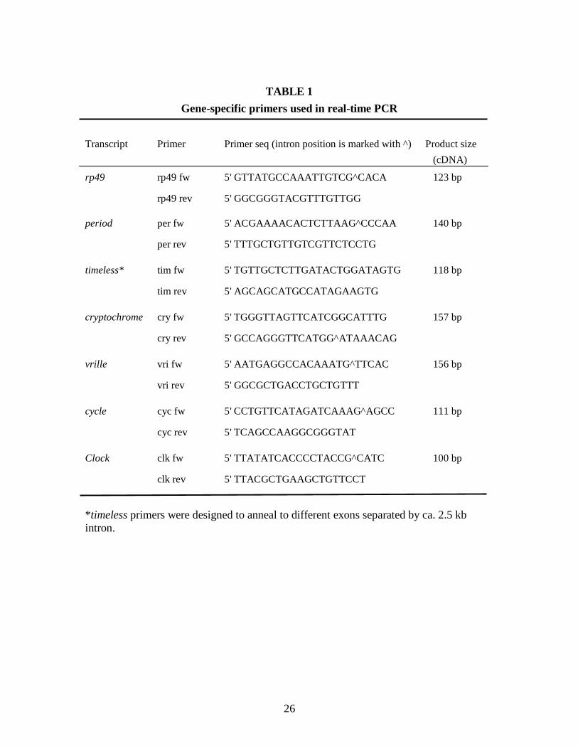
Gene expression analyses: Sequences for Musca domestica circadian genes vri, cyc, Clk and
‘insect’ cryptochrome (cry) were obtained using a degenerate PCR approach with primers
designed according to conserved protein regions from Drosophila and mouse clock gene
orthologs. Short fragments (60-250bp) were then amplified from Musca head cDNA, cloned and
sequenced. Gene sequence was then extended by primer walking or 3' RACE. Musca tim
sequences were obtained by screening a housefly genomic library with D. melanogaster tim as a
probe (E Rosato, J Clayton, B Collins and S Campesan, unpublished). These clock gene
sequences will constitute the subject of a separate publication. The Musca per sequence has been
described previously (Piccin et al., 2000). Real time PCR quantification of reverse-transcribed
6
mRNA from whole head extracts, was carried out for Musca per, tim, vri, cyc, Clk and cry in LD,
DD and LL conditions. The constitutively expressed Musca rp49 gene was used as an
amplification standard. Primers used are listed in Table 1. In one experimental design, fly
cultures (from the final larval stage) were entrained to LD12:12 and collected at 4h
intervals during one final LD cycle and the following day in constant darkness (DD).
Similarly, flies exposed to constant light (LL) from the late larval stage were used to
assess the expression of clock genes in LL. For both experimental designs, at least three
independent animal groups were reared and sacrificed. For each experiment, twenty 3-5
days old males were collected at the appropriate time point, and the heads stored at -80o
until RNA extraction.
Total RNA was isolated with Trizol (Sigma) according to the manufactures instructions.
1µg of total RNA was reverse transcribed using oligodT (24mer) and SuperscriptII reverse
transcriptase (Invitrogen). To safeguard against amplification of possible contaminant genomic
DNA either the primers were designed to anneal only to a template corresponding to the spliced
transcript, or (in one case - timeless amplification) to include a large (2.5 kb) intron in the
genomic template. In the latter case PCR conditions were optimized, so that only the short
(cDNA) product would amplify. This "cDNA specificity" was confirmed in pilot real-time PCRs
with pure genomic DNA as a template (resulting in no product).
5 µl of diluted cDNA were used for a 20 µl PCR reaction ( Hot start Taq polymerase (Takara)
0.32 U, Taq buffer 1x, dNTPs 200 µM each, Syber green 1:25000, primers 400nM each).
Amplifications were carried out on a Rotor-Gene 3000 (Corbet research) for 40 cycles (94o 20s;
60o, 30 s, 72o, 30 s) following an initial denaturation/Taq activation step (95o, 5 min). Clock genes
and rp49 were amplified in separate tubes in duplicates or triplicates for each primer combination
(Table 1) and each cDNA sample in parallel to negative controls. Product size was confirmed by
melting analysis and 2.5 % agarose gel electrophoresis. Data were analyzed and quantified with
7
the Rotor-Gene6 analysis software. Relative values were standardized to rp49 and normalized to
the sample with highest expression. Values represent the mean of three independent experiments
± standard deviation.
For in situ hybridization to Mdper, a 482 bp probe was generated that spanned the MdPER C-
domain (Piccin et al. 2000).
Western blots: Fly heads and thoraces were collected separately in LD12:12 and DD
and western blots run essentially as described previously (Edery et al. 1994; Peixoto et al. 1998).
Primary antibodies used to assess PER and TIM levels were rabbit α-DmPER-I (diluted 1:10,000
a gift from Jeff Hall, Brandeis University), rabbit α-DmPER-II and rat α-DmTIM (both 1:1,000
and gifts from Michael Young, Rockefeller University) and rabbit α-MdPER 774 (1:500,
generated by Seth Racey, Leicester University). An α-rabbit HRP conjugate (Sigma, 1: 6,000),
was employed as a secondary antibody and the reaction was developed by ECL. Non-specific
bands labelled by the primary antibodies were used to correct for loading inaccuracies.
Autoradiographs were quantified with Scion Image software. Immunohistochemistry and
immunofluorescence: Immunohistochemistry was performed on 6µm vertical sections of
paraffin embedded Musca heads, initially using α-DmPER-II (1:1,000), but experiments were
then repeated using α-DmPER-I (1:1,000) and α-MdPER 774 (1: 500). An α-rabbit HRP
conjugate (Sigma, 1: 2,000), was employed as a secondary antibody and the reaction was
developed enzymatically with DAB. Samples were dehydrated and mounted with DPX (Fisher).
Preparations were viewed with a Nikon Eclipse TE 200 inverted microscope and images taken
with a color CCD camera (JVC Ky F50). Immunofluorescence was performed using α-DmPER-I
(1: 10,000), a rabbit α-crab PDH (1: 2,500, gift from Simon Webster, University of Bangor: the
same antibody was also used at the same dilution for DAB staining) and α-DmTIM (1: 1,000) in
conjunction with α-rabbit Cy2 (1: 400, Jackson ImmunoResearch Laboratories) and α-rat Cy3 (1:
400, Jackson) fluorescent secondary antibodies. Whole mount confocal microscopy was
performed on a Zeiss LSM 510 (objective 63x oil NA 1.4, lasers Argon 488and Hene 543, Pin
8
hole 1) and a Leica SP5 (objective 40x oil NA1.25, lasers Argon 488 and DPSS 561, Pin hole 1)
system. The mounting medium was 3% (w/v) propylgallate in 20% PBS and 80% glycerol.
Pictures were adjusted for contrast and brightness in Photoshop (Adobe). The confocal results
described are based on more than 30 brains analysed in three independent experiments with the
Zeiss system and additional 20 brains analysed with the Leica system.
Behaviour: Locomotor behaviour of Musca individuals carrying the white eye mutation was
recorded for 3 day old adults by an infra-red detector that was attached to a Petri dish 9 cm in
diameter. Data were collected for at least 5 days and activity counts were collated in 30 min
BINs. Analysis of the period of locomotor activity was carried out using chi-square periodogram
analysis (Refinetti, 2000). Pupal adult eclosion was monitored by placing individual pupae
homozygous for white and apterous into eppendorf tubes, and adult emergees were counted every
two hours.
Results
Rhythmic Behaviour
We examined locomotor activity rhythms in adult Musca domestica in LD, DD and LL at
25O after prior entrainment to LD12:12 for at least 7 days. Fig 1A illustrates examples of
individual locomotor patterns in each of the three conditions. Under entrainment Musca showed
rhythmic behavior with almost all of the locomotor activity restricted to the day phase (Helfrich
et al. 1985). Under free running conditions the flies showed an average period of 24.4±0.1 h
(N=27). In DD however, locomotor activity began to impinge into the subjective night from the
first day in free-running conditions. In LL, rhythmicity was soon lost (Fig 1A). Pupal/adult
eclosion also showed a clear circadian rhythm in LD and DD with peaks of emergence at ZT2
and CT2. Exhaustion of the observer prevented a prolonged observation period, but a second peak
of emergence occurred ca 24 h after the first in both LD and DD (Fig 1B).
9
mRNA cycles
Quantitative real time PCR (Q-PCR) was performed from total RNA extracts derived
from male Musca heads collected every 4 h both under LD and during the first day of DD in three
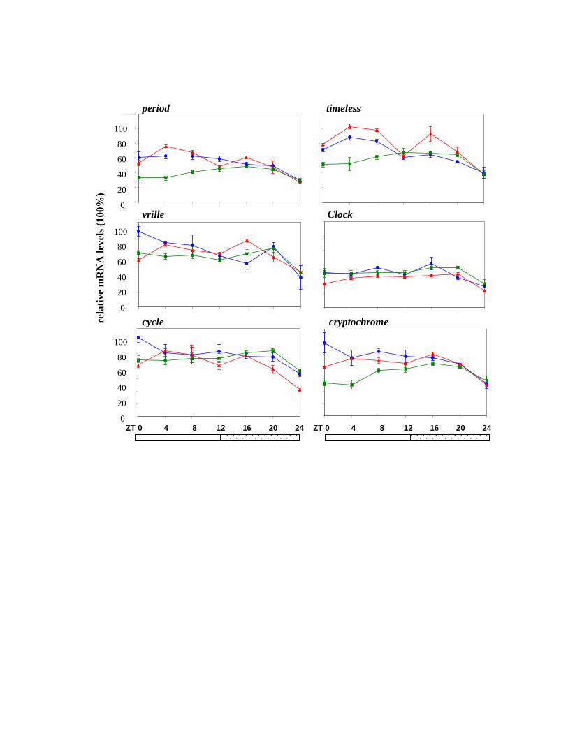
independent experiments. Peaks of mRNA were observed at ZT 12-16 for per, tim and vri and
this pattern was continued into the first cycle of DD. As in Drosophila, MdClk mRNA peaked
with an opposite phase to Mdper, namely at the end of both the actual (LD) and the subjective
(DD) night (Fig 2). The arrhythmic expression of Mdcyc was also consistent with that reported
for the fruitfly, but the absence of robust cycling for Mdcry was surprising (Fig 2). As expected,
exposure to constant light resulted in loss of cycling for all the clock genes (Fig 3) which is in
agreement with the behavioral arrhythmia observed under the same conditions (Fig 1A).
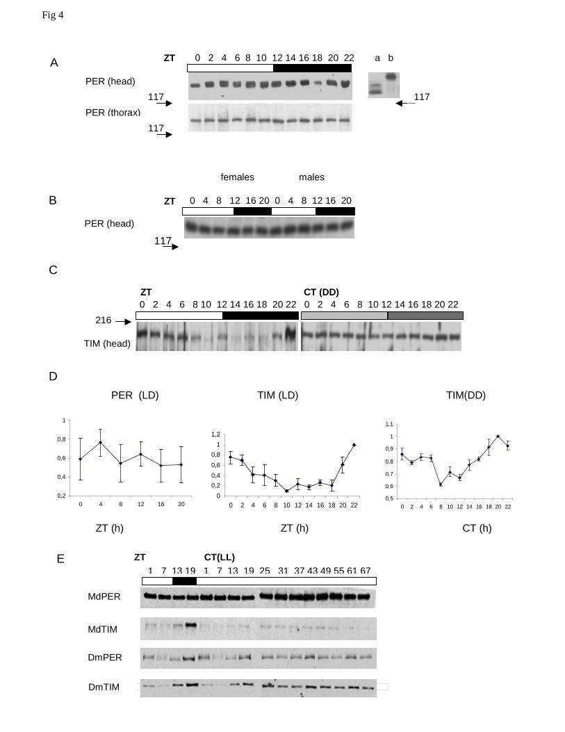
MdPER and MdTIM protein cycles
Figures 4A and B show typical Western blots using α-DmPER-I, but the results were
essentially identical using all three anti-PER antibodies. The MdPER band has a size of about
130-140 kD, consistent with its predicted molecular weight based on primary sequence data
(Piccin et al. 2000) and shows no evidence for any cycling in LD12:12. We repeated the blot
several times, also with other PER antisera and separately for head and thorax (Fig 4A) or male
and female (Fig 4B) extracts, but have never observed cycling (Fig 4D). In all blots examined no
obvious temporal changes in electrophoretic mobility were detected that could be attributed to
circadian modulation of phosphorylation (Edery et al. 1994).
We also examined MdTIM cycling in Musca heads using a rat anti-TIM antibody (α-
DmTIM 1: 1,000) raised against Drosophila TIM. Although weak, we detected a band
approximately of the correct size, that appears to cycle with a peak at the end of the night phase
in LD12:12 and, dampened, also in DD (Fig 4C). We confirmed these finding over three separate
full replicates (Fig 4D). We next investigated expression of MdPER in LL, and found it to be
similarly stable over more than two cycles, with no evidence for light-induced degradation (Fig
10
4E). In contrast, both Drosophila PER and TIM cycle robustly in LD and this cycle dampens in
LL, eventually stabilizing at just above minimal LD levels by the third day in LL (Fig 4E and see
Marrus et al., 1996). MdTIM however, immediately degrades in response to light, implying that
MdPER does not rely on MdTIM for stability (Fig 4E).
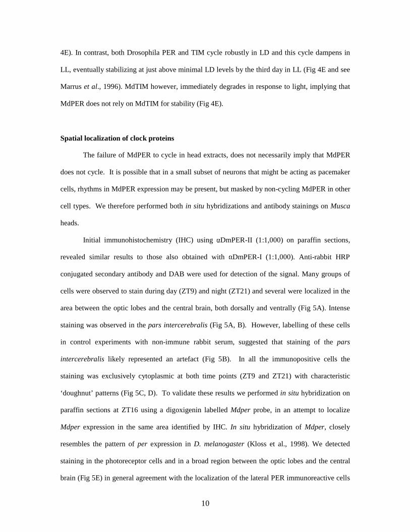
Spatial localization of clock proteins
The failure of MdPER to cycle in head extracts, does not necessarily imply that MdPER
does not cycle. It is possible that in a small subset of neurons that might be acting as pacemaker
cells, rhythms in MdPER expression may be present, but masked by non-cycling MdPER in other
cell types. We therefore performed both in situ hybridizations and antibody stainings on Musca
heads.
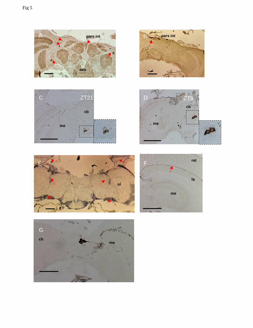
Initial immunohistochemistry (IHC) using αDmPER-II (1:1,000) on paraffin sections,
revealed similar results to those also obtained with αDmPER-I (1:1,000). Anti-rabbit HRP
conjugated secondary antibody and DAB were used for detection of the signal. Many groups of
cells were observed to stain during day (ZT9) and night (ZT21) and several were localized in the
area between the optic lobes and the central brain, both dorsally and ventrally (Fig 5A). Intense
staining was observed in the pars intercerebralis (Fig 5A, B). However, labelling of these cells
in control experiments with non-immune rabbit serum, suggested that staining of the pars
intercerebralis likely represented an artefact (Fig 5B). In all the immunopositive cells the
staining was exclusively cytoplasmic at both time points (ZT9 and ZT21) with characteristic
‘doughnut’ patterns (Fig 5C, D). To validate these results we performed in situ hybridization on
paraffin sections at ZT16 using a digoxigenin labelled Mdper probe, in an attempt to localize
Mdper expression in the same area identified by IHC. In situ hybridization of Mdper, closely
resembles the pattern of per expression in D. melanogaster (Kloss et al., 1998). We detected
staining in the photoreceptor cells and in a broad region between the optic lobes and the central
brain (Fig 5E) in general agreement with the localization of the lateral PER immunoreactive cells
11
(Fig 5A). However, we did not detect staining within photoreceptor cells by IHC, but a structure
at the base of the photoreceptors was strongly labelled at all times (Fig 5F).
We also used a rabbit anti-crab PDH antibody that in Drosophila recognises PDF
expressing cells, and identified some immunoreactive neurons between the central brain and the
optic lobes (Fig 5G). As we could not perform MdPER-PDH double-staining, we can neither
confirm nor exclude the possibility that these PDH immunoreactive cells correspond to any of the
lateral cells that stain for PER. No staining was achieved for MdTIM, precluding any further
investigation using these methods.
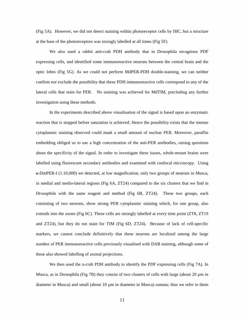
In the experiments described above visualisation of the signal is based upon an enzymatic
reaction that is stopped before saturation is achieved. Hence the possibility exists that the intense
cytoplasmic staining observed could mask a small amount of nuclear PER. Moreover, paraffin
embedding obliged us to use a high concentration of the anti-PER antibodies, raising questions
about the specificity of the signal. In order to investigate these issues, whole-mount brains were
labelled using fluorescent secondary antibodies and examined with confocal microscopy. Using
α-DmPER-I (1:10,000) we detected, at low magnification, only two groups of neurons in Musca,
in medial and medio-lateral regions (Fig 6A, ZT24) compared to the six clusters that we find in
Drosophila with the same reagent and method (Fig 6B, ZT24). These two groups, each
consisting of two neurons, show strong PER cytoplasmic staining which, for one group, also
extends into the axons (Fig 6C). These cells are strongly labelled at every time point (ZT8, ZT19
and ZT24), but they do not stain for TIM (Fig 6D, ZT24). Because of lack of cell-specific
markers, we cannot conclude definitively that these neurons are localized among the large
number of PER immunoreactive cells previously visualised with DAB staining, although some of
these also showed labelling of axonal projections.
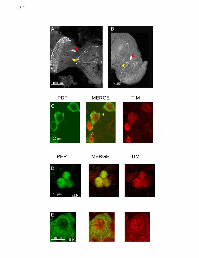
We then used the α-crab PDH antibody to identify the PDF expressing cells (Fig 7A). In
Musca, as in Drosophila (Fig 7B) they consist of two clusters of cells with large (about 20 µm in
diameter in Musca) and small (about 10 µm in diameter in Musca) somata; thus we refer to them
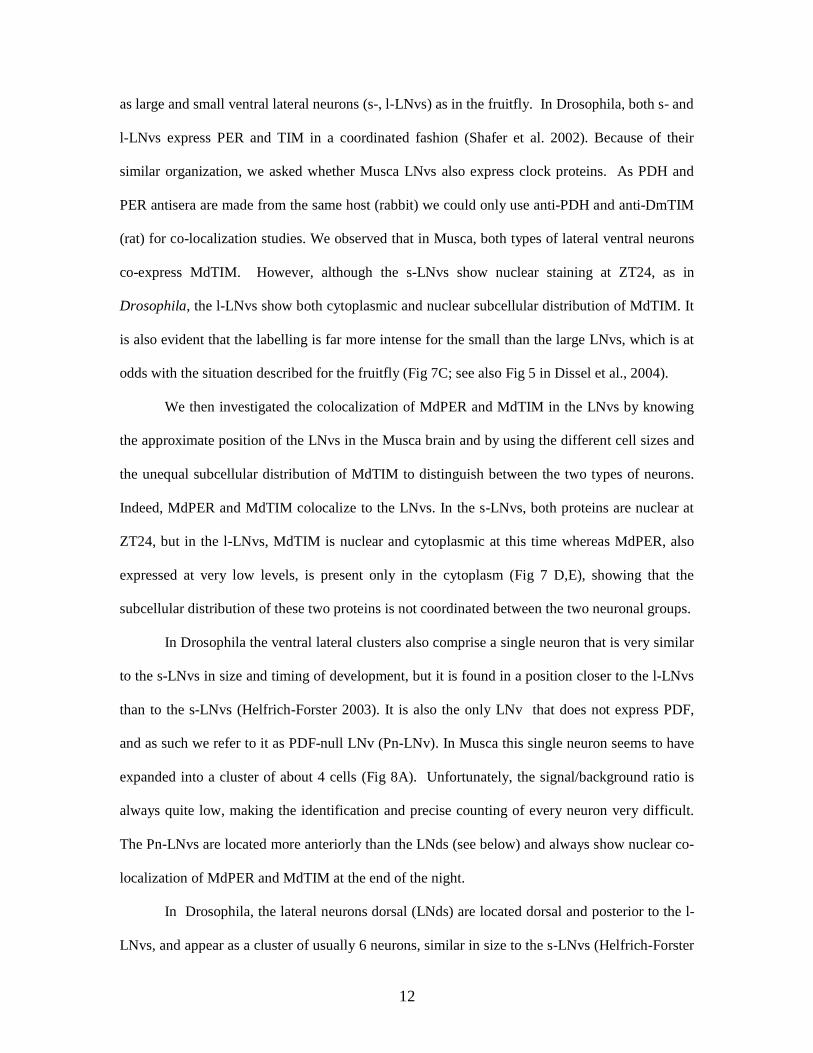
12
as large and small ventral lateral neurons (s-, l-LNvs) as in the fruitfly. In Drosophila, both s- and
l-LNvs express PER and TIM in a coordinated fashion (Shafer et al. 2002). Because of their
similar organization, we asked whether Musca LNvs also express clock proteins. As PDH and
PER antisera are made from the same host (rabbit) we could only use anti-PDH and anti-DmTIM
(rat) for co-localization studies. We observed that in Musca, both types of lateral ventral neurons
co-express MdTIM. However, although the s-LNvs show nuclear staining at ZT24, as in
Drosophila, the l-LNvs show both cytoplasmic and nuclear subcellular distribution of MdTIM. It
is also evident that the labelling is far more intense for the small than the large LNvs, which is at
odds with the situation described for the fruitfly (Fig 7C; see also Fig 5 in Dissel et al., 2004).
We then investigated the colocalization of MdPER and MdTIM in the LNvs by knowing
the approximate position of the LNvs in the Musca brain and by using the different cell sizes and
the unequal subcellular distribution of MdTIM to distinguish between the two types of neurons.
Indeed, MdPER and MdTIM colocalize to the LNvs. In the s-LNvs, both proteins are nuclear at
ZT24, but in the l-LNvs, MdTIM is nuclear and cytoplasmic at this time whereas MdPER, also
expressed at very low levels, is present only in the cytoplasm (Fig 7 D,E), showing that the
subcellular distribution of these two proteins is not coordinated between the two neuronal groups.
In Drosophila the ventral lateral clusters also comprise a single neuron that is very similar
to the s-LNvs in size and timing of development, but it is found in a position closer to the l-LNvs
than to the s-LNvs (Helfrich-Forster 2003). It is also the only LNv that does not express PDF,
and as such we refer to it as PDF-null LNv (Pn-LNv). In Musca this single neuron seems to have
expanded into a cluster of about 4 cells (Fig 8A). Unfortunately, the signal/background ratio is
always quite low, making the identification and precise counting of every neuron very difficult.
The Pn-LNvs are located more anteriorly than the LNds (see below) and always show nuclear co-
localization of MdPER and MdTIM at the end of the night.
In Drosophila, the lateral neurons dorsal (LNds) are located dorsal and posterior to the l-
LNvs, and appear as a cluster of usually 6 neurons, similar in size to the s-LNvs (Helfrich-Forster
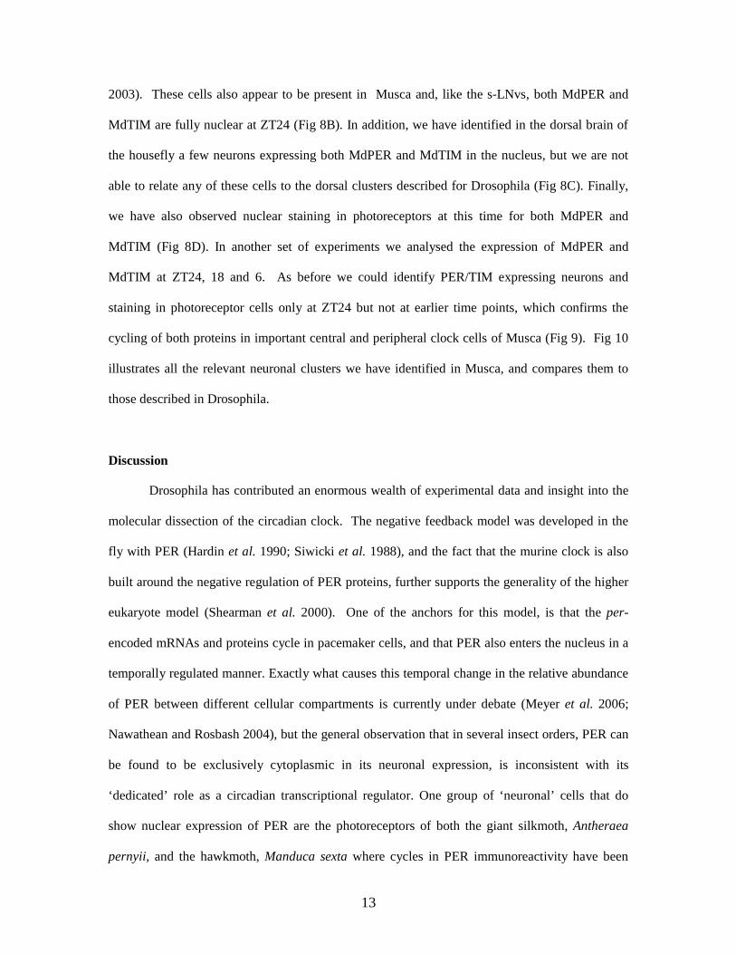
13
2003). These cells also appear to be present in Musca and, like the s-LNvs, both MdPER and
MdTIM are fully nuclear at ZT24 (Fig 8B). In addition, we have identified in the dorsal brain of
the housefly a few neurons expressing both MdPER and MdTIM in the nucleus, but we are not
able to relate any of these cells to the dorsal clusters described for Drosophila (Fig 8C). Finally,
we have also observed nuclear staining in photoreceptors at this time for both MdPER and
MdTIM (Fig 8D). In another set of experiments we analysed the expression of MdPER and
MdTIM at ZT24, 18 and 6. As before we could identify PER/TIM expressing neurons and
staining in photoreceptor cells only at ZT24 but not at earlier time points, which confirms the
cycling of both proteins in important central and peripheral clock cells of Musca (Fig 9). Fig 10
illustrates all the relevant neuronal clusters we have identified in Musca, and compares them to
those described in Drosophila.
Discussion
Drosophila has contributed an enormous wealth of experimental data and insight into the
molecular dissection of the circadian clock. The negative feedback model was developed in the
fly with PER (Hardin et al. 1990; Siwicki et al. 1988), and the fact that the murine clock is also
built around the negative regulation of PER proteins, further supports the generality of the higher
eukaryote model (Shearman et al. 2000). One of the anchors for this model, is that the per-
encoded mRNAs and proteins cycle in pacemaker cells, and that PER also enters the nucleus in a
temporally regulated manner. Exactly what causes this temporal change in the relative abundance
of PER between different cellular compartments is currently under debate (Meyer et al. 2006;
Nawathean and Rosbash 2004), but the general observation that in several insect orders, PER can
be found to be exclusively cytoplasmic in its neuronal expression, is inconsistent with its
‘dedicated’ role as a circadian transcriptional regulator. One group of ‘neuronal’ cells that do
show nuclear expression of PER are the photoreceptors of both the giant silkmoth, Antheraea
pernyii, and the hawkmoth, Manduca sexta where cycles in PER immunoreactivity have been
14
documented in the former species, but not the latter (Sauman and Reppert 1996; Wise et al.
2002). However, in the hawkmoth, nuclear staining of PER was consistently observed in four
neurons within each hemisphere in the pars lateralis, a neurosecretory region, although no
circadian cycling of PER abundance was noted (Wise et al. 2002). In contrast, in A. pernyii, a
similar group of four cells express exclusively cytoplamic PER, which cycles in concert with
TIM (Sauman et al. 1996).
While it can be argued that in some of these studies, perhaps the antigenicity did not
reflect PER in these insects, or that PER does indeed enter the nucleus at low undetectable levels
to engage the negative feedback loop, it is odd that ‘PER’ antigenicity, whether cycling in the
firebrat, (Zavodska et al. 2003a), or not in the hawkmoth (Wise et al. 2002) or beetle (Frisch et
al. 1996), or ‘not known’ in a variety of other insects (Zavodska et al. 2003b), does not usually
appear to be nuclear. In fact, based on our initial results with Musca, we might have been tempted
to add the housefly to the list of species with ‘non-canonical’ patterns of PER regulation. This
would have been based on the failure of MdPER to (1) cycle in Musca heads in Western blots and
its associated inability to ‘degrade’ in response to constant light, (2) show the extensive
phosphorylation patterns that are observed in Drosophila, (3) translocate into the nucleus, with its
apparently exclusive cytoplasmic localization in several groups of neurons, based on enzymatic
staining methods. Such a conclusion would have been surprising given that MdPER strongly
rescues circadian locomotor rhythms in Drosophila per01 transformants (Piccin et al. 2000), and
that Mdper, Mdtim, Mdvri and MdClk mRNAs cycle quite robustly, with similar phases to those
in the fruitfly, in LD and DD and become arrhythmic in LL, further revealing their clock-like
nature. Of interest was that in contrast to Drosophila, Musca cry, unexpectedly, did not cycle
robustly. The cry gene plays a pivotal switch role in the evolution of the clock because in the
central clock mechanism, it can either act predominantly as a photoreceptor, as in Drosophila, or
as a negative regulator (as in the mouse, Shearman et al 2000). However, cry in the fly can also
act as a negative regulator in peripheral neuronal tissue, indicating that cell specific trans-acting
15
factors can modulate the light sensitive function of this molecule (Collins et al 2006). The two
types of CRY’s can be clustered phylogenetically, and the Musca cry identified by our screen is a
Drosophila-like molecule. The distribution of the two types of cry’s do not follow simple
phylogenetic rules, in that mouse-like cry is found exclusively in some insects such as bees and
Tribolium, whereas in others, such as the dipteran Anopheles gambiae or in the monarch butterfly
Danaus plexippus, both types of cry have been identified (Zhu et al, 2005; Rubin et al, 2006;
Yuan et al, 2007). At present we cannot state conclusively whether a mouse like cry is present in
the Musca genome. Nevertheless, it is intriguing that Musca cry does not appear to cycle at the
mRNA level, at least at the level of whole head preparations, but it is too early to say whether this
may imply a different function compared to Drosophila cry.
As mentioned above, the absence of MdPER cycling in Western blots, further contradicts
the Drosophila model. Although the formal possibility exists that the band we are detecting
represents a cross-reacting protein, we are confident that we are reliably observing MdPER in
Western blots for the following reasons. We identified the same band with several antibodies and
separately in heads and thoraxes of both males and females. In per01 D. melanogaster
transformants carrying the Mdper transgene (Piccin et al. 2000), the same antibodies identify a
band of similar size (Fig 4A). α-DmPER-II is able to recognised the cycling PER of the blowfly
Lucilia cuprina (Warman et al., 2000), further confirming that the polyclonal antibodies we used
recognize regions of PER that are highly conserved in diptera
That our initial conclusion (which we held for several years, namely that Musca
represented a non-canonical type of circadian clock), was premature and incorrect, came to light
once we applied immunofluorescence with confocal microscopy. We observed medial and medio-
lateral neurons that expressed high levels of cytoplasmic MdPER but not MdTIM, in night and
day, as we had with the enzymatic method. On closer scrutiny, less dramatic PER and TIM co-
labelling of groups of neurons became apparent, even though the staining was much fainter. This
might have been due to a general low level of expression of MdPER and MdTIM in these

16
neurons, to a low affinity for Musca proteins of our anti-Drosophila antibodies, to an increased
difficulty for the antibodies to penetrate a much bigger brain, or, most likely, to a combination of
all these reasons. Whatever the cause, the immunostaining was very faint with both antisera but
especially with anti-TIM, making the identification of Musca ‘clock neurons’ technically very
challenging. Nevertheless, we identified many cells showing immunoreactivity for either anti-
PER or anti-TIM or both. However, in this study we have only considered as putative clock
neurons those cells where co-localization of the two immunosignals was clearly evident, as we do
not have any other reliable criterion to judge the specificity of the labelling produced by each of
the antibodies. We could not detect any reliable TIM and PER double staining at earlier times
than ZT24 both in central clock neurons and in photoreceptor cells. Therefore, we suggest that in
clock cells there is a cycle in the abundance of the two proteins which might be mirrored by a
cycle in their subcellular localization.
Thus in contrast to our initial conclusions, neuronal clusters in Musca largely correspond
to those in Drosophila, suggesting we have identified homologous structures (see Fig 7-9). It is
interesting to note that in Musca at ZT24, the l-LNvs are the only group of putative clock cells to
show cytoplasmic PER and nuclear-cytoplasmic TIM, and both types of reagents give much
weaker signals. This might suggest that this neuronal cluster might have a special function in the
circadian network, as suggested for Drosophila (Collins et al., 2005). Furthermore, in Drosophila
the l-LNvs are the only group of neurons where, under particular environmental conditions, for
example in constant conditions, PER and TIM nuclear accumulation can be decoupled (Shafer et
al. 2002; Yang and Sehgal 2001; Rieger et al, 2006). These cells possibly represent a strategic
point in the neuronal network of diptera where a physiological response to a combination of
environmental variables might be amplified for entrainment (Collins et al., 2005). As for the
more nuclear subcellular distribution of MdTIM in the l-LNvs of Musca compared to MdPER,
this could reflect a more prominent role for MdTIM as a negative regulator in the housefly, as has
17
been suggested for A. pernyii (Chang et al. 2003).
As in Drosophila, PDF localizes with the two groups of lateral neurons in Musca,
providing a helpful additional marker for these putative clock neurons. In both Drosophila and
Musca these cells are located within the accessory medulla (also termed anterior base of the
medulla, Fig 7A, B) and their patterns of projections from the LNvs are also similar, as has been
previously reported (Helfrich-Forster 1995; Miskiewicz et al. 2004; Pyza and Meinertzhagen
1997). Our results with anti-PER and TIM reagents therefore support the long-standing
suspicions of Pyza and coworkers, that the PDF expressing neurons in Musca are in fact ‘clock’
cells (Miskiewicz et al. 2004; Pyza and Meinertzhagen 1997).
In other insect orders PDF does not colocalize with PER or TIM antigens (Zavodska et al.
2003b), although some colocalization of PER and PDF may be present in the beetle
Pachymorpha sexguttata (Frisch et al. 1996). It may be therefore that the diptera compared with
these other insects have a fundamental difference in this aspect of clock neuronal biology. Even
so, in Antheraea for example, although the PDH and PER/TIM expressing neurons are not the
same, in terms of their relative anatomical positions, they could be functionally related (Sauman
and Reppert 1996). We did however find a number of PER and TIM expressing neurons that
were localized in the region of the Musca l-LNvs that were PDF-null. In Drosophila, one such
neuron has been identified (Helfrich-Forster 1995; Kaneko et al. 1997), and its role in the
circadian mechanism is being clarified (Rieger et al., 2006).
One outstanding issue involves why the MdPER protein does not cycle in head Western
blots ? In the muscid, Lucilia cuprina (sheep blow fly), cycles in both gene products are found
(Warman et al. 2000), yet in the medfly, Ceratitis capitata, a similar situation to Musca is
observed, with cycles in per mRNA expression, but no PER cycling in Westerns (Mazzotta et al.
2005). We suspect that the Musca PER protein that is highly and apparently constitutively
expressed in the medio and medio-lateral neurons, if indeed it is PER, plays a different role to
18
that found in the LNs. Microarray studies in Drosophila have revealed that per does more than
simply control cycling output transcripts, and that a large number of mRNAs that do not cycle,
are either up or down-regulated in per-null mutants (Claridge-Chang et al. 2001; Lin et al.
2002b). This in turn would suggest that there are downstream functions for PER which do not
require cycling per products. Consequently, the apparently stable PER in these neurons may play
a different role to the familiar negative regulator theme, particularly given the apparent lack of
TIM in these cells. Given PER’s intimate association with the DOUBLETIME kinase, which
earmarks it for degradation (Kloss et al. 1998; Price et al. 1998), it would be interesting to see
whether DBT, or indeed other kinases such as casein kinase 2, which have been implicated with
PER stability (Akten et al. 2003; Lin et al. 2002a), are also localized in these non-rhythmic
MdPER-expressing neurons.
Future work will be aimed at elucidating the roles of the various clock molecules in
Musca. Obviously, transgenic Musca, in which misexpression of MdPER, targeted or not, would
be very helpful for functional studies (Hediger et al. 2001), and the development of such
techniques is underway in our laboratories. Musca will therefore prove to be a useful model for
studying the evolution of the circadian system as it is phylogenetically far enough away from
Drosophila to be interesting, yet close enough to have at least the possibility of being studied by
using some of the techniques that are available in the fruitfly. One final thought relates to the
findings in other insect orders, namely that PER is only observed to be cytoplasmic in brain
neurons (Zavodska et al. 2003b). All of these studies have used enzymatic IHC reactions except
one, which also used confocal microscopy, but only for analysis of PDF expressing neurons. Had
we concluded our analyses without confocal microscopy, we would have come to similar
conclusions as these other laboratories, even to the point of suggesting that PDF expressing cells
did not colocalize with PER (compare Fig 5D with 5G). However, we observe that in fact Musca
appears to have similar anatomical substrates for the PER/TIM and PDF molecules as Drosophila,
albeit with some intriguing differences in the way that MdPER behaves in westerns, and in the
19
numbers of neurons that express these clock proteins. Perhaps a more detailed reanalysis of
spatial and temporal clock gene product expression in other insect brains might be timely.
Acknowledgements:
We thank the BBSRC for a David Philips Fellowship to ER and a committee PhD studentship to
VC. We also thank the European Community for grants to CPK and RC, and the Wellcome Trust
for a grant to CPK and ER and for a program grant to EL. CPK acknowledges the Royal Society
for a Wolfson Research Merit Fellowship. This work was supported in part by the Grant Agency
of the Czech Republic, grant 204/04/0862 (IS) and Ministry of Education of the Czech Republic,
project 2B06129 (IS)
20
References
Akten, B., E. Jauch, G. K. Genova, E. Y. Kim, I. Edery et al., 2003 A role for CK2 in the
Drosophila circadian oscillator. Nat Neurosci 6: 251-257.
Benito, J., Zheng, H., Hardin, P.E. 2007 PDP1epsilon functions downstream of the
circadian oscillator to mediate behavioral rhythms. J Neurosci. 27: 2539-47.
Ashmore, L. J., S. Sathyanarayanan, D. W. Silvestre, M. M. Emerson, P. Schotland et al., 2003
Novel insights into the regulation of the timeless protein. J Neurosci 23: 78107819.
Chang, D. C., H. G. McWatters, J. A. Williams, A. L. Gotter, J. D. Levine et al., 2003
Constructing a feedback loop with circadian clock molecules from the silkmoth, Antheraea
pernyi. J Biol Chem 278: 38149-38158.
Claridge-Chang, A., H. Wijnen, F. Naef, C. Boothroyd, N. Rajewsky et al., 2001 Circadian
regulation of gene expression systems in the Drosophila head. Neuron 32: 657-671.
Collins, B.H., Dissel, S., Gaten, E., Rosato, E., and C.P. Kyriacou, 2005 Disruption of
Cryptochrome partially restores circadian rhythmicity to the arrhythmic period mutant of
Drosophila. Proc Natl Acad Sci USA 102:19021-6.
Collins, B.H., Mazzoni E. O., Stanewsky, R., and J. Blau, 2006 Drosophila CRYPTOCHROME
is a circadian transcriptional repressor l. Curr Biol 16:441-449.
Collins, B.H., and J. Blau, 2007 Even a stopped clock tells the right time twice a day: circadian
timekeeping in Drosophila. Pflugers Arch Eur J Physiol 454:857-867
Cyran, S. A., A. M. Buchsbaum, K. L. Reddy, M. C. Lin, N. R. Glossop et al., 2003 vrille, Pdp1,
and dClock form a second feedback loop in the Drosophila circadian clock. Cell 112: 329-341.
Edery, I., L. J. Zwiebel, M. E. Dembinska and M. Rosbash, 1994 Temporal phosphorylation of

21
the Drosophila period protein. Proc Natl Acad Sci USA 91: 2260-2264.
Fang, Y., Sathyanarayanan, S., and A. Sehgal, 2007 Post-translational regulation of the
Drosophila circadian clock requires protein phosphatase 1 (PP1). Genes Dev 15: 1506-1518.
Frisch, B., G. Fleissner, C. Brandes and J. C. Hall, 1996 Staining in the brain of Pachymorpha
sexguttata mediated by an antibody against a Drosophila clock-gene product: labeling of cells
with possible importance for the beetle's circadian rhythms. Cell Tissue Res 286: 411-429.
Hall, J. C., 2003 Genetics and molecular biology of rhythms in Drosophila and other insects.
Adv Genet 48: 1-280.
Hardin, P. E., J. C. Hall and M. Rosbash, 1990 Feedback of the Drosophila period gene
product on circadian cycling of its messenger RNA levels. Nature 343: 536-540.
Hediger, M., M. Niessen, E. A. Wimmer, A. Dubendorfer and D. Bopp, 2001 Genetic
transformation of the housefly Musca domestica with the lepidopteran derived
transposon piggyBac. Insect Mol Biol 10: 113-119.
Helfrich, C., B. Cymborowski and W. Engelmann, 1985 Circadian activity rhythm of the
house fly continues after optic tract severance and lobectomy. Chronobiol Int 2: 19-32.
Helfrich-Forster, C., 1995 The period clock gene is expressed in central nervous system neurons
which also produce a neuropeptide that reveals the projections of circadian pacemaker cells
within the brain of Drosophila melanogaster. Proc Natl Acad Sci U S A 92: 612-616.
Helfrich-Forster, C., and U. Homberg, 1993 Pigment-dispersing hormone-immunoreactive
neurons in the nervous system of wild-type Drosophila melanogaster and of several mutants
with altered circadian rhythmicity. J Comp Neurol 337: 177-190.
Helfrich-Forster, C., 2003 The Neuroarchitecture of the circadian clock in the brain of
Drosophila melanogaster. Microsc Res Tech 62: 94-102
Hennig, W. 1981. Insect phylogeny. Piman Press, Bath, England
Kaneko, M., C. Helfrich-Forster and J. C. Hall, 1997 Spatial and temporal expression of the
period and timeless genes in the developing nervous system of Drosophila: newly identified
22
pacemaker candidates and novel features of clock gene product cycling. J Neurosci 17: 6745-
6760.
Kloss, B., J. L. Price, L. Saez, J. Blau, A. Rothenfluh et al., 1998 The Drosophila clock gene
double-time encodes a protein closely related to human casein kinase Iepsilon. Cell
94: 97-107.
Levine, J. D., I. Sauman, M. Imbalzano, S. M. Reppert and F. R. Jackson, 1995 Period protein
from the giant silkmoth Antheraea pernyii functions as a circadian clock element in Drosophila
melanogaster. Neuron 15: 147-157.
Lin, J. M., V. L. Kilman, K. Keegan, B. Paddock, M. Emery-Le et al., 2002a A role for casein
kinase 2α in the Drosophila circadian clock. Nature 420: 816-820.
Lin, Y., M. Han, B. Shimada, L. Wang, T. M. Gibler et al., 2002b Influence of the period-
dependent circadian clock on diurnal, circadian, and aperiodic gene expression in Drosophila
melanogaster. Proc Natl Acad Sci USA 99: 9562-9567.
Mazzotta, G. M., F. Sandrelli, M. A. Zordan, M. Mason, C. Benna et al., 2005 The clock
gene period in the medfly Ceratitis capitata. Genet Res 86: 13-30.
Meyer, P., L. Saez and M. W. Young, 2006 PER-TIM interactions in living Drosophila cells:
an interval timer for the circadian clock. Science 311: 226-229.
Miskiewicz, K., E. Pyza and F. W. Schurmann, 2004 Ultrastructural characteristics of
circadian pacemaker neurones, immunoreactive to an antibody against a pigment-dispersing
hormone in the fly's brain. Neurosci Lett 363: 73-77.
Naidoo, N., W. Song, M. Hunter-Ensor and A. Sehgal, 1999 A role for the proteasome in the
light response of the timeless clock protein. Science 285: 1737-1741.
Nassel, D. R., S. Shiga, C. J. Mohrherr and K. R. Rao, 1993 Pigment-dispersing hormone-like
peptide in the nervous system of the flies Phormia and Drosophila: immunocytochemistry and
partial characterization. J Comp Neurol 331: 183-198.
Nawathean, P., and M. Rosbash, 2004 The doubletime and CKII kinases collaborate to
23
potentiate Drosophila PER transcriptional repressor activity. Mol Cell 13: 213-223.
Peixoto, A. A., J. M. Hennessy, I. Townson, G. Hasan, M. Rosbash et al., 1998 Molecular
coevolution within a Drosophila clock gene. Proc Natl Acad Sci USA 95: 44754480.
Piccin, A., M. Couchman, J. D. Clayton, D. Chalmers, R. Costa et al., 2000 The clock gene
period of the housefly, Musca domestica, rescues behavioral rhythmicity in Drosophila
melanogaster. Evidence for intermolecular coevolution? Genetics 154: 747-758.
Price, J. L., J. Blau, A. Rothenfluh, M. Abodeely, B. Kloss et al., 1998 double-time is a novel
Drosophila clock gene that regulates PERIOD protein accumulation. Cell 94: 83-95.
Pyza, E., and I. A. Meinertzhagen, 1997 Neurites of period-expressing PDH cells in the fly's
optic lobe exhibit circadian oscillations in morphology. Eur J Neurosci 9: 1784-1788.
Refinetti, R. 2000, Circadian Physiology, CRC Press, Boca Raton, 184 pp
Reppert, S. M., T. Tsai, A. L. Roca and I. Sauman, 1994 Cloning of a structural and
functional homolog of the circadian clock gene period from the giant silkmoth Antheraea
pernyi. Neuron 13: 1167-1176.
Rieger, D., Shafer, O.T., Tomioka, K. and C. Helfrich-Forster 2006 Functional analysis
of circadian pacemaker neurons in Drosophila melanogaster. J Neurosci. 26: 2531-2543.
Rosato, E., Tauber, E., and C. P. Kyriacou, 2006 Molecular genetics of the fruit-fly
circadian clock. Eur J Hum Genet 14:729-738
Rubin, E. B., Shemesh, Y., Cohen, M., Elgavish, S., Robertson, H. M., et al, 2006 Molecular
and phylogenetic analyses reveal mammalian-like clockwork in the honey bee (Apis mellifera)
and shed new light on the molecular evolution of the circadian clock. Genom Res 16:1352-1365
Sandrelli, F., Cappellozza, S., Benna C., Saviane A., Mastella, A., et al. 2007
Phenotypic effects induced by knock-down of the period clock gene in Bombyx mori.
Genet Res 89:73-84
Sathyanarayanan S, Zheng X, Xiao R, and A. Sehgal, 2004 Posttranslational regulation
24
of Drosophila PERIOD protein by protein phosphatase 2A. Cell 116: 603-15.
Sauman, I., and S. M. Reppert, 1996 Circadian clock neurons in the silkmoth Antheraea
pernyi: novel mechanisms of Period protein regulation. Neuron 17: 889-900.
Sauman, I., T. Tsai, A. L. Roca and S. M. Reppert, 1996 Period protein is necessary for
circadian control of egg hatching behavior in the silkmoth Antheraea pernyi. Neuron
17: 901-909.
Shafer, O. T., M. Rosbash and J. W. Truman, 2002 Sequential nuclear accumulation of the
clock proteins period and timeless in the pacemaker neurons of Drosophila melanogaster. J
Neurosci 22: 5946-5954.
Shearman, L. P., S. Sriram, D. R. Weaver, E. S. Maywood, I. Chaves et al., 2000 Interacting
molecular loops in the mammalian circadian clock. Science 288: 1013-1019.
Siwicki, K. K., C. Eastman, G. Petersen, M. Rosbash and J. C. Hall, 1988 Antibodies to the
period gene product of Drosophila reveal diverse tissue distribution and rhythmic changes in
the visual system. Neuron 1: 141-150.
Warman, G. R., R. D. Newcomb, R. D. Lewis and C. W. Evans, 2000 Analysis of the circadian
clock gene period in the sheep blow fly Lucilia cuprina. Genet Res 75: 257267.
Wise, S., N. T. Davis, E. Tyndale, J. Noveral, M. G. Folwell et al., 2002 Neuroanatomical
studies of period gene expression in the hawkmoth, Manduca sexta. J Comp Neurol 447: 366-
380.
Yang, Z., and A. Sehgal, 2001 Role of molecular oscillations in generating behavioral
rhythms in Drosophila. Neuron 29: 453-467.
Yuan, Q., Metterville, D., Briscoe, A.D., and S.M Reppert, 2007 Insect cryptochromes:
gene duplication and loss define diverse ways to construct insect circadian clocks. Mol
Biol & Evol 24: 948-55.
Zavodska, R., I. Sauman and F. Sehnal, 2003a The cycling and distribution of PER-like
antigen in relation to neurons recognized by the antisera to PTTH and EH in Thermobia
25
domestica. Insect Biochem Mol Biol 33: 1227-1238.
Zavodska, R., I. Sauman and F. Sehnal, 2003b Distribution of PER protein, pigment-
dispersing hormone, prothoracicotropic hormone, and eclosion hormone in the cephalic
nervous system of insects. J Biol Rhythms 18: 106-122.
Zeng, H., Z. Qian, M. P. Myers and M. Rosbash, 1996 A light-entrainment mechanism for the
Drosophila circadian clock. Nature 380: 129-135.
Zhu, H,, Yuan, Q., Briscoe, A.D., Froy, O., Casselman, A., et al., 2005 The two CRYs of the
butterfly. Curr Biol. 15: R953-4.
26
TABLE 1
Gene-specific primers used in real-time PCR
Transcript
Primer
Primer seq (intron position is marked with ^)
Product size
(cDNA)
rp49 rp49 fw
rp49 rev
5' GTTATGCCAAATTGTCG^CACA
5' GGCGGGTACGTTTGTTGG
123 bp
period per fw
per rev
5' ACGAAAACACTCTTAAG^CCCAA
5' TTTGCTGTTGTCGTTCTCCTG
140 bp
timeless* tim fw
tim rev
5' TGTTGCTCTTGATACTGGATAGTG
5' AGCAGCATGCCATAGAAGTG
118 bp
cryptochrome cry fw
cry rev
5' TGGGTTAGTTCATCGGCATTTG
5' GCCAGGGTTCATGG^ATAAACAG
157 bp
vrille vri fw
vri rev
5' AATGAGGCCACAAATG^TTCAC
5' GGCGCTGACCTGCTGTTT
156 bp
cycle cyc fw
cyc rev
5' CCTGTTCATAGATCAAAG^AGCC
5' TCAGCCAAGGCGGGTAT
111 bp
Clock clk fw
clk rev
5' TTATATCACCCCTACCG^CATC
5' TTACGCTGAAGCTGTTCCT
100 bp
*timeless primers were designed to anneal to different exons separated by ca. 2.5 kb intron.
27
Figure legends
Figure 1. Rhythmic phenotypes in Musca domestica. A Locomotor activity of individual flies is
double plotted so that day 1 and day 2 activity is placed on the first horizontal line, day 2 and day
3 on the second line, etc. Left panel, LD12:12. Middle panel, four days in LD12:12 followed
(open arrow) by eight days in DD. Right panel, five days in LD12:12 after which (open arrow)
the flies were maintained in LL for four days. B Circadian pupal-adult eclosion. Newly emerging
flies were observed in dim red light after previous entrainment in LD12:12. Eclosions were
followed every 2 h.
Figure 2. Clock gene expression in Musca heads in LD and DD. Real time PCR was performed
for the six clock genes in LD followed by DD. Light regime appears below each column of
figures. Each color represents a different biological replicate.
Figure 3. Clock gene expression in Musca heads in constant light (LL). Real time PCR was
performed for the six clock genes as in Fig 2. Each color represents a different biological
replicate.
Figure 4. PER and TIM expression in Musca. A Western blots in LD 12:12 of heads and
thoraces separately, showing that MdPER does not cycle in either tissue although it is more
highly expressed in the former than in the latter. The numbers ‘117’ and ‘216’ and their
associated arrows represent kilodaltons and show the positions of the molecular weight markers.
To the right is a Western blot of the Drosophila MM1 transformant heads that carry the null
mutation per01, plus the Mdper transgene under the control of Drosophila per 5’ sequences
(Piccin et al. 2001). Lane ‘a’ is the MM1 transformant, and lane ‘b’ represents wild-type
Drosophila heads harvested at ZT 8.

28
B Western blot of male and female heads taken in 4 h intervals in LD12:12. No evidence for
cycling in the abundance of MdPER is observed, suggesting that there are no sex-specific
differences in this trait. Molecular weight markers in kD are arrowed. C Representative MdTIM
head blots in LD 12:12 and DD. The arrow represents a molecular weight marker in kD. D Mean
(+/-sem) levels of MdPER (LD12:12) and MdTIM (LD12:12 and DD) in Musca heads. Loading
differences were corrected using constitutive non-specific cross-reacting bands as a comparison.
Data were normalised to the highest level of clock protein on each Western. Several replicates
were performed for each experiment. E Musca PER is stable in constant light. Western blots are
shown for Musca and Drosophila PER and TIM in 67 h of constant bright light after previous
entrainment in LD12:12. Note the stability of MdPER compared to MdTIM and to dPER and
dTIM, which show levels slightly above the minimum levels in LD (see text).
Figure 5. Clock protein expression in head sections of Musca. A shows a section at ZT21
labelled with α-DmPER-I and represents a general staining pattern obtained with all available
anti-PER antibodies. Several groups of cells are labelled (arrows) including the pars
intercerebralis and a dorsal and ventral group of neurons that are lateral to the central brain. B
Replacing the α-DmPER-I antibody with rabbit normal serum results in staining of pars
intercerebralis cells (arrow), suggesting that these are non-specific for PER staining. CD Both
during the night (ZT21) and the day (ZT9) anti-PER immunoreactivity was exclusively
cytoplasmic with cells showing a characteristic ‘doughnut’ shape E. in situ hybridization to
Mdper. Arrows denote hybridization to regions where the dorsal and lateral PER-positive neurons
are located and to the photoreceptors. F. Although no staining is observed in photoreceptor
nuclei at night a structure at the base of the photoreceptor can be seen to stain strongly at all times
(arrow). G PDF expressing cells and their projections. Scale bars = 100 µm. Key: pars int,
pars intercerebralis; oes, oesophagus; me, medulla; la, lamina; ret, retina; ol, optic lobe.
29
Figure 6 Confocal microscopy of a whole mount Musca brain. A. PER within the Musca brain
stained with α-Dm PER-I antibody at ZT24. Two medial neurons (green arrowhead) and two
medio-lateral neurons (blue arrowhead) show intense staining at this magnification. B D.
melanogaster shows six clusters of PER immunoreactive neurons (α-Dm PER-I) in lateral and
dorsal regions of the brain at ZT24 (this is the same as Fig 2 in Rosato et al., 2006). C-D Musca
medial and medio-lateral neurons show intense PER cytoplamic immunoreactivity but do not
show staining with α-DmTIM antibodies at ZT24.
Figure 7 Confocal images of PDF expressing neurons within Drosophila and Musca brains. A
Musca adult brain at ZT24. Small (s-LNv )and large (l-LNv) ventral lateral neurons are marked
with yellow and red arrowheads respectively. B Drosophila adult brain at ZT24 with s-LNv
(yellow) and l-LNv neurons (red) expressing PDF. C Two s-LNvs (^) and two l-LNvs (*) neurons
in Musca co-expressing PDF and TIM at ZT24. TIM is predominantly nuclear at this time,
although cytoplasmic staining is also observed in the l-LNvs. D s-LNv neurons in Musca co-
expressing PER and TIM at ZT24. Both antigens are nuclear. E l-LNv neurons in Musca co-
expressing PER and TIM at ZT24. TIM is found both in the nucleus and in the cytoplasm,
whereas PER is only cytoplasmic.
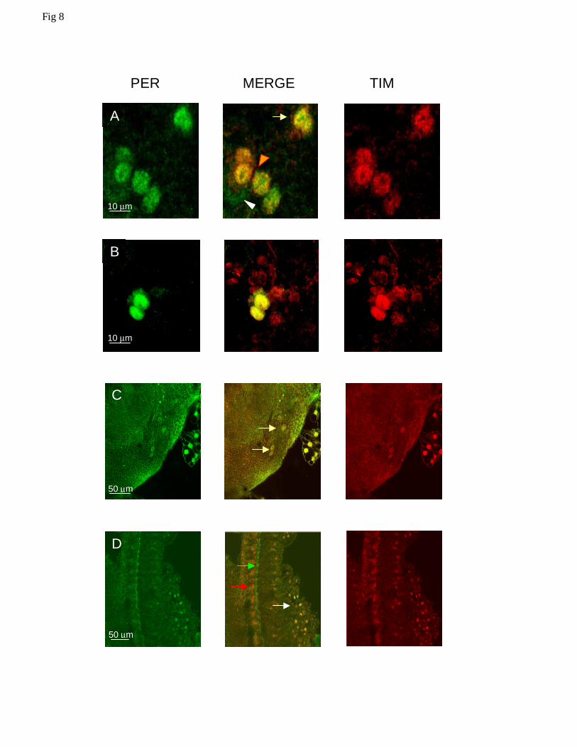
Fig 8. Confocal images of PER and TIM expressing neurons in Musca brain at ZT24. A Four
PDF null LNvs (Pn-LNvs, orange arrowhead) are interspersed among the l-LNvs (white
arrowhead, note that only PER staining is visible). Two LNds are also visible (yellow arrow). As
in the sLNvs, the staining intensity for LNds and Pn-LNvs is higher than for the l-LNvs. B
Nuclear staining for PER and TIM in a 6-8 cell cluster equivalent to the Drosophila LNds. Only
two neurons are in the same focal plane. C Dorsal neurons expressing PER and TIM (arrows).
30
The staining is nuclear for both proteins. D Photoreceptor staining in Musca. MdPER and
MdTIM co-localize within the photoreceptor nuclei (white arrow). PER staining can once again
be seen in a structure at the base of the photoreceptors (green arrow, see also Figure 4F), TIM
does not give staining in this structure although strong staining for TIM can be seen in cells lying
underneath (red arrow).
Fig 9. Confocal images of PER and TIM expressing neurons at different time points in
Musca. A. The PER expressing medial lateral neurons (ml) are in proximity of the PER
and TIM expressing s-LNvs, here shown arrowed and boxed. B. Larger magnification of
the boxed region of A showing nuclear co-localization of PER and TIM in sLNvs at
ZT24. C and D. At ZT6 and 18, the same region shows the brightly stained medial
lateral neurons but indicates the absence of PER and TIM staining in the sLNvs.
Fig 10. Neurons expressing PER in Musca and Drosophila brains. Key: DN1, DN2 and DN3,
dorsal neurons; PLNs, posterior lateral neurons; LNds, lateral neurons dorsal; s-LNvs, small
lateral neurons ventral; l-LNvs, large lateral neurons ventral; Pn-LNv, PDF-null lateral neuron
ventral; MNs, medial neurons; MLNs, medio-lateral neurons.
Fig 1
A B
LD LD - DD LD - LL
0.00
20.00
40.00
60.00
80.00
100.00
120.00
Zt0 LD
Zt4 Zt8 Zt12 Zt16 Zt20 Zt24 Zt4 DD
Zt8 Zt12 Zt16 Zt20 Zt24
0.00
20.00
40.00
60.00
80.00
100.00
120.00
Zt0 LD
Zt4 Zt8 Zt12 Zt16 Zt20 Zt24 Zt4 DD
Zt8 Zt12 Zt16 Zt20 Zt24
0.00
20.00
40.00
60.00
80.00
100.00
120.00
140.00
Zt0 LD
Zt4 Zt8 Zt12 Zt16 Zt20 Zt24 Zt4 DD
Zt8 Zt12 Zt16 Zt20 Zt24
0.00
20.00
40.00
60.00
80.00
100.00
120.00
Zt0 LD
Zt4 Zt8 Zt12 Zt16 Zt20 Zt24 Zt4 DD
Zt8 Zt12 Zt16 Zt20 Zt24
0.00
20.00
40.00
60.00
80.00
100.00
120.00
Zt0 LD
Zt4 Zt8 Zt12 Zt16 Zt20 Zt24 Zt4 DD
Zt8 Zt12 Zt16 Zt20 Zt24
0.00
20.00
40.00
60.00
80.00
100.00
120.00
Zt0 LD
Zt4 Zt8 Zt12 Zt16 Zt20 Zt24 Zt4 DD
Zt8 Zt12 Zt16 Zt20 Zt24
rela
tive
mR
NA
leve
lsperiod timeless
vrille
cycle
Clock
cryptochrome
100
80
60
40
20
0 100
80
60
40
20
0
100
80
60
40
20
0
rela
tive
mR
NA
leve
ls (
100%
)
ZT 0 4 8 12 16 20 24 CT4 8 12 16 20 24 ZT 0 4 8 12 16 20 24 CT4 8 12 16 20 24
0.00
20.00
40.00
60.00
80.00
100.00
120.00
140.00
Zt0 Zt4 Zt8 Zt12 Zt16 Zt20 Zt24
0.00
20.00
40.00
60.00
80.00
100.00
120.00
140.00
Zt0 Zt4 Zt8 Zt12 Zt16 Zt20 Zt24ZT 0 4 8 12 16 20 24
0.00
20.00
40.00
60.00
80.00
100.00
120.00
Zt0 Zt4 Zt8 Zt12 Zt16 Zt20 Zt24
ZT 0 4 8 12 16 20 24
period timeless
vrille
cycle
Clock
cryptochrome
0.00
20.00
40.00
60.00
80.00
100.00
120.00
Zt0 Zt4 Zt8 Zt12 Zt16 Zt20 Zt24
rela
tive
mR
NA
leve
l
0.00
20.00
40.00
60.00
80.00
100.00
120.00
Zt0 Zt4 Zt8 Zt12 Zt16 Zt20 Zt24
0.00
20.00
40.00
60.00
80.00
100.00
120.00
Zt0 Zt4 Zt8 Zt12 Zt16 Zt20 Zt24
100
80
60
40
20
0 100
80
60
40
20
0
100
80
60
40
20
0
rela
tive
mR
NA
leve
ls (
100%
)
A
117
117
0 2 4 6 8 10 12 14 16 18 20 22 ZT
PER (head)
PER (thorax)
117
a b
0,2
0,4
0,6
0,8
1
0 4 8 12 16 200
0,2
0,4
0,6
0,8
1
1,2
0 2 4 6 8 10 12 14 16 18 20 220,5
0,6
0,7
0,8
0,9
1
1,1
0 2 4 6 8 10 12 14 16 18 20 22
PER (LD) TIM (LD) TIM(DD)
ZT (h) ZT (h) CT (h)
117
B
PER (head)
females males
ZT 0 4 8 12 16 20 0 4 8 12 16 20
ZT
C
0 2 4 6 8 10 12 14 16 18 20 22 0 2 4 6 8 10 12 14 16 18 20 22
216
TIM (head)
CT (DD)
D
E 1 7 13 19 1 7 13 19 25 31 37 43 49 55 61 67
MdPER
DmPER
MdTIM
DmTIM
ZT CT(LL)
Fig 4
pars int B
pars int
oes
A
ret
la
me
F
ZT21
cb
me
C ZT9
me
D
G ol
E
me cb
G
cb
Fig 5

A
100µm 50µm
B
20 µm
C PER D TIMM
20 µm
Fig 6
50 µm 100 µm
A B
*
20 µm ^
PDF MERGE TIM
C
PER MERGE TIM
10 µm
sLNv
s
lLNv
s
10 µm
D
E
Fig 7
PER MERGE TIM
10 µm
A
B
10 µm
50 µm
C
50 µm
D
Fig 8


Fig 10
Musca DN LNds
l-LNvs
Pn-LNvs
s-LNvs
MNs MLNs
Drosophila DN1
DN2
DN3
PLNs
LNds
l-LNvs
Pn-LNv
s-LNvS
Related Documents