Cimicifuga species identification by high performance liquid chromatography-photodiode array/mass spectrometric/ evaporative light scattering detection for quality control of black cohosh products Kan He a , Guido F. Pauli b , Bolin Zheng a,d , Huikang Wang c , Naisheng Bai a , Tangsheng Peng a , Marc Roller a , and Qunyi Zheng a,*,e a Department of Research & Development, Naturex/Pure World Botanicals, Inc., 375 Huyler Street, South Hackensack, New Jersey 07606, USA b UIC/NIH Center for Botanical Dietary Supplements Research, PCRPS and Department of Medicinal Chemistry and Pharmacognosy (MC781), College of Pharmacy, The University of Illinois at Chicago, 833 S. Wood St., Chicago, IL 60612 c AndroScience Corporation, 11175 Flintkote Ave., Suite F, San Diego, CA 92121 Abstract Black cohosh has become one of the most important herbal products in the U.S. dietary supplements market. It is manufactured from roots and rhizomes of Cimicifuga racemosa (Ranunculaceae). Botanical identification of the raw starting material is a key step in the quality control of black cohosh preparations. The present report summarizes a fingerprinting approach based on HPLC-PDA/MS/ ELSD that has been developed and validated using a total of ten Cimicifuga species. These include three North American species, C. racemosa, C. americana, C. rubifolia, and seven Asian species, C. acerina, C. biternat, C. dahurica, C. heracleifolia, C. japonica, C. foetida, and C. simplex. The chemotaxonomic distinctiveness of the HPLC fingerprints allows identification of all ten Cimicifuga species. The triterpene glycosides cimigenol-3-O-arabinoside (3), cimifugin (12), and cimifugin-3-O-glucoside (18) were determined to be suitable species-specific markers for the distinction of C. racemosa from the other Cimicifuga species. In addition to identification, the fingerprint method provided insight into chemical interconversion processes occurring between the diverse triterpene glycosides contained in black cohosh. The reported method has proven its usefulness in the botanical standardization and quality control of black cohosh products. Keywords Black cohosh; Cimicifuga; Cimigenol arabinoside; Cimifugin; Species identification; Botanical standardization 1. Introduction The dried rhizomes and roots of black cohosh, Cimicifuga racemosa L. Nutt (Ranunculaceae) (syn. Actaea racemosa L.) are widely used as a herbal dietary supplement. The plant is native to the eastern United States, growing as far south as Florida. Historically, Native American * Corresponding author: Tel +1 201 440 5000; fax +1 201 342 8000 E-mail address: [email protected]. d Current address: 2487 South Revere Way, Aurora, CO 80014, U.S.A. e Current address: 6 Fox Hill Drive, Wayne, NJ 07470, U.S.A. NIH Public Access Author Manuscript J Chromatogr A. Author manuscript; available in PMC 2007 April 21. Published in final edited form as: J Chromatogr A. 2006 April 21; 1112(1-2): 241–254. NIH-PA Author Manuscript NIH-PA Author Manuscript NIH-PA Author Manuscript

Welcome message from author
This document is posted to help you gain knowledge. Please leave a comment to let me know what you think about it! Share it to your friends and learn new things together.
Transcript

Cimicifuga species identification by high performance liquidchromatography-photodiode array/mass spectrometric/evaporative light scattering detection for quality control of blackcohosh products
Kan Hea, Guido F. Paulib, Bolin Zhenga,d, Huikang Wangc, Naisheng Baia, TangshengPenga, Marc Rollera, and Qunyi Zhenga,*,e
a Department of Research & Development, Naturex/Pure World Botanicals, Inc., 375 Huyler Street, SouthHackensack, New Jersey 07606, USA
b UIC/NIH Center for Botanical Dietary Supplements Research, PCRPS and Department of MedicinalChemistry and Pharmacognosy (MC781), College of Pharmacy, The University of Illinois at Chicago, 833S. Wood St., Chicago, IL 60612
c AndroScience Corporation, 11175 Flintkote Ave., Suite F, San Diego, CA 92121
AbstractBlack cohosh has become one of the most important herbal products in the U.S. dietary supplementsmarket. It is manufactured from roots and rhizomes of Cimicifuga racemosa (Ranunculaceae).Botanical identification of the raw starting material is a key step in the quality control of black cohoshpreparations. The present report summarizes a fingerprinting approach based on HPLC-PDA/MS/ELSD that has been developed and validated using a total of ten Cimicifuga species. These includethree North American species, C. racemosa, C. americana, C. rubifolia, and seven Asian species,C. acerina, C. biternat, C. dahurica, C. heracleifolia, C. japonica, C. foetida, and C. simplex. Thechemotaxonomic distinctiveness of the HPLC fingerprints allows identification of all tenCimicifuga species. The triterpene glycosides cimigenol-3-O-arabinoside (3), cimifugin (12), andcimifugin-3-O-glucoside (18) were determined to be suitable species-specific markers for thedistinction of C. racemosa from the other Cimicifuga species. In addition to identification, thefingerprint method provided insight into chemical interconversion processes occurring between thediverse triterpene glycosides contained in black cohosh. The reported method has proven itsusefulness in the botanical standardization and quality control of black cohosh products.
KeywordsBlack cohosh; Cimicifuga; Cimigenol arabinoside; Cimifugin; Species identification; Botanicalstandardization
1. IntroductionThe dried rhizomes and roots of black cohosh, Cimicifuga racemosa L. Nutt (Ranunculaceae)(syn. Actaea racemosa L.) are widely used as a herbal dietary supplement. The plant is nativeto the eastern United States, growing as far south as Florida. Historically, Native American
* Corresponding author: Tel +1 201 440 5000; fax +1 201 342 8000 E-mail address: [email protected] address: 2487 South Revere Way, Aurora, CO 80014, U.S.A.eCurrent address: 6 Fox Hill Drive, Wayne, NJ 07470, U.S.A.
NIH Public AccessAuthor ManuscriptJ Chromatogr A. Author manuscript; available in PMC 2007 April 21.
Published in final edited form as:J Chromatogr A. 2006 April 21; 1112(1-2): 241–254.
NIH
-PA Author Manuscript
NIH
-PA Author Manuscript
NIH
-PA Author Manuscript
women have used black cohosh for the relief of pain during menses and childbirth. It has alsobeen used for the treatment of general malaise, kidney ailments, rheumatism and malaria. Blackcohosh was introduced to Europe in the late 19th century, and became an establishedphytomedicine in the early 20th century. Today, black cohosh is widely used in Europe and theUnited States for various female complaints, including symptoms associated with premenstrualsyndrome, dysmenorrhea and menopause [1].
Raw material of black cohosh is mainly collected from plants growing in the wild in the BlueRidge mountains. Other scattered collections are made in Indiana, Michigan, Illinois andNorthern Pennsylvania. Two closely related Cimicifuga species, C. americana Michx. (syn.Actaea podocarpa DC.) and C. rubifolia Kearney (syn. A. cordifolia DC.), share the samehabitat with black cohosh as they all grow in wooded areas of the eastern United States [2].Their collection is done in batches of various sizes and has not been specifically documented.The Asian Cimicifuga species, including C. dahurica (Turcz.) Maxim., C. foetida L., and C.heracleifolia Kom., are known in traditional Chinese medicine as Sheng-Ma, and are used fortheir antipyretic, analgesic and anti-inflammatory properties. In Japan, C. simplex Wormsk.,C. japonica (Thunb.) Sprengel, and C. acerina (Sieb. et Zucc.) C. Tanaka have been widelyused for the same purposes [3].
Black cohosh is one of the most extensively studied herbal products, and is used as an herbaldietary supplement for the alleviation of menopausal symptoms [4–9]. A considerable amountof clinical research has been documented for a commercial product, Remifemin®, which ismanufactured by Schaper & Brümmer in Germany, and distributed in the United States byGlaxo Smith Kline. Recent pharmacological studies of C. racemosa indicate that the biologicaleffects of black cohosh are due to substances with dopaminergic [10] and serotonergic [11–12] activity rather than an estrogen-like activity.
Triterpenoid saponins and hydroxycinnamic or piscidic acid derivatives are the dominatingtwo groups of chemical constituents in Cimicifuga species. The triterpenoids belong to the 9,19-cycloartanol series and are typically glycosidated at the C-3 position. So far, D-xylose andL-arabinose are the predominant sugar moieties found in the triterpenes of all of theCimicifuga species. Glycosides containing D-glucose and D-galactose were only isolated fromsome species, in particular C. simplex. Hydroxycinnamic acid derivatives are the second groupof common constituents of the rhizomes/roots of Cimicifuga species. These phenoliccomponents include caffeic, ferulic, isoferulic, fukinolic, fukiic and cimicifugic acids and theiresters. Cimifugin and cimifugin glucoside are found in many Asian Cimicifuga species [3],but not in black cohosh. Cimiracemates A–D, however, are minor constituents in black cohosh[13]. Recent reports on the presence of formononetin, an estrogenic isoflavone, remaincontroversial. While we and others could not confirm its presence in black cohosh [14–16],there is one recent positive report [17]. Actein, 23-epi-26-deoxyactein (syn. 27-deoxyactein),acetylshengmanol-3-O-xyloside, cimigenol-3-O-arabinoside, and cimigenol-3-O-xylosiderepresent the major triterpene glycosides found in black cohosh [18–22].
The use of herbal dietary supplement containing black cohosh as an alternative to conventionalestrogen therapy is gaining popularity among perimenopausal women. In 2004, black cohoshranked eighth among herbal supplements sold in mainstream retail outlets in the U.S. [23]. InSeptember 2005, the American Herbal Products Association’s trade recommendations on blackcohosh adulteration came into effect in the herbal industry. This addresses a concern raisedwith the documentation of instances of black cohosh being adulterated with other species.Typical adulterants are C. foetida, C. dahurica and C. heracleifolia.
Most clinical trials with black cohosh have been using products made originating from C.racemosa alcoholic extract that were standardized to a certain level of content of total triterpene
He et al. Page 2
J Chromatogr A. Author manuscript; available in PMC 2007 April 21.
NIH
-PA Author Manuscript
NIH
-PA Author Manuscript
NIH
-PA Author Manuscript

glycosides. Numerous efforts have been made to establish effective methods for theidentifications of chemical constituents in black cohosh, including HPLC-UV/MS [24–28],HPLC-UV/ELSD [15–16,24,29–32], HPTLC [33], GC-MS [17] and random amplifiedpolymorphic DNA (RAPD) analyses [34]. While most of the published chemical methodsalready focus on the identification of triterpene glycosides, it still remains difficult to reliablydistinguish Cimicifuga species due to the large number of triterpene glycosides contained in asingle species. Typically, peaks for more than 30 triterpenes are apparent in HPLCchromatograms, making it impossible to completely resolve these components and identifythem unambiguously. Previously, we have developed an LC-PDA/MS method that allowsdifferentiating between C. racemosa and C. foetida and C. simplex [24]. In the present report,this method is refined by adding an evaporative light scattering (ELSD) as a detection methodand by extending the coverage to a total of ten Cimicifuga species. These include the threeNorth American species, C. racemosa, C. americana, C. rubifolia, and the seven Asian species,C. acerina, C. biternat, C. dahurica, C. heracleifolia, C. japonica, C. foetida and C. simplex.Based on comprehensive HPLC fingerprints of all ten Cimicifuga species, unambiguousspecies identification can be achieved.
2. Experimental2.1. Plant materials and standards
Nine pure Cimicifuga triterpene glycosides and eight phenolic compounds were used asauthentic standards. The triterpene glycosides were actein (1), 23-epi-26-deoxyactein (2),cimigenol-3-O-arabinoside (3), cimigenol-3-O-xyloside (4), 25-O-acetylcimigenol-3-O-xyloside (5), acetyshengmanol-3-O-xyloside (6), 24-O-acetylhydroshengmanol-3-O-xyloside(7), cimiracemoside A (8), and cimicifugoside H-1 (9). The phenolic constituents werecimicifugic acids A (10) and B (11), cimifugin (12), cimiracemates A (13) and B (14), caffeicacid (15), ferulic acid (16), and isoferulic acid (17). Compounds 1–7 and 10–12 were isolatedfrom the Cimicifuga species by procedures described in the literature [3,24], and were identifiedby NMR spectroscopy. Compound 8 was purchased from Chromadex (Santa Ana, CA), 9 fromHerbstandard Inc. (Clarendon Hills, IL), and 15–17 from Sigma Chemical Co. (St. Louis, MO).Compounds 13 and 14 were isolates from C. racemosa and kindly provided by Dr. Shao-NongChen (UIC/NIH Center for Botanical Dietary Supplements Research, University of Illinois atChicago, Chicago, IL). The chemical structures of the standards are listed in Fig. 1, and theirpurities were better than 90% as determined to by HPLC and NMR. The identification of furtherCimicifuga constituents by LC-PDA/MS is discussed below.
The plant samples used in this study include: three C. racemosa, two C. americana and twoC. rubifolia collected from North Carolina, Tennessee and Virginia in 1999, and identified bythe New York Botanical Garden; two C. simplex (C. simplex-1 from Municipality of Enzanand C. simplex-2 from Mutsu, Skayu) and one C. biternat collected from Japan in 1999, andidentified by T. Koyama at Nihon University, and one C. foetida collected from China in 1998and identified by Prof. Zhaoyun Wu at Shanghai University of Traditional Chinese Medicine(SUTCM); one specimen each of C. dahurica, C. foetida and C. heracleifolia collected fromChina in 2001, and identified by Prof. Zheng-Tao Wang at the SUTCM and Prof. Dao-FengChen at the School of Pharmacy, Fudan University; one specimen each of C. simplex, C.japonica and C. acerina collected from Japan in 2002 and identified by Dr. Seiji Nagumo atHoshi University. Voucher samples are deposited in the New York Botanical Garden, NihonUniversity College Museum of Bioresource Sciences, SUTCM and AndroScience’sherbariums, respectively. The commercial products analyzed include Remifemin®, purchasedfrom a health store in New Jersey, CimiPure®, manufactured by Pure World Botanicals, Inc.,and products A and B, which were obtained from the marketplace.
He et al. Page 3
J Chromatogr A. Author manuscript; available in PMC 2007 April 21.
NIH
-PA Author Manuscript
NIH
-PA Author Manuscript
NIH
-PA Author Manuscript

2.2. ApparatusHPLC analyses were carried out on an Agilent 1100 series instrument equipped with aquaternary pump, a four-channel-online degasser, an autosampler, a column oven, a photodiodearray detector (PDA), a mass detector, and an evaporative light scattering detector (ELSD).Data collection was handled by ChemStation software (Agilent). ELSD chromatograms wererecorded on a Sedere Sedex-75 (Alfortville, France). Mass spectra were recorded on an LCQ(Thermo-Finnigan, San Jose, CA) instrument equipped with an atmospheric pressureelectrospray (ESI) and chemical ionization (APCI) sources.
2.3. Chromatography and detectionChromatography was performed on a Zorbax DBS (5 micron, 4 mm ID × 250) column with aflow rate of 1.0 mL/min. Two solvent systems were used. Solvent system I was used for theseparation of phenolic components. It consisted of a step gradient starting with 5% (v/v)aqueous HPLC-grade acetonitrile (A) in 0.1% acetic acid, increasing to 100% acetonitrile over55 minutes in the following step cycle: 0–18 min, 5–28% A; 18–36 min, 28–35% A; 36–45min, 35–55% A; 45–55 min, 55–75% A; 55–56 min, 75–100% A. At the end of the run, 100%of acetonitrile was allowed to flush the column for 10 minutes, and an additional 10 minutesof post run time were set to allow for equilibration of the column with the starting eluant.Solvent system II, derived from the Institute for Nutraceutical Advancement (INA) Method113.001 [29], was used for the triterpene analysis. This system consisted of a step gradientstarting with 30% A in 1% formic acid with water, increasing to 40% A between 0–30 min,then increasing to 60% A between 30–60 min, and resetting to 30% A for column equilibriumbetween 60–70 min. The column temperature was constant at 25°C in the solvent system I,and 30°C in the solvent system II. The sample volume injected was 10 μL. The UV spectraldata was collected at 203 nm for triterpene glycosides and 320 nm for phenolic constituents.For ELSD detection, nitrogen was used as the nebulizing gas at a pressure of 2.6 bar and atemperature of 50°C. Signal gain was set to 7 with an analog output at 20,000 units/Volt. HPLCgrade acetonitrile and water were purchased from Fisher Scientific (Morris Plans, NJ).Atmospheric pressure chemical ionization (APCI) was performed by setting the dischargevoltage to 5.0 kV. The vaporizer and capillary temperatures were set to 450 and 150°C,respectively. Both the sheath gas and the auxiliary gas were nitrogen and had flow rates of 90and 10 units, respectively. A mass range of 50–1500 amu was scanned.
2.4. Sample preparationThe samples were prepared by precisely weighing 300.0–400.0 mg of powdered plant materialor extract into a 10 mL volumetric flask. Approximately 7 mL of 75% methanol was added,and the solution was sonicated for 30 minutes for extracts and 6 hours for crude powders,respectively. The solution was allowed to cool to room temperature before filling up to thefinal volume of 10.00 ml, and was then filtered through a 0.45 μm membrane filter (NylonAcrodisc® 13, Pall Corporation) before the HPLC injection.
3. Results and discussion3.1. Analysis of phenolic constituents
The spectrum of phenolic acid derivatives contained in Cimicifuga species is characterized bythe predominance of caffeic acids and their esters. Both the acids and esters show similar UVspectra mainly due to the conjugated caffeoyl system. The spectrum of caffeic acid derivativesusually comprises the strong caffeoyl absorption bands with maxima around 220–226, 246–252 (sh), 296–300 (sh), and 320–330 nm. The two shoulder (sh) absorptions at the inner sideof the spectrum make these spectra characteristically different from other non-caffeiccomponents (Fig. 2). Since there is less influence of the mobile phase on the long wavelength
He et al. Page 4
J Chromatogr A. Author manuscript; available in PMC 2007 April 21.
NIH
-PA Author Manuscript
NIH
-PA Author Manuscript
NIH
-PA Author Manuscript
absorption, the use of wavelengths above 300 nm is preferred in structural analysis. Compounds15–17 exhibited maxima around 322–324 nm, while compounds 10, 11, 13, and 14 showedmaxima around 326–329 nm. The caffeic acid derivatives 10, 11, and 15–17 are contained inmost of the 10 Cimicifuga species, but in different ratios. Their identification was achieved bya direct comparison with standards, and was confirmed by MS. Other compounds, includingcimifugin glucoside (18), were identified by a combination of UV and MS. In the APCI mode,as expected, negative ionization resulted in higher sensitivity than positive ionization whenanalyzing phenolic components. Therefore, all mass spectral data discussed here was obtainedin (−) APCI mode. All of the caffeic acid derivatives typically displayed molecular ions andacetic acid adducts, along with fragments of the caffeoyl units. Fukinolic acid (19) wasidentified by comparing the relative retention time (Rt.) with that reported in literature and byits characteristic UV spectrum maximum absorption at 330 nm, representing the longestwavelength of all caffeic acid derivatives discussed here. Its molecular ion was observed at m/z 433 [M−H]− and confirmed by m/z at 493 [M+OAc]−, 867 [2M−H]− and 1301 [3M−H]−. Thefragments of m/z 271 [M-caffeoyl]−, 253 [M-caffeic acid-H]−, 179, the molecular ion of caffeicacid [M−H]− and its decarboxylation product 135 [Mcaffeic acid -H-CO2]− provided furtherstructural information of 19. The above should be considered a plausible but tentativefragmentation pattern. The two peaks at Rt. around 21.2 and 21.6 min, with UV spectra similarto those of 10 and 11, showed a pseudomolecular ion m/z 431 [M−H]−, and fragmental ions atm/z 255 [M-feruloyl or isoferuloyl]−, 237 [M-ferulic or isoferulic acid-H]− and 193 due to themolecular ions of ferulic and/or isoferulic acid [M−H]−. Their structures were tentativelyassigned as 2-feruloyl piscidic acid (20) and 2-isoferuloyl piscidic acid (21), respectively. Thesequence of chromatographic elution was assigned as 20 ahead of 21. This is in line with therelative elution order of 10, and 11 and also with a chromatogram reported in the literature[3]. Compounds 20 and 21 are very abundant in C. dahurica. In C. simplex, there was one peakappearing at Rt. around 23 min, which exhibited a molecular ion at m/z 461 [M−H]− and agroup of fragments at m/z 271 [M-dimethoxycinnamoyl]−, 253 [M-dimethoxycinnamic acid-H]−, 207 due to the pseudomolecular ion of dimethoxycinnamic acid [M−H]−, and 191[Mdimethoxycinnamic acid-OH]−. Based on its mass spectral behavior, the structure of thiscompound is proposed to be 2-dimethoxycinnamoyl piscidic acid (22), and represents aphenolic that could only be detected in C. simplex. However, 3, 4-dimethoxycinnamic acid haspreviously been isolated from C. simplex [35]. Although the identification of this compoundremains to be confirmed by isolation, it is likely that 22 can serve as a specific marker of C.simplex. Compounds 13 and 14 were observed in C. racemosa at around Rt. 31–32 min as verysmall peaks, and could not be detected in other species, leading to the conclusion that theywere either absent or present in trace amounts only. Through UV and MS analysis, theindividual caffeic acid derivative could be unambiguously identified (Fig. 3). Our previousstudy had indicated that two chromones, cimifugin (12) and cimifugin glucoside (18), werecontained in C. foetida and C. simplex only, but not in C. racemosa. In the HPLC profiles ofthe current study, 12 and 18 eluted around Rt. 17 and 14 min, respectively. Their UV spectraexhibited absorption maxima at 298 and 300 nm, respectively, with a symmetrical shape, whicheasily distinguished them from the caffeic acid derivatives (Fig. 2). Compounds 12 and 18displayed strong molecular ions in both positive and negative APCI modes. In (+)-APCI,protonated molecular ions of m/z 307 [M+H]+ for 12 and 467 [M+H]+ for 18 were observed.Compound 18 also showed a deglucosidated fragment ion at m/z 307 [M-glucosyl]+. Theapplication of 12 and 18 for species identification will be discussed below. All of the identifiedphenolic constituents in the ten Cimicifuga species are summarized in Table 1.
3.2. Analysis of triterpene glycosidesThe Cimicifuga triterpene glycosides were analyzed by LC/MS in APCI mode, with thevaporization temperature set to 450°C. At this temperature, the highly oxygenatedCimicifuga triterpene glycosides were decomposed during ionization by the loss of neutral
He et al. Page 5
J Chromatogr A. Author manuscript; available in PMC 2007 April 21.
NIH
-PA Author Manuscript
NIH
-PA Author Manuscript
NIH
-PA Author Manuscript
molecules such as water, sugar, and acetic acid. The generated molecular ion, along with agroup of fragmental ions, was passed through the heated capillary into the mass analyzer. Asa result, both molecular ion adducts and fragmental ions were observed, simultaneously,without performing the MS/MS analysis. It was found that all of the fragmental ions generatedfrom triterpene glycosides could be observed in a positive APCI mode, while less or nofragmentation was detected in negative mode. Nine triterpene glycoside standards were usedin this study, belonging to six aglycone types: compounds 1 and 2 are based on acetyl-acteol,3–5 on cimigenol, 6 on shengmanol, 7 on hydroshengmanol, 8 on cimiracerol, and 9 on 16,23-diketo-shengmanol. All of the nine standards contain a pentose (xyl or ara) monosaccharideattached to the C-3-OH position of the aglycone. This gave rise to losses of 132 [M-arabinosylor xylosyl-H2O]+ or the 150 [M-arabinose or xylose]+ in (+)-APCI spectra. Six of the 9triterpene standards contained an acetyl group, which lead to very characteristic fragmentsresulting from the loss of acetic acid [M-60]+. Examples are the fragment ions m/z 617 [M+H-60]+ of 1 and 8, m/z 601 [M+H -60]+ of 2 and m/z 603 [M+H -60]+ of 5, 6 and 7. Theintensity of fragment signals turned out to be a helpful diagnostic tool. For example, structureswith an acetyl group usually generate a stronger fragment resulting from the loss of water, suchas the dehydrated ion at m/z 659 in 1 and 8, and the fragment at m/z 645 observed in the APCIMS spectra of 5, 6, and 7. While the intensity of the individual dehydration fragments variesslightly between individual acquisitions, their relative intensities within the full MS spectra istypically maintained. The mass spectral features discussed above are summarized in Fig. 4.
Of the more than 40 triterpene glycosides that have been reported to date from C. racemosa,acetyl-acteol, cimigenol and shengmanol represent the major aglycones. The epimericaldopentoses arabinose and xylose are the two dominant sugar moieties in Cimicifugatriterpenes, and usually give rise to a pair of glycosides with close chromatographic retentiontime. While representing a chromatographical challenge, this relationship can also provideuseful information for compound identification, like for the pair of 3 and 4. By directlycomparing retention times and mass spectra with those of the standards, 1–8 could be identifiedwithout ambiguity. Other triterpene glycosides, including acetylshengmanol-3-O-arabinoside(23) and 25-O-acetylcimicigenol-3-O-arabinoside (24), were tentatively assigned based on thesimilarity of their mass spectra with those of 6 and 5, respectively, and due to their relativeretention time being similar to those of the pair of 3 and 4. In two C. foetida samples, 1, 2, 4,6 and 9 could be identified. C. simplex had been extensively studied for its chemicalconstituents. More than 50 triterpene glycosides have been isolated from this species [3], withthe majority of triterpenoids reported belonging to the shengmanol, hydroshengmanol, andcimigenol series. However, in the two C. simplex samples collected and tested here, none ofthe major peaks seen in the total ion current trace (TIC) showed a mass spectrum identical toone of the 9 standards, although they all displayed triterpene glycoside fragment patterns. Themajor peak at Rt. of 33 min displayed a protonated molecular ion at m/z 793 [M+H]+ and aseries of fragments that represent two xylosyl or arabinosyl moieties. Since no such structurehas been reported from this species, the structure of the major triterpene peak seen in the TICremains unconfirmed. The major peaks in C. dahurica, however, can be identified as 2, 6, and4. In C. heracleifolia, 4 was shown to be one of the major components. The peak at Rt. 40 min,eluting right before 4, produced a protonated molecular ion adduct at m/z 619 [M+H]+ with aseries of fragmental ions of 601 [M-H2O]+, 583 [M-2H2O]+, 469 [M-xylose]+. Due to thesimilarity of its fragment ion pattern to that of 4, and augmented by the fact that every fragmentwas 2 amu less than the corresponding ion in 4, it is reasonable to assume that this peakrepresents a dehyo-derivative of 4. One triterpene that fulfills the structural criteria is 24-epi-7, 8-didehydrocimigenol-3-O-xyloside (25), a compound previously isolated from thisspecies [36]. Two peaks that appeared at Rt. around 60 and 62 min showed protonatedmolecules at m/z 487 [M+H]+ and 489 [M+H]+, respectively, with no sugar or acetyl groupfragments, suggesting that they were triterpene aglycones.
He et al. Page 6
J Chromatogr A. Author manuscript; available in PMC 2007 April 21.
NIH
-PA Author Manuscript
NIH
-PA Author Manuscript
NIH
-PA Author Manuscript

Compounds 4, 6, and 7 were found to be the major peaks in C. japonica. In C. acerina, only6 could be identified unambiguously. Since no chemical constituents have been previouslyreported from C. biternat, C. americana, and C. rubifolia, structural assignments remaintentative until the compounds are isolated. However, compounds 4 and were undoubtedlyidentified in C. biternat. As with C. heracleifolia, the peak eluting at Rt. 40 min was assignedto 25 based on identical mass spectra. The peak at Rt. 34 min fully resembled the mass fragmentpattern of 6, except that all ions were 2 amu less compared to 6. Therefore, the structure ofacetyl-didehydroshengmanol-3-O-xyloside (26) is suggested for this peak. Regarding the twoNorth American Cimicifuga species, upon injection of the extract of C. americana all of themajor peaks eluted earlier than 30 minutes, indicating that they are less likely to containcimigenol as the aglycone. All peaks displayed the characteristic fragmentation patterns oftriterpenes, but were devoid of acetyl groups and contained only one sugar moiety. The majorpeak found in C. rubifolia was identified as 3 on the basis of retention time (38 min) andidentical mass spectra. Up to date, except for C. racemosa, this is the only other species inwhich 3 has been detected. The total ion current trace (TIC) obtained from 10 Cimicifugaspecies by (+)-APCI is shown in Fig. 5. All triterpene glycosides identified from the teninvestigated Cimicifuga species are summarized in Table 1.
3.3. Chemical conversion of triterpene glycosidesDuring the production process of black cohosh botanical extracts, noteworthy changes wereobserved in the HPLC chromatograms, in particular with respect to the triterpene glycosidesfingerprints. When analyzing the native 75% ethanol extract, a large variety of triterpene peakswas observed in the small retention time window between 37 and 43 min. However, after aconcentration step, in which the extract was concentrated under the influence of heat, the peakdiversity in this HPLC window was reduced, and only 3 and 4 appeared as the two dominatingpeaks and with increased intensity (Fig. 6). A stability test for a 75% ethanol/water extract atroom temperature showed that there were no significant profile changes in the HPLCchromatograms after 10 days, while changes started to occurr after two months. Fig. 6 (A) and(B) show the comparison of the TIC obtained from LC-MS (+)-APCI with fresh extract andafter 24 months of storage. The peak intensities of 3 and 4 were significantly increased in theaged compared to the freshly prepared extract. In addition, new peaks appeared at Rt. around60 to 65 min. Two of the major peaks were identified as the 25-O-acetyl-cimigenol glycosides5 and 24. The chemical conversion involved in the interconversion of 3–5 and 24 apparentlyoccurs at the cimigenol aglycone level. According to literature, however, the open chainaglycones shengmanol and hydroshengmanol are biogenetically related to the spiroketalcimigenol. In studies towards the characterization of the genuine aglycone precursor oftriterpene glycosides from C. simplex, 23-O-acetylshengmanol xyloside was isolated andsubjected to hydrolysis with acid and base. As a result, not only hydroshengmanol, but alsocimigenol were obtained (Fig. 7). Therefore, Kusano et al. hypothesized that acetyl-shengmanol glycoside is the parent component of acetyl-hydroshengmanol, 25-O-acetylcimigenol and 25-O-methylcimigenol glycosides in Cimicifuga species [3,37]. Thisproposed chemical conversion pathway has led us to focus on the 23-O-acetyl-shengmanolxyloside 6 and its related compounds as stability markers to indicate changes in triterpenepattern during the production process. Freshly prepared black cohosh 75% ethanol extract wasprepared with extracts heated to 90–95°C for 3 and 48 hours, respectively. After a 3 hourtreatment, (Fig. 6(C)), many triterpene peaks were either reduced or had even disappeared fromthe chromatogram. Heating made the peaks of 3 and 4 the dominant triterpenes in the 37–43min region. Small peaks of the rearrangement products 5 and 24 were also observed, and it isreasonable to conclude that they were produced from the same precursor(s) by chemicalconversion. After 48 hours of heat treatment (Fig. 6(D)), the peak intensities of 3, 4, 5 and24 were significantly increased, while the peaks of 6, 7 and 23 were further reduced or hadeven disappeared. Furthermore, two new peaks were observed at Rt. around 35.3 and 35.9 min,
He et al. Page 7
J Chromatogr A. Author manuscript; available in PMC 2007 April 21.
NIH
-PA Author Manuscript
NIH
-PA Author Manuscript
NIH
-PA Author Manuscript
respectively. These compounds showed fragmentation patterns similar to those of 6, suggestingthat they may represent further rearrangement products of 6 and 23. In summary, the heattreatment experiments demonstrated that chemical conversion can readily occurr and involvesthe triterpenes 3–6, 23, and 24.
In order to further confirm the occurrence of chemical conversion under heat treatment andpossibly storage, 6 was treated in the same way as the extract at 90–95°C for 15 hours. As aresult, the formation of major 4 and minor 5 was observed. The principle mechanism of thechemical conversion under heat is described in Fig. 7 [3,37]. Other acetylated triterpenes mightbe the source of the acetyl reagent in the formation of the 25-O-acetyl derivatives 5 and 24.This experiment confirms that 23-O-acetyl-shengmanol and 23-O-acetyl-hydroshengmanolglycosides can convert into cimigenol glycosides in solution. While the reaction is acceleratedby heat, it most likely occurs at room temperature and potentially affects the long-term stabilityof black cohosh extracts. Finally, it is noteworthy that we did not observe the analogousconversion to occurr with 9, which differs from 6 by bearing a carbonyl at C-23 instead of anacetyl group. This observation is on line with the lack of a C-23 hydroxyl function as thefunctional precursor for C-16/23 hemiacetal formation.
3.4. Cimicifuga species identification by phenolic components and triterpene glycosideprofiles
In the current analysis, cimifugin (12) and its glucoside 18 were readily detected in our newlycollected C. foetida and C. simplex. Fig. 8 shows the selected ion chromatograms (SIC)obtained at m/z 307 [M+H]+, representing 12 and a fragmental ion of 18. In a combination ofHPLC-UV chromatograms (Fig. 3) with SIC at m/z 307, 12 and 18 can readily be detected.Their presence was also observed in other Cimicifuga species including C. biternat, C.dahurica, and C. rubifolia, but they were not detected in C. acerina, C. heracleifolia, C.japonica, and C. americana. It is noteworthy to mention (Fig. 3 (E) and (F)) that 12 and 18give rise to very small peaks on the HPLC-UV chromatogram, which can easily be missed,while SIC clearly demonstrates the presence of the two components. C. rubifolia wasdetermined to contain 12 as the only one of the three North American Cimicifuga species.
As discussed above, 3 could represent a conversion product from acetyl-hydroshengmanolarabinoside or acetyl-shengmanol arabinoside. Acetylhengmanol-3-O-xyloside (6) has beenisolated from many Cimicifuga species, including C. simplex, C. acerina, C. japonica [3], andC. racemosa. Except for C. racemosa and C. dahurica, we are unaware of any literaturereporting the isolation of acetyl-shengmanol arabinoside or cimigenol arabinoside from theabovementioned Cimicifuga species. Although 3 was reported from C. dahurica, its abundancewas below the limit of detection in our current study. We did not observe an increase in thepeak intensity of 3 after heating a sample of C. dahurica. Due to the presence of 12 and 18,C. dahurica can still be distinguished from C. racemosa. The SIC at m/z 621 [M+H]+
corresponding to 3 and 4 is shown on Fig. 9. In the fresh extract, both 3 and 4 were observedin C. racemosa, albeit with a low signal-to-noise ratio due to interference with other triterpeneglycosides. After heating the sample to 50–60°C for 2–3 hours, the signal-to-noise ratio wasimproved significantly and displayed a pair of peaks similar to what is typically observed forblack cohosh. Compound 3 was also found in a significant amount in C. rubifolia. Except forthese two species, none of the remaining 8 species contained a detectable amount of 3 as shownin Fig. 9. To exclude the heat formation as a source of 3, as seen in C. racemosa, all of the 8species underwent a heating process. As a result, 3 was not found to be increased to a detectablelevel in any of the 8 Cimicifuga species. Therefore, and in summary of the above discussionof the LC profiles, the triterpene 3, and the furanochromones 12 and 18 were determined to bethree reliable markers for the identification of all 10 relevant Cimicifuga species.
He et al. Page 8
J Chromatogr A. Author manuscript; available in PMC 2007 April 21.
NIH
-PA Author Manuscript
NIH
-PA Author Manuscript
NIH
-PA Author Manuscript
After the HPLC profiles of 10 different Cimicifuga species had been established, the newspecies identification protocol was applied to commercial black cohosh products,Remifemin®, CimiPure®, and products A and B, by comparing the phenolic and triterpenefingerprints to those of the authentic Cimicifuga species. It was clearly determined thatRemifemin®, CimiPure® and product A were manufactured using C. racemosa, while productB was manufactured using C. foetida. Product A exhibited a typical C. racemosa profile (Fig.10 (C)) with all the key constituents, including 1–8. Furthermore, the abnormal triterpeneabundance with unusually large amounts of 5 and 24 suggested that the product wasinadequately manufactured, probably involving overheating of the product, which causedtriterpene conversion as discussed above. The use of harsh extraction conditions was alsoindicated by the fact that isoferulic acid (17) was the only phenolic compound detected in theHPLC-UV chromatogram, considering that 17 is likely to be the main degradation product ofthe condensed Cimicifuga isoferulic acid derivatives (Fig. 3 (N)).
3.5. Fingerprints of HPLC-ELSD of Cimicifuga speciesIt is a widely accepted method for black cohosh products to be chemically standardized to theirtotal triterpene glycoside content. Because the Cimicifuga triterpenoids essentially lackchromophores, UV detection provides very poor sensitivity and will not result in acceptablechromatograms, especially when working without previous cleanup or enrichment procedures.This problem can be overcome by using an LC-MS method, which is a powerful tool todistinguish C. racemosa from a possible adulterant using other Cimicifuga species. However,application of LC-MS as routine analytical method is limited by the fact that mass detectionis still not a routine setup in the botanical and the dietary supplemental industry. However, inorder to overcome the low UV sensitivity of the triterpenoids, an HPLC with an in-lineevaporative light scattering detection (ELSD) has been developed in our laboratory. In additionto the LC-MS studies, HPLC-ELSD fingerprints were acquired using the established Institutefor Nutraceutical Advancement (INA) method for the ten investigated Cimicifuga species. Thereason for the selection of the INA method is that several laboratories, including our own, havevalidated it, and that it has found wide acceptance for the determination of triterpene glycosidein the herbal supplement industry. In order to match the results of the ELSD with the LC-MSanalyses, and to provide most significant reference for future studies, all of the LC-MS analysesof triterpene glycosides in this work were performed using the INA method (solvent systemII) as described in the experimental section. For reference purposes, the HPLC-ELSDfingerprints of the ten different Cimicifuga species are shown in Fig. 11. Interestingly, theELSD fingerprints are quite similar to the TIC/MS traces. A typical HPLC-ELSDchromatogram of C. racemosa should include (in the order of elution) 8, 1, 2, 23, 6, 3, and 4as the major components. Compounds 3 and 8 are very important markers, and must beconsidered positive markers for C. racemosa identification.
Today, the most likely form of adulteration of black cohosh products found in the U.S.marketplace is C. foetida, in which 9 is the major component. ELSD detection showed highsensitivity in the analysis of these triterpene glycosides, and generally provided cleanchromatograms with low baseline noise that were perfectly suitable to monitor the quality ofblack cohosh products. The main limitation of ELSD is its lack of structural information, whichin turn limits its use in species differentiation, especially when analyzing products mixtures.
4. ConclusionsDespite of the fact that black cohosh represents one of the clinically most extensively studiedbotanicals, it remains a complex scientific entity with regard to its phytoconstituents andbotanical species assignment. Historically, many different Cimicifuga species, containing thevery similar or even identical constituents, have been used medicinally for various applications
He et al. Page 9
J Chromatogr A. Author manuscript; available in PMC 2007 April 21.
NIH
-PA Author Manuscript
NIH
-PA Author Manuscript
NIH
-PA Author Manuscript

and indications. However, correct species identification and exclusion of adulteration is a keystep in any clinical trial, including those that investigate how black cohosh extracts can alleviateperimenopausal and postmenopausal symptoms. This includes the establishment of reliablesources of raw material, as the use of proper and authentic plant materials is crucial toreproducible clinical results. One practical limitation that has to be taken into account is thatthe botanical industry buys a high volume of raw material directly from farmers and collectors,and that only the pre-processed roots and rhizomes are shipped to the manufacturer. This makesroutine botanical species identification an unfeasible approach, and chemical analysis anecessity in the quality monitoring of black cohosh raw materials. Due to the complexity andvariation of the chemical constituents in authentic black cohosh to start with, it has to be realizedthat fingerprinting methods have intrinsic limitations, especially when it comes to theidentification of small amounts of adulterated product mixed into authentic material. In thisstudy, however, hyphenated MS introduced molecular weight to the fingerprints, thus addinganother layer of information to the chromatographical window for compound identification.Using LC/MS in (+)-APCI mode is preferred because APCI generates less molecular ionadducts and more fragment ions, thus providing more valuable structural information. Ourresults confirm that cimigenol-3-O-arabinoside (3), cimifugin (12) and cimifugin glucoside(18) can serve as the three species-specific marker compounds to distinguish authentic C.racemosa from nine other Cimicifuga species. The finding of chemical conversion among thetriterpene glycosides in black cohosh provides a draft guideline for the maintenance of original“native” phytochemical profiles throughout the manufacturing process. Overall, the presentedHPLC-PDA/MS/ELSD methods should be useful for Cimicifuga species identification and forquality control of black cohosh botanical products.
Acknowledgements
The authors are very thankful to Charlotte Lindqvist, Victor Albert, and Dr. Scott Mori of New York Botanical Garden,T. Koyama, Nihon University, Prof. Zhaoyun Wu, Prof. Zheng-Tao Wang, Shanghai University of Traditional ChineseMedicine, Prof. Dao-Feng Chen, School of Pharmacy, Fudan University, and Dr. Seiji Nagumo, Hoshi University,for Cimicifuga species plant collection and identification. The authors wish to thank Dr. Shao-Nong Chen, UIC/NIHCenter for Dietary Supplements Research, Chicago (IL), for generously providing authentic standards. Our final thanksextend to Mrs. Nancy Madis for her proof reading of this manuscript. GFP gratefully acknowledges funding by NIHthrough grant #P50 AT00155 (UIC/NIH Botanical Center) from NCCAM, ODS, NIGMS, and ORWH.
References1. Foster S. Herbal Gram 1999;45:36.2. Applequist WL. Flora 2003;198:358.3. Kusano G. Yakugaku Zasshi 2001;121:497. [PubMed: 11494597]4. Mahady, GB.; Fong, HS.; Farnsworth, NR. Botanical Dietary Supplements: Quality, Safety and
Efficacy. Swets; Zeitlinger, BV., editors. Lisse; The Netherlands: 2001. p. 275. Barrett, M. The Handbook of Clinically Tested Herbal Remedies. The Haworth Press; New York:
2004. p. 1856. Borrelli F, Ernst E. Eur J Clin Pharmacol 2002;58:235. [PubMed: 12136368]7. Kronenberg F, Fugh-Berman A. Ann Intern Med 2002;137:805. [PubMed: 12435217]8. Dog TL, Powell KL, Weisman SM. Menopause 2003;10:299. [PubMed: 12851513]9. Wuttke W, Seidlova-Wuttke D, Gorkow C. Maturitas 2003;44:S67. [PubMed: 12609561]Suppl 110. Borrellia F, Izzoa AA, Ernst E. Life Sciences 2003;73:1215. [PubMed: 12850238]11. Burdette JE, Liu J, Chen SN, Fabricant DS, Piersen CE, Barker EL, Pezzuto JM, Mesecar A, van
Breemen RB, Farnsworth NR, Bolton JL. J Agric Food Chem 2003;51:5661. [PubMed: 12952416]12. Fabricant D, Nikolic D, Lankin D, Chen SN, Jaki B, Krunic A, van Breemen RB, Fong HHS,
Farnsworth NR, Pauli GF. J Nat Prod 2005;68:1266. [PubMed: 16124775]13. Chen SN, Fabricant DS, Lu ZZ, Zhang H, Fong HS, Farnsworth N. Phytochemistry 2002;61:409.
[PubMed: 12377235]
He et al. Page 10
J Chromatogr A. Author manuscript; available in PMC 2007 April 21.
NIH
-PA Author Manuscript
NIH
-PA Author Manuscript
NIH
-PA Author Manuscript

14. Struck D, Tegtmeier M, Harnischfeger G. Planta Med 1997;63:289. [PubMed: 17252360]15. Li W, Chen S, Fabricant D, Angerhofer CK, Fong HHS, Farnsworth NR, Fitzloff JF. Analytica
Chimica Acta 2002;471:61.16. Jiang B, Kronenberg F, Balick MJ, Kennelly EJ. Phytomedicine. 2005(In press)17. Panossian A, Danielyan A, Mamikonyan G, Wikman G. Phytochem Anal 2004;15:100. [PubMed:
15116940]18. Bedir E, Khan IA. Chem Pharm Bull 2000;48:425. [PubMed: 10726870]19. Shao Y, Harris A, Wang M, Zhang H, Cordell GA, Bowman M, Lemmo E. J Nat Prod 2000;63:905.
[PubMed: 10924163]20. Chen SN, Li W, Fabricant DS, Santarsiero BD, Mesecar A, Fitzloff JF, Fong HHS, Farnsworth NR.
J Nat Prod 2002;65:601. [PubMed: 11975513]21. Chen SN, Fabricant DS, Lu ZZ, Fong HS, Farnsworth NR. J Nat Prod 2002;65:1391. [PubMed:
12398533]22. Watanabe K, Mimaki Y, Sakagami H, Sashida Y. Chem Pharm Bull 2002;50:121. [PubMed:
11824572]23. Blumenthal M. Herbal Gram 2005;66:66.24. He K, Zheng B, Kim CH, Rogers L, Zheng Q. Planta Med 2000;66:635. [PubMed: 11105569]25. Ganzera M, Bedir E, Khan IA. Chromatographia 2000;52:301.26. Onorato J, Henion JD. Anal Chem 2001;73:4704. [PubMed: 11605850]27. Li W, Sun Y, Liang W, Fitzloff JF, van Breemen RB. Rapid Commun Mass Spectrom 2003;17:978.
[PubMed: 12717772]28. Wang HK, Sakurai N, Shih CY, Lee KH. J Agric Food Chem 2005;53:1379. [PubMed: 15740010]29. www.nsf.org/business/ina/blackcohosh.asp?program=INA30. Kong L, Li X, Zou H, Wang H, Mao X, Zhang Q, Ni J. J Chromatogr A 2001;936:111. [PubMed:
11760993]31. Schmidt AH. Journal of Liquid Chromatography & Related Technologies 2005;28:871.32. Pharmacopoeial Forum, The United States Pharmacopeial Convention, Inc. 2002;28:1455.33. Upton, E., editor. Black Cohosh Rhizome, monograph. American Herbal Pharmacopoeia; Scotts
Valley, CA: 2002.34. Xu H, Fabricant DS, Piersen CE, Bolton JL, Pezzuto JM, Fong H, Totura S, Farnsworth NR,
Constantinou AI. Phytomedicine 2002;9:757. [PubMed: 12587700]35. Takahira M, Kusano A, Shibano M, Nagai M, Miyase T. Chem Pharm Bull 1998;46:362.36. Li JX, Kadota S, Hattori M, Yoshimachi S, Shiro M, Oogami N, Mizuno H, Namba T. Chem Pharm
Bull 1993;41:832.37. Sakurai N, Nagai M. Yakugaku Zasshi 1996;116:850. [PubMed: 8981829]
He et al. Page 11
J Chromatogr A. Author manuscript; available in PMC 2007 April 21.
NIH
-PA Author Manuscript
NIH
-PA Author Manuscript
NIH
-PA Author Manuscript
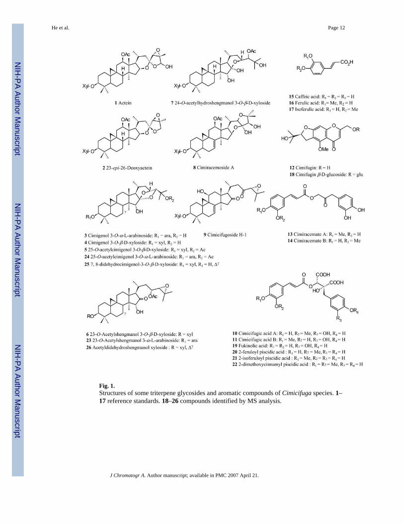
Fig. 1.Structures of some triterpene glycosides and aromatic compounds of Cimicifuga species. 1–17 reference standards. 18–26 compounds identified by MS analysis.
He et al. Page 12
J Chromatogr A. Author manuscript; available in PMC 2007 April 21.
NIH
-PA Author Manuscript
NIH
-PA Author Manuscript
NIH
-PA Author Manuscript

Fig. 2.Typical UV spectra of aromatic Cimicifuga compounds, obtained on-line in mobile phase aspart of the HPLC fingerprint of Cimicifuga species. (A) Isoferulic acid (17); (B) Cimicifugicacid B (11); (C) Cimifugin (12).
He et al. Page 13
J Chromatogr A. Author manuscript; available in PMC 2007 April 21.
NIH
-PA Author Manuscript
NIH
-PA Author Manuscript
NIH
-PA Author Manuscript
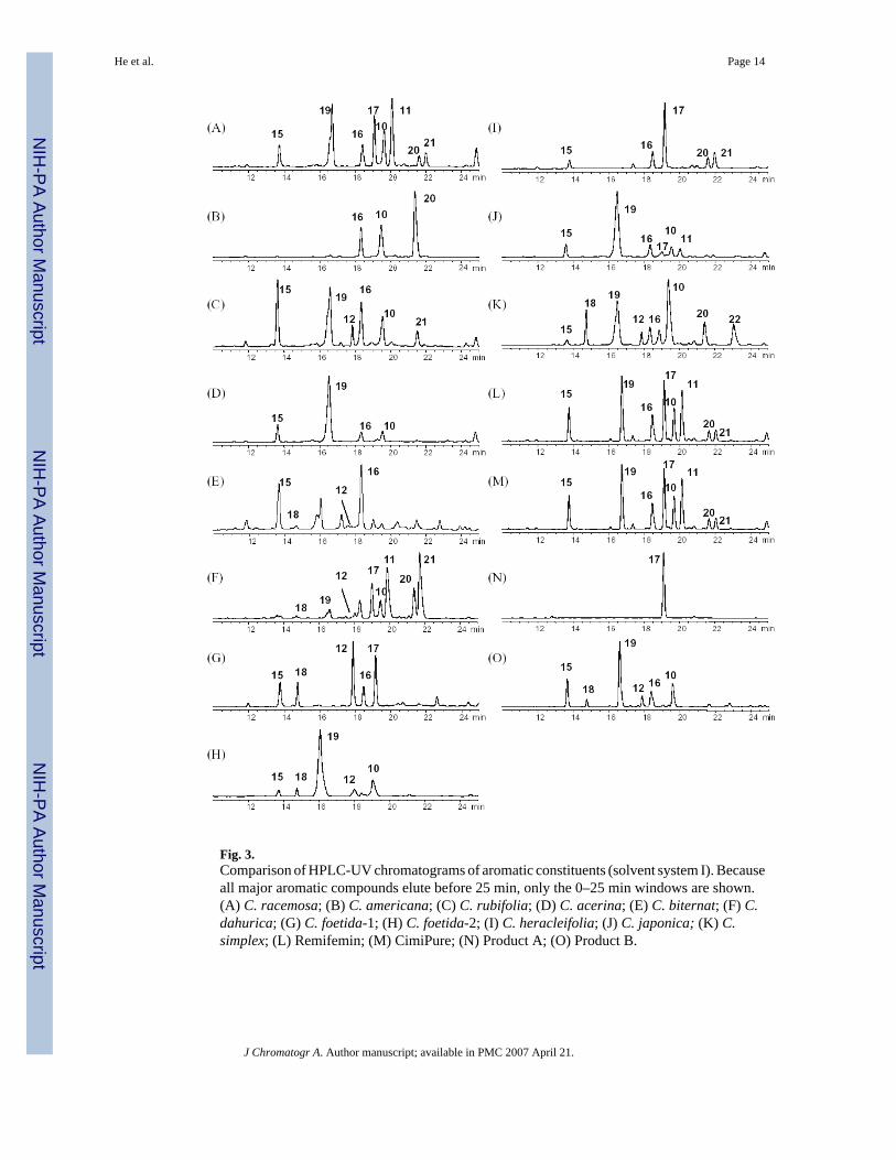
Fig. 3.Comparison of HPLC-UV chromatograms of aromatic constituents (solvent system I). Becauseall major aromatic compounds elute before 25 min, only the 0–25 min windows are shown.(A) C. racemosa; (B) C. americana; (C) C. rubifolia; (D) C. acerina; (E) C. biternat; (F) C.dahurica; (G) C. foetida-1; (H) C. foetida-2; (I) C. heracleifolia; (J) C. japonica; (K) C.simplex; (L) Remifemin; (M) CimiPure; (N) Product A; (O) Product B.
He et al. Page 14
J Chromatogr A. Author manuscript; available in PMC 2007 April 21.
NIH
-PA Author Manuscript
NIH
-PA Author Manuscript
NIH
-PA Author Manuscript

Fig. 4.TIC and mass spectrum of reference standards obtained by hyphenated LC-MS separation. (A)TIC; (B) 1 Actein; (C) 2 23-epi-26-Deoxyactein; (D) 3 Cimigenol3-O-arabinoside; (E) 5 25-O-Acetylcimigenol xyloside; (F) 6 Acetylshengmanol3-O-xyloside; (G) 7 24-O-Acetylhydroshengmanol3-O-xyloside; (H) 8 Cimiracemoside A; (I) 9 Cimicifugoside H-1. The massspectrum of 4 is not displayed as it is almost identical to that of 3; xyl: xylose, ara: arabinose.
He et al. Page 15
J Chromatogr A. Author manuscript; available in PMC 2007 April 21.
NIH
-PA Author Manuscript
NIH
-PA Author Manuscript
NIH
-PA Author Manuscript

Fig. 5.Comparison of the total ion current (TIC) traces of Cimicifuga species (solvent system II). (A)C. racemosa; (B) C. americana; (C) C. rubifolia; (D) C. acerina; (E) C. biternat; (F) C.dahurica; (G) C. foetida-1; (H) C. foetida-2; (I) C. heracleifolia; (J) C. japonica; (K) C.simplex-1; (L) C. simplex-2 (see Experimental for botanical details).
He et al. Page 16
J Chromatogr A. Author manuscript; available in PMC 2007 April 21.
NIH
-PA Author Manuscript
NIH
-PA Author Manuscript
NIH
-PA Author Manuscript
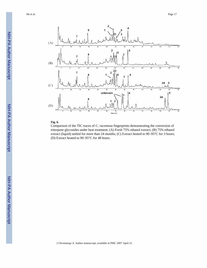
Fig. 6.Comparison of the TIC traces of C. racemosa fingerprints demonstrating the conversion oftriterpene glycosides under heat treatment. (A) Fresh 75% ethanol extract; (B) 75% ethanolextract (liquid) settled for more than 24 months; (C) Extract heated to 90–95°C for 3 hours;(D) Extract heated to 90–95°C for 48 hours.
He et al. Page 17
J Chromatogr A. Author manuscript; available in PMC 2007 April 21.
NIH
-PA Author Manuscript
NIH
-PA Author Manuscript
NIH
-PA Author Manuscript

Fig. 7.Hypothetic conversion of triterpenes: acetyl-shengmanol type triterpenes are first convertedinto hydroshengmanol and subsequently into cimigenol type triterpenes [3,37].
He et al. Page 18
J Chromatogr A. Author manuscript; available in PMC 2007 April 21.
NIH
-PA Author Manuscript
NIH
-PA Author Manuscript
NIH
-PA Author Manuscript

Fig. 8.Comparison of selected ion chromatograms (SIC) at m/z 307 for cimifugin (12) and aglyconefragment of cimifugin glucoside (18). (A) C. racemosa; (B) C. americana; (C) C. rubifolia;(D) C. acerina; (E) C. biternat; (F) C. dahurica; (G) C. foetida; (H) C. heracleifolia; (I) C.japonica; (J) C. simplex; (K) Remifemin; (L) CimiPure; (M) Product B.
He et al. Page 19
J Chromatogr A. Author manuscript; available in PMC 2007 April 21.
NIH
-PA Author Manuscript
NIH
-PA Author Manuscript
NIH
-PA Author Manuscript
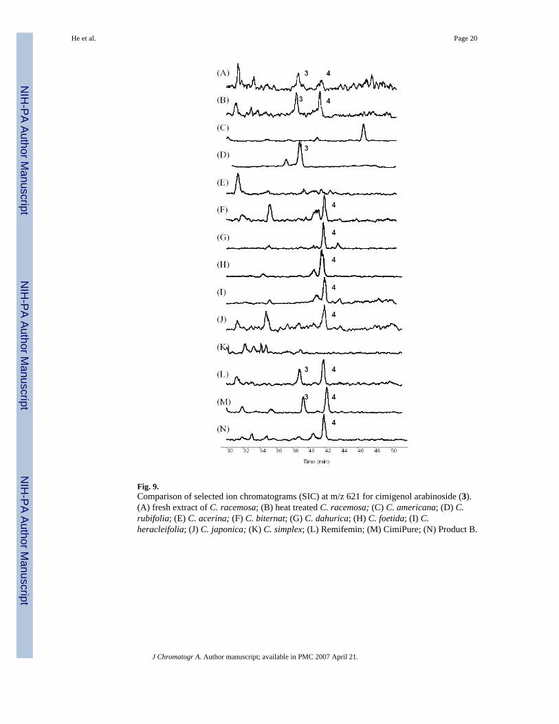
Fig. 9.Comparison of selected ion chromatograms (SIC) at m/z 621 for cimigenol arabinoside (3).(A) fresh extract of C. racemosa; (B) heat treated C. racemosa; (C) C. americana; (D) C.rubifolia; (E) C. acerina; (F) C. biternat; (G) C. dahurica; (H) C. foetida; (I) C.heracleifolia; (J) C. japonica; (K) C. simplex; (L) Remifemin; (M) CimiPure; (N) Product B.
He et al. Page 20
J Chromatogr A. Author manuscript; available in PMC 2007 April 21.
NIH
-PA Author Manuscript
NIH
-PA Author Manuscript
NIH
-PA Author Manuscript
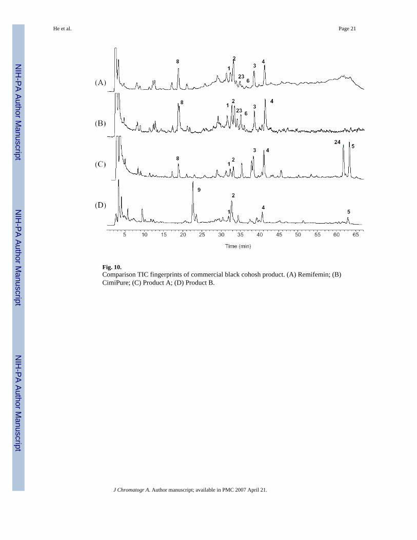
Fig. 10.Comparison TIC fingerprints of commercial black cohosh product. (A) Remifemin; (B)CimiPure; (C) Product A; (D) Product B.
He et al. Page 21
J Chromatogr A. Author manuscript; available in PMC 2007 April 21.
NIH
-PA Author Manuscript
NIH
-PA Author Manuscript
NIH
-PA Author Manuscript

Fig. 11.HPLC-ELSD fingerprint chromatograms of the Cimicifuga triterpene glycosides (INA method,solvent system II). (A) C. racemosa; (B) C. americana; (C) C. rubifolia; (D) C. acerina; (E)C. biternat; (F) C. dahurica; (G) C. foetida-1; (H) C. foetida-2; (I) C. heracleifolia; (J) C.japonica; (K) C. simplex-1; (L) C. simplex-2; (M) Remifemin; (N) CimiPure; (O) Product B.
He et al. Page 22
J Chromatogr A. Author manuscript; available in PMC 2007 April 21.
NIH
-PA Author Manuscript
NIH
-PA Author Manuscript
NIH
-PA Author Manuscript
NIH
-PA Author Manuscript
NIH
-PA Author Manuscript
NIH
-PA Author Manuscript
He et al. Page 23
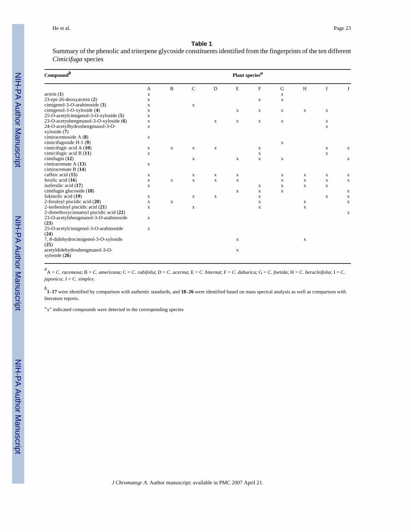
Table 1Summary of the phenolic and triterpene glycoside constituents identified from the fingerprints of the ten differentCimicifuga species
Compoundb Plant speciesa
A B C D E F G H I Jactein (1) x x23-epi-26-deoxyactein (2) x x xcimigenol-3-O-arabinoside (3) x xcimigenol-3-O-xyloside (4) x x x x x x25-O-acetylcimigenol-3-O-xyloside (5) x23-O-acetyshengmanol-3-O-xyloside (6) x x x x x x24-O-acetylhydroshengmanol-3-O-xyloside (7)
x x
cimiracemoside A (8) xcimicifugoside H-1 (9) xcimicifugic acid A (10) x x x x x x xcimicifugic acid B (11) x x xcimifugin (12) x x x x xcimiracemate A (13) xcimiracemate B (14)caffeic acid (15) x x x x x x x xferulic acid (16) x x x x x x x x xisoferulic acid (17) x x x x xcimifugin glucoside (18) x x x xfukinolic acid (19) x x x x x x2-feruloyl piscidic acid (20) x x x x x2-isoferuloyl piscidic acid (21) x x x x2-dimethoxycinnamyl piscidic acid (22) x23-O-acetylshengmanol-3-O-arabinoside(23)
x
25-O-acetylcimigenol-3-O-arabinoside(24)
x
7, 8-didehydrocimigenol-3-O-xyloside(25)
x x
acetyldidehydroshengmanol-3-O-xyloside (26)
x
aA = C. racemosa; B = C. americana; C = C. rubifolia; D = C. acerina; E = C. biternat; F = C. dahurica; G = C. foetida; H = C. heracleifolia; I = C.
japonica; J = C. simplex.
b1–17 were identified by comparison with authentic standards, and 18–26 were identified based on mass spectral analysis as well as comparison with
literature reports.
“x” indicated compounds were detected in the corresponding species
J Chromatogr A. Author manuscript; available in PMC 2007 April 21.
Related Documents