For Review Only Chronic Obstructive Pulmonary Disease: Clinical Review and Update on Consensus Guidelines Journal: Hospital Practice Manuscript ID: Draft Manuscript Type: Review Article Therapeutic Area: Respiratory, Chronic lung disease, Chronic obstructive pulmonary disease, Emphysema, Chronic bronchitis, Pulmonary, Diagnosis, Therapeutics Keywords – Click <a href="http://www.ncbi.nlm.nih.gov/mesh" target="_blank">here</a> to find your MeSH keywords.: Pulmonary Disease, Chronic Obstructive, Practice Guideline, Chronic bronchitis, Emphysema, COPD, Diagnosis, Therapeutics http://mc04.manuscriptcentral.com/hprac Hospital Practice

Welcome message from author
This document is posted to help you gain knowledge. Please leave a comment to let me know what you think about it! Share it to your friends and learn new things together.
Transcript

For Review O
nly
Chronic Obstructive Pulmonary Disease: Clinical Review and
Update on Consensus Guidelines
Journal: Hospital Practice
Manuscript ID: Draft
Manuscript Type: Review Article
Therapeutic Area: Respiratory, Chronic lung disease, Chronic obstructive pulmonary disease, Emphysema, Chronic bronchitis, Pulmonary, Diagnosis, Therapeutics
Keywords – Click <a href="http://www.ncbi.nlm.nih.gov/mesh" target="_blank">here</a> to find your
MeSH keywords.:
Pulmonary Disease, Chronic Obstructive, Practice Guideline, Chronic bronchitis, Emphysema, COPD, Diagnosis, Therapeutics
http://mc04.manuscriptcentral.com/hprac
Hospital Practice

For Review O
nly
1
Chronic Obstructive Pulmonary Disease: Clinical Review and Update on Consensus Guidelines
Abstract
In the past two decades, chronic obstructive pulmonary disease (COPD) has gained increasing
recognition as a major public health problem. Since the introduction of the Global Initiative for Chronic
Obstructive Lung Disease (GOLD) in 1998, increasing interest in the pathogenesis and management of
COPD has led to notable improvements in patient care and quality of life. Despite increasing recognition
surrounding the importance of this common preventable disease and the development of major
therapeutic advances during this period, the global impact of COPD is still strikingly large. We aim to
provide an evidence-based clinical review of the diagnosis, management, and treatment of COPD with a
focus on internists as the target audience. We will also summarize key updates from the GOLD executive
summary and joint clinical practice guideline on diagnosis and management of stable COPD from the
American College of Physicians (ACP), American College of Chest Physicians (ACCP), American
Thoracic Society (ATS), and European Respiratory Society (ERS).
Keywords: Chronic obstructive pulmonary disease; COPD; chronic bronchitis; practice guidelines;
emphysema; diagnosis; therapeutics; GOLD guidelines.
Page 1 of 29
http://mc04.manuscriptcentral.com/hprac
Hospital Practice
123456789101112131415161718192021222324252627282930313233343536373839404142434445464748495051525354555657585960
For Review O
nly
2
INTRODUCTION
Chronic obstructive pulmonary disease (COPD) places a significant socioeconomic burden on the
healthcare system. It is now projected to rank third in terms of leading causes of mortality by the year
2020.1,2
Despite major therapeutic advances in the management of COPD during the past two decades
the disease remains a major public health problem. The Global Initiative for Chronic Obstructive Lung
Disease (GOLD) was formed in 1998 in an effort to bring attention to the management and prevention of
this important disease. In February of 2013, GOLD released its second 5-year revision to its consensus
statement, Global Strategy for the Diagnosis, Management, and Prevention of COPD, originally published
in 2001.3,4
We aim to provide clinicians with a review of new updates to the 2013 GOLD executive
summary as well as the 2011 joint clinical practice guideline on diagnosis and management of stable
COPD from the ACP, ACCP, ATS, and ERS. The article will focus on internists as the target audience.
We also provide an evidence-based clinical review of COPD, highlighting the importance of inpatient and
outpatient management of this important disease.
MATERIALS AND METHODS
We performed a comprehensive literature search of the available evidence was performed by the authors
using PubMed, MEDLINE, and the Cochrane Database (English Language). Articles were critically
evaluated by the authors and/or were independently rated as high quality by an established evaluation
process, such as Cochrane Collaboration or cited by evidence-based consensus statements on COPD
from GOLD, ACP, ACCP, ATS, or ERS.
OVERVIEW
Definitions
COPD is a preventable and treatable condition characterized by progressive airflow limitation caused by
airway and/or parenchymal lung inflammation.5 Clinically, chronic bronchitis is defined as the presence of
chronic productive cough for at least three consecutive months in two consecutive years. Pathologically, it
is the end-result of obstructive bronchiolitis involving small airways caused by a chronic inflammatory
response to the exposure of noxious inhaled agents such as cigarette smoke, industrial fumes, and other
Page 2 of 29
http://mc04.manuscriptcentral.com/hprac
Hospital Practice
123456789101112131415161718192021222324252627282930313233343536373839404142434445464748495051525354555657585960

For Review O
nly
3
environmental pollutants. Emphysema refers to an abnormal enlargement of the airspaces distal to the
terminal bronchioles accompanied by destruction of their walls without obvious fibrosis visualized on
computed tomography or in pathologic analysis of lung specimens. GOLD defines COPD as a disease
state characterized by persistent airflow limitation that is usually progressive and associated with an
enhanced inflammatory response in the airways and the lung to noxious particles or gases.3
Risk Factors
Cigarette smoking remains the most common and important risk factor for the development of COPD.
Smoking has been shown to accelerate the rate of decline in lung function over time; however, individual
susceptibility to the harmful effects of smoking varies considerably from person to person. Clinically, lung
function is measured by use of spirometry and severity of obstructive lung disease is classified based on
forced expiratory volume in 1 second (FEV1) percent predicted. It remains unclear why some smokers
develop an exaggerated loss of FEV1 and others do not. It is postulated that differences in susceptibility to
tobacco smoke injury may be related to individual genetic differences.6 Major risk factors for the
development of COPD are summarized in Table 1.
Epidemiology and Natural History
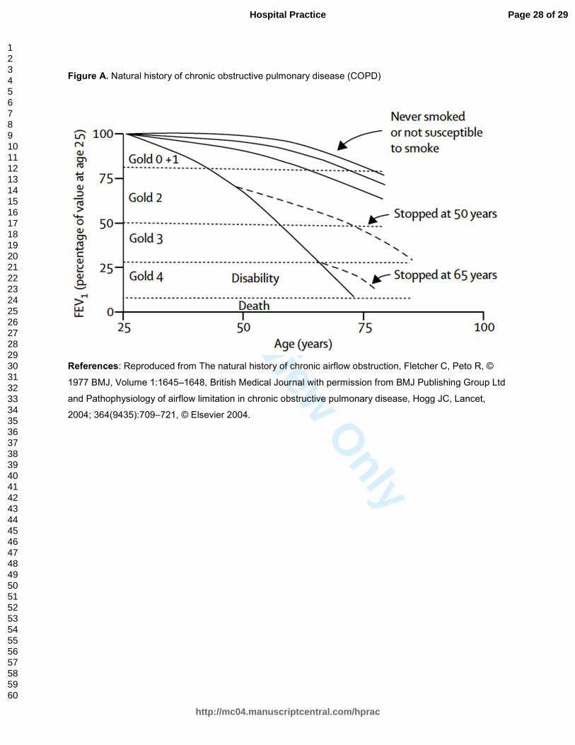
Data collected by Fletcher et al in the 1970s estimated that approximately 15% of smokers
eventually develop COPD (a figure corresponding with moderate disease by current GOLD staging).3,7
However, it is likely that 15% is an underestimate of those truly affected by the disease.8 This is
supported by population-based studies indicating that approximately 15 million Americans report a
diagnosis of COPD, but as many as 50% of those affected by the disease are unaware of it.9,10
Furthermore, COPD is an independent risk factor for cardiac mortality and many patients who are heavy
smokers may die as a result of complications related to atherosclerotic vascular disease, leaving the
diagnosis of COPD in these individuals largely unknown.11–13
In normal nonsmoking individuals, pulmonary function peaks in the early to mid-20s and gradually
declines in a predictable linear fashion over the course of a lifetime. After the age of 35 years old, the
average decline in FEV1 is approximately 25-30 mL/year. Decline in FEV1 is used as a marker for
Page 3 of 29
http://mc04.manuscriptcentral.com/hprac
Hospital Practice
123456789101112131415161718192021222324252627282930313233343536373839404142434445464748495051525354555657585960
For Review O
nly
4
progression of COPD and in active smokers loss of FEV1 occurs in a dose-dependent fashion with regard
to pack-years smoked, averaging approximately 50-55 mL/year, a rate nearly twice that seen in
nonsmokers.14,15
However, there is marked variation in the susceptibility of smokers with regard to loss of
lung function. Additionally, in most persons with a history of smoking fewer than 20 pack-years, there is
little, if any noticeable change in FEV1.13,16
Moreover, in persons with COPD that stop smoking, the rate of
decline in FEV1 returns to that seen in nonsmokers.17
A graphical comparison of the natural history of
pulmonary function in smokers and nonsmokers is depicted in Figure A.
Pathophysiology
Chronic inflammation in the walls and lumens of airways in response to repeated exposures to inhaled
particles—from tobacco smoke or other noxious gases—are the main underlying cause of the pathologic
changes, symptoms, complications, and pulmonary function abnormalities in COPD. The chronic
inflammatory response appears to be a pathologic enhancement of the normal, physiological,
inflammatory response of the respiratory tract to chronic irritants. This process is amplified in persons who
develop COPD for poorly understood reasons. Ultimately, the process leads to destruction of
parenchymal lung tissue and obstructive bronchiolitis resulting in emphysema and fibrotic narrowing of
small airways and a state of progressive obstructive airflow limitation and air trapping.3,6,18
Causes of Mortality
It was once thought that risk of death in patients with COPD was closely correlated with measured FEV1,
as reported by Fletcher and Peto’s landmark 1977 study.17
Since then, numerous other factors have been
identified as important causes of mortality in patients with COPD. Furthermore, the presence of certain
co-morbid medical conditions have been identified with increasing prevalence in those affected by COPD,
including coronary artery disease, lung cancer, osteoporosis, myopathy, anemia, and mood disorders, all
playing an important role in the natural course of the disease.19,20
Their presence adds increasing
complexity in the prediction of mortality among individual patients.
The Lung Health Study,21
sponsored by the National Heart, Lung, and Blood Institute (NHLBI)
served a prominent role in the identification of causes of mortality in persons affected by mild to moderate
Page 4 of 29
http://mc04.manuscriptcentral.com/hprac
Hospital Practice
123456789101112131415161718192021222324252627282930313233343536373839404142434445464748495051525354555657585960

For Review O
nly
5
COPD. The cohort included nearly 6,000 men and women who were cigarette smokers between 35 and
60 years of age and found the most common causes of mortality were cancer (33%), cardiovascular
disease (22%), and respiratory disease (8%). In patients with more severe lung disease, such as those
identified in the TOward a Revolution in COPD Health (TORCH) study, the leading cause of mortality was
respiratory illness (35%), followed by cardiovascular disease (27%), and cancer (21%).22
ASSESSMENT AND DIAGNOSIS
Clinical Manifestations
COPD is characterized by symptoms of dyspnea, chronic cough, and chronic sputum production in
persons with a history of exposure to risk factors, such as smoking cigarettes. Dyspnea is initially
intermittent and only with exertion, but later becomes persistent. Chronic cough can be intermittent,
productive, or non-productive. Any pattern of sputum production may be described.3
Several questionnaires are available to assess severity of symptoms. GOLD recommends use of
the Modified British Medical Research Council (MMRC) questionnaire on breathlessness23
or the COPD
Assessment Test (CAT)24
for this purpose.3 A high symptom burden is suggested by an MMRC grade ≥ 2
or a CAT score ≥ 10. The MMRC questionnaire on breathlessness categorizes severity of symptoms in
grades 0 to 4 based on answers to the following statements:
• MMRC Grade 0: I only get breathless with strenuous exercise;
• MMRC Grade 1: I get short of breath when hurrying on level ground or walking up a slight hill;
• MMRC Grade 2: I walk slower than people of the same age on level ground because of
breathlessness, or I have to stop for breath when walking on my own pace on level ground;
• MMRC Grade 3: I stop for breath after walking about 100 meters or after a few minutes on level
ground; and
• MMRC Grade 4: I am too breathless to leave the house or I am breathless when dressing or
undressing.
Page 5 of 29
http://mc04.manuscriptcentral.com/hprac
Hospital Practice
123456789101112131415161718192021222324252627282930313233343536373839404142434445464748495051525354555657585960
For Review O
nly
6
History and Examination
Patients with suspected COPD should have a detailed medical history and physical examination. History
of present illness should include review of current respiratory symptoms and their development over time.
It should also include assessment of physical activity level but clinicians are cautioned to be especially
aware of have gradually limited their activity level over time to compensate due to symptoms of dyspnea.
Medical history should focus on prior history of lung disease, exposure to risk factors, family history of
chronic respiratory disease or COPD, pattern of symptom development, prior history of hospitalizations
for exacerbations or other respiratory disorders, medical co-morbidities, and risk factor reduction such as
smoking cessation.3
Clinicians should also determine each patient’s cumulative number of pack-years smoked (packs
of cigarettes smoked per day multiplied by the number of years) when evaluating patients as the
magnitude of decline in FEV1 is dose-dependent.13
A reported history of smoking ≥ 70 pack-years of
cigarettes is the single most specific indicator of airway obstruction (positive likelihood ratio [LR] =
8.0).25,26
However, a combination of findings is more useful for diagnosing airflow obstruction than any
individual sign, symptom, or piece of historical information. However, the presence of two or more of the
following findings (history of smoking ≥ 70 pack-years, history of COPD, and/or the auscultation of
decreased breath sounds) is more useful for predicting airflow obstruction than any individual sign,
symptom, or piece of historical information (positive LR = 34).26
Moreover, absence of all three items
strongly suggests against underlying obstructive airway disease (negative LR = 0.02).26,27
A systematic review28
found the following historical elements to have the strongest independent
diagnostic value for detecting COPD: > 40 pack years of smoking (positive LR = 11.6); self-reported
history of COPD (positive LR = 4.4); wheezing (odds ratio [OR] = 4.4); and symptoms provoked by
allergens (OR = 4.5). Examination tests found to have strong independent diagnostic value include
prolonged expiration (OR = 3.7), forced expiratory time > 9 seconds (positive LR =4.6), and maximum
laryngeal height (the distance between the top of the thyroid cartilage and suprasternal notch) of < 4 cm
(positive LR = 3.6).28
Page 6 of 29
http://mc04.manuscriptcentral.com/hprac
Hospital Practice
123456789101112131415161718192021222324252627282930313233343536373839404142434445464748495051525354555657585960

For Review O
nly
7
The majority of characteristic exam findings associated with COPD are typically not seen until late
in the course of disease. This emphasizes the importance of obtaining spirometry for screening purposes
in smokers ≥ 40 years old with respiratory symptoms. Early diagnosis of COPD may result in improved
patients outcomes through lifestyle changes such as smoking cessation, by use of appropriate
pharmacological therapies, and implementation of preventative health practices such as immunizations
against influenza and pneumococcal infection.3
Ancillary Testing
GOLD recommends pulse oximetry should be used to assess all stable patients with FEV1 < 35% of
predicted or those with clinical signs suggestive of respiratory failure or right heart failure.3 In patients with
an oxygen saturation < 92%, arterial blood gas (ABG) measurement is suggested because pulse oximetry
loses sensitivity for detecting hypoxia below this cut-off.29
ABG measurement is useful in determining
need for chronic oxygen therapy, ascertaining the presence of hypercapnia, and classifying severity of
acute exacerbations and/or need for non-invasive or invasive ventilatory strategies during exacerbations.3
An electrocardiogram (ECG) can aid in the diagnosis of coexisting cardiac problems. Chest radiography
may show evidence of hyperinflation, flattening of the diaphragmatic contour, bullous disease, or
increased depth of the retrosternal air space.
Co-morbidities
Co-morbidities in patients with COPD should be routinely screened for and treated in the same manner
as in patients without COPD. Commonly encountered co-morbid medical conditions include depression
and anxiety, osteoporosis, cardiovascular disease, diabetes, and lung cancer.3 Osteoporosis is present in
up to 60% of patients with advanced COPD.30
Accelerated bone loss is associated with smoking, use of
glucocorticoids, vitamin D deficiency, low body mass index, hypogonadism, and sedentary lifestyle.
Recognition and screening for the presence of underlying bone disease in patients with COPD is
important because many patients remain undiagnosed until their first fracture. Management involves
vitamin D and calcium supplementation and bisphosphonates when indicated.30
Page 7 of 29
http://mc04.manuscriptcentral.com/hprac
Hospital Practice
123456789101112131415161718192021222324252627282930313233343536373839404142434445464748495051525354555657585960
For Review O
nly
8
Alpha 1-Antitrypsin Deficiency
COPD is the most prevalent clinical disorder associated with alpha 1-antitrypsin (AAT) deficiency, an
inherited disorder characterized by panacinar basilar emphysema, often presenting at a young age (< 45
years old). Up to 3% of patients with COPD have AAT deficiency and among Caucasians in North
America, AAT deficiency is as common as cystic fibrosis.31
Approximately 95% of patients with severe
AAT deficiency are homozygotes for the protease inhibitor (PI), type ZZ allele (designated PI*ZZ).
Cigarette smoking is the most significant risk factor for the development of emphysema in patients with
AAT deficiency, usually developing by the third or fourth decade of life. However, in homozygotes who
are nonsmokers, few develop clinically significant lung disease and the disorder often goes clinically
unrecognized.32
The World Health Organization (WHO) recommends screening for the disorder in
patients with COPD from areas with a high prevalence of AAT deficiency (particularly Caucasians of
Northern European ancestry) using a quantitative test. Serum levels of AAT in PI*ZZ homozygotes is
approximately 15% of normal (30-40 mg/dL—compared with normal serum values of 150-350 mg/dL).
Those with abnormal results on screening should undergo PI typing and if positive should be offered
genetic counseling.33
Spirometry
Post-bronchodilator spirometry is recommended for the diagnosis of COPD in patients with respiratory
symptoms and a history of exposure to risk factors by GOLD and the ACP, ACCP, ATS, and ERS joint
practice guideline.3,26
The presence of a post-bronchodilator FEV1/FVC ratio < 0.70 is characteristic of
obstructive airway disease. The ACP, ACCP, ATS, and ERS joint practice guideline recommend against
use of spirometry to screen for airflow obstruction in individuals without respiratory symptoms.26
GOLD
recommends that spirometry should be performed in individuals > 40 years of age with respiratory
symptoms suggestive of COPD. Spirometry should be repeated annually in patients diagnosed with
COPD.3
Measurements of lung volumes and diffusing capacity are not essential to patient management,
but may help to identify air trapping or concomitant restrictive lung disease and characterize the severity
of alveolar destruction. Early in the course of disease, patients with COPD exhibit gas trapping, reflected
Page 8 of 29
http://mc04.manuscriptcentral.com/hprac
Hospital Practice
123456789101112131415161718192021222324252627282930313233343536373839404142434445464748495051525354555657585960
For Review O
nly
9
by an increased residual volume. As airflow limitation progresses, static hyperinflation occurs, reflected by
an increase in total lung capacity. Destruction of lung parenchyma, as occurs in emphysema, may be
reflected by a reduced diffusing capacity of carbon monoxide.
Classifying Severity of Disease
In patients with a post-bronchodilator FEV1/FVC ratio of < 0.70, the degree of airflow obstruction in COPD
is determined by spirometric measurement of post-bronchodilator FEV1. Accordingly, GOLD organizes
patients into the following stages of severity:3
• GOLD 1 (Mild): FEV1 ≥ 80% predicted;
• GOLD 2 (Moderate): FEV1 50-79% predicted;
• GOLD 3 (Severe): FEV1 30-49% predicted; or,
• GOLD 4 (Very Severe): FEV1 < 30% predicted.
Although it is important to determine the magnitude of reduction in FEV1 to determine disease
severity, other disease severity indices such as the BODE index (Table 2) may be more reliable
predictors of hospitalization from COPD than FEV1 alone.34,35
Recognizing that worsening airflow
limitation alone is not the most reliable means of assessing disease severity, a major revision was
introduced in the updated GOLD guideline, recommending use of a novel tool, the combined COPD
assessment (Figure B), which utilizes an MMRC grade ≥ 2 or a CAT score ≥ 10 as an indication of
severe symptom burden.3 Categorization of patients in this manner allows practitioners to stratify
individual patients according to risk of exacerbations and apply recommended therapeutic strategies for
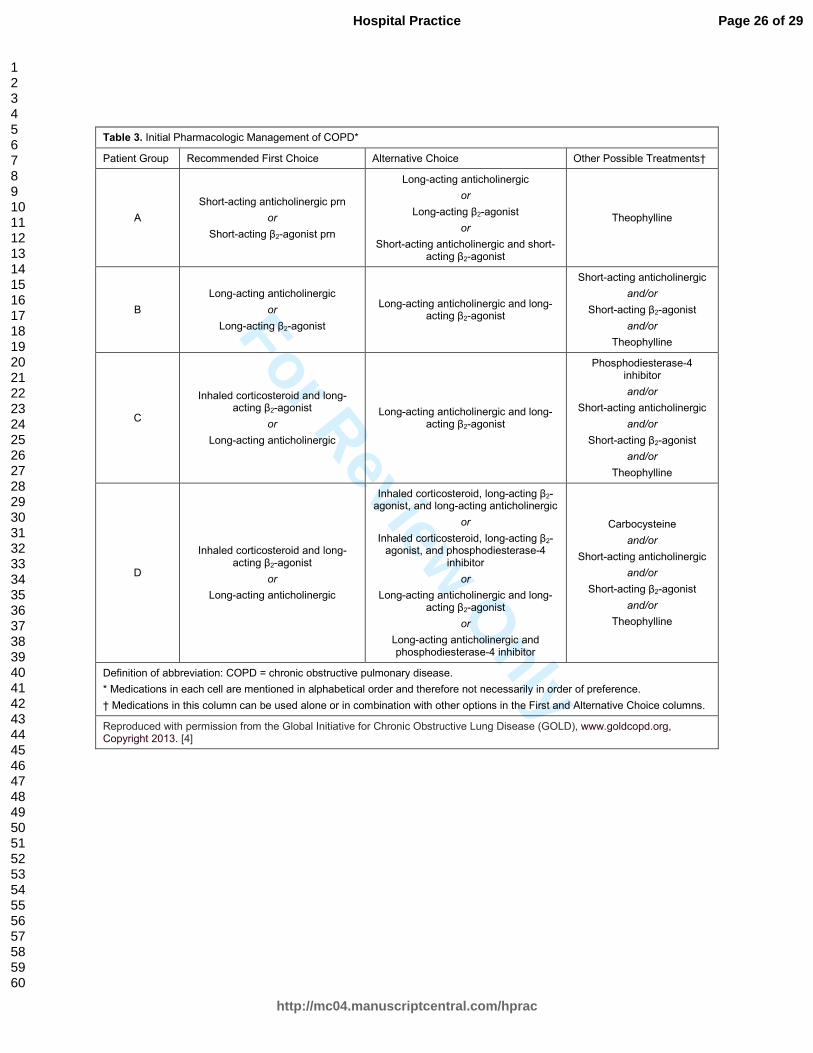
each patient group, as recommended by GOLD in Table 3. According to GOLD,3 in patients where there
is a discrepancy between risk category assessed by spirometry and that derived from exacerbation
history and symptoms based on the combined COPD assessment, the highest risk group should be
chosen. Based on the combined COPD assessment, groups can be organized into the following
categories:
• Patient group A—low risk, less symptoms: GOLD 1-2 (mild or moderate airflow limitation) and
0-1 exacerbations per year and MMRC grade 0-1 or CAT score < 10.
Page 9 of 29
http://mc04.manuscriptcentral.com/hprac
Hospital Practice
123456789101112131415161718192021222324252627282930313233343536373839404142434445464748495051525354555657585960

For Review O
nly
10
• Patient group B—low risk, more symptoms: GOLD 1-2 (mild or moderate airflow limitation)
and 0-1 exacerbations per year and MMRC grade ≥ 2 or CAT score ≥10.
• Patient group C—high risk, less symptoms: GOLD 3-4 (severe or very severe airflow
limitation) and/or ≥ 2 exacerbations per year and/or ≥ 1 hospitalized exacerbations per year and
MMRC grade 0-1 or CAT score < 10.
• Patient group D—high risk, more symptoms: GOLD 3-4 (severe or very severe airflow
limitation) and/or ≥ 2 exacerbations per year and/or ≥ 1 hospitalized exacerbations per year and
MMRC grade ≥ 2 or CAT score ≥ 10.
MANAGEMENT
Stable COPD
GOLD now recommends that management strategy of stable COPD should consider individualized
symptoms and future risk for exacerbation in addition to FEV1, as FEV1 alone is a poor descriptor of
disease status.3 Use of the new Combined COPD Assessment tool from GOLD can be used for this
purpose (Figure B). Treatment of stable COPD should be largely focused at reducing symptoms in an
effort to improve quality of life, minimizing risk factors, and preventing exacerbations. Interventions shown
to be effective at reducing exacerbations include, evaluating knowledge of current therapy, such as
proper inhaler technique, smoking cessation, annual influenza vaccination, pulmonary rehabilitation, and
maintenance medications including long-acting bronchodilators (long-acting beta agonists [LABAs] or
long-acting anticholinergics such as tiotropium), inhaled corticosteroids (ICS), and phosphodiesterase
type-4 inhibitors (roflumilast).3
Inhaled Bronchodilators
Inhaled bronchodilators are preferred over oral bronchodilators and long-acting formulations are preferred
over short-acting formulations for both anticholinergics and β2-agonists.3 Long-acting bronchodilators
including LABAs and/or long-acting anticholinergics (tiotropium) are recommended in patients with
moderate to very severe disease. LABAs can be used alone or in combination with ICS. Use of LABAs as
monotherapy has been shown to reduce frequency in exacerbations and slow decline in FEV1.22
Page 10 of 29
http://mc04.manuscriptcentral.com/hprac
Hospital Practice
123456789101112131415161718192021222324252627282930313233343536373839404142434445464748495051525354555657585960

For Review O
nly
11
In patients with moderate to very severe disease, addition of tiotropium (18 mcg inhaled once
daily) to standard medications used to treat COPD (excluding the short-acting anticholinergic
ipratropium), has been shown to reduce the risk of exacerbations, related hospitalizations, and respiratory
failure.36
When used as monotherapy, tiotropium is superior to ipratropium.37
The ACP, ACCP, ATS, and
ERS joint guideline recommends treatment with inhaled bronchodilators for stable COPD patients with
respiratory symptoms and FEV1 < 60% predicted.26
They also recommend that monotherapy using either
long-acting inhaled anticholinergics or inhaled LABAs should be prescribed to symptomatic patients with
COPD and FEV1 < 60% predicted with choice of specific monotherapy based on patient preference, cost,
and adverse effect profile.26
Inhaled Corticosteroids
Use of inhaled corticosteroids as maintenance therapy in COPD has been shown to reduce the frequency
of exacerbations in patients with an FEV1 < 50% of predicted. Their use may also slow decline in FEV1
when used alone, but more so when used in combination with a long-acting β2-agonist. Unlike asthma
therapy, long-term monotherapy with ICS is not recommended in patients with COPD because it is less
effective than the combination of ICS with LABAs.3 Despite the benefits seen with ICS use in COPD
management, their use (alone or in combination with LABA) has been linked to an increased risk of
pneumonia.38
GOLD recommends that long-term treatment with ICS is recommended for patients with
FEV1 < 50% of predicted and/or frequent exacerbations that are not adequately controlled by long-acting
bronchodilators.3 The ACP, ACCP, ATS, ERS joint guideline suggests administering combination inhaled
therapies (long-acting inhaled anticholinergics, LABAs, or ICS) for symptomatic patients with stable
COPD and FEV1 < 60% predicted.26
Continuous Oxygen
Supplemental oxygen therapy has been shown to be improve survival when used for ≥ 15 hours daily to
maintain an arterial PaO2 > 60 mm Hg in patients with COPD who have severe resting hypoxemia (mean
resting arterial PaO2 ≤ 55 mm Hg) or arterial oxygen saturation ≤ 88%.39,40
Continuous oxygen therapy is
recommended by both guidelines for patients with COPD with a resting PaO2 ≤ 55 mm Hg or SaO2 ≤ 88%
Page 11 of 29
http://mc04.manuscriptcentral.com/hprac
Hospital Practice
123456789101112131415161718192021222324252627282930313233343536373839404142434445464748495051525354555657585960
For Review O
nly
12
(during rest, exercise, or sleep).3,26
Long-term continuous oxygen therapy should also be considered for
patients with a resting PaO2 of 56 to 59 mm Hg and evidence of right heart failure (cor pulmonale) and/or
secondary polycythemia (hematocrit > 55%).3
Roflumilast
Addition of roflumilast (a type-4 phosphodiesterase inhibitor) to standard therapy can be considered in
patients with an FEV1 < 50% predicted, chronic bronchitis, and frequent exacerbations (two or more per
year) that are not adequately controlled by long-acting bronchodilators.3 In this patient population, it has
been shown to reduce frequency of exacerbations.41,42
Common adverse reactions (≥ 2%) associated
with its use include nausea, diarrhea, weight loss, and psychiatric disturbance (including suicidal ideation
and behavior).43
Because some patients develop significant weight loss with its use, it is recommended
that weight be monitored regularly with its use. Its use is contraindicated in patients with moderate to
severe liver impairment.43
Smoking Cessation
Smoking cessation remains the most important preventative strategy in the management of COPD. In
addition to improving respiratory symptoms, smoking cessation has been shown to slow disease
progression, as evidenced by a decrease in the rate of decline in the FEV1, and decrease long-term
mortality in patients with COPD.15,21,44
Patients should be counseled on smoking cessation at regular
intervals during follow up visits.
Nicotine replacement therapy in any form (e.g., nicotine gum, inhaler, nasal spray, sublingual
tablet, lozenge, or transdermal patch) has been shown to increase long-term smoking cessation rates.45–
47 Other pharmacologic options that have been shown to improve long-term smoking cessation rates
include varenicline, bupropion, and nortriptyline.48–50
Many patients are anxious to try cigarette
substitutes, or “electronic cigarettes”, but safety of the propellant has not been established, though
presumably it is safer than the known toxins of inhaled tobacco smoke.51
Page 12 of 29
http://mc04.manuscriptcentral.com/hprac
Hospital Practice
123456789101112131415161718192021222324252627282930313233343536373839404142434445464748495051525354555657585960
For Review O
nly
13
Vaccinations
Annual influenza vaccination is recommended in all persons age 50 or older and those with chronic
pulmonary disease (including COPD and asthma).52
Annual vaccination against influenza can reduce the
risk of serious lower respiratory tract infections and death in patients with COPD.3 Pneumococcal
vaccination is recommended for all persons 65 years of age or older, all persons with COPD, and all
active smokers. Vaccination decreases risk of serious invasive disease with the pathogen Streptococcus
pneumoniae.53
Pulmonary Rehabilitation
Maintenance of physical activity and pulmonary rehabilitation is clinically useful in all patients with
symptomatic COPD and after exacerbations. Exercise rehabilitation has been shown to improve exercise
tolerance, decrease symptoms of dyspnea and fatigue, and improve quality of life in patients with
COPD.54,55
Regular physical activity is recommended for all patients with COPD.3 The ACP, ACCP, ATS,
ERS joint guideline recommends that pulmonary rehabilitation be prescribed for symptomatic patients
with an FEV1 < 50% predicted and considered for symptomatic or exercise-limited patients with an FEV1
> 50% predicted.26
Perioperative Risk Assessment
Increased severity of postoperative complications in COPD may vary with underlying baseline disease
severity; however, the most important predictor of outcome is proximity of the surgical site to the
diaphragm. The closer the incision is to the diaphragm, the higher the risk. Most reports conclude that
epidural or spinal anesthesia have lower risk in comparison to general anesthesia, although results are
not uniform. Postoperative complications include lung infections, atelectasis, and worsened airflow
limitation that may lead to respiratory failure.3
Acute COPD Exacerbations
An exacerbation of COPD is defined as an acute event characterized by a worsening of the patient’s
respiratory symptoms that is beyond normal day-to-day variations requiring a change in medication.56–58
Page 13 of 29
http://mc04.manuscriptcentral.com/hprac
Hospital Practice
123456789101112131415161718192021222324252627282930313233343536373839404142434445464748495051525354555657585960
For Review O
nly
14
Nearly 50% of patients with COPD experience at least one exacerbation annually.59
The hallmark of
exacerbations is a change in the character and/or severity in one or more of the three cardinal symptoms
of COPD—dyspnea, chronic cough, and/or sputum production. Exacerbations have numerous
precipitating causes but are most commonly related to viral and/or bacterial respiratory tract infections.3,5
Frequent COPD exacerbations are defined as ≥ 2 exacerbations per year and are seen in
approximately 10% of patients with COPD.59
Although frequent exacerbations are more common with
increasing severity of the disease, a considerable number of patients with moderate disease (i.e., FEV1 >
50% predicted, GOLD stage 2) are affected by frequent exacerbations.59
The single best predictor for risk
of frequent exacerbations, across all GOLD stages of COPD, is a history of previously treated
exacerbations.59
Patients should be considered at high risk for exacerbations if they meet GOLD 3 or
GOLD 4 (severe to very severe disease) severity airflow obstruction, or if they have a history of ≥ 2
exacerbations in the preceding year.3
Exacerbations play a significant role in the course of disease and are associated with significant
mortality. It may take several weeks or even months following an exacerbation for patients to improve to
baseline from a symptomatic and pulmonary function standpoint.3 The in-hospital mortality is
approximately 10% for patients admitted with exacerbations complicated by acidosis and hypercapnic
respiratory failure. In-hospital mortality rates are as high as 20% among those admitted to the ICU.60,61
Exacerbations of COPD are commonly managed with a combination of inhaled short-acting
bronchodilators, systemic corticosteroids, and antibiotics. Severity of exacerbations is usually classified
as mild when respiratory symptoms require change of inhaled treatment by the patient, moderate when
respiratory symptoms require medical interventions such as antibiotics and/or oral corticosteroids, and
severe when respiratory symptoms require hospitalization for management.3
Potential indications for hospital admission for management of exacerbation include severe
baseline underlying disease, onset of new signs suggestive of severe disease, frequent exacerbations,
older age, insufficient home support, failure to respond to outpatient medical management, and presence
of serious co-morbidities such as heart failure or arrhythmias.3 Signs suggestive of severe exacerbations
include use of accessory respiratory muscles, paradoxical chest wall movements, worsening or new onset
central cyanosis, development of peripheral edema, hemodynamic instability, and altered mental status.3
Page 14 of 29
http://mc04.manuscriptcentral.com/hprac
Hospital Practice
123456789101112131415161718192021222324252627282930313233343536373839404142434445464748495051525354555657585960

For Review O
nly
15
Indications for ICU admission include hemodynamic instability, need for invasive mechanical ventilation,
changes in mental status (e.g., confusion, lethargy, coma), persistent or worsening hypoxemia (arterial
PaO2 < 40 mm Hg) and/or severe or worsening respiratory acidosis (pH < 7.25) despite supplemental
oxygen and NIV.3
Systemic Corticosteroids
The beneficial effects of corticosteroid use for the treatment of exacerbations was established in 1999
with the Department of Veterans Affairs Cooperative Study Group Trial.62
Beneficial effects of
corticosteroids include a reduction in exacerbation treatment failure, shorter hospital stays, improvement
in lung function and oxygenation, and shortened recovery times.33,63,64
Although their role in COPD
management remains important, corticosteroids are associated with significant adverse effects including
hyperglycemia, myopathy, adrenal suppression, cataract formation, peptic ulcer disease, psychosis,
hyperadrenocorticism, increased risk of infection, and osteoporosis.65
Thus, duration and dosage required
for an adequate clinical response is of clinical importance but remains a matter of debate.
GOLD recommends prednisone (or equivalent) 40 mg/day by mouth for 10-14 days during
exacerbations.3 The REDuction in the Use of Corticosteroids in Exacerbated COPD (REDUCE)
66 trial
found a short-term (5 days) course of corticosteroids non-inferior to conventional (14 days) treatment. The
main outcome measure of the trial was time to next exacerbation within 180 days, which was similar in
both groups. The REDUCE trial provides strong evidence supporting use of a 5-day course of
corticosteroids (irrespective of disease severity) for treatment of exacerbations.66
In most circumstances,
corticosteroids used for a duration of < 2 weeks can be discontinued safely without tapering.62
Antibiotics
Although conflicting data exists regarding the role of antibiotics in exacerbations, antibiotics are often
utilized empirically, even in the absence of supportive data implicating a bacterial pathogen.67
The most
common bacterial pathogens implicated in exacerbations include Streptococcus pneumoniae,
Haemophilus influenzae, and Moraxella catarrhalis.68
In patients with more severe disease (GOLD 3 and
GOLD 4), Pseudomonas aeruginosa can be implicated as a causative pathogen in exacerbations.3
Page 15 of 29
http://mc04.manuscriptcentral.com/hprac
Hospital Practice
123456789101112131415161718192021222324252627282930313233343536373839404142434445464748495051525354555657585960

For Review O
nly
16
GOLD recommends that antibiotics should be given to patients with COPD exacerbations who
have three cardinal symptoms (increase in dyspnea, sputum volume, and sputum purulence); patients
with two of the cardinal symptoms, if increased purulence of sputum is present; or those who require
invasive or noninvasive mechanical ventilation.3 Addition of antibiotics (e.g., a respiratory quinolone) to
standard treatment in patients with severe exacerbations requiring need for mechanical ventilation
(noninvasive or invasive) has been shown to reduce the risk of inhospital death, shorten the duration of
mechanical ventilation and hospital stay, and reduce the need for additional courses of antibiotics.69
The
recommended duration of antibiotics is 5-10 days and selection should be based on local bacterial
resistance patterns.3 Evidence supports a shorter antibiotic course (≤ 5 days) in patients with mild to
moderate exacerbations.70
Inhaled Bronchodilators
Short-acting bronchodilators play a key role in symptom management in patients with exacerbations.
Their use is associated with acute improvements in FEV1, but have little effect on FEV1 with chronic use.
Commonly utilized short-acting bronchodilators include albuterol sulfate, a short-acting β2-agonist
(SABA), and the short-acting anticholinergic ipratropium bromide. Both provide acute symptomatic
improvement with their use. With long-term use, there is no significant difference in outcomes when
comparing metered dose inhalers with nebulizers. However, both are commonly administered via
nebulized preparations during exacerbations. No studies have evaluated the use of inhaled long-acting
bronchodilators with/without ICS during exacerbations.3
SABAs produce bronchodilation primarily due to their ability to relax airway smooth muscle.
Systemic absorption of the drug can cause unwanted side effects, most commonly tachycardia, and
palpitations.71
Their short half-lives limits their use in the chronic treatment of COPD, though they are
commonly used as rescue medications during acute exacerbations. Controversy exists regarding optimal
dosing of inhaled SABA, and ideally they should be utilized at the lowest dose and frequency required to
achieve clinical response.71
Anticholinergics such as ipratropium bromide also work by inhibiting
bronchoconstriction resulting in symptomatic improvement in addition to reducing mucus secretion. The
two drugs can be used alone or in combination, and produce similar effects in terms of bronchodilation
Page 16 of 29
http://mc04.manuscriptcentral.com/hprac
Hospital Practice
123456789101112131415161718192021222324252627282930313233343536373839404142434445464748495051525354555657585960

For Review O
nly
17
and combination of SABA and ipratropium do not appear to increase the effect on FEV1 more than either
used alone.72
Noninvasive Ventilation
Acute hypercapnic respiratory failure in the setting of COPD exacerbation is the most common indication
for noninvasive positive-pressure ventilation (NIPPV).73
Use of NIPPV has been shown to improve blood
gas exchange, reduce the need for intubation, and decrease mortality when properly applied as a first line
treatment for respiratory failure resulting complicating COPD exacerbations. Its use has also been shown
to decrease length of hospital stay, reduce ventilator-induced lung injury, and lower rates of nosocomial
infections (e.g., ventilator associated pneumonia).74,75
The ideal candidate for NIPPV is an alert patient
with increased work of breathing, tachypnea, and moderate respiratory acidosis (pH 7.10–7.35).74
Indications for initiation of NIPPV in patients with hypercapnic respiratory failure include one or
more of the following: respiratory acidosis (arterial pH ≤ 7.35 and/or PaCO2 ≥ 45 mm Hg) or severe
dyspnea with signs of respiratory muscle fatigue and/or increased work of breathing (e.g., use of
accessory respiratory muscles, paradoxical abdominal motion, or intercostal muscle retraction).3,73–77
Contraindications to NIV include altered mental status, nausea/vomiting, upper gastrointestinal
hemorrhage or recent abdominal surgery, hemodynamic instability, and inability to clear secretions.76
Proper NIPPV settings and early institution of the therapy is of utmost importance in order to
achieve successful outcomes with its use. We suggest use of Bi-level Positive Airway Pressure (BiPAP)
mode with the following initial settings: Inspiratory Positive Airway Pressure (IPAP) at 8 cm H2O,
Expiratory Positive Airway Pressure at 4 cm H2O, Fraction of inspired oxygen (FiO2) set at 60% and
titrated to obtain a goal oxygen saturation of 88-92%. IPAP should be titrated upwards in 2 cm H2O
increments at the bedside in order to obtain a goal tidal volume of 6-8 mL/kg of ideal body weight (IBW).
Invasive Mechanical Ventilation
Indications for invasive mechanical ventilation (IMV) include: failure of an initial trial of NIV; respiratory or
cardiac arrest; massive aspiration; diminished or loss of consciousness; respiratory pauses with loss of
consciousness; psychomotor agitation inadequately controlled by sedation; inability to remove/tolerate
Page 17 of 29
http://mc04.manuscriptcentral.com/hprac
Hospital Practice
123456789101112131415161718192021222324252627282930313233343536373839404142434445464748495051525354555657585960

For Review O
nly
18
secretions; hemodynamic instability; severe ventricular arrhythmias; and life-threatening hypoxemia in
patients unable to tolerate NIV.3,78
Major complications of IMV in patients with COPD include development of barotrauma, failure to
wean to spontaneous ventilation, and risk of ventilator-acquired pneumonia.3 When IMV is required for
the management of respiratory failure complicating COPD exacerbations, the most appropriate setting is
in the intensive care unit in consultation with appropriately trained specialists. We recommend intubation
with a relatively large endotracheal tube (internal diameter 7.5-8.5 mm) and use assist-control mode with
the following initial ventilator settings: delivered tidal volume of 8 mL/kg of IBW; respiratory rate 10-14
breaths per minute; FiO2 of 35% (titrated to maintain a goal oxygen saturation of 88-92%); positive end-
expiratory pressure (PEEP) of 5 cm H2O. Moderate sedation may be achieved with agents such as
propofol, fentanyl, or midazolam, with scheduled withdrawal of sedatives on a daily basis. The use of
paralytics should be avoided.78
In all patients with severe exacerbations requiring NIV or IMV, ABG measurements, chest
radiographs, fluid balance, and nutritional status should be monitored closely. Usual therapy for
exacerbations should be continued, including as systemic corticosteroids, antibiotics, and inhaled
bronchodilators. Additionally, co-morbid medical conditions (e.g., heart failure, arrhythmias, infections)
should the same as in patients without COPD. Venous thromboembolism prophylaxis should be utilized in
all patients without contraindications and the head of the bed should be kept elevated at ≥ 45 degrees at
all times to minimize the risk of aspiration.3,78
Weaning and discontinuation of ventilatory support should
take place via a standardized protocol.
Hospital Discharge and Outpatient Follow Up
Prior to discharge, patients should be started on long-acting bronchodilators (anticholinergics and/or β2-
agonists), with or without ICS. Inhaler technique should be reassessed to ensure proper use. Patients
should be educated about their maintenance regiment and given instructions regarding completion of
steroid therapy and antibiotics, if prescribed. Counseling on smoking cessation should also take place
prior to discharge, and routinely at all follow up visits. Pulse oximetry or ABG measurements should be
utilized to assess need for long-term oxygen therapy. Patients should also be screened for need of a
Page 18 of 29
http://mc04.manuscriptcentral.com/hprac
Hospital Practice
123456789101112131415161718192021222324252627282930313233343536373839404142434445464748495051525354555657585960

For Review O
nly
19
home nebulizer.3 Early outpatient pulmonary rehabilitation after hospitalization for exacerbations should
also be considered. Its use is safe and has been shown to result in clinically significant improvements in
exercise capacity and health status at 3 months.79
Finally, vaccination status should be updated and a
plan for management and follow up of co-morbidities should be arranged.3
Patients hospitalized for exacerbations should be seen in the outpatient setting four to six weeks
after discharge. Outpatient visits should focus on smoking cessation counseling, proper inhaler
techniques, symptoms, and status of co-morbidities. The MMRC or CAT can be utilized to re-assess
symptom severity. Spirometry should be utilized for measurement of post-bronchodilator FEV1. Patients
should be questioned about their ability to cope in their normal living environment, current exercise
capacity, and physical ability to perform activities of daily living. Screening for need of long-term oxygen
therapy and/or a home nebulizer should also take place.3
CONCLUSION
COPD remains an important preventable cause of death and places a significant socioeconomic burden
on the healthcare system. Management of the disease is complex, often involving a multidisciplinary
approach to achieve optimal outcomes. Patients often have multiple co-morbid medical conditions that
impact mortality and pose a significant challenge to practitioners managing the disease. A new GOLD
combined COPD assessment tool has been introduced, which serves to help practitioners in the
assessment of disease severity, estimate risk of future exacerbations, and guide selection of appropriate
pharmacotherapy. GOLD now requires use of spirometry and characteristic findings in the diagnosis of
COPD. Despite the introduction of multiple new drug therapies for COPD in the past two decades,
smoking cessation remains the single most important method of reducing mortality.
REFERENCES
1. Halbert RJ, Natoli JL, Gano A, Badamgarav E, Buist AS, Mannino DM. Global burden of COPD: systematic review and meta-analysis. Eur Respir J. 2006;28(3):523–532. doi:10.1183/09031936.06.00124605.
2. World Health Organization (WHO). Burden of COPD. World Heal Organ Who. Available at: http://www.who.int/respiratory/copd/burden/en/index.html. Accessed June 21, 2013.
3. Vestbo J, Hurd SS, Agustí AG, et al. Global strategy for the diagnosis, management, and prevention of chronic obstructive pulmonary disease: GOLD executive summary. Am J Respir Crit Care Med. 2013;187(4):347–365. doi:10.1164/rccm.201204-0596PP.
Page 19 of 29
http://mc04.manuscriptcentral.com/hprac
Hospital Practice
123456789101112131415161718192021222324252627282930313233343536373839404142434445464748495051525354555657585960

For Review O
nly
20
4. Pauwels RA, Buist AS, Calverley PM, Jenkins CR, Hurd SS, GOLD Scientific Committee. Global strategy for the diagnosis, management, and prevention of chronic obstructive pulmonary disease. NHLBI/WHO Global Initiative for Chronic Obstructive Lung Disease (GOLD) Workshop summary. Am J Respir Crit Care Med. 2001;163(5):1256–1276. doi:10.1164/ajrccm.163.5.2101039.
5. Barnes PJ. Chronic Obstructive Pulmonary Disease. N Engl J Med. 2000;343(4):269–280. doi:10.1056/NEJM200007273430407.
6. Kohansal R, Martinez-Camblor P, Agustí A, Buist AS, Mannino DM, Soriano JB. The natural history of chronic airflow obstruction revisited: an analysis of the Framingham offspring cohort. Am J Respir Crit Care Med. 2009;180(1):3–10. doi:10.1164/rccm.200901-0047OC.
7. Fletcher C, Peto R, Tinker C, Speizer F. The natural history of chronic bronchitis and emphysema. Oxford: Oxford University Press; 1976.
8. Rennard SI, Vestbo J. COPD: the dangerous underestimate of 15%. Lancet. 2006;367(9518):1216–1219. doi:10.1016/S0140-6736(06)68516-4.
9. Mannino DM, Gagnon RC, Petty TL, Lydick E. Obstructive lung disease and low lung function in adults in the United States: data from the National Health and Nutrition Examination Survey, 1988-1994. Arch Intern Med. 2000;160(11):1683–1689.
10. Chronic Obstructive Pulmonary Disease Among Adults — United States, 2011. Available at: http://www.cdc.gov/mmwr/preview/mmwrhtml/mm6146a2.htm. Accessed July 2, 2013.
11. Mannino D, Buist A, Petty T, Enright P, Redd S. Lung function and mortality in the United States: data from the First National Health and Nutrition Examination Survey follow up study. Thorax. 2003;58(5):388–393. doi:10.1136/thorax.58.5.388.
12. Lange P, Nyboe J, Jensen G, Schnohr P, Appleyard M. Ventilatory function impairment and risk of cardiovascular death and of fatal or non-fatal myocardial infarction. Eur Respir J. 1991;4(9):1080–1087.
13. Ashley F, Kannel WB, Sorlie PD, Masson R. Pulmonary function: relation to aging, cigarette habit, and mortality. Ann Intern Med. 1975;82(6):739–745.
14. Celli BR, Thomas NE, Anderson JA, et al. Effect of pharmacotherapy on rate of decline of lung function in chronic obstructive pulmonary disease: results from the TORCH study. Am J Respir Crit Care Med. 2008;178(4):332–338. doi:10.1164/rccm.200712-1869OC.
15. Anthonisen NR, Connett JE, Murray RP. Smoking and Lung Function of Lung Health Study Participants after 11 Years. Am J Respir Crit Care Med. 2002;166(5):675–679. doi:10.1164/rccm.2112096.
16. Kuperman AS, Riker JB. The variable effect of smoking on pulmonary function. Chest J. 1973;63(5):655–660. doi:10.1378/chest.63.5.655.
17. Fletcher C, Peto R. The natural history of chronic airflow obstruction. Br Med J. 1977;1(6077):1645–1648.
18. Hogg JC. Pathophysiology of airflow limitation in chronic obstructive pulmonary disease. Lancet. 2004;364(9435):709–721. doi:10.1016/S0140-6736(04)16900-6.
19. Barnes PJ, Celli BR. Systemic manifestations and comorbidities of COPD. Eur Respir J. 2009;33(5):1165–1185. doi:10.1183/09031936.00128008.
20. Celli BR. Predictors of mortality in COPD. Respir Med. 2010;104(6):773–779. doi:10.1016/j.rmed.2009.12.017.
21. Anthonisen NR, Skeans MA, Wise RA, et al. The effects of a smoking cessation intervention on 14.5-year mortality: a randomized clinical trial. Ann Intern Med. 2005;142(4):233–239.
22. Calverley PMA, Anderson JA, Celli B, et al. Salmeterol and Fluticasone Propionate and Survival in Chronic Obstructive Pulmonary Disease. N Engl J Med. 2007;356(8):775–789. doi:10.1056/NEJMoa063070.
23. Stenton C. The MRC breathlessness scale. Occup Med. 2008;58(3):226–227. doi:10.1093/occmed/kqm162.
24. Jones PW, Harding G, Berry P, Wiklund I, Chen W-H, Leidy NK. Development and first validation of the COPD Assessment Test. Eur Respir J. 2009;34(3):648–654. doi:10.1183/09031936.00102509.
Page 20 of 29
http://mc04.manuscriptcentral.com/hprac
Hospital Practice
123456789101112131415161718192021222324252627282930313233343536373839404142434445464748495051525354555657585960

For Review O
nly
21
25. Badgett RG, Tanaka DJ, Hunt DK, et al. Can moderate chronic obstructive pulmonary disease be diagnosed by historical and physical findings alone? Am J Med. 1993;94(2):188–196.
26. Qaseem A, Wilt TJ, Weinberger SE, et al. Diagnosis and management of stable chronic obstructive pulmonary disease: a clinical practice guideline update from the American College of Physicians, American College of Chest Physicians, American Thoracic Society, and European Respiratory Society. Ann Intern Med. 2011;155(3):179–191. doi:10.1059/0003-4819-155-3-201108020-00008.
27. Holleman DR Jr, Simel DL, Goldberg JS. Diagnosis of obstructive airways disease from the clinical examination. J Gen Intern Med. 1993;8(2):63–68.
28. Broekhuizen BD, Sachs AP, Oostvogels R, Hoes AW, Verheij TJ, Moons KG. The diagnostic value of history and physical examination for COPD in suspected or known cases: a systematic review. Fam Pract. 2009;26(4):260–268. doi:10.1093/fampra/cmp026.
29. Kelly AM, McAlpine R, Kyle E. How accurate are pulse oximeters in patients with acute exacerbations of chronic obstructive airways disease? Respir Med. 2001;95(5):336–340. doi:10.1053/rmed.2001.1046.
30. Biskobing DM. COPD and osteoporosis. Chest. 2002;121(2):609–620.
31. American Thoracic Society, European Respiratory Society. American Thoracic Society/European Respiratory Society statement: standards for the diagnosis and management of individuals with alpha-1 antitrypsin deficiency. Am J Respir Crit Care Med. 2003;168(7):818–900. doi:10.1164/rccm.168.7.818.
32. Seersholm N, Kok-Jensen A. Clinical features and prognosis of life time non-smokers with severe alpha 1-antitrypsin deficiency. Thorax. 1998;53(4):265–268.
33. Alpha 1-antitrypsin deficiency: memorandum from a WHO meeting. Bull World Health Organ. 1997;75(5):397–415.
34. Ong K-C, Earnest A, Lu S-J. A multidimensional grading system (BODE index) as predictor of hospitalization for COPD. Chest. 2005;128(6):3810–3816. doi:10.1378/chest.128.6.3810.
35. Celli BR, Cote CG, Marin JM, et al. The Body-Mass Index, Airflow Obstruction, Dyspnea, and Exercise Capacity Index in Chronic Obstructive Pulmonary Disease. N Engl J Med. 2004;350(10):1005–1012. doi:10.1056/NEJMoa021322.
36. Tashkin DP, Celli B, Senn S, et al. A 4-Year Trial of Tiotropium in Chronic Obstructive Pulmonary Disease. N Engl J Med. 2008;359(15):1543–1554. doi:10.1056/NEJMoa0805800.
37. Barr RG, Bourbeau J, Camargo CA, Ram FSF. Inhaled tiotropium for stable chronic obstructive pulmonary disease. Cochrane Database Syst Rev Online. 2005;(2):CD002876. doi:10.1002/14651858.CD002876.pub2.
38. Crim C, Calverley PMA, Anderson JA, et al. Pneumonia risk in COPD patients receiving inhaled corticosteroids alone or in combination: TORCH study results. Eur Respir J. 2009;34(3):641–647. doi:10.1183/09031936.00193908.
39. Long term domiciliary oxygen therapy in chronic hypoxic cor pulmonale complicating chronic bronchitis and emphysema. Report of the Medical Research Council Working Party. Lancet. 1981;1(8222):681–686.
40. Continuous or nocturnal oxygen therapy in hypoxemic chronic obstructive lung disease: a clinical trial. Nocturnal Oxygen Therapy Trial Group. Ann Intern Med. 1980;93(3):391–398.
41. Fabbri LM, Calverley PMA, Izquierdo-Alonso JL, et al. Roflumilast in moderate-to-severe chronic obstructive pulmonary disease treated with longacting bronchodilators: two randomised clinical trials. Lancet. 2009;374(9691):695–703. doi:10.1016/S0140-6736(09)61252-6.
42. Rabe KF, Bateman ED, O’Donnell D, Witte S, Bredenbröker D, Bethke TD. Roflumilast--an oral anti-inflammatory treatment for chronic obstructive pulmonary disease: a randomised controlled trial. Lancet. 2005;366(9485):563–571. doi:10.1016/S0140-6736(05)67100-0.
43. Daliresp (Roflumilast) [Prescribing Information]. © 2013 Forest Laboratories, Inc. September 2011. Available at: http://www.frx.com/pi/Daliresp_pi.pdf. Accessed: 26 June 2013.
Page 21 of 29
http://mc04.manuscriptcentral.com/hprac
Hospital Practice
123456789101112131415161718192021222324252627282930313233343536373839404142434445464748495051525354555657585960
For Review O
nly
22
44. Willemse BWM, Postma DS, Timens W, ten Hacken NHT. The impact of smoking cessation on respiratory symptoms, lung function, airway hyperresponsiveness and inflammation. Eur Respir J. 2004;23(3):464–476.
45. A clinical practice guideline for treating tobacco use and dependence: A US Public Health Service report. The Tobacco Use and Dependence Clinical Practice Guideline Panel, Staff, and Consortium Representatives. Jama J Am Med Assoc. 2000;283(24):3244–3254.
46. Tønnesen P, Mikkelsen K, Bremann L. Nurse-conducted smoking cessation in patients with COPD using nicotine sublingual tablets and behavioral support. Chest. 2006;130(2):334–342. doi:10.1378/chest.130.2.334.
47. Lancaster T, Stead L, Silagy C, Sowden A. Effectiveness of interventions to help people stop smoking: findings from the Cochrane Library. BMJ. 2000;321(7257):355–358.
48. Tashkin D, Kanner R, Bailey W, et al. Smoking cessation in patients with chronic obstructive pulmonary disease: a double-blind, placebo-controlled, randomised trial. Lancet. 2001;357(9268):1571–1575.
49. Tashkin DP, Rennard S, Hays JT, Ma W, Lawrence D, Lee TC. Effects of varenicline on smoking cessation in patients with mild to moderate COPD: a randomized controlled trial. Chest. 2011;139(3):591–599. doi:10.1378/chest.10-0865.
50. Jorenby DE, Leischow SJ, Nides MA, et al. A Controlled Trial of Sustained-Release Bupropion, a Nicotine Patch, or Both for Smoking Cessation. N Engl J Med. 1999;340(9):685–691. doi:10.1056/NEJM199903043400903.
51. Cobb NK, Byron MJ, Abrams DB, Shields PG. Novel Nicotine Delivery Systems and Public Health: The Rise of the “E-Cigarette.” Am J Public Health. 2010;100(12):2340–2342. doi:10.2105/AJPH.2010.199281.
52. Bridges CB, Harper SA, Fukuda K, et al. Prevention and control of influenza. Recommendations of the Advisory Committee on Immunization Practices (ACIP). Mmwr Recomm Reports Morb Mortal Wkly Rep Recomm Reports Centers Dis Control. 2003;52(RR-8):1–34; quiz CE1–4.
53. Prevention of pneumococcal disease: recommendations of the Advisory Committee on Immunization Practices (ACIP). Mmwr Recomm Reports Morb Mortal Wkly Rep Recomm Reports Centers Dis Control. 1997;46(RR-8):1–24.
54. Ries AL, Bauldoff GS, Carlin BW, et al. Pulmonary Rehabilitation: Joint ACCP/AACVPR Evidence-Based Clinical Practice Guidelines. Chest. 2007;131(5 Suppl):4S–42S. doi:10.1378/chest.06-2418.
55. Berry MJ, Rejeski WJ, Adair NE, Zaccaro D. Exercise rehabilitation and chronic obstructive pulmonary disease stage. Am J Respir Crit Care Med. 1999;160(4):1248–1253.
56. Rodriguez-Roisin R. Toward a consensus definition for COPD exacerbations. Chest. 2000;117(5 Suppl 2):398S–401S.
57. Burge S, Wedzicha JA. COPD exacerbations: definitions and classifications. Eur Respir J Suppl. 2003;41:46s–53s.
58. Celli BR, Barnes PJ. Exacerbations of chronic obstructive pulmonary disease. Eur Respir J. 2007;29(6):1224–1238. doi:10.1183/09031936.00109906.
59. Hurst JR, Vestbo J, Anzueto A, et al. Susceptibility to exacerbation in chronic obstructive pulmonary disease. N Engl J Med. 2010;363(12):1128–1138. doi:10.1056/NEJMoa0909883.
60. Connors AF Jr, Dawson NV, Thomas C, et al. Outcomes following acute exacerbation of severe chronic obstructive lung disease. The SUPPORT investigators (Study to Understand Prognoses and Preferences for Outcomes and Risks of Treatments). Am J Respir Crit Care Med. 1996;154(4 Pt 1):959–967. doi:10.1164/ajrccm.154.4.8887592.
61. Seneff MG, Wagner DP, Wagner RP, Zimmerman JE, Knaus WA. Hospital and 1-year survival of patients admitted to intensive care units with acute exacerbation of chronic obstructive pulmonary disease. Jama J Am Med Assoc. 1995;274(23):1852–1857.
62. Niewoehner DE, Erbland ML, Deupree RH, et al. Effect of systemic glucocorticoids on exacerbations of chronic obstructive pulmonary disease. Department of Veterans Affairs
Page 22 of 29
http://mc04.manuscriptcentral.com/hprac
Hospital Practice
123456789101112131415161718192021222324252627282930313233343536373839404142434445464748495051525354555657585960
For Review O
nly
23
Cooperative Study Group. N Engl J Med. 1999;340(25):1941–1947. doi:10.1056/NEJM199906243402502.
63. Walters JA, Gibson PG, Wood-Baker R, Hannay M, Walters EH. Systemic corticosteroids for acute exacerbations of chronic obstructive pulmonary disease. Cochrane Database Syst Rev Online. 2009;(1):CD001288. doi:10.1002/14651858.CD001288.pub3.
64. Sayiner A, Aytemur ZA, Cirit M, Unsal I. Systemic glucocorticoids in severe exacerbations of COPD. Chest. 2001;119(3):726–730.
65. McEvoy CE, Niewoehner DE. Adverse effects of corticosteroid therapy for copd : A critical review. Chest J. 1997;111(3):732–743. doi:10.1378/chest.111.3.732.
66. Leuppi JD, Schuetz P, Bingisser R, et al. Short-term vs conventional glucocorticoid therapy in acute exacerbations of chronic obstructive pulmonary disease: the REDUCE randomized clinical trial. Jama J Am Med Assoc. 2013;309(21):2223–2231. doi:10.1001/jama.2013.5023.
67. Ball P, Tillotson G, Wilson R. Chemotherapy for chronic bronchitis. Controversies. Presse Médicale Paris Fr 1983. 1995;24(3):189–194.
68. Hirschmann JV. Do bacteria cause exacerbations of COPD? Chest. 2000;118(1):193–203.
69. Nouira S, Marghli S, Belghith M, Besbes L, Elatrous S, Abroug F. Once daily oral ofloxacin in chronic obstructive pulmonary disease exacerbation requiring mechanical ventilation: a randomised placebo-controlled trial. Lancet. 2001;358(9298):2020–2025. doi:10.1016/S0140-6736(01)07097-0.
70. El Moussaoui R, Roede BM, Speelman P, Bresser P, Prins JM, Bossuyt PMM. Short-course antibiotic treatment in acute exacerbations of chronic bronchitis and COPD: a meta-analysis of double-blind studies. Thorax. 2008;63(5):415–422. doi:10.1136/thx.2007.090613.
71. Cazzola M, Page CP, Rogliani P, Matera MG. β2-agonist therapy in lung disease. Am J Respir Crit Care Med. 2013;187(7):690–696. doi:10.1164/rccm.201209-1739PP.
72. McCrory DC, Brown CD. Anti-cholinergic bronchodilators versus beta2-sympathomimetic agents for acute exacerbations of chronic obstructive pulmonary disease. Cochrane Database Syst Rev Online. 2002;(4):CD003900. doi:10.1002/14651858.CD003900.
73. Hill NS, Brennan J, Garpestad E, Nava S. Noninvasive ventilation in acute respiratory failure. Crit Care Med. 2007;35(10):2402–2407. doi:10.1097/01.CCM.0000284587.36541.7F.
74. Plant P, Owen J, Elliott M. Non-invasive ventilation in acute exacerbations of chronic obstructive pulmonary disease: long term survival and predictors of in-hospital outcome. Thorax. 2001;56(9):708–712. doi:10.1136/thorax.56.9.708.
75. Lightowler JV. Non-invasive positive pressure ventilation to treat respiratory failure resulting from exacerbations of chronic obstructive pulmonary disease: Cochrane systematic review and meta-analysis. BMJ. 2003;326(7382):185–185. doi:10.1136/bmj.326.7382.185.
76. Clinical indications for noninvasive positive pressure ventilation in chronic respiratory failure due to restrictive lung disease, copd, and nocturnal hypoventilation—a consensus conference report*. Chest J. 1999;116(2):521–534. doi:10.1378/chest.116.2.521.
77. Ram FSF, Picot J, Lightowler J, Wedzicha JA. Non-invasive positive pressure ventilation for treatment of respiratory failure due to exacerbations of chronic obstructive pulmonary disease. Cochrane Database Syst Rev Online. 2004;(1):CD004104. doi:10.1002/14651858.CD004104.pub2.
78. Conti G, Antonelli M, Navalesi P, et al. Noninvasive vs. conventional mechanical ventilation in patients with chronic obstructive pulmonary disease after failure of medical treatment in the ward: a randomized trial. Intensive Care Med. 2002;28(12):1701–1707. doi:10.1007/s00134-002-1478-0.
79. Man WD-C, Polkey MI, Donaldson N, Gray BJ, Moxham J. Community pulmonary rehabilitation after hospitalisation for acute exacerbations of chronic obstructive pulmonary disease: randomised controlled study. BMJ. 2004;329(7476):1209. doi:10.1136/bmj.38258.662720.3A.
Page 23 of 29
http://mc04.manuscriptcentral.com/hprac
Hospital Practice
123456789101112131415161718192021222324252627282930313233343536373839404142434445464748495051525354555657585960

For Review O
nly
1
Table 1. Risk factors for COPD
Risk factor Comments
Cigarette smoking The most common and important risk factor, exposure seen in 85% to 95% of cases. Less commonly, inhalation of pipe and cigar smoke.
Passive exposure to smoke Also referred to as “second hand smoke” or environmental tobacco smoke.
Occupational exposures Professions with exposure to dusts, gases and/or fumes (coal workers, hard rock miners, tunnel workers, and concrete manufacturing).
AAT deficiency Over 90% caused by homozygous phenotype.
Childhood illnesses Low birth weight, respiratory infections, and childhood asthma
Indoor and outdoor air pollutants
Low socioeconomic status
Reference: Data from Mannino DM, Buist AS. Global burden of COPD: risk factors, prevalence, and future trends. The Lancet. 1;370(9589):765–773. doi:10.1016/S0140-6736(07)61380-4.
Page 24 of 29
http://mc04.manuscriptcentral.com/hprac
Hospital Practice
123456789101112131415161718192021222324252627282930313233343536373839404142434445464748495051525354555657585960

For Review O
nly
Table 2. Variables and Point Values Used for the Computation of the Body-Mass Index, Degree of Airflow Obstruction and Dyspnea, and Exercise Capacity (BODE) Index.*
Variable Points on BODE Index
0 1 2 3
FEV1 (% of predicted) ≥ 65 50-64 36-64 ≤ 35
Distance walked in 6 min (m) ≥ 350 250-349 150-249 ≤ 149
MMRC dyspnea scale 0-1 2 3 4
Body-mass index (kg/m2) > 21 ≤ 21
* The cutoff values for the assignment of points are shown for each variable. The total possible values range from 0 to 10. FEV1 denotes forced expiratory volume in one second.
† The forced expiratory volume in 1 second (FEV1) categories are based on stages identified by the American Thoracic Society.
‡ Scores on the modified Medical Research Council (MMRC) dyspnea scale can range from 0 to 4, with a score of 4 indicating that the patient is too breathless to leave the house or becomes breathless when dressing or undressing.
§ The values for body-mass index were 0 or 1 because of the inflection point in the inverse relation between survival and body-mass index at a value of 21.
Reference: Reproduced with permission from Celli BR, Cote CG, Marin JM, et al. The Body-Mass Index, Airflow Obstruction, Dyspnea, and Exercise Capacity Index in Chronic Obstructive Pulmonary Disease. N Engl J Med. 2004;350(10):1005–1012. doi:10.1056/NEJMoa021322.
Page 25 of 29
http://mc04.manuscriptcentral.com/hprac
Hospital Practice
123456789101112131415161718192021222324252627282930313233343536373839404142434445464748495051525354555657585960
For Review O
nly
Table 3. Initial Pharmacologic Management of COPD*
Patient Group Recommended First Choice Alternative Choice Other Possible Treatments†
A
Short-acting anticholinergic prn
or
Short-acting β2-agonist prn
Long-acting anticholinergic
or
Long-acting β2-agonist
or
Short-acting anticholinergic and short-acting β2-agonist
Theophylline
B
Long-acting anticholinergic
or
Long-acting β2-agonist
Long-acting anticholinergic and long-acting β2-agonist
Short-acting anticholinergic
and/or
Short-acting β2-agonist
and/or
Theophylline
C
Inhaled corticosteroid and long-acting β2-agonist
or
Long-acting anticholinergic
Long-acting anticholinergic and long-acting β2-agonist
Phosphodiesterase-4 inhibitor
and/or
Short-acting anticholinergic
and/or
Short-acting β2-agonist
and/or
Theophylline
D
Inhaled corticosteroid and long-acting β2-agonist
or
Long-acting anticholinergic
Inhaled corticosteroid, long-acting β2-agonist, and long-acting anticholinergic
or
Inhaled corticosteroid, long-acting β2-agonist, and phosphodiesterase-4
inhibitor
or
Long-acting anticholinergic and long-acting β2-agonist
or
Long-acting anticholinergic and phosphodiesterase-4 inhibitor
Carbocysteine
and/or
Short-acting anticholinergic
and/or
Short-acting β2-agonist
and/or
Theophylline
Definition of abbreviation: COPD = chronic obstructive pulmonary disease.
* Medications in each cell are mentioned in alphabetical order and therefore not necessarily in order of preference.
† Medications in this column can be used alone or in combination with other options in the First and Alternative Choice columns.
Reproduced with permission from the Global Initiative for Chronic Obstructive Lung Disease (GOLD), www.goldcopd.org, Copyright 2013. [4]
Page 26 of 29
http://mc04.manuscriptcentral.com/hprac
Hospital Practice
123456789101112131415161718192021222324252627282930313233343536373839404142434445464748495051525354555657585960

For Review O
nly
225x136mm (72 x 72 DPI)
Page 27 of 29
http://mc04.manuscriptcentral.com/hprac
Hospital Practice
123456789101112131415161718192021222324252627282930313233343536373839404142434445464748495051525354555657585960
For Review O
nly
Figure A. Natural history of chronic obstructive pulmonary disease (COPD)
References: Reproduced from The natural history of chronic airflow obstruction, Fletcher C, Peto R, ©
1977 BMJ, Volume 1:1645–1648, British Medical Journal with permission from BMJ Publishing Group Ltd
and Pathophysiology of airflow limitation in chronic obstructive pulmonary disease, Hogg JC, Lancet,
2004; 364(9435):709–721, © Elsevier 2004.
Page 28 of 29
http://mc04.manuscriptcentral.com/hprac
Hospital Practice
123456789101112131415161718192021222324252627282930313233343536373839404142434445464748495051525354555657585960

For Review O
nly
Figure B. Model of Symptom/Risk of Evaluation of COPD [4]
When assessing risk, choose the highest risk according to GOLD grade or exacerbation history.
(One or more hospitalizations for COPD exacerbations should be considered high risk.)
Patient Characteristic Spirometric Classification
Exacerbations per year
mMRC CAT
A Low Risk
Less Symptoms GOLD 1-2 ≤ 1 0-1 < 10
B Low Risk
More Symptoms GOLD 1-2 ≤ 1 ≥ 2 ≥ 10
C High Risk
Less Symptoms GOLD 3-4 ≥ 2 0-1 < 10
D High Risk
More Symptoms GOLD 3-4 ≥ 2 ≥ 2 ≥ 10
Abbreviations: CAT, COPD Assessment Test; COPD, chronic obstructive pulmonary disease; GOLD, the Global Initiative for Chronic Obstructive Lung Disease; mMRC, Modified British Medical Research Council scale.
Reference: Reprinted with permission from the Global Strategy for Diagnosis, Management and Prevention of COPD 2011, http://www.goldcopd.org. © 2011 Global Initiative for Chronic Obstructive Lung Disease, all rights reserved.
Page 29 of 29
http://mc04.manuscriptcentral.com/hprac
Hospital Practice
123456789101112131415161718192021222324252627282930313233343536373839404142434445464748495051525354555657585960
Related Documents

![Chronic Obstructive Pulmonary Diseaseopenaccessebooks.com/chronic-obstructive-pulmonary...Chronic Obstructive Pulmonary Disease 5 a-MCI is made [32]. COPD patients without significant](https://static.cupdf.com/doc/110x72/5f853ccf82a2412fd65b9e28/chronic-obstructive-pulmonary-dis-chronic-obstructive-pulmonary-disease-5-a-mci.jpg)