O M I C S P u blishin g G r o u p J Blood Disord Transfus ISSN: 2155-9864 JBDT, an open access journal Journal of Blood Disorders & Transfusion - Open Access Case Report OPEN ACCESS Freely available online doi:10.4172/2155-9864.1000102 Volume 1• Issue 1•1000102 Chronic Neutrophilic Leukemia: A Rare and Difficult Diagnosis of Exclusion James Ziai 1,2 , Richard Torres 1,2 and Christopher A. Tormey 1,2 * 1 Pathology and Laboratory Medicine Service, VA Connecticut Healthcare System, 950 Campbell Avenue, West Haven, CT 06513 2 Department of Laboratory Medicine, Yale University School of Medicine, 333 Cedar Street, PO Box 208035, New Haven, CT 06520 *Corresponding author: Christopher A. Tormey, Assistant Professor of Laboratory Medicine, Yale University School of Medicine, 333 Cedar Street, PO Box 208035, New Haven, CT 06520, Tel:203-932-5711 ext. 2964; Fax: 203-937-4746; E-mail: [email protected] Received September 28, 2010; Accepted November 11, 2010; Published November 13, 2010 Citation: Ziai J, Torres R, Tormey CA (2010) Chronic Neutrophilic Leukemia: A Rare and Difficult Diagnosis of Exclusion. J Blood Disord Transfus 1:102. doi:10.4172/2155-9864.1000102 Copyright: © 2010 Ziai J, et al. This is an open-access article distributed under the terms of the Creative Commons Attribution License, which permits unrestricted use, distribution, and reproduction in any medium, provided the original author and source are credited. Abstract Chronic neutrophilic leukemia (CNL) is an extremely rare myeloproliferative disorder that presents diagnostic challenges for both pathologists and treating clinicians. Because this disease entity is very rare, and because it is typically a diagnosis of exclusion, it is important for pathologists and hematologists to be familiar with CNL when approaching the patient with a myeloproliferative clinical picture. Thus, the objectives of this report are: 1) to detail the clinical case of a 59 year old male veteran with initial presentation of hyperleukocytosis, 2) to review the differential diagnosis of a granulocytic myeloproliferative presentation and demonstrate the laboratory and clinical criteria utilized to establish a diagnosis of CNL in this case, and 3) to briefly review the current literature on the diagnosis and treatment of CNL. Keywords: Chronic neutrophilic leukemia; A typical chronic myelogenous leukemia; Myeloproliferative diseases Introduction Chronic neutrophilic leukemia (CNL) is a rare myeloproliferative neoplasm characterized primarily by leukocytosis, but often lacking distinct clinical, laboratory, and molecular features [1]. Assessing the patient with an atypical myeloproliferative picture and correctly making the diagnosis of CNL can be challenging for pathologists and clinicians alike. The aims of this report are to detail the clinical case of a 59-year-old male veteran with initial presentation of hyperleukocytosis in order to demonstrate the laboratory and clinical criteria utilized to establish a diagnosis of CNL. We also briefly review the current literature on the diagnosis and treatment of CNL. Case Report A 59-year-old male veteran presented to our facility (VA Connecticut Healthcare System, West Haven, CT, USA) with chief complaints of shortness of breath (SOB) and fatigue of approximately two weeks duration. The patient had been diagnosed with hypertension in the past and was taking amlodipine, labetalol, and aspirin. He had no relevant family history and was a non-smoker. On review of systems the patient denied wheezing, cough, orthopnea, syncope, chest pain, fever or any particular exacerbating or remitting factors for his current condition. Pertinent physical exam findings included a lethargic appearance, mild splenomegaly, and a 2x2x1cm fixed painless, hard mass on the right anterior mandible. The patient reported that two months prior he was admitted to an outside, non-VA hospital because of SOB. The workup at that facility revealed a non-ST elevation myocardial infarction as well as profound leukocytosis (reported at 260x10 9 /L). By history, previous evidence of leukocytosis had been documented at that facility two years earlier with several white blood cell (WBC) counts in the 20-30x10 9 /L range, but left undiagnosed and untreated. As a result of the hyperleukocytosis, and with symptoms including SOB, the patient underwent leukapheresis for presumed leukostasis and was placed on hydroxyurea for presumed acute leukemia. However, a bone marrow biopsy performed at the time was non-diagnostic. By report, the patient did not demonstrate a translocation involving the Philadelphia chromosome. The patient was not completely compliant with treatment and was subsequently lost to follow-up after discharge without a firm diagnosis ever having been established and without receiving any ongoing therapy. In total, approximately 3 years had elapsed between the patient’s initial presentation with mild leukocytosis (in the 20-30x10 9 /L range) and his ultimate presentation and diagnosis at our facility. As a result of this complex medical history, several laboratory tests were ordered during the initial work-up at our facility. Most notable among the first set of studies was a grossly abnormal WBC count of 137x10 3 /µL (reference range 4.5-11.0x10 3 /µL); the differential for this WBC count was 96% neutrophils, 3% lymphocytes, and <1% monocytes, eosinophils, and basophils. No circulating myeloblasts or other immature leukocytes were noted; other cell indices, including platelet count, were within normal limits. Additional abnormal laboratory findings at presentation included: hemoglobin 7.9 g/dL (reference range 14-18 g/dL) and B-natriuretic peptide 1682 pg/mL (reference 5-100 pg/mL). An EKG showed non-specific ST changes with no definitive evidence of myocardial infarction. After consultation with the hematology/oncology service and based on the history and presentation, the patient was thought to most likely have a myeloproliferative process, although a massive leukemoid reaction could not be definitively ruled out. Specifically, the differential diagnosis at admission included: chronic myelogenous leukemia (CML), atypical CML, BCR-ABL1 negative (based on historical reports of a negative Philadelphia chromosome test), CNL, and reactive leukocytosis. A more extensive work-up was pursued. Pathologist interpretation of the peripheral smear was leukocytosis consisting nearly entirely of mature granulocytes and band forms with no dysplastic changes noted in any lineage. A battery of other laboratory tests, including total/direct bilirubin, haptoglobin, lactate dehydrogenase (LDH), troponin I, and urinalysis were performed to

Welcome message from author
This document is posted to help you gain knowledge. Please leave a comment to let me know what you think about it! Share it to your friends and learn new things together.
Transcript

OM
ICS Publishing GroupJ Blood Disord Transfus
ISSN: 2155-9864 JBDT, an open access journal
Journal of Blood Disorders & Transfusion - Open AccessCase Report
OPEN ACCESS Freely available onlinedoi:10.4172/2155-9864.1000102
Volume 1• Issue 1•1000102
Chronic Neutrophilic Leukemia: A Rare and Difficult Diagnosis of ExclusionJames Ziai1,2, Richard Torres1,2 and Christopher A. Tormey1,2*1Pathology and Laboratory Medicine Service, VA Connecticut Healthcare System, 950 Campbell Avenue, West Haven, CT 065132Department of Laboratory Medicine, Yale University School of Medicine, 333 Cedar Street, PO Box 208035, New Haven, CT 06520
*Corresponding author: Christopher A. Tormey, Assistant Professor of Laboratory Medicine, Yale University School of Medicine, 333 Cedar Street, PO Box 208035, New Haven, CT 06520, Tel:203-932-5711 ext. 2964; Fax: 203-937-4746; E-mail: [email protected]
Received September 28, 2010; Accepted November 11, 2010; Published November 13, 2010
Citation: Ziai J, Torres R, Tormey CA (2010) Chronic Neutrophilic Leukemia: A Rare and Difficult Diagnosis of Exclusion. J Blood Disord Transfus 1:102. doi:10.4172/2155-9864.1000102
Copyright: © 2010 Ziai J, et al. This is an open-access article distributed under the terms of the Creative Commons Attribution License, which permits unrestricted use, distribution, and reproduction in any medium, provided the original author and source are credited.
AbstractChronic neutrophilic leukemia (CNL) is an extremely rare myeloproliferative disorder that presents diagnostic
challenges for both pathologists and treating clinicians. Because this disease entity is very rare, and because it is typically a diagnosis of exclusion, it is important for pathologists and hematologists to be familiar with CNL when approaching the patient with a myeloproliferative clinical picture. Thus, the objectives of this report are: 1) to detail the clinical case of a 59 year old male veteran with initial presentation of hyperleukocytosis, 2) to review the differential diagnosis of a granulocytic myeloproliferative presentation and demonstrate the laboratory and clinical criteria utilized to establish a diagnosis of CNL in this case, and 3) to briefly review the current literature on the diagnosis and treatment of CNL.
Keywords: Chronic neutrophilic leukemia; A typical chronicmyelogenous leukemia; Myeloproliferative diseases
IntroductionChronic neutrophilic leukemia (CNL) is a rare myeloproliferative
neoplasm characterized primarily by leukocytosis, but often lacking distinct clinical, laboratory, and molecular features [1]. Assessing the patient with an atypical myeloproliferative picture and correctly making the diagnosis of CNL can be challenging for pathologists and clinicians alike. The aims of this report are to detail the clinical case of a 59-year-old male veteran with initial presentation of hyperleukocytosis in order to demonstrate the laboratory and clinical criteria utilized to establish a diagnosis of CNL. We also briefly review the current literature on the diagnosis and treatment of CNL.
Case ReportA 59-year-old male veteran presented to our facility (VA
Connecticut Healthcare System, West Haven, CT, USA) with chief complaints of shortness of breath (SOB) and fatigue of approximately two weeks duration. The patient had been diagnosed with hypertension in the past and was taking amlodipine, labetalol, and aspirin. He had no relevant family history and was a non-smoker. On review of systems the patient denied wheezing, cough, orthopnea, syncope, chest pain, fever or any particular exacerbating or remitting factors for his current condition. Pertinent physical exam findings included a lethargic appearance, mild splenomegaly, and a 2x2x1cm fixed painless, hard mass on the right anterior mandible.
The patient reported that two months prior he was admitted to an outside, non-VA hospital because of SOB. The workup at that facility revealed a non-ST elevation myocardial infarction as well as profound leukocytosis (reported at 260x109/L). By history, previous evidence of leukocytosis had been documented at that facility two years earlier with several white blood cell (WBC) counts in the 20-30x109/L range, but left undiagnosed and untreated. As a result of the hyperleukocytosis, and with symptoms including SOB, the patient underwent leukapheresis for presumed leukostasis and was placed on hydroxyurea for presumed acute leukemia. However, a bone marrow biopsy performed at the time was non-diagnostic. By report, the patient did not demonstrate a translocation involving the Philadelphia chromosome. The patient was not completely compliant with treatment and was subsequently lost to follow-up after discharge without a firm diagnosis ever having been established and without receiving any ongoing therapy. In total, approximately 3
years had elapsed between the patient’s initial presentation with mild leukocytosis (in the 20-30x109/L range) and his ultimate presentation and diagnosis at our facility.
As a result of this complex medical history, several laboratory tests were ordered during the initial work-up at our facility. Most notable among the first set of studies was a grossly abnormal WBC count of 137x103/µL (reference range 4.5-11.0x10³/µL); the differential for this WBC count was 96% neutrophils, 3% lymphocytes, and <1% monocytes, eosinophils, and basophils. No circulating myeloblasts or other immature leukocytes were noted; other cell indices, including platelet count, were within normal limits. Additional abnormal laboratory findings at presentation included: hemoglobin 7.9 g/dL (reference range 14-18 g/dL) and B-natriuretic peptide 1682 pg/mL (reference 5-100 pg/mL). An EKG showed non-specific ST changes with no definitive evidence of myocardial infarction.
After consultation with the hematology/oncology service and based on the history and presentation, the patient was thought to most likely have a myeloproliferative process, although a massive leukemoid reaction could not be definitively ruled out. Specifically, the differential diagnosis at admission included: chronic myelogenous leukemia (CML), atypical CML, BCR-ABL1 negative (based on historical reports of a negative Philadelphia chromosome test), CNL, and reactive leukocytosis. A more extensive work-up was pursued. Pathologist interpretation of the peripheral smear was leukocytosis consisting nearly entirely of mature granulocytes and band forms with no dysplastic changes noted in any lineage. A battery of other laboratory tests, including total/direct bilirubin, haptoglobin, lactate dehydrogenase (LDH), troponin I, and urinalysis were performed to

Citation: Ziai J, Torres R, Tormey CA (2010) Chronic Neutrophilic Leukemia: A Rare and Difficult Diagnosis of Exclusion. J Blood Disord Transfus 1:102. doi:10.4172/2155-9864.1000102
OM
ICS Publishing GroupJ Blood Disord Transfus
ISSN: 2155-9864 JBDT, an open access journal Volume 1• Issue 1•1000102
Page 2 of 4
rule out other causes of leukemoid reaction such as hemolysis and myocardial infarction. With the exception of LDH (values ranging from 331-585 U/L; reference range 125-243 U/L), all other laboratory test results were within normal limits. Notably, blood and urine cultures collected on admission were both negative after five days of growth.
Imaging studies were also performed to investigate for the possibility of other malignant or reactive causes for the patient’s leukocytosis. Chest x-rays, performed on admission and on several days during the course of the patient’s hospital stay, showed some evidence of pulmonary congestion consistent with mild congestive heart failure, but without evidence of an infectious infiltrate or malignant process. A CT scan of the thorax performed on day 4 of admission showed mild, “non-specific” mediastinal adenopathy, without other significant findings. Retroperitoneal ultrasound demonstrated echogenic kidneys and a 4 cm hepatic cyst cluster, but no other significant abnormal findings. In the absence of any evidence for a reactive cause of the leukocytosis, a bone marrow aspirate and biopsy were performed.
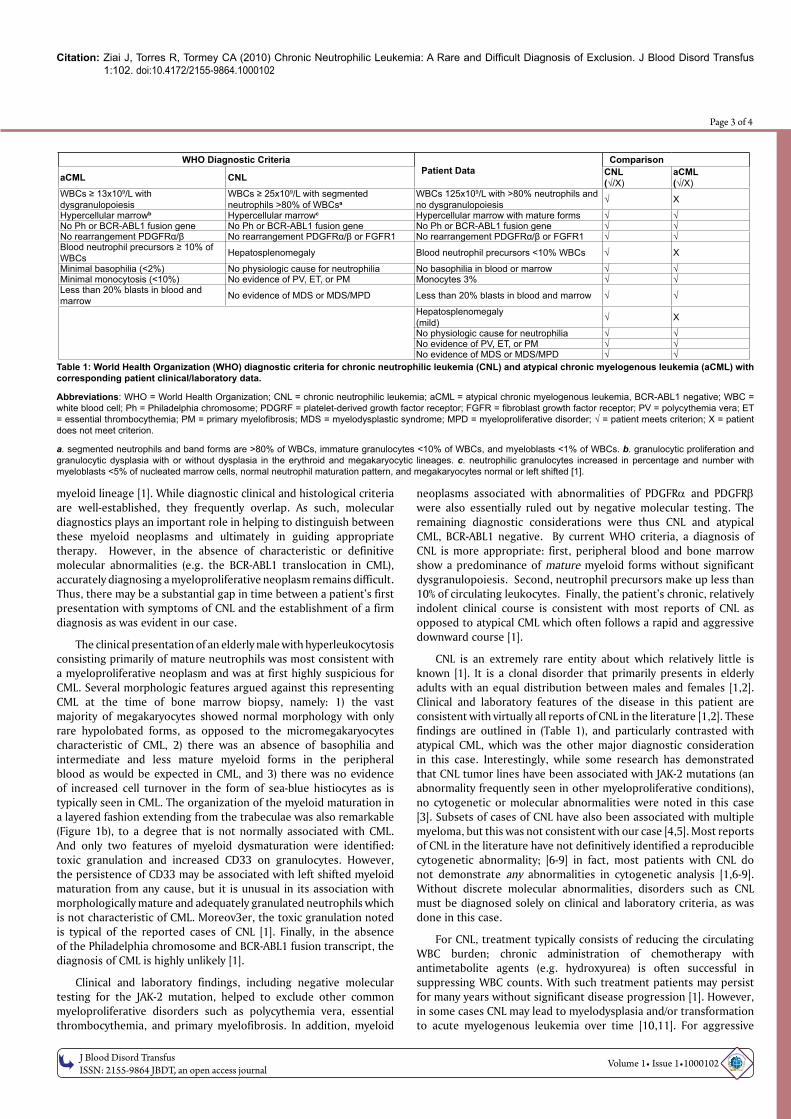
The bone marrow core biopsy was markedly hypercellular for age with a cellularity estimated at 90% (Figure 1a). Myeloid maturation was sequential with a predominance of mature granulocytes, mainly localized in interstitial areas (Figure 1b and 1c). Immature myeloid cells showed organized lining of paratrabecular regions with maturing cells extending towards interstitial areas in a layered fashion (Figure 1b). Erythroid island architecture was disrupted by the neutrophilic proliferation but otherwise appeared normal and megakaryocytes were mildly increased with normal morphology (Figure 1b). There was no relative increase in myeloblasts, eosinophils, basophils, or mast cells. Moderate focal reticulin fibrosis was noted in paratrabecular regions, but did not extend to cortical areas (Figure 1d). The bone marrow aspirate reiterated the core biopsy findings. It was a cellular specimen with sequential myeloid maturation, a predominance of mature neutrophils, and no significant relative increase in blasts. There was toxic granulation noted on neutrophils and some hypersegmentation noted on peripheral smear, but otherwise no abnormalities including no pelgeroid forms (Figure
2a and 2b). The erythroid series was sparsely represented but did not show any morphologic abnormalities, and the majority of megakaryocytes were normal sized, with minimal hypolobation of some forms, but otherwise normal morphology (Figure 2c). Increases in eosinophils, basophils, plasma cells, or mast cells were not noted. Sea-blue histiocytes were not seen. Stainable iron was markedly reduced without any ringed sideroblasts. Flow cytometric studies on the aspirate showed that CD34+ CD117+ myeloblasts constituted less than 1% of total marrow nucleated cells, mature granulocytes demonstrated persistent expression of CD33, and there was no evidence for T- or B-cell clonopathy or any other aberrant findings. The conventional marrow karyotype was 46, XY with no abnormalities noted. A t(9;22) was not identified by either PCR or fluorescence-in-situ hybridization (FISH) methods. JAK-2 mutation analysis and PDGFRα/PDGFRβ mutation analysis were similarly negative.
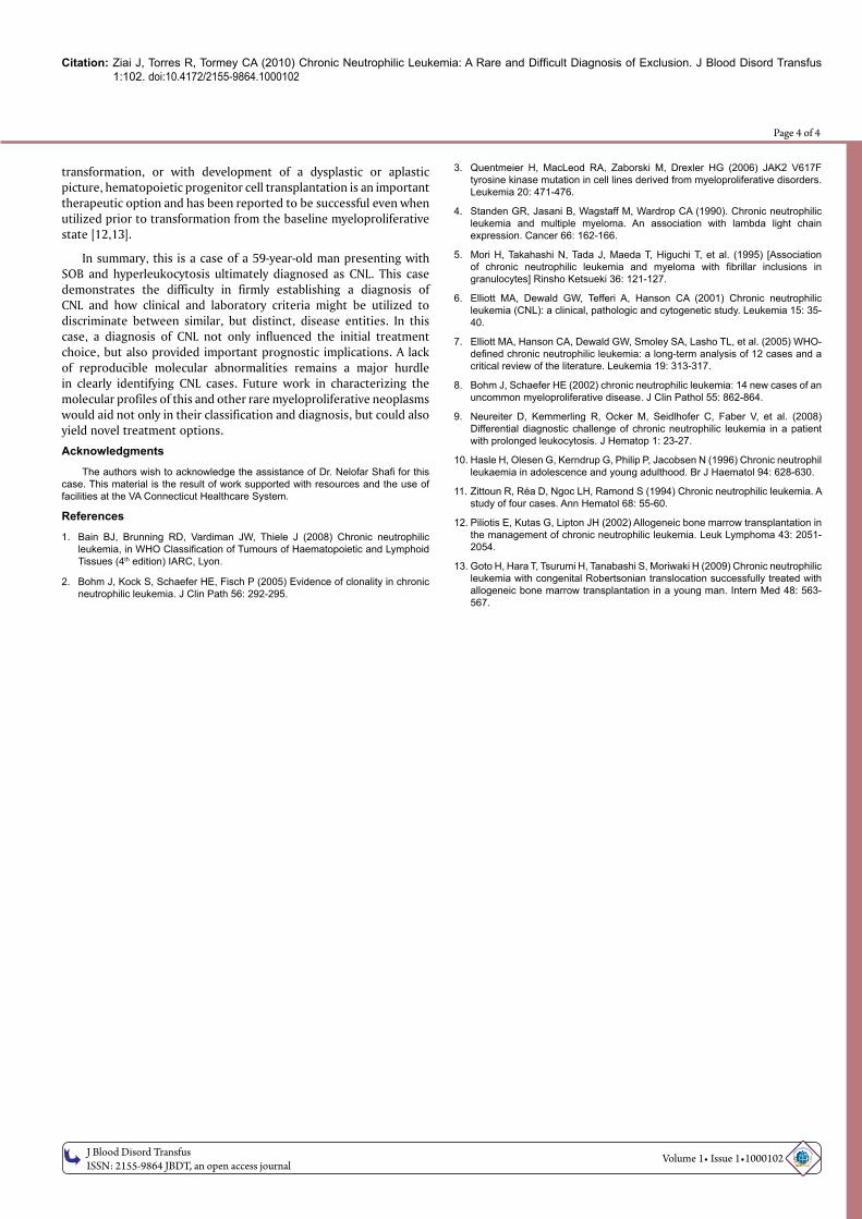
With the additional data provided by the above tests, the patient’s initial differential diagnosis was further restricted to atypical CML, BCR-ABL 1 negative versus CNL. Utilizing rigorous criteria as defined by the World Health Organization (WHO), a head-to-head comparison of both clinical and laboratory features of the case was made [1]. The patient met all criteria for a diagnosis of CNL, while a diagnosis of atypical CML was less likely given the lack of evidence of overt neutrophilic dysgranulopoiesis, a virtual absence of circulating immature myeloid cells, and the demonstration of only mild splenomegaly by physical exam. The final diagnosis was chronic neutrophilic leukemia.
Based on the firm establishment of this diagnosis, hydroxyurea was re-initiated and a subsequent improvement in leukocytosis was immediately noted; the patient’s WBC count corrected to less than 11x103/µL within 9 days of re-starting this medication. To date, he is maintained on chronic monotherapy with hydroxyurea with adequate suppression of his leukocyte count. Although rare spikes in WBC count have been noted, these changes have been entirely attributable to lack of compliance with the chemotherapeutic regimen as correction of counts has occurred with re-initiation of hydroxyurea in each instance. Of note, several family members have been evaluated as potential allogeneic hematopoietic progenitor cell donors for this patient should myeloablative chemotherapy with salvage stem cell transplantation ever needed in the course of the illness.
DiscussionMyeloproliferative neoplasms are clonal hematopoietic stem cell
disorders characterized by proliferation of one or more elements of the
Figure 1: Bone marrow core biopsy. a. Low power view (4X, H&E) showing high cellularity estimated at 90%. b. Myeloid maturation extends sequentially from trabeculae in an organized fashion with thickened immature layer. Erythroid islands are somewhat disorganized. Megakaryocytes appear normal (20X, H&E). c. Mature granulocytes predominate in interstitial areas (40X, H&E). d. Moderate reticulin fibrosis is restricted to paratrabecular areas (20X, Reticulin).
Figure 2: Bone marrow aspirate. a. Myeloid maturation showing hypergranulation but is complete to granulocytic stage without dysplasia (40X, Wright-Giemsa). b. Some degree of hyperlobulation was evident on mature granulocytes (40X, Wright-Giemsa). c. Megakaryocytes appeared normal with only minor hypolobulation on a subset of cells (40X, Wright-Giemsa).
Citation: Ziai J, Torres R, Tormey CA (2010) Chronic Neutrophilic Leukemia: A Rare and Difficult Diagnosis of Exclusion. J Blood Disord Transfus 1:102. doi:10.4172/2155-9864.1000102
OM
ICS Publishing GroupJ Blood Disord Transfus
ISSN: 2155-9864 JBDT, an open access journal Volume 1• Issue 1•1000102
Page 3 of 4
myeloid lineage [1]. While diagnostic clinical and histological criteria are well-established, they frequently overlap. As such, molecular diagnostics plays an important role in helping to distinguish between these myeloid neoplasms and ultimately in guiding appropriate therapy. However, in the absence of characteristic or definitive molecular abnormalities (e.g. the BCR-ABL1 translocation in CML), accurately diagnosing a myeloproliferative neoplasm remains difficult. Thus, there may be a substantial gap in time between a patient’s first presentation with symptoms of CNL and the establishment of a firm diagnosis as was evident in our case.
The clinical presentation of an elderly male with hyperleukocytosis consisting primarily of mature neutrophils was most consistent with a myeloproliferative neoplasm and was at first highly suspicious for CML. Several morphologic features argued against this representing CML at the time of bone marrow biopsy, namely: 1) the vast majority of megakaryocytes showed normal morphology with only rare hypolobated forms, as opposed to the micromegakaryocytes characteristic of CML, 2) there was an absence of basophilia and intermediate and less mature myeloid forms in the peripheral blood as would be expected in CML, and 3) there was no evidence of increased cell turnover in the form of sea-blue histiocytes as is typically seen in CML. The organization of the myeloid maturation in a layered fashion extending from the trabeculae was also remarkable (Figure 1b), to a degree that is not normally associated with CML. And only two features of myeloid dysmaturation were identified: toxic granulation and increased CD33 on granulocytes. However, the persistence of CD33 may be associated with left shifted myeloid maturation from any cause, but it is unusual in its association with morphologically mature and adequately granulated neutrophils which is not characteristic of CML. Moreov3er, the toxic granulation noted is typical of the reported cases of CNL [1]. Finally, in the absence of the Philadelphia chromosome and BCR-ABL1 fusion transcript, the diagnosis of CML is highly unlikely [1].
Clinical and laboratory findings, including negative molecular testing for the JAK-2 mutation, helped to exclude other common myeloproliferative disorders such as polycythemia vera, essential thrombocythemia, and primary myelofibrosis. In addition, myeloid
neoplasms associated with abnormalities of PDGFRα and PDGFRβ were also essentially ruled out by negative molecular testing. The remaining diagnostic considerations were thus CNL and atypical CML, BCR-ABL1 negative. By current WHO criteria, a diagnosis of CNL is more appropriate: first, peripheral blood and bone marrow show a predominance of mature myeloid forms without significant dysgranulopoiesis. Second, neutrophil precursors make up less than 10% of circulating leukocytes. Finally, the patient’s chronic, relatively indolent clinical course is consistent with most reports of CNL as opposed to atypical CML which often follows a rapid and aggressive downward course [1].
CNL is an extremely rare entity about which relatively little is known [1]. It is a clonal disorder that primarily presents in elderly adults with an equal distribution between males and females [1,2]. Clinical and laboratory features of the disease in this patient are consistent with virtually all reports of CNL in the literature [1,2]. These findings are outlined in (Table 1), and particularly contrasted with atypical CML, which was the other major diagnostic consideration in this case. Interestingly, while some research has demonstrated that CNL tumor lines have been associated with JAK-2 mutations (an abnormality frequently seen in other myeloproliferative conditions), no cytogenetic or molecular abnormalities were noted in this case [3]. Subsets of cases of CNL have also been associated with multiple myeloma, but this was not consistent with our case [4,5]. Most reports of CNL in the literature have not definitively identified a reproducible cytogenetic abnormality; [6-9] in fact, most patients with CNL do not demonstrate any abnormalities in cytogenetic analysis [1,6-9]. Without discrete molecular abnormalities, disorders such as CNL must be diagnosed solely on clinical and laboratory criteria, as was done in this case.
For CNL, treatment typically consists of reducing the circulating WBC burden; chronic administration of chemotherapy with antimetabolite agents (e.g. hydroxyurea) is often successful in suppressing WBC counts. With such treatment patients may persist for many years without significant disease progression [1]. However, in some cases CNL may lead to myelodysplasia and/or transformation to acute myelogenous leukemia over time [10,11]. For aggressive
WHO Diagnostic CriteriaPatient Data
Comparison
aCML CNL CNL(√/X)
aCML(√/X)
WBCs ≥ 13x109/L with dysgranulopoiesis
WBCs ≥ 25x109/L with segmented neutrophils >80% of WBCsa
WBCs 125x109/L with >80% neutrophils and no dysgranulopoiesis √ X
Hypercellular marrowb Hypercellular marrowc Hypercellular marrow with mature forms √ √No Ph or BCR-ABL1 fusion gene No Ph or BCR-ABL1 fusion gene No Ph or BCR-ABL1 fusion gene √ √No rearrangement PDGFRα/β No rearrangement PDGFRα/β or FGFR1 No rearrangement PDGFRα/β or FGFR1 √ √Blood neutrophil precursors ≥ 10% of WBCs Hepatosplenomegaly Blood neutrophil precursors <10% WBCs √ X
Minimal basophilia (<2%) No physiologic cause for neutrophilia No basophilia in blood or marrow √ √Minimal monocytosis (<10%) No evidence of PV, ET, or PM Monocytes 3% √ √Less than 20% blasts in blood and marrow No evidence of MDS or MDS/MPD Less than 20% blasts in blood and marrow √ √
Hepatosplenomegaly(mild) √ X
No physiologic cause for neutrophilia √ √No evidence of PV, ET, or PM √ √No evidence of MDS or MDS/MPD √ √
Table 1: World Health Organization (WHO) diagnostic criteria for chronic neutrophilic leukemia (CNL) and atypical chronic myelogenous leukemia (aCML) with corresponding patient clinical/laboratory data.
Abbreviations: WHO = World Health Organization; CNL = chronic neutrophilic leukemia; aCML = atypical chronic myelogenous leukemia, BCR-ABL1 negative; WBC = white blood cell; Ph = Philadelphia chromosome; PDGRF = platelet-derived growth factor receptor; FGFR = fibroblast growth factor receptor; PV = polycythemia vera; ET = essential thrombocythemia; PM = primary myelofibrosis; MDS = myelodysplastic syndrome; MPD = myeloproliferative disorder; √ = patient meets criterion; X = patient does not meet criterion.
a. segmented neutrophils and band forms are >80% of WBCs, immature granulocytes <10% of WBCs, and myeloblasts <1% of WBCs. b. granulocytic proliferation and granulocytic dysplasia with or without dysplasia in the erythroid and megakaryocytic lineages. c. neutrophilic granulocytes increased in percentage and number with myeloblasts <5% of nucleated marrow cells, normal neutrophil maturation pattern, and megakaryocytes normal or left shifted [1].
Citation: Ziai J, Torres R, Tormey CA (2010) Chronic Neutrophilic Leukemia: A Rare and Difficult Diagnosis of Exclusion. J Blood Disord Transfus 1:102. doi:10.4172/2155-9864.1000102
OM
ICS Publishing GroupJ Blood Disord Transfus
ISSN: 2155-9864 JBDT, an open access journal Volume 1• Issue 1•1000102
Page 4 of 4
transformation, or with development of a dysplastic or aplastic picture, hematopoietic progenitor cell transplantation is an important therapeutic option and has been reported to be successful even when utilized prior to transformation from the baseline myeloproliferative state [12,13].
In summary, this is a case of a 59-year-old man presenting with SOB and hyperleukocytosis ultimately diagnosed as CNL. This case demonstrates the difficulty in firmly establishing a diagnosis of CNL and how clinical and laboratory criteria might be utilized to discriminate between similar, but distinct, disease entities. In this case, a diagnosis of CNL not only influenced the initial treatment choice, but also provided important prognostic implications. A lack of reproducible molecular abnormalities remains a major hurdle in clearly identifying CNL cases. Future work in characterizing the molecular profiles of this and other rare myeloproliferative neoplasms would aid not only in their classification and diagnosis, but could also yield novel treatment options.
Acknowledgments
The authors wish to acknowledge the assistance of Dr. Nelofar Shafi for this case. This material is the result of work supported with resources and the use of facilities at the VA Connecticut Healthcare System.
References
1. Bain BJ, Brunning RD, Vardiman JW, Thiele J (2008) Chronic neutrophilic leukemia, in WHO Classification of Tumours of Haematopoietic and Lymphoid Tissues (4th edition) IARC, Lyon.
2. Bohm J, Kock S, Schaefer HE, Fisch P (2005) Evidence of clonality in chronic neutrophilic leukemia. J Clin Path 56: 292-295.
3. Quentmeier H, MacLeod RA, Zaborski M, Drexler HG (2006) JAK2 V617F tyrosine kinase mutation in cell lines derived from myeloproliferative disorders. Leukemia 20: 471-476.
4. Standen GR, Jasani B, Wagstaff M, Wardrop CA (1990). Chronic neutrophilic leukemia and multiple myeloma. An association with lambda light chain expression. Cancer 66: 162-166.
5. Mori H, Takahashi N, Tada J, Maeda T, Higuchi T, et al. (1995) [Association of chronic neutrophilic leukemia and myeloma with fibrillar inclusions in granulocytes] Rinsho Ketsueki 36: 121-127.
6. Elliott MA, Dewald GW, Tefferi A, Hanson CA (2001) Chronic neutrophilic leukemia (CNL): a clinical, pathologic and cytogenetic study. Leukemia 15: 35-40.
7. Elliott MA, Hanson CA, Dewald GW, Smoley SA, Lasho TL, et al. (2005) WHO-defined chronic neutrophilic leukemia: a long-term analysis of 12 cases and a critical review of the literature. Leukemia 19: 313-317.
8. Bohm J, Schaefer HE (2002) chronic neutrophilic leukemia: 14 new cases of an uncommon myeloproliferative disease. J Clin Pathol 55: 862-864.
9. Neureiter D, Kemmerling R, Ocker M, Seidlhofer C, Faber V, et al. (2008) Differential diagnostic challenge of chronic neutrophilic leukemia in a patient with prolonged leukocytosis. J Hematop 1: 23-27.
10. Hasle H, Olesen G, Kerndrup G, Philip P, Jacobsen N (1996) Chronic neutrophil leukaemia in adolescence and young adulthood. Br J Haematol 94: 628-630.
11. Zittoun R, Réa D, Ngoc LH, Ramond S (1994) Chronic neutrophilic leukemia. A study of four cases. Ann Hematol 68: 55-60.
12. Piliotis E, Kutas G, Lipton JH (2002) Allogeneic bone marrow transplantation in the management of chronic neutrophilic leukemia. Leuk Lymphoma 43: 2051-2054.
13. Goto H, Hara T, Tsurumi H, Tanabashi S, Moriwaki H (2009) Chronic neutrophilic leukemia with congenital Robertsonian translocation successfully treated with allogeneic bone marrow transplantation in a young man. Intern Med 48: 563-567.
Related Documents