Chronic Musculoskeletal Conditions and Neuroplasticity in the Central Nervous System

Welcome message from author
This document is posted to help you gain knowledge. Please leave a comment to let me know what you think about it! Share it to your friends and learn new things together.
Transcript

Chronic Musculoskeletal Conditions and Neuroplasticity in the Central
Nervous System
Pelletier et al. BMC Musculoskeletal Disorders (2015) 16:25 DOI 10.1186/s12891-015-0480-y
DEBATE Open Access
Is neuroplasticity in the central nervous systemthe missing link to our understanding of chronicmusculoskeletal disorders?René Pelletier1*, Johanne Higgins1,2 and Daniel Bourbonnais1,2
Abstract
Background: Musculoskeletal rehabilitative care and research have traditionally been guided by a structuralpathology paradigm and directed their resources towards the structural, functional, and biological abnormalitieslocated locally within the musculoskeletal system to understand and treat Musculoskeletal Disorders (MSD).However the structural pathology model does not adequately explain many of the clinical and experimentalfindings in subjects with chronic MSD and, more importantly, treatment guided by this paradigm fails to effectivelytreat many of these conditions.
Discussion: Increasing evidence reveals structural and functional changes within the Central Nervous System (CNS)of people with chronic MSD that appear to play a prominent role in the pathophysiology of these disorders. Theseneuroplastic changes are reflective of adaptive neurophysiological processes occurring as the result of alteredafferent stimuli including nociceptive and neuropathic transmission to spinal, subcortical and cortical areas withMSD that are initially beneficial but may persist in a chronic state, may be part and parcel in the pathophysiology ofthe condition and the development and maintenance of chronic signs and symptoms. Neuroplastic changes withindifferent areas of the CNS may help to explain the transition from acute to chronic conditions, sensory-motorfindings, perceptual disturbances, why some individuals continue to experience pain when no structural cause canbe discerned, and why some fail to respond to conservative interventions in subjects with chronic MSD. We arguethat a change in paradigm is necessary that integrates CNS changes associated with chronic MSD and that thesefindings are highly relevant for the design and implementation of rehabilitative interventions for this population.
Summary: Recent findings suggest that a change in model and approach is required in the rehabilitation ofchronic MSD that integrate the findings of neuroplastic changes across the CNS and are targeted by rehabilitativeinterventions. Effects of current interventions may be mediated through peripheral and central changes but maynot specifically address all underlying neuroplastic changes in the CNS potentially associated with chronic MSD.Novel approaches to address these neuroplastic changes show promise and require further investigation toimprove efficacy of currents approaches.
Keywords: Musculoskeletal disorders, Chronic low back pain, Osteoarthritis, Neuroplasticity, Periaqueductal grey,Rostral ventromedial medulla, Rehabilitation, Primary somatosensory cortex, Primary motor cortex, Limbic, Pre-frontal,Pain
* Correspondence: [email protected]École de réadaptation, Faculté de médecine, Université de Montréal, C.P.6128, succursale Centre-ville, Montréal H3C 3 J7, Québec, CanadaFull list of author information is available at the end of the article
© 2015 Pelletier et al.; licensee BioMed Central. This is an Open Access article distributed under the terms of the CreativeCommons Attribution License (http://creativecommons.org/licenses/by/4.0), which permits unrestricted use, distribution, andreproduction in any medium, provided the original work is properly credited. The Creative Commons Public DomainDedication waiver (http://creativecommons.org/publicdomain/zero/1.0/) applies to the data made available in this article,unless otherwise stated.

Pelletier et al. BMC Musculoskeletal Disorders (2015) 16:25 Page 2 of 13
BackgroundThe treatment of Musculoskeletal Disorders (MSD) hasbeen guided by a structural-pathology paradigm wherethe source of dysfunctions associated with the injury areto be found locally at the site of injury, the premise of“end organ dysfunction” [1]. The structural-pathologyparadigm helps to comprehend and guide treatmenteffectively in acute MSD. There are however many un-answered questions and discrepant findings with chronicMSD where the structural-pathology paradigm fails as aworking model for comprehension, research and intreatment. These include allusive questions such aswhy diagnostic findings correlate poorly with pain anddysfunction, the presence of bilateral findings withunilateral injuries, why a large proportion of personswith damage to musculoskeletal structures are asymp-tomatic, why some persons heal and others developchronic MSD, and persisting sensory motor abnormalities[2-6]. In an attempt to better understand the clinical andexperimental manifestations of these disorders researchershave expanded their scope of inquiry to include neuro-physiological processes and plasticity within the CentralNervous System (CNS) associated with MSD.Neuroplasticity is an intrinsic fundamental neuro-
physiological feature that refers to changes in structure,function and organisation within the nervous systemthat occurs continuously throughout a person’s lifetime[7-10]. Recent studies have revealed structural and func-tional changes within the CNS of people with chronicMSD. These changes are believed to be reflective ofadaptive neurophysiological processes occurring withMSD that are initially beneficial and aid in the healingprocess by protecting the injured structures from furtherinsult. In a chronic state, the structural pathology para-digm dictates that that these neuroplastic changes asso-ciated with chronic MSD are secondary to the injuryand result from ongoing altered sensory transmissionarising from the area of the musculoskeletal injury. Clin-ical and experiment findings however challenge this be-lief and demonstrate that neurophysiological adaptationsmay persist and be implicated in the development andmaintenance of chronic signs and symptoms, possibly inlieu of healing to the peripheral musculoskeletal structuresor co-existing with peripheral mechanisms [1,11]. It hasrecently been proposed that chronic pain associated withMSD is the result of imprinting, an implicit and/or explicitlearned response that has formed a maladaptive memorysustaining the persistence of chronic pain [12-15]. Accord-ing to this hypothesis, associative learning resulting fromthe initial trauma and subsequent events that reinforcesthe concurrent pairing between movement and painresults in an aversive association that is reflected andmaintained by plastic changes in the meso-limbic andprefrontal areas [15].
This article will argue that neuroplastic adaptationsand their effects may initially result from structural in-jury, but in chronic conditions contribute to the patho-physiology of the condition possibly even in the absenceof any continued anatomical/structural insult to muscu-loskeletal structures. These neuroplastic changes explainmany of the experimental and clinical findings presentin subjects with chronic MSD. These changes result insensory amplification [16], changes in sensory and motorrepresentations [17-19] resulting in perceptual changesin body image [20,21], changes in motor control [22], bi-lateral experimental findings [23-25], the persistence andamplification of pain [16,26], and why some individualstransit from acute to chronic disorders [27,28]. Furtherevidence arguing to the importance of these neurophysio-logical adaptations stem from recent studies targetingneuronal processes appear to restore function and de-crease pain [14,29,30]. These findings are highly relevantfor the design and implementation of rehabilitative inter-ventions for MSD which when guided by the structural-pathology paradigm have limited success in the treatmentof many of these chronic conditions [31]. If neuroplasticchanges in the CNS are not simply an epiphenomenonbut are part and parcel to the pathophysiological processin chronic MSD, interventions that target these underlyingpathophysiological mechanisms have the greatest chanceof success [32]. Current conventional interventions in re-habilitation do not usually address underlying neuroplasticchanges in the CNS associated with MSD [32] and theincapacity to effectively treat these chronic MSD stem asthey are incomplete and/or misdirected [1,31,33-35].
DiscussionThe structural pathology paradigm is guided by theinherent belief that pain and other neurophysiologicalchanges are secondary to local structural insult to mus-culoskeletal structures. Both in animal and human stud-ies, it is apparent that local and systemic inflammatoryresponses, cellular and vascular proliferative changes aswell as degeneration and fibrosis are all hallmarks ofchronic and overuse MSD [34,36-41]. Injury to muscu-loskeletal structures, inflammatory mediators, and sub-sequent fibrosis change the mechanics of muscles andconnective tissues affecting their physical propertiesand these in turn impact sensory receptor activity andtransmission [11,34,42-46]. Under the structural-pathologyparadigm neurophysiological consequences, with the ex-ception of damage to the nerve(s), is secondary and shoulddisappear when normal tissue properties are restored andreceptor activity, sensory transmission, and perceptionshould renormalize to reflect the state of the healed struc-ture(s). Within this paradigm pain is simply a symptomand reflects the degree of damage to the musculoskeletalstructure and associated biological responses locally in the

Pelletier et al. BMC Musculoskeletal Disorders (2015) 16:25 Page 3 of 13
area of injury. This viewpoint is supported by the findingsthat demonstrates the reversal of some, but not all CentralNervous System (CNS) changes when anatomical insult tomusculoskeletal structures and pain disappears [47,48].This paradigm however fails to explain many of the
experimental findings with chronic MSD. For example,on a population level anatomical insult to musculoskel-etal structures correlates poorly with diagnostic findingsand these across a wide range of musculoskeletal disorders[2-6]. Therefore structural damage to musculoskeletalstructures alone cannot always fully explain the presenceof signs and symptoms in chronic MSD. Cognitive basedinterventions that involve education of pain processingand faulty beliefs regarding pain and movement yield bet-ter outcomes, between 10-20% improvement in disabilityand performance scales [49], than interventions involvingeducation of anatomical and structural basis of injury[49-52] suggesting that central rather than peripheral in-fluences play a key role in the clinical and experimentalmanifestation of at least some chronic MSD [51], and thatclinical interventions aimed to modify the central process-ing of pain should be further evaluated and compared toclinical interventions targeting peripheral mechanisms.
Principles of experience dependent plasticityNeuroplasticity refers to changes in neuronal properties,structure and organization and is the manner in which thenervous system encodes new experiences. Neuroplasticchanges have been demonstrated in response to experi-ence and behaviour [53-56], motor learning [57-62], pain[17,63-65], injury [66,67], sensory stimuli [68-71], andcognitive processes [53,56,72,73]. Changes can be transi-ent, reflecting the adaptability of the sensorimotor systemto respond to internal and environmental demands andcan occur over short training periods [74,75]. Neuroplasticchanges in sensory-motor areas are stimulus driven andresult in lasting neuroplastic changes when the internaland external pressures are repetitive, salient, involvelearning and require sustained attention [7,53,54,76-78].Neuroplastic changes have been observed in differentareas of the CNS including the spinal cord, subcorticaland cortical areas.
Plasticity in the spinal cord and brain stem withchronic MSDSensory testing has demonstrated changes in sensorytransmission and processing across a number of MSDincluding osteoarthritis (OA) [79,80], Patella-FemoralPain Syndrome (PFPS) [81], tendinitis [82], Lateral Epi-condylitis (LE) [83], Carpal Tunnel Syndrome (CTS) [84],lumbar [85] and cervical injuries including whiplash [86].These studies include findings of changes in perceptionthreshold to noxious and innocuous stimuli, but also othersensory alterations including stimuli being processed more
slowly, incorrect localization, and decreased accuracy inrecognition of tactile stimulation [43,79,81-85,87-92]. Thesechanges have been demonstrated bilaterally and in sites re-mote to the initial injury [81,83,93]. Proprioceptive deficitsinclude increased errors in repositioning [94-96], decreasedposition sense and ability to detect joint motion [97,98],difficulty to adopt postures seen on a photograph [87,89]across a number of MSD.Although not all studies involving subjects with chronic
MSD demonstrate altered sensory transmission [99] manystudies with chronic MSD demonstrate augmented noci-ceptive transmission involving responsiveness to normallysub threshold nociceptive stimuli that results in hyperla-gesia, an increase in nociceptive transmission and painperception, indicative of an altered stimulus–responserelationship to nociceptive stimuli, a process called Cen-tral Sensitization [16,26,83-85,100-102]. This is a nor-mal, adaptive and reversible process that is biologicallyadvantageous to protect the injured structure from furtherinsult and is a consistent notion within the structural-pathology paradigm [26].Neurophysiological changes also result in the amplifi-
cation of noxious and innocuous stimuli within the dor-sal horn of the spinal cord that persist in chronic painstates. These changes are reflective of processes similarto experience dependent plasticity and result from seg-mental, spinal and supraspinal processes that modulatemembrane excitability and affect inhibitory and facilitatoryprocesses within the spinal cord (see [16]). Some dorsalhorn nociceptive neurons develop increased receptor fieldsize (wide-dynamic range neurons) responding to noci-ceptive and cutaneous stimuli that results in secondaryhyperalgesia and allodynia (spread and perception of painwith innocuous stimulation) [16].The supraspinal influences on dorsal horn nocicep-
tive transmission include descending pain modulatorysystems including the Periaqueductal grey (PAG)-RostralVentromedial (RVM) pathway. Under normal circum-stances these systems inhibit the transmission of nocicep-tive stimuli in the dorsal horn of the spinal cord [103].There exists convincing evidence in animal models thatthese descending modulatory systems are disrupted inchronic pain subjects shifting from a state of inhibition toa mal-adaptive state of facilitation amplifying the trans-mission of nociceptive stimuli, contributing to the processof central sensitization, and perpetuating the augmentedtransmission of neuropathic stimuli [16,45,103]. For ex-ample, an increase in activity of cells that project to thedorsal horn of the spinal cord from the RVM that facilitatethe transmission of noxious stimuli is present only in ani-mals with neuropathic pain behaviours [104]. The micro-injection of lidocaine into the RVM, causing a temporarycessation of neuronal activity, and an ipsilateral lesion ofthe dorsal lateral funiculus that house neuronal projections

Pelletier et al. BMC Musculoskeletal Disorders (2015) 16:25 Page 4 of 13
from the RVM towards the dorsal horn both decrease thethreshold to elicit withdrawal reflexes, indicative of in-creased pain perception and that neuronal activity of theRVM is facilitating the transmission of nociceptive/neuro-pathic stimuli [105]. Electrical stimulation of the RVMpaired with cutaneous stimulation recorded from secondorder spinal nociceptive neurons results in a 130% in-crease in neuronal activity [106]. In CLBP patients there isa decrease in PAG cerebral blood flow not seen in healthycontrol subjects suggestive of decreased neuronal activity[107]. In humans there is evidence that the a test noxiousstimulus, under normal circumstances, is inhibited by apreceding noxious conditioning stimulus, a process calledConditioned Pain Modulation [108], and is disturbed insubjects in some MSD and chronic pain states [108,109].Collectively the results from these studies demonstratethat the PAG-RVM pathway not only facilitates nocicep-tive transmission in the dorsal horn of the spinal cord butactually perpetuates the transmission of pain. This arguesagainst a peripherally driven source of augmented noci-ceptive/neuropathic transmission and for a centrally medi-ated mechanism perpetuating the transmission of afferentstimuli that is inconsistent with the structural-pathologyparadigm.Neuroplastic changes amplifying sensory transmission
have functional implications. Subjects demonstratingcentral sensitization, hypersensitivity and allodynia havea poorer prognosis to treatment including surgical inter-ventions for varied MSD [12,100,110,111]. Furthermore,studies in both animals and humans demonstrate thataltered sensory transmission may result in changes inneuronal properties and organization within differentsubcortical and cortical areas including the thalamus,primary somatosensory cortex (S1) and the primary motorcortex (M1) implicated in sensory transmission, percep-tion and motor control [112,113].
Neuroplastic changes in the primary somatosensorycortex and perceptual changes with MSDStudies of cortical properties and organisation within thesensorimotor areas have been performed with subjectswith PFPS [114], anterior cruciate ligament (ACL) deficiencyand reconstruction [33,115-117], CLBP [17-19,118-121],cervical pain and whiplash injury [91,122], rotator cuff tears[123,124], dystonia [125-129] and CTS [130-133]. Thesestudies suggest that neuronal properties, organization, andmorphometric changes are present in subjects with chronicMSD. For example, subjects with CLBP demonstrate a2.5 cm shift of the somatotopic representation in S1[17,121] and grey matter volume changes that correlatewith chronicity of symptoms [134,135]. Studies in subjectswith CTS reveal changes along the afferent pathway in thespinal cord, brain stem and S1 [131], a decrease in greymatter volume [133] and a loss of spatially segregated
representations of digits 2 and digits 3 in the contralateralS1 that correlate with changes in nerve conduction velocity[131,133,136]. Somatotopic re-organisation in CTS subjectsare specific to the nature of sensory stimuli as the represen-tation of the digits in S1 is decreased with pain and in-creased with paraesthesia [130].In the perspective of the structural-pathology paradigm,
these changes in S1 associated with MSD may simply bereflective of altered peripheral sensory transmission re-flective of altered afferent peripheral sensory stimuli andtransmission occurring as the result of insult to muscu-loskeletal structures and inflammation. Studies in non-human primates with peripheral de-afferentation andspinal cord injury demonstrate degeneration in the cu-neate nucleus of the brainstem, an area that containsaxons from the dorsal root ganglion transmitting cuta-neous and proprioceptive stimuli, as well as somatoto-pic reorganization in an area of the thalamus (ventralposterior lateral nucleus) that transmits sensory afferentstimuli to S1. The changes in S1 in these studies mirrorthe changes found in the thalamus suggesting that thechanges in sensory afference including noxious, cutane-ous, and possibly proprioceptive afferent transmissionare implicated in S1 reorganization [112,113]. However,should altered afferent transmission persist, potentiatedby functional changes in the brain stem and the spinalcord, neurophysiological changes appear to result in be-havioural and functional implications that are not sim-ply a reflection of altered sensory afference.There is growing evidence that pain associated with
MSD such as osteoarthritis and CLBP may be, at least inpart, the result of the plasticity of the sensory representa-tion of the body and perceptual disturbances [137-139].Distortions in body image have been found in a range ofconditions where cortical reorganization in S1 are presentincluding Phantom Limb Pain (PLP), Complex RegionalPain Syndrome (CRPS) and in CLBP [14,20,140-142].These changes include the sensation of abnormal size,shape, swelling, and position [30]. Perceptual changes mayalso arise from abnormal or conflicting sensory and/ormotor inputs [143,144]. Perceptual changes also havefunctional implications. Incongruence and manipulationbetween sensory and motor input has been shown tocause sensory disturbances, and aggravate symptomsand pain [145]. Modulation of the shape and size of alimb can impact tactile acuity and pain [146]. Visual dis-tortion of the hands in subjects with osteoarthritis helpsto decrease pain [137]. Interventions targeting changesin somatotopic reorganization through the use of sen-sory discriminative training and visual distortion canrenormalize the S1 representation and decrease pain[30,139,147-149]. The modulation of the size of the limbcan alter subjective feelings of pain and motor imagerycan cause an increase in pain and swelling that cannot

Pelletier et al. BMC Musculoskeletal Disorders (2015) 16:25 Page 5 of 13
be attributed to increased peripheral sensory afferencearising from nociceptors or peripheral neural injury[142,150]. The persistence of abnormal motor imageryin recurrent low back subjects is also believed to be re-flective of ongoing disruption of cortical maps even inthe absence of pain [151]. These findings support thebelief that structural injury to musculoskeletal struc-tures are not the only driver of pain and dysfunction,CNS changes play an active role in the pathophysiologyof chronic pain conditions, and interventions that targetthese CNS changes may decrease pain, improve function,and even affect mechanisms involved in the local bio-logical response to injured structures such as swelling.
Changes in primary motor cortex associated with MSDStudies that investigate changes in the properties, functionand organisation within the primary motor cortex (M1) ofsubjects with different MSD have been performed, ofwhich the majority utilise Transcranial Magnetic Stimu-lation (TMS). TMS produces a high intensity electricalpulse resulting in a magnetic field perpendicular to thestimulating coil. The magnetic pulse traverses the skulland when applied over the motor cortex with sufficientintensity, can depolarize corticospinal neurons directly orindirectly. This stimulation results in the depolarization ofdifferent motoneuron pools within the spinal cord and anelectromyographic response, the Motor Evoked Potential(MEP) can be recorded. Utilising different parameters ofstimulation and experimental protocols, TMS allowsfor the appreciation of corticospinal excitability, in-hibitory and facilitatory processes, and somatotopicorganization of corticospinal neurons. Studies of corti-cospinal excitability have been performed in subjects withvarious MSD including PFPS [114], ACL deficiency [117],CLBP [18,19,119,120,152,153], and Rotator Cuff Tears[123,124]. Collectively these studies demonstrate changesin corticospinal excitability that correlate with pain anddisability scores. Changes in motor behaviour that arepresent in subjects with CMSD appear to be largely medi-ated by changes in the cortical areas including M1. Inhib-ition of corticospinal output is increased in experimentallyinduced muscle pain resulting in decreased motor re-sponses to TMS at rest [154] and increased corticospinaloutput during forceful muscle contractions [155,156].Findings from these studies appear to be consistent withthe experimental findings that demonstrate variable motorcontrol changes including reorganization of motor unitrecruitment both within and between muscles in an at-tempt to minimize the motor consequences associatedwith chronic MSD (see [11,22,65]), co-activation of mus-cles and overlapping of muscle/movement representationsin M1 [19,22], and variations in corticospinal output in anattempt to maintain constant force under painful condi-tions and compensate for increased inhibition [155].
In a series of experiments Tsao and his colleagues in-vestigated the properties and organization of the repre-sentation of muscles in the lumbar spine within M1 insubjects with CLBP. They demonstrated that the area ofcorticospinal recruitment of muscles of the lumbar spinein M1 is altered in CLBP subjects [18]. These changescorrelate with changes in motor recruitment [18,19].Motor skill learning involving exercises to specifically re-cruit the transverse abdominus muscle, but not a walk-ing exercise, could restore the representation within M1and EMG activation pattern in CLBP subjects to thatseen in healthy controls [118]. The changes in the repre-sentation of the movements elicited by the trunk mus-cles in M1 are associated with the impaired activation ofthese muscles and may underpin changes in motor acti-vation, specifically the inability to selectively recruitthese muscles. This, in turn is consistent with the in-creased activation of superficial muscles in this popula-tion when performing movements [157] and the alteredactivation of the multifidus that has been demonstratedin patients with recurrent LBP [158,159]. These studiesdemonstrate that neuronal properties and organisationwithin M1 are modified in CLBP subjects and that inter-vention specifically targeting these representational changesimprove function and decrease pain.The relationship between the plastic changes in the
spinal cord, brain stem and cortical sensori-motor areasare complex. Experimental findings suggest the possibilityof two-way causality, where altered sensory input includ-ing enhanced nociceptive/neuropathic stimuli, altered cu-taneous and proprioceptive input affects sensorimotororganisation and processes within the CNS, and thesechanges in turn affect perception, pain, and motor controlprocesses contributing to the pathophysiology of the con-dition [137,138]. If these processes remain present for asubstantial period of time they may result in lasting neuro-physiological adaptations that may become imprinted andcan outlive the insult to peripheral musculoskeletal struc-tures [14,15]. It is important to note that a return to be-fore injury sensory transmission and the performance ofrepetitive strengthening exercises may not be sufficient toreturn the neuronal properties and organization withinthe sensorimotor areas to a pre-injury state [160]. Specificinterventions addressing these neuroplastic changes insensorimotor areas appear to be required. Repetitive un-skilled movements do not result in neuroplastic changesin M1 [57,76]. Motor skill training however has provensuccessful in the treatment of some musculoskeletalconditions, improves task performance and helps pro-mote neuroplastic changes in M1 [53,118,161-165]. Thesefindings are suggestive that the neuroplastic changes inthe sensory-motor areas are implicated in the pathophysi-ology of some chronic MSD and should impact rehabilita-tive treatments.

Pelletier et al. BMC Musculoskeletal Disorders (2015) 16:25 Page 6 of 13
Role of pain in CNS plasticityFindings from experimental studies do provide convin-cing evidence that pain provides an impetus for CNSchanges with MSD. Experimentally induced pain im-pacts neuronal properties and organisation in S1 andM1 [153,166] and subjects with chronic pain associatedwith unilateral herpes simplex virus have a decreasedrepresentation between digits 1–5 in the contralateralS1 [167]. Although the causal relationship between painand cortical reorganization has not been definitivelyestablished with MSD, the evidence suggests that pain isa driver of cortical re-organization. In other conditionswhere re-organisation in S1 is present there is a renor-malisation with the attenuation of pain [168,169] andsome, but not all, of the morphological changes in braingrey matter volume and changes in cortical somatotopyreturn to those seen in normal healthy subjects whenpain is eliminated [47,48,168,169].However pain alone is neither necessary nor sufficient
to drive neuroplastic changes. Dystonia and CTS areboth conditions where researchers have demonstratedneuroplastic changes in M1 and S1 in the absence ofpain. Focal hand dystonia involves a loss of individualcontrol of the digits of the hand that results from rapidrepetitive motor actions of the fingers. These movementsresult in blurring of the representation of the digits withloss of spatial segregation [127-129]. Subjects with recur-rent low back pain continue to demonstrate abnormalmotor control in the absence of pain possibly reflectingcontinued reorganisation of neuronal properties and or-ganisation in M1 [170-172]. Behavioural interventions thathelp to restore somatotopic organisation also improvefunction and decrease pain suggesting the possibility oftwo way causality between pain and sensorimotor repre-sentations [173].Although pain provides an impetus for neuroplastic
changes in the CNS, other forms of stimuli, cognitiveprocesses and behaviours can induce plastic changes.Studies in animals, healthy human and neurologicallycompromised human subjects have demonstrated thatrepetition and attention/salience are important factors in-ducing neuroplastic changes in S1 and M1 [7,77,174,175].The limbic and prefrontal structures are the cortical areasresponsible for these aspects of behaviour and findingshave demonstrated important changes in these areas inchronic pain states including some MSD [13,15].
Neuroplastic changes in meso-limbic and prefrontalstructures in chronic pain statesOf all the areas of the CNS with documented changesoccurring in association with chronic MSD, the meso-limbic and prefrontal structures are the most impressiveand possibly the most important as changes in theseareas demonstrate strong correlations with chronicity
[13], and furthermore can be predictive and possiblyeven determine who will transit from acute to chronicpain [15,27,28]. Experimentally induced pain results inthe activation of characteristic cortical regions includingS1, S2, insula, cingulate cortex, amygdala, and prefrontalcortex in what is commonly referred to as the painmatrix, but is possibly more reflective of a salience net-work as these structures are not only active with painfulstimuli but also in conditions involving increased atten-tion/salience [176,177].Experimental findings suggest that the structure and
function of the brains of subjects with chronic pain in-cluding CLBP and OA are different from healthy controlsand this is most important in the meso-limbic and pre-frontal areas (see [13]). When experimentally induced painis applied to subjects with CLBP and osteoarthritis (OA)while performing a fMRI, both CLBP and OA subjectsdemonstrate spontaneous fluctuations of pain that is nottime locked to the experimental noxious stimuli and arenot present in healthy control subjects [178,179]. Spon-taneous pain engages pre-frontal and limbic areas import-ant for the processing and cognitive response to incomingstimuli [13,178,180]. FMRI studies have demonstrated thatsubjects with chronic MSD, specifically CLBP and OA,demonstrate abnormal activity in the cingulate cortex,the amygdala, the insula, nucleus accumbens (NAc) andpre-frontal areas including the medial prefrontal cortex(mPFC) and the dorsolateral prefrontal cortex (dlPFC)[13,134,178]. These mesolimbic-prefrontal areas areinvolved in the cognitive affective aspects of pain andinjury including the behavioural response to these, theprocessing of fear, emotions, negative conditioning andattention [181,182]. One result of the abnormal activityin these areas is increased vigilance and a decreasedability to disengage from pain [12]. These limbic struc-tures have direct and indirect connections with both thesensorimotor areas and the brain stem and may providethe substrate of attention and salience necessary for theinduction of neuroplastic changes in these areas [180,183].Furthermore, these structures influence descending painmodulatory systems including the PAG-RVM pathwaywhere, as discussed earlier, compelling evidence suggestsis disrupted in chronic pain subjects and perpetuate theongoing abnormal augmented pain transmission originat-ing from nociceptive and non-nociceptive peripheral re-ceptors in the dorsal horn of the spinal cord [103,184].The brain derived biomarkers from abnormal activity
in the mesolimbic and prefrontal areas correlate stronglywith clinical measures in patients with CLBP and correl-ate better with clinical findings than do structural andpsychosocial findings [13,184]. Increased insular activationis correlated with pain duration, while mPFC activation iscorrelated with pain intensity in CLBP subjects [13]. Ab-normal increased connectivity between the mPFC and the

Pelletier et al. BMC Musculoskeletal Disorders (2015) 16:25 Page 7 of 13
NAc is highly predictive (90%) of who will go on to de-velop CLBP suggesting that there may be pre-disposingbiomarkers for the development of chronicity [28,179].For a more thorough overview of changes in the meso-limbic and prefrontal areas associated with CMSD excel-lent reviews have been published [13,15,184].The complex interrelationship between pain, cortical
reorganization, disability, and abnormal motor behaviouris compounded by the implication of psychological factorsassociated with chronic pain and injury. Catastrophization(“tendency to focus and magnify pain sensation, and tofeel helpless in the face of pain”) and fear play a role inthe etiology and prognosis of chronic pain conditions[185-188]. Psychosocial factors predict variance in pain,gait velocity, and psychological disability in OA subjects,appear to increase pain and disability (see [185,186]), im-pact pain perception in healthy controls [186,189], andmay result in a learned avoidance behaviour perpetuatingthe disability [185,190]. These changes in the pre-frontalcortex activation are also consistent with fMRI studiesthat have correlated changes in prefrontal activity withpsychosocial variables involved in CLBP and OA, includ-ing dlPFC activity being negatively correlated with PainCatastrophizing Scores and mPFC activity correlated withfear-avoidance/anxiety [12,191,192]. Pain catastrophizingand fear-avoidance cause behavioural changes and may beresponsible for changes in neuronal properties and soma-totopic reorganization because of disuse similar to learnednon-use in stroke patients [193,194]. Neural circuits notactively engaged in task performance for an extendedperiod of time begin to degrade [7,195]. Prolonged non-use of the affected limb may lead to a vicious cyclewhereby immobility, changes in cortical representation,and atrophic changes re-enforce each other.
Integrating CNS changes into a more comprehensivemodel of chronic MSDIt would appear that behavioural changes and psycho-logical processes in chronic pain subjects involve activityin the meso-limbic and pre-frontal areas that influencepain perception and behaviour. Although speculative,the behavioural changes associated with these changesin meso-limbic and pre-frontal areas may therefore bereflective of salience and increased attention directed to-wards the injury and associated pain. The meso-limbicand prefrontal structures influence descending modula-tory pathways and facilitate the transmission of noxiousstimuli which perpetuates the altered transmission ofsensory stimuli and appear to influence sensorimotorrepresentations and neuronal properties. It is possiblethat these changes collectively result in a vicious cyclewhere injury, pain, altered sensory transmission, sensori-motor changes, behavioural changes, salience, attention,and fear-avoidance may feed off one another perpetuating
the disability. It has been hypothesized that the neuroplas-tic cortical changes in the meso-limbic prefrontal areas as-sociated with chronic pain states are reflective of learnedoperant and classic conditioning resulting in the forma-tion of a “pain” memory [12-14,196]. Providing supportfor this hypothesis are findings where imagery affects pain,swelling, and cortical excitability [150]. Consistent withthe implication of altered neuronal activity in the meso-limbic and prefrontal areas in the pathophysiology ofchronic MSD are the findings from educational and cogni-tive based interventions. Educational programs explainingthe neurophysiological mechanisms of pain have provenmore effective than back schools (which emphasizeend organ dysfunction and behavioural changes to de-crease loading of anatomical structures) in CLBP patients[49-51,197]. These educational programs attack faultypain beliefs which lead to fear-avoidance often presentwith chronic MSD [198]. The findings that altered func-tional connectivity in these areas are the best predictors ofchronicity in the transition from acute to CLBP furthersupports the argument as to the importance of thechanges in these areas in the pathophysiology of MSD[28]. These findings are inconsistent with a structural-pathology paradigm of a solely peripherally driven sourceof dysfunction in chronic MSD. Chronic MSD such asOA and CLBP, and possibly other MSD may have promin-ent CNS contributions with peripheral and central factors,cortical and limbic areas, all playing a role in the pain anddysfunction they produce [11,45]. Collectively these find-ings of changes in meso-limbic and prefrontal structuresprovide compelling evidence that CNS changes contributeto the pathophysiology of at least some chronic MSD andconversely, that the structural-pathology paradigm of localtissue compromise being solely at the root of chronicMSD is at the very least incomplete and insufficient. Amodel integrating central neurophysiological modifica-tions must be integrated into the present paradigm tobroaden its scope and be further investigated.
Impact of CNS plasticity in the rehabilitation of chronicMSDRestoration of motor activity and function are integral tocurrent practice in rehabilitation [51,199]. The notion ofaddressing neuroplastic changes is well established inneurological rehabilitation [32]. Interventions presentlyutilized in conventional rehabilitative care may resultfrom peripheral and central mechanisms and it remainsa challenge to distinguish their relative contribution. Forexample, resistance training in subjects with non-specificshoulder and neck pain increased local and distal pressurepain thresholds suggestive a central mechanism under-lying these effects [200]. However studies also demon-strate that specific types of interventions may be bettersuited at inducing neuroplastic changes [10,62,76,160].
Pelletier et al. BMC Musculoskeletal Disorders (2015) 16:25 Page 8 of 13
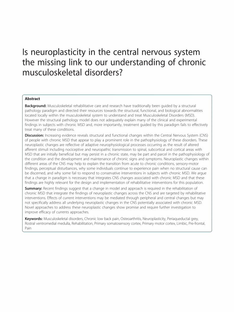
Rehabilitative interventions specifically addressing neuro-physiological changes, in addition to peripheral end organdysfunction, may prove to be an important avenue of in-vestigation in the hope to improve treatment success inthe rehabilitation of musculoskeletal injuries [10,32]. Stud-ies in animal models have demonstrated that the neuro-plastic changes in S1 and M1 occur concurrently withtissue damage, inflammation, and motor impairment andtherefore would need to be addressed early on in the re-habilitation process [34,201]. Addressing neurophysio-logical changes would involve interventions in an attemptto minimize and/or normalize structure, function andorganization to that found in uninjured healthy controlsby explicitly targeting and priming neuronal structuresand processes including those in the sensorimotor, meso-limbic and pre-frontal areas. These could include incorp-orating approaches to present conventional care such aseducation of neuronal and pain processes [51,197], cogni-tive based interventions such as Cognitive BehaviouralTherapy [202,203] and Mindfulness Based Stress Reduc-tion [204,205] which have been associated with changes inpre-frontal and meso-limbic structures [206-211], mentalimagery [29], peripheral sensory and electrical stimulation[63,147], visual distortion and the use of non-invasivebrain stimulation such as Transcranial Direct CurrentStimulation and TMS for example to alter neuronal pro-cesses [212-214]. Effect sizes of rehabilitation approachesare consistently small regardless of intervention in manyMSD and therefore multiple and progressive interventionsmay be warranted [51].
ResearchResearch investigating changes in S1 and M1 across alarge range of MSD, including changes in responsiveness,inhibitory processes, and somatotopic organization wouldhelp elucidate the mechanisms and their presence in MSD.Subsequent studies evaluating novel treatment approachessuch as motor skill training, mental imagery, action obser-vation, mirror therapy, peripheral sensory stimulation andcortical stimulation as adjuncts to traditional rehabilitativecare for MSD to impact neuronal responsiveness andreorganization are needed. Research in changes in neur-onal processes and organization of techniques presentlyutilized in rehabilitation, such as manual therapies, mayhelp elucidate the physiological mechanisms of action andlead to more effective application and outcomes. Furtherresearch of the plastic changes occurring in meso-limbicand prefrontal areas and the complex interrelationship be-tween structures and connections on these areas, corticalsensorimotor areas, descending modulatory processes, andpsychological traits and behaviours associated with CMSDwill not only increase our comprehension, but help guidethe development of more effective pharmacological, behav-ioural and rehabilitative interventions.
SummaryIn our opinion the present structural-pathology paradigmguiding treatment for MSD is at the very least incompleteas it fails to integrate recent findings of important neuro-physiological changes associated with chronic MSD andthat appear to be involved in the pathophysiology of theseconditions either in isolation or co-existing with periph-eral mechanisms. Musculoskeletal injury, in addition tothe local damage to anatomical structures and inflamma-tion, results in changes in sensory stimuli, transmissionand processing including neuroplastic changes along theneuroaxis of pain within the spinal cord and brain stem,in the properties and functions of neurons within S1 andM1. There are associated changes also found in the meso-limbic pre-frontal areas in subjects with chronic MSDsome which may pre-dispose the injury. The neuroplasticchanges may occur rapidly in response to injury causingadaptive changes that may help in the protection andhealing response. However, these changes may persist andno longer perform their intended function contributing tothe development of chronic disability and dysfunctionalpain with enduring neuroplastic changes along theneuroaxis of pain resulting in peripheral and centralsensitization, in the sensorimotor areas affecting perceptionand motor behavior, and in the meso-limbic prefrontalareas influencing emotional, attentional and cognitive pro-cesses [11,31,44]. In some musculoskeletal conditions theresponsiveness and somatotopic organization in S1 andM1, including changes in excitability, the blurring of therepresentation of anatomical structures and a shift in therepresentation of muscles within somatotopic representa-tions are present. These changes in properties, functionand organization within the CNS often correlate with theseverity and duration of pain, functional changes includingaspects of motor control, psychological traits associatedwith the chronic pain states, and can be predictive ofprognosis. These findings have important implications inthe rehabilitation of MSD. Many questions remain to beanswered including the specific nature of the contributionof these neuroplastic changes to the clinical condition spe-cifically in relation to causation and how widespread thesechanges are with different MSD. In this respect, we are inagreement with the hypothesis that failure of rehabilitativeand medical interventions to treat these chronic musculo-skeletal conditions effectively may stem from failure to ad-dress these neuroplastic cortical changes and are of theopinion that the elaboration and evaluation of rehabilita-tive interventions, some presently utilised in neurologicalrehabilitation, in the prevention and treatment of chronicMSD are desirable [31,32].
AbbreviationsMSD: Musculoskeletal disorders; CNS: Central nervous system;SEP: Somatosensory evoked potentials; CLBP: Chronic low back pain;OA: Osteoarthritis; PFPS: Patella-femoral pain syndrome; LA: Lateral

Pelletier et al. BMC Musculoskeletal Disorders (2015) 16:25 Page 9 of 13
epicondylitis; CTS: Carpal tunnel syndrome; PAG: Periaqueductal gray;RVM: Rostral ventromedial; S1: Primary somatosensory cortex; M1: Primarymotor cortex; ACL: Anterior cruciate ligament; fMRI: Functional magneticresonance imaging; SEP: Somatosensory evoked potentials; PLP: Phantomlimb pain; CRPS: Complex regional pain syndrome; TMS: Transcranialmagnetic stimulation; MEP: Motor evoked potential; NAc: Nucleusaccumbens; mPFC: Medial prefrontal cortex; dlPFC: Dorsolateral prefrontalcortex.
Competing interestsThe authors declare that they have no competing interests.
Authors’ contributionsRP was the principal author for the manuscript. JH and DB participated inthe elaboration, content and drafting of the manuscript. All authors read andapproved the final manuscript.
Authors’ informationRP is an osteopath and has been working as a clinical therapist for morethan 20 years with individuals suffering from chronic musculoskeletaldisorders. He is presently enrolled in a PhD program involved in researchlooking at neuroplastic changes in the primary motor cortex associated withchronic wrist and hand conditions including carpal tunnel syndrome andosteoarthritis. His PhD supervisors are JH and DB.JH research interests include the study of neuroplasticity in the primarymotor cortex to assess and improve upper extremity function in strokepatients utilizing Transcranial Magnetic Stimulation.DB has performed research in the kinetic and electromyographic assessmentof hand and finger function in neurological and musculoskeletal disorders.
Author details1École de réadaptation, Faculté de médecine, Université de Montréal, C.P.6128, succursale Centre-ville, Montréal H3C 3 J7, Québec, Canada. 2Centre forInterdisciplinary Research in Rehabilitation of Greater Montreal (CRIR), Institutde réadaptation Gingras-Lindsay-de-Montréal, Montréal, Québec, Canada.
Received: 10 June 2014 Accepted: 27 January 2015
References1. Wand BM, Parkitny L, O’Connell NE, Luomajoki H, McAuley JH, Thacker M,
et al. Cortical changes in chronic low back pain: current state of the art andimplications for clinical practice. Man Ther. 2011;16:15–20.
2. Stadnik TW, Lee RR, Coen HL, Neirynck E, Buisseret TS, Osteaux M. Annulartears and disk herniation: prevalence and contrast enhancement on MRimages in the absence of low back pain or sciatica. Radiology.1998;206:49–55.
3. Tempelhof S, Rupp S, Seil R. Age-related prevalence of rotator cuff tears inasymptomatic shoulders. J Shoulder Elbow Surg. 1999;8:296–9.
4. Teresi L, Lufkin R, Reicher M, Moffit B, Vinuela F, Wilson G, et al.Asymptomatic degenerative disk disease and spondylosis of the cervicalspine: MR imaging. Radiology. 1987;164:83–8.
5. Mazanec DJ, Benzel EC. Acute low back pain and radiculopathy: MRimaging findings and their prognostic role and effect on outcome.Radiology. 2005;237:597–604.
6. Finan PH, Buenaver LF, Bounds SC, Hussain S, Park RJ, Haque UJ, et al.Discordance between pain and radiographic severity in knee osteoarthritis:findings from quantitative sensory testing of central sensitization. ArthritisRheum. 2013;65:363–72.
7. Kleim JA, Jones TA. Principles of experience-dependent neural plasticity:implications for rehabilitation after brain damage. J Speech Lang Hear Res.2008;51:S225–39.
8. Pascual-Leone A, Freitas C, Oberman L, Horvath JC, Halko M, Eldaief M, et al.Characterizing brain cortical plasticity and network dynamics across theage-span in health and disease with TMS-EEG and TMS-fMRI. Brain Topogr.2011;24:302–15.
9. Sanes JN, Donoghue JP. Plasticity and primary motor cortex. Annu RevNeurosci. 2000;23:393–415.
10. Boudreau SA, Farina D, Falla D. The role of motor learning andneuroplasticity in designing rehabilitation approaches for musculoskeletalpain disorders. Man Ther. 2010;15:410–4.
11. Coombes BK, Bisset L, Vicenzino B. A new integrative model of lateralepicondylalgia. Br J Sports Med. 2009;43:252–8.
12. Davis KD, Moayedi M. Central mechanisms of pain revealed throughfunctional and structural MRI. J Neuroimmune Pharmacol.2013;8:518–34.
13. Apkarian AV, Hashmi JA, Baliki MN. Pain and the brain: specificity andplasticity of the brain in clinical chronic pain. Pain. 2011;152:S49.
14. Moseley GL, Flor H. Targeting cortical representations in the treatment ofchronic pain a review. Neurorehabil Neural Repair. 2012;26:646–52.
15. Mansour A, Farmer M, Baliki M, Apkarian AV. Chronic pain: the role oflearning and brain plasticity. Restor Neurol Neurosci. 2014;32:129–39.
16. Latremoliere A, Woolf CJ. Central sensitization: a generator of painhypersensitivity by central neural plasticity. J Pain. 2009;10:895–926.
17. Flor H, Braun C, Elbert T, Birbaumer N. Extensive reorganization of primarysomatosensory cortex in chronic back pain patients. Neurosci Lett.1997;224:5–8.
18. Tsao H, Galea M, Hodges P. Reorganization of the motor cortex isassociated with postural control deficits in recurrent low back pain. Brain.2008;131:2161–71.
19. Tsao H, Danneels LA, Hodges PW. ISSLS prize winner: smudging the motorbrain in young adults with recurrent low back pain. Spine (Phila Pa 1976).2011;36:1721–7.
20. Bray H, Moseley GL. Disrupted working body schema of the trunk in peoplewith back pain. Br J Sports Med. 2011;45:168–73.
21. Lotze M, Moseley GL. Role of distorted body image in pain. Curr RheumatolRep. 2007;9:488–96.
22. Hodges PW, Tucker K. Moving differently in pain: a new theory to explainthe adaptation to pain. Pain. 2011;152:S90–8.
23. Forget N, Piotte F, Arsenault J, Harris P, Bourbonnais D. Bilateral thumb’sactive range of motion and strength in de Quervain’s disease: comparisonwith a normal sample. J Hand Ther. 2008;21:276–84.
24. Heales L, Lim E, Hodges P, Vicenzino B. Sensory and motor deficits exist onthe non-injured side of patients with unilateral tendon pain and disability—implications for central nervous system involvement: a systematic reviewwith meta-analysis. Br J Sports Med. 2013;48:1400–6.
25. Koltzenburg M, Wall PD, McMahon SB. Does the right side know what theleft is doing? Trends Neurosci. 1999;22:122–7.
26. Woolf CJ. Central sensitization: implications for the diagnosis and treatmentof pain. Pain. 2011;152:S2–15.
27. Baliki MN, Petre B, Torbey S, Herrmann KM, Huang L, Schnitzer TJ, et al.Corticostriatal functional connectivity predicts transition to chronic backpain. Nat Neurosci. 2012;15:1117–9.
28. Mansour AR, Baliki MN, Huang L, Torbey S, Herrmann KM, Schnitzer TJ, et al.Brain white matter structural properties predict transition to chronic pain.Pain. 2013;154:2160–8.
29. Bowering KJ, O'Connell NE, Tabor A, Catley MJ, Leake HB, Moseley GL, et al.The effects of graded motor imagery and its components on chronic pain:a systematic review and meta-analysis. J Pain. 2012;14:3–13.
30. Moseley GL. Graded motor imagery is effective for long-standing complexregional pain syndrome: a randomised controlled trial. Pain. 2004;108:192–8.
31. Wand BM, O’Connell NE. Chronic non-specific low back pain - sub-groupsor a single mechanism? BMC Musculoskelet Disord. 2008;9:11.
32. Snodgrass SJ, Heneghan NR, Tsao H, Stanwell P, Rivett DA, van Vliet PM.Recognising neuroplasticity in musculoskeletal rehabilitation: a basis forgreater collaboration between musculoskeletal and neurologicalphysiotherapists. Man Ther. 2014;19:614–7.
33. Kapreli E, Athanasopoulos S, Gliatis J, Papathanasiou M, Peeters R,Strimpakos N, et al. Anterior cruciate ligament deficiency causes brainplasticity: a functional MRI study. Am J Sports Med. 2009;37:2419–26.
34. Barr AE, Barbe MF, Clark BD. Work-related musculoskeletal disorders of thehand and wrist: epidemiology, pathophysiology, and sensorimotor changes.J Orthop Sports Phys Ther. 2004;34:610–27.
35. Barr AE. Tissue pathophysiology, neuroplasticity and motor behaviouralchanges in painful repetitive motion injuries. Man Ther. 2006;11:173–4.
36. Barbe M, Barr A, Gorzelany I, Amin M, Gaughan J, Safadi F. Chronicrepetitive reaching and grasping results in decreased motor performancewidespread tissue responses in a rat model of MSD. J Orthop Res.2003;21:167–76.
37. Barr A, Barbe M. Inflammation reduces physiological tissue tolerance in thedevelopment of work-related musculoskeletal disorders. J ElectromyogrKinesiol. 2004;14:77–85.

Pelletier et al. BMC Musculoskeletal Disorders (2015) 16:25 Page 10 of 13
38. Fredberg U, Stengaard‐Pedersen K. Chronic tendinopathy tissue pathology,pain mechanisms, and etiology with a special focus on inflammation. ScandJ Med Sci Sports. 2008;18:3–15.
39. Barbe MF, Barr AE. Inflammation and the pathophysiology of work-relatedmusculoskeletal disorders. Brain Behav Immun. 2006;20:423–9.
40. Barr A, Safadi F, Garvin R, Popoff S, Barbe M. Evidence of progressive tissuepathophysiology and motor degradation in a rat model of work relatedmusculoskeletal disease. In: Proceedings of the IAE/HFES Congress. SanDiego, CA: Human Factors and Ergonomic Society; 2000.
41. Sandell LJ, Aigner T. Articular cartilage and changes in arthritis. Anintroduction: cell biology of osteoarthritis. Arthritis Res. 2001;3:107–13.
42. Petersen-Felix S, Curatolo M. Neuroplasticity–an important factor in acuteand chronic pain. Swiss Med Wkly. 2002;132:273–8.
43. Wilder-Smith OH, Tassonyi E, Arendt-Nielsen L. Preoperative back pain isassociated with diverse manifestations of central neuroplasticity. Pain.2002;97:189–94.
44. Costigan M, Scholz J, Woolf CJ. Neuropathic pain: a maladaptive responseof the nervous system to damage. Annu Rev Neurosci. 2009;32:1–32.
45. Phillips K, Clauw DJ. Central pain mechanisms in chronic pain states–maybeit is all in their head. Best Pract Res Clin Rheumatol. 2011;25:141–54.
46. Langevin HM, Sherman KJ. Pathophysiological model for chronic low backpain integrating connective tissue and nervous system mechanisms. MedHypotheses. 2007;68:74–80.
47. Rodriguez-Raecke R, Niemeier A, Ihle K, Ruether W, May A. Brain gray matterdecrease in chronic pain is the consequence and not the cause of pain.J Neurosci. 2009;29:13746–50.
48. Seminowicz DA, Wideman TH, Naso L, Hatami-Khoroushahi Z, Fallatah S,Ware MA, et al. Effective treatment of chronic low back pain in humansreverses abnormal brain anatomy and function. J Neurosci. 2011;31:7540–50.
49. Moseley GL, Nicholas MK, Hodges PW. A randomized controlled trial ofintensive neurophysiology education in chronic low back pain. Clin J Pain.2004;20:324–30.
50. Koes BW, van Tulder MW, van der Windt DA, Bouter LM. The efficacy ofback schools: a review of randomized clinical trials. J Clin Epidemiol.1994;47:851–62.
51. Nijs J, Meeus M, Cagnie B, Roussel N, Dolphens M, Van Oosterwijck J, et al.A modern neuroscience approach to chronic spinal pain: combining painneuroscience education with cognition-targeted motor control training.Phys Ther. 2014;94:730–8.
52. Louw A, Diener I, Butler DS, Puentedura EJ. The effect of neuroscienceeducation on pain, disability, anxiety, and stress in chronic musculoskeletalpain. Arch Phys Med Rehabil. 2011;92:2041–56.
53. Pascual-Leone A, Nguyet D, Cohen LG, Brasil-Neto JP, Cammarota A, HallettM. Modulation of muscle responses evoked by transcranial magneticstimulation during the acquisition of new fine motor skills. J Neurophysiol.1995;74:1037–45.
54. Tyc F, Boyadjian A, Devanne H. Motor cortex plasticity induced by extensivetraining revealed by transcranial magnetic stimulation in human. Eur JNeurosci. 2005;21:259–66.
55. Recanzone GH, Merzenich MM, Jenkins WM, Grajski KA, Dinse HR.Topographic reorganization of the hand representation in cortical area 3bowl monkeys trained in a frequency-discrimination task. J Neurophysiol.1992;67:1031–56.
56. Hasenkamp W, Barsalou LW. Effects of meditation experience on functionalconnectivity of distributed brain networks. Front Hum Neurosci. 2012;6:1–14.
57. Bayona NA, Bitensky J, Teasell R. Plasticity and reorganization of theuninjured brain. Top Stroke Rehabil. 2005;12:1–10.
58. Nudo RJ, Milliken GW, Jenkins WM, Merzenich MM. Use-dependent alterationsof movement representations in primary motor cortex of adult squirrelmonkeys. J Neurosci. 1996;16:785–807.
59. Kleim JA, Barbay S, Nudo RJ. Functional reorganization of the rat motorcortex following motor skill learning. J Neurophysiol. 1998;80:3321–5.
60. Plautz EJ, Milliken GW, Nudo RJ. Effects of repetitive motor training onmovement representations in adult squirrel monkeys: role of use versuslearning. Neurobiol Learn Mem. 2000;74:27–55.
61. Kleim JA, Barbay S, Cooper NR, Hogg TM, Reidel CN, Remple MS, et al.Motor learning-dependent synaptogenesis is localized to functionallyreorganized motor cortex. Neurobiol Learn Mem. 2002;77:63–77.
62. Adkins DL, Boychuk J, Remple MS, Kleim JA. Motor training inducesexperience-specific patterns of plasticity across motor cortex and spinalcord. J Appl Physiol. 2006;101:1776–82.
63. Flor H. The modification of cortical reorganization and chronicpain by sensory feedback. Appl Psychophysiol Biofeedback.2002;27:215–27.
64. Mercier C, Léonard G. Interactions between pain and the motor cortex:insights from research on phantom limb pain and complex regional painsyndrome. Physioth Can. 2010;63:305–14.
65. Bank PJ, Peper CE, Marinus J, Beek PJ, van Hilten JJ. Motor consequences ofexperimentally induced limb pain: a systematic review. Eur J Pain. 2012.
66. Hamilton RH, Pascual-Leone A. Cortical plasticity associated with Braillelearning. Trends Cogn Sci. 1998;2:168–74.
67. Elbert T, Rockstroh B. Reorganization of human cerebral cortex: the range ofchanges following use and injury. Neuroscientist. 2004;10:129–41.
68. Merzenich MM, Jenkins WM. Reorganization of cortical representations ofthe hand following alterations of skin inputs induced by nerve injury, skinisland transfers, and experience. J Hand Ther. 1993;6:89–104.
69. Merzenich MM, Kaas J, Wall J, Nelson R, Sur M, Felleman D. Topographicreorganization of somatosensory cortical areas 3b and 1 in adult monkeysfollowing restricted deafferentation. Neuroscience. 1983;8:33–55.
70. Merzenich MM, Nelson RJ, Stryker MP, Cynader MS, Schoppmann A, ZookJM. Somatosensory cortical map changes following digit amputation inadult monkeys. J Comp Neurol. 1984;224:591–605.
71. Hamdy S, Rothwell JC, Aziz Q, Singh KD, Thompson DG. Long-termreorganization of human motor cortex driven by short-term sensorystimulation. Nat Neurosci. 1998;1:64–8.
72. Fourkas AD, Bonavolontà V, Avenanti A, Aglioti SM. Kinesthetic imagery andtool-specific modulation of corticospinal representations in expert tennisplayers. Cereb Cortex. 2008;18:2382–90.
73. Schwartz JM. A role for volition and attention in the generation of newbrain circuitry. Toward a neurobiology of mental force. J Conscious Stud.1999;6:8–9.
74. Classen J, Liepert J, Wise SP, Hallett M, Cohen LG. Rapid plasticity of humancortical movement representation induced by practice. J Neurophysiol.1998;79:1117–23.
75. Hayashi S, Hasegawa Y, Kasai T. Transcranial magnetic stimulation study ofplastic changes of human motor cortex after repetitive simple musclecontractions. Percept Mot Skills. 2002;95:699–705.
76. Remple M, Bruneau R, VandenBerg P, Goertzen C, Kleim J. Sensitivity ofcortical movement representations to motor experience: evidence thatskilled learning but not strength training induces cortical reorganisation.Behav Brain Res. 2001;123:133–41.
77. Jenkins WM, Merzenich MM, Ochs MT, Allard T, Guic-Robles E. Functionalreorganization of primary somatosensory cortex in adult owl monkeys afterbehaviorally controlled tactile stimulation. J Neurophysiol. 1990;63:82–104.
78. Byl NN, Melnick M. The neural consequences of repetition: clinicalimplications of a learning hypothesis. J Hand Ther. 1997;10:160–74.
79. Stanton TR, Lin C-WC, Bray H, Smeets RJ, Taylor D, Law RY, et al. Tactileacuity is disrupted in osteoarthritis but is unrelated to disruptions in motorimagery performance. Rheumatol. 2013;52:1509–19.
80. Sofat N, Smee C, Hermansson M, Howard M, Baker EH, Howe FA, et al.Functional MRI demonstrates pain perception in hand osteoarthritis hasfeatures of central pain processing. J Biomed Graph Comput. 2013;3:20–6.
81. Jensen R, Kvale A, Baerheim A. Is pain in patellofemoral pain syndromeneuropathic? Clin J Pain. 2008;24:384–94.
82. Wilgen C, Konopka K, Keizer D, Zwerver J, Dekker R. Do patients withchronic patellar tendinopathy have an altered somatosensory profile?–AQuantitative Sensory Testing (QST) study. Scand J Med Sci Sports.2011;23:149–55.
83. Fernandez-Carnero J, Fernandez-de-Las-Penas C, de la Llave-Rincon AI, GeHY, Arendt-Nielsen L. Widespread mechanical pain hypersensitivity as signof central sensitization in unilateral epicondylalgia: a blinded, controlledstudy. Clin J Pain. 2009;25:555–61.
84. Fernandez-de-las-Penas C, de la Llave-Rincon AI, Fernandez-Carnero J,Cuadrado ML, Arendt-Nielsen L, Pareja JA. Bilateral widespread mechanicalpain sensitivity in carpal tunnel syndrome: evidence of central processing inunilateral neuropathy. Brain. 2009;132:1472–9.
85. Giesecke T, Gracely RH, Grant MA, Nachemson A, Petzke F, Williams DA,et al. Evidence of augmented central pain processing in idiopathic chroniclow back pain. Arthritis Rheum. 2004;50:613–23.
86. Chien A, Eliav E, Sterling M. Whiplash (grade II) and cervical radiculopathyshare a similar sensory presentation: an investigation using quantitativesensory testing. Clin J Pain. 2008;24:595–603.

Pelletier et al. BMC Musculoskeletal Disorders (2015) 16:25 Page 11 of 13
87. Luomajoki H, Moseley GL. Tactile acuity and lumbopelvic motor control inpatients with back pain and healthy controls. Br J Sports Med.2011;45:437–40.
88. Wand BM, Di Pietro F, George P, O’Connell NE. Tactile thresholds arepreserved yet complex sensory function is impaired over the lumbar spineof chronic non-specific low back pain patients: a preliminary investigation.Physiotherapy. 2010;96:317–23.
89. Moseley GL, Gallagher L, Gallace A. Neglect-like tactile dysfunction inchronic back pain. Neurology. 2012;79:327–32.
90. Sharma L, Pai Y. Impaired prorioception and osteoathritis. Curr OpinRheumatol. 1997;9:253–8.
91. Tinazzi M, Fiaschi A, Rosso T, Faccioli F, Grosslercher J, Aglioti M.Neuroplastic changes related to pain occur at multiple levels of the humansomatosensory system: a somatosensory-evoked potentials study in patientswith cervical radicular pain. J Neurosci. 2000;20:9277–83.
92. Brumagne S, Cordo P, Verschueren S. Proprioceptive weighting changes inpersons with low back pain and elderly persons during upright standing.Neurosci Lett. 2004;366:63–6.
93. Smeulders MJ, Kreulen M, Hage JJ, Ritt MJ, Mulder T. Motor controlimpairment of the contralateral wrist in patients with unilateral chronicwrist pain. Am J Phys Med Rehabil. 2002;81:177–81.
94. Brumagne S, Lysens R, Spaepen A. Lumbosacral position sense during pelvictilting in men and women without low back pain: test development andreliability assessment. J Orthop Sports Phys Ther. 1999;29:345–51.
95. O’Sullivan P, Burnett A, Floyd A, Gadson K, Logiudice J, Miller D, et al.Lumbar repositioning deficit in a specific low back pain population. Spine.2003;28:1074–9.
96. Huysmans MA, Hoozemans MJ, van der Beek AJ, de Looze MP, van DieenJH. Position sense acuity of the upper extremity and tracking performancein subjects with non-specific neck and upper extremity pain and healthycontrols. J Rehabil Med. 2010;42:876–83.
97. Gill K, Callaghan M. The measurement of lumbar proprioception inindividuals with and without low back pain. Spine. 1998;23:371–7.
98. Field J. Back pain: what is happening when it becomes chronic? ClinChiropractic. 2009;12:45–51.
99. Baliki MN, Geha PY, Fields HL, Apkarian AV. Predicting value of pain andanalgesia: nucleus accumbens response to noxious stimuli changes in thepresence of chronic pain. Neuron. 2010;66:149–60.
100. Sterling M, Treleaven J, Edwards S, Jull G. Pressure pain thresholds inchronic whiplash associated disorder: further evidence of altered centralpain processing. J Musculoskelatal Pain. 2002;10:69–81.
101. Lee YC, Lu B, Bathon JM, Haythornthwaite JA, Smith MT, Page GG, et al.Pain sensitivity and pain reactivity in osteoarthritis. Arthritis Care Res.2011;63:320–7.
102. Arendt-Nielsen L, Nie H, Laursen MB, Laursen BS, Madeleine P, SimonsenOH, et al. Sensitization in patients with painful knee osteoarthritis. Pain.2010;149:573–81.
103. Heinricher M, Tavares I, Leith J, Lumb B. Descending control of nociception:specificity, recruitment and plasticity. Brain Res Rev. 2009;60:214–25.
104. De Felice M, Sanoja R, Wang R, Vera-Portocarrero L, Oyarzo J, King T, et al.Engagement of descending inhibition from the rostral ventromedialmedulla protects against chronic neuropathic pain. Pain. 2011;152:2701–9.
105. Wang R, King T, De Felice M, Guo W, Ossipov MH, Porreca F. Descendingfacilitation maintains long-term spontaneous neuropathic pain. J Pain.2013;14:845–53.
106. Porreca F, Ossipov MH, Gebhart GF. Chronic pain and medullary descendingfacilitation. Trends Neurosci. 2002;25:319–25.
107. Giesecke T, Gracely R, Clauw D, Nachemson A, Dück M, Sabatowski R, et al.[Central pain processing in chronic low back pain. Evidence for reducedpain inhibition]. Schmerz. 2006;20:411–4. 416–417.
108. Yarnitsky D. Conditioned pain modulation (the diffuse noxious inhibitorycontrol-like effect): its relevance for acute and chronic pain states. Curr OpinAnesthesiol. 2010;23:611–5.
109. Kosek E, Ordeberg G. Lack of pressure pain modulation by heterotopicnoxious conditioning stimulation in patients with painful osteoarthritisbefore, but not following, surgical pain relief. Pain. 2000;88:69–78.
110. Gwilym S, Oag H, Tracey I, Carr A. Evidence that central sensitisation ispresent in patients with shoulder impingement syndrome and influencesthe outcome after surgery. J Bone Joint Surg (Br). 2011;93:498–502.
111. Farrell M, Gibson S, McMeeken J, Helme R. Pain and hyperalgesia inosteoarthritis of the hands. J Rheumatol. 2000;27:441–7.
112. Kambi N, Halder P, Rajan R, Arora V, Chand P, Arora M, et al. Large-scalereorganization of the somatosensory cortex following spinal cord injuries isdue to brainstem plasticity. Nat Commun. 2014;5:1–10.
113. Jones EG, Pons TP. Thalamic and brainstem contributions to large-scaleplasticity of primate somatosensory cortex. Science. 1998;282:1121–5.
114. On AY, Uludag B, Taskiran E, Ertekin C. Differential corticomotor control of amuscle adjacent to a painful joint. Neurorehabil Neural Repair. 2004;18:127–33.
115. Ochi M, Iwasa J, Uchio Y, Adachi N, Sumen Y. The regeneration of sensoryneurones in the reconstruction of the anterior cruciate ligament. J BoneJoint Surg. 1999;81:902–6.
116. Ochi M, Iwasa J, Uchio Y, Adachi N, Kawasaki K. Induction of somatosensoryevoked potentials by mechanical stimulation in reconstructed anteriorcruciate ligaments. J Bone Joint Surg. 2002;84:761–6.
117. Héroux M, Tremblay F. Corticospinal excitability associated with unilateralknee dysfunction secondary to anterior cruciate ligament injury. Knee SurgSports Traumatol Arthrosc. 2006;14:823–33.
118. Tsao H, Druitt TR, Schollum TM, Hodges PW. Motor training of the lumbarparaspinal muscles induces immediate changes in motor coordination inpatients with recurrent low back pain. J Pain. 2010;11:1120–8.
119. Strutton PH, Catley M, McGregor AH, Davey NJ. Corticospinal excitability inpatients with unilateral sciatica. Neurosci Lett. 2003;353:33–6.
120. Strutton PH, Theodorou S, Catley M, McGregor AH, Davey NJ. Corticospinalexcitability in patients with chronic low back pain. J Spinal Disord Tech.2005;18:420–4.
121. Lloyd D, Findlay G, Roberts N, Nurmikko T. Differences in low back painbehavior are reflected in the cerebral response to tactile stimulation of thelower back. Spine (Phila Pa 1976). 2008;33:1372–7.
122. Falla D, Farina D. Neuromuscular adaptation in experimental and clinicalneck pain. J Electromyogr Kinesiol. 2008;18:255–61.
123. Berth A, Pap G, Neuman W, Awiszus F. Central neuromuscular dysfunctionof the deltoid muscle in patients with chronic rotator cuff tears. J OrthopTraumatol. 2009;10:135–41.
124. Berth A, Pap G, Neumann W, Awiszus F. Altered neuromuscular control of ahand muscle in chronic rotator cuff tears. Arch Orthop Trauma Surg.2010;130:705–10.
125. Byl N, Merzenich M, Cheung S, Bedenbaugh P, Nagarjan S, Jenkins W. Aprimate model for studying focal hand dystonia and repetitive strain injury:effects on the primary somatosensory cortex. Phys Ther. 1997;77:269–84.
126. Byl N, Wilson F, Merzenich M, Melnick M, Scott P, Oakes A, et al. Sensorydysfunction associated with repetitive strain injuries of tendinitis and focal handdystonia: a comparative study. J Orthop Sports Phys Ther. 1996;23:234–44.
127. Byl N, McKenzie A, Nagarajan S. Differences in somatosensory handorganization in a healthy flutist and a flutist with focal hand dystonia: a casereport. J Hand Ther. 2000;13:302–9.
128. Byl N, Nagarjan S, Merzenich M, Roberts T, McKenzie A. A correlation ofclinical neuromusculoskeletal and central somatosensory performance:variability in controls and patients with severe and mild focal handdystonia: a comparative study. Neural Plast. 2002;9:177–203.
129. Butterworth S, Francis S, Kelly E, McGlone F, Bowtell R, Sawle G. Abnormalcortical sensory activation in dystonia: an fMRI study. Mov Disord.2003;18:673–82.
130. Tecchio F, Padua L, Aprile I, Rossini PM. Carpal tunnel syndrome modifiessensory hand cortical somatotopy: a MEG study. Hum Brain Mapp.2002;17:28–36.
131. Tinazzi M, Zanette G, Volpato D, Testoni R, Bonato C, Manganotti P, et al.Neuophysiological evidence of neuroplasticity at multiple levels of thesomatosensory system in patients with carpal tunnel syndrome. Brain.1998;121(Pt 9):1785–94.
132. Druschky K, Kaltenhauser M, Hummel C, Drushky A, Huk WJ, Stephan H,et al. Alteration of the somatosensory cortical map in peripheral neuropathydue to capal tunnel syndrome. Neuroreport. 2000;11:3925–30.
133. Maeda Y, Kettner N, Sheehan J, Kim J, Cina S, Malatesta C, et al. Alteredbrain morphometry in carpal tunnel syndrome is associated with mediannerve pathology. NeuroImage Clin. 2013;2:313–9.
134. Apkarian AV, Sosa Y, Sonty S, Levy RM, Harden RN, Parrish TB, et al. Chronicback pain is associated with decreased prefrontal and thalamic gray matterdensity. J Neurosci. 2004;24:10410–5.
135. Schmidt-Wilcke T, Leinisch E, Ganssbauer S, Draganski B, Bogdahn U,Altmeppen J, et al. Affective components and intensity of pain correlatewith structural differences in gray matter in chronic back pain patients. Pain.2006;125:89–97.

“This course was developed and edited from the document: Is Neuroplasticity in the Central
Nervous System the Missing Link to our Understanding of Chronic Musculoskeletal Disorders? -
Pelletier et al. BMC Musculoskeletal Disorders (2015) 16:25 DOI 10.1186/s12891-015-0480-y,
used under the Creative Commons Attribution License.”
Related Documents


![University of Groningen Chronic musculoskeletal disorders … · 2016-03-09 · Table 1 Disability adjusted life years for musculoskeletal dysfunctions [3] Daly’s (x 1000) Osteoarthritis](https://static.cupdf.com/doc/110x72/5f04c2617e708231d40f9030/university-of-groningen-chronic-musculoskeletal-disorders-2016-03-09-table-1-disability.jpg)