Chronic hypobaric hypoxia mediated skeletal muscle atrophy: role of ubiquitin–proteasome pathway and calpains Pooja Chaudhary • Geetha Suryakumar • Rajendra Prasad • Som Nath Singh • Shakir Ali • Govindsamy Ilavazhagan Received: 18 August 2011 / Accepted: 21 December 2011 / Published online: 4 January 2012 Ó Springer Science+Business Media, LLC. 2012 Abstract The most frequently reported symptom of exposure to high altitude is loss of body mass and decreased performance which has been attributed to altered protein metabolism affecting skeletal muscles mass. The present study explores the mechanism of chronic hypobaric hypoxia mediated skeletal muscle wasting by evaluating changes in protein turnover and various proteolytic pathways. Male Sprague–Dawley rats weighing about 200 g were exposed to hypobaric hypoxia (7,620 m) for different durations of exposure. Physical performance of rats was measured by treadmill running experiments. Protein synthesis, protein degradation rates were determined by 14 C-Leucine incorporation and tyro- sine release, respectively. Chymotrypsin-like enzyme activity of the ubiquitin–proteasome pathway and calpains were studied fluorimetrically as well as using western blots. Declined physical performance by more than 20%, in terms of time taken in exhaustion on treadmill, fol- lowing chronic hypobaric hypoxia was observed. Com- pared to 1.5-fold increase in protein synthesis, the increase in protein degradation was much higher (five-folds), which consequently resulted in skeletal muscle mass loss. Myo- fibrillar protein level declined from 46.79 ± 1.49 mg/g tissue at sea level to 37.36 ± 1.153 (P \ 0.05) at high altitude. However, the reduction in sarcoplasmic proteins was less as compared to myofibrillar protein. Upregulation of Ub-proteasome pathway (five-fold over control) and calpains (three-fold) has been found to be important fac- tors for the enhanced protein degradation rate. The study provided strong evidences suggesting that elevated protein turnover rate lead to skeletal muscle atrophy under chronic hypobaric hypoxia via ubiquitin–proteasome pathway and calpains. Keywords Hypobaric hypoxia Á Muscle atrophy Á Protein turnover Á Ubiquitin–proteasome pathway Introduction It has now become a well known fact that physical per- formance of people decreases on ascending to high altitude [1]. People ascending to high altitude expose themselves to a number of challenging environmental conditions such as low air humidity, low temperature, high UV radiations, and most importantly hypoxia. High altitude hypoxia has been reported to be an important factor in skeletal muscle atrophy at moderate altitudes [2]. High altitude mediated hypobaric hypoxia initiates many physiological changes with loss of body mass and protein stores being the most inevitable changes [3]. The decreased physical perfor- mance observed at high altitude [4–6] is likely to be a consequence of the dwindled protein stores. Sustained exposure to severe hypoxia has detrimental effects on muscle structure [7]. Some studies showed that muscle cross-sectional area in the thigh decreased by 10% after sojourns went to the Himalayas. Morphologically this loss in muscle mass appeared as a decrease in muscle fiber size mainly due to a loss of myofibrillar proteins [8]. Muscle lipofuscin, a degradation product of lipid peroxidation and P. Chaudhary Á G. Suryakumar (&) Á R. Prasad Á S. N. Singh Á G. Ilavazhagan Defence Institute of Physiology and Allied Sciences, Lucknow Road, Timarpur, New Delhi 110054, India e-mail: [email protected] S. Ali Department of Biochemistry, Jamia Hamdard, New Delhi 110062, India 123 Mol Cell Biochem (2012) 364:101–113 DOI 10.1007/s11010-011-1210-x

Welcome message from author
This document is posted to help you gain knowledge. Please leave a comment to let me know what you think about it! Share it to your friends and learn new things together.
Transcript
Chronic hypobaric hypoxia mediated skeletal muscle atrophy:role of ubiquitin–proteasome pathway and calpains
Pooja Chaudhary • Geetha Suryakumar •
Rajendra Prasad • Som Nath Singh •
Shakir Ali • Govindsamy Ilavazhagan
Received: 18 August 2011 / Accepted: 21 December 2011 / Published online: 4 January 2012
� Springer Science+Business Media, LLC. 2012
Abstract The most frequently reported symptom of
exposure to high altitude is loss of body mass and
decreased performance which has been attributed to
altered protein metabolism affecting skeletal muscles
mass. The present study explores the mechanism of
chronic hypobaric hypoxia mediated skeletal muscle
wasting by evaluating changes in protein turnover and
various proteolytic pathways. Male Sprague–Dawley rats
weighing about 200 g were exposed to hypobaric hypoxia
(7,620 m) for different durations of exposure. Physical
performance of rats was measured by treadmill running
experiments. Protein synthesis, protein degradation rates
were determined by 14C-Leucine incorporation and tyro-
sine release, respectively. Chymotrypsin-like enzyme
activity of the ubiquitin–proteasome pathway and calpains
were studied fluorimetrically as well as using western
blots. Declined physical performance by more than 20%,
in terms of time taken in exhaustion on treadmill, fol-
lowing chronic hypobaric hypoxia was observed. Com-
pared to 1.5-fold increase in protein synthesis, the increase
in protein degradation was much higher (five-folds), which
consequently resulted in skeletal muscle mass loss. Myo-
fibrillar protein level declined from 46.79 ± 1.49 mg/g
tissue at sea level to 37.36 ± 1.153 (P \ 0.05) at high
altitude. However, the reduction in sarcoplasmic proteins
was less as compared to myofibrillar protein. Upregulation
of Ub-proteasome pathway (five-fold over control) and
calpains (three-fold) has been found to be important fac-
tors for the enhanced protein degradation rate. The study
provided strong evidences suggesting that elevated protein
turnover rate lead to skeletal muscle atrophy under chronic
hypobaric hypoxia via ubiquitin–proteasome pathway and
calpains.
Keywords Hypobaric hypoxia � Muscle atrophy �Protein turnover � Ubiquitin–proteasome pathway
Introduction
It has now become a well known fact that physical per-
formance of people decreases on ascending to high altitude
[1]. People ascending to high altitude expose themselves to
a number of challenging environmental conditions such as
low air humidity, low temperature, high UV radiations, and
most importantly hypoxia. High altitude hypoxia has been
reported to be an important factor in skeletal muscle
atrophy at moderate altitudes [2]. High altitude mediated
hypobaric hypoxia initiates many physiological changes
with loss of body mass and protein stores being the most
inevitable changes [3]. The decreased physical perfor-
mance observed at high altitude [4–6] is likely to be a
consequence of the dwindled protein stores. Sustained
exposure to severe hypoxia has detrimental effects on
muscle structure [7]. Some studies showed that muscle
cross-sectional area in the thigh decreased by 10% after
sojourns went to the Himalayas. Morphologically this loss
in muscle mass appeared as a decrease in muscle fiber size
mainly due to a loss of myofibrillar proteins [8]. Muscle
lipofuscin, a degradation product of lipid peroxidation and
P. Chaudhary � G. Suryakumar (&) � R. Prasad �S. N. Singh � G. Ilavazhagan
Defence Institute of Physiology and Allied Sciences, Lucknow
Road, Timarpur, New Delhi 110054, India
e-mail: [email protected]
S. Ali
Department of Biochemistry, Jamia Hamdard,
New Delhi 110062, India
123
Mol Cell Biochem (2012) 364:101–113
DOI 10.1007/s11010-011-1210-x
an indicator of muscle fiber damage, has been shown to
increase by more than two-fold after an expedition to the
Himalayas [9]. Human subjects climbing to Peak Lenin
(7,134 m) showed a significant decrease in fat-free mass
after their return [10]. The study by Bigard et al. [11],
clearly demonstrate that hypobaric hypoxia decreases
growth rate in rats and increasing the dietary protein
intakes in rat had no effect on the depression of muscle
growth related to high altitude but had deleterious effects
on glycogen deposition in liver and fast muscle.
Decreased protein synthesis is suggested as the under-
lying mechanism for the loss of skeletal muscle mass [12–
14]. Since these studies are acute, presumably, the short
duration exposure to hypoxia does not allow sufficient time
for relevant hypoxic responses to occur and thus does not
determine whether and how the chronic hypoxia affects
protein synthesis rate. Only a limited number of chronic
studies reporting protein turnover under hypobaric hypoxia
have been published. Most of these studies have reported
attenuated protein synthesis rate based on indirect mea-
surements such as reduction in mammalian target of rap-
amycin (mTOR), a marker of protein synthesis after
hypobaric hypoxic exposure [15].
Thus, declined protein synthesis has been shown to be
detrimental for skeletal muscle mass in numerous studies.
However, contrasting results have also been reported.
Imoberdorf and co-workers [16] assessed muscle protein
synthesis rate after exposure to high altitude and showed
that in a group of subjects, investigated acutely after active
ascent to high-altitude, muscle protein synthesis rate was
higher compared to in a group that was flown to the altitude
and compared to the rate at sea level. Similarly, resting
skeletal muscle myocontractile protein synthesis rate was
shown to be concomitantly elevated by high-altitude
induced hypoxia [17].
Increased excretion of proteins in the urine of native
high landers [18] provides a clue for enhanced muscle
protein catabolism at high altitude. Skeletal muscle atro-
phy under various catabolic conditions such as cachexia,
burn, and sepsis has been linked to upregulation of dif-
ferent proteolytic pathways like ubiquitin–proteasome
pathway [19, 20], lysosomal pathways [21], and calpains
[22, 23]. However, the role of these pathways is still not
clear in the hypobaric hypoxia mediated skeletal muscle
loss.
Inconsistency of the earlier results and lack of conclu-
sive evidences for the mechanism of hypobaric hypoxia
induced skeletal muscle atrophy led us to design the
present study. The purpose of this study was to evaluate the
effect of chronic hypobaric hypoxia on skeletal muscle
mass and protein turnover and to find out the exact
mechanism of loss of skeletal muscle mass under these
conditions.
Materials and methods
Ethical approval
The study was approved by the Animal Ethical Committee
of our institute in accordance with Committee for the
Purpose of Control and Supervision on Experiments on
Animals (CPCSEA) of the government of India.
Experiments were conducted on Male Sprague–Dawley
rats, weighing about 180–200 g. Rats were maintained at
25 ± 2�C in Animal facility, DIPAS, India, and given food
and water ad libitum. The animals were housed three rats
per cage, and maintained on a 12 h, day–night cycle.
The rats were randomly allocated to two groups. The
groups were control: C and hypoxia treated: H. Hypoxia
group was further divided into three batches: hypoxia
exposure for 3, 7, and 14 days. These batches (n = 12)
were exposed to hypoxia at a simulated altitude of 7,620 m
in a hypobaric chamber. Since food intake decreases during
hypoxia exposure, one group was pair fed to the intake of
hypoxia exposed rats. Control group rats were maintained
in the normoxic condition within the same laboratory.
Physical performance measurements: treadmill exercise
Before starting the exposure experiments, all the animals
were familiarized for 2 weeks on a motorized treadmill
(0% grade) at 25 m/min for 45 min daily. Fatigue time of
rats on the treadmill was recorded before and after expo-
sure of rats to hypoxia.
Histology
The animals (n = 12) were anesthesized by injection of
sodium pentobarbital (50 mg/kg, i.p.), and perfused intra-
cardially with 0.1 M PBS (pH 7.4) followed by 4% form-
aldehyde. The muscles were then carefully dissected out
and postfixed in the same fixative overnight. Paraffin
blocks were then prepared after dehydration, clearing, and
wax impregnation. Sections of 5 lm were cut with a rotary
microtome, deparaffinized in xylene, and stained with
Hematoxylin and Eosin.
Protein synthesis and protein degradation rates
Rats were killed by cervical dislocation and hind limb
gastrocnemius muscles were excised and immediately
placed in Krebs-Henseleit bicarbonate buffer for incubation
as described [24, 25]. The muscles were quickly rinsed and
incubated in Krebs-Henseleit buffer consisting of 120 mM
NaCl, 4 mM KCl, 25 mM NaHCO3, 2.5 mM CaCl2,
1.2 mM KH2PO4, and 1.2 mM MgSO4 (pH 7.4), supple-
mented with 5 mM glucose, 5 mM HEPES, 0.1% (w/v)
102 Mol Cell Biochem (2012) 364:101–113
123
BSA, 0.17 mM leucine, 0.20 mM valine, and 0.10 mM
isoleucine.
Protein synthesis rate was measured as previously descri-
bed [25, 26]. Muscles were first preincubated at 37�C for
30 min. After the preincubation period, fresh Krebs-Henseleit
bicarbonate buffer supplemented with 0.10 lCi/ml 14C-leu-
cine were added to the skeletal muscle, and incubated for a
further 60 min. At the end of the incubation, muscles were
removed from the incubation buffer, washed with cold buffer
and homogenized in 10% (w/v) ice-cold TCA. The homoge-
nate was centrifuged at 10,0009g for 10 min at 4�C. The
supernatant was decanted and the pellet was suspended in 1 M
NaOH and incubated at 37�C for 30 min. Aliquots of this
mixture were used to quantify the radioactivity based on liquid
scintillation counting of b emission. The rate of protein syn-
thesis was expressed as nmoles of leucine incorporated per
hour per milligram of muscle protein.
Protein degradation rate was determined by the release
of tyrosine over a period of 2 h as described previously [24,
25]. Because tyrosine is neither synthesized nor degraded
by muscles, its release from muscle into the incubation
medium reflects the net protein degradation rate. Tyrosine
was assayed fluorometrically [27]. The rate of protein
degradation was expressed as nmoles of tyrosine released
per 2 h per milligram of muscle protein.
Protein degradation pathways
20 S proteasome activity of Ub-proteasome pathways
The ubiquitin proteasome pathway was studied by assaying
the chymotrypsin-like enzyme activity of 20 S Proteasome,
as described earlier [28]. The fluorogenic peptide succinyl-
Leu-Leu-Val-Tyr-7-amido-4-methylcoumarin (Suc-LLVY-
AMC) served as substrate for the chymotrypsin-like
activity. The muscle extracts containing 60 lg protein were
incubated for 30 min at 37�C in 50 ll of a buffer con-
taining 100 mM Tris–HCl (pH 8.0), 1 mM DTT, 5 mM
MgCl2, 1 mM Suc-LLVY-MCA, 2 mg/ml ovalbumin, and
0.07% SDS. The reaction was terminated by 25 ll of 10%
SDS and diluted by 2 ml of 0.1 M Tris–HCl (pH 9.0).
Fluorescence of the liberated amidomethylcoumarin was
monitored in a Perkin-Elmer fluorometer at excitation
380 nm, emission 460 nm. Chymotrypsin-like enzyme
activity was expressed arbitrary units per minute per
microgram of muscle protein.
Calpain assay
Calpains, calcium activated proteases, were measured in the
homogenate using N-succinyl-Leu-Tyr-7-amido-4-methyl-
coumarin (SLY-AMC) as a substrate [29]. A stock solution of
50 mM SLY-AMC was prepared in dimethyl sulfoxide and
stored at -20�C. Muscles were homogenized in buffer having
50 mM Tris–HCl (pH 7.6), 150 mM NaCl, 10 mM NaH2PO4,
1% nonidet P-40, and 0.4 mM sodium orthovanadate.
Homogenate was then centrifuged at 13,0009g for 15 min at
4�C. The following procedure was used for measuring calpain
activity in muscle extracts: 30 ll muscle extract was incu-
bated for 60 min at 37�C in a buffer solution (pH 7.4) con-
taining 25 mM HEPES (pH 7.5), 0.1% CHAPS, 10% sucrose,
10 mM DTT, 0.1 mg/ml ovalbumin. After addition of 5 ml of
the substrate solution, buffer was added to adjust the volume
of the assay to 2 ml. Fluorescence of the liberated AMC was
monitored in a Perkin-Elmer fluorimeter (LS-45) at excitation
380 nm, emission 460 nm. Calpain activity, Ca2? dependent
cleavage of SLY-AMC, was expressed as picomole AMC
released per microgram of muscle protein.
Lysosomal enzymes assay
Acid phosphatase activity, marker enzyme of lysosomes, was
determined using the p-nitrophenyl-phosphate method [30].
An aliquot of 20 ll of the homogenate was incubated at 37�C
for 10 min in 2.5 mM sodium acetate buffer, pH 5.0 and
0.5 mM p-nitrophenyl phosphate. The reaction was stopped
by adding 0.2 ml NaOH (5 N) and the absorption was read at
405 nm using UV–Vis spectrophotometer, (BioRad, USA).
One unit of the enzyme was defined as the amount of enzyme
that liberates 1 mol of p-nitrophenol per hour.
Total protein estimation
Total Protein in skeletal muscle homogenate (10% w/v in
150 mM KCl) was assayed using Lowry’s method [31].
Results were expressed as milligram of protein per gram
wet tissue weight.
Myofibrillar and sarcoplasmic protein content
Myofibrillar and Sarcoplasmic fractions from rat skeletal
muscle were obtained by slight modification in the method
described earlier [32]. In brief, muscle samples were homog-
enized in a 5% ice-cold buffer containing 0.25 M sucrose,
2 mM EDTA, and 10 mM Tris–HCl (pH 7.4). The homogenate
was centrifuged at low speed (6009g) and the pellet containing
myofibrillar protein was collected. From the supernatant, the
sarcoplasmic protein fraction was isolated after centrifugation
at 100,0009g for 60 min at 4�C. Protein content in both the
fractions was assayed using Lowry’s method [31].
Estimation of hormones
IGF-1 and catecholamines in rat skeletal muscle homoge-
nate and plasma, respectively, were estimated using
Mol Cell Biochem (2012) 364:101–113 103
123
commercially available kits (Rat/Mouse IGF-1 ELISA,
Novozymes, UK and 2-CAT ELISA, LDN, Germany,
respectively).
CPK activity
Creatine Phosphokinase (CPK) activity was measured in
rat skeletal muscle homogenate using commercially
available kit (Randox CK-NAC, UK) as per manufacturer’s
instructions. CPK activity was expressed as mIU per mg
tissue weight.
Glutaminase enzyme activity
Glutaminase enzyme activity in rat muscle homogenate
was measured as described earlier [33]. In brief, GDH
(Sigma; 42 units/mg) was incubated in buffer containing L-
Glutamine (100 mM), oxoglutarate (50 mM), phosphate
buffer (1 M), EDTA (2 mM), and NADH for 5 min at
25�C. Initial absorbance was read at 340 nm. Tissue
homogenate was added to it and incubated again for 2 min
at 25�C. Final absorbance was again read at 340 nm.
Activity was expressed as micromole per minute per mil-
ligram muscle protein.
Glutamine synthetase activity
Glutamine synthetase activity in rat muscle homogenate
was measured as described earlier [34]. In brief, tissue
homogenate was incubated with buffer containing Tris
(0.1 M), MgSO4 (20 Mm), sodium glutamate (80 mM),
hydroxylamine (6 mM), and ATP (8 Mm) for 5–15 min at
37�C. Reaction was terminated by adding ferric chloride
(0.37 M) and absorbance was read at 540 nm using UV–
Vis spectrophotometer, (BioRad, USA). Glutamine syn-
thetase activity was expressed as katal units per minute per
milligram of muscle protein.
Western blot: ubiquitin, calpain and HIF-1a expressions
The time dependent expression of ubiquitinated proteins,
calpain, and HIF-1a on exposure to hypobaric hypoxia was
determined by western blot. Primary anti-ubiquitin antibody
was obtained from Santa Cruz and anti-l-calpain antibody
and anti-HIF-1a antibody were purchased from Sigma.
Muscles were dissected at 4 �C on completion of hypoxia
exposure. Ten percent tissue homogenate was prepared in
ice cold- lysis buffer (10 mM Tris–HCl, 100 mM NaCl,
0.1 mM dithiothreitol, 1 mM EDTA, 0.1% NaN3, 100 lg/
ml PMSF, protease inhibitor cocktail, pH 7.6). The
homogenate was centrifuged at 1,0009g for 10 min at 4�C
and the supernatant was used for further studies. Total
protein content was estimated by Lowry’s method. Fifty lg
of sample protein was then resolved by SDS-PAGE and
transferred to nitrocellulose membrane pre-soaked in
transfer buffer (20% methanol, 0.3% Tris and 1.44% gly-
cine) using a semidry transblot module (BIORAD). The
membranes were blocked with 5% non-fat milk, washed
with PBST (0.01 M phosphate buffer saline, pH 7.4, 1 ml of
0.01% Tween 20), and probed with primary antibody
(1:1000 dilution) for 3 h at room temperature. The mem-
branes were then washed with PBST and incubated for 2 h
at room temperature in secondary antibody (Santa Cruz)
diluted in 3% non-fat milk. Membranes were then finally
washed with PBST and the bands were developed on X-ray
film using chemiluminescent substrate (Sigma). The bands
thus obtained on the films were quantified by densitometry
to determine expression of the protein.
Oxidative stress markers
Free radical generation
The production of free radicals was determined by using
2,7-dichlorofluoroscein diacetate (DCFH-DA) as described
earlier [35]. In brief, 150 ll of muscle homogenate was
incubated with (10 ll) 100 lM DCFH-DA for 30 min in
dark. Fluorescence was read using a fluorimeter (Perkin
Elmer, UK) with excitation at 485 nm and emission at
535 nm. Percentage change in free radical generation was
expressed as result.
Lipid peroxidation
Malondialdehyde (MDA), a marker for lipid peroxidation,
was measured in muscle tissue homogenates as described
by Buege and Aust [36]. In brief, 100 mg tissue was
homogenized in 15% (w/v) TCA and 0.355% (w/v) TBA
and then incubated in boiling water bath for 30 min. It was
then centrifuged and absorbance was read at 535 nm using
UV–Vis spectrophotometer, (BioRad, USA). Results were
expressed as percentage change in lipid peroxidation.
Statistical analysis
All the results are presented as mean ± SEM. The exper-
iments were conducted on two different occasions and the
data was analyzed using ANOVA followed by Student–
Newman–Keuls (SNK) test. Significance level was set at
P \ 0.05. All statistical analyses were performed using the
Statistical Package for the Social Sciences (SPSS Inc ver-
sion 15.0). Multi regression analysis was also performed
between the physical performance and other biochemical
parameters to understand the dependency of these
variables.
104 Mol Cell Biochem (2012) 364:101–113
123
Results
Effect of chronic hypobaric hypoxia on physical
performance of rats
Exposure of rats to chronic hypobaric hypoxia decreased
their ability to perform physical activity as shown by
decline in their fatigue time on treadmill running. The
decline in physical activity was time dependent, showing a
significant reduction after 3 days of exposure, continuing
to 7 and 14 days. A significant reduction of 24%
(P \ 0.05) in the fatigue time was observed in rats exposed
to 14 days of hypoxia (Table 1). Pair fed group did not
show significant difference as compared to the control
group (data not shown).
Effect of chronic hypobaric hypoxia on rat
gastrocnemius muscle weight/tibial length
With increasing duration of exposure to hypobaric hypoxia,
the ratio of rat gastrocnemius muscle weight/tibial length
decreased significantly. The ratio decreased by 25%
(P \ 0.05) following 7 days of exposure and the decre-
ment moved up to 34% (P \ 0.05) following 14 days of
hypoxia exposure (Table 2).
Histological effects of chronic hypobaric hypoxia
on skeletal muscle
Histological examination of sections from gastrocnemius
muscles were used to evaluate the effects of chronic
hypobaric hypoxia on their integrity. Muscles were
removed from hypobaric hypoxia-treated and control rats
and analyzed for the potential presence of histopathological
signs. The 409 High power photomicrograph of muscle
biopsy showed transverse cut muscle fibers with uniform
size and shape revealing the thin, delicate endomysial
connective tissue, fibers of uniform size, and the periph-
erally placed nuclei. After exposure to hypoxia for 3 days
the muscle fibers were uniform in size and shape. However,
the fibers were smaller in size and had relatively more
space between them as compared to control fibers. The
photomicrograph of muscle tissue from rats exposed to
7 days of hypoxia exposure showed both atrophic and
hypertrophic muscle fibers. Photomicrograph of muscle
tissue from rat exposed to 14 days of hypobaric hypoxia,
showed further atrophy of muscle fibers with irregularity in
fiber size, which had relatively more space between them,
compared to 7 days exposed groups. However, no necrotic
fiber or cell splitting could be observed in any of the
micrographs (Fig. 1).
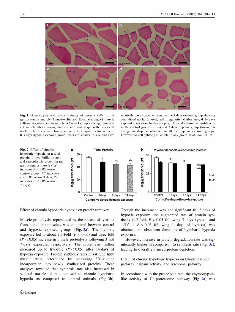
Effect of chronic hypobaric hypoxia on total protein,
myofibrillar and sarcoplasmic protein content
A reduction in protein levels is expected under conditions
where skeletal muscle atrophy is observed. Predictably,
total protein content in the skeletal muscle homogenates
of rats exposed to chronic hypobaric hypoxia of different
durations showed a significant decrease. Following
3 days of exposure, 13% (P \ 0.05) reduction is obtained
in the total protein content of skeletal muscle homoge-
nates of rats followed by 18 and 22% (P \ 0.05)
reduction over the control rats after 7 and 14 days,
respectively (Fig. 2a).
On subfractionation of the total protein, we found a
significant decrease of 30% (P \ 0.05) in the myofibrillar
protein content in the 14 days exposed rats (Fig. 2b). The
7% decrease in sarcoplasmic protein (Fig. 2b), was much
less as compared to the loss of myofibrillar protein.
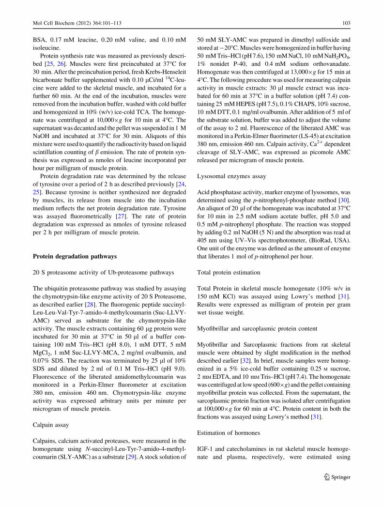
Table 1 Effect of chronic hypobaric hypoxia on physical performance of rats on treadmill
Parameter Control rats 3 days hypoxia 7 days hypoxia 14 days hypoxia
Fatigue time on treadmill running (min) 96.28 ± 3.01 86.56 ± 2.5a 80.07 ± 3.04a, b 73.85 ± 3.57a, b, c
a P \ 0.05 versus control groupb P \ 0.05 versus 3 daysc P \ 0.05 versus 7 days
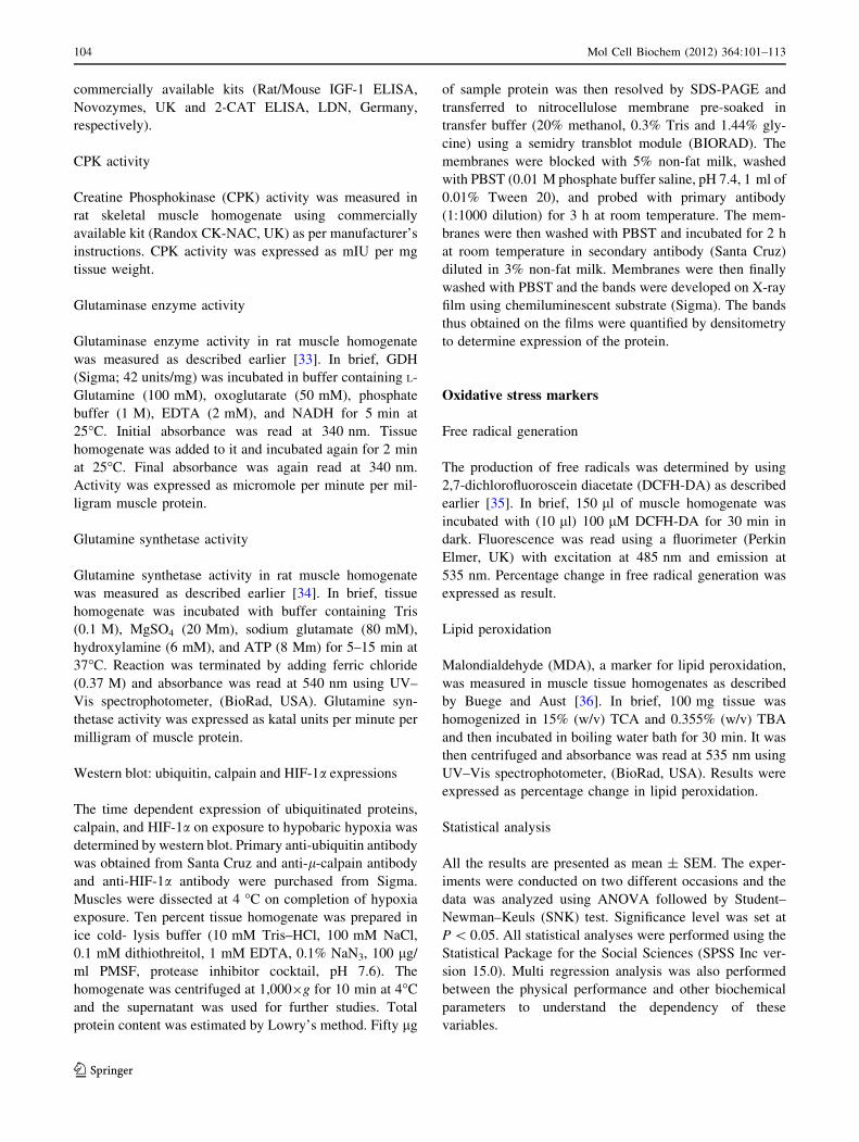
Table 2 Effect of chronic hypobaric hypoxia on rat gastrocnemius muscle weight/tibial length ratio
Parameter Control rats 3 days hypoxia 7 days hypoxia 14 days hypoxia
Muscle weight/tibial length ratio (mg/mm) 41.729 ± 0.2 35.16 ± 0.3a 30.91 ± 0.2a, b 27.729 ± 0.2a, b, c
a P \ 0.05 versus control groupb P \ 0.05 versus 3 daysc P \ 0.05 versus 7 days
Mol Cell Biochem (2012) 364:101–113 105
123
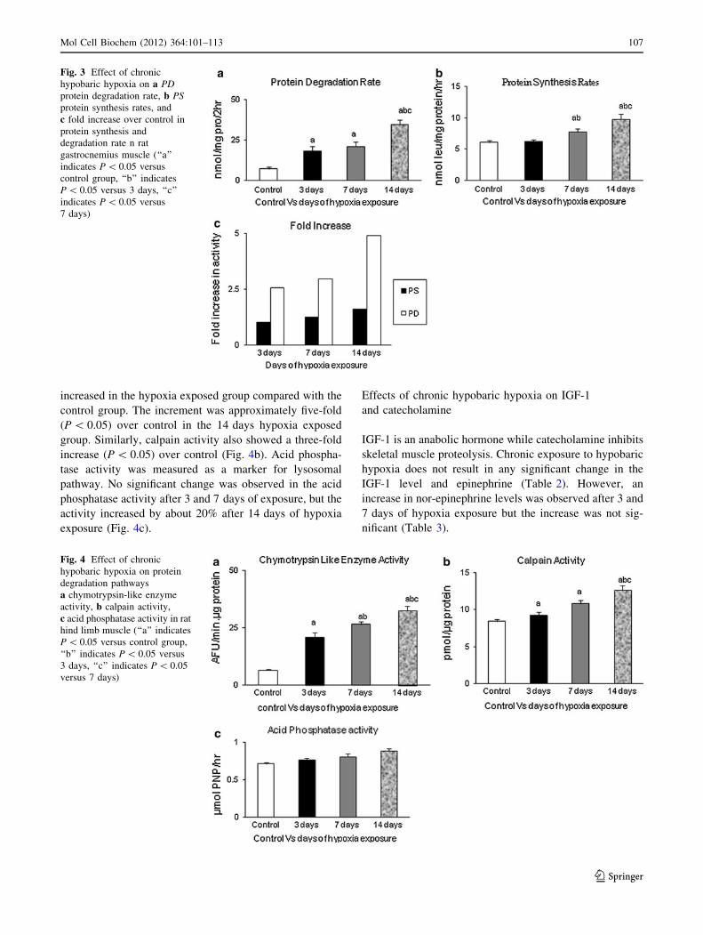
Effect of chronic hypobaric hypoxia on protein turnover
Muscle proteolysis, represented by the release of tyrosine
from hind limb muscles, was compared between control
and hypoxia exposed groups (Fig. 3a). The hypoxic
exposure led to about 2.5-Fold (P \ 0.05) and three-fold
(P \ 0.05) increase in muscle proteolysis following 3 and
7 days exposure, respectively. The proteolysis further
increased up to five-fold (P \ 0.05) after 14 days of
hypoxia exposure. Protein synthesis rates in rat hind limb
muscle were determined by measuring 14C-leucine
incorporation into newly synthesized proteins. These
analyses revealed that synthesis rate also increased in
skeletal muscle of rats exposed to chronic hypobaric
hypoxia as compared to control animals (Fig. 3b).
Though the increment was not significant till 3 days of
hypoxia exposure, the augmented rate of protein syn-
thesis (1.2-fold, P \ 0.05 following 7 days hypoxia and
1.5-fold, P \ 0.05 following 14 days of hypoxia) was
obtained on subsequent durations of hypobaric hypoxia
exposure.
However, increase in protein degradation rate was sig-
nificantly higher in comparison to synthesis rate (Fig. 3c),
leading to overall enhanced protein depletion.
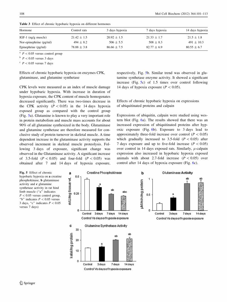
Effect of chronic hypobaric hypoxia on Ub-proteasome
pathway, calpain activity, and lysosomal pathway
In accordance with the proteolytic rate, the chymotrypsin-
like activity of Ub-proteasome pathway (Fig. 4a) was
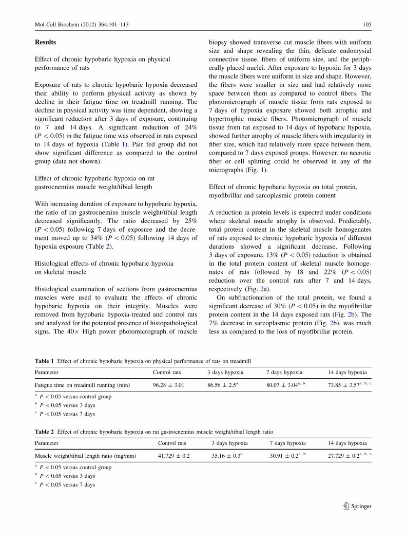
Fig. 1 Hematoxylin and Eosin staining of muscle cells in rat
gastrocnemius muscle. Hematoxylin and Eosin staining of muscle
cells in rat gastrocnemius muscle: a Control group showing transverse
cut muscle fibers having uniform size and shape with peripheral
nuclei. The fibers are closely set with little space between them;
b 3 days hypoxia exposed group fibers are smaller in size and have
relatively more space between them; c 7 days exposed group showing
centralized nuclei (arrow), and irregularity of fiber size; d 14 days
exposed fibers show further atrophy. Thin endomysium is visible only
in the control group (arrow) and 3 days hypoxia group (arrow). A
change in shape is observed in all the hypoxia exposed groups,
however no cell splitting is visible in any group. Scale bar 10 lm
Fig. 2 Effect of chronic
hypobaric hypoxia on a total
protein, b myofibrillar protein
and sarcoplasmic protein in rat
gastrocnemius muscle (‘‘a’’
indicates P \ 0.05 versus
control group, ‘‘b’’ indicates
P \ 0.05 versus 3 days, ‘‘c’’
indicates P \ 0.05 versus
7 days)
106 Mol Cell Biochem (2012) 364:101–113
123
increased in the hypoxia exposed group compared with the
control group. The increment was approximately five-fold
(P \ 0.05) over control in the 14 days hypoxia exposed
group. Similarly, calpain activity also showed a three-fold
increase (P \ 0.05) over control (Fig. 4b). Acid phospha-
tase activity was measured as a marker for lysosomal
pathway. No significant change was observed in the acid
phosphatase activity after 3 and 7 days of exposure, but the
activity increased by about 20% after 14 days of hypoxia
exposure (Fig. 4c).
Effects of chronic hypobaric hypoxia on IGF-1
and catecholamine
IGF-1 is an anabolic hormone while catecholamine inhibits
skeletal muscle proteolysis. Chronic exposure to hypobaric
hypoxia does not result in any significant change in the
IGF-1 level and epinephrine (Table 2). However, an
increase in nor-epinephrine levels was observed after 3 and
7 days of hypoxia exposure but the increase was not sig-
nificant (Table 3).
Fig. 3 Effect of chronic
hypobaric hypoxia on a PDprotein degradation rate, b PSprotein synthesis rates, and
c fold increase over control in
protein synthesis and
degradation rate n rat
gastrocnemius muscle (‘‘a’’
indicates P \ 0.05 versus
control group, ‘‘b’’ indicates
P \ 0.05 versus 3 days, ‘‘c’’
indicates P \ 0.05 versus
7 days)
Fig. 4 Effect of chronic
hypobaric hypoxia on protein
degradation pathways
a chymotrypsin-like enzyme
activity, b calpain activity,
c acid phosphatase activity in rat
hind limb muscle (‘‘a’’ indicates
P \ 0.05 versus control group,
‘‘b’’ indicates P \ 0.05 versus
3 days, ‘‘c’’ indicates P \ 0.05
versus 7 days)
Mol Cell Biochem (2012) 364:101–113 107
123
Effects of chronic hypobaric hypoxia on enzymes CPK,
glutaminase, and glutamine synthetase
CPK levels were measured as an index of muscle damage
under hypobaric hypoxia. With increase in duration of
hypoxia exposure, the CPK content of muscle homogenates
decreased significantly. There was two-times decrease in
the CPK activity (P \ 0.05) in the 14 days hypoxia
exposed group as compared with the control group
(Fig. 5a). Glutamine is known to play a very important role
in protein metabolism and muscle mass accounts for about
90% of all glutamine synthesized in the body. Glutaminase
and glutamine synthetase are therefore measured for con-
clusive study of protein turnover in skeletal muscle. A time
dependent increase in the glutaminase activity supports the
observed increment in skeletal muscle proteolysis. Fol-
lowing 3 days of exposure, significant change was
observed in the Glutaminase activity. A significant increase
of 3.5-fold (P \ 0.05) and four-fold (P \ 0.05) was
obtained after 7 and 14 days of hypoxia exposure,
respectively, Fig. 5b. Similar trend was observed in glu-
tamine synthetase enzyme activity. It showed a significant
increase (Fig. 5c) of 1.5 times over control following
14 days of hypoxia exposure (P \ 0.05).
Effects of chronic hypobaric hypoxia on expressions
of ubiquitinated proteins and calpain
Expressions of ubiquitin, calpain were studied using wes-
tern blot (Fig. 6a). The results showed that there was an
increased expression of ubiquitinated proteins after hyp-
oxic exposure (Fig. 6b). Exposure to 3 days lead to
approximately three-fold increase over control (P \ 0.05)
which gradually increased to 3.5-fold (P \ 0.05) after
7 days exposure and up to five-fold increase (P \ 0.05)
over control in 14 days exposed rats. Similarly, l-calpain
expression also increased in hypobaric hypoxia exposed
animals with about 2.7-fold increase (P \ 0.05) over
control after 14 days of hypoxia exposure (Fig. 6c).
Table 3 Effect of chronic hypobaric hypoxia on different hormones
Hormone Control rats 3 days hypoxia 7 days hypoxia 14 days hypoxia
IGF-1 (ng/g muscle) 21.42 ± 1.5 20.92 ± 1.5 21.33 ± 1.7 21.5 ± 1.8
Nor-epinephrine (pg/ml) 494 ± 8.2 506 ± 5.5 508 ± 8.3 491 ± 10.3
Epinephrine (pg/ml) 78.88 ± 3.8 86.66 ± 7.5 82.77 ± 6.9 80.55 ± 6.7
a P \ 0.05 versus control groupb P \ 0.05 versus 3 daysc P \ 0.05 versus 7 days
Fig. 5 Effect of chronic
hypobaric hypoxia on a creatine
phosphokinase, b glutaminase
activity and c glutamine
synthetase activity in rat hind
limb muscle (‘‘a’’ indicates
P \ 0.05 versus control group,
‘‘b’’ indicates P \ 0.05 versus
3 days, ‘‘c’’ indicates P \ 0.05
versus 7 days)
108 Mol Cell Biochem (2012) 364:101–113
123
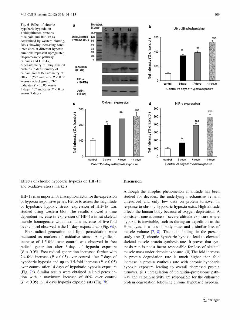
Effects of chronic hypobaric hypoxia on HIF-1aand oxidative stress markers
HIF-1a is an important transcription factor for the expression
of hypoxia responsive genes. Hence to assess the magnitude
of hypobaric hypoxic stress, expression of HIF-1a was
studied using western blot. The results showed a time
dependent increase in expression of HIF-1a in rat skeletal
muscle homogenate with maximum increase of five-fold
over control observed in the 14 days exposed rats (Fig. 6d).
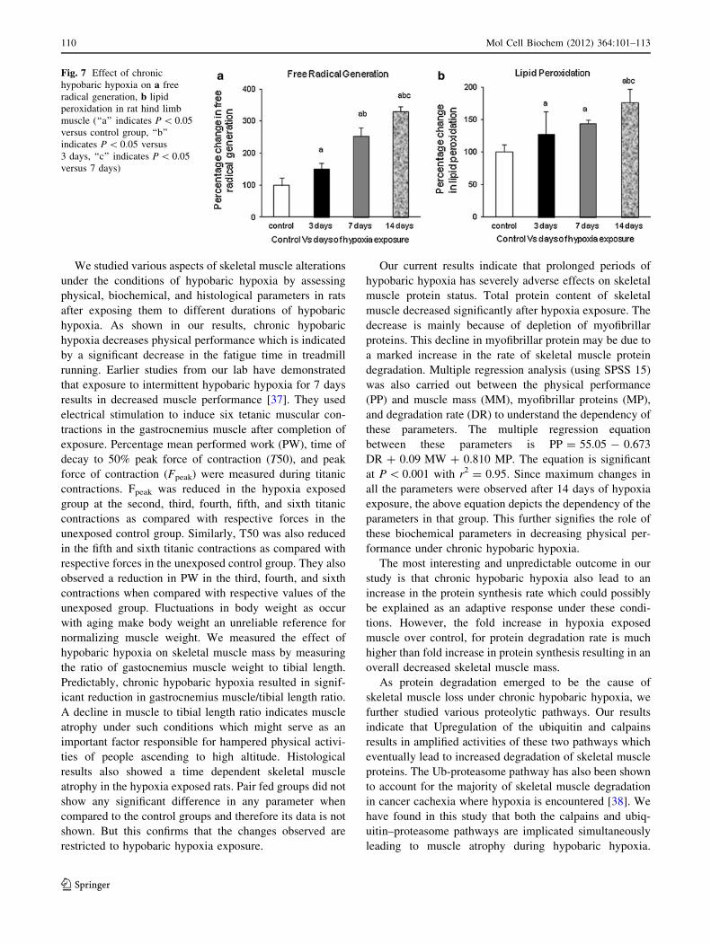
Free radical generation and lipid peroxidation were
measured as markers of oxidative stress. A significant
increase of 1.5-fold over control was observed in free
radical generation after 3 days of hypoxia exposure
(P \ 0.05). Free radical generation increased further with
2.4-fold increase (P \ 0.05) over control after 7 days of
hypobaric hypoxia and up to 3.5-fold increase (P \ 0.05)
over control after 14 days of hypobaric hypoxia exposure
(Fig. 7a). Similar results were obtained in lipid peroxida-
tion with a maximum increase of 80% over control
(P \ 0.05) in 14 days hypoxia exposed rats (Fig. 7b).
Discussion
Although the atrophic phenomenon at altitude has been
studied for decades, the underlying mechanisms remain
unresolved and only few data on protein turnover in
response to chronic hypobaric hypoxia exist. High altitude
affects the human body because of oxygen deprivation. A
consistent consequence of severe altitude exposure where
hypoxia is inevitable, such as during an expedition to the
Himalayas, is a loss of body mass and a similar loss of
muscle volume [7, 8]. The main findings in the present
study are: (i) chronic hypobaric hypoxia lead to elevated
skeletal muscle protein synthesis rate. It proves that syn-
thesis rate is not a factor responsible for loss of skeletal
muscle mass under chronic exposure. (ii) The fold increase
in protein degradation rate is much higher than fold
increase in protein synthesis rate with chronic hypobaric
hypoxic exposure leading to overall decreased protein
turnover. (iii) upregulation of ubiquitin–proteasome path-
way and calpain activity are responsible for the enhanced
protein degradation following chronic hypobaric hypoxia.
Fig. 6 Effect of chronic
hypobaric hypoxia on
a ubiquitinated proteins,
l-calpain and HIF-1a as
determined by western blotting.
Blots showing increasing band
intensities at different hypoxia
durations represent upregulated
ub-proteasome pathway,
calpains and HIF-1a,
b densitometry of ubiquitinated
proteins, c densitometry of
calpain and d Densitometry of
HIF-1a (‘‘a’’ indicates P \ 0.05
versus control group, ‘‘b’’
indicates P \ 0.05 versus
3 days, ‘‘c’’ indicates P \ 0.05
versus 7 days)
Mol Cell Biochem (2012) 364:101–113 109
123
We studied various aspects of skeletal muscle alterations
under the conditions of hypobaric hypoxia by assessing
physical, biochemical, and histological parameters in rats
after exposing them to different durations of hypobaric
hypoxia. As shown in our results, chronic hypobaric
hypoxia decreases physical performance which is indicated
by a significant decrease in the fatigue time in treadmill
running. Earlier studies from our lab have demonstrated
that exposure to intermittent hypobaric hypoxia for 7 days
results in decreased muscle performance [37]. They used
electrical stimulation to induce six tetanic muscular con-
tractions in the gastrocnemius muscle after completion of
exposure. Percentage mean performed work (PW), time of
decay to 50% peak force of contraction (T50), and peak
force of contraction (Fpeak) were measured during titanic
contractions. Fpeak was reduced in the hypoxia exposed
group at the second, third, fourth, fifth, and sixth titanic
contractions as compared with respective forces in the
unexposed control group. Similarly, T50 was also reduced
in the fifth and sixth titanic contractions as compared with
respective forces in the unexposed control group. They also
observed a reduction in PW in the third, fourth, and sixth
contractions when compared with respective values of the
unexposed group. Fluctuations in body weight as occur
with aging make body weight an unreliable reference for
normalizing muscle weight. We measured the effect of
hypobaric hypoxia on skeletal muscle mass by measuring
the ratio of gastocnemius muscle weight to tibial length.
Predictably, chronic hypobaric hypoxia resulted in signif-
icant reduction in gastrocnemius muscle/tibial length ratio.
A decline in muscle to tibial length ratio indicates muscle
atrophy under such conditions which might serve as an
important factor responsible for hampered physical activi-
ties of people ascending to high altitude. Histological
results also showed a time dependent skeletal muscle
atrophy in the hypoxia exposed rats. Pair fed groups did not
show any significant difference in any parameter when
compared to the control groups and therefore its data is not
shown. But this confirms that the changes observed are
restricted to hypobaric hypoxia exposure.
Our current results indicate that prolonged periods of
hypobaric hypoxia has severely adverse effects on skeletal
muscle protein status. Total protein content of skeletal
muscle decreased significantly after hypoxia exposure. The
decrease is mainly because of depletion of myofibrillar
proteins. This decline in myofibrillar protein may be due to
a marked increase in the rate of skeletal muscle protein
degradation. Multiple regression analysis (using SPSS 15)
was also carried out between the physical performance
(PP) and muscle mass (MM), myofibrillar proteins (MP),
and degradation rate (DR) to understand the dependency of
these parameters. The multiple regression equation
between these parameters is PP = 55.05 - 0.673
DR ? 0.09 MW ? 0.810 MP. The equation is significant
at P \ 0.001 with r2 = 0.95. Since maximum changes in
all the parameters were observed after 14 days of hypoxia
exposure, the above equation depicts the dependency of the
parameters in that group. This further signifies the role of
these biochemical parameters in decreasing physical per-
formance under chronic hypobaric hypoxia.
The most interesting and unpredictable outcome in our
study is that chronic hypobaric hypoxia also lead to an
increase in the protein synthesis rate which could possibly
be explained as an adaptive response under these condi-
tions. However, the fold increase in hypoxia exposed
muscle over control, for protein degradation rate is much
higher than fold increase in protein synthesis resulting in an
overall decreased skeletal muscle mass.
As protein degradation emerged to be the cause of
skeletal muscle loss under chronic hypobaric hypoxia, we
further studied various proteolytic pathways. Our results
indicate that Upregulation of the ubiquitin and calpains
results in amplified activities of these two pathways which
eventually lead to increased degradation of skeletal muscle
proteins. The Ub-proteasome pathway has also been shown
to account for the majority of skeletal muscle degradation
in cancer cachexia where hypoxia is encountered [38]. We
have found in this study that both the calpains and ubiq-
uitin–proteasome pathways are implicated simultaneously
leading to muscle atrophy during hypobaric hypoxia.
Fig. 7 Effect of chronic
hypobaric hypoxia on a free
radical generation, b lipid
peroxidation in rat hind limb
muscle (‘‘a’’ indicates P \ 0.05
versus control group, ‘‘b’’
indicates P \ 0.05 versus
3 days, ‘‘c’’ indicates P \ 0.05
versus 7 days)
110 Mol Cell Biochem (2012) 364:101–113
123
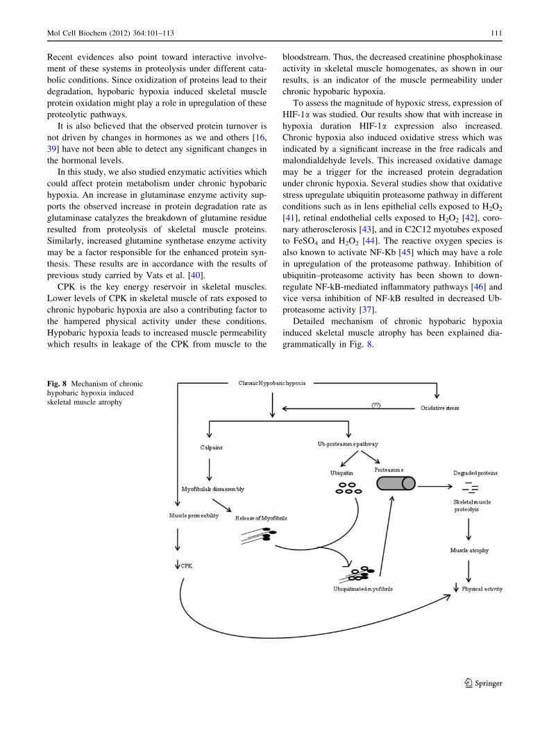
Recent evidences also point toward interactive involve-
ment of these systems in proteolysis under different cata-
bolic conditions. Since oxidization of proteins lead to their
degradation, hypobaric hypoxia induced skeletal muscle
protein oxidation might play a role in upregulation of these
proteolytic pathways.
It is also believed that the observed protein turnover is
not driven by changes in hormones as we and others [16,
39] have not been able to detect any significant changes in
the hormonal levels.
In this study, we also studied enzymatic activities which
could affect protein metabolism under chronic hypobaric
hypoxia. An increase in glutaminase enzyme activity sup-
ports the observed increase in protein degradation rate as
glutaminase catalyzes the breakdown of glutamine residue
resulted from proteolysis of skeletal muscle proteins.
Similarly, increased glutamine synthetase enzyme activity
may be a factor responsible for the enhanced protein syn-
thesis. These results are in accordance with the results of
previous study carried by Vats et al. [40].
CPK is the key energy reservoir in skeletal muscles.
Lower levels of CPK in skeletal muscle of rats exposed to
chronic hypobaric hypoxia are also a contributing factor to
the hampered physical activity under these conditions.
Hypobaric hypoxia leads to increased muscle permeability
which results in leakage of the CPK from muscle to the
bloodstream. Thus, the decreased creatinine phosphokinase
activity in skeletal muscle homogenates, as shown in our
results, is an indicator of the muscle permeability under
chronic hypobaric hypoxia.
To assess the magnitude of hypoxic stress, expression of
HIF-1a was studied. Our results show that with increase in
hypoxia duration HIF-1a expression also increased.
Chronic hypoxia also induced oxidative stress which was
indicated by a significant increase in the free radicals and
malondialdehyde levels. This increased oxidative damage
may be a trigger for the increased protein degradation
under chronic hypoxia. Several studies show that oxidative
stress upregulate ubiquitin proteasome pathway in different
conditions such as in lens epithelial cells exposed to H2O2
[41], retinal endothelial cells exposed to H2O2 [42], coro-
nary atherosclerosis [43], and in C2C12 myotubes exposed
to FeSO4 and H2O2 [44]. The reactive oxygen species is
also known to activate NF-Kb [45] which may have a role
in upregulation of the proteasome pathway. Inhibition of
ubiquitin–proteasome activity has been shown to down-
regulate NF-kB-mediated inflammatory pathways [46] and
vice versa inhibition of NF-kB resulted in decreased Ub-
proteasome activity [37].
Detailed mechanism of chronic hypobaric hypoxia
induced skeletal muscle atrophy has been explained dia-
grammatically in Fig. 8.
Fig. 8 Mechanism of chronic
hypobaric hypoxia induced
skeletal muscle atrophy
Mol Cell Biochem (2012) 364:101–113 111
123
Conclusion
Excessive protein degradation during exposure to chronic
hypobaric hypoxia resulted in skeletal muscle atrophy
which could be detrimental for performing any physical
task under these conditions. The majorly affected protein is
myofibrillar protein and the pathways responsible for this
loss of skeletal muscle mass are Ub-proteasome pathway
and calpains. Attenuation of these pathways could be sig-
nificant for preventing or impeding the observed skeletal
muscle atrophy at high altitude and under various catabolic
conditions mimicking the symptoms of high-altitude mal-
adies such as COPD. Our results also concluded that
increased oxidative stress induced at high altitude may also
be important factor responsible for the skeletal muscle loss.
Acknowledgments This study was supported and funded by the
Defence Research and Development organization, Ministry of
Defence, Government of India. The authors are grateful to the
Director, Defence Institute of Physiology and Allied Sciences, Delhi
for providing facilities to carry out these investigations.
References
1. Fulco CS, Friedlander AL, Muza SR, Rock PB, Robinson S,
Lammi E, Baker R, Fulco CJ, Lewis SF, Cymerman A (2002)
Energy intake deficit and physical performance at altitude. Aviat
Space Environ Med 73:758–765
2. Bharadwaj H, Prasad J, Pramanik SN, Kishnani S, Zachariah T,
Chaudhary KL, Sridharan K, Srivastava KK (2000) Effect of
prolonged exposure to high altitude on skeletal muscles of Indian
soldiers. Def Sci J 50:167–176
3. Macdonald JH, Oliver SJ, Hillyer K, Sanders S, Smith Z, Wil-
liams C, Yates D, Ginnever H, Scanlon E, Roberts E, Murphy D,
Lawley J, Chichester E (2009) Body composition at high altitude:
a randomized placebo-controlled trial of dietary carbohydrate
supplementation. Am J Clin Nutr 90:1193–1202
4. Sridharan K, Mukherjee AK, Grover SK, Kumaria MML, Arora BS,
Rai RM (1987) Assessment of nutritional status and physical work
capacity of road construction workers at altitude of 2150–2750 m
on two different ration scales. Nutr Rep Int 35:1269–1277
5. Brouns F (1992) Nutritional aspects of health and performance at
lowland and altitude. Int J Sports Med 13:S100–S106
6. Schols AM (2002) Pulmonary cachexia. Int J Cardiol 85:101–110
7. Hoppeler H, Vogt M (2001) Muscle tissue adaptations to
hypoxia. J Exp Biol 204:3133–3139
8. Hoppeler H, Kleinert E, Schlegel C, Claassen H, Howald H,
Kayar SR, Cerretelli P (1990) Muscular exercise at high altitude.
II. Morphological adaptation of skeletal muscle to chronic
hypoxia. Int J Sports Med 11:S3–S9
9. Martinelli M, Winterhalder R, Cerretelli P, Howald H, Hoppeler
H (1990) Muscle lipofuscin content and satellite cell volume is
increased after high altitude exposure in humans. Experientia
46:672–676
10. Kung-tung C, Yu-yawn C, Huey-june W, Chen-kang C, Wen-
tsung L, Yen-yuan L, Chieh-chung L, Rong-sen Y, Jung-charng L
(2008) Decreased anaerobic performance and hormone adapta-
tion after expedition to Peak Lenin. Chin Med J 121:2229–2233
11. Bigard AX, Douce P, Merino D, Lienhard F, Guezennec CY
(1996) Changes in dietary protein intake fail to prevent decrease
in muscle growth induced by severe hypoxia in rats. J Appl
Physiol 80:208–215
12. Preedy VR, Smith DM, Sugden PH (1985) The effects of 6 hr
hypoxia on protein synthesis in rat tissue in vivo & in vitro.
Biochem J 228:179–185
13. Preedy VR, Sugden PH (1989) The effects of fasting or
hypoxia on rates of protein synthesis in vivo in subcellular
fractions of rat heart and gastrocnemius muscle. Biochem J
257:519–527
14. Iioka TK, Sugito K, Moriya T, Kuriyama T (2002) Effects of
insulin like growth factor on nitrogen balance during hypoxic
exposure. Eur Respir J 20:293–299
15. Vigano A, Ripamonti M, Palma SD, Capitanio D, Vasso M, Wait
R, Lundby C, Cerretelli P, Gelfi C (2008) Proteins modulation in
human skeletal muscle in the early phase of adaptation to
hypobaric hypoxia. Proteomics 8:4668–4679
16. Imoberdorf R, Garlick PJ, McNurlan MA, Casella GA, Marini
JC, Turgay M, Bartsch P, Ballmer PE (2006) Skeletal muscle
protein synthesis after active or passive ascent to high altitude.
Med Sci Sports Exerc 38:1082–1087
17. Holm L, Haslund ML, Robach P, van Hall G, Calbet JA,
Saltin B, Lundby C (2010) Skeletal muscle myofibrillar and
sarcoplasmic protein synthesis rates are affected differently by
altitude-induced hypoxia in native lowlanders. PLoS ONE
20:e15606
18. Rennie MJ (1985) Muscle protein turnover and the wasting due to
injury and disease. Br Med Bull 41:257–264
19. Cai D, Lee KK, Li M, Tang MK, Chan KM (2004) Ubiquitin
expression is upregulated in human and rat skeletal muscles
during aging. Arch Biochem Biophys 425:42–50
20. Reid MB (2005) Response of the ubiquitin–proteasome pathway
to changes in muscle activity. Am J Physiol Regul Integr Comp
Physiol 288:R1423–R1431
21. Mammucari C, Milan G, Romanello V, Masiero E, Rudolf R,
Piccolo PD, Burden SJ, Lisi RD, Sandri C, Zhao J, Goldberg AL,
Schiaffino S, Sandri M (2007) FoxO3 controls autophagy in
skeletal muscle in vivo. Cell Metab 6:458–471
22. Attaix D, Mosoni L, Dardevet D, Combaret L, Mirand PP, Gri-
zard J (2005) Altered responses in skeletal muscle protein turn-
over during aging in anabolic and catabolic periods. Int J
Biochem Cell Biol 37:2098–2114
23. Enns DL, Raastad T, Ugelstad I, Belcastro AN (2007) Calpain/
calpastatin activities and substrate depletion patterns during
hindlimb unweighting and reweighting in skeletal muscle. Eur J
Appl Physiol 100:445–455
24. Dardevet D, Sornet C, Vary T, Grizard J (1996) Phosphotidyli-
nositol 3-kinase and p70 S6 kinase participate in the regulation of
protein turnover in skeletal muscle by insulin and insulin-like
growth factor I. Endocrinology 137:4089–4094
25. Vary TC, Dardevet D, Grizard J, Voisin L, Buffiere C, Denis P,
Breuille D, Obled C (1998) Differential regulation of skeletal
muscle protein turnover by insulin and IGF-1 after bacteremia.
Am J Physiol Endocrinol Metab 275:E584–E593
26. Ventrucci G, Mello MAR, Marcondes G (2004) Proteasome
activity is altered in skeletal muscle tissue of tumour-bearing rats
fed a leucine-rich diet. Endocr Relat Cancer 11:887–895
27. Waalkes TP, Udenfriend S (1957) A fluorimetric method for the
estimation of tyrosine in plasma and tissues. J Lab Clin Med
50:733–736
28. Hepple RT, Qin M, Nakamoto H, Goto S (2008) Caloric
restriction optimizes the proteasome activity. Am J Physiol Regul
Integr Comp Physiol 295:R1231–R1237
29. Mastrocola R, Reffo P, Penna F, Tomasinelli CE, Boccuzzi G,
Baccino FM, Aragno M, Costelli P (2008) Muscle wasting in
diabetic and in tumor bearing rats: role of oxidative stress. Free
Radic Biol Med 44:584–593
112 Mol Cell Biochem (2012) 364:101–113
123
30. Oron U (1990) Proteolytic enzyme activity in rat hind limb
muscle in fetus and during post natal development. Int J Dev Biol
34:457–460
31. Lowry OH, Rosebrough NJ, Farr AL, Randall RJ (1951) Protein
measurement with the folin phenol reagent. J Biol Chem
193:265–275
32. Koopman R, Gehrig SM, Leger B, Walrand S, Murphy KT,
Lynch GS (2010) Cellular mechanisms underlying temporal
changes in skeletal muscle protein synthesis and breakdown
during chronic b-adrenoceptor stimulation in mice. J Physiol
588:4811–4823
33. Kvamme E, Torgner IA, Svenneby G (1985) Glutaminase from
mammalian tissue. Methods Enzymol 113:241–244
34. Elliott WH (1955) Glutamine synthesis. In: Colowick SP, Kaplan
NO (eds) Methods enzymol II. pp 337–339
35. Cathcart R, Schwiers E, Ames BN (1983) Detection of pico mole
levels of hyderoperoxides using fluorescent dichlorofluoroscein
assay. Anal Biochem 134:111–116
36. Buege JA, Aust SD (1978) Microsomal lipid peroxidation.
Methods Enzymol 52:302–310
37. Dutta A, Ray K, Singh VK, Vats P, Singh SN, Singh SB (2008)
L-carnitine supplementation attenuates intermittent hypoxia-
induced oxidative stress and delays muscle fatigue in rats. Exp
Physiol 93:1139–1146
38. Tisdale MJ (2005) The Ub-proteasome pathway as a therapeutic
target for muscle wasting. J Support Oncol 3:209–217
39. Howald H, Pette D, Simoneau JA, Uber A, Hoppeler H, Cerretelli
P (1990) Effect of chronic hypoxia on muscle enzyme activities.
Int J Sports Med S10–S14
40. Vats P, Mukherjee AK, Kumria MM, Singh SN, Patil SK,
Rangnathan S, Sridharan K (1999) Changes in activity levels of
glutamine synthetase, glutaminase and glycogen synthetase in
rats subjected to hypoxic stress. Int J Biometeorol 42:205–209
41. Shang F, Gong Taylor A (1997) Activity of ubiquitin-dependent
pathway in response to oxidative stress. Ubiquitin-activating
enzyme is transiently up-regulated. J Biol Chem 272:23086–
23093
42. Fernandes R, Ramalho J, Pereira P (2006) Oxidative stress
upregulates ubiquitin proteasome pathway in retinal endothelial
cells. Mol Vis 12:1526–1535
43. Hermann J, Gulati R, Napoli C, Woodrum LLO, Porcel MR, Sica
V, Simari RD, Ciechanover A, Lerman A (2003) Oxidative
stress-related increase in ubiquitination in early coronary ath-
erogenesis. FASEB 17:1730–1732
44. Gomes M, Maria CC, Tisdale MJ (2002) Induction of protein
catabolism and the ubiquitin proteasome pathway by mild oxi-
dative stress. Cancer Lett 180:69–74
45. Sagi SKS, Patir H, Mishra C, Pradhan G, Mastoori SR, Ila-
vazhagan G (2008) Role of oxidative stress and NFkB in hypoxia
induced pulmonary edema. Exp Biol Med 233:1088–1098
46. Marfella R, Amico MD, Filippo CD, Baldi A, Siniscalchi M,
Sasso FC, Portoghese M, Carbonara O, Crescenzi B, Sangiuolo P,
Nicoletti GF, Rossiello R, Ferraraccio F, Cacciapuoti F, Verza M,
Coppola L, Rossi F, Paolisso G (2006) Increased activity of the
ubiquitin-proteasome system in patients with symptomatic car-
otid disease is associated with enhanced inflammation and may
destabilize the atherosclerotic plaque: effects of rosiglitazone
treatment. JACC 47:2444–2455
Mol Cell Biochem (2012) 364:101–113 113
123
Related Documents
![Effects of Prolonged Exposure to Hypobaric Hypoxia on ... · conditions (chamber, high-altitude) [16]. Operation Everest II (simulated ascent of Mount Everest over 40 days in a hypobaric](https://static.cupdf.com/doc/110x72/5f6274c4768114196b61bb97/effects-of-prolonged-exposure-to-hypobaric-hypoxia-on-conditions-chamber-high-altitude.jpg)