Sleep, 18(8):617-634 © 1995 American Sleep Disorders Association and Sleep Research Society Sleep-disordered Breathing State of the Art Review Chronic Alveolar Hypoventilation: A Review for the Clinician Thomas J. Martin and Mark H. Sanders Division of Pulmonary, Allergy and Critical Care Medicine, University of Pittsburgh and Oakland VA Medical Centers, Pittsburgh, Pennsylvania, U.S.A. The term hypoventilation defines a condition in which alveolar ventilation (VA) is insufficient to meet the individual's metabolic demands, resulting in an inappropriately high arterial carbon dioxide tension (Paco 2 ). The normal range ofPaco 2 at rest is generally accepted to be between 35 and 45 mm Hg. Alveolar hypoventilation may be an acute or chronic manifes- tation of a variety of pathologic processes. Although patients with acute alveolar hypoventilation generally present in a clinically obvious fashion, chronic alveolar hypoventilation may be characterized by an insidi- ously progressive course. In the following review, the pathophysiology of chronic alveolar hypo ventilation (a term used inter- changeably with chronic ventilatory failure) will be examined and specific disorders discussed to illustrate important concepts. Emphasis will be placed on the contributory role of the physiologic changes that occur during sleep. Currently available diagnostic and man- agement techniques will be examined, with particular emphasis placed on new therapeutic modalities that are proving to be valuable in the long-term manage- ment of patients with chronic alveolar hypoventila- tion. PATHOPHYSIOLOGY Under steady state conditions in normal individuals, Paco2 is maintained within tightly controlled limits. The Paco 2 reflects the balance between CO 2 production Accepted for publication May 1995. Address correspondence to Mark H. Sanders, M.D., Division of Pulmonary, Allergy and Critical Care Medicine, Montefiore Uni- versity Hospital, Room S-643, 3459 Fifth Avenue, Pittsburgh, PA 15213. by the body (V co 2 ) and its elimination through the alveolar ventilation, as expressed in the relationship: Paco 2 = k(VC02/V A) where Paco2 is the arterial carbon dioxide tension (mm Hg), V cO 2 is the rate of metabolic production of carbon dioxide (milliliters per minute), and k is a constant. Increased V C02 generally does not lead to chronic hypercapnia without concomitant decreases in VA due to coexistent ventilatory drive abnormalities, restric- tive or obstructive thoracic abnormalities and/or ab- normal transduction of the central nervous system (CNS) neural respiratory signal into mechanical activ- ity. Thus decreased VA is the ultimate mechanism of increased Paco 2 in chronic alveolar hypoventilation. The VA is a reflection of the total volume of gas breathed over time (minute ventilation, VE) and the portion of each breath that ventilates lung units where gas ex- change occurs. Ventilated portions of the respiratory system where oxygen and CO 2 are not exchanged are defined as dead space (VD). The fraction of each breath that fills the dead space and is therefore "wasted" is the dead space-to-tidal volume ratio (VDIVT). Either reduced VT or increased VD can increase VD/VT and contribute to alveolar hypoventilation. Clinically, these aberrations may cause chronic ventilatory failure if the breathing rate is not increased sufficiently to allow aug- mentation of VE to compensate for the reduction in VA. It is useful to group disorders causing chronic al- veolar hypoventilation on a clinical basis into those in which the patient "Can't Breathe" (i.e. because ofneu- romuscular weakness or excessive work of breathing) and those in which the patients "Won't Breathe" (i.e. due to inadequate drive from the ventilatory centers in the CNS) (Table 1). Many disorders include features 617 Downloaded from https://academic.oup.com/sleep/article/18/8/617/2749686 by guest on 13 January 2022

Welcome message from author
This document is posted to help you gain knowledge. Please leave a comment to let me know what you think about it! Share it to your friends and learn new things together.
Transcript
Sleep, 18(8):617-634 © 1995 American Sleep Disorders Association and Sleep Research Society
Sleep-disordered Breathing
State of the Art Review
Chronic Alveolar Hypoventilation: A Review for the Clinician
Thomas J. Martin and Mark H. Sanders
Division of Pulmonary, Allergy and Critical Care Medicine, University of Pittsburgh and Oakland VA Medical Centers, Pittsburgh, Pennsylvania, U.S.A.
The term hypoventilation defines a condition in which alveolar ventilation (VA) is insufficient to meet the individual's metabolic demands, resulting in an inappropriately high arterial carbon dioxide tension (Paco2). The normal range ofPaco2 at rest is generally accepted to be between 35 and 45 mm Hg. Alveolar hypoventilation may be an acute or chronic manifestation of a variety of pathologic processes. Although patients with acute alveolar hypoventilation generally present in a clinically obvious fashion, chronic alveolar hypoventilation may be characterized by an insidiously progressive course.
In the following review, the pathophysiology of chronic alveolar hypo ventilation (a term used interchangeably with chronic ventilatory failure) will be examined and specific disorders discussed to illustrate important concepts. Emphasis will be placed on the contributory role of the physiologic changes that occur during sleep. Currently available diagnostic and management techniques will be examined, with particular emphasis placed on new therapeutic modalities that are proving to be valuable in the long-term management of patients with chronic alveolar hypoventilation.
PATHOPHYSIOLOGY
Under steady state conditions in normal individuals, Paco2 is maintained within tightly controlled limits. The Paco2 reflects the balance between CO2 production
Accepted for publication May 1995. Address correspondence to Mark H. Sanders, M.D., Division of
Pulmonary, Allergy and Critical Care Medicine, Montefiore University Hospital, Room S-643, 3459 Fifth Avenue, Pittsburgh, PA 15213.
by the body (V co2) and its elimination through the alveolar ventilation, as expressed in the relationship:
Paco2 = k(V C02/V A)
where Paco2 is the arterial carbon dioxide tension (mm Hg), V cO2 is the rate of metabolic production of carbon dioxide (milliliters per minute), and k is a constant.
Increased V C02 generally does not lead to chronic hypercapnia without concomitant decreases in VA due to coexistent ventilatory drive abnormalities, restrictive or obstructive thoracic abnormalities and/or abnormal transduction of the central nervous system (CNS) neural respiratory signal into mechanical activity. Thus decreased VA is the ultimate mechanism of increased Paco2 in chronic alveolar hypoventilation. The VA is a reflection of the total volume of gas breathed over time (minute ventilation, VE) and the portion of each breath that ventilates lung units where gas exchange occurs. Ventilated portions of the respiratory system where oxygen and CO2 are not exchanged are defined as dead space (VD). The fraction of each breath that fills the dead space and is therefore "wasted" is the dead space-to-tidal volume ratio (VDIVT). Either reduced VT or increased VD can increase VD/VT and contribute to alveolar hypoventilation. Clinically, these aberrations may cause chronic ventilatory failure if the breathing rate is not increased sufficiently to allow augmentation of VE to compensate for the reduction in VA.
It is useful to group disorders causing chronic alveolar hypoventilation on a clinical basis into those in which the patient "Can't Breathe" (i.e. because ofneuromuscular weakness or excessive work of breathing) and those in which the patients "Won't Breathe" (i.e. due to inadequate drive from the ventilatory centers in the CNS) (Table 1). Many disorders include features
617
Dow
nloaded from https://academ
ic.oup.com/sleep/article/18/8/617/2749686 by guest on 13 January 2022
618 T. 1. MARTIN AND M. H. SANDERS
TABLE 1. Etiologies for chronic hYPol'entilation
Won't breathe Impaired ventilatory drive
Primary alveolar hypoventilation Structural defects
CNS tumor Bulbar poliomyelitis Amold-Chiari malformation Cerebrovascular accident
Metabolic Severe metabolic alkalosis
Medications (alone and/or in combination) Narcotics, sedati ves. ethanol
Can't breathe Neuromuscular disorders
Neuropathic processes Amyotrophic lateral sclerosis Spinal cord injury Diaphragmatic paralysis/weakness
Neuromuscular junction disorders Myasthenia gravis Eaton-Lambert syndrome
Myopathy Muscular dystrophy Myotonic dystrophy Metabolic (e.g. acid maltase deficiency) Congenital
Restrictive chest abnormalities Kyphoscoliosis
Idiopathic, congenital Secondary (post-polio, neuromuscular, connective tissue
diseases) Interstitial lung disease (late manifestation) Post-tuberculosis (thoracoplasty) Pulmonary resection
Mixed (may have both features) Mvxedema Ai~way obstruction
Chronic obstructive pulmonary disease Obesity-hypoventilation syndrome
of both groups, and consequently not all disease processes associated with chronic hypoventilation can be easily categorized. Some disorders may have well documented effects on both the ventilatory muscles and chemoresponsiveness (myxedema). In other disorders, where abnormal pulmonary or chest wall mechanics increase the work required to maintain a given level of ventilation, a compensatory increase in the ventilatory drive may not occur, and in fact decreased ventilatory responsiveness may exist. If reduced responsiveness does exist, this may be physiologically important as it may allow such individuals to decrease their work of breathing, albeit at the expense of developing alveolar hypoventilation.
CONSEQUENCES OF CHRONIC HYPOVENTILA TION
The manifestations of hypercapnia are dependent upon the rapidity of its onset. The presentation of in-
SI('ep. r·of. 18, No.8, 1995
TABLE 2. Clinical consequences of chronic hYPol'entilation
Central nervous system effects Altered mentation Hypersomnolence Headache
Pulmonary artery hypertension Cor pulmonale
Elevated serum bicarbonate Impaired diaphragm function (possible) Disability
dividuals with chronic hypo ventilation can be nonspecific and may be accentuated by coexistent hypoxemia. Hypercapnia, either directly or indirectly, affects the CNS, ventilatory system, heart and kidneys (Table 2). The CNS effects of chronic hypercapnia include hypersomnolence, altered mentation and headache. Whereas diaphragm contractility is impaired in the presence of acute hypercapnia (1,2), whether contractility in the chronic state is similarly influenced has not been studied. Pulmonary artery pressures may be elevated in patients with chronic hypercapnia, although to a large extent this is related to concomitant hypoxemia. Pulmonary artery hypertension may lead to cor pulmonale, reduced cardiac output, disability and death. Increased resorption of urinary bicarbonate by the kidneys serves to buffer the existing respiratory acidosis.
CONTROL OF VENTILATION AND CHANGES WITH SLEEP
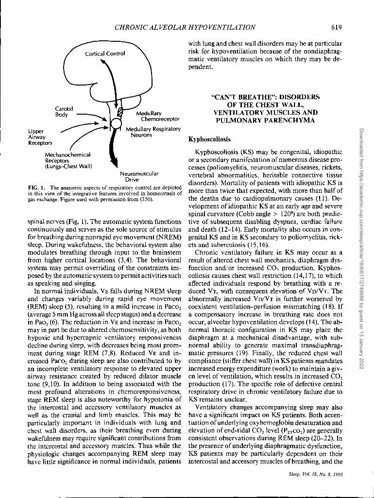
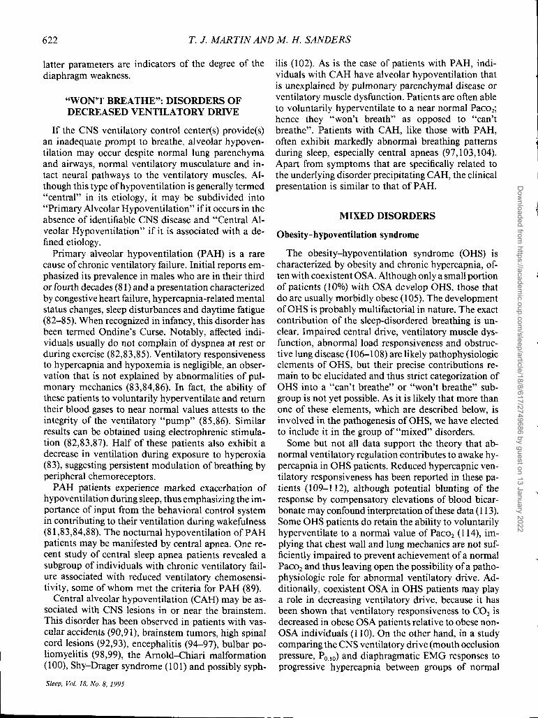
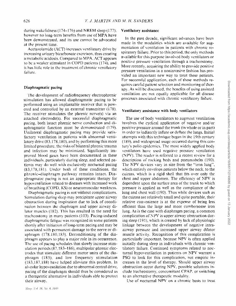
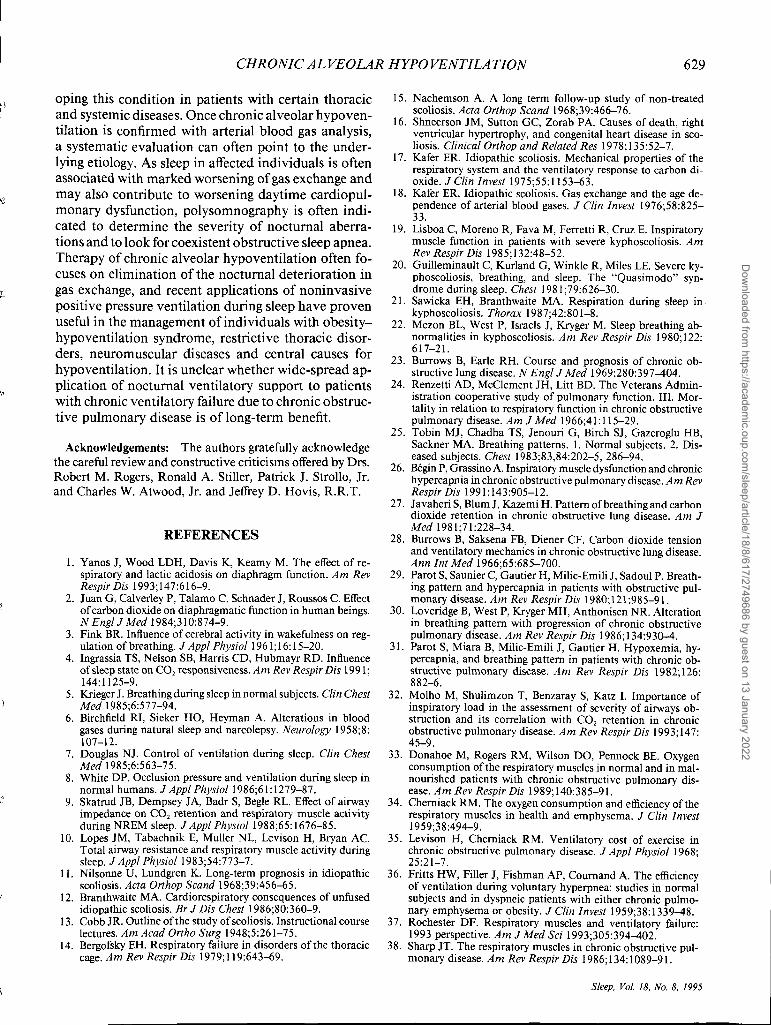
Sleep is a particularly critical period for patients with chronic alveolar hypoventilation, as significant aberrations in ventilation may occur and potentially contribute to hypercapnia during wakefulness. During wakefulness, arterial blood gases are normally maintained within narrow limits by output from two control systems, the "automatic" and the "behavioral". The automatic ventilatory control system is located in the brainstem, and its inherent rhythmogenicity is maintained by complex interactions within and between the dorsal and ventral respiratory groups of respiratory neurons. This automatic system receives input from central (medullary) chemoreceptors, which are primarily responsive to CO2 tension (through changes in hydrogen ion concentration), and from peripheral chemoreceptors (carotid and aortic bodies), which are sensitive to both arterial oxygen tension (Pao2 ) and, to a lesser degree, Paco2• Feedback to the central rhythmgenerating centers, which is important for respiratory timing and coordination, is also provided to the automatic system from pulmonary receptors (stretch, juxtacapillary, irritant) via the vagus nerve as well as from chest wall receptors (joint, tendon, muscle) via
Dow
nloaded from https://academ
ic.oup.com/sleep/article/18/8/617/2749686 by guest on 13 January 2022
CHRONIC ALVEOLAR HYPO VENTILATION 619
Upper Airway Receptors
Carotid Body
Mechanochemical Receptors (Lungs-Chest Wall)
Medullary Respiratory Neurons
Neuromuscu lar Drive
FIG. 1. The anatomic aspects of respiratory control are depicted in this view of the integrative features involved in homeostasis of gas exchange. Figure used with permission from (250).
spinal nerves (Fig. 1). The automatic system functions continuously and serves as the sole source of stimulus for breathing during nonrapid eye movement (NREM) sleep. During wakefulness, the behavioral system also modulates breathing through input to the brainstem from higher cortical locations (3,4). The behavioral system may permit overriding of the constraints imposed by the automatic system to permit activities such as speaking and singing.
In normal individuals, VE falls during NREM sleep and changes variably during rapid eye movement (REM) sleep (5), resulting in a mild increase in Paco2
(average 5 mm Hg across all sleep stages) and a decrease in Pao2 (6). The reduction in VE and increase in Paco2
may in part be due to altered chemosensitivity, as both hypoxic and hypercapnic ventilatory responsiveness decline during sleep, with decreases being most prominent during stage REM (7,8). Reduced VE and increased Paco2 during sleep are also contributed to by an incomplete ventilatory response to elevated upper airway resistance created by reduced dilator muscle tone (9,10). In addition to being associated with the most profound alterations in chemoresponsiveness, stage REM sleep is also noteworthy for hypotonia of the intercostal and accessory ventilatory muscles as well as the cranial and limb muscles. This may be particularly important in individuals with lung and chest wall disorders, as their breathing even during wakefulness may require significant contributions from the intercostal and accessory muscles. Thus while the physiologic changes accompanying REM sleep may have little significance in normal individuals, patients
with lung and chest wall disorders may be at particular risk for hypoventilation because of the nondiaphragmatic ventilatory muscles on which they may be dependent.
"CAN'T BREATHE": DISORDERS OF THE CHEST WALL,
VENTILATORY MUSCLES AND PULMONARY PARENCHYMA
Kyphoscoliosis
Kyphoscoliosis (KS) may be congenital, idiopathic or a secondary manifestation of numerous disease processes (poliomyelitis, neuromuscular diseases, rickets, vertebral abnormalities, heritable connective tissue disorders). Mortality of patients with idiopathic KS is more than twice that expected, with more than half of the deaths due to cardiopulmonary causes (11). Development of idiopathic KS at an early age and severe spinal curvature (Cobb angle> 120°) are both predictive of subsequent disabling dyspnea, cardiac failure and death (12-14). Early mortality also occurs in congenital KS and in KS secondary to poliomyelitis, rickets and tuberculosis (15,16).
Chronic ventilatory failure in KS may occur as a result of altered chest wall mechanics, diaphragm dysfunction and/or increased CO2 production. Kyphoscoliosis causes chest wall restriction (14,17), to which affected individuals respond by breathing with a reduced VT, with consequent elevation of VD/VT. The abnormally increased VD/VT is further worsened by coexistent ventilation-perfusion mismatching (18). If a compensatory increase in breathing rate does not occur, alveolar hypo ventilation develops (14). The abnormal thoracic configuration in KS may place the diaphragm at a mechanical disadvantage, with subnormal ability to generate maximal transdiaphragmatic p'ressures (19). Finally, the reduced chest wall compliance (stiffer chest wall) in KS patients mandates increased energy expenditure (work) to maintain a given level of ventilation, which results in increased CO2
production (17). The specific role of defective central respiratory drive in chronic ventilatory failure due to KS remains unclear.
Ventilatory changes accompanying sleep may also have a significant impact on KS patients. Both accentuation of underlying oxyhemoglobin desaturation and elevation of end-tidal CO2 level (PET co2) are generally consistent observations during REM sleep (20-22). In the presence of underlying diaphragmatic dysfunction, KS patients may be particularly dependent on their intercostal and accessory muscles of breathing, and the
Sleep. Vol. 18. No.8. 1995
Dow
nloaded from https://academ
ic.oup.com/sleep/article/18/8/617/2749686 by guest on 13 January 2022
620 T. J. MARTIN AND M. H. SANDERS
expected reduction in activity ofthese muscles during REM sleep can have a devastating effect on the ability to maintain adequate ventilation. Additionally, KS patients may be prone to central, mixed and obstructive apneas and hypopneas during REM sleep (20-22). Limited evidence suggests that these nocturnal events playa significant role in the development of daytime cardiopulmonary dysfunction (22), but the precise nature of the nocturnal ventilatory changes cannot be predicted from the awake status, and thus polysomnography (PSG) may be required.
Chronic obstructive pulmonary disease
Increased severity of chronic obstructive pulmonary disease (COPD), as measured by gas exchange and ventilatory function, is predictive of decreased long-term survival (23,24). In eucapnic COPD patients, VE is above normal levels (25), providing compensation for the increased VD/VT created by parenchymal lung disease. As airflow limitation worsens, however, further increases in VE cannot be maintained and hypercapnia develops (26). The rapid, shallow breathing pattern adopted by COPD patients further increases VD/VT and contributes to alveolar hypoventilation (27-31).
Historically, patients with COPD have been categorized as being either "blue bloaters" (patients who have chronic bronchitis) or "pink puffers" (patients who have emphysema), although most patients present with clinical and pathologic manifestations of both subgroups. "Blue bloaters" are typically obese, hypoxemic during wakefulness and prone to develop chronic hypercapnia. On the other hand, "pink puffers" are of normal or subnormal body weight, usually have radiographic evidence of lung hyperinflation, maintain a normal or low Paco2 and generally do not develop cor pulmonale until the disease is far advanced. The increased propensity for hypercapnia in chronic bronchitis patients may be partly due to their relatively fixed lower airway narrowing, in contrast to the more compliant and collapsible airways in emphysema patients with similar degrees of airflow limitation (32). Concurrent obesity and/or obstructive sleep apnea (OSA) in the chronic bronchitis patients may also contribute to the development of chronic alveolar hypoventilation.
Investigators have examined ventilatory muscle function in COPD patients to gain further insight into the mechanisms of hypoventilation. Increased lower airway resistance in COPD increases the energy cost and work of breathing (33-37). Flattening of the diaphragm's normal dome-shaped configuration places the muscle at a mechanical disadvantage and decreases its pressure generating efficiency (37-39). Impairment may
Sleep. Vol. 18. No.8. 1995
be accentuated by a decreased blood supply to the muscle (40). Thus the combination of increased work load and contractile limitation of the diaphragm predispose to the development of muscle failure and alveolar hypo ventilation. Support for ventilatory muscle dysfunction in COPD, unexplained by the mechanical disadvantage imposed by pulmonary hyperinflation, is found in the association of reduced maximal inspiratory pressure (MIP) with elevated Paco2 (41). Additionally, although stable COPD patients can be made to augment their ventilation for brief periods of time (42,43), assessment of their resting diaphragmatic tension-time index reveals that they have little ventilatory reserve (44) and display evidence of inspiratory muscle fatigue (45). The diaphragm tension-time index takes into account the diaphragm contraction time as well as the magnitude of the pressure generated relative to the muscle's maximum pressure generating capacity (40).
Altered ventilatory drive has also been considered to be a potential pathogenic factor in the development of chronic ventilatory failure in COPD. However, it is not feasible to directly measure the output of the CNS respiratory center(s) in humans, and evaluation ofventilatory chemoresponsiveness (augmented ventilation in the presence of hypoxemia or hypercapnia) may inaccurately reflect the drive to breathe in the setting oflower airways obstruction. There are conflicting data regarding CO2 responsiveness in COPD patients. Although there is some evidence indicating that hypercapnic COPD patients have diminished ventilatory responses to CO2 (46,47), central chemoresponsiveness to CO2 , as assessed by diaphragmatic and intercostal electromyogram (EMG), has been shown to be increased in hypercapnic COPD patients (48). Additionally, although some data suggest that hypoxic drive is decreased in hypoxemic COPD patients (49), most of the study subjects were also hypercapnic, potentially leading to significant bias. If abnormal chemoresponsiveness is present, it may in part be attributable to familial factors, as healthy offspring of hypercapnic COPD patients have been variably shown to have decreased chemoresponsiveness (50).
Central respiratory drive may also be diminished on an acquired basis, and it is with this notion that the concept of "central fatigue" has been developed (51-53). The development of central fatigue in COPD patients has been postulated to serve as a protective mechanism in individuals whose ventilatory muscle work is dangerously close to a fatiguing level, placing them at risk of catastrophic respiratory failure. This could also apply to patients with restrictive or neuromuscular diseases. If central fatigue allows ventilation to decrease, the work of breathing could be decreased and theoretically the patient distanced from
Dow
nloaded from https://academ
ic.oup.com/sleep/article/18/8/617/2749686 by guest on 13 January 2022
f\ ,I
CHRONIC ALVEOLAR HYPO VENTILATION 621
START ....
CHEST WALL MOVEMENT
~"~\~V~MN~VWV\NW'WV\MNWW~W~WVWVV~'VV~'V\lVVV OIiAL AIR FI.O! )Q w.
76 ...
CONTINUED ....
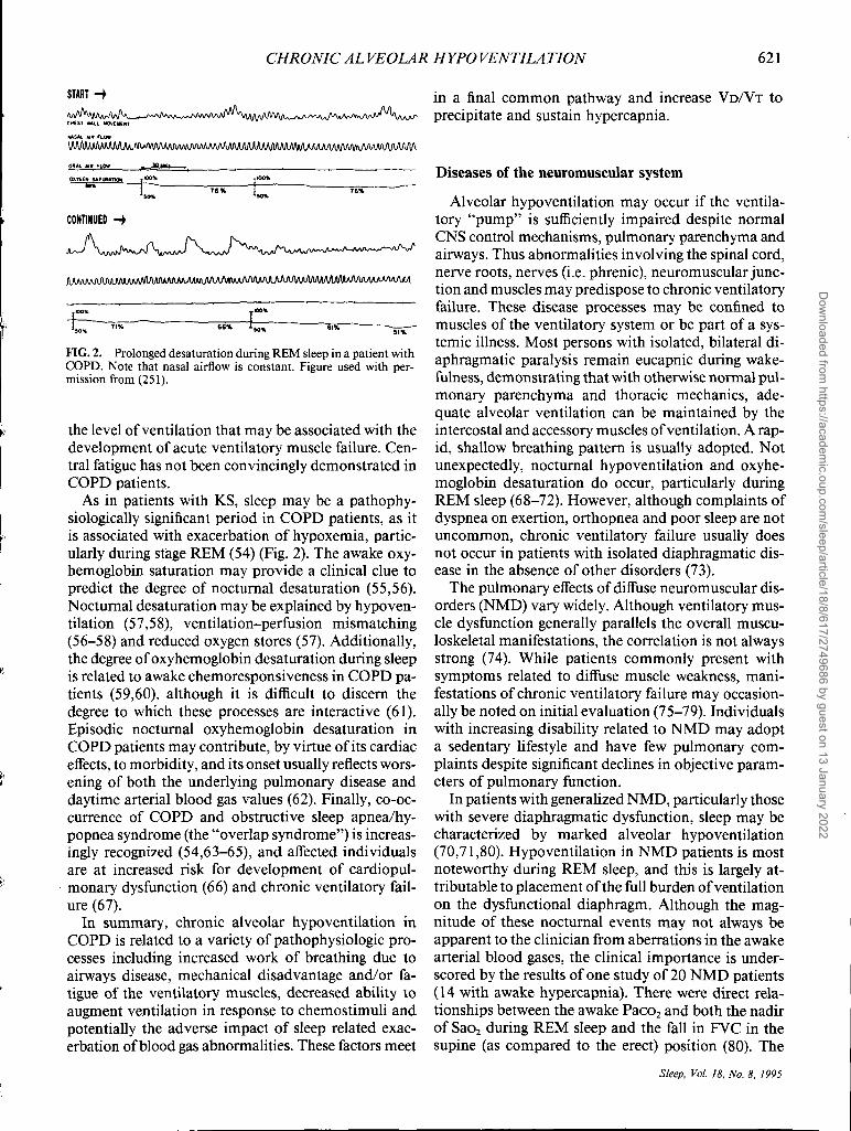
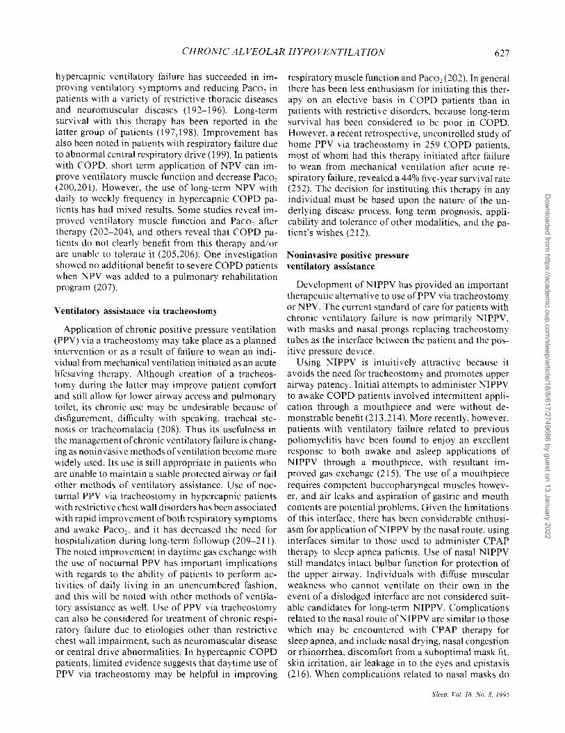
FIG. 2. Prolonged desaturation during REM sleep in a patient with COPD. Note that nasal airflow is constant. Figure used with permission from (251).
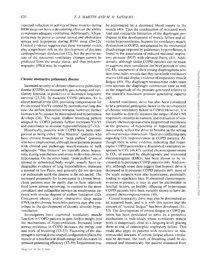
the level of ventilation that may be associated with the development of acute ventilatory muscle failure. Central fatigue has not been convincingly demonstrated in eOPD patients.
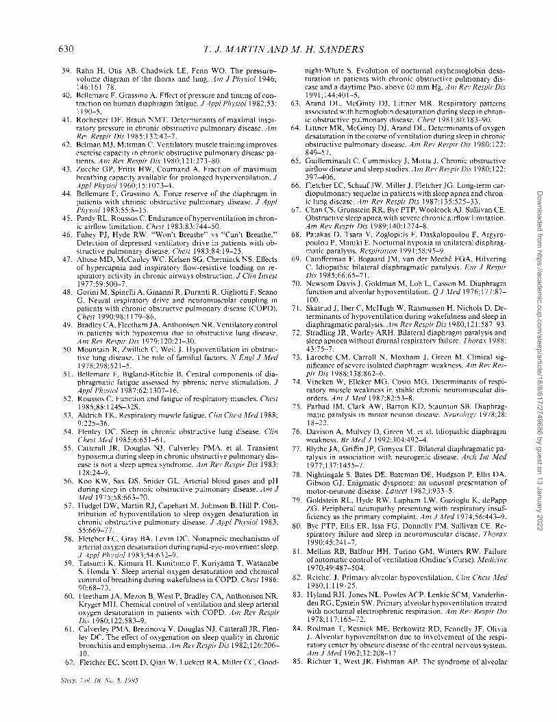
As in patients with KS, sleep may be a pathophysiologically significant period in COPD patients, as it is associated with exacerbation of hypoxemia, particularly during stage REM (54) (Fig. 2). The awake oxyhemoglobin saturation may provide a clinical clue to predict the degree of nocturnal de saturation (55,56). Nocturnal desaturation may be explained by hypoventilation (57,58), ventilation-perfusion mismatching (56-58) and reduced oxygen stores (57). Additionally, the degree of oxyhemoglobin desaturation during sleep is related to awake chemoresponsiveness in COPD patients (59,60), although it is difficult to discern the degree to which these processes are interactive (61). Episodic nocturnal oxyhemoglobin desaturation in eOPD patients may contribute, by virtue of its cardiac effects, to morbidity, and its onset usually reflects worsening of both the underlying pulmonary disease and daytime arterial blood gas values (62). Finally, co-occurrence of eOPD and obstructive sleep apnea/hypopnea syndrome (the "overlap syndrome") is increasingly recognized (54,63-65), and affected individuals are at increased risk for development of cardiopulmonary dysfunction (66) and chronic ventilatory failure (67).
In summary, chronic alveolar hypoventilation in eOPD is related to a variety of pathophysiologic processes including increased work of breathing due to airways disease, mechanical disadvantage and/or fatigue of the ventilatory muscles, decreased ability to augment ventilation in response to chemostimuli and potentially the adverse impact of sleep related exacerbation of blood gas abnormalities. These factors meet
in a final common pathway and increase VD/VT to precipitate and sustain hypercapnia.
Diseases of the neuromuscular system
Alveolar hypoventilation may occur if the ventilatory "pump" is sufficiently impaired despite normal eNS control mechanisms, pulmonary parenchyma and airways. Thus abnormalities involving the spinal cord, nerve roots, nerves (i.e. phrenic), neuromuscular junction and muscles may predispose to chronic ventilatory failure. These disease processes may be confined to muscles of the ventilatory system or be part of a systemic illness. Most persons with isolated, bilateral diaphragmatic paralysis remain eucapnic during wakefulness, demonstrating that with otherwise normal pulmonary parenchyma and thoracic mechanics, adequate alveolar ventilation can be maintained by the intercostal and accessory muscles of ventilation. A rapid, shallow breathing pattern is usually adopted. Not unexpectedly, nocturnal hypoventilation and oxyhemoglobin de saturation do occur, particularly during REM sleep (68-72). However, although complaints of dyspnea on exertion, orthopnea and poor sleep are not uncommon, chronic ventilatory failure usually does not occur in patients with isolated diaphragmatic disease in the absence of other disorders (73).
The pulmonary effects of diffuse neuromuscular disorders (NMD) vary widely. Although ventilatory muscle dysfunction generally parallels the overall musculoskeletal manifestations, the correlation is not always strong (74). While patients commonly present with symptoms related to diffuse muscle weakness, manifestations of chronic ventilatory failure may occasionally be noted on initial evaluation (75-79). Individuals with increasing disability related to NMD may adopt a sedentary lifestyle and have few pulmonary complaints despite significant declines in objective parameters of pulmonary function.
In patients with generalized NMD, particularly those with severe diaphragmatic dysfunction, sleep may be characterized by marked alveolar hypoventilation (70,71,80). Hypoventilation in NMD patients is most noteworthy during REM sleep, and this is largely attributable to placement of the full burden of ventilation on the dysfunctional diaphragm. Although the magnitude of these nocturnal events may not always be apparent to the clinician from aberrations in the awake arterial blood gases, the clinical importance is underscored by the results of one study of 20 NMD patients (14 with awake hypercapnia). There were direct relationships between the awake Paco2 and both the nadir of Sao2 during REM sleep and the fall in FVC in the supine (as compared to the erect) position (80). The
Sleep, Vol. 18, No.8, 1995
Dow
nloaded from https://academ
ic.oup.com/sleep/article/18/8/617/2749686 by guest on 13 January 2022
622 T. J. MARTIN AND M. H. SANDERS
latter parameters are indicators of the degree of the diaphragm weakness.
"WON'T BREATHE": DISORDERS OF DECREASED VENTILATORY DRIVE
If the CNS ventilatory control center(s) provide(s) an inadequate prompt to breathe, alveolar hypoventilation may occur despite normal lung parenchyma and airways, normal ventilatory musculature and intact neural pathways to the ventilatory muscles. Although this type of hypo ventilation is generally termed "central" in its etiology, it may be subdivided into "Primary Alveolar Hypoventilation" ifit occurs in the absence of identifiable CNS disease and "Central Alveolar Hypoventilation" if it is associated with a defined etiology.
Primary alveolar hypo ventilation (PAH) is a rare cause of chronic ventilatory failure. Initial reports emphasized its prevalence in males who are in their third or fourth decades (81) and a presentation characterized by congestive heart failure, hypercapnia-related mental status changes, sleep disturbances and daytime fatigue (82-85). When recognized in infancy, this disorder has been termed Ondine's Curse. Notably, affected individuals usually do not complain of dyspnea at rest or during exercise (82,83,85). Ventilatory responsiveness to hypercapnia and hypoxemia is negligible, an observation that is not explained by abnormalities of pulmonary mechanics (83,84,86). In fact, the ability of these patients to voluntarily hyperventilate and return their blood gases to near normal values attests to the integrity of the ventilatory "pump" (85,86). Similar results can be obtained using electrophrenic stimulation (82,83,87). Half of these patients also exhibit a decrease in ventilation during exposure to hyperoxia (83), suggesting persistent modulation of breathing by peripheral chemoreceptors.
PAH patients experience marked exacerbation of hypoventilation during sleep, thus emphasizing the importance of input from the behavioral control system in contributing to their ventilation during wakefulness (81,83,84,88). The nocturnal hypoventilation of PAH patients may be manifested by central apnea. One recent study of central sleep apnea patients revealed a subgroup of individuals with chronic ventilatory failure associated with reduced ventilatory chemosensitivity, some of whom met the criteria for PAH (89).
Central alveolar hypoventilation (CAR) may be associated with CNS lesions in or near the brainstem. This disorder has been observed in patients with vascular accidents (90,91), brainstem tumors, high spinal cord lesions (92,93), encephalitis (94-97), bulbar poliomyelitis (98,99), the Arnold-Chiari malformation (100), Shy-Drager syndrome (101) and possibly syph-
Sleep. Vol. 18. No.8. 1995
ilis (102). As is the case of patients with PAH, individuals with CAH have alveolar hypoventilation that is unexplained by pulmonary parenchymal disease or ventilatory muscle dysfunction. Patients are often able to voluntarily hyperventilate to a near normal Paco2;
hence they "won't breath" as opposed to "can't breathe". Patients with CAH, like those with PAH, often exhibit markedly abnormal breathing patterns during sleep, especially central apneas (97,103,104). Apart from symptoms that are specifically related to the underlying disorder precipitating CAH, the clinical presentation is similar to that of PAH.
MIXED DISORDERS
Obesity-hypoventilation syndrome
The obesity-hypoventilation syndrome (OHS) is characterized by obesity and chronic hypercapnia, often with coexistent OSA. Although only a small portion of patients (10%) with OSA develop OHS, those that do are usually morbidly obese (105). The development ofOHS is probably multifactorial in nature. The exact contribution of the sleep-disordered breathing is unclear. Impaired central drive, ventilatory muscle dysfunction, abnormal load responsiveness and obstructive lung disease (106-108) are likely pathophysiologic elements of OHS, but their precise contributions remain to be elucidated and thus strict categorization of OHS into a "can't breathe" or "won't breathe" subgroup is not yet possible. As it is likely that more than one of these elements, which are described below, is involved in the pathogenesis of OHS, we have elected to include it in the group of "mixed" disorders.
Some but not all data support the theory that abnormal ventilatory regulation contributes to awake hypercapnia in OHS patients. Reduced hypercapnic ventilatory responsiveness has been reported in these patients (109-112), although potential blunting of the response by compensatory elevations of blood bicarbonate may confound interpretation of these data (113). Some OHS patients do retain the ability to voluntarily hyperventilate to a normal value of Paco2 (114), implying that chest wall and lung mechanics are not sufficiently impaired to prevent achievement of a normal Paco2 and thus leaving open the possibility of a pathophysiologic role for abnormal ventilatory drive. Additionally, coexistent OSA in OHS patients may play a role in decreasing ventilatory drive, because it has been shown that ventilatory responsiveness to CO2 is decreased in obese OSA patients relative to obese nonOSA individuals (110). On the other hand, in a study comparing the CNS ventilatory drive (mouth occlusion pressure, PO. IO) and diaphragmatic EMG responses to progressive hypercapnia between groups of normal
Dow
nloaded from https://academ
ic.oup.com/sleep/article/18/8/617/2749686 by guest on 13 January 2022
I
,1
CHRONIC ALVEOLAR HYPOVENTILATION 623
TABLE 3. Modalitiesfor evaluation of chronic alveolar hypoventilation
Arterial blood gas analysis-the confirmatory test Physical examination-with emphasis on thoracic and neurologic
examinations Thyroid function studies Serum electrolytes, calcium, magnesium, phosphorus, hematocrit Chest radiograph Pulmonary function studies
Spirometry Maximal inspiratory and expiratory pressures Maximal voluntary ventilation
Diaphragmatic studies fluoroscopy Transdiaphragmatic pressure measurement-with or without
twitch stimulation Chemoresponsiveness evaluation
Progressive hyperoxic hypercapnia Progressive eucapnic hypoxia
Central nervous system evaluation Computed tomography Magnetic resonance imaging Lumbar puncture
Polysomnography
subjects, obese subjects, obese eucapnic OSA patients and patients with OHS, Lopata and coworkers were unable to distinguish the eucapnic OSA patients from the OHS patients (115). Furthermore, OHS patients may return to eucapnia following therapy (continuous positive airway pressure or tracheostomy) without a change in their hypercapnic responsiveness (112), suggesting that some abnormality other than or in addition to altered ventilatory drive must be present.
Conceptually, patients with OHS are susceptible to ventilatory muscle fatigue because of obesity-related alteration of thoracic mechanics, decreased ventilatory efficiency, underlying lung disease and, if there is coexistent OSA, repetitive nocturnal airway occlusions. OHS patients do have markedly reduced chest wall compliance (116,117), which leads to increased energy costs of breathing (118). In addition, ventilatory muscle performance is impaired in obesity because of abnormal ventilatory system mechanics and metabolic inefficiency of the ventilatory muscles (116, 119-121). Conceivably these factors contribute to the declines in MIP and maximal voluntary ventilation (MVV) observed in OHS patients (117). When OSA or upper airway resistance syndrome (122) coexists with OHS, it confers a predisposition to ventilatory muscle fatigue because of repetitive inspiratory efforts against an occluded upper airway. The significance of ventilatory muscle dysfunction in the pathogenesis of OHS is reinforced by the observation that weight loss is associated with increased MVV and forced vital capacity (FVC) as well as reduced Paco2 despite little change in ventilatory system compliance (117). Along these lines, significant improvement in hypercapnic ventilatory responsiveness may occur within 24 hours of initiating
positive airway pressure in OHS patients (123), and the rapidity of this response may be consistent with relief of ventilatory muscle fatigue.
OHS patients are clearly subjected to abnormal ventilatory loads including those imposed by chest wall mass, increased elastance ofthe ventilatory system and possibly increased upper airway resistance (124,125). Load compensation, the action taken to defend alveolar ventilation in the presence of a mechanical stress on the system, may be abnormal in individuals with OHS, but its presence in this disorder and its role in the development of hypercapnia has yet to be defined.
EVALUATION OF PATIENTS WITH CHRONIC ALVEOLAR
HYPOVENTILA TION
There is no uniform clinical presentation that identifies patients with alveolar hypo ventilation with a high degree of sensitivity and specificity. Clinical clues regarding the presence of hypercapnia may be provided by such signs and symptoms as morning headache, sleep disruption, daytime fatigue, mental status changes and the development of dependent edema, orthopnea or other manifestations of pulmonary vascular hypertension and cor pulmonale that are not readily explained by other diagnostic entities. As noted previously, dyspnea is not uniformly present. Clinicians must maintain a high index of suspicion in order to perform the definitive test for alveolar hypoventilation, arterial blood gas analysis. The subsequent evaluation may include a number of examinations which are detailed below and outlined in Table 3.
Alveolar hypo ventilation should not be considered a disease, but rather a manifestation of a disease process that in tum should be the object of diagnostic efforts. The physical examination may help guide the evaluation and close attention should be paid to chest wall configuration, signs of pulmonary parenchymal disease, and the functional integrity of the ventilatory muscles. Neurologic examination may also provide insight into the etiology of the hypoventilation. A chest radiograph may be useful in defining the contributions of underlying lung disease, thoracic cage abnormalities or diaphragmatic dysfunction to the development of hypo ventilation or in documenting the presence of pulmonary hypertension.
Spirometric evaluation and measurement of lung volumes, MVV, MIP and maximal expiratory pressures (MEP) yield information regarding lung and ventilatory muscle function. If performed with maximal effort, measurement of the MIP and MEP may provide an early indication of ventilatory muscle weakness in patients with NMD (74,126-130). In contrast, the FVC is reduced only after there has been significant im-
Sleep, Vol. 18, No.8, 1995
Dow
nloaded from https://academ
ic.oup.com/sleep/article/18/8/617/2749686 by guest on 13 January 2022
624 T. J. MARTIN AND M. H. SANDERS
pairment of muscle function. In patients with known disorders, these tests may also be of use in monitoring the progression of disease (131). Although these measurements may not definitively predict development of ventilatory failure in individual patients, awake hypercapnia appears to be particularly likely in NMD patients when the FVC falls below 55% of predicted values (129,132). The MIP and MEP also have prognostic utility in NMD, with hypercapnia particularly likely when ventilatory muscle strength falls below 30% of predicted (132). The MVV is theoretically an estimate of an individual's maximum sustainable ventilation (133), and abnormalities may indicate impending ventilatory failure. As with the MIP and MEP, it is critical that the patient be motivated and maximal effort made in order for the MVV to provide an accurate reflection of ventilatory muscle function.
A significant reduction in FVC when changing from the erect to supine position in patients with NMD and/ or diaphragm dysfunction and an MIP less than 60 cm H 20 in amyotrophic lateral sclerosis patients reflect a degree of diaphragm weakness which predisposes to nocturnal oxyhemoglobin desaturation (80,134). Relative sparing of expiratory muscle strength is often noted in patients with polyneuropathy or distal muscle diseases (74), as opposed to the more profound expiratory weakness associated with amyotrophic lateral sclerosis, Duchenne's muscular dystrophy, myotonic dystrophy and myasthenia gravis (135-138).
Individuals in whom there is concern about isolated diaphragmatic weakness or paralysis require specific evaluation. Fluoroscopic imaging of diaphragm motion has been used for this purpose, but in patients with bilateral diaphragmatic paralysis, pseudo-normal diaphragm movement may lead to test misinterpretation by the inexperienced clinician and thus diminish its value (139). Direct measurement of transdiaphragmatic pressure (POI) using esophageal and gastric balloons is the most precise quantitative technique for assessing diaphragmatic function. Measurements may be obtained during maximal inspiratory efforts, sniffing maneuvers, and inspiration to maximum volume. Measurement of POI during sniffing maneuvers in individuals with normal lung mechanics appears to be particularly useful because of ease of performance and reproducibility of results (140-143).
Although generally not widely performed outside of research facilities (144-146), there may be considerable utility in measuring POI during electrical twitch stimulation of the phrenic nerves. Performance of maximal inspiratory maneuvers during airway occlusion with and without supramaximal phrenic twitch stimulation may prove to be clinically useful in diagnosing "central" respiratory fatigue (51,147,148), although further studies are needed to determine if this
SI~ef1, 1'01. 18, So, 8, 1995
somewhat invasive test offers distinct advantages over the more widely available conventional tests.
In patients with chronic hypoventilation that is unexplained by airway obstruction, a restrictive process or NMD, abnormal central control of respiration should be considered. Demonstration of the ability to voluntarily "hyperventilate" down to a normal Paco2
may provide indirect evidence of a central defect ("won't breathe") (82,85,86). Tests of hypoxic and hypercapnic chemoresponsiveness can be performed in specialized laboratories and may provide diagnostic insight. A search for eNS disease (Table 1) when an acquired central defect in chemoresponsiveness is suspected may include brainstem magnetic resonance imaging, cranial computed tomography and lumbar puncture.
In the absence of other obvious etiologies of alveolar hypoventilation, an assessment of thyroid function should be conducted to exclude hypothyroidism. Screening for serum calcium, potassium (hypokalemic periodic paralysis), magnesium and phosphorus abnormalities or acid maltase deficiency may reveal the etiology of chronic muscular weakness in selected patients. Measuring the hematocrit may provide information regarding the chronicity and physiologic impact of hypo ventilation or the presence of isolated nocturnal hypoventilation and desaturation.
The decision to perform nocturnal PSG in an individual with chronic hypoventilation must be made in response to the clinical suspicion of nocturnal alveolar hypo ventilation/de saturation or concurrent OSA in conjunction with a reasonable expectation that the study will provide clinically useful information. An increasingly large body of knowledge supporting the therapeutic utility of nocturnal noninvasive positive pressure ventilation (NIPPV), particularly in patients with restrictive and central causes for hypoventilation, heightens the value of efforts to identify nocturnal hypoventilation and its etiology. Assessing the magnitude of hypo ventilation and oxyhemoglobin desaturation during sleep may also yield insight regarding the likelihood of developing cardiopulmonary dysfunction in patients who are left untreated.
Some investigators have advocated monitoring oxyhemoglobin saturation alone as a screening tool to detect or exclude patients with sleep-disordered breathing (149). However, the usefulness of oximetry in quantifying hypoventilation may be significantly limited by the sigmoidal shape of the oxyhemoglobin dissociation curve, on which minimal degrees of desaturation may be recorded despite substantial reductions in Pao2 associated with hypoventilation. The diagnostic utility of oximetry may further be limited in patients receiving supplemental oxygen during sleep. Thus direct assessment of nocturnal Paco2 is a desir-
Dow
nloaded from https://academ
ic.oup.com/sleep/article/18/8/617/2749686 by guest on 13 January 2022
CHRONIC ALVEOLAR HYPO VENTILATION 625
'" c CD E CD
" '" '" CD ::;;
50
40
0-5 6-10 11-15 16-20
• Capnograph R
IZZl Capnograph S
El Capnograph N
20+
Error In PTCC02 Measurement ( mmHg )
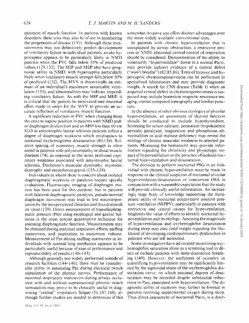
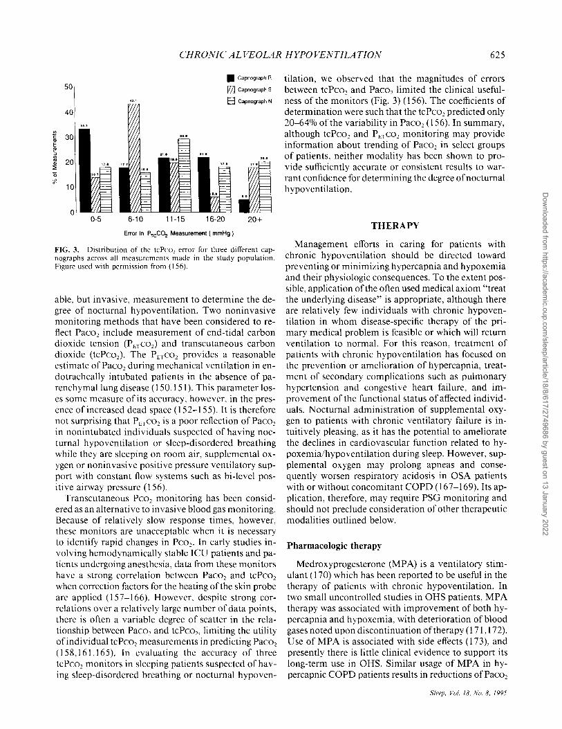
FIG. 3. Distribution of the tcPco, error for three different capnographs across all measurements made in the study population. Figure used with permission from (156).
able, but invasive, measurement to determine the degree of nocturnal hypoventilation. Two noninvasive monitoring methods that have been considered to reflect Paco2 include measurement of end-tidal carbon dioxide tension (PETC02) and transcutaneous carbon dioxide (tcPco2). The PETC02 provides a reasonable estimate ofPaco2 during mechanical ventilation in endotracheally intubated patients in the absence of parenchymallung disease (150,151). This parameter loses some measure of its accuracy, however, in the presence of increased dead space (152-155). It is therefore not surprising that PETC02 is a poor reflection ofPaco2
in nonintubated individuals suspected of having nocturnal hypoventi1ation or sleep-disordered breathing while they are sleeping on room air, supplemental oxygen or noninvasive positive pressure ventilatory support with constant flow systems such as bi-level positive airway pressure (156).
Transcutaneous Pco2 monitoring has been considered as an alternative to invasive blood gas monitoring. Because of relatively slow response times, however, these monitors are unacceptable when it is necessary to identify rapid changes in Pco2 • In early studies involving hemodynamically stable leu patients and patients undergoing anesthesia, data from these monitors have a strong correlation between Paco2 and tcPco2
when correction factors for the heating of the skin probe are applied (157-166). However, despite strong correlations over a relatively large number of data points, there is often a variable degree of scatter in the relationship between Paco2 and tcPcob limiting the utility of individual tcPco2 measurements in predicting Paco2
(158,161,165). In evaluating the accuracy of three tcPco2 monitors in sleeping patients suspected ofhaving sleep-disordered breathing or nocturnal hypoven-
tilation, we observed that the magnitudes of errors between tcPco 2 and Paco2 limited the clinical usefulness of the monitors (Fig. 3) (156). The coefficients of determination were such that the tcPco2 predicted only 20-64% of the variability in Paco2 (156). In summary, although tcPco2 and PETC02 monitoring may provide information about trending of Paco2 in select groups of patients, neither modality has been shown to provide sufficiently accurate or consistent results to warrant confidence for determining the degree of nocturnal hypoventilation.
THERAPY
Management efforts in caring for patients with chronic hypo ventilation should be directed toward preventing or minimizing hypercapnia and hypoxemia and their physiologic consequences. To the extent possible, application of the often used medical axiom "treat the underlying disease" is appropriate, although there are relatively few individuals with chronic hypoventilation in whom disease-specific therapy of the primary medical problem is feasible or which will return ventilation to normal. For this reason, treatment of patients with chronic hypoventilation has focused on the prevention or amelioration of hypercapnia, treatment of secondary complications such as pulmonary hypertension and congestive heart failure, and improvement of the functional status of affected individuals. Nocturnal administration of supplemental oxygen to patients with chronic ventilatory failure is intuitively pleasing, as it has the potential to ameliorate the declines in cardiovascular function related to hypoxemia/hypoventilation during sleep. However, supplemental oxygen may prolong apneas and consequently worsen respiratory acidosis in OSA patients with or without concomitant COPD (167-169). Its application, therefore, may require PSG monitoring and should not preclude consideration of other therapeutic modalities outlined below.
Pharmacologic therapy
Medroxyprogesterone (MPA) is a ventilatory stimulant (170) which has been reported to be useful in the therapy of patients with chronic hypoventilation. In two small uncontrolled studies in OHS patients, MPA therapy was associated with improvement of both hypercapnia and hypoxemia, with deterioration of blood gases noted upon discontinuation of therapy (171,172). Use of MPA is associated with side effects (173), and presently there is little clinical evidence to support its long-term use in OHS. Similar usage of MPA in hypercapnic COPD patients results in reductions ofPaco2
Sleep. Vol. 18, No.8, 1995
Dow
nloaded from https://academ
ic.oup.com/sleep/article/18/8/617/2749686 by guest on 13 January 2022
626 T. 1. MARTIN AND M. H. SANDERS
during wakefulness (J 74-176) and NREM sleep (177); however no long-term benefits from use ofMPA have been demonstrated, and its use cannot be advocated at the present time.
Acetazolamide (ACT) increases ventilatory drive by increasing urinary bicarbonate excretion, thus creating a metabolic acidosis. Compared to MPA, ACT appears to be a weaker stimulant in COPD patients (174), and it has little role in the treatment of chronic ventilatory failure.
Diaphragmatic pacing
The development of radiofrequency electrophrenic stimulation has allowed diaphragmatic pacing to be performed using an implantable receiver that is powered and controlled by an external transmitter (178). The receiver stimulates the phrenic nerve(s) via an attached electrode(s). For successful diaphragmatic pacing, both intact phrenic nerve conduction and diaphragmatic function must be demonstrated (179). Unilateral diaphragmatic pacing may provide satisfactory ventilation in patients with abnormal ventilatory drive (83, 178, 180), and by performing this more limited procedure, the risks of bilateral phrenic trauma and infection may be minimized. Significantly improved blood gases have been documented in these individuals, particularly during sleep, and selected patients may do well with exclusively nocturnal pacing (83,178,181). Under both of these conditions, the phrenic-diaphragm pathway remains intact. Diaphragmatic pacing is not an appropriate therapy for hypoventilation related to diseases with increased work of breathing (COPD, KS) or neuromuscular weakness.
Diaphragmatic pacing is not without complications. Stimulation during sleep may precipitate upper airway obstruction during inspiration due to lack of coordination between the diaphragm and upper airway dilator muscles (182). This has resulted in the need for tracheostomy in many patients (103). Pacing-induced diaphragmatic fatigue was recognized in some patients shortly after initiation oflong-term pacing and may be associated with permanent damage to the nerve or diaphragm (178,180,183). Deconditioning of the diaphragm appears to playa major role in its occurrence. The use of pacing schedules that slowly increase stimulation periods (87,183-186), multipolar phrenic electrodes that stimulate different motor units of the diaphragm (185), and low frequency stimulation (183,187,188) have helped alleviate this problem. In alveolar hypoventilation due to abnormal central drive, pacing of the diaphragm should thus be considered as a therapeutic alternative in individuals able to protect their airway.
Sleep. j·ol. lB. So. B. 1995
Ventilatory assistance
In the past decade, significant advances have been made in the modalities which are available for augmentation of ventilation in patients with chronic respiratory failure. Prior to this period, the only methods available for this purpose involved body ventilators or positive pressure ventilation through a tracheostomy. More recently, acquiring the ability to provide positive pressure ventilation in a noninvasive fashion has provided an important new way to treat these patients. For successful application, each of these methods requires careful patient selection and monitoring oftherapy. As will be discussed, the benefits of using assisted ventilation are not equally applicable for all disease processes associated with chronic ventilatory failure.
Ventilatory assistance with body ventilators
The use of body ventilators to augment ventilation involves the cyclical application of negative and/or positive pressure around the trunk (in whole or in part) in order to indirectly inflate or deflate the lungs. Initial attempts with this technique began in the 19th century (J 89), and widespread usage occurred during this century's polio epidemics. The most widely applied body ventilators have used negative pressure ventilation (NPV). The reader is referred to a recent review for a description of rocking beds and pneumobelts (190). The NPV devices vary in size from the "iron lung", which entirely envelops patients below the neck, to the cuirass, which is a rigid shell that fits over only the chest and upper abdomen. The efficiency of NPV is dependent upon the surface area of the trunk to which pressure is applied as well as the compliance of the lung and chest wall (190). Thus while devices such as the cuirass are relatively small and quite portable, their relative convenience is at the expense of being less efficient than the large and more cumbersome iron lung. As is the case with diaphragm pacing, a common complication ofNPV is upper airway obstruction during sleep (191), which is created by lack of physiologic linkage between the development of negative intraairway pressure and increased upper airway dilator muscle activity. Recognition of this complication is particularly important, because NPV is often applied initially during sleep in individuals with chronic ventilatory failure. Continued symptoms related to nocturnal hypoventilation in patients on NPV warrant a PSG to look for this complication, not empiric increases in the level of therapy. Should upper airway obstruction occur during NPV, possible solutions include tracheostomy, concomitant CPAP, or switching to an alternative therapeutic modality.
Use of nocturnal NPV on a chronic basis to treat
Dow
nloaded from https://academ
ic.oup.com/sleep/article/18/8/617/2749686 by guest on 13 January 2022
CHRONIC ALVEOLAR HYPOVENTILATION 627
hypercapnic ventilatory failure has succeeded in improving ventilatory symptoms and reducing Paco2 in patients with a variety of restrictive thoracic diseases and neuromuscular diseases (192-196). Long-term survival with this therapy has been reported in the latter group of patients (197,198). Improvement has also been noted in patients with respiratory failure due to abnormal central respiratory drive (199). In patients with COPD, short term application of NPV can improve ventilatory muscle function and decrease Paco2
(200,201). However, the use of long-term NPV with daily to weekly frequency in hypercapnic CO PO patients has had mixed results. Some studies reveal improved ventilatory muscle function and Paco2 after therapy (202-204), and others reveal that COPD patients do not clearly benefit from this therapy and/or are unable to tolerate it (205,206). One investigation showed no additional benefit to severe CO PO patients when NPV was added to a pulmonary rehabilitation program (207).
Ventilatory assistance via tracheostomy
Application of chronic positive pressure ventilation (PPV) via a tracheostomy may take place as a planned intervention or as a result of failure to wean an individual from mechanical ventilation initiated as an acute lifesaving therapy. Although creation of a tracheostomy during the latter may improve patient comfort and still allow for lower airway access and pulmonary toilet, its chronic use may be undesirable because of disfigurement, difficulty with speaking, tracheal stenosis or tracheomalacia (208). Thus its usefulness in the management of chronic ventilatory failure is changing as noninvasive methods of ventilation become more widely used. Its use is still appropriate in patients who are unable to maintain a stable protected airway or fail other methods of ventilatory assistance. Use of nocturnal PPV via tracheostomy in hypercapnic patients with restrictive chest wall disorders has been associated with rapid improvement of both respiratory symptoms and awake Paco2 , and it has decreased the need for hospitalization during long-term followup (209-211). The noted improvement in daytime gas exchange with the use of nocturnal PPV has important implications with regards to the ability of patients to perform activities of daily living in an unencumbered fashion, and this will be noted with other methods of ventilatory assistance as well. Use of PPV via tracheostomy can also be considered for treatment of chronic respiratory failure due to etiologies other than restrictive chest wall impairment, such as neuromuscular disease or central drive abnormalities. In hypercapnic COPD patients, limited evidence suggests that daytime use of PPV via tracheostomy may be helpful in improving
respiratory muscle function and Paco2 (202). In general there has been less enthusiasm for initiating this therapy on an elective basis in COPD patients than in patients with restrictive disorders, because long-term survival has been considered to be poor in COPD. However, a recent retrospective, uncontrolled study of home PPV via tracheostomy in 259 CO PO patients, most of whom had this therapy initiated after failure to wean from mechanical ventilation after acute respiratory failure, revealed a 44% five-year survival rate (252). The decision for instituting this therapy in any individual must be based upon the nature of the underlying disease process, long term prognosis, applicability and tolerance of other modalities, and the patient's wishes (212).
Noninvasive positive pressure ventilatory assistance
Development ofNIPPV has provided an important therapeutic alternative to use ofPPV via tracheostomy or NPV. The current standard of care for patients with chronic ventilatory failure is now primarily NIPPV, with masks and nasal prongs replacing tracheostomy tubes as the interface between the patient and the positive pressure device.
Using NIPPV is intuitively attractive because it avoids the need for tracheostomy and promotes upper airway patency. Initial attempts to administer NIPPV to awake CO PO patients involved intermittent application through a mouthpiece and were without demonstrable benefit (213,214). More recently, however, patients with ventilatory failure related to previous poliomyelitis have been found to enjoy an excellent response to both awake and asleep applications of NIPPV through a mouthpiece, with resultant improved gas exchange (215). The use of a mouthpiece requires competent buccopharyngeal muscles however, and air leaks and aspiration of gastric and mouth contents are potential problems. Given the limitations of this interface, there has been considerable enthusiasm for application ofNIPPV by the nasal route, using interfaces similar to those used to administer CPAP therapy to sleep apnea patients. Use of nasal NIPPV still mandates intact bulbar function for protection of the upper airway. Individuals with diffuse muscular weakness who cannot ventilate on their own in the event of a dislodged interface are not considered suitable candidates for long-term NIPPV. Complications related to the nasal route ofNIPPV are similar to those which may be encountered with CPAP therapy for sleep apnea, and include nasal drying, nasal congestion or rhinorrhea, discomfort from a suboptimal mask fit, skin irritation, air leakage in to the eyes and epistaxis (216). When complications related to nasal masks do
Sleep. Vol. 18, No.8, 1995
Dow
nloaded from https://academ
ic.oup.com/sleep/article/18/8/617/2749686 by guest on 13 January 2022
628 T. J. MARTIN AND M. H. SANDERS
occur they are generally easily treated (217). If there is a mask leak of sufficient magnitude to preclude adequate ventilatory assistance, an alternative interface such as an oral-nasal mask may be used (218,219).
Because worsening hypoventilation is recognized to occur during sleep in patients with awake hypoventilation, efforts have focused on administering NIPPY during sleep. Initially, small series and case reports attested to successful nocturnal NIPPY with volumecycled ventilators in patients with NMD, OHS, restrictive disorders and central ventilatory defects (217,220-231,253). As with other modalities ofnocturnal ventilatory assistance, many patients had significantly improved awake arterial blood gases with use of nasal NIPPY. Subsequently, larger studies over longer follow-up periods (years) have confirmed the findings of the initial reports (216,232,233).
NIPPY may be provided by pressure or volumelimited machines. Bi-Ievel positive airway pressure devices which provide a form of pressure support therapy are an alternative to NIPPY with portable, volumecycled ventilators (234). With a bi-Ievel positive airway pressure device, inspiratory and expiratory positive airway pressures can be independently adjusted to augment alveolar ventilation and, if needed, to maintain upper airway patency during sleep. Currently available bi-level positive airway pressure devices are relatively leak tolerant and cycle properly in the presence ofleaks that are inherent with mask or nasal prong ventilation (i.e. at the mask-skin interface or through th~ mouth). Early small studies of noninvasively appbed nocturnal bi-Ievel positive airway pressure therapy in patients with chronic ventilatory failure due to NMD, OHS, restrictive disorders and central ventilatory defects have been promising, with improved arterial blood gases and subjective symptom improvements being reported (234-237). Prescribing NIPPY to carefully selected patients with hypoventilation due to neuro~uscular or chest wall disorders is appropriate to alleVIate symptoms and may also prolong life (233,238).
There is no consistent evidence that NIPPY provides effectiv~ t~erapy for COPD patients with chronic hypoventllahon. COPD patients are often intolerant of the mask or prong interface, thus precluding application of therapy (239,240). In addition, although there are reports of improved gas exchange and Paco2 after chronic ~IPPY therapy in COPD patients, these studies are small and generally uncontrolled (220,240,241). Long-term study of NIPPY in COPD patients has been plagued by high dropout rates (216). Until such a time when more compelling evidence is available to support its use, advocating widespread application of NIPPY in COPD patients is not appropriate.
Sleep, Vol. 18, No.8, 1995
Ventilatory assistance: mechanism of action and titration of therapy
The mechanism(s) by which application of nocturnal ventilatory assistance result(s) in improved ventilatory symptoms and Paco2 in patients with hypoventilation due to NMD, OHS, restrictive disorders and central ventilatory defects is unclear. The mechanism may -:ary a~ong the patient groups but, in general, may mclude Improved ventilatory muscle function due to relief of chronic fatigue, improved ventilatory muscle function due to relief of chronic hypercapnia and hypoxemia, and normalization of chronically blunted central ventilatory drive. With regard to ventilatory muscle function, use of these therapies may acutely reduce diaphragmatic EMG activity (204,206,242-247), perhaps reflecting muscle rest, with resultant improvements in ventilatory muscle strength and/or endurance (192,200-204,225,228,248). It is not possible, however, to exclude changes in central ventilatory drive occurring in conjunction with this therapy.
Because of the uncertainty regarding the mechanism for improved gas exchange in these patients after institution of nocturnal NIPPY, there is uncertainty regarding the best method for establishing the optimal level of ventilatory assistance. If muscle rest is the therapeutic goal, nocturnal therapy should be adjusted with the aid of diaphragm and sternocleidomastoid EMG monitoring (249). However, use of surface electrodes to monitor the diaphragm EMG is technically difficult in these patients, who often have distortion of the chest wall. Using an esophageal electrode to monitor diaphragm function and needle electrodes to assess accessory muscle activity is invasive and not widely used in the clinical setting. Ifreversal of blunted central ventilatory drive is desired, then therapy could be specifically directed towards preventing hypercapnia during sleep. To accomplish this during titration of nocturnal NIPPY, Paco2, or an accurate reflection ofPaco2, must be assessed. Such monitoring of this variable will not only assure that treatment goals are met, but also that potentially dangerous hypocapnia and respiratory alkalosis are not inadvertently precipitated. In the absence of systematic data to provide guidance, the objective of titration in our laboratory is to establish a level of ventilatory assistance that reduces the Paco2 to no more than 10 mm Hg below the awake (hypercapnic) value or at least prevents sleep-related increases in Paco2.
SUMMARY
Chronic alveolar hypoventilation may present in an insidious fashion with nonspecific manifestations. The clinician should be aware of the potential for devel-
Dow
nloaded from https://academ
ic.oup.com/sleep/article/18/8/617/2749686 by guest on 13 January 2022
!
~
J.
CHRONIC ALVEOLAR HYPO VENTILATION 629
oping this condition in patients with certain thoracic and systemic diseases. Once chronic alveolar hypoventilation is confirmed with arterial blood gas analysis, a systematic evaluation can often point to the underlying etiology. As sleep in affected individuals is often associated with marked worsening of gas exchange and may also contribute to worsening daytime cardiopulmonary dysfunction, polysomnography is often indicated to determine the severity of nocturnal aberrations and to look for coexistent 0 bstructi ve sleep apnea. Therapy of chronic alveolar hypoventilation often focuses on elimination of the nocturnal deterioration in gas exchange, and recent applications of noninvasive positive pressure ventilation during sleep have proven useful in the management of individuals with obesityhypo ventilation syndrome, restrictive thoracic disorders, neuromuscular diseases and central causes for hypoventilation. It is unclear whether wide-spread application of nocturnal ventilatory support to patients with chronic ventilatory failure due to chronic obstructive pulmonary disease is of long-term benefit.
Acknowledgements: The authors gratefully acknowledge the careful review and constructive criticisms offered by Drs. Robert M. Rogers, Ronald A. Stiller, Patrick J. Strollo, Jr. and Charles W. Atwood, Jr. and Jeffrey D. Hovis, R.R.T.
REFERENCES
1. Yanos J, Wood LDH, Davis K, Kearny M. The effect of respiratory and lactic acidosis on diaphragm function. Am Rev Respir Dis 1993;147:616-9.
2. Juan G, Calverley P, Talamo C, Schnader J, Roussos C. Effect of carbon dioxide on diaphragmatic function in human beings. N Engl J Med 1984;310:874-9.
3. Fink BR. Influence of cerebral activity in wakefulness on regulation of breathing. J Appl Physiol 1961; 16: 15-20.
4. Ingrassia TS, Nelson SB, Harris CD, Hubmayr RD. Influence of sleep state on CO2 responsiveness. Am Rev Respir Dis 1991; 144:1125-9.
5. Krieger J. Breathing during sleep in normal subjects. Clin Chest Med 1985;6:577-94.
6. Birchfield RI, Sieker HO, Heyman A. Alterations in blood gases during natural sleep and narcolepsy. Neurology 1958;8: 107-12.
7. Douglas NJ. Control of ventilation during sleep. Clin Chest Med 1985;6:563-75.
8. White DP. Occlusion pressure and ventilation during sleep in normal humans. J Appl PhysioI1986;61:1279-87.
9. Skatrud JB, Dempsey JA, Badr S, Begle RL. Effect of airway impedance on CO2 retention and respiratory muscle activity during NREM sleep. J Appl PhysioI1988;65:1676-85.
10. Lopes JM, Tabachnik E, Muller NL, Levison H, Bryan AC. Total airway resistance and respiratory muscle activity during sleep. J Appl Physio!1983;54:773-7.
11. Nilsonne U, Lundgren K. Long-term prognosis in idiopathic scoliosis. Acta Orthop Scand 1968;39:456-65.
12. Branthwaite MA. Cardiorespiratory consequences of unfused idiopathic scoliosis. Br J Dis Chest 1986;80:360-9.
13. Cobb JR. Outline ofthe study of scoliosis. Instructional course lectures. Am Acad Ortho Surg 1948;5:261-75.
14. Bergofsky EH. Respiratory failure in disorders of the thoracic cage. Am Rev Respir Dis 1979; 119:643-69.
15. Nachemson A. A long term follow-up study of non-treated scoliosis. Acta Orthop Scand 1968;39:466-76.
16. Shneerson JM, Sutton GC, Zorab PA. Causes of death, right ventricular hypertrophy, and congenital heart disease in scoliosis. Clinical Orthop and Related Res 1978;135:52-7.
17. Kafer ER. Idiopathic scoliosis. Mechanical properties of the respiratory system and the ventilatory response to carbon dioxide. J Clin Invest 1975;55: 1153-63.
18. Kafer ER. Idiopathic scoliosis. Gas exchange and the age dependence of arterial blood gases. J Clin Invest 1976;58:825-33.
19. Lisboa C, Moreno R, Fava M, Ferretti R, Cruz E. Inspiratory muscle function in patients with severe kyphoscoliosis. Am Rev Respir Dis 1985;132:48-52.
20. Guilleminault C, Kurland G, Winkle R, Miles LE. Severe kyphoscoliosis, breathing, and sleep. The "Quasimodo" syndrome during sleep. Chest 1981 ;79:626-30.
21. Sawicka EH, Branthwaite MA. Respiration during sleep in. kyphoscoliosis. Thorax 1987;42:801-8.
22. Mezon BL, West P, Israels J, Kryger M. Sleep breathing abnormalities in kyphoscoliosis. Am Rev Respir Dis 1980;122: 617-21.
23. Burrows B, Earle RH. Course and prognosis of chronic obstructive lung disease. N Eng! J Med 1969;280:397-404.
24. Renzetti AD, McClement JH, Litt BD. The Veterans Administration cooperative study of pulmonary function. III. Mortality in relation to respiratory function in chronic obstructive pulmonary disease. Am J Med 1966;41:115-29.
25. Tobin MJ, Chadha TS, Jenouri G, Birch SJ, Gazeroglu HB, Sackner MA. Breathing patterns. I. Normal subjects. 2. Diseased subjects. Chest 1983;83,84:202-5, 286-94.
26. Begin P, Grassino A. Inspiratory muscle dysfunction and chronic hypercapnia in chronic obstructive pulmonary disease. Am Rev Respir Dis 1991;143:905-12.
27. Javaheri S, Blum J, Kazemi H. Pattern of breathing and carbon dioxide retention in chronic obstructive lung disease. Am J Med 1981;71:228-34.
28. Burrows B, Saksena FB, Diener CF. Carbon dioxide tension and ventilatory mechanics in chronic obstructive lung disease. Ann In! Med 1966;65:685-700.
29. Parot S, Saunier C, Gautier H, Milic-EmiliJ, Sadoul P. Breathing pattern and hypercapnia in patients with obstructive pulmonary disease. Am Rev Respir Dis 1980;121:985-91.
30. Loveridge B, West P, Kryger MH, Anthonisen NR. Alteration in breathing pattern with progression of chronic obstructive pulmonary disease. Am Rev Respir Dis 1986; 134:930-4.
31. Parot S, Miara B, Milic-Emili J, Gautier H. Hypoxemia, hypercapnia, and breathing pattern in patients with chronic obstructive pulmonary disease. Am Rev Respir Dis 1982;126: 882-6.
32. Molho M, Shulimzon T, Benzaray S, Katz I. Importance of inspiratory load in the assessment of severity of airways obstruction and its correlation with CO2 retention in chronic obstructive pulmonary disease. Am Rev Respir Dis 1993; 147: 45-9.
33. Donahoe M, Rogers RM, Wilson DO, Pennock BE. Oxygen consumption of the respiratory muscles in normal and in malnourished patients with chronic obstructive pulmonary disease. Am Rev Respir Dis 1989; 140:385-91.
34. Cherniack RM. The oxygen consumption and efficiency of the respiratory muscles in health and emphysema. J Clin Invest 1959;38:494-9.
35. Levison H, Cherniack RM. Ventilatory cost of exercise in chronic obstructive pulmonary disease. J App/ Physio/ 1968; 25:21-7.
36. Fritts HW, Filler J, Fishman AP, Cournand A. The efficiency of ventilation during voluntary hyperpnea: studies in normal subjects and in dyspneic patients with either chronic pulmonary emphysema or obesity. J Clin Invest 1959;38:1339-48.
37. Rochester OF. Respiratory muscles and ventilatory failure: 1993 perspective. Am J Med Sci 1993;305:394-402.
38. Sharp JT. The respiratory muscles in chronic obstructive pulmonary disease. Am Rev Respir Dis 1986; 134: 1089-91.
Sleep. Vol. 18, No.8, 1995
Dow
nloaded from https://academ
ic.oup.com/sleep/article/18/8/617/2749686 by guest on 13 January 2022
630 T. J. MARTIN AND M. H. SANDERS
39. Rahn H, Otis AB, Chadwick LE, Fenn WOo The pressurevolume diagram of the thorax and lung. Am J Physio/ 1946; 146:161-78.
40. Bellemare F. Grassino A. Effect of pressure and timing of contraction on human diaphragm fatigue . .1 App/ Physio/1982;53: 1190-5.
41. Rochester DF, Braun NMT. Determinants of maximal inspiratory pressure in chronic obstructive pulmonary disease. Am ReI' Respir Dis 1985;132:42-7.
42. Belman MJ, Mittman C Ventilatory muscle training improves exercise capacity in chronic obstructive pulmonary disease patients. Am Rev Respir Dis 1980; 121 :273-80.
43. Zocche GP, Fritts HW, Cournand A. Fraction of maximum breathing capacity available for prolonged hyperventilation . .1 App/ Physio/ 1960; 15: 1073-4.
44. Bellemare F, Grassino A. Force reserve of the diaphragm in patients with chronic obstructive pulmonary disease . .1 App/ Physio/1983:55:8-15.
45. Pardy RL, Roussos C Endurance of hyperventilation in chronic airflow limitation. Chest 1983:83:744-50.
46. Fahey PJ, Hyde RW. "Won't Breathe" vs "Can't Breathe." Detection of depressed ventilatory drive in patients with obstructive pulmonary disease. Chest 1983:84: 19-25.
47. Altose MD, McCauley We, Kelsen SG. Cherniack NS. Effects of hypercapnia and inspiratory flow-resistive loading on respiratory activity in chronic airways obstruction . .1 Clin Invest 1977:59:500-7.
48. Gorini M, Spinelli A, Ginanni R. Duranti R. Gigliotti F, Scano G. Neural respiratory drive and neuromuscular coupling in patients with chronic obstructve pulmonary disease (COPD). Chest 1990:98: 1179-86.
49. Bradley CA, Fleetham JA, Anthonisen NR. Ventilatory control in patients with hypoxemia due to obstructive lung disease. Am Rev Respir Dis 1979; 120:21-30.
50. Mountain R, Zwillich C, Weil J. Hypoventilation in obstructive lung disease. The role of familial factors. N Eng/ .1 Med 1978;298:521-5.
51. Bellemare F, Bigland-Ritchie B. Central components of diaphragmatic fatigue assessed by phrenic nerve stimulation . .1 App/ Physio/1987;62: 1307-16.
52. Roussos C Function and fatigue of respiratory muscles. Chest 1985;88: 124S-32S.
53. Aldrich TK. Respiratory muscle fatigue. Clin Chest Med 1988; 9:225-36.
54. Flenley DC Sleep in chronic obstructive lung disease. Clin Chest Med 1985;6:651-61.
55. Catterall JR, Douglas NJ, Calverley PMA. et al. Transient hypoxemia during sleep in chronic obstructive pulmonary disease is not a sleep apnea syndrome. Am ReI' Respir Dis 1983: 128:24-9.
56. Koo KW, Sax DS, Snider GL. Arterial blood gases and pH during sleep in chronic obstructive pulmonary disease. Am J Med 1975:58:663-70.
57. Hudgel DW, Martin RJ, Capehart M. Johnson B, Hill P. Contribution of hypoventilation to sleep oxygen de saturation in chronic obstructive pulmonary disease . .1 App/ Physio/ 1983; 55:669-77.
58. Fletcher Ee, Gray BA, Levin DC Nonapneic mechanisms of arterial oxygen de saturation during rapid-eye-movement sleep. .1 Appl PhrsioI1983:54:632-9.
59. Tatsumi K, Kimura H, Kunitomo F, Kuriyama T, Watanabe S. Honda Y. Sleep arterial oxygen desaturation and chemical control of breathing during wakefulness in COPD. Chest 1986: 90:68-73.
60. Fleetham JA, Mezon B, West P, Bradley CA, Anthonisen NR, Kryger MH. Chemical control of ventilation and sleep arterial oxygen desaturation in patients with COPD. Am Rev Respir Dis 1980;122:583-9.
61. Calverley PMA. Brezinova V. Douglas NJ, Catterall JR, Flenley DC The effect of oxygenation on sleep quality in chronic bronchitis and emphysema. Am Re\' Respir Dis 1982; 126:206-10.
62. Fletcher EC, Scott D, Qian W. Luckett RA, Miller CC, Good-
Slce!'. J d. 18. So. 8. 1995
night-White S. Evolution of nocturnal oxyhemoglobin desaturation in patients with chronic obstructive pulmonary disease and a daytime Pao, above 60 mm Hg. Alii ReI' Respir Dis 1991;144:401-5.
63. Arand DL, McGinty DJ. Littner MR. Respiratory patterns associated with hemoglobin desaturation during sleep in chronic obstructive pulmonary disease. Chest 1981 :80: 183-90.
64. Littner MR, McGinty DJ, Arand DL. Determinants of oxygen desaturation in the course of ventilation during sleep in chronic obstructive pulmonary disease. Am Rei' Respir Dis 1980: 122: 849-57.
65. Guilleminault C, Cummiskey J, Motta J. Chronic obstructive airflow disease and sleep studies. Am ReI' Respir Dis 1980; 122: 397-406.
66. Fletcher EC, Schaaf JW, Miller J, Fletcher JG. Long-term cardiopulmonary sequelae in patients with sleep apnea and chronic lung disease. Am Rei' Respir Dis 1987; 135:525-33.
67. Chan CS, Grunstein RR, Bye PTP, Woolcock AJ, Sullivan CEo Obstructive sleep apnea with severe chronic airflow limitation. Am Rev Respir Dis 1989; 140: 1274-8.
68. Patakas D. Tsara V, Zoglopitis F. Daskalopoulou E, Argyropoulou P. Maniki E. Nocturnal hypoxia in unilateral diaphragmatic paralysis. Respiration 1991 ;58:95-9.
69. Camfferman F, Bogaard JM, van der Meche FGA, Hilvering C Idiopathic bilateral diaphragmatic paralysis. Eur .1 Respir Dis 1985;66:65-71.
70. Newsom Davis J, Goldman M. Loh L, Casson M. Diaphragm function and alveolar hypoventilation. Q.I J,fed 1976; 177:87-100.
71. Skatrud J, Iber C, McHugh W, Rasmussen H, Nichols D. Determinants of hypo ventilation during wakefulness and sleep in diaphragmatic paralysis. Am Rev Respir Dis 1980; 121:587-93.
72. Stradling JR, Warley ARH. Bilateral diaphragm paralysis and sleep apnoea without diurnal respiratory failure. Thorax 1988; 43:75-7.
73. Laroche CM, Carroll N, Moxham J. Green M. Clinical significance of severe isolated diaphragm weakness. Am Rev Respir Dis 1988; 138:862-6.
74. Vincken W, Elleker MG, Cosio MG. Determinants of respiratory muscle weakness in stable chronic neuromuscular disorders. Am J Med 1987:82:53-8.
75. Parhad 1M, Clark A W, Barron KD, Staunton SB. Diaphragmatic paralysis in motor neuron disease. Neurology 1978;28: 18-22.
76. Davison A, Mulvey D, Green M, et al. Idiopathic diaphragm weakness. Br Med J 1992;304:492-4.
77. Blythe JA, Griffin JP, Gonyea EF. Bilateral diaphragmatic paralysis in association with neurogenic disease. Arch Int Med 1977; 137: 1455-7.
78. Nightingale S, Bates DE, Bateman DE, Hudgson P, Ellis DA. Gibson GJ. Enigmatic dyspnoea: an unusual presentation of motor-neurone disease. Lancet 1982;i:933-5.
79. Goldstein RL, Hyde RW, Lapham LW, Gazioglu K, dePapp ZG. Peripheral neuropathy presenting with respiratory insufficiency as the primary complaint. Am.l },.jed 1974;56:443-9.
80. Bye PTP, Ellis ER, Issa FG. Donnelly PM, Sullivan CE. Respiratory failure and sleep in neuromuscular disease. Thorax 1990;45:241-7.
81. Mellins RB, Balfour HH. Turino GM. Winters RW. Failure of automatic control of ventilation (Ondine's Curse). Medicine 1970;49:487-504.
82. Reichel 1. Primary alveolar hypoventi1ation. Clin Chest /vfed 1980;1:119-25.
83. Hyland RH, Jones NL. Powles ACP, Lenkie SCM, Vanderlinden RG. Epstein SW. Primary alveolar hypo ventilation treated with nocturnal electrophrenic respiration. Am Re\' Respir Dis 1978;117:165-72.
84. Rodman T, Resnick ME, Berkowitz RD. Fennelly JF, Olivia J. Alveolar hypoventilation due to involvement of the respiratory center by obscure disease of the central nervous system. Am.l Med 1962:32:208-17.
85. Richter T. West JR, Fishman AP. The syndrome of alveolar
Dow
nloaded from https://academ
ic.oup.com/sleep/article/18/8/617/2749686 by guest on 13 January 2022
CHRONIC ALVEOLAR HYPOVENTILATION 631
hypoventilation and diminished sensitivity of the respiratory center. N Engl.! /v/ed 1957:256:1165-70.
86. Rhoads GG, Brody JS. Idiopathic alveolar hypoventilation: clinical spectrum. Ann Int "'fed 1969:71:271-8.
87. Wilcox PG, Pare PD, Fleetham JA. Conditioning of the diaphragm by phrenic nerve pacing in primary alveolar hypoventilation. Thorax 1988:43: 1017-8.
88. Barlow PB, Bartlett D, Hauri P. et al. Idiopathic hypoventilation syndrome: importance of preventing nocturnal hypoxemia and hypercapnia. Am Rev Respir Dis 1980:\21:141-5.
89. Bradley TD. McNicholas WT, Rutherford R, Popkin J, Zamel N, Phillipson EA. Clinical and physiologic heterogeneity of the central sleep apnea syndrome. Am Rev Respir Dis 1986; 134: 217-21.
90. Levin BE, Margolis G. Acute failure of automatic respirations secondary to a unilateral brainstem infarct. Ann Neuro/1977; 1:583-6.
91. Deveraux MW, Keane JR, Davis RL. Automatic respiratory failure associated with infarction of the medulla. Arch Neurol 1973;29:46-52.
92. Krieger AJ. Rosomoff HL. Sleep-induced apnea. Part I: A respiratory and autonomic dysfunction syndrome following bilateral percutaneous cervical cordotomy. Part 2: Respiratory failure after anterior spinal surgery . .! Neurosurg 1974;39: 168-80,181-5.
93. Severinghaus JW, Mitchell RA. Ondine's Curse-failure of respiratory center automaticity while awake. Clin Res 1962; 10:122.
94. White DP, Miller F. Erickson RW. Sleep apnea and nocturnal hypoventilation after Western Equine Encephalitis. Am Rev Respir Dis 1983; 127: 132-3.
95. Cohn JE, Kuida H. Primary alveolar hypoventilation associated with Western Equine Encephalitis. Annlnt ""fed 1962;56: 633-44.
96. Jensen TH, Hansen PB, Brodersen P. Ondine's Curse in Listeria monocytugenes brain stem encephalitis. Acta Neurol Scand 1988;77:505-6.
97. Brouillette RT, Hunt CEo Gallemore GE. Respiratory dysrhythmia: a new cause of central alveolar hypoventilation. Am Rev Respir Dis 1986; 134:609-11.
98. Solliday NH, Gaensler EA, Schwaber JR, Parker TF. Impaired central chemoreceptor function and chronic hypoventilation many years following poliomyelitis. Respiration 1974;31: 177-92.
99. Plum F, Swanson AG. Abnormalities in central regulation of respiration in acute and convalescent poliomyelitis. Arch Neu-1'01 Psych 1958;80:267-85.
100. Montserrat JM, Picado C Agusti-Vidal A. Arnold-Chiari malformation and paralysis of the diaphragm. Respiration 1988: 53: 128-31.
101. Chester CS, Gottfried SB, Cameron DI, Strohl KP. Pathophysiologic findings in a patient with Shy-Drager and alveolar hypoventilation syndromes. Chest 1988;94:212-4.
102. Rodman T, Close HP. The primary hypoventilation syndrome. Am'! Med 1959:26:808-17.
103. Glenn WWL, Gee JBL, Cole DR, Farmer WC, Shaw RK, Beckman CB. Combined central alveolar hypoventilation and upper airway obstruction. Am .! Med 1978;64:50-60.
104. Bradley TD, Day A. Hyland RH, et al. Chronic ventilatory failure caused by abnormal respiratory pattern generation during sleep. Am Rev Respir Dis 1984:130:678-80.
\05. Alexander lK. Amad KH, Cole VW. Observations on some clinical features of extreme obesity, with particular reference to cardiorespiratory effects. Am .! JJed 1962;32:512-24.
106. Bradley TD. Rutherford R, Lue F, et al. Role of diffuse airway obstruction in the hypercapnia of obstructive sleep apnea. Am Rev Respir Dis 1986; 134:920-4.
107. Leech JA. Onal E, Baer P, Lopata M. Determinants of hypercapnia in occlusive sleep apnea syndrome. Chest 1987;92:807-13.
\08. Krieger J, Sforza E, Apprill M, Lampert E, Weitzenblum E, Ratomaharo J. Pulmonary hypertension, hypoxemia, and hy-
percapnia in obstructive sleep apnea patients. Chest 1989:96: 729-37.
109. Garay SM, Rapoport D, Sorkin B, Epstein H. Feinberg I, Goldring RM. Regulation of ventilation in the obstructive sleep apnea syndrome. Am Rev Respir Dis 1981;124:451-7.
110. Gold AR, Schwartz AR. Wise RA. Smith PL. Pulmonary function and respiratory chemosensitivity in moderately obese patients with sleep apnea. Chest 1993; I 03: 1325-9.
Ill. Zwillich CWo Sutton FD, Pierson DJ. Creagh EM. Weil JV. Decreased hypoxic ventilatory drive in the obesity-hypoventilation sydrome. Am .! .tfed 1975:59:343-8.
112. Rapoport DM, Garay SM. Epstein H. Goldring RM. Hypercapnia in the obstructive sleep apnea syndrome. Chest 1986: 89:627-35.
113. Heinemann HO, Goldring RM. Bicarbonate and the regulation of ventilation. Am .! ."fed 1974:57:361-70.
114. Leech J, Onal E, Aronson R, Lopata M. Voluntary hyperventilation in obesity hypoventilation. Chest 1991;1 00: 1334-8.
115. Lopata M, Onal E. Mass loading. sleep apnea, and the pathogenesis of obesity hypoventilation. Am Rev Respir Dis 1982; 126:640-5.
116. Naimark A, Chcrniack RM. Compliance of the respiratory system and its components in health and obesity . .! Appl Physiol 1960;15:377-82.
117. Rochester DF, Enson Y. Current concepts in the pathogenesis of the obesity-hypoventilation syndrome. Am'! ."fed 1974:57: 402-20.
118. Kaufman BJ, Ferguson MH. Cherniack RM. Hypoventilation in obesity . .! Clin Invest 1959:38:500-7.
119. Gilbert R, Sipple JH, Auchincloss JH. Respiratory control and work of breathing in obese subjects . .! Appl PhysioI1961;16: 21-6.
120. Ray CS, Sue DY. Bray G, Hansen JE. Wasserman K. Effects of obesity on respiratory function. Am Rev Respir Dis 1983: 128:501-6.
121. Sharp JT, Henry JP. Sweany SK, Meadows WR, Pietras RJ. The total work of breathing in normal and obese men . .! Clin 1I1Vest 1964;43:728-39.
122. Guilleminault C, Stoohs R, Clerk A, Cetc! M. Maistros P. A cause of excessive daytime sleepiness. The upper airway resistance syndrome. Chest 1993;104:781-7.
123. Berthon-lones M. Sullivan CEo Time course of change in ventilatory response to CO, with long-term CPAP therapy for obstructive sleep apnea. Am Rev Respir Dis 1987: 135: 144-7.
124. Anch AM, Remmers JE, Bunce H. Supraglottic airway resistance in normal subjects and patients with occlusive sleep apnea . .! Appl Physio/1982:53: 1158-63.
125. Bradley TD, Brown IG, Grossman RF. et al. Pharyngeal size in snorers, non snorers, and patients with obstructive sleep apnea. N Engl .! Med 1986;315: 1327-31.
126. Black LF, Hyatt RE. Maximal static respiratory pressures in generalized neuromuscular disease. Am Rev Respir Dis 1971; 103:641-50.
127. Demedts M, Beckers J, Rochette F, Bulcke J. Pulmonary function in moderate neuromuscular disease without respiratory complaints. Eur.! Respir Dis 1982;63:62-7.
128. Griggs RC, Donohoe KM, Utell MJ, Goldblatt D, Moxley RT. Evaluation of pulmonary function in neuromuscular disease. Arch Neuro/1981;38:9-12.
129. Braun NMT, Rochester DF. Muscular weakness and respiratory failure. Am Rev Respir Dis 1979; 119: 123-5.
130. Inkley SR, Oldenburg FC Vignos PJ. Pulmonary function in Duchenne muscular dystrophy related to stage of disease. Am .! Med 1974;56:297-306.
131. Libby DM, Briscoe W A, Boyce B, Smith JP. Acute respiratory failure in scoliosis or kyphosis. Prolonged survival and treatment. Am .!Wed 1982:73:532-8.
132. Braun NMT. Arora NS, Rochester DF. Resoiratorv muscle and pulmonary function in polymyositis and' other proximal myopathies. Thorax 1983;38:616-23.
133. Rochester DF, Arora NS. Respiratory muscle failure. Med Clin N Am 1983;67:573-97.
134. Gay PC Westbrook PR, Daube JR, Litchy WJ, Windebank
Sleep, Vol. 18, No.8, 1995
Dow
nloaded from https://academ
ic.oup.com/sleep/article/18/8/617/2749686 by guest on 13 January 2022
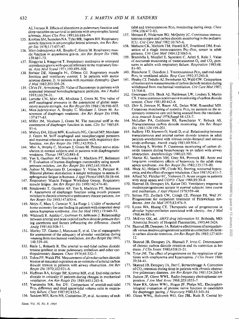
632 T. J. MARTIN AND M. H. SANDERS
AJ, Iverson R. Effects of alterations in pulmonary function and sleep variables on survival in patients with amyotrophic lateral sclerosis. Mayo Clin Proc 1991 ;66:686-94.
135. Kreitzer SM, Saunders NA, Tyler HR, Ingram RH. Respiratory muscle function in amyotrophic lateral sclerosis. Am Rev Respir Dis 1978;117:437-47.
136. Mier-Jedrzejowicz AK, Brophy C, Green M. Respiratory muscle function in myasthenia gravis. Am Rev Respir Dis 1988; 138:867-73.
137. Ringqvist I, Ringqvist T. Respiratory mechanics in untreated myasthenia gravis with special reference to the respiratory forces. Acta Med Scand 1971;190:499-508.
138. Serisier DE, Mastaglia FL, Gibson GJ. Respiratory muscle function and ventilatory control. I. In patients with motor neurone disease. II. In patients with myotonic dystrophy. Quart J Med 1982;202:205-26.
139. Ch'en IY, Armstrong JD. Value of fluoroscopy in patients with suspected bilateral hemidiaphragmatic paralysis. Am J Radiol 1993; 160:29-31.
140. Laroche CM, Mier AK, Moxham J, Green M. The value of sniff esophageal pressures in the assessment of global inspiratory muscle strength. Am Rev Respir Dis 1988; 138:598-603.
141. Mier-Jedrzejowicz A, Brophy C, Moxham J, Green M. Assessment of diaphragm weakness. Am Rev Respir Dis 1988; 137:877-83.
142. Miller JM, Moxham J, Green M. The maximal sniff in the assessment of diaphragm function in man. Clin Sci 1985;69: 91-6.
143. Mulvey DA, Elliott MW, Koulouris NG, Carroll MP, Moxham J, Green M. Sniff esophageal and nasopharyngeal pressures and maximal relaxation rates in patients with respiratory dysfunction. Am Rev Respir Dis 1991;143:950-3.
144. Mier A, Brophy C, Moxham J, Green M. Phrenic nerve stimulation in normal subjects and in patients with diaphragmatic weakness. Thorax 1987;42:885-8.
145. Yan S, Gauthier AP, Similowski T, Macklem PT, Bellemare F. Evaluation of human diaphragm contractility using mouth pressure twitches. Am Rev Respir Dis 1992; 145: 1064-9.
146. Aubier M, Murciano D, Lecocguic Y, Viires N, Pariente R. Bilateral phrenic stimulation: a simple technique to assess diaphragmatic fatigue in humans. J Appl PhysioI1985;58:58-64.
147. Respiratory Muscle Fatigue Workshop Group. Respiratory muscle fatigue. Am Rev Respir Dis 1990; 142:474-80.
148. Similowski T, Gauthier AP, Yan S, Macklem PT, Bellemare F. Assessment of diaphragm function using mouth pressure twitches in chronic obstructive pulmonary disease patients. Am Rev Respir Dis 1993;147:850-6.
149. Series F, Marc I, Cormier Y, La Forge J. Utility of nocturnal home oximetry for case finding in patients with suspected sleep apnea hypopnea syndrome. Ann Int Med 1993; 119:449-53.
150. Whitesell R, Asiddo C, Gollman D, Jablonski J. Relationship between arterial and peak expired carbon dioxide pressure during anesthesia and factors influencing the difference. Anesth Analg 1981;60:508-11.
151. Morley TF, Giamo J, Maroszan E, et al. Use of capnography for assessment of the adequacy of alveolar ventilation during weaning from mechanical ventilation. Am Rev Respir Dis 1993; 148:339-44.
152. Hade L, Rokseth R. The arterial to end-tidal carbon dioxide tension gradient in acute pulmonary embolism and other cardiopulmonary diseases. Chest 1974;66:352-7.
153. Tulou PP, Walsh PM. Measurement of alveolar carbon dioxide tension at maximal expiration as an estimate of arterial carbon dioxide tension in patients with airway obstruction. Am Rev Respir Dis 1970;102:921-6.
154. Hoffman RA, Krieger BP, Kramer MR, et al. End-tidal carbon dioxide in critically ill patients during changes in mechanical ventilation. Am Rev Respir Dis 1989; 140: 1265-8.
155. Yamanaka MK, Sue DY. Comparison of arterial-end-tidal Peo, difference and dead space/tidal volume ratio in respiratory failure. Chest 1987;92:832-5.
156. Sanders MH, Kern NB, Costantino JP, et al. Accuracy of end-
Sleep, Vol. 18, No.8, 1995
tidal and transcutaneous Peo, monitoring during sleep. Chest 1994;106:472-83.
157. Monaco F, Nickerson BG, McQuitty Je. Continuous transcutaneous oxygen and carbon dioxide monitoring in the pediatric ICU. Crit Care Med 1982;10:765-6.
158. Mahutte CK, Michiels TM, Hassell KT, Trueblood DM. Evaluation of a single transcutaneous Po,-Peo, sensor in adult patients. Crit Care Med 1984;12:1063-6.
159. Brambilla I, Micallef E, Sacerdoti C, Arlati S, Rol0 J. Value of nocturnal monitoring of transcutaneous 0, and CO, pressures in adults with respiratory failure. Respiration 1985;48: 81-90.
160. Blanchette T, Dziodzio J. Transcutaneous Peo, and end-tidal Pco, in ventilated adults. Resp Care 1992;37:240-8.
161. Healey CJ, Fedullo AJ, Swinburne AJ, Wahl GW. Comparison of noninvasive measurements of carbon dioxide tension during withdrawal from mechanical ventilation. Crit Care Med 1987; 15:764-8.
162. Greenspan GH, Block AJ, Haldeman LW, Lindsey S, Martin CS. Transcutaneous noninvasive monitoring of carbon dioxide tension. Chest 1981 ;80:442-6.
163. Eletr S, Jimison H, Ream AK, Dolan WM, Rosenthal MH. Cutaneous monitoring of systemic Peo, on patients in the respiratory intensive care unit being weaned from the ventilator. Acta Anaesth Scand 1978;Suppl 68: 123-7.
164. McLellan PA, Goldstein RS, Ramcharan V, Rebuck AS. Transcutaneous carbon dioxide monitoring. Am Rev Respir Dis 1981; 124: 199-201.
165. Rafferty TD, Marrero 0, Nardi D, et al. Relationship between transcutaneous and arterial carbon dioxide tension in adult patients anesthetized with nitrous oxide-fentanyl and nitrous oxide-enflurane. Anesth Analg 1981 ;60:504-7.
166. Weinberg S, Werbin P. Cutaneous monitoring of carbon dioxide tension during bronchoscopy in an infant with airway obstruction. Anesthesiology 1986;65:703.
167. Martin RJ, Sanders MH, Gray BA, Pennock BE. Acute and long-term ventilatory effects of hyperoxia in the adult sleep apnea syndrome. Am Rev Respir Dis 1982; 125:175-80.
168. Block AJ, Hellard DW, Cicale MJ. Snoring, nocturnal hypoxemia, and the effect of oxygen inhalation. Chest 1987;92:411-7.
169. Alford NJ, Fletcher EC, Nickeson D. Acute oxygen in patients with sleep apnea and COPD. Chest 1986;89:30-8.
170. Skatrud JB, Dempsey JA, Kaiser DG. Ventilatory response to medroxyprogesterone acetate in normal subjects: time course and mechanism. J Appl PhysioI1978;939-44.
171. Sutton FD, Zwillich CW, Creagh CE, Pierson DJ, Weil JV. Progesterone for outpatient treatment of Pickwickian syndrome. Ann Int Med 1975;83:476-9.
172. Lyons HA, Huang CT. Therapeutic use of progesterone in alveolar hypoventilation associated with obesity. Am J Med 1968;44:881-8.
173. McEvoy GK, ed. AHFS drug information 93. Bethesda, MD: American Society of Hospital Pharmacists, 1993;44:2424.
174. Skatrud JB, Dempsey JA. Relative effectiveness ofacetazolamide versus medroxyprogesterone acetate in correction of chronic carbon dioxide retention. Am Rev Respir Dis 1983; 127:405-12.
175. Skatrud JB, Dempsey JA, Bhansali P, Irvin e. Determinants of chronic carbon dioxide retention and its correction in humans. J Clin Invest 1980;65:813-21.
176. Tyler JM. The effect of progesterone on the respiration of patients with emphysema and hypercapnia. J Clin Invest 1960; 39:34-41.
177. Skatrud JB, Dempsey JA, Iber C, Berssenbrugge A. Correction of CO, retention during sleep in patients with chronic obstructive pulmonary diseases. Am Rev Respir Dis 1981; 124:260-8.
178. Judson JP, Glenn WWL. Radio-frequency electrophrenic respiration. JAm Med Assoc 1968;203:1033-7.
179. Shaw RK, Glenn WWL, Hogan JF, Phelps ML. Electrophysiological evaluation of phrenic nerve function in candidates for diaphragm pacing. J Neurosurg 1980;53:345-54.
180. Glenn WWL, Holcomb WG, Gee JBL, Rath R. Central hy-
Dow
nloaded from https://academ
ic.oup.com/sleep/article/18/8/617/2749686 by guest on 13 January 2022
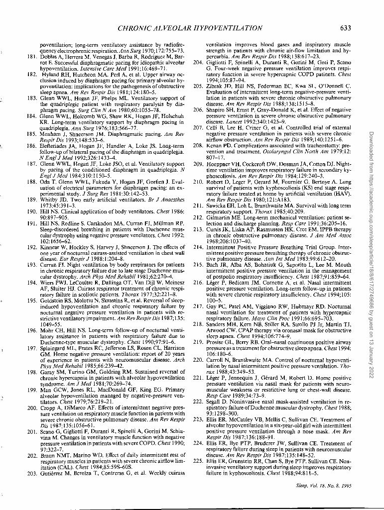
I,
CHRONIC ALVEOLAR HYPOVENTILATION 633
poventilation; long-term ventilatory assistance by radiofrequency electrophrenic respiration. Ann Surg 1970; 172:755-73.
181. Dobias A, Herrera M, Venegas J, Barba R, Rodriguez M, Barrot E. Successful diaphragmatic pacing for idiopathic alveolar hypoventilation. Intensive Care Med 1991; 16:469-71.
182. Hyland RH, Hutcheon MA, Perl A, et al. Upper airway occlusion induced by diaphragm pacing for primary alveolar hypoventilation: implications for the pathogenesis of obstructive sleep apnea. Am Rev Respir Dis 1981; 124: 180-5.
183. Glenn WWL, Hogan JF, Phelps ML. Ventilatory support of the quadriplegic patient with respiratory paralysis by diaphragm pacing. Surg Clin N Am 1980;60:1055-78.
184. Glenn WWL, Holcomb WG, Shaw RK, Hogan JF, Holschuh KR. Long-term ventilatory support by diaphragm pacing in quadriplegia. Ann Surg 1976;183:566-77.
185. Moxham J, Shneerson JM. Diaphragmatic pacing. Am Rev Respir Dis 1993;148:533-6.
186. Elefteriades JA, Hogan JF, Handler A, Loke JS. Long-term follow-up of bilateral pacing of the diaphragm in quadriplegia. N Engl J Med 1992;326:1433-4.
187. Glenn WWL, Hogan JF, Loke JSO, et al. Ventilatory support by pacing of the conditioned diaphragm in quadriplegia. N Engl J Med 1984;310: 1150-5.
188. Oda T, Glenn WWL, Fukuda Y, Hogan JF, Gorfien J. Evaluation of electrical parameters for diaphragm pacing: an experimental study. J Surg Res 1981;30:142-53.
189. Whitby JD. Two early artificial ventilators. Br J Anaesthes 1973;45:391-3.
190. Hill NS. Clinical application of body ventilators. Chest 1986; 90:897-905.
191. Hill NS, Redline S, Carskadon MA, Curran FJ, Millman RP. Sleep-disordered breathing in patients with Duchenne muscular dystrophy using negative pressure ventilators. Chest 1992; 102: 1656-62.
192. Kinnear W, Hockley S, Harvey J, Shneerson J. The effects of one year of nocturnal cuirass-assisted ventilation in chest wall disease. Eur Respir J 1988; 1 :204-8.
193. Curran FJ. Night ventilation by body respirators for patients in chronic respiratory failure due to late stage Duchenne muscular dystrophy. Arch Phys Med Rehabill981 ;62:270-4.
194. Wiers PWJ, LeCoultre R, Dallinga OT, Van Dijl W, Meinesz AF, Sluiter Hl Cuirass respirator treatment of chronic respiratory failure in scoliotic patients. Thorax 1977;32:221-8.
195. Goldstein RS, Molotiu N, Skrastins R, et al. Reversal of sleepinduced hypoventilation and chronic respiratory failure by nocturnal negative pressure ventilation in patients with restrictive ventilatory impairment. Am Rev Respir Dis 1987; 135: 1049-55.
196. Mohr CH, Hill NS. Long-term follow-up of nocturnal ventilatory assistance in patients with respiratory failure due to Duchenne-type muscular dystrophy. Chest 1990;97:91-6.
197. Splaingard ML, Frates RC, Jefferson LS, Rosen CL, Harrison GM. Home negative pressure ventilation: report of 20 years of experience in patients with neuromuscular disease. Arch Phys Med RehabilI985;66:239-42.
198. Garay SM, Turino GM, Goldring RM. Sustained reversal of chronic hypercapnia in patients with alveolar hypoventilation syndrome. Am J Med 1981;70:269-74.
199. Man GCW, Jones RL, MacDonald GF, King EG. Primary alveolar hypoventilation managed by negative-pressure ventilators. Chest 1979;76:219-21.
200. Cropp A, DiMarco AF. Effects of intermittent negative pressure ventilation on respiratory muscle function in patients with severe chronic obstructive pulmonary disease. Am Rev Respir Dis 1987; 135: 1056-61.
20 I. Scano G, Gigliotti F, Duranti R, Spinelli A, Gorini M, Schiavina M. Changes in ventilatory muscle function with negative pressure ventilation in patients with severe COPD. Chest 1990; 97:322-7.
202. Braun NMT, Marino WD. Effect of daily intermittent rest of respiratory muscles in patients with severe chronic airflow limitation (CAL). Chest 1984;85:59S-60S.
203. Gutierrez M, Beroiza T, Contreras G, et al. Weekly cuirass
ventilation improves blood gases and inspiratory muscle strength in patients with chronic air-flow limitation and hypercarbia. Am Rev Respir Dis 1988; 138:617-23.
204. Gigliotti F, Spinelli A, Duranti R, Gorini M, Goti P, Scano G. Four-week negative pressure ventilation improves respiratory function in severe hypercapnic COPD patients. Chest 1994; 105:87-94.
205. Zibrak JD, Hill NS, Federman EC, Kwa SL, O'Donnell C. Evaluation of intermittent long-term negative-pressure ventilation in patients with severe chronic obstructive pulmonary disease. Am Rev Respir Dis 1988; 138: 1515-8.
206. Shapiro SH, Ernst P, Gray-Donald K, et al. Effect of negative pressure ventilation in severe chronic obstructive pulmonary disease. Lancet 1992;340:1425-9.
207. Celli B, Lee H, Criner G, et al. Controlled trial of external negative pressure ventilation in patients with severe chronic airflow obstruction. Am Rev Respir Dis 1989;140:1251-6.
208. Kenan PD. Complications associated with tracheostomy: prevention and treatment. Otolaryngol Clin North Am 1979;12: 807-17.
209. Hoeppner VH, Cockcroft DW, Dosman JA, Cotton DJ. Nighttime ventilation improves respiratory failure in secondary kyphoscoliosis. Am Rev Respir Dis 1984;129:240-3.
210. Robert D, Leger P, Gerard M, Fournier G, Bertoye A. Long survival of patients with kyphoscoliosis (KS) end stage respiratory failure treated at home by artificial ventilation (HA V). Am Rev Respir Dis 1980;121:AI83.
211. Sawicka EH, Loh L, Branthwaite MA. Survival with long term respiratory support. Thorax 1985;40:209.
212. Gilmartin ME. Long-term mechanical ventilation: patient selection and discharge planning. Resp Care 1991 ;36:205-16.
213. Curtis JK, Liska AP, Rasmussen HK, Cree EM. IPPB therapy in chronic obstructive pulmonary disease. JAm Med Assoc 1968;206: 1037-40.
214. Intermittent Positive Pressure Breathing Trial Group. Intermittent positive pressure breathing therapy of chronic obstructive pulmonary disease. Ann Int Med 1983;99:612-20.
215. Bach JR, Alba AS, Bohatiuk G, Saporito L, Lee M. Mouth intermittent positive pressure ventilation in the management of postpolio respiratory insufficiency. Chest 1987;91 :859-64.
216. Leger P, Bedicam JM, Cornette A, et al. Nasal intermittent positive pressure ventilation. Long-term follow-up in patients with severe chronic respiratory insufficiency. Chest 1994; 105: 100-5.
217. Gay PC, Patel AM, Viggiano RW, Hubmayr RD. Nocturnal nasal ventilation for treatment of patients with hypercapnic respiratory failure. Mayo Clin Proc 1991;66:695-703.
218. Sanders MH, Kern NB, Stiller RA, Strollo PJ Jr, Martin TJ, Atwood CWo CPAP therapy via oronasal mask for obstructive sleep apnea. Chest 1994;106:774-9.
219. Prosise GL, Berry RB. Oral-nasal continuous positive airway pressure as a treatment for obstructive sleep apnea. Chest 1994; 106:180-6.
220. Carroll N, Branthwaite MA. Control of nocturnal hypoventilation by nasal intermittent positive pressure ventilation. Thorax 1988;43:349-53.
221. Leger P, Jennequin J, Gerard M, Robert D. Home positive pressure ventilation via nasal mask for patients with neuromuscular weakness or restrictive lung or chest-wall disease. Resp Care 1989;34:73-9.
222. Segall D. Noninvasive nasal mask-assisted ventilation in respiratory failure ofDuchenne muscular dystrophy. Chest 1988; 93:1298-300.
223. Ellis ER, McCauley VB, Mellis C, Sullivan CEo Treatment of alveolarhypoventilation in a six-year-oldgirl with intermittent positive pressure ventilation through a nose mask. Am Rev Respir Dis 1987;136:188-91.
224. Ellis ER, Bye PTP, Bruderer JW, Sullivan CEo Treatment of respiratory failure during sleep in patients with neuromuscular disease. Am Rev Respir Dis 1987;135:148-52.
225. Ellis ER, Grunstein RR, Chan S, Bye PTP, Sullivan CEo Noninvasive ventilatory support during sleep improves respiratory failure in kyphoscoliosis. Chest 1988;94:811-5.
Sleep, Vol. 18, No.8, 1995
Dow
nloaded from https://academ
ic.oup.com/sleep/article/18/8/617/2749686 by guest on 13 January 2022
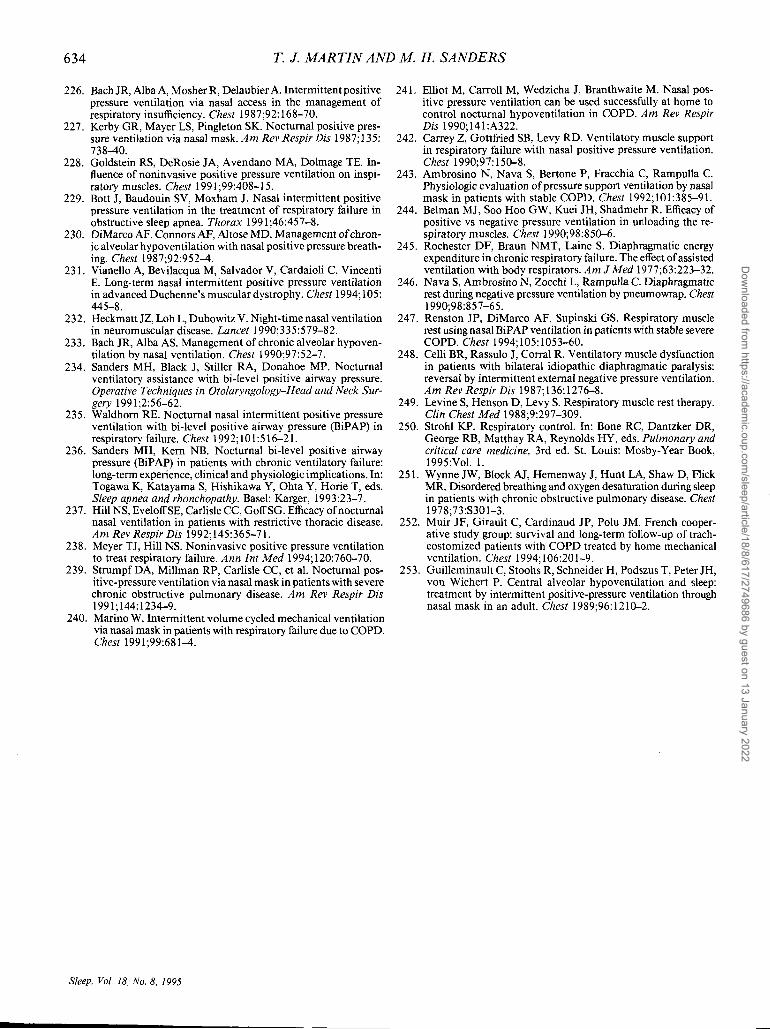
b
634 T. J. MARTIN AND M. H. SANDERS
226. Bach JR, Alba A, Mosher R, Delaubier A. Intermittent positive pressure ventilation via nasal access in the management of respiratory insufficiency. Chest 1987;92: 168-70.
227. Kerby GR, Mayer LS, Pingleton SK. Nocturnal positive pressure ventilation via nasal mask. Am Rev Respir Dis 1987; 135: 738-40.
228. Goldstein RS, DeRosie JA, Avendano MA, Dolmage TE. Influence of noninvasive positive pressure ventilation on inspiratory muscles. Chest 1991;99:408-15.
229. Bott J, Baudouin SV, Moxham J. Nasal intermittent positive pressure ventilation in the treatment of respiratory failure in obstructive sleep apnea. Thorax 1991 ;46:457-8.
230. DiMarco AF, Connors AF, Altose MD. Management of chronic alveolar hypoventilation with nasal positive pressure breathing. Chest 1987;92:952-4.
231. Vianello A, Bevilacqua M, Salvador V, Cardaioli C, Vincenti E. Long-term nasal intermittent positive pressure ventilation in advanced Duchenne's muscular dystrophy. Chest 1994; 105: 445-8.
232. HeckmattJZ, Loh L, Dubowitz V. Night-time nasal ventilation in neuromuscular disease. Lancet 1990;335:579-82.
233. Bach JR, Alba AS. Management of chronic alveolar hypoventilation by nasal ventilation. Chest 1990;97:52-7.
234. Sanders MH, Black J, Stiller RA, Donahoe MP. Nocturnal ventilatory assistance with bi-Ievel positive airway pressure. Operative Techniques in Otolaryngology-Head and Neck Surgery 1991;2:56-62.
235. Waldhorn RE. Nocturnal nasal intermittent positive pressure ventilation with bi-Ievel positive airway pressure (BiPAP) in respiratory failure. Chest 1992; 10 I :516-21.
236. Sanders MH, Kern NB. Nocturnal bi-Ievel positive airway pressure (BiPAP) in patients with chronic ventilatory failure: long-term experience, clinical and physiologic implications. In: Togawa K, Katayama S, Hishikawa Y, Ohta Y, Horie T, eds. Sleep apnea and rhonchopathy. Basel: Karger, 1993:23-7.
237. Hill NS, EveloffSE, Carlisle CC, GoffSG. Efficacy of nocturnal nasal ventilation in patients with restrictive thoracic disease. Am Rev Respir Dis 1992;145:365-71.
238. Meyer TJ, Hill NS. Noninvasive positive pressure ventilation to treat respiratory failure. Ann In! Med 1994;120:760-70.
239. StrumpfDA, Millman RP, Carlisle CC, et al. Nocturnal positive-pressure ventilation via nasal mask in patients with severe chronic obstructive pulmonary disease. Am Rev Respir Dis 1991;144: 1234-9.
240. Marino W. Intermittent volume cycled mechanical ventilation via nasal mask in patients with respiratory failure due to COPD. Chest 1991;99:681-4.
Sleep, Vol. 18, No.8, 1995
241. Elliot M, Carroll M, Wedzicha J, Branthwaite M. Nasal positive pressure ventilation can be used successfully at home to control nocturnal hypoventilation in COPD. Am Rev Respir Dis 1990;141:A322.
242. Carrey Z, Gottfried SB, Levy RD. Ventilatory muscle support in respiratory failure with nasal positive pressure ventilation. Chest 1990;97:150-8.
243. Ambrosino N, Nava S, Bertone P, Fracchia C, Rampulla C. Physiologic evaluation of pressure support ventilation by nasal mask in patients with stable COPD. Chest 1992;101:385-91.
244. Belman MJ, Soo Hoo GW, Kuei JH, Shadmehr R. Efficacy of positive vs negative pressure ventilation in unloading the respiratory muscles. Chest 1990;98:850-6.
245. Rochester DF, Braun NMT, Laine S. Diaphragmatic energy expenditure in chronic respiratory failure. The effect of assisted ventilation with body respirators. Am J Med 1977;63:223-32.
246. Nava S, Ambrosino N, Zocchi L, Rampulla C. Diaphragmatic rest during negative pressure ventilation by pneumowrap. Chest 1990;98:857-65.
247. Renston JP, DiMarco AF, Supinski GS. Respiratory muscle rest using nasal BiPAP ventilation in patients with stable severe COPD. Chest 1994; 105: 1053-60.
248. Celli BR, Rassulo J, Corral R. Ventilatory muscle dysfunction in patients with bilateral idiopathic diaphragmatic paralysis: reversal by intermittent external negative pressure ventilation. Am Rev Respir Dis 1987;136:1276-8.
249. Levine S, Henson D, Levy S. Respiratory muscle rest therapy. Clin Chest Med 1988;9:297-309.
250. Strohl KP. Respiratory control. In: Bone RC, Dantzker DR, George RB, Matthay RA, Reynolds HY, eds. Pulmonary and critical care medicine, 3rd ed. St. Louis: Mosby-Year Book, 1995:Vol. 1.
251. Wynne JW, Block AJ, Hemenway J, Hunt LA, Shaw D, Flick MR. Disordered breathing and oxygen desaturation during sleep in patients with chronic obstructive pulmonary disease. Chest 1978;73:S30 1-3.
252. Muir JF, Girault C, Cardinaud JP, Polu JM. French cooperative study group: survival and long-term follow-up oftracheostomized patients with COPD treated by home mechanical ventilation. Chest 1994;106:201-9.
253. Guilleminault C, Stoohs R, Schneider H, Podszus T, Peter JH, von Wichert P. Central alveolar hypoventilation and sleep: treatment by intermittent positive-pressure ventilation through nasal mask in an adult. Chest 1989;96: 1210-2.
Dow
nloaded from https://academ
ic.oup.com/sleep/article/18/8/617/2749686 by guest on 13 January 2022
Related Documents