CHROMOSOMAL REARRANGEMENTS INTHE ONION FLYHYLEMYA ANTIQVA (ME I GEN), INDUCEDANDISOLATED FOR GENETIC INSECT CONTROL PURPOSES Studies oncytogenetics and fertility, with emphasis on anX-linked translocation CENTRALE LANDBOUWCATALOGUS 0000 0092 0724

Welcome message from author
This document is posted to help you gain knowledge. Please leave a comment to let me know what you think about it! Share it to your friends and learn new things together.
Transcript

CHROMOSOMAL REARRANGEMENTS IN THE ONION FLY HYLEMYA ANTIQVA (ME I GEN),
INDUCED AND ISOLATED FOR GENETIC INSECT CONTROL PURPOSES
Studies on cytogenetics and fertility, with emphasis on an X-linked
translocation
CENTRALE LANDBOUWCATALOGUS
0000 0092 0724

Dit proefschrift met stellingen van
CORNEL IS VAN HEEMERT,
landbouwkundig ingenieur, geboren te Leeuwarden op 28 april 19̂ *̂ * > is goedge-
keurd door de promotor, dr.ir. J. Sybenga, lector in de erfelijkheidsleer.
De Rector Magnificus van de Landbouwhogeschooi
H.A. Leniger
Wageningen, 7 december 197**

NA' diot 6tf Q
C. van Heemert
Chromosomal rearrangements in the onion fly
Hylemya antiqua (Meigen), induced and isolated for
genetic insect control purposes
Studies on cytogenetics and fertil ity, with emphasis on
an X-linked translocation
Proefschrift
ter verkrijging van de graad van
doctor in de landbouwwetenschappen,
op gezag van de rector magnificus, prof. dr. ir. J.P.H. van der Want
hoogleraar in de virologie
in net openbaar te verdedigen
op donderdag 27 maart 1975 des namiddags te vier uur
in de aula van de Landbouwhogeschool te Wageningen
B I B L I O T H B E K -DEB
•LAKDBOTJTiTHOOE!3CHOOl, WAG ^':;'!•:••£ ;_::•?
ISH^IO-! •!?; l

OH CHROMOSOMES
(melodie: 0 denneboom)
Oh chromosomes, my chromosomes,
How faithful is thy mission.
Oh chromosomes, my chromosomes,
Thou bringest my condition.
You make my eyes look brown or blue.
My blood group, too, depends on you,
Meiosis brings us something new,
Not gained by simple fission.
Oh chromosomes, my chromosomes,
We've learned to know you better,
We know the code of DNA
We can translate each letter.
Our thymine must have adenine,
Our guanine mates with cytosine;
Their messenger, pure RNA
Puts our proteins together.
Oh chromosomes, my chromosomes,
How faithful is thy mission.
Oh chromosomes, my chromosomes,
How sad is my condition.
My grandsire's gift for singing well
Has gone to some lost polar cell.
That's why I sing this doggerel;
I can do no better.
G.L. Stebbins 1969

aan Janny
Judith
Marcel

Voorwoord
Het is voor mij een genoegen dat ik via dit voorwoord in de gelegenheid
ben om verschi1lende personen en instanties te bedanken voor hun steun en
bijdrage bij het tot stand komen van dit proefschrift.
In de eerste plaats gaat mijn dank uit naar mijn ouders die mijn studie
en studietijd mogelijk maakten.
Mijn promotor Dr. Ir. J. Sybenga wi1 ik in het bijzonder dankzeggen
voor de inzet en tijd gegeven aan dit onderzoek. Beste Jaap, enkele jaren
neb ik onder jouw cytogenetische hoede mogen werken. Zonder de voortdurende
kritische toets en de grote vrijheid van onderzoeken zou dit proefschrift
niet zijn ontstaan. Verschi1lende manuscripten hebben pas na uitvoerige be-
sprekingen hun uiteindelijke vorm gekregen. Door de vele brieven en telefoon-
tjes heeft jouw bemoeienis het mogelijk gemaakt dat het onderzoek steeds weer
financieel gehonoreerd kon worden. De overstap van plant naar insekt die ik
destijds maakte heb ik niet betreurd en ik heb gemerkt dat chromosomen zich
wat hun gedrag en vlijt betreft van dit verschi] weinig aantrekken.
Mijn gastheer, Prof. Dr. Ir. J.H. van der Veen, wi1 ik dankzeggen voor
de periode die ik op de afdeling Erfelijkheidsleer verbleef. Ik zal zeer goe-
de herinneringen aan deze tijd overhouden, maar de herinnering loopt nog ver-
der terug. Beste Jaap, als student maakt ik onder jouw supervisie een be-
langrijke fase door. Jouw geTnspireerdheid in het vak genetica bleek op mij
besmettelijk te werken, zoals ondervonden tijdens het mutatieonderzoek in
Arabidopsis en de inzichtelijke colleges populatie-genetica. Ondanks mijn
vertrek naar het ITAL hoop ik nog regelmatig van de gastvrijheid gebruik te
mogen maken om het noodzakelijke contact te behouden.
De periode van samenwerking met mevrouw Drs. K.J.A. Wijnands-Stab (ITAL)
wil ik hier tevtns memoreren. Beste Clary, ik zie terug op een plezierige
tijd van ons gemeenschappelijk onderzoek aan de uievlieg. Een deel van deze
dissertatie draagt ook jouw naam, hetgeen ik zie als een nuttig resultaat van
onze cooperatie.
To Dr. A.S. Robinson (ITAL) I would like to speak my appreciation for
the continuous contact we have since he joined the project. Dear Alan, you

fjfiJoXW, *n
STELLINGEN
De kleine acrocentrische chromosomen van de uievlieg Hylemya antiqua (Meigen)
zijn de geslachtschromosomen.
Dit proefschrift
Het induceren van structurele chromosoommutaties voor het gebruik bij de gene-
tische insektenbestrijding dient bij een zo 1aag mogelijke stralingdosis te
gebeuren. De genetische achtergrondschade en het risico van te complexe struc
turele mutaties worden hierdoor geminimaliseerd.
Dit proefschrift
Dupiicatie-deficientie gameten van translocatie heterozygote of translocatie
trisome vaders van de in dit proefschrift beschreven X-gekoppelde translocatie
zijn alle in staat tot bevruchting.
Dit proefschrift
IV
Afwezigheid van chiasmata bij mannetjes van de uievlieg wordt bevestigd door
het normaal fertiel zijn van (pericentrische) inversie heterozygote mannetjes.
Dit proefschri ft
De inductie van androgenese bij insekten kan met een groter effect geschieden
door middel van een temperatuurschok dan door middel van rontgenstraling.
Heemert, C. van (1973). Nature
New Biology 246, 149: 21-22
VI
De somatische synapsis zoals door Halfer en Barigozzi beschreven voor
Drosophila melanogaster is niet representatief voor de somatische paring in
Diptera in het algemeen.
Halfer, C. and Barigozzi, C.
(1973). Chromosomes today Vol. h:
181-186

VI I
De fertiliteit van een translocatie heterozygoot uitgedrukt als net percentage
"egg-hatch" is alleen een maat voor de "alternate" segregatie indien duplicatie-
deficientie karyotypen post-embryonaal niet levensvatbaar zijn. De door Curtis
en Hill in nun modellen gebruikte factor W voor fertiliteit kan hierdoor onder-
schat zijn.
Curtis, C.F. and Hill, W.G. (197D-
Theor.Pop.Biol.2:71-90
VIII
De postulering van Dennhofer dat een enkele mendelende factor "sg" de verhou-
ding tussen de verschi1lende orientatietypen in een translocatie heterozygoot
bepaalt is zeer onwaarschijnlijk.
Dennhofer, L. (197*0. Theor.Appl.
Gen. 44:311-323
Eventuele introductie van genetische methoden bij de bestrijding van de uie-
vlieg in Nederland dient van overheidswege te geschieden.
Ontwikkeling van de wetenschap hangt niet alleen af van de geboden materiele
mogelijkheden en de vakkennis van de onderzoeker, maar tevens van diens
intuTtie -door Ramsey beschouwd als "disclosure": het d66rbreken van een
nieuw idee- en diens capaciteit om dit idee uit te werken. In het kader van
het wetenschapsbeleid dient hieraan meer aandacht besteed te worden.
Ramsey, I.T. (1963). Religious
Language, New York, Macmillan
Paperback Edition
XI
Het zich te geTsoleerd in zijn specialisme opstellen van de wetenschappelijke
onderzoeker is een rem op de ontwikkeling van de geTntegreerde bestrijding van
ziekten en plagen in land- en tuinbouw.
Proefschrift van C. van Heemert
Wageningen, 27 maart 1975

made a quick start in a new field and it was surprising how fast you could
take over Clary's work. I hope we will soon be able to solve the puzzle of
the translocation homozygotes.
Zonder assistentie voor de kweek en de cytologie zou het werk zeer veel
langer hebben geduurd. Dankbaarheid ben ik dan ook verschuldigd aan Dorothee
Botje (l.A.E.A. budget) en aan Willem van den Brink (T.N.O. budget).
Alle medewerkers van de afdeling Erfelijkheidsleer wi1 ik gaarne dank-
zeggen voor de goede contacten die ik de afgelopen jaren mocht hebben. In het
bijzonder wi1 ik de Heer K. Knoop bedanken voor zijn steeds weerkerende foto-
grafische en technische hulp en het vervaardigen van tekeningen en tabellen.
Manuscripten en brieven werden door Henriet Boelema met veel typevaardigheid
behandeld. De klimatologische beheersing van de kweekruimte door de Heer P.L.
Visser werd zeer op prijs gesteld.
De leden van de uievliegclub op het I.P.O., Drs. Thijs Loosjes, Winold
Noordink Ing., Jan Noorlander, Ir. Jan Theunissen en Dr.lr. Jan Ticheler dank
ik voor de goede connecties, waardoor nieuwe feiten of ideeen verkregen werden.
De Centrale Organisatie van T.N.O., Sectie Landbouwkundig Onderzoek
maakte het mij mogelijk ruim vier en een half jaar aan de uievlieg te werken.
Tevens werd ik in de gelegenheid gesteld om regelmatig congressen te bezoe-
ken. Verder dank ik T.N.O. voor de financiele bijdrage in de kosten van dit
proefschrift.
De afdeling tekstverwerking van de Landbouwhogeschool ben ik erkente-
1ijk voor het typen van enkele manuscripten.
Graag vermeld ik hier de studenten die aan het onderzoek hun bijdrage
hebben gegeven: Annelies van Tiggele, Peter Engels, Jacqueline van Spronsen,
Cor van Silfhout, Bart Vosselman en Nel de Haan.
Dr. D. de Zeeuw ben ik zeer erkentelijk voor zijn inspanning om het
lopende onderzoek op het I.T.A.L. te kunnen voortzetten.
Ir. P. de Boer wi1 ik graag noemen als goede buur op het laboratorium.
Beste Peter, de vele discussies, wetenschappelijk en niet-wetenschappelijk,
zijn voor mij van betekenis geweest. We hebben tegelijkertijd in harmonie ons
verhaaltje mogen schrijven en ik vind het plezierig dat ons duo nu op dezelf-
de dag een duet mag blazen. Dat we afwisselend elkaars paranymph zijn en
daardoor afwisselend de eerste en tweede partij spelen, zie ik als een lo-
gisch gevolg.
Last, but not least, wil ik mijn vrouw Janny bedanken voor haar bijzon-
dere bijdragen. Ook erg veel dank voor het uittypen van het proefschrift.
Graag draag ik dit proefschrift aan jou en de kinderen op.

Contents
Voorwoord (Dutch) ( v i )
I n t r o d u c t i o n 1
A r t i c l e s :
1. Radiation induced semi-steri 1 ity for genetic contro] pur- 3
poses in the onion fly Hylemya antiqua (Meigen). I. Iso
lation of semi-sterile stocks and their cytogenetical pro
perties. 197**. Theor. Appl . Genet, kk: 1 1 1 — 1 19.
II. Radiation induced semi-steri1ity for genetic control pur- 12
poses in the onion fly Hylemya antiqua. (Meigen). II. Induc
tion, isolation and cytogenetic analysis of new chromoso
mal rearrangements. 1975- In press in Theor. Appl. Genet.
III. Preliminary radiobiological studies on Hylemya antiqua 23
(Meigen) and data on three radiation induced (0.5 krad)
chromosomal rearrangements. 1975- In press in Proc. Symp.
Innsbruck, 197^. I.A.E.A./F.A.O.
IV. Meiotic disjunction, sex-chromosome determination and em- 37
bryonic lethality in an X-linked 'simple' translocation of
the onion fly Hylemya antiqua (Meigen). 197**- Chromosoma
hi: 45-60.
V. Meiotic disjunction and embryonic lethality in trisomies 53
derived from an X-linked translocation in the onion fly
Hylemya antiqua (Meigen). 197^- Chromosoma k7: 237"251•
General discussion 68
Summary 77
Samenvatting (Dutch) 79
Curriculum vitae (Dutch) 82

Introduction
Mankind is continuously confronted with insects causing considerable da
mage in agriculture or endangering public health especially in tropical coun
tries. For different reasons alternatives for chemical insecticides were de
veloped. Firstly, as a result of the chronic use of these agents resistance
against insecticides appeared in several insect species. Secondly, persis
tence of the chemicals used in the field is considered by most people to be an
important case of environmental pollution. Thirdly, the non-specificity of
many insecticides used is an argument for investigations on a more selective
method of insect control not affecting beneficial insects and other organisms.
It is for these reasons that entomologists and geneticists have started
studies on a number of insect species to investigate if their reproduction
can be cut down by the use of a genetic or other autocidal system causing ste
rility. The onion fly, Hylemya antiqua (Meigen) being an important pest spe
cies attacking the onion crop in the Netherlands was chosen as an object in
this country. Fortunately no other important insects attack the onion crop,
otherwise it would be difficult to test the damage caused exclusively by the
onion fly after a control program. About ten years ago at the Institute for
Phytopathological Research (IPO) a number of studies was started on,among
others,mass-rearing,radiation effects, and ecology. The sterile insect release
method (SIRM) was the main purpose in the first few years. With this method
a recurrent release of a large number of sterilized insects many times per
generation and during many years has to be carried out. Some of the most im
portant factors on which success of this method depends are good competitivi-
ty and longevity of the released complete sterile insects compared to the
native flies in the field. Mass-rearing must be carried out without problems.
Further, geographic isolation is very important to prevent fertile immigrants
moving into the treated areas. The sterile insect principle is based on the
induction of 100% dominant lethality in the gametes of the irradiated parents.
As a consequence, the fertilized eggs die in an early stage and do not hatch.
A different approach was started (1969) at the ITAL and the Department
of Genetics by developing strains with structural chromosome mutations (trans-
1

locations, inversions or compound chromosomes) causing "semi"-steri1ity. By
the principle of using chromosomal rearrangements in contrast to the sterile
insect method, the sterility of the strain is hereditary and will show up a-
gain in later generations. In the first instance it was decided to concen
trate on radiation-induced chromosomal rearrangements rather than on other
genetic means (cytoplasmic incompatibility, hybrid sterility, meiotic drive,
deleterious genes or sex-ratio distortion).
A chromosomal rearrangement can be used in an other way viz. as a gene
tic transporting mechanism. Negative heterosis or underdominance of the struc
tural heterozygotes can lead to elimination of one of the two homotypes (ei
ther the normal or the translocation homozygous karyotype) in the case the
population is in a disequilibrium. By linking a conditional lethal gene with
the rearrangement one can replace the original population by the modified
population and subsequently eliminate the new population by the effect of the
introduced gene. Here, this method has not been given further attention.
In absence of data for the onion fly on the optimal radiation dose and
conditions for inducing chromosomal rearrangements causing "semi"-steri1ity,
first the methodology was developed. The first three articles of the under
lying thesis cover the irradiation work, the selection on the basis of "se-
mi"-steri1ity after testcrosses and the isolation of suitable strains. Fur
ther, a cytological description of the isolated "semi"-sterile strains is
presented. This author is responsible primarily for the parts on cytogenetics
and fert i1i ty.
In the fourth and fifth paper we deal with an unusual X-linked translo
cation. This translocation appeared to be very suitable for basic studies on
meiotic disjunction of the chromosomes of the translocation complex as ap
peared from observations on H II (males) and young eggs (males and females).
Studies on the relationship between embryonic lethality ("semi"-steri1ity)
and the duplication/deficiency karyotypes are of fundamental importance. As
the result of meiotic numerical non-disjunction in translocation heterozy
gous females, several different karyotypes !ike translocation trisomies, pri
mary trisomies, tertiary trisomies and duplication/deficiency karyotypes oc
curred. Many of these were viable into the adult stage. The meiotic behavi
our and embryonic lethality of these aneuploids is discussed.
For an ordinary X-linked translocation only females can become homozy
gous. However, we have found both viable translocation homozygous males and
females which can reproduce. These had one or two additional sex-chromosomes.
This will be further considered in the general discussion.

Radiation Induced Semi-Sterility for Genetic Control Purposes in the Onion Fly Hylemya antiqua (Meigen)
i . Isolation of Semi-Sterile Stocks and their Cytogenetical Properties
K. J. A. WIJNANDS-STAB and C. VAN HEEMERT
Institute for Atomic Sciences in Agriculture, Wageningen and Department of Genetics, Agricultural University, Wageningen (The Netherlands)
Summary. In the preliminary stages of a study into the use of translocations for genetic control of the onion fly Hylemya antiqua (Meigen), irradiations were carried out in order to obtain chromosomal rearrangements. Several irradiation experiments, with X-rays or fast neutrons, were carried out on pupae and adults of both sexes at sub-sterilizing doses below 3.0 krad, to establish a favourable way of induction.
Because no visible markers are available for the genetic screening of induced rearrangements, and the reciprocal translocations or inversions in demand express themselves in the heterozygous condition by reduced fertility, a total of 237 Fj individuals of both sexes were checked for reduced fertility. 50 F t individuals were suspected of carrying a translocation or inversion when they produced an egg hatch of between 30 and 60% (semi-sterility).
This category was passed for cytogenetic analysis. In the progeny of 25 suspect Fj individuals, 9 different rearrangements were established, of which 7 were translocations. This means a yield of 4% for all the tested F^
After a discussion of the normal karyotype, some of the observed rearrangements are described. Irradiation of males with 1.0 krad of X-rays is advised for the production of semi-sterile stocks carrying trans
locations. Fast neutrons were not found to be better than X-rays. At doses higher than 1.0 krad complex rearrangements and/or fragments were observed.
A translocation homozygote could be isolated in the case of an X-autosomal translocation, and this stock will be used for further genetic control purposes.
Introduction
The onion fly Hylemya antiqua (Meigen) was chosen by the Dutch Government in 1965 as a model for the development of genetic control methods (Ticheler and Noordink, 1968). It is an important pest in the Netherlands, and also in many other onion-growing countries in the Northern temperate zone. The feeding larvae cause losses in (export) quality and quantity of the crop. In most places the fly is resistant to chlorinated hydrocarbons, and in some areas it is also resistant to organophosphates. It lives in monocultures as the sole insect threat to the crop. The species belongs to the family Anthomyidae, which also includes other agricultural pests such as Hylemya brassicae (Bouche), Hylemya cilicrura (Rond) and Psila rosae (F).
The sterile release method was given primary attention. A method for continuous rearing of the onion fly has already been developed (Ticheler, 1971). A dose-effect curve for sterilization with X-rays has been determined (Noordink, 1971). The sterilizing dose is 3 krad for -males and 2 krad for females. Untreated and irradiated gonads have been studied histologically (Theunissen, 1971). Population dynamics in onion fields is being studied by M. Loosjes (unpublished). Allied to this research team the authors are investigating the possibility of obtaining
chromosomal rearrangements which could be useful in control programmes because of the genetic load they can introduce into the population in the field (Serebrovski, 1940). For example, a translocation induced in a field population at a suitable ratio puts a lasting genetic load on the insect population, which may slow down the rate of increase (Curtis and Hill, 1971) or even prevent the number of insects from increasing. Double translocations may enhance the genetic load on the population in the field (Curtis and Robinson, 1971). Still more complex genetic engineering has been suggested, such as the combination of multiple translocations with conditional lethals (Whitten, 1971). The authors were directly stimulated by Laven's work (Laven, 1969) and lectures.
Laven (1967) suggests the use of natural incompatibility as a means of genetic control, but no indication of natural incompatibility between geographic strains was found in the onion fly. The Dutch X Canadian onion fly cross and the reciprocal were fully fertile and produced fertile offspring. Chromosomal rearrangements, such as reciprocal translocations and inversions, also cause a fertility barrier. Irradiation facilities to induce chromosomal rearrangements were available. In the absence of visible genetic markers for genetic screening of induced rearrangements, it was necessary to design a

selection procedure on t he basis of reduced fertility of t he heterozygotes (Laven et al., 1971).
Cytogenetic me thods could be applied for definite proof of a r ear rangement . The onion fly has 5 pairs of large dist inguishable chromosomes (Boyes, 1954) and 2 or 3 small sex chromosomes. Somatic pair ing enables cytogenetic screening to be carried out .
Pupae and adul ts of bo th sexes were i r radia ted wi th X- rays a t sub-sterilizing doses to invest igate t he conditions for efficient p roduct ion of t ranslocat ions. Eventua l ly , fast neu t rons were used to confirm the i r expected high R B E (relative biological effectiveness) compared wi th X- rays and, in p re l iminary experiments , t o invest igate whe ther fast neu t rons are advisable for the induct ion of t ranslocat ions .
Materials and Methods
Experimental work with the onion fly was started in October 1969- The insect was reared for 6 or more generations under laboratory conditions at the Institute for Phytopathological Research, Wageningen. Hundreds of pupae of this stock were used. The offspring of irradiated parents and of the control groups were reared in small 8 cm 0 perspex cages. 16 cm high, in a climate room with 21 ° C - 2 3 °C, 80% R. A. H. and l8hr light per day of 1300 lux. Fresh flies had been collected from onion fields on the island of Goeree, June 1971. Their offspring were reared in small colonies of 15 — 20 flies in larger cages.
When the pupae are 4 — 7 days old and their cuticle has hardened, they can be stored at 2 °C, 90% R. A. H. They may be kept for a year but eclosion percentages will decrease in time. The flies were irradiated at different stages: pupae just before eclosion; 13 days old at 23 °C; and newly emerged males or females. Late pupal stage is the most suitable for manipulation in mass irradiation. Irradiation is usually carried out at this stage for the sterile release method as the pupae can withstand high doses without immediate effects on fitness, and after release the males are able to compete for females and inseminate them. Spermatozoa are already present, but all preceding stages of spermatogenesis are also present (Theunissen, 1971).
Due to the unstable way of storing the pupae, their developmental stage is not precisely defined. At the moment of eclosion all flies have reached the same stage of development. Flies in the first 6 hours after eclosion are therefore better suited for the comparison of irradiation effects.
When females are irradiated, either as old pupae or as young adults, their ovaries are still developing (Theunissen, 1971).
The following apparatus was used for irradiation: X-rays were applied with a Philips 250/25 deep
therapy apparatus, operating at 250 kVp and 15 mA, without an additional filter. The dose rate applied was 200 rad/min. X-ray doses were determined with a Philips Universal Dosimeter connected to a hose-shaped intra-cavity ionization chamber. A van de Graaff electron generator, producing X-rays at an energy of 1.5 MeV, was used as a substitute for the X-ray machine.
Fast neutron irradiation was carried out in the BARN (Biological Agricultural Reactor Netherlands) reactor. Fast neutron doses were determined using acetylene equivalent and muscle tissue equivalent ionization chambers. The fast neutron spectrum has an average energy of 1:7 MeV. The y-contamination amounts to 80 rad/h.
The material was irradiated in flat boxes so tha t the dose was distributed equally, and in ordinary air. The doses applied were all below the sterilizing dose of 3 krad as established by Noordink (1971). Dose rate was as high as possible in order to exclude dose-rate effects.
After i r radiat ion t he adul ts were crossed wi th non-i r radia ted mates , e i ther individually 1 <J x 3 ? $ or in small g roups of 5 i r radia ted t o 10 non- i r radia ted mates . When t he i r radia ted pupae had emerged, t he sexes were separa ted and t he tes ted sex was out -crossed to un t r ea t ed ma tes . Eggs were collected after a pre-oviposition period of 7—10 days , 3 t imes a week, and incubated a t 23 °C, 8 0% R. A. H. , for 2—3 days , dur ing which t ime embryonic development is usually complete.
The percentage emp ty eggs of all collected eggs {% e gg hatch) has been used as a measure of ferti l i ty of t he t r ea ted flies and their offspring. The remaining full eggs may consist of:
1. defective eggs, often very small and glassy; 2. non-fertilized eggs, which preserve the i r white
colour; 3 . fertilized eggs
a. w i thout any observable embryonic development , these eggs are also wh i te ;
b . wi th short embryonic life, t he eggs being somewha t coloured;
c. w i th a clear embryonic development . Segmentation and/or jaws are visible, b u t t he larvae die before or dur ing ha tching. These eggs are brown in colour.
The percentage of unfertilized eggs f luc tuates ; i t is relat ively h igh in t he first egg ba tch , t hen decreases, and increases as t he female grows older. I n t he first selection series, unfertilized eggs were included while calculat ing t h e % egg ha tch . The n umbe r of defective eggs also increases as the females grow older. Defective eggs were excluded from calculation. The percentage egg h a t ch used is t he mean of egg h a t ch dur ing t he 2—4 weeks of egg collection, no t corrected to the control va lue.
The symbol P is used for i r radia ted flies and the i r un t r ea t ed ma t e s ; the i r offspring are called F1} a nd were backcrossed to un t rea ted ma t e s (B1 cross), t o yield t he B1 generat ion. The following backcross is called B 2 e tc .
The first score gives the immedia te effects of i r radiat ion on t he reproduct ive capaci ty of t he P generat ion. The fertilized full eggs are t hough t to represent dominan t le thal mu ta t ions in which embryonic development usual ly ceases a t an early s tage. A t t emp t s were made to backcross 25—30 individuals of t he i7! w i th control ma tes for each t r ea tmen t , in order to invest igate their individual fertility by scoring t he egg ha t ch .
Stocks of B1 crosses w i th 60—30% egg h a t ch (semi-sterile) were passed for cytogenetic investigat ion. Stocks w i th an egg h a t ch of between 75—60%
Theoret. Appl. Genetics. Vol. 44, No. 3

and a high percentage of brown eggs, or wi th a very low fertility (30—15%) b u t wi th enough larvae, were also analyzed cytogenetically. In suspected cases, or when too few offspring could be obtained, a B2
backcross was made w i th 5 B1 males and 5 B x females individually, so as to enlarge t h e stock and/or to see if t he reduced ferti l i ty was s table (in some of t he offspring), or sex-linked.
For cytogenetic screening, testes and ovaries were used just after eclosion Of the adults, and brains from 7 — 9 day-old larvae were used for analyzing the karyotypes.
After anaesthetizing the males with chloroform vapour thej r were put into a soap solution for a few minutes to promote wetting of the cuticle. The caudal 4 — 5 segments of the abdomen were torn away and the testes were dissected in a physiological saline solution under a dissecting microscope (12 X magn.) with a pair of fine needles (Theunissen, 1971). Distilled water was added for 5 — 10 minutes in order to spread the chromosomes, after which staining was carried out in 2% lacto-acetic-orceine, overnight, at room temperature. Squash preparations were then made in 45% acetic acid. Larval brains, ovaries and young eggs (11 hours old at 24 °C after oviposition) could be prepared in the same way, but the tissue had to be crushed with fine needles before squashing. If larval brain tissue was used, the larvae were supplied with additional onion two days before, to ensure t ha t they were in good condition. Most photographs were taken with a Zeiss Photo-microscope on Agfa Copex Ortho high-contrast negative film.
Resul t s A survey of t he t r e a tmen t s , fertility scores and
cytogenetic d a t a of t he exper iments u p to d a t e is
<S> egg hatch of B i cross {•/•)
1 0 0 6 0 3 0
dose of code egg hatch " X-rays ofP(%>
2.8 krad
2.0 krad
1.5 krad
IPO
0 1
IPO
D
< 2
as 3
10.6
D 17,5 10 krad Juli 12.5
D 52.4 05 krad m 46
given in t able 1. This scheme i l lustrates t he i r radiat ion procedure, selection and cytogenetic analysis. I t can be seen t h a t there were few individuals per t r ea tmen t , and sometimes interest ing s tocks could no t be main ta ined for fur ther analysis. T r ea tmen t s a imed for comparison were often carried ou t on different ma te r ia l wi th different an tecedents .
Fertility of the F1 Generation
The ferti l i ty of B1 crosses ranged from nearly 100% egg h a t ch to complete s teri l i ty. The var ia t ion in egg ha tch of t he Bt crosses is i l lustrated in fig. 1, on t he left for i r radia ted males, on the r ight for i r radia ted females. A small exper iment w i th fast neu t rons has been omi t ted from th is figure. The figure a t t he t op r ight h and corner shows t he range of egg h a t ch in control crosses.
Al though semi-sterili ty is generally considered t o be a p roper ty of individuals carrying a reciprocal t ranslocat ion or pericentric inversion, chromosomal r ear rangements may be carried b y individuals wi th an a lmost no rmal egg ha tch . However , t he g roup wi th an apparen t ly reduced ferti l i ty is more interesting w i th regard to any applicat ion of t h e rearrangements . Too high a degree of s teri l i ty impedes the rearing of t he s tock. If a mean egg h a t ch of between 60 and 3 0% was found, t he t es ted pa ren t was suspected of carrying a chromosomal rear rangement .
egg hatch of Bi cross (*W 100 6 0 3 0
^ontrcj)
dose of code egg hatch Xrays ofP(M)
D 90.6 0 0 krad juli 76
111
mm 022S
mm •i|f|l i l i y ^ ^
©
1.5 krad D 53.5
1.0 krad 0 4 8 5
1.0 krad juli 61
0.5 krad D 82.6
legends: 1 tested F| <f i
1 tested F, o i an 1 tested o with few data • • • both sexes tested
e 1 tested o with few data
Fig. 1. Diagram of the range of egg hatch of B1 crosses after different irradiation treatments of P $ or P o. Dotted areas and area between 60—30% E. H. contain the F1 stocks suspected of carrying a chromosomal rearrangement. Shaded
area contains the failures
Theoret. Appl. Genetics, Vol. 44, No. 3

3fr H-
•3? hi
•aH- H-
S M I I
T - m T - • *
"^Tt*-)0O 04
+ + SP+2+S2
3 reci
p
tran
s (r
ecip
re
cip
O \ 0 0 -r- ^«
a—fc
cu
ndit
d ca
se
e fi
gure
s du
ced f
e o eg
gs la
D
rder
line
H - $ 0 i
fcS B (S CO
o o o o o I O T - 0 \ CO r o TJ- ^ J -
o" o" o* o cT cT o~
NCN<N N ( S r O M n „-• W •r- •<- T- T- CN ^ •
aaaatitiaHaHggoQoooo,,. w M _ , „ , - , , - , I H l - l M l - l M H< U< g g
J I- -i I 1 | I I | L..
T " T > ^ w m N N
- *•£> m • * Tf • CN
« £
p. P.
- •!• T
r*-j T - u-i o )
•r- M ^ , -
fN <N 3 ^ N
-r» T - O ^O ""> f*"> W> O
m IN (N
00 00 "*•
oo m ON
ftftft S 3 3 ftftft
T 3 T J T 3 m r o r o
CO \ 0 ""> "J" Vl-l U-l \ £
n O N N f o o O fN T- <*M LO u-> ^ - o0
cd cfi ct3 co rd co cd ft ft ft ft ft ft ft P S 3 3 S P 3 ft ft ft ft ft ft ft
*o fl'O ' d ' d fl 'O
vO O OS
O C \ CM T -
4-> -+J
cc eo
T3 T )
rt . o
rn
(1)
s c
cd
a T3
>> £
is . £ •?
o g
en (ffl
£ £ XX o ^ ci -̂
in in i/i
&»>>>» IH )-. U
XXX 0 0 \ O O fN CN c-i
* o * o ' : o * 1 o Of O f Of tfl III Kl Ul III U) V) ^ * ^ 1 *̂> ^ 1 ^ 1 ^ 1 p*~l ri co ccj co rt co o i l-« »H lH (H )-i l-l 1-1
O k> ^ S/ S^ ^ M rS rS rS rS rS rS KS
en cfi
I I « O to O
X X £ £ £ £ o o o g o g T - ^ T - a •<- c
c o o
I I -1
A v l rt rt ct ri —
J3 J3 J3 J5 •§
O S?5«S?S?
Theoret. Appl. Genetics, Vol. 44, No. 3

Fig. 2. Photographs of the normal and a — Normal karyotype, diplotene in male meiosis, 5 autosomal
pairs and one complex of three sex chromosomes;
b — Normal karyotype, spermatogonial metaphase, 2 n — 13, clear somatic pairing;
c — Translocation heterozygote III 9 a, diplotene in male, see cross figure, cell incomplete;
d — Translocation heterozygote RA, mitotic metaphase in larval brain cell, exchange 5'— 6s;
translocated karyotypes in the onion fly. e — Translocation heterozygote I 31 6, diplotene in male,
exchange 3'—5^ — 6'; f — Translocation heterozygote I 31 <5, mitotic metaphase in
larval brain cell; g — Translocation heterozygote RE, mitotic metaphase in
larval brain cell, exchange 3l— X. Chromosome X and X3 acrocentric;
h — Translocation homozygote RE, mitotic metaphase in larval brain cell, chromosomes 3-* and X3 in duplo. See the Y chromosome
The do t t ed areas wi th egg ha tches of 75—60% and 30—15% also show s t ruc tu ra l r ear rangements , as had been proved cytogenetically. Scores in t he do t t ed areas are especially suspect when reduced ferti l i ty was accompanied by b rown eggs. This is an expression of l a te embryonic dea th , p robably due to unbalanced genotypes. I n t he case of a t ranslocat ion, this feature is the result of adjacent meiotic segregat ions of t he chromosomes in t he t es ted pa ren t .
The shaded area does no t give much informat ion on the tes ted pa ren ts , e i ther because t h e y were fully sterile (S—), had no egg h a t ch a t all ( S + ) , or h ad a very low egg ha tch ( < 10% E . H.) , which is not reliable. This class of failure is r a the r large. Al though
it may be a delayed effect of i r radiat ion, failure to ma te mus t have played a role in the high frequency, because the controls also contained a h igh number of failures.
Normal Karyotype of Hylemya antiqua In order to obtain a clear p ic ture of t he normal
ka ryo type and somatic pairing, many larval b ra in cells were s tudied. Boyes (1954) has described the ka ryo type of Hylemya antiqua in detai l . His classification of the different chromosomes was used. The chromosome number is 12—13, depending on t he sex. Each pair of the five pairs of l a rge-sub-metacentr ic chromosomes, 9— \ 2 / i long a t mi to t ic me taphase ,
Theoret. Appl. Genetics, Vol, 44, No. 3

could be distinguished by looking at the total chromosome length, arm ratio and secondary or tertiary constrictions (fig. 2 b).
Two or three very small chromosomes, presumably the sex-chromosomes, + 2 p long, were also present (figs. 2 a and b). Boyes asserts: Twelve chromosomes were regularly present in larvae studied, of which a small pair of chromosomes are considered to be the sex chromosomes. Contrary to Boyes findings three small chromosomes were usually found in approximately 50% of the larvae checked cytogenetically, and two small chromosomes in the other 50%. In larvae with 13 chromosomes, in spermatogonial metaphases as well as in male meiosis, it could be seen that there were nearly always two acrocentric chromosomes present, as well as a somewhat smaller metacentric one. Sometimes only the 2 acrocentric chromosomes were visible, but the metacentric one was not detectable probably due to its small size. Oogonial metaphases always showed only two small acrocentric chromosomes.
In male meiosis the three small chromosomes sometimes form a trivalent, so a multiple sex determination system is being considered, the male being the heterogametic sex (fig. 2 a).
Presumably there is a XXY/XX system and not a Y jY^ /XX system involved, as could be concluded from a translocation (RE) of the acrocentric chromosome and one of the large chromosomes (unpublished) .
Cytogenetic Analysis of Chromosomal Rearrangements When an asymmetrical exchange between two pairs was induced by irradiation, the translocation could be established fairly easily and sometimes a somatic quadrivalent could be seen. When the exchanged
chromosome segments had approximately the same length and were rather short, or if a multiple or cyclic translocation or an inversion was involved, pachytene or diplotene were the only stages at which the presence of a structural mutation could be established cytogenetically.
As seen in table 1, some of the semi-sterile stocks originating from 25 suspected F1 males or females, which were cytogenetically analyzed, carry a reciprocal translocation. Eight different translocations were found, six of them being reciprocal translocations, I 31 <5 a cyclic one in which three pairs were involved (figures 2e and f), and the RE translocation appeared to be an X-autosome translocation (figures 2g and h).
In some cases it could be established which chromosomes or arms were involved and it could also be seen whether the translocated arms became longer (+) or shorter (—).
III9<%: 3"-» - 5s(+) fig. 2c RA : 5'(+) - 6s<-> fig. 2d I 31 d : 3!<+> - 5 / (+) - 6'<-> fig. 2e and f RE : 3«-> - X<+> fig. 2g and h
In three cases the presence of a translocation was not convincing, because of the bad quality of the slides or insufficient material. In one case, RG, a pericentric inversion appeared to be the reason for the semi-sterility. In some slides it could be seen that the karyotype had also been changed for some other chromosomes.
The RC stock probably carries both a translocation and a pericentric inversion or sometimes the translocation only.
In 13 of the 25 investigated cases no structural rearrangements were found.
Table 2. Results of different radiation treatments of males and females (comprimated), as measured by the egg hatch in Bl crosses, and the number of observed rearrangements
P 3 t r e a t e d X - r a y s
G r o u p > 1.1
G r o u p +_ 1.0
P 3 t r e a t e d f as t n e u t r o n s G r o u p fN
P 9 t r e a t e d X - r a y s
Con t r o l g r o u p
Dose k rad
2.8 2 .0 1.5
1.1 1.0
0.5
1.0 0.25
1.5 1.0 0.5
F j fertility
n tes ted
1Q6" 106" + 1 5
60" + 11 2 38 16 6* 22 6" + 18 9 56 37 0" + 6 9 43
9<J + 4 9 9cJ + 9 9
31
3 * + 5 9 22 6" + 21 9 10 a" + 2 5 9
20 d + 6 9 + 10 p a i r s
normal
n
5 8 2
15 6
11 17
17
5 5
10
1 5
-
14
% 50.0 72.7 11.6 39-5 37-5 27-5 30.4
3 9 6
38.4 27 .8 32 .2
12.5 11.6
—
38.9
suspected
n
2
— 3 5 4
13 17
14
2 4 6
1 8 4
7
% 20.0
— 17.4 13-2 25.0 32.5 30.4
32.5
15.4 22 .2 19-4
12.5 18.6 11.4
19-5
Cytogenetic analysis
n result
2
1 3 3 6 9
7
1
— 1
— 4 1
—
— — 2 4 + 1 ? 6 4 - 1 ?
2 + 1 ?
1
1
— — 1 ?
—
failures
n
3 3
12 18 6
16 22
12
6 9
15
6 30 31
15
% 30.0 27 .3 71.0 47-3 37-5 40.0 3 9 2
27-9
46 .2 50.0 48.4
75.0 69 .8 88.6
41 .6
Theoret. Appl, Genetics, Vol. 44, No. 3

Results of Different Radiation Experiments
Table 2 gives a summary of the results. The treatments are compressed together, neglecting differences in growth stage, for males with X-rays, males with fast neutrons, and females with X-rays. The X-irra-diations of males are summarized in three groups, one with doses above 1.1 krad, one with doses of 1.1 krad together with 1.0 krad and one with a dose of 0.5 krad.
In the fast neutron treatments an irradiation of 0.25 krad is mentioned, which is still being investigated. Values for control crosses are also represented. The tested F1 was divided into a class with more than 75% egg hatch (normal), a class with an egg hatch of between 60 and 30% o r suspect on account of many brown eggs, and a class (failures) with an egg hatch of below 10%, no egg hatch or even no eggs.
For each class the percentage of the total number of tested F1 for that dose is listed. Some strains of the suspected class have been analyzed, and the number with observed chromosomal rearrangements is noted under results.
Altogether the fertility of 146 F1 males was tested, of which definite rearrangements were found in seven stocks while in 2 stocks the conclusion is not clear (table 1) 9-1 F1 females were also tested, with 2 proved rearrangements and one indistinct case. The total output of the 23 7-' tested Fx was 4% clearly visible rearrangements. Table 3, in which the percentages are calculated on the numbers of respondents (being the total tested minus failures), is even more compressed.
Conclusions and Discussion The data obtained so far prove that it is possible
to induce translocations and/or inversions in the onion fly, which express themselves in heterozygotes by the reduction of fertility. These results resemble those of Laven et al. (1971) for Culex pipiens L. To induce these rearrangements, X-rays can be applied to males and females either as late pupae just before eclosion or as young adults (table 1).
As far as semi-steriles in the F1 are concerned, it makes no difference whether late male pupae or young adult males are irradiated, probably because the early spermatids, expected to be the most sensitive for translocation induction, are already present in late male pupae (Sobels, 1969).
Irradiation of females causes serious reduction of fecundity. When females are irradiated as old pupae or as young adults their ovaries are still developing (Theunissen, 1971). Irradiation with the doses used at these stages results in a loss of fecundity. The whole oogenesis ceases in young females. Apart from lethal mutations in the germ line, the disturbed activity of nurse cells causes egg production to be stopped (Theunissen, 1971). Roughly, it may be stated that irradiation of females is not advisable for the production of semi-sterile stocks because little Fj is produced and many F1 individuals are sterile or nearly sterile, whereas the number of visible rearrangements among the respondents is not decisively higher (fig. 1, table 2 and 3). In the back-crosses both sexes can be used, the males being better respondents (fig. 1).
Irradiation of males with 1.0 krad fast neutrons induces more dominant lethals than 1.0 krad X-rays (table 1, P generation % egg hatch). In the smaller Fj-pool that is left, the number of carriers of a structural rearrangement might be higher than for X-rays. The few data represented here do not allow any conclusions to be drawn. All conclusions on the efficiency of the irradiation treatments are given without statistical proof. The variation of the material used and the few data on each treatment are not suited to statistical analysis.
From fig. 1, it is apparent that most of the induced translocations were observed in the suspect class which had an egg hatch of between 60 and 30%, but some, such as RG, I II 31 and RE, could be found in the dotted area between 75 and 60% and between 30 and 15%- In the experiments reported, cyto-logical analysis of stocks originating from an F1 with a normal egg hatch was omitted. In later experiments, some material from stocks with a normal fertility in F,, and also from stocks of controls with a suspected % egg hatch was analyzed. Chromosomal rearrangements were never found, but this does not prove that rearrangements will not occur in these categories.
Egg hatch B1 Egg hatch B3-4
I 31 8 40% RE 20% RA 50%
45% 60-70% 70%
Table 3. Number of proved structural rearrangements related to number of semi-sterile Fx and their percentage from tested Fx with an egg hatch above 10%, grouped for males treated
with X-rays or fast neutrons and females with X-rays, and the control values
Irradiated sex
6"
5 6 + 9
Treatment
X-rays fast neutrons X-rays control
tested Fx n
137 31 83 36
respondent n
85 16 19 21
suspected
n %
36 6
12 7
42.4 37-5 63.1 33-3
rearrangement
n %
8 ( + 2?) 9.4(11.7) 1 6.35 1? 5-3
Theoret. Appl. Genetics, Vol. 44, No. 3

In la ter generat ions of some of the t ranslocat ions reared for cytogenetic purposes, a higher fertility has sometimes been achieved.. This is p robably influenced by a change of incubat ion me thod for t he eggs: three ins tead of two days ana a t empera tu re of 29 °C instead of 23 °C. This change permits a b e t t e r d ist inction between unfertilized eggs and late embryonic dea ths . The unfertilized eggs were, from t ha t t ime, deducted while calculating t he % egg ha tch . In th is way t he ferti l i ty of the control crosses is also essentially higher, because incomplete fertilization falls out . A na tu ra l selection against induced recessive lethals or o ther effects of i r radiat ion might also have influenced egg ha tch . The increase in fertility is favourable for the rear ing of t he insect in small numbers .
The clear d ist inction of b rown eggs was favourable for cytogenetic invest igation. For example, in the case of R E the cross of the t ranslocat ion heterozygote wi th t he normal ma tes scored an average of 3 0% brown eggs. If t ranslocat ion heterozygotes were intercrossed, 5 1 % brown eggs was observed. I n th is case, t ranslocat ion homozygotes were de tected for t he first t ime among t he progeny (fig. 2h) . Exper iments will be carried out to establish a s tock of homozygotes so t h a t cage exper iments may be s t a r ted wi th these t ranslocat ion homozygotes released in to a normal populat ion stock.
Several k inds of chromosomal rear rangements were observed. Al though the efficiency of induct ion and selection is not very high (10% of respondents a t l .Ok rad -X-rays on males), it should be possible to isolate t he k ind of chromosomal rear rangement desirable for genetic control in the onion fly.
A problem which still has to be solved is t he design of a p rocedure for genetic control in which chromosomal r ear rangements a t least fit theoretically (Wij-nands-Stab and Frissel, 1973). The combinat ion of r ear rangements wi th condit ional lethals should now be s tudied experimental ly.
Another question is t he relat ionship between radiat ion dose and chromosome-breakage events . I n creasing the dose results in a higher chance of breakage so t ha t complicated rear rangements and/or f ragments will occur. This was observed in t he cases I 31 <5 and I 6 a after 1.1 k rad of X- rays on la te male pupae. I n 1 3 1 5 , a clear cyclic t ranslocat ion was found because of a three-hi t -event in which three chromosome pairs were involved. Three r a ther long chromosome segments were exchanged (figs. 2e a nd / ) . This s tock perfectly resembles t he theoret ical description by Curtis and Robinson (1971) of a three-chromosome double t ranslocat ion. I 6 a showed an ex t ra f ragment. Doses of about l .Ok r ad of X-rays give a reasonable result . At lower doses the p ropor t ion of normal individuals in t he F1 r ises. This fact increases the amount of work necessary. Improving t he mat ing conditions, which is being s tudied
a t present, would increase the efficiency of t he selection procedures.
I t was observed t h a t most of the chromosomes were involved several t imes in one of t he reciprocal t ranslocat ions. Chromosome 3 was involved in t r ans locations RE , I 31 <5 and I I I 9 a , chromosome 5. in I 31 d, I I I 9 a and RA, chromosome 6 in RA, I 31 6 and in the pericentric inversion of RG. The length of t he chromosome is one of the factors which determines t he chance of becoming involved in a r ear rangement , so i t is s t range t ha t the small X-chromosome was found in the R E t ranslocat ion. I t is also interesting to no te where the breaks occurred, on t he chromosomes. I n t he case of chromosome 3 , t he long a rm J1 was a lways involved and there is some indication t h a t the initial b reaks were often more or less in the middle of ')', as observed in I I I 9 a , I 31 d a nd R E .
In many somatic metaphases one or two t e r t i a ry (heterochromatic) constrict ions were seen in t he middle of chromosome a rm 3 ! (Boyes, 1954). For example, Wh i t t i ngham and Stebbins (1969) pointed out t h a t b reakage positions in t ranslocat ions are usually located within or a t t he end of he terochromatic regions. This agrees wi th our observat ions t h a t t he breaks in the long a rm of chromosome 3 as well as the b reak in t he X-chromosome are close to or inside the he terochromat ic region.
Acknowledgements
The authors thank Prof. Dr. W. Scharloo and Dr. J. Sybenga for criticizing the manuscript.
Literature
1. Boyes, J. W.: Somatic chromosomes of higher dipt-era. I I I . Interspecific and intraspecific variation in the genus Hylemya. Can. J. of Zool. 32, 39 — 63 (1954). — 2. Curtis, C. F., Hill, W. G.: Theoretical Studies on the use of translocations for the control of tsetse flies and other disease vectors. Theor. Pop. Biol. 2, 71 —90 (1971). — 3- Curtis, C. F., Robinson, A. S.: Computer simulation of the use of double translocations for pest control. Genetics 69, 92—113 (1971). — 4. Laven, H.: Eradication of Culex pipiens fatigans through cytoplasmatic incompatibility. Nature 216, 383 — 384 (1967). — 5. Laven, H . : Eradicating mosquitoes using translocations. Nature 221, 958 — 959 (1969)- — 6. Laven, H., et al.: Semisterility for insect control. In : "Sterility principle for insect control or eradication" (Proc. Symp. Athens, 1970), p. 415 —424. Vienna: I. A. E. A./F. A. O. 1971. — 7. Noordink, J. Ph. W.: Irradiation, competitiveness and the use of radioisotopes'in sterile-male studies with the onion fly,Hylemya antiqua (Meigen). In : "Sterility principle for insect control or eradication" (Proc. Symp. Athens, 1970), p. 323 — 328. Vienna: I. A. E. A./F. A. O. 1971 • — 8. Serebrovski, A. S.: On the possibility of a new method for the control of insect pests (Russian). Zool. Zh. 19, 618 — 630 (1940). — 9- Sobels, F. H . : A study of the causes underlying the differences in radiosensitivity between mature spermatozoa and late spermatids in Drosophila. Mut. Res. 8, 111—125 (1969). — 10. Theu-nissen, J . : Radiation pathology in Hylemya antiqua (Meigen). In : "Sterility principle for insect control or eradication" (Proc. Symp. Athens, 1970) p. 329—340. Vienna: I. A. E. A./F. A. O. 1971. — 11. Ticheler, J . :
Theoret. Appl. Genetics, Vol. 44, No. 3
10

Rearing the onion fly Hylemya antiqua (Meigen), with a view to release of sterilized insects. In : "Sterility principle for insect control or eradication" (Proc. Symp. Athens 1970) p. 341 — 346. Vienna: I. A. E. A./F. A. O. 1971. — 12. Ticheler, J., Noordink, J. Ph. W.: Application of the sterile male technique on the onion fly Hylemya antiqua (Meigen), in the Netherlands. Progress Report "Radiation, Radioisotopes and rearing methods in the control of insect pests" {Proc. Panel Tel Aviv, 1966) p. 111-115- Vienna: I. A. E. A./F. A. O. 1968. -13. Whitten, M. J . : Use of chromosomal rearrangements
for mosquito control. I n : "Sterility principle for insect control or eradication" (Proc. Symp. Athens, 1970) p . 399-418 . Vienna: I. A. E. A./F. A. O. 1971- — 14. Whittingham, A. D., Stebbins, G. L.: Chromosomal rearrangements in Plantago insularis Eastw. Chromosoma 26, 4 49 -468 (1969). — 15- Wijnands-Stab, K. J. A., Frissel, M. J . : Computer simulation for genetic control of the onion fly Hylemya antiqua (Meigen). In : "Computer models and application of the sterile male technique", p . 9 5 - 111 . Vienna: I. A. E. A./F. A. O. 1973-
Received March 7, 1973
Communicated by H. Stubbe
C. van Heemert Department of Genetics of the Agricultural University 53 Generaal Foulkesweg Wageningen (The Netherlands)
K. J. A. Wijnands-Stab Institute for Atomic Sciences in Agriculture Wageningen (The Netherlands)
Note added to the proof:
From later experiments in the X-linked translocation stock (RE) it appeared t ha t a normal XY^jXX^ sex-determination system is involved. The third small (metacentric) chromosome doesn't pair usually with the two acrocentric sex chromosomes. In the control series we could get rid of this chromosome in both sexes and therefore should be considered as a B-chromosome.
Theoret. Appl. Genetics, Vol. 44, No. 3
V/12/6 0,410 Printed in Germany
11

Radiation induced semi-sterility for genetic control
purposes in the onion fly Hylemya antiqua (Meigen)
II. Induction, Isolation and Cytogenetic Analysis of
New Chromosomal Rearrangements
C. van Heemert and K.J.A. Wijnands-Stab
Department of Genetics, Agricultural University, Wageningen and
Institute for Atomic Sciences in Agriculture, Wageningen (The Netherlands)
SUMMARY
The study of the radiobiological and cytogenetic aspects of induced semi
sterility for the application in genetic control of the onion fly Hylemya antiqua
(Meigen) has been continued. Doses of 1.5 krad of X-rays or 0.25 krad of fast
neutrons were applied to males and 1.0 krad of X-rays or 0.25 krad of fast neu
trons to seven days old females. On the basis of semi-steri1ity (between 60% and
30% egg hatch) in backcrosses to normal flies, eleven strains were suspected of
carrying a chromosomal rearrangement. Seven had a reciprocal translocation and
two from a 1.5 krad X-ray treatment, showed complex rearrangements. In two strains
no rearrangements were found. Combining experimental data of earlier experiments
with the new results we concluded that the irradiation of males with low doses,
0.5 krad of X-rays or 0.25 krad of fast neutrons is suitable for the induction
of chromosomal rearrangements. Strains carrying rearrangements from such low
dose treatments will be further used for the genetic control experiments of the
onion fly.
INTRODUCTION
The induction of chromosomal rearrangements for the control of the onion fly,
Hylemya antiqua (Meigen), has been described by Wijnands-Stab and van Heemert,
(1974). More data were needed which are presented in this report. When males
were irradiated with 1.0 krad of X-rays, this dose appeared suitable for the
12

production of semi-sterile translocation stocks. In order to increase the yield
of rearrangements, this time a dose of 1.5 krad of X-rays was decided upon.
Females were treated with 1.0 krad of X-rays when seven days old, instead of one
day old females which had a very low fecundity after irradiation due to disturbed
ovarian development (Theunissen, 1971). Fast neutrons were administered at a
lower dose (0.25 krad) than previously (1.0 krad) to both sexes in order to in
crease the parental fertility.
As described in the previous paper the selection for semi-sterility was
carried out in the Fl generation (see Materials and Methods). Crosses with an
egg hatch between 60% and 30% mainly were used for cytological analysis. This
category of semi-sterile Fl-crosses will be further named suspected. In contrast
to the previous paper the crosses in which no eggs were produced and the crosses
of which no eggs hatched are called failures.
In general chromosomal rearrangements can be found in the whole fertility
range from 0% to 100% (Searle et al , 197**). The fertility is positively corre
lated with the percentage of alternate orientations of translocation multivalents.
Mainly strains with a 60%-30% egg hatch were selected because these can still be
reared efficiently and may be useful for genetic control purposes.
Variations in fertility had been found in the irradiated generation (Wijnands-
Stab and van Heemert, 197*0 at each irradiation treatment. We assumed that the va
riation in radiation sensitivity of the material related to the method of rearing
might cause a part of this variation. Therefore the same treatment was given to
males from a continuous reared stock without inducing diapause and to males from
an earlier generation which had been stored for several months as pupae in dia
pause. No difference in radiosensitivity was found and the results of both groups
are therefore combined in this report.
MATERIALS AND METHODS
In general the same materials and methods were used as decribed in Wijnands-
Stab and van Heemert, 197^. After initial collection of larvae from the field,
the onion flies had been reared for three to five generations in the laboratory
with avoidance of inbreeding. The male flies were irradiated on the first day
of their adult life. The female flies were irradiated on the seventh day after
emergence. On the seventh day the ovaries are full-grown and the females mate
and subsequently oviposite quickly following massmating. F. flies were individ
ually testcrossed in the first backcross (B.). In this paper like in the previous
13

one the symbol P is used for the irradiated flies and their untreated mates;
their offspring is called F , and is backcrossed to untreated mates (B. cross),
to yield the B. generation. The following backcross is called B„, etc. The fer
tility was determined by measuring the percentage hatched eggs over the total
number of eggs deposited. This percentage is not corrected for the reduction in
fertility as measured from the control crosses.
For scoring hatchability and browning in B and B. crosses the eggs were
incubated for three days at 29°C and at a high R.A.H. This permits a good dis
crimination of brown eggs (late embryonic lethals) from the unfertilized and the
hatched (empty) eggs (van Heemert, 1973)- The percentage of brown eggs versus D
empty eggs ( -5—=• x 100) is used as another criterion for the selection of semi-
steri le stocks.
The offspring of semi-sterile B1 crosses was preserved for further rearing
and/or cytological analysis. If possible five sons and five daughters were test-
crossed individually in a second backcross (B.) , to see if any sex-linkage is
present. The fertility in the control in general is about 85%, however in a few
cases a rather low fertility was found. For cytologic analysis testes were pre
ferred, since primarily due to meiotic pairing the presence of a rearrangement
can be established even when small segments are exchanged or a symmetrical ex
change is present. Larval brains were useful for cytology as well although diffi
culties in the analysis may arise when the exanged segments were small and/or
s imilar in s ize.
RESULTS
A survey of the treatments and most fertility scores and cytogenetic ana
lyses is given in table 1. The fertility of the irradiated parents in crosses
with normal mates expressed as the percentage of egg hatch is rather low (h% -
15%)- Strains suspected of carrying a chromosomal rearrangement (60%-30% E H . )
are listed with the percentage of egg hatch and the percentage of brown eggs
(late embryonic lethals). In a control cross usually the percentage of brown eggs
is below 10%. Mostly semi-steri1ity in the B1 and the B_ is correlated with a
percentage of brown eggs of 20%-60|. The frequency of rearrangements found in a
particular stock is given as the number of individuals with the rearrangement
divided by the total number analyzed. A short indication of the kind of rear
rangement is given in the table.

Table 1. The effect of different radiations on the onion fly in terms of fertility and structural mutations
in the 8 and B Only the strains which are analyzed cytologically are mentioned.
P generation
Dose in krad sex
1.5 X-rays </
1.0 X-rays cj
0.25 fast <J neutrons
E.H.
4
15
10
tested on ferti1i ty
33
24
25
B. cross
tested
B. code
Fe 1
Fe 2
Fe 3
Fe 4
Ha 4
Ma 5
Ma 1
Ma 7
Ma 8
Ma 10
Ma 11
cyto
sex
<S
c/
o +
¥
?
% o + a"
ff-
ogica
E.H.
31
25
54
30
Mi
63
57
38
39
58
34
lly
B.E.
43
60
20
67
47 <
7
18
53
18
47
testec
B. code
f B 2 a B 2 b
U2C B 2
j-B2a B 2 b
B 2 c
l B 2 d
B2
\ B 2 a
I B2
b
B2
-
B_ cross
cyto
sex
</ c +
¥ ¥
¥
? o + o +
ogica
E.H.
hi
hi
57
32
31
35
7"i
26
27
65
37
51
lly
B.E.
49
42
31
15
53
37
15
61
55
26
38
50
Structural
mutat ions
3/6 1 3/6 I 3/6 J 1/6
15/23
6/8
1/1 1
0/3 1 1/6 |
2/3 J 0/1
2/6
4/121
o/3 / 0/9
1/1
2/2
Comments
j'l-'-i'W
, K - ) . 61(+)
three pairs in complex
chrom. 3,6 and X or Y in complex
41 - 6 1
2K-)_6s(+)
2 K - ) _ (s(i]
recipr. transl.
31 - 6 ' 0.25 fast
neutrons
Legends table 1
E.H. X egg hatch ratios are numbers of individuals with „ r a, . rearrangements divided by total number B.E. % brown eggs , . ,.3. , , c . ' . ... of individuals from each stock, which
ib strains
Eleven strains suspected of having a rearrangement were analyzed cytologi-
cally for the presence of chromosomal rearrangements and in nine of these a re
arrangement was found. Five F. stocks which were suspected were lost during the
rearing before cytologic analysis was carried out.
In fig. la the normal karyotype is shown. In the progeny of backcrossed
flies from the Fe 1 stock, a reciprocal translocation was found between chromo
somes 3 and 6 in mitotic as well as meiotic stages. The long arm of chromosome
3 had become shorter and the short arm of chromosome 6 had gained in length:
3 - 6 . Fig. lb shows a mitotic metaphase in a larval brain cell of a
heterozygote for this translocation. Among larvae from B2 crosses of Fe 2 in one
case out of six a rearrangement between 3 and 6 was found. The other five
appeared to be normal. Complex rearrangements were found in the offspring of Fe 3
and Fe k. Three pairs of chromosomes were involved (Fe h see fig. 1c) and even
trisomy for chromosome 3 could sometimes be observed in a few larvae. One reci
procal translocation was found in the progeny of B„ crosses (Ma k). Testes pre-
15

frife
10 JJ
a
XX or xy
t 6Q

parations showed that the interchanged segments of this tranlocation (k -6 ) are
of an equal length. After irradiation with X-rays of seven days old females, one
translocation was found (Ma 1) between chromosomes 2 and 6: 2 - 6 , fig.
Id, This is the first clear case in which we have obtained a translocation in the
progeny of an irradiated female. Two different translocations, Ma 10 and Ma 11,
were found in the B. cross progeny of a male irradiated with fast neutrons,
0.25 krad. Although only three testes preparations could be analyzed, we were
able to establish that the chromosome arms 3 and 6 are involved in Ma 11. In
two B. crosses of a male treated with fast neutrons (0.25 krad) only for Ma 7
a rearrangement (2 - 6 ) was found (figs. If and Ig) in the testes of the
male progeny. Data on sibcrosses involving translocations Ma 1 and Ma 7 will be
discussed below. No individuals suspected to have a rearrangement were found in
the progeny of females irradiated with fast neutrons (tabel 1).
In fig. 2 we have presented the data in a slightly different way as presented
in fig. 1 of the previous paper. As mentioned above only the B. crosses in which
no eggs or no hatching eggs were scored were considered as failures. The sterility
area most relevant for genetic control with translocation stocks (60%-30%) is
indicated. The range of percentages of egg hatch in the B crosses with progeny
is shown for the various treatments (table 1). The rearrangements which were found
are marked in the figure. As seen in fig. 2, the fertility of the B.. crosses after
the 1.5 krad X-ray treatment of males has values mainly in the middle of the scale
between 60% and 30%. Strains in this fertility range almost all had structural re
arrangements. One strain with a translocation isolated after this treatment (Fe 2)
was found aside of the 60%-30% suspected area. In the case of males treated with
0.25 krad of fast neutrons the B^ fertility scores are spread over the whole scale.
In the progeny of the suspected B. crosses chromosomal rearrangements were seen in
three cases. Irradiation of females (X-rays or fast neutrons) resulted in B crosses
showing a rather high fertility. In one case after 1.0 krad of X-rays on females,
Figure 1. Photomicrographs of normal and translocated karyotypes of the onion fly (Hylemya antiqua). a. Normal karyotype. Spermatogonia! metaphase. 2n = 12 + B. The chromosome designations have.been indicated aside the centromeres, b. Fe 1. Translocation heterozygote 3'("/ - 6 S ( + ) . Larval brain cell. Mitotic metaphase. c. Fe h. Complex rearrangement. 3' - 6' - X. Mitotic metaphase. Duplicated for a large segment of the long arm of chromosome 3. d. Ma 1. Translocation heterozygote 2' (") - 6 S ' + ) . Larval brain cell. Mitotic metaphase. e. Ma 1. Translocation homozygote. Spermatogonial metaphase. f. Ma 7. Translocation heterozygote 2' |"| - 6S|+|- Spermatogonial metaphase. g. Ma 7. Translocation heterozygote 21("> - 6 S ( + ). Diakines i s/Prometaphase </. Crossfigure. h. Ma 7. Translocation homozygote. Larval brain cell. Mitotic metaphase. The arrows indicate the translocated chromosome arms.
17

a B cross with 571 egg hatch had a translocation (Ma 1). There is about an equa'
distribution of the rearrangements over the F males and females. Five of the 55
backcrossed F males (9%) and four of the 39 F females (10%) had a chromosomal
rearrangement.
Egg hatch of B1 crosses (%)
suspected Failures with
Legends: 1 tested d*
1 tested o
•m 1 tested d* with few data
a 1 tested 9 with few data
Fig. 2. Diagrammatic summary of B| data. The marked area covers the region between 60% and 30% E.H. suspected of carrying chromosomal rearrangements which are checked cytologically (compare tabel 1).
DISCUSSION
In table 2 the most relevant data of the reported experiments are combined
with comparable data published previously (Wi jnands-Sta"b and van Heemert, \37h) ,
Although sample size per treatment is relatively small, a few comments can be
made. At a dose of 1.5 krad of X-rays (on males) 34% of the B^ crosses produced
progeny and many were failures (66%). About half of these fell in the fertility
range of 60%-30% and generally carried a chromosomal rearrangement. At doses of
1.0 (and 1.1) krad of X-rays (on males) the percentage of reproductive F -flies
U

Table 2. Combined results from irradiation experiments and cytogenetic analysis, as published by Wi jnands-Stab
and van Heemert (197M I, and from experiments decribed in this report (ll).
P g e n e r a t i o n
r a d i a t ion
dose ( k rad ) t ype sex
1.5 X - rays </
1.1 and 1.0 X - rays c /
0 .5 X - rays &"
1.0 f a s t neu t rons cr '
0 .25 f a s t neu t rons o^
1.0 X - r a y s 0 1 day o l d
1.0 X - r a y s o 7 days o lc
r e p o r t
l + l 1
1 1
I I
t o t a l number t e s t e d
50
56
<<3
13
25
<0
Ik
B c ross
number and pe rcen t w i t h progeny
n %
17 5h
39 70
31 72
9 69
l<t 56
16 37
17 71
60£-
n
8
8
6
2
5
5
1
3 0 * E.H.
5 , * *
hi
20
19
22
36
31
6
*** Cytogene t i c
ana l y s i s
n r e a r r .
5 k
6 C-)5
2 ( l - ) 2
1 1
* 3
ll 0
1 1
Fai
n
33
17
12
k
11
27
7
u res
°/*
66
30
28
31
kh
63
29
Of total number tested in B1 cross
Of the number with progeny
Cytogenetic analysis of the 60%-30% E.H. category
is considerably higher (70%). Only twenty percent of these is suspected and again
in most of those analyzed structural mutations were present. At 0.5 krad of X-
rays (on males) 72% of the B. crosses produces progeny. Although the picture has
changed in favour of the class with a normal fertility (> 75% E,H,), still 19%
is suspected.
In general it can be stated that for the irradiation of males with X-rays
a decrease of the dose will considerably enlarge the percentage of B. crosses
with a normal fertility and the percentage of suspected crosses decreases. The
high percentage of hj after 1.5 krad X-rays may look acceptable, but gives more
complicated rearrangements and many other deleterious effects. The high percen
tage of failures compared to all other treatments points in the same direction.
The data on the fast neutron treatments of males with a dose of 1.0 krad
resemble most those of the 1.0 (and 1.1) krad X-ray treatment of males. The
treatment of males with 0.25 krad of fast neutrons even seems to give more strains
which carry chromosomal rearrangements.
Comparing the results of B. crosses for the treatment with 1.0 krad of X-rays
of females on the first day after emergence (Wijnands-Stab and van Heemert, 197*0
with the same treatment of females on the seventh day of their adult life, a
large difference can be noticed. Young females apparently are very sensitive for
19

the induction of mutations. Although many failures (63%) were observed the per
centage of suspected strains is not low (31%)- However, the cytological analysis
of four strains was negative. For the treated seven days old females a much lower
percentage (29%) of failures was scored, but a very low percentage (6%) of sus
pected B. crosses was found. Only one translocation could be traced in this case
Ma 1, fig. Id), The egg production of the P females irradiated on the seventh
day after emergence is essentially better than of females irradiated on the first
day. Nevertheless the egg production is reduced to about 1/5 of the normal pro
duction of a control series. After ten days oviposition ceases, while normally
it may go on for a month.
In two cases (Ma A and Fe 1) males as well as females were backcrossed in
a second backcross (B.). No sex-linkage of the semi-sterile strains was found as
can be seen in table 1,
We have used unirradiated flies from the control stock as if they were ir
radiated. They were mated in small groups as usually was done in B. crossings.
At the start of oviposition the females were separated. Rather a lot of these
control crosses were failures. Their egg hatches predominately were between 100%
and 75%. A few strains revealed semi-sterility, even accompagnied by about 25%
of brown eggs. However no chromosomal rearrangement could be found. This fact
must be taken into account in the interpretations of all egg hatch data. Onion
flies massmated in larger numbers and not separated before oviposition score a
much better average egg hatch of about 85%.
We wish to emphasize that the output of strains with a structural mutation
is a minimum score. In the first place only a small sample of the produced F.
is backcrossed to measure fertility. Secondly for several reasons carriers of a
structural mutation can be lost. For instance the scoring for rearrangements is
done on B crosses with severely reduced fertility and/or a high percentage of
late embryonic lethals. Due to rearing difficulties at the larval stage some
suspected strains were lost. As can be seen in table 1 some samples for cyto
logical analysis were rather small (e.g. Ma 5). As expected in the case of a
translocation carrier, half of the larval offspring will be normal and half will
have the translocation. For statistical reasons rearrangements may go undetected,
although in general wherever possible at least 6 individuals were taken for ana
lysis to obtain about a 98% probability for at least finding one translocation
carrier. In addition we may have overlooked translocations with a very small or
a symmetrical exchange.
With translocations Ma 1 and Ma 7 (both between 2 and 6 ) we have started
a sibcross programme to isolate homozygous flies. These two translocations are
20

very similar in respect to the segments exchanged. We have obtained a few homo
zygous translocation flies in the adult stage (fig. le) in the Ma 1 stock, but
in Ma 7 only in the larval stage (fig. lb). The fertility of both is rather high
in backcrosses and consequently the fertility of a T/+ x T/+ cross is still high
enough to obtain sufficient offspring for the isolation of translocation of homo-
zygotes. The work on the isolation of homozygotes of Ma 1 and Ma 7 will be con
tinued.
It is striking that as in the case of Ma 1 and Ma 7 in nearly all rearrange
ments we have found, the largest chromosome (6) is involved. Both arms of this
chromosome are involved in an equal frequency. The long arm of chromosome 3 is
involved rather often as well. Like we have found before (Wijnands-St3b and
van Heemert, 197*0 the length of the chromosome (-arms) probably plays a role
in the chance of becoming involved in a rearrangement.
Finally we can conclude that for the induction and isolation of semi-sterile
strains carrying a chromosomal rearrangement, males are more suitable than females.
A dose of 1.5 krad of X-rays on males seems to induce too much genetic damage to
obtain fully viable semi-sterile strains and there is a good chance of getting
rearrangements which give too many complications (Searle at al, 197*0 to be used
for genetic control purposes. The large class of failures (66?) in B crosses
probably contains many sublethal mutations. At a dose of 1.0 (and 1.1) krad of
X-rays a much lower percentage (30?) of failures and a relatively high number
of rearrangements was found. The results of 0.5 krad of X-rays on males seems
rather similar to the irradiation with J.O (and 1.1) krad. Nevertheless we be
lieve that a dose of 0.5 krad must be advised, because it can be assumed that
the obtained semi-sterile strains have less genetic damage from the irradiation
(Robinson and van Heemert, 1975).
For the study of the performance of semi-sterile strains a translocation
which can simply be recognized cytologically is important, to monitor the fre
quency of the translocation in cage experiments. For application in genetic con
trol programmes we hope that strains with a low fertility of the translocation
heterozygote, but a normal competitiveness can be obtained, and isolated as a
homozygote. Release of one or more translocation stocks will then be carried out
to establish the effect on the natural population, singly or in combination.
Since single translocations only can be made homozygous if the fertility is not
too low (> 60%), combinations of two or more stocks each with a moderate ste
rility should be used, to reach a sufficient sterility for genetic control.
21

ACKNOWLEGDEMENTS
We thank Dr.lr. J. Sybenga for his critical reading and discussing of the manu
script. Dr. A,S, Robinson made some valuable suggestions. The technical assis
tance of Mr. J, Eikelenstam, Mr. K, Knoop and Mr. G, Schelling and the typing by
Miss H. Boelema is also much appreciated.
LITERATURE
1. Heemert, C. van: Isolation of a translocation homozygote in the onion fly Hylemya antiqua (Meigen) with a cytogenetic method in combination with the determination of the percentage late embryonic lethals. Genen en Phaenen 16, 17-18 (1973).
2. Robinson, A.S. and Heemert, C. van: Preliminary radiobiological studies on Hylemya antiqua (Meigen) and data on three radiation induced (0.5 krad) chromosomal rearrangements. (Proc. Symp. Innsbruck, 197't), Vienna: I.A.E. A/F.A.O. In press.
3. Searle, A.G., Ford, C.E., Evans, E.P., Beechey, C.U., Burtenshaw, M.D. and Clegg, H.M.: The induction of translocations in mouse spermatozoa. I. Kinetics of dose response with acute X-i rrad iat ion. Mut. Res. 22: 157"'7't (1974).
k. Theunissen, J.: Radiation pathology in Hylemya antiqua (Meigen). In: "Sterility principle for insect control or eradication" (Proc. Symp. Athens, 1970) p. 329-3^0. Vienna: I.A.E.A./F.A.O. (1971).
5. Wijnands-Stab, K.J.A. and Heemert, C. van: Radiation induced semi-sterility for genetic control purposes in the onion fly Hylemya antiqua (Meigen). I. Isolation of semi-sterile stocks and their cytogenetical properties. Theoretical and Applied Genetics kk, 111-119 (197't).
22

Preliminary radiobiological studies on Hylemya antiqua
(Meigen) and data on three radiation induced
(0.5 krad) chromosomal rearrangements
A.S. Robinson and C. van Heemert
Association Euratom-ITAL, Wageningen and
Department of Genetics, Agricultural University, Wageningen (The Netherlands)
ABSTRACT
Using varying doses of X-rays (500-3000 rad) , seven day old adult male
onion f'lies, Hylemya antiqua, were irradiated and mated to virgin females.
In eggs from individual females the % egg hatch and the % of late enbryonic
lethals was assessed. Late embryonic lethality could be observed by the brown
appearance of the eggs. The sperm of the onion fly is relatively sensitive to
the radiation-induction of dominant lethal mutations. Three Krad gave almost
complete sterility. The X of late embryonic lethals showed an initial rise
with dose to a peak at 1 Krad, thereafter there was a decline. Arguments are
put forward as to the merits of using a low radiation dose for the induction
of chromosomal rearrangements for insect control. To test this males were ir
radiated with 500 rad and the F. progeny were screened for reduced fertility.
Out of a total of 7** test-crossed F. males and females three rearrangements
have been isolated and confirmed cytological1y to the present time, two reci
procal translocations and one pericentric inversion. The duplication/defi
ciency gametes from such rearrangements following fusion with normal gametes
lead to late embryonic lethality and hence produce brown eggs. However, in
the two translocation stocks viable duplication/deficiency larvae (7 - 9 days
old) have been observed. The fertility and cytology of the three rearrange
ments are described. Only the female carriers of the inversion showed reduced
fertility. The fertility of both translocations was in excess of 50% of the
wild-type value. Preliminary inbreeding has been undertaken but as yet no ho
mozygous stock has been established.
23

I. INTRODUCTION
Hylemya antiqua Meigen is the main insect pest attacking the onion crop
in the Netherlands. In 1972, 7361 ha. were planted with onion and the export
value of the crop was $ 28.7 M.Mass rearing (1), ecology (Loosjes, unpublished
data), radiobiology and cytology (2) and population modelling (3) of the in
sect have already been well studied thus providing an excellent base for a
possible genetic control method using radiation induced chromosomal rearrange
ments.
The use of chromosomal rearrangements for insect control, specifically
translocations, first proposed by Serebrovskii (k), was developed indepen
dently by Curtis (5), since when a host of publications both theoretical (6,
7,8,9) and practical (10,11,12,13) have indicated the potential of the tech
nique. Chromosomal translocations can function in two interrelated ways in an
insect control programme.
Firstly, by subjecting the natural population to a high genetic load and se
condly, as a transport/replacement system for the incorporation of conditio
nal lethal factors or mutants which render the replacements innocuous.
The degree of genetic load necessary to produce an actual decrease in
population numbers is influenced by many factors including density dependent
regulation, immigration and emigration and the reproductive capacity of the
natural population.
The work reported here is a continuation of the experiments begun by
Wijnands-Stab and van Heemert (2) and the short term aim is the construction
of a strain of Hylemya antiqua through the use of homozygous rearrangements
which will generate a high genetic load when released either as a multiple
homozygote or heterozygote into a native population.
I I . RELEVANT LABORATORY DATA OF HYLEMYA ANTIQUA
The generation interval in the laboratory is k - 6 weeks. Mated females
can produce eggs over a period of 3 " 4 weeks and up to 600 eggs have been re-
24

corded from one female. Following emergence there is a pre-oviposition period
of about one week. In control matings the eggs from fertilized females, after
incubation at 29 C and 80% humidity for three days, can be differentiated by
three biological end points: a) empty hatched eggs, b) unhatched 'brown' eggs
i.e. late embryonic lethals and c) unhatched white eggs i.e. unfertilized eggs
or early embryonic lethals. Following mass mating of control males and fe
males, individually egged females gave the following percentages of eggs in
the three categories: 93.4 + 3-9%, 3-0 + 1.3% and 3-5 + 2.9%, respectively.
Egg hatch (%) Brown eggs (%)
400 800 1200 1600 2000 2400 2800 3200
X-ray dose (rad)
-•) and late en Figure 1. Dose response curves for dominant lethality (• embryonic lethal i tyx (•- — -•) induced in mature sperm of Hylemya ant-iqua, "Values followed by the same letter are not significantly different from each other at the 5% level as determined by Duncan/s multiple range test.
III. DOSE RESPONSE CURVES FOR DOMINANT LETHALITY AND LATE EMBRYONIC LETHALS
INDUCED IN X-IRRADIATED SPERM
As mature sperm provides a homogeneous cell sample and is sensitive to
the induction of chromosomal rearrangements, seven day old adult males were
25

irradiated. The males were treated with varying doses of X-rays at a dose rate
of 300 rad/min and mated in mass to an equal number of ten day old virgin fe
males for three days. The males were then discarded and females caged indi
vidually. In the eggs from these females the % egg hatch and the % brown eggs
were measured. The results are shown in Fig. 1: the sperm of H. antiqua is
relatively radiosensitive compared with other Diptera i.e. a dose of three
Krad caused almost complete sterility, it was possible to rear and mate the
few surviving F. progeny from the males given three Krad. (The F. generation
is defined as the progeny of irradiated males following outcrossing to con
trol females.) The virtual straight line plot of the graph, log % egg hatch
against dose, indicated that single hit kinetics were involved for the majo
rity of the dose range. The dose-response curve for the % of brown eggs i.e.
late embryonic lethals showed an initial rise with increasing dose to a peak
at one Krad, thereafter there was a decline because at higher doses it be
came increasingly probable that an egg had at least one early acting dominant
lethal, which could forestall the expression of later acting embryonic domi
nant lethals. There were significant differences in the % of brown eggs for
the different doses (F = 5-71 d.f. 9 and 40). Von Borstel and Rekemeyer
(]h) recorded a similar dose response curve for late embryonic lethals follo
wing irradiation of Habrobraaon sperm, although the peak was at four Krad. The
brown appearance of the eggs is probably produced by the tyrosinase system
which in Drosophila is active in embryos older than six hours (15).
IV. THE CHOICE OF A RADIATION DOSE FOR THE INDUCTION OF CHROMOSOMAL REAR
RANGEMENTS
Radiobiological data indicate that the higher the dose of radiation gi
ven to the parental generation the higher the frequency of rearrangements re
covered in the F. generation. However, for insect control purposes quality is
of more importance than quantity and one of the most important aspects (for
genetic control) of quality is the fitness of the rearrangement as a homozy-
gote. In most insect species so far studied a large proportion of induced
translocations are either lethal when made homozygous or show severe fitness
reductions e.g. Drosophila (16,1 7),Aedes aegypti (18) and Luaillia cuprina
(19). However, on the positive side there are reports in pest insects of via
ble translocation homozygotes (20,21,22). The reduced viability of homozygous
translocations can be due to position effects or damage at the translocation
26

breakpoints or to radiation induced or naturally occurring recessive lethals.
Sobels (17) nas calculated the relative importance of these different aspects
with translocations in Drosophila. It has been claimed that by a series of
backcrosses, radiation induced recessive lethals can be removed but it is
highly improbable that genetic engineering will remove the effect due to a
true position effect or damage at the translocation breakpoint. Relevant to
this argument is the observation that in maize (23) and Drosophila (2k) cros
sing-over is reduced in the region of a translocation breakpoint and it is
conceivable that the absence of close pairing during meiosis in the region of
the breakpoint would make extremely difficult the removal, by backcross ing,
of recessive lethals within this particular chromosomal segment. The higher
the dose of radiation used the higher is the probability that a recessive
lethal would be included in the non-crossover region. Two other points indi
cated that at least as far as the onion fly is concerned, the removal of re
cessive lethals by backcrossing would not be a worthwhile procedure.
a) In H.antiqua males there is no recombination and in females there are as
yet no data on the frequency of recombination.
b) With a generation time of six weeks, backcrossing is an extremely laborious
and time consuming process.
In a second experiment with these considerations in mind a very low ra
diation dose, 500 rads X-rays given to seven day old males, was used; it was
considered that this dose would give a detectable frequency of translocated
F. individuals and that it was rather improbable that the same individuals
would carry many induced recessive lethals. From Fig. 1, 500 rads would lead
to about 50% egg hatch in the eggs fertilized by sperm from the irradiated
males.
V. RECOVERY OF REARRANGEMENTS IN THE F} GENERATION
There are no marker genes available in Hylemya antiqua, so initially the
presence of rearrangements is ascertained by reduced fertility in outcrosses
of F individuals to control insects. It is also known that the duplications
and deficiencies from translocations can act as late embryonic lethals and
hence produce brown eggs (25).
Since single pair matings are not very successful in Hylemya antiqua the
following mating techniques were used; F. females were confined in mass with
control males and subsequent to mating they were placed in individual cages;
27

P-
F-j males
TA +A
v / ppn pn flj
F-| females
TA
_, //_, w 10 50 60
+A
-P-
D individual F-j
^ progeny examined cytologically
TA: translocation heterozygote
I/+: inversion heterozygote
+/+: normal karyotype
70 80 90 100
Egg hatch (%)
Figure 2. Fertility of test-crossed male and female Hylemya antiquaF,'s following radiation of parental males, with 500 rads of X-rays.
F. males were placed individually in separate cages with three control fe
males. Using these techniques it is possible that the females do not always
receive a full supply of sperm, (especially when F. males are tested) and
therefore a varying proportion of unfertilized eggs may be laid. Consequent
ly, if the % egg hatch is calculated from the total number of eggs laid then
large variations can occur. To reduce the variation the white unhatched eggs
were deducted from the total before calculating the egg hatchabi1ity.
Fig. 2 shows the distribution of the % egg hatch for Ik F,'s testcrossed to
control insects. Larvae from six stocks were examined cytologically for the
presence or absence of rearrangements in mitotic preparations.
As a routine procedure larval brains of 7"9 day old larvae were used for
cytological analysis. Techniques were as described in a previous paper (2).
Somatic pairing makes it easy to identify homologous chromosomes (Fig. 3a).
It also facilitates the detection of differences in length between the nor
mal and the rearranged chromosomes. Mitotic prophases, because of their pro
nounced telomeric pairing, were also used. Meiotic pairing was studied in
diakinesis/prometaphase stages in the testes of young males in order to obtain
the critical evidence for the presence of rearrangements. The identification
of rearrangements in mitotic preparations greatly increases the efficiency of
the screening process. It would also be valuable in field experiments.
28

As indicated in Fig. 2, three rearrangements were identified, two reciprocal
translocations and one pericentric inversion. However, the progeny of one
semi-sterile male showed no visible aberration. It is possible in this case
that the exchanged segments were symmetrical and/or that the exchanged seg
ments were very small. Both these conditions make it impossible for the trans
locations to be observed in mitotic preparations.
VI. DATA ON THE THREE REARRANGEMENTS
The fertility and the segregation ratio of the three rearrangements are
shown in Table I.
a) T (3, 5) 6
It was isolated in the progeny of an F, female. The long arm of chromo
some 3 lost a large segment and gained a short segment from the short arm of
chromosome 5 (Fig. 3b). The arm ratio in a normal chromosome 5 is 1.4 and the
arm ratio of the translocated chromosome 5 is 1.0. The relative small new
chromosome 3 was used as a marker in an inbreeding programme for the isola
tion of homozygotes. In this stock duplication deficiency karyotypes (3 3
5 5) were regularly found in the larval stage.
Table I. Fertility and Segregation Ratio of 3 Radiation Induced Rearrangements in H. antiqua when test-crossed to Control Insects.
FEMALE MALE
FERTILITY SEGREGATION FERTILITY SEGREGATION RAT 10 RAT 10
T (3, 5) 6
T (2, 6) 5
In (6) 1
Control
\.% EGG HATCH;
62.2 + 9.2
81.9 + 10.9
73.9 + 6.7
96.7 + 3.1
+/+
10
9
30
HET
12
12
31
U EGG HATCH)
61.7+ 7.1
74.9+ 5.6
97.4 + 2.8
+/+
11
11
49
HET
6
13
48
There was no difference in the fertility of males and females carrying
this stock when they were outcrossed to control insects. The fertility was in
excess of 50% of the control value that would be expected following random
alternate and adjacent segregation from a translocation heterozygote. It in-
29

^**± 6
4
• y
a to' a* jg
I
4*?
t * •
» ^

dicates that there was either preferential alternate segregation as found in
Blatella germaniaa (26) and in males of Coohliomyia hominivovax (27) or there
was survival to the larval stage of a large proportion of the duplication/de
ficiency karyotypes. In Glossina austeni (21) and Musca domestioa (28) all
translocations appear to have a fertility very close to the expected value
of 50%. The reasons for these species differences are not clear.
Preliminary inbreeding work has been carried out in order to produce a
homozygous stock. Following inbreeding in mass of a translocation stock it is
expected that a proportion of the matings will be between heterozygotes and
consequently translocation homozygotes can be generated. If the only viable
zygotes from these matings are produced from genetically balanced sperm and
eggs and if the translocation homozygote is not egg-lethal then the expected
fertility of such crosses (from Tabl e I ) woul d be —'•—j-prr '•— = 38.4%.
However, the preliminary data show a much higher fertility (50-55%) of such
sib-crosses indicating that a proportion of unbalanced sperm and eggs comple
ment each other's duplications and deficiencies and so produce viable geno
types (29). Using the translocated 3rd chromosome as a marker, the transloca
tion homozygote karyotype has been observed in the larval stage but as yet
not in the adult stage. This fact together with the observation that the %
pupation in such crosses is reduced might indicate larval lethality of the
translocation homozygous genotype. Only a small number of sib-crosses have
yet been tested and a much larger inbreeding programme is underway.
b) In 6(1)
This inversion was isolated in the progeny of an F. female showing a %
egg hatch of 76%. It is a pericentric inversion i.e. the centromere is inclu-
Figure 3- a. Normal karyotype of the onion fly. Mitotic metaphase (2n = 12) in a larval brain cell. b. Mitotic metaphase of a translocation heterozygous larva of T (3,5)6. Arrows indicate the translocated arms. c. Mitotic prophase of an inversion heterozygote of In (6)1 in a larval brain cell. Arrow indicates position of the centromeres of chromosome pair 6. Cell incomplete. Note the loop. d. Mitotic metaphase of an inversion heterozygous larva. Arrow indicates the position of the two centromeres of chromosome pair 6. Note the disturbed somatic pairing in this pair (arms curved), e. Diakinesis/prometaphase in the testes of an inversion heterozygous male. Arrow indicates the position of the centromeres of chromosome pair 6. Note the loop. f. Spermatogonia! metaphase of a translocation heterozygous male of T (2,6) 5- Arrows indicate the translocated arms. g. Multivalent in a diakinesis/prometaphase stage of a translocation heterozygous male of T (2,6) 5- Arrows indicate the paired arms involved in the translocation, h. Mitotic metaphase of a larva with a large duplication (see arrow) in chromosome 2.
31

ded within the inverted segment.
In late mitotic prophases a ring configuration can be seen in chromosome pair
6 (Fig. 3c). Mitotic metaphases showed the presence of a typical inversion
configuration in chromosome pair 6 in that both arms of one of the pair al
ways have a curve in about the middle (Fig. 3d). An explanation of these ob
servations can be given by considering the way somatic pairing is acting du
ring the mitotic cycle. Centromeric pairing is of equal strength in prophase
and metaphase. However, telomeric pairing is much stronger in the prophase.
During the transition from prophase to metaphase the homologous areas around
the centromeres proximal to the inversion breakpoints stay paired. Subse
quently the chromosome ends distal to the breakpoint begin to loose their
pairing because of the breakdown of telomeric attraction. In achiasmate male
meiosis, pairing is rather complete over the total chromosome length and as
expected a loop is observed in diakinesis/prometaphase stages (Fig. 3e).
Two observations would indicate that the inversion breakpoints are equi
distant from the centromere. Firstly, the arm ratio of the normal and inver
ted chromosome 6 is the same and secondly, the centromere is positioned in
the middle of the inversion loop (Fig. 3c,e). Rough estimations indicate that
the breakpoint positions are at the middle of the short arm and at 1/3 of the
length of the long arm from the centromere.
As indicated in Fig. 3e linear pairing during the meiotic sequence is
achieved by the formation of an inversion loop. Crossing-over within the loop
leads to the formation of duplication/deficiency gametes (30) and hence to a
reduction in fertility. Such cross-over products have been cytologically ob
served in the eggs from test-crossed females of this inversion stock. Ferti
lity reductions implicating inversions have also been observed in Aedes
aegypti (31), Culex pipiens (32), Culex tritaeniorhynahus (33) and Musaa
domestioa {"ik).
With In (6)1 only the female carriers of the inversion exhibit reduced
fertility, the male inversion heterozygotes have normal fertility (Table I).
We conclude that this is strong evidence for the absence of crossing-over in
the male of the onion fly: this is in agreement with data for other Cyclor-
rhaphid Diptera (35,36) and confirms the previous observation of achiasmate
male meiosis (37). Both sexes showed the expected 1 : 1 segregation of the
inversion and wild-type gametes as determined by cytological analysis of the
larvae from test-crosses. Inbreeding has been carried out with this stock
in an attempt to obtain the inversion as a homozygote. However, there are
three difficulties apparent. Firstly, as the inversion heterozygous male does
32

not show reduced fertility it is impossible to differentiate the matings be
tween the inversion females and wild-type males from those between inversion
males and females. However, if the inversion homozygote is egg lethal then
the fertility of the latter matings would be reduced by an additional 25%.
Secondly, as the only gametes which are recovered from an inversion female
are non-crossover types it is impossible to remove radiation induced or na
turally occurring recessive lethals within the inverted segment.
Thirdly, as indicated above, the inversion homozygote cannot be differentia
ted cytologically from the wild-type karyotype.
c) T (2, 6) 5
This was isolated in the progeny of an F, male and the shortest and lar
gest autosomes are involved. The short arm of chromosome 6 lost a large piece
and received a small piece from the short arm of chromosome 2. Both of the
translocated chromosomes can be easily differentiated from the other chromo
somes in the karyotype (Fig. 3f,g). Translocation homozygotes, if viable will
be very easy to discriminate from translocation heterozygotes and normal ka
ryotypes by looking for the presence of two, one or none translocated chromo
somes 6.
Further evidence for the asymmetry of the exchange can be obtained by
the occurrence of duplication karyotypes 22 66 in the larval stage (Fig. 3h).
Such individuals have a duplication for chromosome 6 and a very small defi
ciency for chromosome 2. The survival of such duplication types is perhaps
the reason why the observed % egg hatch of this translocation is high (see
Table I). Using the translocated chromosome 6 as a marker, this translocation
could be observed in the late larval stage as a homozygote, but as yet not
in the adult stage.
VI I. CONCLUSIONS
Several tentative conclusions can be drawn from these preliminary stu
dies.
a) Chromosome rearrangements useful for genetic control can be induced by low
doses of radiation. However, because of the small number of rearrangements so
far tested for homozygous viability a conclusion as to the merits of a low
v.s.a high dose of radiation has still to be established.
b) Cytological observation of mitotic chromosomes in larval preparations great-
33

ly reduces the time involve^ in the isolation of rearrangements. In order to
use this technique a transiocation involving the exchange of asymmetrical pie
ces is necessary in order to differentiate between the translocation homozy-
gote, the translocation heterozygote and the wild-type karyotype.
c) Because of the survival of duplication/deficiency karyotypes to the late
larval stage, it is important for control of the onion fly that translocations
are used which involve the exchange of large segments of the chromosomes.
d) The use of inversions for exerting a genetic load in a wild population
would appear to be limited as the males do not show reduced fertility because
of the absence of crossing-over.
e) When inbreeding to produce homozygotes as large a number of individuals as
possible should be used in order to maximize the size of the inevitable gene
tic bottleneck through which the homozygotes must pass.
ACKNOWLEDGEMENTS
We thank Dr. C.F. Curtis and Dr. J. Sybenga for their critical reading
of the manuscript. The technical assistance of Mr. G. Schelling and Mr. W.
van de Brink and the photographic skill of Mr. K. Knoop is also acknowledged.
REFERENCES
(1) Ticheler, J. Rearing of the onion fly Hylemya antiqua with a view to release to sterilized insects 'Sterility Principle for Insect Control on Eradication1 (Proc. Symp. Athens 1970), I.A.E.A., Vienna (1971) pp. 341 -346.
(2) Wi jnands-Stab, K.J.A., and van Heemert, C. Radiation induced semi-steri1i • ty for genetic control purposes in the onion fly Hylemya antiqua (Meigen) I. Isolation of semi-sterile stocks and their cytogenetical properties. Theoret. Appl. Genetics 44 (1974) 111-119.
(3) Wijnands-Stab, K.J.A., and Frissel, M.J. Computer simulation for genetic control of the onion fly Hylemya antiqua (Meigen), Computer Models and Application of the Sterile Male Technique, I.A.E.A., Vienna (1973) 95" 111.
(4) Serebrovski i, A.S. On the possibility of a new method for the control of insect pests. Zool. Zh. 19 (1940) 618-630 (Russian).
(5) Curtis, C.F. Possible use of translocations to fix desirable genes in insect pest populations. Nature, 218 (1968) 368-369.
(6) Curtis, C.F. and Hill, W.G. Theoretical studies on the use of translocations for the control of tsetse flies and other disease vectors. Theoret. Pop. Biol. 2 (1971) 71-90.
(7) Curtis, C.F. and Robinson, A.S. Computer simulation of the use of double translocations for pest control. Genetics 64 (1971) 97-113.
34

(8) McDonald, P.T. and Rai, K.S. Population control potential of heterozygous translocations as determined by computer simulations. Bull. Wld. Hlth. Org. 44 (1971) 824-845.
(9) Whitten, M.J. Insect control by genetic manipulation of natural populations. Science 171 (1971) 682-684.
(10) Laven, H., Cousserans, J. and Guille, G. Eradicating mosquitoes using translocations: a first field experiment. Nature 236 (1972) 456-457.
(11) Wagoner, D.E., Johnson, O.A. and Nickel, C.A. Fertility reduced in a caged native house fly strain by the introduction of strains bearing heterozygous chromosomal translocations. Nature 234 (1971) 473"475.
(12) Robinson, A.S. and Curtis, C.F. Controlled crosses and cage experiments with a translocation in Drosophila. Genetica 44 (1973) 591-601.
(13) Rai, K.S., Grover, K.K. and Suguna, S.G. Genetic manipulation of Aedes: incorporation and maintainance of a genetic marker and a chromosomal translocation in natural populations. Bull. Wld. Hlth. Org. 48 (1973) 44-56.
(14) Von Borstel, R.C. and Rekemeyer, M.L. Radiation induced and genetically contrived dominant lethality in Habrobracon and Drosophila.Genetics 44 (1959) 1053.
(15) Hanly, E.W. Lack of melanin formation in Drosophila embryo extract. Dros. Inf. Serv. 39 (1964) 100.
(16) Patterson, J.T., Stone, W. , Bedichek, P.S. and Suche, M. The production of translocations in Drosophila. Am. Nat. 68 (1934) 354-364.
(17) Sobels, F.H. The viability of ll-lll translocations in homozygous condition. Dros. Inf. Serv. 48 (1972) 117-
(18) Rai, K.S., McDonald, T.P. and Astnan Sr. M. Cytogenetics of two radiation induced, sex-linked translocations in the yellow fever mosquito Aedes aegypti. Genetics 66 (1970) 635-651-
(19) Foster, G.G. and Whitten, M.J. The development of genetic methods of controlling the australian sheep blowfly, Lucilla cuprina. In 'The use of Genetics in Insect Control1 Eds. R. Pal and M.J. Whitten. Elsevier in press. 1974.
(20) Lorimer, N., Hallinan, E. and Rai, K.S. Translocation homozygotes in the yellow fever mosquito, Aedes aegypti. J. Hered. 63 (1971) 159-166.
(21) Curtis, C.F., Southern, D.I., Pell, P.E. and Craig-Cameron, T.A. Chromosome translocations in Glossina austeni. Genet. Res. 20 (1972) 101-113-
(22) McDonald, I.C. and Overland, D.E. House Fly Genetics. II Isolation of a heat-sensitive translocation homozygote. J. Hered. 64 (1973) 253-256.
(23) Burnham, C.R. Chromosomal interchanges in maize: Reduction of crossing over and the association of non-homologous parts. Amer. Nat. 68 (1934) 81-87-
(24) Dobzhansky, T. The decrease of crossing-over observed in translocations and its probable explanation. Amer. Nat. 65 (1931) 214-232.
(25) Heemert, C. van. Isolation of a translocation homozygote in the onion fly Hylemya antiqua with a cytogenetic method in combination with the determination of the percentage of late embryonic lethals. Genen Phaenen 16 (1973) 17-18.
(26) Ross, M.H. and Cochran, D.G. German cockroach genetics and its possible use in control measures. Patna J. Med. 47 (1973) 325-327-
(27) LaChance, L.E., Riemann, J.G. and Hopkin, D.E. A reciprocal translocation in Cochliomyia hominivorax. Genetical and cytogenetical evidence for preferential segregation in males. Genetics 49 (1966) 959"972.
(28) Wagoner, D.E., Nickel, C.A. and Johnson, O.A. Chromosomal translocation heterozygotes in the house fly. J. Hered. 60 (1969) 301-304.
(29) Muller, H.J. and Settles, F. The non-functioning of genes in spermatozoa. Z. Ind. Abstr. Vererbungsl. 43 (1927) 285"301.
35

(30) Alexander, M.L. The effect of two pericentric inversions upon crossing-over in Drosophila melanogaster. Univ. Texas Publ. 5204 (1952) 219-226.
(30 McGivern, J.J. and Rai, K.S. A radiation induced paracentric inversion in Aedes aegypti (L.) Cytogenetic and interchromosomal effects. J. Hered. 63 (1972) 247-255.
(32) Jost, E. and Laven, H. Meiosis in translocation heterozygotes in the mosquito Culex pipiens. Chromosoma (Berl.) 35 (1971) 184-205.
(33) Baker, R., Sakai, R.K. and Mian, A. Linkage group-chromosome correlation in a mosquito. Inversions in Culex tritaeniorhynchus. J. Hered. 62 (1971) 31-36.
(34) McDonald, I.C. The isolation of cross-over suppressors on house fly autosomes and their possible use in isolating recessive genetic factors. Can. J. Genet. Cytol. 12 (1970) 860-875-
(35) LaChance, L.E., Dawkins, C. and Hopkins, D.E. Mutants and linkage groups of the screw worm fly. J. econ. Ent. 59 (1966) 1493-1496.
(36) Wagoner, D.E. Linkage group-karyotype correlation in the house fly determined by cytological analysis of X-ray induced translocations. Genetics 57 (1967) 729-739.
(37) Heemert, C. van. Meiotic disjunction, sex determination and embryonic lethality in an X-linked 'simple' translocation in the onion fly. Chromosoma 47 (1974) 45-60.
36

Meiotic Disjunction, Sex-Determination and Embryonic Lethality in
an X-linked "Simple" Translocation of the Onion Fly, Hylemya antiqua (Meigen)
C. van Heemer t
Department of Genetics, Agricultural University, Wageningen
Abstract. I t was shown that the translocation in study is X-linked. After test-crossing translocation heterozygous males they generally only produce translocation heterozygous daughters and normal sons. The small acrocentric chromosomes involved in the translocation appeared to be the sex-chromosomes. The X-chromo-some has a secondary constriction which is missing in the (male determining) Y-chromosome. Meiotic orientation was studied in translocation heterozygous males and females. The alternate and adjacent I orientations were found in about equal frequencies. Further, numerical meiotic non-disjunction (two types) occurred in translocation heterozygous males (about 2 %), but is much higher in females (18.7 %). In (achiasmate) males the homologous centromeres predominantly regulate meiotic pairing, coorientation and disjunction, apparently independently of the chromosomal rearrangement. Disturbed telomere pairing in particular leading to reduced chiasma frequency most probably explains the high numerical non-disjunction in chiasmate females. A rather good relationship exists between the percentage'' semi'' -sterility (28%), scored as late embryonic lethals (eggs, 72 hrs.) and the percentage karyotypes (20%) in young eggs (8-16 hrs.) with a large chromosomal deficiency. The remaining sterility (8 %) can be explained by the somewhat decreased viability of tertiary trisomies and duplication karyotypes at the end of the egg stage. This translocation behaves like a "simple" one.
Introduction
One out of a series of t ranslocations recently induced in t he onion fly, Hylemya antiqua (Meigen) (Wijnands-Stab and van Heemer t , 1974) differed from the others in genetic behaviour. I n test-crosses, heterozygous males generally produced only normal sons and transloca-tion-heterozygous daughters , whereas heterozygous females produced bo th normal and t ranslocat ion sons as well as daughters . This suggests X-linkage. Because of th i s l inkage, t h e t ranslocat ion offers a good opportun i ty t o check Boyes ' (1954) assumpt ion t h a t t h e pair of small acrocentric chromosomes in t he onion fly is t h e sex-chromosome pair.
I n general a t meiosis in a t ranslocat ion heterozygote a mul t ivalent is formed, which m a y show irregularities in chromosome disjunction.
37

With random orientation about half of the gametes are expected to receive an unbalanced set of chromosomes, leading to lethality in the progeny, usually during embryogenesis (semi-sterility). Some abnormal karyotypes are viable even in the adult stage and a few can even reproduce. One of the purposes of this study was to relate the frequencies of the different balanced and unbalanced embryonic types in the onion fly to meiotic orientation and disjunction, as well as to the degree of sterility.
Materials and Methods
The translocation studied here, was induced by irradiating newly emerged males with 1.0 Krad of X-rays (Wijnands-Stab and van Heemert, 1974). I t was selected from backcross material after screening for semi-sterility. In the embryonic, the larval as well as the adult progeny of suspect Bx and B2 crosses we could easily recognize the translocation chromosomes cytologically. For testing males for the presence of the translocation we placed one male in a small cage with three normal virgin females. The onion fly hardly breeds in single pairs (10% success). Virgin females to be tested were mass mated with normal males. About 15 females from the suspected translocation stock and 15 males from the control stock were usually put in one cage. Females were separated in small cages when they started producing eggs. When a sufficient number (50) had been laid, the eggs were incubated for three days at 29° C and nearly 100% r.a.h. The eggs were classified as white (unfertilized), empty (hatched) and brown (late embryonic lethals) with a stereomicroscope (12 X). In general half of the males and females tested appeared to be normal: almost all the eggs hatched (95%; 2.5% were white, 2.5% brown). The other half had the translocation and about 25%-35% of the eggs did not hatch and turned brown and again only 2%-3% remained white. These eggs are non-fertilized as we have observed cytologically and the same percentage was found in the control.
We have made cytological observations on the translocation among the embryonic, larval and adult offspring grown from testcrossed "semi "-sterile parents. Young eggs (8-16 hours), larvae (9-11 days), young males and females (1 day) were used for cytological analysis. After completion of the progeny test the karyotype of the male was determined from spermatogonial metaphases. Lack of suitable tissues makes it difficult to analyze the karyotype of the females after eggproduction. Females which carried the translocation could therefore only be distinguished from their normal sibs by analyzing the offspring.
Eggs were placed into a drop of lacto acetic orcein (LAO) and dechorionated with a pair of fine needles. The vitelline membrane was ruptured and the embryonic tissue was stained at least one hour. We have used 2% LAO as a fixing-staining medium made according to the following procedure: dissolve 1.0 g of natural orcein in 10.42 ml lactid acid (90% pure), add 24.38ml glacial acetic acid and 15.21 ml distilled water. Boil gently with a reflux cooler for one hour, cool slowly and filtrate. Larval brains from well fed larvae were dissected in Levy's saline solution. The composition is: 90.0 g NaCl, 7.08 g KC1 and 4.58 g CaCl2 dissolved in 1 liter of distilled water. The brains were fixed and stained directly in LAO. Testes from newly emerged males were dissected in Levy and an excess of distilled water was added. After five minutes the swollen testes were then put in LAO. Ovaries from newly emerged females were put in LAO directly.
After staining for a maximum of two days the tissues were squashed in 45% acetic acid. Cytological analysis was carried out immediately after squashing.
38

Photographs were made from temporary preparations with a Zeiss photomicroscope on a high contrast Agfa-Gevaert ortho negative film (12 DIN) or on an Ilford pan film (18 DIN).
The flies were reared at 23° C, appr. 70% r.a.h. and 16 hours of light a day. The translocation stock used in the testcrosses (B t and B2) as presented here, had passed trough six generations after induction. The control stock used has been reared in our laboratory for over ten generations. We have taken care to avoid inbreeding. Originally the stock came from a Dutch field population and a sufficient number of pupae was introduced into the laboratory.
In the stocks used, B-chromosomes were occasionally observed. They have about the same size as the small acrocentric chromosomes. In the testcrosses we have consistently used control mates without B-chromosomes.
Results
The t ranslocat ion is be tween chromosome 3 and one of t he small acrocentric chromosomes. The exchange resulted in a t ranslocated chromosome 3~, which lost about half t he length of t h e long a rm. I t seems not t o have gained any chromosomal mater ia l from t h e small (2-3 \x) acrocentric chromosomes. The size of t he t en large chromosomes is about 9-12 \x, as based on mitot ic measurements . The t ranslocated acrocentric chromosome X 3 gained considerably in length a l though the presence of a t i ny end segment of t h e acrocentric chromosome a t t he breakage end of 3~ can no t be excluded. Fig. l a d iagrammatical lyshows the four chromosomes involved. Two submetacentr ic and two acrocentric chromosomes, all different in size, can easily be recognized in mitot ic and some meiotic stages. I n Fig. 1 b i t can be seen how t hey might pair in a typical pachytene stage. However, in the testes of males which are achiasmate we have never seen this t ype of pairing, bu t usually the t ranslocation complex dur ing diakinesis/prometaphase took t h e shape of Fig. l c . We have never seen any pairing between chromosome 3 " and t h e small acrocentric chromosome.
This meiotic pairing behaviour in combination with the very asymmetrical exchange as observed a t mitosis allow two different explanat ions. First ly, t he t ranslocation is a simple one and there is no terminal segment of t he acrocentric chromosome a t tached t o t h e breakage end of chromosome 3~. The free breakage end of 3~ becomes s table. Secondly, a t i ny segment of t h e acrocentric chromosome, no t identifiable a t mitosis or meiosis, has been exchanged with the large segment of chromosome 3. This t iny te rminal segment apparent ly is unable to pair wi th the unal tered acrocentric chromosome. I n practice th is t ranslocation can be considered as a " s i m p l e " one.
According t o Boyes t h e small acrocentrics p resumably are t h e sex-chromosomes, mainly because t hey sometimes appear t o be heteromor-phic. I n testes of normal onion flies i t can hardly be seen if t he small
39

3 X 3 3 — • / \ > 3 " — • r
X 3» Alternate N Adjacent
4
J J Lx 3" X3 3 " X
3
3
" - • X 3 3 X3X t -f
Hon- \ Non-disjunction ^ disjunction
1 \ n c 3" d 3" X 3 X 3"
Fig. 1. (a) Idiogram of the chromosomes involved in the translocation complex of a TN female carrying the T(ranslocation) and N(ormal) genomes, (b) Cross-figure as expected in early meiotic prophase I, diagrammatically. (c) Meiotic pairing as usually observed in diakinesis/prometaphase. (d) The four different orientation
types (A I) (see Tables 1-3). In the case of TN males replace X by Y
pair of acrocentric chromosomes is heteromorphic. Eggs and larvae appeared to be the most suitable for studying chromosome morphology, due to a more pronounced spreading of the chromosomes and the chromatids. Unfortunately we were not able to sex the individuals at these developmental stages. About half of the eggs and larvae in most of the cells analyzed have one of the acrocentrics with a secondary constriction in the vicinity of the centromere, while in the other half most cells analyzed have both the acrocentrics with a secondary constriction. One of the two acrocentrics in Fig. 2d can be seen to have a secondary con-constriction, which is missing in the other one. In Fig. 3f, e.g., both such a constriction.
An extra very small chromosome present in many individuals could generally be distinguished from the two acrocentric chromosomes, because it is metacentric and is not somatically paired with the acrocentrics. This chromosome most probably is a B-chromosome (see Fig. 2d, f and g). If two B-chromosomes were present they exclusively paired with each other somatically or meiotically and not with the acrocentrics.
Table 1 shows M I I and testcross segregations of TN males [carying a T(ranslocation) and a N(normal) genome] with the chromosomes 33~X3Y (Fig. 1 a). Cytological observations of the progeny include egg, larval and adult stages (Bt generation). Among 143 young eggs from testcrosses involving heterozygous males 5 different karyotypes (33~X3X, 33XY, 33-XY, 33X3X and 33~X) were observed. In 62 larvae descended from three males, three different karyotypes (33-X3X, 33XY and 33X3X)
AO

', 44xWL „ 3 ^ ! ^ Fig. 21. (a) Normal (NN) karyotype (33XY) of the onion fly. Spermatogonial meta-phase. Note strong somatic pairing, (b) NN(33XY) karyotype. Diakinesis/prometa-phase (J. (c) NN(33XY) karyotype. AI $. X(Y) means X or Y. (d) NN karyotype + a metacentric B-chromosome. Mitotic anaphase from a larva, presumably male. (e) TN(33~X3Y) karyotype. Diakinesis/prometaphase <J. (f) TN karyotype + B-chromosome. Mitotic metaphase from a larva, presumably male, (g) TN karyo-type+B-chromosome. Mitotic anaphase from a larva, presumably male, (h) T N + X
(33~X3XY) karyotype. Spermatogonial metaphase.
1 The bars on all photomicrographs represent 10 \j.m.
4 Chromosoma (Berl.), Bd. 47

were observed. The 33~XY karyotype observed at a young egg stage has a large deficiency for about half the length of the long arm of chromosome 3, and most probably dies in a late embryonic stage. This embryonic lethality will be considered again in the discussion more extensively.
In the column of the B2 generation are indicated the results of individual testcrosses of Bj males and females. This constitutes an additional check on the Bx karyotypes. The presence of TN(33~X3X) females and normal (33XY) males in the Bx generation was confirmed. Table 1 further shows that the daugthers of TN fathers either have the translocation (TN) or an abnormal duplication karyotype 33X3X which is probably viable in the adult stage, but not fertile. In the larval stage we have seen many karyotypes with 33X3 plus one normal small acrocentric chromosome. Since no 33X3Y males were ever found after analysis of a sufficient number of adult males, such larvae must have been females with a 33X3X karyotype. On the basis of expected segregations these females should not have late embryonic lethals among the progeny (brown eggs). However, all the testcrossed Bx females showed "semi"-sterility (brown eggs) and must have been TN(33~X3X). Therefore we conclude that the 33X3X karyotypes if reaching the adult stage can not reproduce.
Out of 38 Bj sons of TN males 37 were normal (33XY); one was a T N + X j(33~X3XY). This trisomic karyotype results from numerical meiotic non-disjunction: Chromosomes Y and X3 go to the same pole. The numerically deviant karyotypes as observed in the M I I of TN males can also be concluded to be due to non-disjunction (Table 1). A 33~X3XX (TN + X) female is not expected from a TN father unless spontaneous meiotic non-disjunction for the X-chromosome occurred in the 33XX mother.
Table 2 gives the results of the screening of eggs, larvae and adult progeny from TN 33~X3X females. The second metaphase was not accessible. In 118 eggs from 5 testcrossed TN mothers, 8 different karyotypes were observed. In 72 larvae from two backcrossed females we have scored 6 different karyotypes. Apparently the 33~XX or 33~XY (Fig. 3f) and 33~X or 33~Y (Fig. 3e) karyotypes observed in young eggs are lethal in the larval and late embryonic stages. Adult males (110 from 6 females) but no females were analyzed and 6 different karyotypes were observed, as in the larval stage. Besides NN, TN and TN + X sons we have scored 33X3Y, 33X3XY and 33Y sons. In particular the viability of the 33Y (Fig. 3 b and c) karyotype is striking, such males even produce sperm.
We came to the conclusion after M I I analysis of TN males and analysis of young eggs of TN males and females (Tables 1 and 2), that 4 different meiotic orientation types of the translocation complex occur
hi
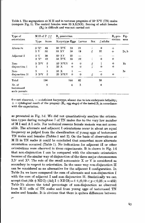
Table 1. The segregations at M II and in testcross progenies of 33~X3Y (TN) males (compare Fig. 1). The control females were 33 XX(NN). Scoring of adult females
(Bj) is difficult and was not carried out
Type of
orientation
Alternate
Adjacent 1
Nondisjunction I
Nondisjunction 11
Total
Nr. of testcrossed male parents
M II of P <$J
Type Score
3 X 3
3 Y
3 V 3 X3
3-X'Y 3
3 3 X3Y
49 55
38 42
2 1
1 2
190
6
Bx genera ion
Karyotype Eggs
3 3 X 3 X 33 XY
33 XY 33 X3X
3 3 X 3 X Y 33 X
33 X 33 X3XY
33 38
37 35
0 0
1 0
144
4
Larvae
21 19
— 22
0 0
— 0
62
3
Sex
X
3
*
* ?
0
cJ adults
0 37
— 0
1 0
— 0
38
4
B2 generation
+ + — 0
0 0
— 0
Fig
ures
2a, b
l h
3e
0 = not observed, — = deficient karyotype, absent due to late embryonic lethality; + = cytological result of the progeny (B2, egg stage) of the tested Br in accordance with the expectation.
as presented in Fig. 1 d. We did not quantitatively analyze the orientation types during metaphase I of TN males due to the very low number of M I and A I cells. For technical reasons female meiosis was not accessible. The alternate and adjacent I orientations occur in about an equal frequency as judged from the classification of young eggs of testcrossed TN males and females (Tables 1 and 2). On the basis of observations at M I I in TN males it could be concluded that somewhat more alternate orientation occurred (Table 1). No indications for adjacent I I or other orientations were observed in these experiments. I t is shown in Fig. I d that non-disjunction I can be compared with the alternate orientation because of the similar way of disjunction of the three major chromosomes 3,3_ and X3. The role of the small acrocentric X or Y is considered as secondary in respect to orientation. In the same way non-disjunction I I can be considered as an alternative for the adjacent I configuration. In Table 3 a we have compared the sum of alternate and non-disjunction I with the sum of adjacent I and non-disjunction I I . Statistically we can acceptthat(Alt + NDI) : (AdjI + N D I I ) = l : l , ( 0 , 1 0 < p < 0 , 2 5 ; w = 4 5 2 ) Table 3 b shows the total percentage of non-disjunction as observed from M I I cells of TN males and from young eggs of testcrossed TN males and females. I t is obvious that there is quite a difference between
43

$*$£ 38& tfjg .f
i 1 d ^mm § ^^* v .
3r* ^ f^/^}i Fig. 3. (a) 33X3XY karyotype. Spermatogonia! metaphase. (b) 33Y karyotype. Spermatogonial metaphase. (c) 33Y karyotype. Diakinesis/prometaphase Q. (d) 33X3Y karyotype. Spermatogonial metaphase. (e) 33~X karyotype. Mitotic metaphase from a young embryo, presumably female, (f) 33~XX karyotype. Mitotic metaphase from a young embryo, presumably female on the basis of chromosome
morphology. The bars on the photomicrographs represent 10 |/.m
T N males and females. I n T N females 18.7% of t he or ientat ion types are non-disjunction types . Both types (I and I I ) occur in about equal frequencies. TN (33~X3Y) males showed 3.2% and 0 .8% non-disjunction (I + I I ) as judged from M I I cells and young eggs respectively. I n (viable) 33X 3Y males, wi th a duplication for half t he length of t he long a rm of chromosome 3, we have scored 3.8% non-disjunction (only one t ype possible) from M I I cells (Table 3b) . This corresponds with t he percentage of 3.2 for T N males.
"Semi" - s te r i l i ty scored as brown eggs of T N males and females in testcrosses is between 25 % and 35 %. I n 3 TN females we have compared
't'l

Table 2. The segregations in testcross progenies of 33_X3X (TN) females (compare Fig. 1). The control males were 33XY (NN). Scoring of adult females (Bj) is difficult and was not carried out. The B2 generation was not investigated. See
further Table 1
Type of M I I Bj generation Fig-orientation type ures
P $ $ Karyotype Eggs Larvae Sex <$ adults
X ? 3-X3 33-X3 H-or 25 21
Y c? 28 2e, 2f Alternate
X ? 3 X 33 X + o r 26 24
Y cJ 38 2a, 2b
X 3f 3-X 33-X + o r 22 —
Y — Adjacent I
X 2 3 X3 33 X3 + o r 23 13
Y <J 21 3d
X ? 3-X3X 33-X3X + or 5 7
Non- Y <J 12 2h disjunction I X $
3 33 + o r 8 5 Y c? 6 3b, 3c
X 3e 3~ 3 3 - + o r 4 —
Non- Y — disjunction I I X 2
3 X3X 33 X3X + or 5 2 Y (J 5 3a
Total 118 72 110
Nr. of testcrossed female parents 5 2 6
the percentage "semi"-sterility or late embryonic lethality (eggs, 72 hours) with the percentage of cytologically determined deficient karyotypes in young (still viable, 8-16 hours) eggs (Table 4). Simultaneous tests were carried out for cytology (a total of 80 eggs) and for "semi"-sterility (397 eggs). The "semi"-sterility scored was about 28%, which is 8% higher than the percentage deficient karyotypes (20%) as scored in young eggs, and this 8 % may be due to a reduced viability of deviant other karyotypes.
hS

Table 3. a. The number of gametic karyotypes as scored a t M I I or in young eggs. Al ternate and non-disjunction I combined, and adjacent I and non-disjunction I I
combined, average rat io 1.15:1.00
b . Numerical non-disjunction as scored on TN males and females and 33X3Y males. Y ( X ) means :Y or X
Orientation
Alt. + NO 1
Adj.! + ND II
Gametic karyotypes 3 3 X 3 Y (TN $) 33-X 3 X(TN?)
M 11 Young eggs Young eggs ^-stage
(3 X3 + 3Y) + ( 3 X 3 Y + 3) 107 71
( 3 Y + 3X3) + (3 +3X 3 Y) 83 73
(>4
54
b
Alt. + Adj. 1
ND 1 + ND 11
33-X3Y (TN S)
M i l Young eggs
184 143
6 1
% Numerical non-disjunction (1 + 11), 3.2% 0.8% i.e. (X3 + Y(X) to the same pole)
3 3 X 3 X ( T N t) 33X 3Y(^)
Young eggs M 11
96 149
22 6
18.7% 3.8%
Table 4. Relation between percentage late embryonic lethality ("semi"-sterility) as scored in eggs of 72 hours and the percentage deficient karyotypes in young eggs
(8-16 hours)
Karyo- Nr. of Nr. of % deficient Nr. of % sterility Average % Nr. of Nr. of type crosses eggs for karyotypes eggs for (eggs, sterility eggs cros-tested cytology in young eggs sterility 72 hours) from other ses
(8-16 hours) score crosses
TN t: 3 Control 10
80 100
20% 0%
397 28% 500 2 -3%
31% 2 -3%
> 1 0 0 0 6 >2000 > 2 0
Conclusions and Discussion Boyes (1954) advanced four arguments for the view that the acro
centric chromosomes are the sex-chromosomes. Only one seems relevant: the acrocentric chromosomes sometimes differ in morphology, a phenomenon normally considered as characteristic for the sex-chromosomes. The acrocentric chromosomes in some related Hylemya species may be rather different in morphology and size. In Hylemya fugax, e.g., the acrocentric chromosomes appeared to be involved in a multiple sex-determination system, XjXgY/XjXjX^j.
46

From our experiments it can be concluded rather definitively that the small acrocentric chromosomes are the sex-chromosomes. The acrocentrics were seen to be different in morphology, due to the presence (in the X) or absence (in the Y) of a constriction in the vicinity of the (terminal) kinetochore (Figs. 2d, 3e and f). Secondly it was shown that this translocation between chromosome 3 and one of the acrocentrics is sex-linked. We can conclude that the acrocentrics are the sex-chromosomes, because no sex-linkage could be found for translocations in which chromosome 3 but not one of the acrocentrics in involved. Further it is clear that the translocation is X-linked and not Y-linked, as in that case it could not occur in the female sex. A XY (CJ)/XX( $) sex-determination system is considered as the most likely. Some more information on the sex-determination can be extracted from data presented elsewhere (van Heemert, 1974) where it is shown that 33XXY males appear after test-crossing translocation trisomic males (33~X3XY). No 33XXX karyotypes were expected and indeed no normal females with an extra X-chro-mosome were observed. This strongly suggests that the Y-chromosome as such is male determining. Several authors, e.g. Ullerich (1963), Ullerich etal. (1964) and Hiroyoshi (1964) stated that in many Diptera the Y-chromosome is male-determining as in mammals, in contrast to Droso-phila in which the X/A balance determines the sex. The distinct secondary constriction is located in the vicinity of the centromere of the X-chromosome but not in the Y-chromosome. I t can also clearly be seen in the translocated acrocentric chromosome X3 (Fig. 2g).
Table 2 shows that karyotypes monosomic for the sex-chromosomes could be derived from TN females. Six adult males (33Y) were seen which only had one sex-chromosome, presumably a Y-chromosome (Fig. 3 b, c). In the adult stage it could not be proven cytologically, that this chromosome is the Y-chromosome. There was no certainty in respect to the presence or absence of a secondary constriction. The X-chromosome that is missing in this karyotype probably is genetically inert, and it may have only a function in the female. Males of the 33Y constitution produce spermatozoa but no information about fertility is available.
The presence of very small metacentric B-chromosomes which do not pair somatically nor meiotically with the sex-chromosomes was not mentioned by Boyes (1954). However, the Canadian material we investigated and which Boyes used as well, possessed the same type of B-chromosomes. For the backcross experiments we have used a control series in which the B-chromosome was absent, to exclude misinterpretations, which could arise from the superficial similarity of the sex- and B-chromosomes.
Concerning the coorientation and disjunction in this translocation we can see from Tables 1 and 3 a that alternate, adjacent I, nondisjunc-
47

tion I and I I , but no adjacent I I were found. Nor were the nondisjunction types corresponding to adjacent I I observed. As appeared from the literature adjacent I I is considered a relatively infrequent event in animals. In mice e.g., Searle etal. (1971) indicated that the percentage of adjacent I I in three different translocations is 13% on the average. Jost and Laven (1971) showed in Culex pipiens that in ten different translocations studied, adjacent I I is the least frequent orientation. John and Hewitt (1963) have shown in Chorthippus brunneus that in an asym-
. metrical translocation between a small acrocentric and a large metacentric chromosome adjacent I I only occurred in a few cases. The results of Jaylet (1971) from two translocations in the newt show that no adjacent I I was found and that the homologous centromeres always disjoin in the first anaphase. Curtis et al. (1972) investigated 5 translocations in the tsetse fly and there were no indications for the adjacent I I orientation. La Chance etal. (1964) assumed adjacent I I to be absent in TN males as well as in TN females. John and Lewis (1965) in their review of the results of La Chance, could agree with this if translocation heterozygous males are concerned and considered it to be a result of achiasmate meiosis. In such an achiasmate situation homologous chromosomes have a prolonged pairing up to the first metaphase, and as Jaylet (1971) assumed, homologous centromeres disjoin in the first anaphase and no adjacent I I occurs.
John and Lewis (1965) have argued that the results of La Chance in the case of translocation heterozygous females should fit the expectations better when adjacent I I (25%) is assumed. Lewis and John (1963) even showed in one spontaneous translocation of Chorthippus brunneus that about 50% adjacent I I might occur.
In general, however, when there is no preference for alternate orientation, a 1:1 ratio of alternate: adjacent (I -\- II) is expected, adjacent I I being relatively infrequent. Apparently in this translocation there is no preference for alternate orientation (Tables 1 and 3a). From the data by Brink and Cooper (1932) on a (presumably) "simple" translocation in maize we could conclude that there were only two orientation types, alternate and adjacent I. These occurred in a ratio of 1:1, corresponding to what we have found in our translocation in the onion fly. There is no certainty that the fact that these translocations are "simple" is the cause of this 1:1 ratio. Only chains of four (Fig. 1) occur and no coorienta-tion of the centromeres of 3~ and X or Y is possible. We conclude that primarily homologous centromeres disjoin in this translocation and that therefore no adjacent I I coorientation was found. From Table 3a it can be seen that even when alternate and non-disjunction I are combined and adjacent I with non-disjunction I I , the 1:1 ratio has not changed. The two non-disjunction types can be considered as modifications of
48

alternate and adjacent I. This is particularly clear in the case of TN females where ND I and ND I I occur in about equal frequencies (Table 2). This indicates that both the X3 and the X orientate randomly, which can be explained by different mechanisms.
I t is rather peculiar that there is such a difference in numerical nondisjunction between TN males and females (Table 3b). In the case of TN females 18.7 % was found, in the case of TN males about 2 %. Ullerich (1964) has found numerical non-disjunction in Phormia regina (males achiasmate). About 1% was found in a Y-linked translocation as the result of a modified alternate orientation (like ND I in our case). The Y-chromosome is very small and acrocentric, while the translocated Y is much longer than the normal Y. This is analogous to our translocation in which the X3 is much longer than the X-chromosome. In fact Ullerich's translocation can be considered as a "simple" translocation as well. Of course, numerical non-disjunction could only be observed in males, because females do not have the translocation. Their score of about 1% is in agreement with our result of 2%. In general sex-chromosomes show a somewhat higher numerical non-disjunction than autosomes (Wurgler and Liitolf, 1972).
Several explanations may be given for the higher numerical nondisjunction in TN females than in TN males. One explanation might be disturbed meiotic pairing, which occurs at the time the X 3 - and the X-chromosomes start their pairing (in TN females). Very frequently meiotic pairing starts from the telomeric end of the chromosome arms. In this translocation the X3 and X do not usually find each other probably because the X-telomere at the place of the breakpoint in X3 in case of a true simple translocation does not function. In the case that a tiny end segment of X is attached to 3~ there is no place where X and X3 can start pairing. I t seems that in achiasmate male meiosis predominantly centromere pairing is responsible for the association and disjunction of the homologous chromosomes. The pairing and disjunction of the X3- and Y-chromosomes will not differ from the pairing and disjunction of the X- and Y-chromosomes in normal males. An other explanation can be the reduced chiasma frequency in thfe "interstitial" segment of the X3- and X-chromosomes in the female. This reduction may be due to the proximity of the breakpoint to the telomere of the X and to the centromeres of the X3 and X (Pig. lb ) . In males chiasmata were never found and therefore these factors do not operate here.
Several authors have observed numerical meiotic non-disjunction in certain translocation stocks of different animal species. These translocations had in common that a relatively small chromosome is involved, as in our X-linked translocation. Jost and Laven (1971) have reported numerical non-disjunction in chiasmate males of Culex pipiens in several
h3

translocations in frequencies up to 30 %. They argued that this is due to early separation of a relatively small sub-metacentric chromosome from the pairing complex. In these cases trivalents and univalents predominated, resulting in the formation of (n + 1) and (n —1) gametes. In mice Eicher (1973) and de Boer (personal communication) found numerical non-disjunction in translocation stocks where one of the translocated chromosomes was relatively small. In these translocations of Culex and mice which have a chiasmate meiosis, the small chromosome may have only a low chiasma frequency and, as a result the coorientation of the chromosomes in the translocation complex may be disturbed. In contrast to these translocations the relatively small chromosome in TN females of the onion fly is not a translocated one, but the original X-chromosome. I t may be assumed to have a reduced chiasma frequency as a result of pairing problems in the critical telomere area where the small X-chromosome pairs with the rest of the translocation complex. The small size of the X-chromosome as such is not the cause of the low chiasma frequency, but it does contribute to a reduced probability of chiasma formation once one of the fundamental conditions (pairing) is disturbed.
Table 4 shows that there is good correspondence between the percentage of embryonic lethals ("semi"-sterility) and the percentage of deficient karyotypes as cytologically determined in young eggs. The karyotypes with a long segment of the long arm of chromosome no. 3 missing, without any chromosomal compensation by an X3 chromosome, were not observed in the larval stage even in very young larvae. These karyotypes are 33~X or 33~Y and 33~XX or 33~XY in the progeny of testcrossed TN females (Table 2). They may cause 20% of brown eggs while the total percentage of brown eggs is 28%. From the literature it can be noted that in several other insect species these so called hypoploids die off in the middle stages of the embryonic development and cause semi-sterility (von Borstel and Rekemeyer, 1959; Imaizumi, 1962; Wright, 1971). The time of death of these hypoploids is very regular (von Borstel, 1963).
The explanation of the remaining 8% can be given in two ways. Firstly, we have found 2.5% brown eggs in the control but no cytologic anomalies were observed in the eggs. This percentage must always be taken into account. The remaining 5.5% of brown eggs is probably caused by the reduced viability of some specific deviant karyotypes. The number of 33X3X or 33X3Y and 33X3XX or 33X3XY karyotypes in the larval and adult stages (Table 2) is much lower than expected from the egg-scores. We have indications that 33X3X or 33X3Yindividuals pupate poorly. Some may die at late embryonic stages. Less viable 33X3X or 33X3Y karyotypes of Table 2 might in fact be mainly 33X3Y, because the 33X3X karyotypes in the larval progeny of TN males appeared to be as viable as the other karyotypes in the larval stage
50

(Table 1). A comparable s i tuat ion was found by Curtis et al. (1972) who found t h a t a certain unbalanced combination originating from a normal gamete and a gamete from an adjacent I or ientation can s tay alive, even in to t he adul t s tage. Such so called hyperploids (duplication karyotypes) most probably contr ibute considerably to the late embryonic lethality, as also suggested by Poulson (1940), Imaizumi (1962) and Scriba (1967). These authors s ta ted t h a t so called hyperploids sometimes also produce post-embryonic lethali ty. This we found in our experiments too. I t is not surprising t h a t in this asymmetrical t ranslocation t he viabili ty of so called hyperploid karyotypes is relatively high. They mainly carry duplications and no deficiencies as normally is t he case in reciprocal t ranslocations. The (monosomic) 33X or 33Y karyotype , deficient for an X-chromosome (Table 2), might contr ibute as well to the browning due to lower viabili ty in t he egg stage. No investigations were carried out on the embryological aspects of t he lethal syndromes.
A surprising observation was t h a t t he duplication types 3 3 X 3 X X or 3 3X 3 XY and 33X 3 X or 33X 3Y apparent ly were no t able to fertilize mates , unlike t he other viable aber ran t karyotypes . We never found any progeny of these types a l though in t he case of males, spermatozoa were found in the testes. I n t he asymmetrical t ranslocation of maize (Brink and Cooper, 1932) one duplication (hyperploid) t ype was able t o produce offspring through the eggs an<* not t he pollen. However this t ype had a r a ther low fertility.
This paper precedes a second one (van Heemert, 1974) about different trisomic types derived from translocation heterozygous parents. There the same aspects as presented here will be discussed.
Acknowledgements. I thank Dr. Ir. J. Sybenga for discussion and careful commenting of the manuscript. Valuable suggestions were made by Ir. P. de Boer. Technical assistance of Willem van den Brink is gratefully acknowledged. This research was supported by T.N.O.
Literature
Borstel, R. C. von: Inherited partial sterility in Habrobracon. Genetics today. I. Proc. XI Int. Congr. Genet. (S. J. Geerts, ed.), p. 124-125 (1963)
Borstel, R. C. von, Bekemeyer, M. L.: Radiation-induced and genetically contrived dominant lethality in Habrobracon and Drosophila. Genetics 44, 1053-1074(1959)
Boyes, J. W.: Somatic chromosomes of higher diptera. I l l Interspecific and intra-specific variation in the genus Hylemya. Canad. J. Zool. 32, 39-63 (1954)
Brink, R. A., Cooper, D. C : A structural change in the chromosomes of maize leading to chain formation. Amer. Naturalist 66, 310-322 (1932)
Curtis, C. F., Southern, D. J., Pell, P. E., Craig-Cameron, T. A.: Chromosome translocations in Glossina austeni. Genet. Res. (Camb.) 20, 101-113 (1972)
Eicher, E. M.: Translocation trisomic mice: production by female but not male translocation carriers. Science 180, 81 (1973)
Heemert, C. van: Isolation of a translocation homozygote in the onionfly Hylemya antiqua (Meigen) with a cytogenetic method in combination with the determination of the percentage late embryonic lethals. Genen Phaenen 16, 17-18 (1973)
51

Heemert, C. van: Meiotic disjunction and embryonic lethality in trisomies derived from an X-linked translocation in the onion fly. Chromosoma (Berl.) (in press, 1974)
Hiroyoshi, T.: Sex-limited inheritance and abnormal sex ratio in strains of the housefly. Genetics 50, 373-385 (1964)
Imaizumi, T.: Recherches sur l'expression des facteurs letaux hereditaires chez l'embryon de la Drosophile. VII. Sur les effects letaux dus a l'hypoploidie et a l'hyperploidie de chromosomes dans les souches d'XXY et de translocation chez Drosophila melanogaster. Cytologia (Tokyo) 27, 212-228 (1962)
Jaylet, A.: Creation d'une lignee homozygote pour une translocation reciproque chez 1'amphibien Pleurodeles waltlii. Chromosoma (Berl.) 34, 383-423 (1971)
John, B., Hewitt, G. M.: A spontaneous interchange in Chorthippus brunneus with extensive chiasma formation in an interstitial segment. Chromosoma (Berl.) 14, 638-650 (1963)
John, B., Lewis, K. R.: The meiotic system. Protoplasmatologia vol. VI/F/1. Wien-New York: Springer 1965
Jost, E., Laven, H.: Meiosis in translocation heterozygotes in the mosquito Culex pipiens. Chromosoma (Berl.) 35, 184-205 (1971)
La Chance, L. E., Riemann, J . G., Hopkins, D. E.: A reciprocal translocation in Cochliomyia hominivorax (Diptera: Calliphoridae). Genetic and cytological evidence for preferential segregation in males. Genetics 49, 959-972 (1964)
Lewis, K. R., John, B.: A spontaneous interchange in Chorthippus brunneus. Chromosoma (Berl.) 14, 618-637 (1963)
Poulson, D. F . : The effects of certain X-chromosome deficiencies on the embryonic development of Drosophila melanogaster. J. exp. Zool. 83, 272-325 (1940)
Scriba, M. E. L.: Embryonale Entwicklungsstorungen bei Defizienz und Tetra-ploidie des 2. Chromosoms von Drosophila melanogaster. Roux' Arch. Entwickl.-Mech. Org. 159, 314-345 (1967)
Searle, A. G., Ford, C. E., Beechy, C. V.: Meiotic disjunction in mouse translocations and the determination of centromere position. Genet. Res. (Camb.) 18, 215-235 (1971)
Ullerich, F . H.: Geschlechtschromosomen und Geschlechtsbestimmung bei einigen Calliphorinen (Calliphoridae, Diptera). Chromosoma (Berl.) 14, 45-110 (1963)
Ullerich, F.-H., Bauer, H., Dietz, R.: Geschlechtsbestimmung bei Tipuliden (Nema-tocera, Diptera). Chromosoma (Berl.) 15, 591-605 (1964)
Wijnands-Stab, K. J . A., Heemert, C. van: Radiation induced semisterility for genetic control purposes in the onionfly Hylemya antiqua (Meigen) I. Isolation of semisterile stocks and their eytogenetical properties. Theor. Appl. Genet. 44,111-119(1974)
Wright, T. R. F . : The genetics of embryogenesis in Drosophila. Advanc. Genet. 15, 262-395 (1971)
Wiirgler, F. E., Lutolf, H.-TJ.: The frequency of meiotic primary nondisjunction of the large autosomes in Drosophila melanogaster. Arch. Genetik 45,126-128 (1972)
Received March 11 — April 5, 1974 / Accepted by H. Bauer Ready for press April 11, 1974
C. van Heemert Department of Genetics Agricultural University 53 Generaal Foulkesweg Wageningen The Netherlands
52

Meiotic Disjunction and Embryonic Lethality in Trisomies Derived from an X-linked Translocation
in the Onion Fly, Hylemya untiqua (Meigen)
C. van Heemert
Department of Genetics, Agricultural University, Wageningen
Abstract. Translocation- and tertiary trisomies (for the X-chromosomes) were obtained after testcrossing translocation heterozygous females of an X-linked "simple" translocation stock. Meiotic disjunction as judged from segregations at M II (males) and in young eggs of testcrosses (males and females) in translocation trisomies was studied. No progeny of tertiary trisomic males and females was found, but male M II could be studied. Six different orientation types appeared in translocation trisomic ( 2 n+ l ) males and these were present in equal frequencies. No adjacent II configurations were found. The small X- and Y-chromosomes and the large translocated X-chromosome of the translocation complex disjoin at random (n and n -f-1 gametes) in both translocation- and tertiary trisomic males. In translocation trisomic females four different orientation types appeared. From the high frequency of two of these (together, 94.5%) it is concluded that the two normal X-chromosomes show preferential pairing and disjunction, while the translocated X-chromosome moves to either one of the two poles at random. Primary trisomic (for the X-chromosome) males (XXY) and females (XXX) were obtained from testcrossed translocation trisomies. Cytological analysis of adult male progeny of testcrossed XXY males showed that no random orientation for the X-, X- and Y-chromosomes occurred because half of the sons was disomic (XY) and half of them trisomic (XXY). A possible mechanism is discussed. Analysis of young eggs of testcrossed XXX females indicated a segregation of 2X:1X = 1 : 1 . The level of "semi"-sterility as scored from testcrosses of translocation trisomies appeared to be as in translocation heterozygotes. Here again a close relation exists between "semi"-sterility and deficiencies in eggs for a large chromosomal segment. The possible use of this translocation for genetic control of insect pests is discussed.
Introduction
In an earlier article an X-linked translocation in the onion fly (2n = 12) was described (van Heemert, 1974). A large submetacentric chromosome (no. 3) and the small acrocentric X-chromosome are involved in this translocation. The translocated chromosome 3~ lost about half the length of the long arm. I t seemed not to have gained any chromosomal material from the X-chromosome. The translocated acrocentric chromosome X3 gained considerably in length. This translocation is considered as a "simple" translocation but may also be a highly unequal reciprocal translocation.
53

V
X Y
n
\ %x3
«x
3
l 4
3 "
3
X 3
^ 1
X Y
X 3 X
.-_« X
V \ i L_
> X3
3
X3 Y
I
XJ Y
— T
d V X V 1— i • ^ U 3 " X 3 X 3 - X
Fig. 1. (a) Idiogram of the chromosomes involved in the translocation complex of a TN -f- X male, (b) Cross-figure as expected in early meiotic prophase I, diagrammati-cally. (c) Meiotic pairing as usually observed in diakinesis/prometaphase. (d) Six different orientation types (A I). In the case of TN + X females replace Y by X. Orientation types 2 and 3 become identical as do types 5 and 6 in the case of TN + X
females
I t appeared that in addition to the alternate and adjacent I orientations, numerical non-disjunction for the X-chromosome occurred. As a result, translocation trisomies, tertiary trisomies, monosomies and duplication karyotypes were produced, besides the normal (NN) and translocation heterozygous individuals (TN). The terminology used is explained in Sybenga (1972). All these aberrant karyotypes reached the adult stage, but only the translocation trisomies were fertile.
Translocation trisomies (TN + X) although having an additional X-chromosome compared to translocation heterozygotes (TN), showed the same percentage (25%-35%) of "semi"-sterility in both sexes. Five chromosomes are involved in one complex at meiosis. In Fig. la we have drawn them diagrammatically for a TN + X male: 3,3~,X3,X and Y. In the case of a TN + X female the Y-chromosome should be replaced by an X-chromosome.
I t has been shown (van Heemert, 1974) that the pattern of inheritance of the translocation heterozygotes (TN) is typically X-linked. Trans-
5k

location heterozygous males after testcrossing produce normal sons (except in one case, where a TN + X son appeared) and "semi"-sterile daughters exclusively. Translocation heterozygous females produced normal as well as translocation heterozygous males and females.
However, we discovered that several "semi"-sterile males after test-crossing produced translocation sons as well as normal sons.
We found another, indirect, indication for this. When flies obtained from an individual testcross of certain "semi "-sterile males were sib -crossed, many matings (about 25 %) showed normal fertility. This is not expected in the case of a normal X-linked translocation, because all the sons of a testcrossed translocation-heterozygous father (TN) will be normal and all the daughters will have the translocation. Therefore all matings between sons and daughters of such a father should show " semi "-sterility. Cytological analysis showed these unusual fathers to be translocation trisomies (TN + X).
The progeny of testcrossed translocation trisomic (TN + X) males and females has been investigated in the egg, larval and adult stages. M I I cells of TN + X males were analyzed as well. Disjunction at meiosis of the X3, X and Y chromosomes of TN -+- X males and the X3, X and X chromosomes of TN + X females with homologous centromeres was studied and compared with the disjunction of X3Y or X3X in TN males and females respectively (van Heemert, 1974). Only insufficient numbers however, of analyzable M I and A I cells were available for quantitative analysis.
Fig. l b shows how the five chromosomes of the translocation trisomic might pair at pachytene. Since the onion fly has achiasmate males, regular diplotene stages are not expected. We have observed diakinesis/ prometaphase pairing in spermatocytes as indicated in Fig. 1 c. Fig. 1 d diagrammatically shows the 6 different orientation types the presence of which was demonstrated by segregations at M I I and in young embryos. There were no indications for adjacent I I or other orientation types. Orientation types 1,2 and 3 in TN ~f- X males, have in common that the major chromosomes 3~ and X3 go to the same pole and chromosome 3 to the opposite pole. The X- and Y-chromosomes can be found together in one pole or in different poles. In the case of orientation types 4, 5 and 6, chromosomes 3 and X3 go to the same pole while chromosome 3~ goes to the other pole. Again the X- and Y-chromosomes can be found together in one pole or in different poles.
Only four different orientation types could be concluded to be present in TN + X females, and are analogous with the types 1, 2 (3), 4 and 5 (6) as shown in Fig. 1 d.
Primary trisomies for the X-chromosome were found among the offspring of testcrossed TN + X flies. The disjunction of the X, X and Y
55

Table 2. The segregations in testcross progenies of 33_X3XX (TN + X) females (compare Fig. 1). The control males were 33XY (NN). Scoring of adult females (Bj) is difficult and only six could be analyzed. No larvae were investigated. For
further explanation see Table 1
Type of
orientation
1
2
3
4
M I I type parental females
3 X 3
3 XX
3~X3X
3 X
3-XX
3 X3
3-X
3 X3X
Total
Nr. of testcrossed female parents
Bx generation
Karyotype
33-X3
33 XX
X + or
Y X
+ or Y
X 33"X3X + or
Y X
33 X + o r Y
33-XX
33 X3
X + or
Y X
+ or Y
X 33-X + or
Y
X 33 X3X + or
Y
Eggs
2
5
34
27
0
0
30
29
127
3
Larvae Sex
$
<? 9
9
9
<?
9
9
<?
Adults
<?
0
1
6
6
1
-
3
17
3
9
0
l
2
2
—
1
—
0
6
3
the 33 XX mother occurs. Indeed no 33X3Y males were observed even though a sufficient number was analyzed. In the same way karyotypes of eggs with 3 3 + three small acrocentric chromosomes were 33XXY (males) exclusively (Table 1).
Table 2 gives the results of the analysis of the progenies of testcrossed females which were TN + X as could be demonstrated by the presence of the 33 XXX or 33XXY karyotypes in young eggs. Six different karyo-
58

types were observed in the egg stage. The karyotypes 33~XXX or 33-XXY and 33X3X or 33X3Y both resulting from orientation type 3 (Fig. 1) were not observed in the eggs. The karyotypes 33~X3X or 33~X3Y and 33 XXX or 33 XXY were present only in a small number. No larvae were scored but young adult flies of both sexes were analyzed. In 17 young newly emerged males we observed five different karyotypes. Four different karyotypes were found in six young females. Although in young eggs the products of orientation type 3 were not observed, yet one 33X3X adult female and one 33X3Y adult male were found, which can only arise as the result of this orientation type 3. Apparently the frequency of this orientation type is very low.
In Table 3 a the frequencies of gametic types corresponding to specific orientation types as scored at M I I and as derived from eggs of testcrosses involving TN + X males and females are combined. The sum of orientation types 1, 2 and 3 were compared with the sum of orientation types 4, 5 and 6 in the case of TN + X males, while the sum or orientation types 1 and 2 and the sum of orientation types 3 and 4 were compared in the case of TN + X females. There was no significant deviation from a 1:1 ratio in TN + X males nor females. As indicated in Table 3 b, the X- and Y-chromosomes of TN + X males go to the same pole and the X3 to the opposite pole in 32.7% (eggs)—34.7% (Mil) of the cases observed. In tertiary trisomic males (33X3XY) a corresponding value (30.7%) was observed at M II . In TN + X females both X-chromosomes go to the same pole and the X3-chromosome to the opposite pole in only 5.5% (eggs) of the cases.
Table 4 shows the percentages of disjunction types for the XXY and XXX trivalents in primary trisomic (NN + X) males and females respectively. These trisomies were originally obtained from testcrossed TN + X parents (Tables 1 and 2). From testcrossed NN + X(33XXX) females we have found half of the eggs with an additional X-chromosome and the other half with the normal number (2) of sex-chromosomes. This clearly demonstrates that the XXX trivalent always disjoins as: 2X/1X. Here the segregation of the sex-chromosomes was analyzed in the testcross progeny (60 eggs) of the homogametic sex ($) and it is not necessary to distinguish between the sexes in the progeny. When, however, as in the case of NN + X males (33 XXY) segregation is analyzed in the test-cross progeny of the heterogametic sex, the sex of the progeny is relevant, and in the onion fly can only be determined in the adult stage, 52.4% of the sons (101) appeared to be 33 XXY and 46.6% 33 XY.
We have investigated the relationship between late embryonic lethality in eggs (72 hours) and deficiency in young eggs (8-16 hours) for a large chromosomal segment. Ninety eggs (8-16 hours old) from testcrossed TN -f- X males were used for cytology, and 18% had the large deficiency.
17 Chromosoma (Berl.), Bd. 47
59

Table 3 a. The number of gametic karyotypes as scored at M II and in young eggs from testcrosses. Orientation types 1, 2 and 3 combined and 4, 5 and 6 combined (see Fig. 1 d). Replace Y by X in the case of TN + X females. Numbers gathered from Tables 1 and 2. — b. Disjunction percentage of X3XY in translocation trisomic males and in tertiary trisomic males and the disjunction percentage of X3XX in
translocation trisomic females X—X3 means: X and Y go to one pole and X3 to the other. X(Y) means: X or Y.
a Orientation type
Gametic karyotypes 33~X3XY(TN + X S) 33-X3XX(TN + X $)
M i l Young eggs
Young eggs
1 + 2 + 3 (3"X3 + 3XY) i - h z + d + (3"X3Y(X) + 3X(Y)) * '
4 + 5 + 6 (3"XY + 3X3) 4 + o + D + (3-Y (X) + 3X3X(Y)) 0 1
31
24 59
b
Dis
junction
X3 1 —Y
X * X3
Y - X
Y - X 3
Transl. tris. males
33-X3XY(TN + X)
M i l (98 cells)
• 65.3%
34.7%
Young eggs (55)
67.3%
32.7%
Tert. tris. males
33X3XY
M i l (52 cells)
69.3%
30.7%
Dis
junction
x3 x
X X
X _ X 3 X A
Transl. tris. females
33-X3XX(TN + X)
Young eggs (127)
94.5%
5.5%
Table 4. Disjunction percentages of XXY and XXX in primary trisomies (for the X-chromosome). In the case of males we have analyzed adult sons from testcrosses. Disjunction in females was established by analyzing young eggs (8-16 hrs.) from
testcrosses Y—X means: X and Y go to one pole and the second X to the other pole.
X Y" X X"
X
Y
33XXY (<J)
52.4%
47.6%
101 sons
X X" X
33XXX (?)
100%
60 young eggs
60

Table 5. Relation between the percentage late embryonic lethality ("semi"-sterility) as scored in eggs (72 hours) and the percentage deficient karyotypes in
young eggs (8-16 hours)
Karyotype tested
TN + X S Control
Nr. of crosses
2 10
Nr. of eggs for cytol
ogy
90 100
% deficient karyotypes in young eggs (8-16 hours)
18 0
Nr. of eggs for sterility score
328 500
% sterility
(eggs 72 hours)
27 2-3
Average % sterility from other crosses
29 2-3
Xr. of eggs
> 1000 > 2 0 0 0
Nr. of crosses
7 > 2 0
Another 328 eggs (72 hours old) from the same batch were scored for the percentage " semi "-sterility (late embryonic lethality): 27 %. The remaining 9% may be due to a reduced viability of other deviant karyotypes. The average percentage of " semi "-sterility in other experiments is about 29% (Table 5).
Conclusions and Discussion
The meiotic behaviour of the chromosomes in translocation heterozygous (TN) males and females could be reasonably well established by analyzing M I I and young eggs (van Heemert, 1974). Particularly the meiotic disjunction of the translocated X-chromosome (X3) and the normal X-chromosome in TN females and the meiotic disjunction of X3
and Y in TN males was studied. I t appeared that i8.7% numerical nondisjunction (X3 and X to the same pole) occurred in TN females and only about 2% in TN males (X3 and Y to the same pole; Table 3, van Heemert, 1974).
Translocation trisomic (TN + X) males and females have an additional X-chromosome compared to TN males and females. Here we compared the coorientation and disjunction of the three chromosomes with homologous centromeres in TN + X males (X3, X and Y) with the behaviour of X3, X and X in TN + X females. The data on disjunction in tertiary trisomic males, 33X3XY (Table 3b), show that the coorientation and disjunction of the X3-, X- and Y-chromosomes is similar to that in translocation trisomic (TN + X) males. This similarity suggests that it does not make any difference whether 33 or 33" is present in combination with the chromosomes X3, X and Y. Further it was concluded from segregations (Mil and eggs) of tertiary trisomic (33X3XY, 2 n + 1) males and of translocation trisomic (TN + X, 2n + 1 ) males and females, that only (n) and(n + 1) gametes are produced and no (n + 2)or(n—-1) gametes. Therefore we assumed that for TN + X males orientation types 1,2 and 3 (Fig. 1)
17*
61

can be combined, as well as orientation types 4, 5 and 6. Similarly, types 1 and 2, and types 3 and 4 can be combined in the case of TN + X females. I t was shown statistically that the sum of orientation types 1, 2 and 3 is not different from the sum of 4, 5 and 6 (0.50 <p<0.75; n = 163) in TN + X males and that 1 + 2 is not different from 3 + 4 (0.25 < p<0 . 50 ; n = 127) in TN + X females (Table 3a). This corresponds to the 1:1 ratio of (Alt + ND I ) : (Adj I + ND II) as observed in TN males and females (Table 3a, van Heemert, 1974). In both TN and TN + X for males as well as for females there is an equal chance for X3 to go to the same pole with chromosome 3 as with chromosome 3~, independently of the presence of one or two normal sex-chromosomes.
Looking at the behaviour of the chromosomes X3, X and Y in TN + X males, we see that in 33.7% of the cases observed (Table 3b) the X- and Y-chromosomes go to the same pole (orientation types 1 and 4, Fig. 1). In about 2/3 of the cases X and Y disjoin and it is plausible that the two types of gametes with X3Y or X (orientation types 2 and 6) and gametes with X3X or Y (orientation types 3 and 5) occur in equal frequencies, 1I3 each. I t can be concluded from these results (M I I and young eggs) that random coorientation and disjunction ( n /n+ 1) occurs for the X3-, X- and Y-chromosomes in TN + X ( 2 n + l ) males. Orientation types (1 + 4): (2 + 6): (3 + 5) = 1 : 1 : 1 . An explanation for this random process might be that the three different chromosomes (X3, X and Y) have homologous centromeres, and because the males have no chiasmata the association of the homologous centromeres determines the coorientation. In spite of the same size of the X- and Y-chromosomes, distributive pairing apparently does not occur, as otherwise we would have found mainly X3Y (X3X) and X (Y) gametes.
In TN + X females a completely different situation was found. In the X3XX group of chromosomes, the two identical X-chromosomes disjoin in 94.5% of the cases observed (Table 3 b) and in only 5.5% of the eggs of testcrossed TN + X females both X-chromosomes from the TN + X female parent are present. Distributive pairing (size-dependent) may be an explanation for this phenomenon, but preferential (homologous) pairing of the two normal X-chromosomes can explain their almost 1:1 disjunction as well. In the latter case chiasmata can occur between the two X-chromosomes and probably are absent between the X3 and X. Apparently the X3-chromosome moves at random to either one of the two poles at the first anaphase (Table 2). As a consequence the gametes 3~X3 and 3 XX (orientation type 1, Fig. 1) are formed and when fusion with a 3X or 3Y gamete of the normal male testcross parent takes place, 33-X3X or 33"X3Y and 33 XXX or 33XXY karyotypes will be found in the eggs (Table 2). No eggs originating from orientation type 3 were found as these would have had the 3 3 X X X (or 33XXY) or 33 X3X (or 33 X3Y)
62

•* Jkf§
w MM ^mm j B f e _ ^ " "
Fig. 2. (a) NN + X (33XXY) karyotype. Spermatogonial metaphase. XXY means X + X + Y. (b) NN + X (33XXY) karyotype. Diakinesis/prometaphase $. (c) TT + XY (3~3~X3X3XY) karyotype. Translocation homozygous male (tetrasomic). Spermatogonial metaphase. The bars on the photomicrographs represent 10 fxm
karyotype. However, after cytological analysis of Bj adults (Table 2) we could conclude that this orientation type must have occurred occasionally.
Primary trisomic males (NN + X) appeared in the progeny of TN + X males and females and not in that of TN. NN + X females were found among the progeny of TN + X females exclusively. NN + X males and females were both very viable and completely fertile. Primary trisomies (NN + X, Fig. 2a and b) and translocation trisomies (TN + X) have in common that only ( n + 1) and (n) gametes occur and that their ratio is 1:1. Table 4 shows that when two X-chromosomes and one Y-chromo-some are present there is no random disjunction. In a random situation twice as many XY and X gametes as XX and Y gametes would be expected. However, equal frequencies of XY (52.4%) and Y (47.6%) were found. In diakinesis/prometaphase in males both X-chromosomes usually are paired very intimately and the Y-chromosome, though paired, can be seen more apart (Fig. 2 b). Both X-chromosomes might act as a " couple " in 50% of the cases and disjoin together from the Y-chromosome as normally one X disjoins from the Y in 33 XY males. In the other 50% the two X-chromosomes disjoin from each other and the Y goes with either one of the two X-chromosomes into a pole. No Y- nor X-chromo-some seems to get lost.
63

females and TN -)- X males show about the same percentage (28 % and 27% respectively) of brown eggs (72 hours) and about the same percentage of deficient karyotypes (20% and 18% respectively), as scored cytologi-cally in young eggs (8-16 hours). TN males and TN-f-X females were not tested. Testcrossed primary trisomic (NN -)- X) males and females did not show any "semi"-sterility and no deficiencies in young eggs were observed.
McDonald and Rai (1971) suggested the use of a sex-linked translocation for genetic control purposes. They concluded from computer simulation studies, that X-linked translocations are more useful than autosomal translocations for release experiments. The use of double heterozygotes which are heterozygous for two different sex-linked translocations seemed even more successful for genetic control in Aedes aegypti. For translocations in general, important limiting factors are competitive ability, degree of the "semi"-sterility and population growth rate. For normal X-linked translocations the theoretical impossibility of homo-zygotes in both sexes is an additional limitation for mass rearing. We were able to isolate homozygotes for this X-linked translocation in the onion fly even in the male sex. This rather unexpected result can be explained as follows. When in a cross between a TN + X male and a TN female 3~X3Y and 3~X3 gametes are formed respectively, we may expect some 3~3 X3X3-|-Y karyotypes, which are translocation homozygous males (TT -f Y). A TN + X male crossed with a TN + X female can give translocation homozygous (TT -\- Y) males as well. Even TT -f XY males are present rather often (Fig. 2 c). Translocation homozygous females could be detected by analysis of their oogonia. They can be 3~3-X3X3 or 3~3~X3X3 + one or two additional X-chromosomes. We maintained the stock in which translocation homozygous flies were observed by full sibmating. Morphologically the translocation homozygotes appeared to be normal and completely competitive with NN individuals. Their fertility was about 75% (control: 94%). The stock was kept homozygous for three generations but unfortunately probably became contaminated with normal flies. Attempts to make it homozygous again are under way. Disjunction of the X3,X3,X and X or X3,X3,X and Y chromosomes of tetrasomic TT + 2 X females or TT + XY males respectively will then be studied as well and compared with the disjunction of the X3,X and Y, and of the X3,X and X chromosomes of the translocation trisomies presented here.
Acknowledgements. I thank Dr. Ir. J. Sybenga for discussion and careful commenting of the manuscript. Valuable suggestions were made by Ir. P. de Boer. Technical assistance of Willem van den Brink and the photography of Mr. K. Knoop are gratefully acknowledged.—This research was supported by T.N.O.
66

References Borstel, R. C. von, Rekemeyer, M. L . : Radiation-induced and genetically con
trived dominant lethality in Habrobracon and Drosophila. Genetics 44,1053-1074 (1959)
Heemert , C. v an : Meiotic disjunction, sex-determination and embryonic lethality in an X-linked „ s imp le" translocation of t he onion fly. Chromosoma (Berl.) 47, 45-60 (1974)
Imaizumu, T . : Recherches sur 1'expression des facteurs letaux hereditaires chez l 'embryon de la Drosophile. VI I . Sur les effects letaux dus a l 'hypoploidie et a l'hyperploi'die de chromosomes dans les souches d ' X X Y et de translocation chez Drosophila melanogaster. Cytologia (Tokyo) 27, 212-228 (1962)
McDonald, P . T., Rai , K. S.: Population control potential of heterozygous t ranslocations as determined by computersimulations. Bull. Wld. Hl th . Org. 44 ,829-845 (1971)
Sybenga, J . : General cytogenetics. Amsterdam: Nor th Holland Publ . Co., Inc. 1972
Wright , T. R. F . : The genetics of embryogenesis in Drosophila. Advanc. Genet. 15, 262-395 (1971)
Received March 11—April 5, 1974 / Accepted by H . Bauer Ready for press April 16, 1974
C. van Heemert Depar tment of Genetics Agricultural University 53 Generaal Foulkesweg Wageningen The Netherlands
67

General discussion
From the results of the first three articles (I, II and III) a few com
ments can be made which may be relevant for future isolations of "semi"-ste-
rile stocks carrying chromosomal rearrangements. Comparison of the different
doses used on males (all below the 100% sterilizing dose of 3 krad of X-rays)
led to the conclusion that a low dose of 0.5 krad of X-rays on males is re-
commendable for the induction of chromosomal rearrangements to be used for
genetic control purposes. The reasons are firstly, that a few or no complex
chromosomal rearrangements causing "semi"-steri1ity are induced at such low
doses. Secondly, the genetic background damage (such as recessive lethals)
will be reduced, which is important for homozygosing the translocations. Yt-
terborn (1970) has shown with Drosophila that translocations induced with
lower radiation doses have a greater chance to be viable as homozygotes. On
the other hand more initial screening in the F. has to be carried out to find
a "semi"-sterile strain, because the frequency of rearrangements is low. Ori
ginally, 13 days old pupae instead of adult males were irradiated because
these seemed to be more manageable. A disadvantage was the ignorance of the
precise developmental stage and also the fact that half of the emerged flies
(the females) had to be discarded. As far as the output of "semi"-sterile
stocks is concerned, no difference between irradiating 13 days old pupae or
young adult males was found. This can be understood when one considers that
early spermatids, expected to be the most sensitive for the induction of
translocations (Sobels, 1969), are already present at late pupal stages. La-
teron it was concluded (article III) that irradiation of about seven days old
males might be more useful because of the presence of a more homogeneous sam
ple of mature sperm. In this case, of course, probably most chromosomal re
arrangements are induced at a less sensitive stage, resulting in a lower yield
of chromosomal rearrangements per unit of dose.
Females appeared to be very sensitive to X-rays, compared to males. Glass
(1956) has shown the same in Drosophila. In particular just emerged females
in which the ovaries still have to develop (Theunissen, 1970, are sensitive
as appeared from the high infecundity in the parental and even the F, genera-
68

tion, and many F, crosses as far as they could be made were sterile. A mino
rity of the irradiated females (P generation) had a relatively high fertili
ty compared to males irradiated with the same dose, probably because eggs
which were laid were rather free of radiation damage and therefore could
hatch. Seven days old females gave much better results, because the fecundi
ty was higher, although the egg production is still reduced to about 1/5 of
the normal production of a control series and stopped earlier. A reasonable
number of F. crosses produced offspring, but most of them had an about nor
mal fertility and were not suitable for our purpose.
The use of fast neutrons on females even at a low dose is probably not
advisable, due to high infecundity in the P and F. generation. However, ir
radiation of young males with fast neutrons gives results comparable to those
of the use of X-rays. At a low dose of 0.25 krad a few reciprocal transloca
tions were found in a rather small sample. Fast neutrons, having a higher
track density, probably cause less genetic background damage than X-rays.
Table A. Distribution of the initial breakpoints (21) of 10 different translocations (table B) over the 11 different chromosome arms (See table B). Arm-length at mitotic metaphase in larval brain cells according to Boyes (1954).
14 13 12 11 10 10 8 7 6 5 4 Armlength (in order; microns)
61 31 41 51 21 6s 5s 2 s 4 s 3s 1 Chromosome arms
1 4 1 3 4 5 1 1 1 Distribution of breakpoints
In the first article (l) we have made a few comments on the positions of
the initial breakpoints of the translocations. We stated that the length of
the chromosome arms probably is one of the factors which determines the chan
ce of becoming involved in a chromosomal rearrangement. From table A giving
the chromosome arms in which the initial breakpoints of the ten transloca
tions (table B) are located it can indeed be seen that the arm length plays
a role. The conclusions of Burnham (1964) in his review of break positions in
maize and Drosophila are in general in agreement with our results. However,
in those organisms the smaller chromosome arms mostly were usually somewhat
more involved in the translocations than expected on the basis of mitotic
length. Most (18) of the initial 21 breakpoints are located in the six of the
eleven chromosome arms which are longer than or equal to 10 ju. It is not
clear why 6 and 4 each are involved only once, although the armlengths are
among the greatest. Chromosome arms 3S and 4 s are not involved in any of the
69

translocations. However, In (3) 2 is a pericentric inversion, thus a break
must have occurred in the short arm of chromosome 3. Apparently no preferen
tial participation of particular chromosome arms during the induction of
translocations could be demonstrated. In the first paper we thought it plau
sible that the initial translocation breakpoints preferentially were located
in achromatic areas of the chromosomes or in secondary constrictions. We can
see now, that there is no relationship between the occurrence of breakpoints
and these achromatic (heterochromatic?) areas. Of course, small heterochro-
matic areas may be present, not discernable with the convential staining tech
niques we have used.
For the recognition and isolation of translocations genetic markers are
generally employed in insect genetics. These are not available in the onion
fly but the use of cytological techniques can be applied without problem. The
karyotype of the onion fly consists of six pairs of chromosomes. Due to so
matic pairing exchanged non-homologous segments of reciprocal translocations
can readily be distinguished, unless these are too short and/or equal in size.
In cases of doubt meiotic analysis (males) as a rule could give evidence.
A standard method of brooding the eggs followed by the scoring of brown
eggs (van Heemert, 1973) proved to be more succesfull for the screening of
the "semi"-steri1ity than the use of egg hatch. Originally, the egg hatch was
measured (at a temperature of 2it°C) by dividing the empty eggs by the total
number of eggs: unhatched white eggs + empty eggs. By brooding the egos at
29 C a part of the unhatched eggs turned brown in colour in the case of a
translocation. Late embryonic lethality (brown eggs) was shown to be related
with duplication/deficiency karyotypes resulting from adjacent orientation in
the translocation heterozygote.
In crosses between two translocation heterozygous (TN) individuals as
expected, we scored higher percentages of brown eggs in comparison with the
testcross, as both parents instead of one produce unbalanced gametes. In most
of the translocation stocks which were sibcrossed we could distinguish three
levels of sterility in spite of a sometimes considerable variation. These
correspond with the sterility from NNxNN, NNxTN and TNxTN crosses. When the
testcross fertility of a translocation is rather high (for instance 85%) or
low (for instance 15%) the difference between the average fertility of a
testcross and of a cross between two heterozygotes is small (in this example
13%). The maximum difference of 25% occurs in the case the testcross fertili
ty is 50%. With a moderate variation a difference of 13% is too small to dis-
70

Table B. Review of the different chromosomal rearrangements induced in the onion fly in relation to the induction dose, chromosome arms involved, egg hatch, homozygosity and larval duplication/deficiency karyotypes. ? not yet analyzed; - not found; + observed; T translocation; In inversion.
Code Original dose Chromosomes Egg-hatch % Homozygous Larval Comments involved test cross as dupl./
larvae/adults def. types
T
T
T
T
T
T
T
T
T
1
2
3
k
5
6
7
8
9
1.0
1.1
1 .0
0.25
0.5
0.5
1.0
"spo
1.5
Krad
Krad
Krad
Krad
Krad
Krad
Krad
X <S
X a*
X o-
fN cr*
X C'
X a*
X cr'
ntaneous"
Krad X </
3
3
5
2
2
3
2
2
3
-1(H
ts( +
c ^
1(H
T 10 1.0 Krad X j
In 1 0.5 Krad X r/ Chr. 6
In 2 "spontaneous" Chr. 3
6s <+
-6 K-) 72
55
70
80
79
62
60
50
55
L
L
L
L
Ik o 97 V 80 o 94 V
X-linked
cyclic
resembles T 10
symmetrical
resembles T U
pericentric, symmetrical
pericentric, asymmetrical
criminate between the two fertility levels. The fertility in the control (NN)
crosses normally has little variation.
From the experiments as described in the first three articles we could
definitively recognize 17 different translocations and two pericentric inver
sions. Ten translocations and the two inversions were kept for further re
search (Table B). The rest was lost or not maintained because of rearing dif
ficulties or because the translocation was not suitable for more detailed cy-
tological analysis. Three out of these ten translocations (T2, T3 and T9) were
removed from the sib program for the following reasons.
Translocation T2 is rather complex. It is a cyclic translocation (three
breakpoints) in which three pairs of chromosomes are involved. No transloca
tion homozygotes were ever seen, even after sibcrosses for at least k gene
rations. This translocation produced very good egg rafts with a sterility of
about 50% and had a good competitiveness with normal flies. These character
istics were the reason why this particular stock was successfully used in a
preliminary field experiment. From a cage in which thousend normal and thou-
send translocation heterozygous flies were released, after three weeks we have
71

recaptured 15 females and these were allowed to oviposite in the laboratory.
An average sterility of 36% was scored, while the control cage showed no ste
rility.
Analysis of T3 indicated that the translocation heterozygous karyotype
could not unequivocally be discriminated from the normal karyotype.
In the case of T9 a rather high dose of 1.5 krad of X-rays was used and
therefore it was assumed that inbreeding would give homozygotes carrying de
trimental factors (recessive lethals) in a homozygous condition. As a check
one sibcross was studied and indeed no homozygote was found in the larval
stage.
Five of the seven remaining translocations have been sibcrossed and the
larval progeny (6 larvae per caged female) was analyzed cytologically for the
presence of translocation homozygotes (TT) in the case a relatively high ste
rility was found. In theory i of the sibcrosses between randomly taken sibs
out of testcross progenies (NN+TN) will show this high level of sterility.
If the presence of translocation homozygotes could be cytologically proven
in the progenies of individually caged females, both parents should have been
heterozygous (TN) for the translocation. Surprisingly in all five transloca
tion stocks which were sibcrossed translocation homozygotes were found cyto
logical ly. In three of these, Tl, T6 and T10, they were viable into the a-
dult stage (Table B). In the translocations T*t and T5 homozygotes could be
found only as older larvae, but further analysis is needed. Of Tl and T10 we
know that the translocation homozygotes are fertile and can produce offspring.
In both cases some of the individual sibcrosses with flies from the first
sibcross progenies were found to have approximately an about normal fertili
ty and had a progeny consisting of 100% translocation heterozygotes. These
must have been the combination of a normal and a translocation homozygous
parent.
The difficulty of obtaining a homozygous stock most probably is due to
the small scale on which sibcrosses could be made. Theoretically about one
out of sixteen crosses with flies from the progeny of a cross between trans
location heterozygous parents will be between two translocation homozygous
individuals. It obviously requires large scale experiments to be able to iso
late a homozygous translocation stock.
An important feature we have observed is the occurrence of duplication/
deficiency karyotypes at the larval stage. Surprisingly these karyotypes were
found only in those translocations of which translocation homozygotes were
observed. In general it is rather uncommon that such karyotypes reach the
72

latest larval stages. In the case of Tl even the adult (with sperm) stage. We
have no explanation for this and further investigation is needed. In spite of
the many references on translocations in insects it is peculiar that only in
a few occasions such viable duplication/deficiency karyotypes were described.
Curtis et al (1972) and Ved Brat and Rai (197M mentioned the presence of
such deviant karyotypes in the larval stage of Tsetse fly and Aedes aegypti
respectively. Most probably other workers have paid insufficient attention
to the larval cytology. Sometimes the distinction of a duplication/deficiency
karyotype from a translocation heterozygous karyotype was difficult. When one
of the two marker (translocated) chromosomes of the translocation heterozy
gous karyotype is similar to one of the other chromosomes of the karyotype
and when at the same time somatic pairing is somewhat less close, confusion
with a duplication/deficiency karyotype is possible. Normally only one of the
two duplication/deficiency karyotypes originating from an adjacent I orien
tation (possessing the larger duplications and the smaller deficiencies) were
found at the larval stage. However, in the case of T5 we have found both pos
sible duplication/deficiency karyotypes from adjacent I among the larval off
spring of a testcrossed translocation heterozygote.
Sibcrbsses of both pericentric inversion stocks have been started. In a
few among a large number of sibcrosses of In 1 a lower fertility was found
compared to the testcross fertility of inversion heterozygous females. Since
inversion heterozygous males are completely fertile (article III) these cros
ses must have been between inversion heterozygous parents and the inversion
homozygotes of the progeny most probably die in the embryonic stage. This as
sumption is supported by the observation of a higher frequency of inversion
heterozygotes (larvae) among the progeny than expected. We do not yet have
sufficient data from inbreeding experiments of the In 2.
Detailed studies on the X-linked translocation Tl are reported (IV and
V ) . The presence of an extra X-chromosome in the translocation trisomic ka
ryotypes (TN+X) caused a completely different meiotic behaviour compared to
translocation heterozygotes (TN) and different composition of the offspring.
In our first attempts to isolate homozygous flies (TT) , carried out concur
rently with the experiments described in articles IV and V, we have found
translocation nomozygotes in both sexes, with one or two additional sex-chro
mosomes. We now know that these must have arisen from crosses between two
translocation trisomic parents (TN+X). Crosses between translocation hetero
zygotes (TN), as can be concluded from tables 1 and 2 from paper IV, will
73

give TT or TT+X females (in a ratio of about k:\) but no TT males. Crosses
between TN+X males and females do give translocation homozygotes for both
sexes and these possess one (or two) extra sex chromosomes: The male is TT+Y
(or TT+X+Y), the female TT+X or TT+2X and occassionaly TT.
In the first step of the homozygosing program we have testcrossed TN+X
females. The progeny has a higher frequency of TN+X sons and daugthers than
testcrossed TN+X males or TN females. Subsequently 63 sibcrosses were made,
of which 32 had an increased sterility (40%-60%) compared to the testcross
sterility (25%~35%). The progenies of these were analyzed cytologically (at
least six larvae per progeny) with the aim of finding TT+2X (or TT+X+Y) ka
ryotypes. NN was present as well. Five out of these 32 sibcrosses contained
TT+2X (or TT+X+Y and sometimes TT+X or TT+Y) among their progeny and an ad
ditional three had TT+X (and no TT+2X). The presence of TT+2X (or X+Y) pro
ves that both parents were TN+X. In exceptional cases they could have been
TN and TN+X. The progeny of these five crosses were again sibcrossed in the
next generation (52 crosses) with the aim of finding only TT+1 or 2 sex-chro
mosomes. Progenies with NN karyotypes were not maintained. Out of these 52
crosses 38 were analyzed cytologically and in five out of these we have found
only TT+1 or 2 sex-chromosomes or TN+X (or TN) karyotypes and no NN types
among the six tested larvae per progeny. In one completely fertile cross all
the larvae (nine) were TN+X, which indicates that one parent was NN and the
other TT+2X (or X+Y). Unfortunately no crosses between translocation homozy
gotes could be demonstrated. Theoretically in this translocation the chance
of finding a cross between translocation homozygotes is approximately 1%,
while in autosomal translocations (without complementation) this is appr. 6%.
It is planned to continue with the progeny expected to be from crosses be
tween a translocation homozygote and a TN+X karyotype. Finally we hope to end
up with an unique strain of translocation homozygotes for this X-linked trans
location. In earlier experiments we were able to isolate a homozygous stock,
but unfortunately this was lost. Perhaps this stock when again homozygous will
turn out to be one with unexpected advantages for genetic control of the
onion fly. When not all the individuals carry the two extra sex-chromosomes
there is the risk that the stock will backslide into a stock wi th only TT
female individuals, which cannot persist.
In two more translocation stocks (T6 and T10) we found adult transloca
tion homozygotes. In the case of T10 we know already that the TT karyotype
is fertile because we have observed one cross (with a normal fertility) and
all the larval offspring (12) analyzed was TN.
74

Little is known about the best way of infiltrating a noxious insect po
pulation with translocation flies. It is generally assumed that in order to
introduce sterility into the population a single translocation stock is not
sufficient. Normally the reproductive capacity of an insect population can
be enormous and therefore the use of one translocation, which normally has a
moderate "semi"-steri1ity can not cut down the population density below an
acceptable level. A moderate sterility on the other hand is essential for the
rearing and homozygosing of the translocation. In the case a single translo
cation would be sufficient, most probably the release of translocation he-
terozygotes, synthesized after combining a NN and TT strain, would be advi
sable rather than the release of translocation homozygotes. The advantages
of releasing only heterozygotes are: sterility occurs at once and possibly
there may be heterosis of the heterozygotes. If homozygotes are released, ste
rility does not occur before the next generation and these may be less viable
due to inbreeding. With both methods it must be further investigated if only
males or both sexes have to be released and how many releases are necessary
each generation. The highest sterility will be obtained with a maximum number
of translocation heterozygotes in the population or when the frequency of Inl
and T-gametes is at the equilibrium of 0.5 (Curtis and Hill, 1971). However,
a Hardy-Weinberg ratio can not be obtained mainly due to the complementation
of "adjacent" gametes. Further, due to the reproductive negative heterosis
of the structural heterozygotes the equilibrium is unstable. If the 1:1 ratio
of normal to translocation gametes is slightly changed and the TT karyotypes
are as fit as the NN types a rapid shift (frequency dependent selection) will
occur either in the direction of a complete TT or NN (original) population
and the fertility will increase concurrently (Li, 1955). To counterbalance
this one must be very keen on translocation heterozygotes with a superior com
petitiveness in order to maintain the sterility in the population as long as
possible. In the case the population becomes homozygous (TT) this is a way to
change the population by incorporation of a particularly useful gene linked
to the translocation.
The release of double translocation heterozygotes as suggested e.g. by
Curtis and Robinson (1971) gives the opportunity of a considerably increased
sterility compared to the use of single translocations. Further population
genetic studies and simulation studies are needed. The future application of
the translocation method to achieve population suppression will depend on co
operative efforts among geneticists, entomologists and ecologists.
75

duator in the selection for "semi"-steri1ity.
Adult fertile translocation trisomies and adult sterile tertiary triso
mies were obtained (both sexes) after meiotic numerical non-disjunction. In
translocation trisomic males the X-, Y- and translocated (extra) X-chromosome
were shown to disjoin at random. In females the two normal X-chromosomes al
most (35%) preferentially disjoin, while the translocated X-chromosome goes
to either one of the poles. Primary trisomic males (XXY) and females (XXX)
were obtained from testcrossed translocation trisomic parents. XXY males pro
duced four types of gametes XY, X, Y and XX in equal numbers. XXX females on
ly gave XX and X gametes in an equal number. Succesfull attempts to obtain
homozygotes for this X-linked translocation are reported. The theoretical
background of genetic insect control is discussed.
78

Samenvatting
Het beschreven onderzoek, gepubliceerd in vijf artikelen (I-V), had tot
doel het isoleren van structurele chromosoom mutaties die "semi"-steri1iteit
veroorzaken waardoor zij voor de genetische bestrijding van de uievlieg, Hy-
lemya antiqua (Meigen) gebruikt kunnen worden. In de eerste drie artikelen
worden de resultaten gegeven van het onderzoek naar de inductie met behulp
van straling, en de selectie, isolatie en cytologische analyse van "semi"-
steriele translocaties en inversies. X-stralen en snelle neutronen werden ge
bruikt in verschi1lende doses bij mannelijke en vrouwelijke vliegen (of pop-
pen) van verschi1lende leeftijden. Het eerste artikel (I) beschrijft o.a. de
wijze van bestralen, het testen van de fertiliteit (ei-uitkomst) in de be-
straalde- (P) en terugkruisings- (F.) generatie, de criteria voor "semi"-ste-
riliteit en cytologisch onderzoek van "semi"-steriele stammetjes. In het twee-
de (II) en derde (ill) artikel wordt ingegaan op de resultaten van bestraling
bij hogere zowel als lagere doses dan gebruikt in het begin-onderzoek (l). Te-
vens werd de leeftijd der te bestralen vliegen als variabele ingevoerd.
Er kon vastgesteld worden (I + ll) dat het gebruik van hogere doses X-
stralen (1.5 krad) op mannetjes relatief veel translocaties maar ook veel com-
plexe chromosoom mutaties oplevert. Uit het oogpunt van het risico van gene
tische achtergrondschade (b.v. recessief (sub-) lethale factoren) is het ge
bruik van hogere doses af te raden. Een lage dosis van 0.5 krad (X-stralen op
mannetjes) 1ijkt het meest aanbevelenswaard in dit stadium van onderzoek (II +
III). Er moet echter relatief veel screening op "semi"-steri1iteit in de F.
uitgevoerd worden om toch nog een aantal translocaties en inversies te kunnen
isoleren. Vrouwtjes die 1 dag oud zijn, zijn zeer gevoelig voor bestraling
(1.0 krad X-stralen). Zeven dagen oude vrouwtjes (1.0 krad X) zijn zowel wat
de fecunditeit (P en F,) als wat de F. reproductie betreft gunstiger (II),
maar in vergelijking met mannelijke vliegen (1.0 krad X) is de opbrengst aan
structurele mutaties te laag. Snelle neutronen op mannetjes geven bij 0.25
krad minstens zulke goede resultaten als bij 1.0 krad gezien het percentage
gevonden translocaties. Verder onderzoek naar het gebruik van neutronen is no-
dig.
79

De door ons ontworpen broedtechniek der eieren, gevolgd door het scoren
van bruine eieren (1aat-embryonaal lethaliteit als gevolg van duplicatie/de-
ficiency karyotypen), bleek goed te functioneren in combinatie met de cytolo-
gische analyse van nakomelingschappen van "semi"-steriele ouders. In totaal
werden 17 verschi1lende translocaties en twee pericentrische inversies gevon-
den. Bij vijf van de acht reciproke translocaties die onderworpen werden aan
een sibcross programma bleken translocatie homozygoten in het larvale stadium
voor te komen en in drie gevallen van deze vijf zelfs in het imaginale sta
dium. Twee van deze drie bleken als homozygoot reproductief te zijn. Verder
was het opvallend dat slechts bij de vijf genoemde translocaties in de nako
mel ingschap (larven) van toetskruisingen duplicatie/deficiency karyotypen af-
komstig van "adjacent I" orientaties optraden. Een reden hiervoor is niet be-
kend. Van de uit "adjacent I" ontstane duplicatie/deficiency karyotypen (twee
typen) weten we dat vooral de typen die een grote duplicatie en een geringe
deficiency bezitten tot ver in het larvale stadium levensvatbaar zijn.
De meeste "breukpunten" bleken op de 6 (van de 11) langste chromosoom-
armen te liggen. GSen preferentiele deelname van bepaalde chromosoomarmen aan
reciproke translocaties werd waargenomen. Uit toetskruisingen van inversie
heterozygote ouders bleek dat alleen vrouwtjes "semi"-steriel zijn vanwege
het voorkomen van chiasma(ta) in de inversielus van de pericentrische inver
sie. Inversie heterozygote mannetjes waren even fertiel als de controle man-
netjes, hetgeen het achiasmatisch zijn van de mannetjes onderstreept.
De laatste twee artikelen (IV en V) bevatten de studies van de meiotische
disjunctie en de embryonaal lethaliteit van een X-gekoppelde translocatie en
van de daarvan verkregen trisome karyotypen. Deze studies zijn van belang voor
de homozygotering van deze translocatie. Er kon overtuigend aangetoond worden
dat de kleine acrocentrische chromosomen de geslachtschromosomen zijn. Van de
translocatie heterozygote karyotypen (IV) werd het gedrag van de chromosomen
van het complex tijdens de meiose geanalyseerd aan de hand van M II (manne
tjes) en jonge eieren (mannetjes en vrouwtjes). In beide geslachten treedt
even vaak de "alternate" als de "adjacent I" orientatie op, terwijl numerieke
non-disjunctie van het kleinste chromosoom van het translocatiecomplex vaak
bij vrouwtjes (18.7%) en weinig bij mannetjes {2%) geconstateerd werd. Ver-
moedelijk is verstoorde telomeer paring, waardoor verminderde chiasmavorming
optreedt, de oorzaak voor de hoge frequentie numerieke non-disjunctie bij
vrouwtjes. Bij de mannetjes wordt zeer waarschi jnl ijk het meiotisch gedrag
der chromosomen door de homologe centromeren bepaald. Cytologische analyse
van jonge eieren (8-16 uur) van toetskruisingen van translocatie heterozygote
80

vrouwtjes wees uit dat ongeveer 20% een grote deficientie bezit, hetgeen re-
del ijk goed overeenkomt met de gevonden "semi"-steri1iteit (laat-embryonale
lethaliteit, bruine eieren).
Als gevolg van de numerieke non-disjunctie bij translocatie heterozygote
vrouwtjes werden translocatie- en tertiaire trisomen (voor het X-chromosoom)
verkregen (V). De tertiaire trisomen, waarbij een groot chromosoom extra voor-
komt, bleken niet reproductief te zijn hoewel de meiose (bij mannetjes) nog
wel bestudeerd kon worden. De meiotische disjunctie in "semi"-steriele trans
locatie trisome mannetjes en vrouwtjes, waarbij slechts een klein X-chromo
soom extra voorkomt, werd (bij de vrouwtjes indirect via analyse van jonge
eieren) bestudeerd. Evenals bij de translocatie heterozygote karyotypen werd
gSfin "adjacent II" gevonden. In translocatie trisome mannetjes blijken de
twee normale geslachtschromosomen (X en Y) en het getransloceerde (extra) X-
chromosoom volledig volgens het toeval te segregeren. Dit werd ook in terti
aire trisome mannetjes (M II) gevonden. In translocatie-trisome vrouwtjes,
waarin naast twee normale X-chromosomen een getransloceerd X-chromosoom aan-
wezig is, blijken de twee normale X-chromosomen in 95% van de gevallen elk
naar een van de twee polen te gaan. In 5% van de gevallen gaan beide normale
X-chromosomen naar een pool, terwijl het getransloceerde X-chromosoom naar
de andere pool gaat. Het percentage "semi"-steri1iteit van toetskruisingen
blijkt redelijk goed overeen te komen met het percentage eieren met een grote
chromosomale deficiency.
Primaire trisome karyotypen (voor het X-chromosoom) werden gevonden na
toetskruising van translocatie trisome karyotypen. De fertiliteit is gelijk
aan die van de controle. De XXY mannetjes leveren vier typen gameten: XY, X,
Y en XX in een verhouding van 1:1:1:1. XXX vrouwtjes leveren twee typen ga
meten: XX en X (1:1).
Tot slot is in de algemene discussie het verloop van de homozygotering
van de X-gekoppelde translocatie besproken en wordt kort ingegaan op de theo-
retische achtergrond van de genetische bestrijding.

Curriculum vitae
Cornells van Heemert
Geboren: 28 april 1 9 ^ te Leeuwarden
Eindexamen H.B.S.-B 1962 te Groningen
Landbouwhogeschool Wageningen van September 1962 - September 1970
Studierichting (Hoofdvak): PIantenveredeling
Keuzevakken: Erfelijkheidsleer (verzwaard)
Algemene Plantenziektekunde
In dienst van de Centrale Organisatie TNO, Sectie Landbouwkundig Onderzoek
C0-TN0, Den Haag, van 15 juni 1970 - 31 december 1974.
In deze periode als gastmedewerker verbonden aan net laboratorium voor Erfe-
1ijkheidsleer van de Landbouwhogeschool, Wageningen.
Vanaf 1 januari 1975 verbonden aan het I.T.A.L. te Wageningen.
82

Ik kan alleen nog regels schrijven
die net als jullie
vliegen blijven.
(Judith Herzberg u i t : Vliegen)
83
Related Documents