Journal of Basic and Clinical Reproductive Sciences · January - June 2014 · Vol 3 · Issue 1 71 Chorangioma of Placenta with High Risk Pregnancy: A Case Series Uma S Andola, Shabnam Karangadan 1 , Sainath K Andola 1 , Rajashekhar Jewargikar 1 Department of Obstetrics and Gynecology, 1 Resident, Professor and Head of Department, Professor, Department of Pathology, Mahadevappa Rampure Medical College, Gulbarga, Karnataka, India ABSTRACT Chorangioma is the most common benign tumor of placenta, with an incidence of approximately 1%. Tumors larger than 5 cm are associated with maternal and fetal complications. We report four cases of chorangioma that were identified in mostly primigravida (75%) with a mean gestational age of 33 wks (Range 28-33 wks). All except one case, complete placenta was received with mean weight of 550 g (Range 400-800 g) and tumor size of 9.4 cm (Range 0.5-23 cm). Microscopy showed capillary proliferation resembling fetal vessels in enlarged villi covered by trophoblastic epithelium. Three cases had preterm delivery. One also presented with abruptio placenta and intrauterine fetal death. Because of limited documentation of cases and dismal prognosis of these pregnancies, especially with large chorangiomas, it’s of utmost importance to correctly diagnose them with the aid of color Doppler flow. In this series, 75% of cases (3/4), babies survived probably due to small size of tumor and attainment of fetal maturity. KEY WORDS: Chorangioma, placenta, placental tumor, vascular tumor KEY MESSAGES: Even though chorangioma is the most common tumor of placenta, tumors arising from placenta are rare. Because of dismal prognosis of these pregnancies with large lesions, it’s important to correctly diagnose them with the aid of color Doppler flow. This case series demonstrates the need for awareness of such lesions. INTRODUCTION Chorangioma of placenta is the most common benign tumor of placenta, with an incidence of approximately 1% of all pregnancies. [1-3] It is a vascular tumor which is usually small in size and only found after careful examination of the placenta. Tumors larger than 5 cm are associated with maternal and fetal complications. [1,2,4] Limited documentation of cases and dismal prognosis of these pregnancies, especially with large chorangiomas, has prompted us to present this case series. CASE REPORT Four cases of placental chorangioma were diagnosed in the Department of Pathology, M. R. Medical College, Gulbarga, between 2011 and 2013. The clinical and pathological findings of the cases are summarized in Table 1. The patient’s age ranged from 23 to 28 yrs (mean 24 yrs). Most of them were primigravida (3/4). Gestational age at diagnosis ranged from 28 to 39 wks (Mean 33 wks). Clinical presentation was in active stage of labor, bleeding PV and with retained bits of placenta post delivery. Size of the tumor ranged from 0.5 to 23 cm (mean 9.4 cm). All except Case 3, complete placenta was received [Figure 1a]. Placental weight of the three cases ranged from 400 to 800 g (mean 550 g). Ultrasound scan report was available in Case 1 which was reported as 10 × 10 cm placental hematoma [Figure 1b]. All cases showed capillary proliferation resembling fetal vessels in enlarged villi covered by trophoblastic epithelium [Figures 1c and 1d]. Lymphoplasmacytic infiltrate along with scattered Hofbauer cells were noted. Areas of myxoid change and calcification were also seen. In between these hemangiomatous areas are seen normal and degenerated villi. All four cases delivered by spontaneous normal vaginal delivery, of which one was still born. Cases 1 and 4’s babies were having low birth weight as well (0.8 and 1.8 kg, respectively). Access this article online Quick Response Code Website: www.jbcrs.org DOI: 10.4103/2278-960X.129291 Case Report Address for correspondence Dr. Sainath K Andola, Department of Pathology, Mahadevappa Rampure Medical College, Gulbarga, Karnataka - 585 105, India. E-mail: [email protected] [Downloaded free from http://www.jbcrs.org on Monday, March 13, 2017, IP: 220.227.255.125]

Welcome message from author
This document is posted to help you gain knowledge. Please leave a comment to let me know what you think about it! Share it to your friends and learn new things together.
Transcript
Journal of Basic and Clinical Reproductive Sciences · January - June 2014 · Vol 3 · Issue 1 71
Chorangioma of Placenta with High Risk Pregnancy: A Case Series Uma S Andola, Shabnam Karangadan1, Sainath K Andola1, Rajashekhar Jewargikar1
Department of Obstetrics and Gynecology, 1Resident, Professor and Head of Department, Professor, Department of Pathology, Mahadevappa
Rampure Medical College, Gulbarga, Karnataka, India
A B S T R A C T
Chorangioma is the most common benign tumor of placenta, with an incidence of approximately 1%. Tumors larger than 5 cm are associated with maternal and fetal complications. We report four cases of chorangioma that were identified in mostly primigravida (75%) with a mean gestational age of 33 wks (Range 28-33 wks). All except one case, complete placenta was received with mean weight of 550 g (Range 400-800 g) and tumor size of 9.4 cm (Range 0.5-23 cm). Microscopy showed capillary proliferation resembling fetal vessels in enlarged villi covered by trophoblastic epithelium. Three cases had preterm delivery. One also presented with abruptio placenta and intrauterine fetal death. Because of limited documentation of cases and dismal prognosis of these pregnancies, especially with large chorangiomas, it’s of utmost importance to correctly diagnose them with the aid of color Doppler flow. In this series, 75% of cases (3/4), babies survived probably due to small size of tumor and attainment of fetal maturity.
KEY WORDS: Chorangioma, placenta, placental tumor, vascular tumor
KEY MESSAGES: Even though chorangioma is the most common tumor of placenta, tumors arising from placenta are rare. Because of dismal prognosis of these pregnancies with large lesions, it’s important to correctly diagnose them with the aid of color Doppler flow. This case series demonstrates the need for awareness of such lesions.
INTRODUCTION
Chorangioma of placenta is the most common benign tumor of placenta, with an incidence of approximately 1% of all pregnancies.[1-3] It is a vascular tumor which is usually small in size and only found after careful examination of the placenta. Tumors larger than 5 cm are associated with maternal and fetal complications.[1,2,4]
Limited documentation of cases and dismal prognosis of these pregnancies, especially with large chorangiomas, has prompted us to present this case series.
CASE REPORT
Four cases of placental chorangioma were diagnosed in the Department of Pathology, M. R. Medical College, Gulbarga, between 2011 and 2013. The clinical and pathological findings of the cases are summarized in Table 1.
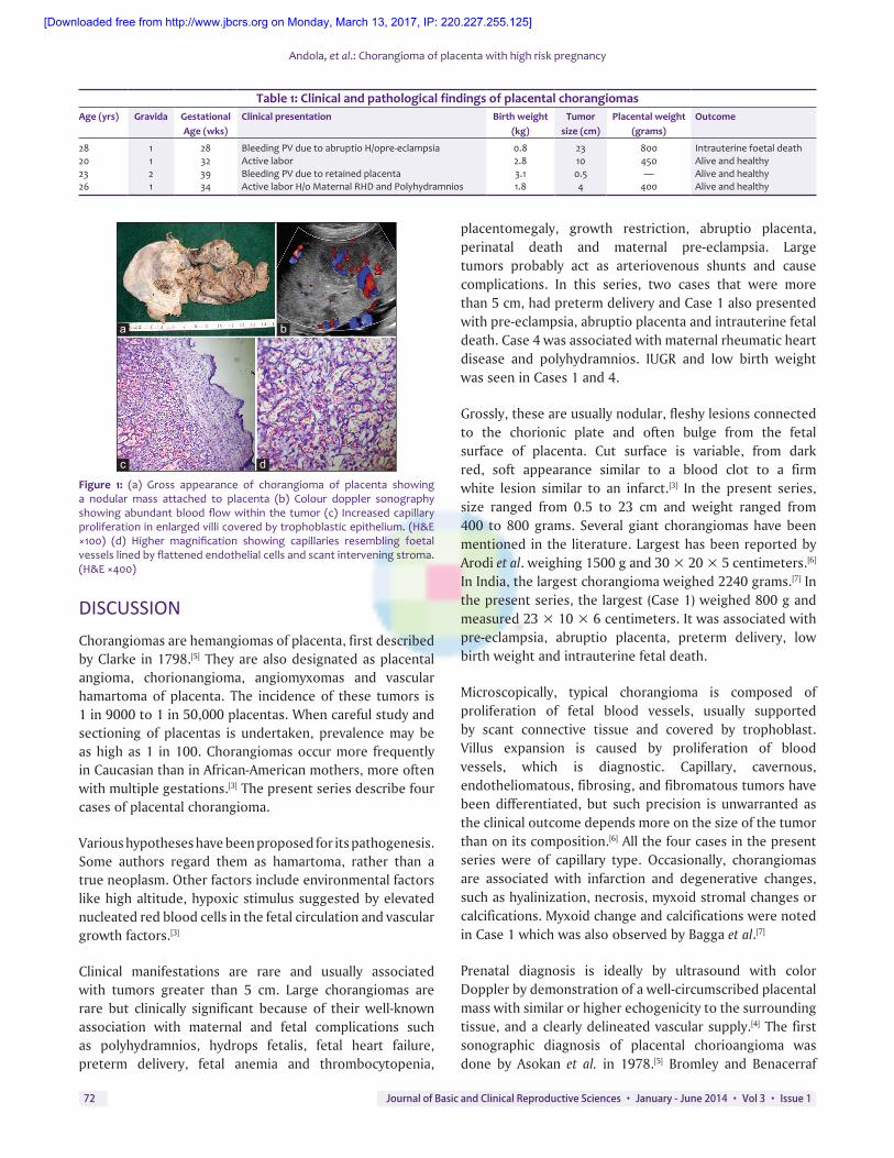
The patient’s age ranged from 23 to 28 yrs (mean 24 yrs). Most of them were primigravida (3/4). Gestational age at diagnosis ranged from 28 to 39 wks (Mean 33 wks). Clinical presentation was in active stage of labor, bleeding PV and with retained bits of placenta post delivery. Size of the tumor ranged from 0.5 to 23 cm (mean 9.4 cm). All except Case 3, complete placenta was received [Figure 1a]. Placental weight of the three cases ranged from 400 to 800 g (mean 550 g). Ultrasound scan report was available in Case 1 which was reported as 10 × 10 cm placental hematoma [Figure 1b].
All cases showed capillary proliferation resembling fetal vessels in enlarged villi covered by trophoblastic epithelium [Figures 1c and 1d]. Lymphoplasmacytic infiltrate along with scattered Hofbauer cells were noted. Areas of myxoid change and calcification were also seen. In between these hemangiomatous areas are seen normal and degenerated villi.
All four cases delivered by spontaneous normal vaginal delivery, of which one was still born. Cases 1 and 4’s babies were having low birth weight as well (0.8 and 1.8 kg, respectively).
Access this article onlineQuick Response Code
Website:www.jbcrs.org
DOI:10.4103/2278-960X.129291
Case Report
Address for correspondence Dr. Sainath K Andola,
Department of Pathology, Mahadevappa Rampure Medical College, Gulbarga, Karnataka - 585 105, India.
E-mail: [email protected]
[Downloaded free from http://www.jbcrs.org on Monday, March 13, 2017, IP: 220.227.255.125]
Andola, et al.: Chorangioma of placenta with high risk pregnancy
Journal of Basic and Clinical Reproductive Sciences · January - June 2014 · Vol 3 · Issue 172
DISCUSSION
Chorangiomas are hemangiomas of placenta, first described by Clarke in 1798.[5] They are also designated as placental angioma, chorionangioma, angiomyxomas and vascular hamartoma of placenta. The incidence of these tumors is 1 in 9000 to 1 in 50,000 placentas. When careful study and sectioning of placentas is undertaken, prevalence may be as high as 1 in 100. Chorangiomas occur more frequently in Caucasian than in African-American mothers, more often with multiple gestations.[3] The present series describe four cases of placental chorangioma.
Various hypotheses have been proposed for its pathogenesis. Some authors regard them as hamartoma, rather than a true neoplasm. Other factors include environmental factors like high altitude, hypoxic stimulus suggested by elevated nucleated red blood cells in the fetal circulation and vascular growth factors.[3]
Clinical manifestations are rare and usually associated with tumors greater than 5 cm. Large chorangiomas are rare but clinically significant because of their well-known association with maternal and fetal complications such as polyhydramnios, hydrops fetalis, fetal heart failure, preterm delivery, fetal anemia and thrombocytopenia,
placentomegaly, growth restriction, abruptio placenta, perinatal death and maternal pre-eclampsia. Large tumors probably act as arteriovenous shunts and cause complications. In this series, two cases that were more than 5 cm, had preterm delivery and Case 1 also presented with pre-eclampsia, abruptio placenta and intrauterine fetal death. Case 4 was associated with maternal rheumatic heart disease and polyhydramnios. IUGR and low birth weight was seen in Cases 1 and 4.
Grossly, these are usually nodular, fleshy lesions connected to the chorionic plate and often bulge from the fetal surface of placenta. Cut surface is variable, from dark red, soft appearance similar to a blood clot to a firm white lesion similar to an infarct.[3] In the present series, size ranged from 0.5 to 23 cm and weight ranged from 400 to 800 grams. Several giant chorangiomas have been mentioned in the literature. Largest has been reported by Arodi et al. weighing 1500 g and 30 × 20 × 5 centimeters.[6] In India, the largest chorangioma weighed 2240 grams.[7] In the present series, the largest (Case 1) weighed 800 g and measured 23 × 10 × 6 centimeters. It was associated with pre-eclampsia, abruptio placenta, preterm delivery, low birth weight and intrauterine fetal death.
Microscopically, typical chorangioma is composed of proliferation of fetal blood vessels, usually supported by scant connective tissue and covered by trophoblast. Villus expansion is caused by proliferation of blood vessels, which is diagnostic. Capillary, cavernous, endotheliomatous, fibrosing, and fibromatous tumors have been differentiated, but such precision is unwarranted as the clinical outcome depends more on the size of the tumor than on its composition.[6] All the four cases in the present series were of capillary type. Occasionally, chorangiomas are associated with infarction and degenerative changes, such as hyalinization, necrosis, myxoid stromal changes or calcifications. Myxoid change and calcifications were noted in Case 1 which was also observed by Bagga et al.[7]
Prenatal diagnosis is ideally by ultrasound with color Doppler by demonstration of a well-circumscribed placental mass with similar or higher echogenicity to the surrounding tissue, and a clearly delineated vascular supply.[4] The first sonographic diagnosis of placental chorioangioma was done by Asokan et al. in 1978.[5] Bromley and Benacerraf
Table 1: Clinical and pathological findings of placental chorangiomasAge (yrs) Gravida Gestational
Age (wks)Clinical presentation Birth weight
(kg)Tumor
size (cm)Placental weight
(grams)Outcome
28 1 28 Bleeding PV due to abruptio H/opre-eclampsia 0.8 23 800 Intrauterine foetal death20 1 32 Active labor 2.8 10 450 Alive and healthy23 2 39 Bleeding PV due to retained placenta 3.1 0.5 — Alive and healthy26 1 34 Active labor H/o Maternal RHD and Polyhydramnios 1.8 4 400 Alive and healthy
Figure 1: (a) Gross appearance of chorangioma of placenta showing a nodular mass attached to placenta (b) Colour doppler sonography showing abundant blood flow within the tumor (c) Increased capillary proliferation in enlarged villi covered by trophoblastic epithelium. (H&E ×100) (d) Higher magnification showing capillaries resembling foetal vessels lined by flattened endothelial cells and scant intervening stroma. (H&E ×400)
a
c
b
d
[Downloaded free from http://www.jbcrs.org on Monday, March 13, 2017, IP: 220.227.255.125]
Andola, et al.: Chorangioma of placenta with high risk pregnancy
Journal of Basic and Clinical Reproductive Sciences · January - June 2014 · Vol 3 · Issue 1 73
stated that the sonographic appearance of a chorioangioma was indistinguishable from that of a placental hemorrhage. They speculated that the use of color Doppler sonography could have helped them in differentiating these lesions.[8] Similar error was made in one of the cases in this series (Case 1), mistaken as placental hematoma.
Chorioangioma with complications before fetal viability requires interventions. Because of the dismal prognosis of these pregnancies, especially with lesions larger than 5 cm, it’s important to correctly diagnose these lesions and to closely follow up these pregnancies, preferably with the aid of color Doppler.[2] Except Case 1, all babies are alive and healthy. The 28 wks gestation fetus was delivered by normal vaginal delivery following severe ante partum hemorrhage due to abruptio placenta. Baby expired due to birth asphyxia. In 3 out of 6 chorangiomas Zalel et al.[2] described pregnancies that ended before completion of 32 wks of gestation.
Chorangiomas can be confused with subamniotic hematoma, placental teratoma, degenerated myoma and placental infarction on gross morphology but microscopy is characteristic.
CONCLUSION
Even though chorangioma is the most common tumor of placenta, tumors arising from placenta are rare. Because of dismal prognosis of these pregnancies, especially with lesions larger than 5 cm, it’s of utmost importance to correctly diagnose them and to closely follow up these
pregnancies, preferably with the aid of color Doppler flow. In the present series, 75% of cases (3/4), babies survived probably due to small tumor size and attainment of fetal maturity. The present case series demonstrates the need for awareness of such lesions, and emphasizes the importance of timely antenatal diagnosis and intervention.
REFERENCES1. Fox H, Sebire NJ. Non-trophoblastic tumors of the placenta. In: Fox H,
Sebire N. Pathology of the Placenta. 3rd ed. Philadelphia: Saunders Elsevier; 2007. p. 401-30.
2. Zalel Y, Weisz B, Gamzu R, Schiff E, Shalmon B, Achiron R. Chorioangiomas of the placenta: Sonographic and doppler flow characteristics. J Ultrasound Med 2002;21:909-13.
3. Baergen RN. Neoplasms. In: Baergen RN. Manual of Benirschke and Kaufmann’s Pathology of the Human Placenta. New York: Springer; 2005. p. 405-15.
4. Zanardini C, Papageorghiou A, Bhide A, Thilaganathan B. Giant placental chorioangioma: Natural history and pregnancy outcome. Ultrasound Obstet Gynecol 2010;35:332-6.
5. Jaffe R, Siegal A, Rat L, Bernheim J, Gruber A, Fejgin M. Placental chorioangiomatosis — a high risk pregnancy. Postgrad Med J 1985;61:453-5.
6. Benirschke K, Kaufmann P, Baergen RN. Benign tumors and chorangiosis. In: Benirschke K, Kaufmann P. Pathology of Human Placenta. 5th ed. New York: Springer; 2006. p. 747-60.
7. Bagga R, Suri V, Srinivasan R, Chadha S, Chopra S, Gupta N. Huge placental myxoid chorangioma presenting with severe antepartum hemorrhage. JNMA J Nepal Med Assoc 2006;45:366-9.
8. Bromley B, Benacerraf BR. Solid masses on the fetal surface of the placenta: Differential diagnosis and clinical outcome. J Ultrasound Med 1994;13:883-6.
How to cite this article: Andola US, Karangadan S, Andola SK, Jewargikar R. Chorangioma of placenta with high risk pregnancy: A case series. J Basic Clin Reprod Sci 2014;3:71-3.
Source of Support: Nil, Conflict of Interest: None declared
[Downloaded free from http://www.jbcrs.org on Monday, March 13, 2017, IP: 220.227.255.125]
Related Documents