Research Article Cholesterol-loaded nanoparticles ameliorate synaptic and cognitive function in Huntington’s disease mice Marta Valenza 1,† , Jane Y Chen 2,† , Eleonora Di Paolo 1,‡ , Barbara Ruozi 3,‡ , Daniela Belletti 3 , Costanza Ferrari Bardile 1 , Valerio Leoni 4,5 , Claudio Caccia 4 , Elisa Brilli 1 , Stefano Di Donato 4,§ , Marina M Boido 6 , Alessandro Vercelli 6 , Maria A Vandelli 3 , Flavio Forni 3 , Carlos Cepeda 2 , Michael S Levine 2 , Giovanni Tosi 3 & Elena Cattaneo 1,* Abstract Brain cholesterol biosynthesis and cholesterol levels are reduced in mouse models of Huntington’s disease (HD), suggesting that locally synthesized, newly formed cholesterol is less available to neurons. This may be detrimental for neuronal function, especially given that locally synthesized cholesterol is implicated in synapse integ- rity and remodeling. Here, we used biodegradable and biocompati- ble polymeric nanoparticles (NPs) modified with glycopeptides (g7) and loaded with cholesterol (g7-NPs-Chol), which per se is not blood–brain barrier (BBB) permeable, to obtain high-rate choles- terol delivery into the brain after intraperitoneal injection in HD mice. We report that g7-NPs, in contrast to unmodified NPs, effi- ciently crossed the BBB and localized in glial and neuronal cells in different brain regions. We also found that repeated systemic delivery of g7-NPs-Chol rescued synaptic and cognitive dysfunction and partially improved global activity in HD mice. These results demonstrate that cholesterol supplementation to the HD brain reverses functional alterations associated with HD and highlight the potential of this new drug-administration route to the diseased brain. Keywords cholesterol; cognition; Huntington’s disease; nanoparticles; synapses Subject Categories Metabolism; Neuroscience DOI 10.15252/emmm.201505413 | Received 6 May 2015 | Revised 20 October 2015 | Accepted 21 October 2015 | Published online 20 November 2015 EMBO Mol Med (2015) 7: 1547–1564 Introduction Huntington’s disease (HD) is a genetic neurological disorder caused by a CAG expansion in the gene encoding the huntingtin (HTT) protein (HDCRG, 1993). Clinically, HD is characterized by motor, cognitive, and psychiatric disturbances (Ross et al, 2014) and is associated with neuronal dysfunction, atrophy of the striatum and other brain regions, and progressive loss of striatal medium-sized spiny neurons (MSNs) and of cortical pyramidal neurons (Vonsattel & DiFiglia, 1998). Several molecular and cellular dysfunctions have been identified (Zuccato et al, 2010), and one affected pathway implicates brain cholesterol. The brain is the most cholesterol-rich organ in the body, with almost all of the cholesterol produced in situ, as circulating choles- terol is not able to cross the BBB (Dietschy & Turley, 2004). A large majority of cholesterol (> 70% of brain cholesterol mass) is present in myelin sheaths. Indeed, the rate of cholesterol synthesis is highest during post-natal stage to build myelin scaffolding. Cholesterol is also a structural component of glial and neuronal membranes and is concentrated in lipid rafts, specialized membrane microdomains that initiate, propagate, and maintain signal transduction events (Paratcha & Ibanez, 2002). Newly synthesized cholesterol is also required for vesicle assembly and fusion (Huttner & Zimmerberg, 2001; Lang et al, 2001), synapse formation, integrity, remodeling (Pfrieger, 2003), and neurotransmitter release (Thiele et al, 2000; Mauch et al, 2001). Accordingly, a breakdown of cholesterol synthe- sis causes brain malformations and impaired cognitive functions (Valenza & Cattaneo, 2006). 1 Department of BioSciences, Centre for Stem Cell Research, Università degli Studi di Milano, Milan, Italy 2 Intellectual and Developmental Disabilities Research Center, Semel Institute for Neuroscience, Brain Research Institute, David Geffen School of Medicine, University of California Los Angeles, Los Angeles, CA, USA 3 Department of Life Sciences, University of Modena and Reggio Emilia, Modena, Italy 4 Neurological Institute C. Besta, Milan, Italy 5 Laboratory of Clinical Chemistry, Ospedale di Circolo e Fondazione Macchi, Varese, Italy 6 Neuroscience Institute Cavalieri Ottolenghi, Neuroscience Institute of Turin, Orbassano, Turin, Italy *Corresponding author. Tel: +39 02 50325842; E-mail: [email protected] † These authors share first authorship ‡ These authors share second authorship § Deceased on 12 November 2015 ª 2015 The Authors. Published under the terms of the CC BY 4.0 license EMBO Molecular Medicine Vol 7 | No 12 | 2015 1547

Welcome message from author
This document is posted to help you gain knowledge. Please leave a comment to let me know what you think about it! Share it to your friends and learn new things together.
Transcript

Research Article
Cholesterol-loaded nanoparticles amelioratesynaptic and cognitive function in Huntington’sdisease miceMarta Valenza1,†, Jane Y Chen2,†, Eleonora Di Paolo1,‡, Barbara Ruozi3,‡, Daniela Belletti3,
Costanza Ferrari Bardile1, Valerio Leoni4,5, Claudio Caccia4, Elisa Brilli1, Stefano Di Donato4,§,
Marina M Boido6, Alessandro Vercelli6, Maria A Vandelli3, Flavio Forni3, Carlos Cepeda2,
Michael S Levine2, Giovanni Tosi3 & Elena Cattaneo1,*
Abstract
Brain cholesterol biosynthesis and cholesterol levels are reduced inmouse models of Huntington’s disease (HD), suggesting that locallysynthesized, newly formed cholesterol is less available to neurons.This may be detrimental for neuronal function, especially giventhat locally synthesized cholesterol is implicated in synapse integ-rity and remodeling. Here, we used biodegradable and biocompati-ble polymeric nanoparticles (NPs) modified with glycopeptides (g7)and loaded with cholesterol (g7-NPs-Chol), which per se is notblood–brain barrier (BBB) permeable, to obtain high-rate choles-terol delivery into the brain after intraperitoneal injection in HDmice. We report that g7-NPs, in contrast to unmodified NPs, effi-ciently crossed the BBB and localized in glial and neuronal cells indifferent brain regions. We also found that repeated systemicdelivery of g7-NPs-Chol rescued synaptic and cognitive dysfunctionand partially improved global activity in HD mice. These resultsdemonstrate that cholesterol supplementation to the HD brainreverses functional alterations associated with HD and highlightthe potential of this new drug-administration route to thediseased brain.
Keywords cholesterol; cognition; Huntington’s disease; nanoparticles;
synapses
Subject Categories Metabolism; Neuroscience
DOI 10.15252/emmm.201505413 | Received 6 May 2015 | Revised 20 October
2015 | Accepted 21 October 2015 | Published online 20 November 2015
EMBO Mol Med (2015) 7: 1547–1564
Introduction
Huntington’s disease (HD) is a genetic neurological disorder caused
by a CAG expansion in the gene encoding the huntingtin (HTT)
protein (HDCRG, 1993). Clinically, HD is characterized by motor,
cognitive, and psychiatric disturbances (Ross et al, 2014) and is
associated with neuronal dysfunction, atrophy of the striatum and
other brain regions, and progressive loss of striatal medium-sized
spiny neurons (MSNs) and of cortical pyramidal neurons (Vonsattel
& DiFiglia, 1998). Several molecular and cellular dysfunctions have
been identified (Zuccato et al, 2010), and one affected pathway
implicates brain cholesterol.
The brain is the most cholesterol-rich organ in the body, with
almost all of the cholesterol produced in situ, as circulating choles-
terol is not able to cross the BBB (Dietschy & Turley, 2004). A large
majority of cholesterol (> 70% of brain cholesterol mass) is present
in myelin sheaths. Indeed, the rate of cholesterol synthesis is highest
during post-natal stage to build myelin scaffolding. Cholesterol is
also a structural component of glial and neuronal membranes and is
concentrated in lipid rafts, specialized membrane microdomains
that initiate, propagate, and maintain signal transduction events
(Paratcha & Ibanez, 2002). Newly synthesized cholesterol is also
required for vesicle assembly and fusion (Huttner & Zimmerberg,
2001; Lang et al, 2001), synapse formation, integrity, remodeling
(Pfrieger, 2003), and neurotransmitter release (Thiele et al, 2000;
Mauch et al, 2001). Accordingly, a breakdown of cholesterol synthe-
sis causes brain malformations and impaired cognitive functions
(Valenza & Cattaneo, 2006).
1 Department of BioSciences, Centre for Stem Cell Research, Università degli Studi di Milano, Milan, Italy2 Intellectual and Developmental Disabilities Research Center, Semel Institute for Neuroscience, Brain Research Institute, David Geffen School of Medicine, University of
California Los Angeles, Los Angeles, CA, USA3 Department of Life Sciences, University of Modena and Reggio Emilia, Modena, Italy4 Neurological Institute C. Besta, Milan, Italy5 Laboratory of Clinical Chemistry, Ospedale di Circolo e Fondazione Macchi, Varese, Italy6 Neuroscience Institute Cavalieri Ottolenghi, Neuroscience Institute of Turin, Orbassano, Turin, Italy
*Corresponding author. Tel: +39 02 50325842; E-mail: [email protected]†These authors share first authorship‡These authors share second authorship§Deceased on 12 November 2015
ª 2015 The Authors. Published under the terms of the CC BY 4.0 license EMBO Molecular Medicine Vol 7 | No 12 | 2015 1547

HD is characterized by abnormal brain cholesterol homeostasis.
Patients with HD show altered cholesterol homeostasis since pre-
and early stages of disease as judged by the plasmatic measure of
24S-hydroxy-cholesterol (24OHC), the brain-specific catabolite of
cholesterol able to cross the blood–brain barrier (BBB) (Leoni et al,
2008, 2013). Reduced cholesterol biosynthesis and levels are also
found in the brain of several HD mouse models (Valenza et al,
2007a,b, 2010). On the contrary, others reported an increased accu-
mulation of free cholesterol in brain tissues of HD mouse models
(Trushina et al, 2006; del Toro et al, 2010) likely due to different
sample preparation and less sensitive methods (colorimetric and
enzymatic assays) to detect and quantify cholesterol compared to
mass spectrometry (Marullo et al, 2012). Of note, more recently,
some of the same groups have reported a decrease of lathosterol
and cholesterol levels in the striatum of a HD mouse model by
means of mass spectrometry (Trushina et al, 2014). Cholesterol
dysregulation occurs in astrocytes (Valenza et al, 2015) and is
linked to a specific action of mutant HTT on sterol regulatory-
element-binding proteins (SREBPs) and its target genes, whose
reduced transcription leads to less brain cholesterol produced and
released and available to be uptaken by neurons (Valenza et al,
2005).
Accordingly, an early decrease of cholesterol production in the
HD brain might be detrimental for neuronal activities. Abnormalities
in synaptic communication within the striatum and between the
cortex and striatum occur long before, or in the absence of, cell
death in HD animal models (Milnerwood & Raymond, 2010) and
cognitive disturbances have been observed decades before predicted
clinical diagnosis in HD gene carriers (Levine et al, 2004; Paulsen &
Long, 2014). Similarly, brain cholesterol biosynthesis is significantly
reduced before the onset of motor symptoms in all the HD animal
models analyzed so far (Valenza et al, 2007a,b) and synaptosomes
—a compartment dedicated to impulse transmission and neuro-
transmitter release—carry suboptimal levels of sterols in the early
stages of HD in one mouse model (Valenza et al, 2010). However, a
link between the reduced level of cholesterol and neuronal dysfunc-
tion in vivo in HD is still missing.
Here, we explored the effects of cholesterol supplementation on
synaptic communication and machinery, motor and cognitive
behaviors, and neuropathology in the R6/2 mouse model, a well-
established early onset transgenic mouse model of HD (Mangiarini
et al, 1996). Since cholesterol does not cross the BBB, cholesterol
was delivered using a new technology for drug administration in the
brain (Vergoni et al, 2009; Tosi et al, 2010), that is, via biodegrad-
able polymeric (polylactide-co-glycolide, PLGA) nanoparticles (NPs)
modified with a glycopeptide (g-7) able to cross the BBB upon
systemic injection in mice (Costantino et al, 2005; Tosi et al, 2007,
2011b). The development of new strategies to enhance brain
delivery based on colloidal carriers is of great importance, since
nanocarriers can protect drugs and deliver them across the BBB to
target brain cells in a non-invasive way (Tosi et al, 2008). Notably,
both FDA and EMA have approved PLGA in various drug delivery
systems in humans (Mundargi et al, 2008), as confirmed by a
number of market products (i.e., Lupron Depot�, Nutropin
Depot �).
We report that, in contrast to unmodified NPs, g7-NPs efficiently
crossed the BBB and within a few hours after systemic injection
reached glial and neuronal cells in different brain regions.
Importantly, repeated systemic delivery of g7-NPs-Chol rescued
synaptic communication, protected from cognitive decline and
partially improved global activity in HD mice.
Results
Chemical–physical and technological optimization of unloadedand cholesterol-loaded Nanoparticles
The chemical formulation and features of unloaded NPs (u-NPs)
herein employed have been largely described (Vergoni et al, 2009;
Tosi et al, 2011a, 2014; Vilella et al, 2014). To optimize the produc-
tion of NPs loaded with cholesterol (NPs-Chol), we first prepared
u-NPs and NPs loaded with different amounts of cholesterol (1, 5,
and 10 mg of Chol per 100 mg of polymer; herein defined as NPs-
Chol1, NPs-Chol2 and NPs-Chol3, respectively) according to the
nanoprecipitation procedure (Minost et al, 2012) (see Materials and
Methods). The composition of different NPs is described in
Appendix Table S1, and details about their optimization and charac-
terization are described in the Appendix.
NPs were characterized by their chemical–physical properties,
summarized in Appendix Table S2. The average diameter
(Z-average) of u-NPs ranged from 170 to 192 nm. Z-average for
NPs-Chol1 and NPs-Chol2 was lower than 210 nm, while size of
NPs-Chol3 ranged between 200 nm and 300 nm. The polydispersity
index (PDI value), a measure of the heterogeneity of NPs, was
0.08 � 0.01 for u-NPs, suggesting a homogeneous and monomodal
distribution population around the mean size. NPs-Chol1 and NPs-
Chol2 showed a PDI value of 0.09 � 0.01 and 0.11 � 0.02, respec-
tively, and a narrow dimension distribution, indicating that they are
monomodal and monodisperse systems. On the contrary, NPs-Chol3
was characterized by a PDI value close to 0.3, accounting for a
marked increase in sample heterogeneity. Zeta-potential (f-pot), afunction of particle surface charges that influences cell interaction,
was negative for all the NPs-Chol samples and similar to those of
u-NPs. Moreover, f-pot of NPs-Chol3 displayed higher standard
deviation (�12 � 10 mV) with respect to those of NPs-Chol1
(�9 � 4 mV) and NPs-Chol2 (�8 � 4 mV), further highlighting the
higher heterogeneity of this sample.
To evaluate whether and how the incorporation of cholesterol
influences the morphology, architecture and surface properties of
NPs, atomic force microscopy (AFM) and transmission electron
microscopy (TEM) analyses were performed on u-NPs and NPs-
Chol (Fig 1A–C). In agreement with the chemical–physical proper-
ties (Appendix Table S2), the “height” AFM image (Fig 1A, left
column), 3D reconstruction (Fig 1A, middle column), and TEM
micrograph (Fig 1A, right column) of u-NPs highlighted well
compact and defined spherical structures (Belletti et al, 2012). The
AFM analysis for NPs-Chol1 confirmed the spherical shape, but
shape and size were less homogeneous if compared with those of
u-NPs (Fig 1B). Particles adopted an irregular frame, evident in
the AFM 3D reconstruction, supporting the hypothesis that alter-
ation of polymer organization and intimate interplay between
cholesterol and PLGA occurred when cholesterol was added to the
formulation. The greater complexity of these samples was con-
firmed by TEM microphotographs (right columns) emphasizing the
less dense and compact structures of NPs-Chol1 with respect to
EMBO Molecular Medicine Vol 7 | No 12 | 2015 ª 2015 The Authors
EMBO Molecular Medicine Cholesterol delivery to the brain is beneficial in HD Marta Valenza et al
1548

u-NPs. NPs-Chol2 showed similar morphology and architecture of
NPs-Chol1 (data not shown). Instead, the AFM images of NPs-
Chol3 showed the presence of irregular structures and unformed
material and a remarkable tendency to aggregate (Fig 1C). With
respect to u-NPs and NPs-Chol1, NPs-Chol3 seemed to promote
the formation of disorganized clusters characterized by heteroge-
neous dimensions (242 � 52 nm) and by a roughness surface with
evident fissuring. Similarly, TEM microphotographs showed the
complexity of NPs-Chol3 that appeared with abundant adsorbed
unformed material (likely unloaded cholesterol) and modified NPs’
morphology.
We also evaluated the content of cholesterol into NPs (loading
capacity, LC%) and the encapsulation efficiency (EE%)
(Appendix Table S2). About 0.7 � 0.1 mg/100 mg of formulation,
corresponding to an EE of 68%, were loaded in the NPs-Chol1, indi-
cating that an important fraction of the initial cholesterol was stably
incorporated into the NPs-Chol1. On the contrary, a decrease in EE
value was observed as the amount of cholesterol used in the prepa-
ration increased. In NPs-Chol2 and NPs-Chol3, the EE remarkably
decreased (about 20%) although the highest value of drug loading
was observed in NPs-Chol3 (2.5 mg of Chol/100 mg of NPs).
However, as previously pointed out, cholesterol in NPs-Chol3 was
A
B
C
D E
Figure 1. Characterization of NPs loaded with different concentrations of cholesterol.
A–C AFM and TEM analysis of unloaded (u-NPs) and cholesterol-loaded NPs (NPs-Chol). AFM “height” images (left column), 3D reconstruction (middle column), andTEM micrograph (right column) of u-NPs (A), NPs-Chol1 (B), and NPs-Chol3 (C).
D Release profile in water of cholesterol (continuous line, —) and NBD-Chol (dotted line, - - -) from NPs-Chol1 and NPs-NBD-Chol1, respectively. The graphrepresents mean ± SEM. Data are from three independent experiments.
E In vitro release of NBD-Chol from NPs at different time intervals in NS cells. Data in the graph represent mean (lg) � SEM of total NBD-Chol (embedded into andreleased from NPs; red columns) and NBD-Chol released after NPs degradation (purple columns) present in the homogenates of NS cells treated with NPs-NBD-Chol1. Data obtained from four independent experiments. N.T.: not treated cells.
ª 2015 The Authors EMBO Molecular Medicine Vol 7 | No 12 | 2015
Marta Valenza et al Cholesterol delivery to the brain is beneficial in HD EMBO Molecular Medicine
1549
not completely embedded, but a remarkable fraction was absorbed
onto the surface. Based on these analyses, NPs-Chol1 formulation
was used in all experiments.
Controlled release of cholesterol from NPs in physiologicalconditions and in vitro
To explore the ability of the system to release cholesterol, we first
carried out release studies in deionized water for 10 days (Fig 1D).
The release profile of cholesterol from NPs-Chol1 (hereafter referred
as NPs-Chol; solid line) showed an initial “burst release” (< 8%)
during the first 3 days, followed by a second slow release phase.
Chol release was detected close to values of 18% over 10 days
owing to the poor water solubility of cholesterol (estimated to be
2 lg/ml). Moreover, during the second phase, the slow linear
release kinetic of Chol from NPs-Chol between day 5 and day 10
could be ascribed to NPs degradation.
In specific experiments, we also adopted a lead formulation
prepared by replacing cholesterol with the fluorescent cholesterol
derivative NBD-Chol to discriminate between endogenous
and exogenous cholesterol released from NPs. We therefore
characterized also the NBD-Chol-loaded NPs (NPs-NBD-Chol) in
terms of their chemical–physical and technological properties
(Appendix Table S2) and morphological features (Appendix Fig S1).
The release of NBD-Chol from NPs in water showed a slow kinetic
profile (Fig 1D, dotted line) similar to that observed for native
cholesterol (Fig 1D, solid line). Similar findings were observed
when the kinetic profile of drug release was evaluated in experi-
ments conducted in cultured cells (Fig 1E). Spectrophotometric
quantification of NBD-Chol in neural stem (NS) cells treated with
3 lg of NPs-NBD-Chol revealed that only 20% of the total NBD-
Chol taken by the cells was released after 24 h (0.05 lg vs. 0.23 lg;Fig 1E, seventh and fourth columns, respectively). At 72 h, the
amount of NBD-Chol released increased to about 35% of the total
NBD-Chol taken up by cells (0.14 lg vs. 0.39 lg; Fig 1E, ninth and
sixth columns, respectively), confirming the slow kinetic profile of
cholesterol release from NPs.
g7-NPs distribution in HD cells and brain
The g7-NPs used in this study are designed to cross the BBB,
and previous studies indicated that about 10% are estimated to
penetrate the brain (Costantino et al, 2005; Tosi et al, 2007,
2011a,b, 2014). To verify that g7-NPs could penetrate HD cells,
primary neurons from R6/2 mice and neurons and astrocytes
from mouse NS cells carrying 140 CAG repeats (NS Q140/7)
were exposed to g7-NPs labeled with rhodamine to allow their
detection with fluorescence microscopy. Appendix Fig S2 shows
that g7-NPs are taken up in vitro by different brain cells express-
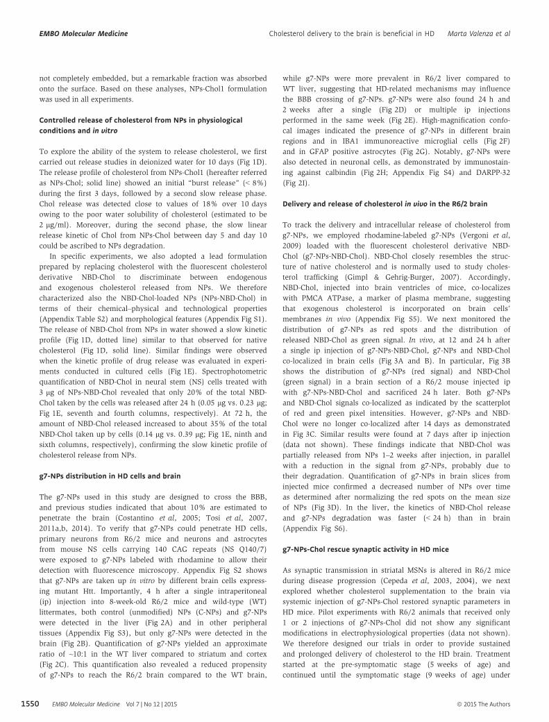
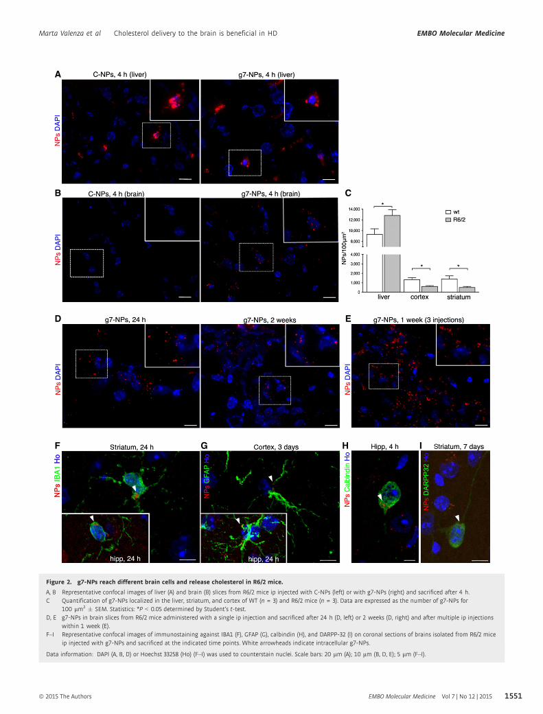
ing mutant Htt. Importantly, 4 h after a single intraperitoneal
(ip) injection into 8-week-old R6/2 mice and wild-type (WT)
littermates, both control (unmodified) NPs (C-NPs) and g7-NPs
were detected in the liver (Fig 2A) and in other peripheral
tissues (Appendix Fig S3), but only g7-NPs were detected in the
brain (Fig 2B). Quantification of g7-NPs yielded an approximate
ratio of ~10:1 in the WT liver compared to striatum and cortex
(Fig 2C). This quantification also revealed a reduced propensity
of g7-NPs to reach the R6/2 brain compared to the WT brain,
while g7-NPs were more prevalent in R6/2 liver compared to
WT liver, suggesting that HD-related mechanisms may influence
the BBB crossing of g7-NPs. g7-NPs were also found 24 h and
2 weeks after a single (Fig 2D) or multiple ip injections
performed in the same week (Fig 2E). High-magnification confo-
cal images indicated the presence of g7-NPs in different brain
regions and in IBA1 immunoreactive microglial cells (Fig 2F)
and in GFAP positive astrocytes (Fig 2G). Notably, g7-NPs were
also detected in neuronal cells, as demonstrated by immunostain-
ing against calbindin (Fig 2H; Appendix Fig S4) and DARPP-32
(Fig 2I).
Delivery and release of cholesterol in vivo in the R6/2 brain
To track the delivery and intracellular release of cholesterol from
g7-NPs, we employed rhodamine-labeled g7-NPs (Vergoni et al,
2009) loaded with the fluorescent cholesterol derivative NBD-
Chol (g7-NPs-NBD-Chol). NBD-Chol closely resembles the struc-
ture of native cholesterol and is normally used to study choles-
terol trafficking (Gimpl & Gehrig-Burger, 2007). Accordingly,
NBD-Chol, injected into brain ventricles of mice, co-localizes
with PMCA ATPase, a marker of plasma membrane, suggesting
that exogenous cholesterol is incorporated on brain cells’
membranes in vivo (Appendix Fig S5). We next monitored the
distribution of g7-NPs as red spots and the distribution of
released NBD-Chol as green signal. In vivo, at 12 and 24 h after
a single ip injection of g7-NPs-NBD-Chol, g7-NPs and NBD-Chol
co-localized in brain cells (Fig 3A and B). In particular, Fig 3B
shows the distribution of g7-NPs (red signal) and NBD-Chol
(green signal) in a brain section of a R6/2 mouse injected ip
with g7-NPs-NBD-Chol and sacrificed 24 h later. Both g7-NPs
and NBD-Chol signals co-localized as indicated by the scatterplot
of red and green pixel intensities. However, g7-NPs and NBD-
Chol were no longer co-localized after 14 days as demonstrated
in Fig 3C. Similar results were found at 7 days after ip injection
(data not shown). These findings indicate that NBD-Chol was
partially released from NPs 1–2 weeks after injection, in parallel
with a reduction in the signal from g7-NPs, probably due to
their degradation. Quantification of g7-NPs in brain slices from
injected mice confirmed a decreased number of NPs over time
as determined after normalizing the red spots on the mean size
of NPs (Fig 3D). In the liver, the kinetics of NBD-Chol release
and g7-NPs degradation was faster (< 24 h) than in brain
(Appendix Fig S6).
g7-NPs-Chol rescue synaptic activity in HD mice
As synaptic transmission in striatal MSNs is altered in R6/2 mice
during disease progression (Cepeda et al, 2003, 2004), we next
explored whether cholesterol supplementation to the brain via
systemic injection of g7-NPs-Chol restored synaptic parameters in
HD mice. Pilot experiments with R6/2 animals that received only
1 or 2 injections of g7-NPs-Chol did not show any significant
modifications in electrophysiological properties (data not shown).
We therefore designed our trials in order to provide sustained
and prolonged delivery of cholesterol to the HD brain. Treatment
started at the pre-symptomatic stage (5 weeks of age) and
continued until the symptomatic stage (9 weeks of age) under
EMBO Molecular Medicine Vol 7 | No 12 | 2015 ª 2015 The Authors
EMBO Molecular Medicine Cholesterol delivery to the brain is beneficial in HD Marta Valenza et al
1550
A
B
D
F G H I
E
C
Figure 2. g7-NPs reach different brain cells and release cholesterol in R6/2 mice.
A, B Representative confocal images of liver (A) and brain (B) slices from R6/2 mice ip injected with C-NPs (left) or with g7-NPs (right) and sacrificed after 4 h.C Quantification of g7-NPs localized in the liver, striatum, and cortex of WT (n = 3) and R6/2 mice (n = 3). Data are expressed as the number of g7-NPs for
100 lm2 � SEM. Statistics: *P < 0.05 determined by Student’s t-test.D, E g7-NPs in brain slices from R6/2 mice administered with a single ip injection and sacrificed after 24 h (D, left) or 2 weeks (D, right) and after multiple ip injections
within 1 week (E).F–I Representative confocal images of immunostaining against IBA1 (F), GFAP (G), calbindin (H), and DARPP-32 (I) on coronal sections of brains isolated from R6/2 mice
ip injected with g7-NPs and sacrificed at the indicated time points. White arrowheads indicate intracellular g7-NPs.
Data information: DAPI (A, B, D) or Hoechst 33258 (Ho) (F–I) was used to counterstain nuclei. Scale bars: 20 lm (A); 10 lm (B, D, E); 5 lm (F–I).
ª 2015 The Authors EMBO Molecular Medicine Vol 7 | No 12 | 2015
Marta Valenza et al Cholesterol delivery to the brain is beneficial in HD EMBO Molecular Medicine
1551

two experimental regimens. One group of R6/2 mice and WT
littermates were administered 0.15 mg g7-NPs/g body weight for
each injection, once every two weeks (three injections total)
while the second experimental group was injected twice a week
(ten injections total) accounting for a total estimated amount of
6.3 lg or 21 lg of cholesterol injected, respectively. Four groups
were compared: WT mice administered with saline (referred to
as WT), R6/2 mice administered with saline (referred to as R6/
2), R6/2 injected with empty g7-NPs (referred to as R6/2-emp),
and R6/2 mice receiving three or ten injections (referred to as
R6/2-Chol). At sacrifice, the presence of g7-NPs was analyzed by
fluorescence microscopy in liver sections from each animal and
in cortical samples taken from the brains before the
electrophysiological recordings (Appendix Fig S7). At the end of
the analyses, data from the two experimental paradigms were
pooled together as no significant differences were found.
HD mouse models have been extensively analyzed for their basic
striatal electrophysiological phenotypes. Similar and robust defects
have been described in striatal MSNs, namely a reduced membrane
capacitance, a decrease in spontaneous excitatory postsynaptic
current (EPSC), and an increase in spontaneous inhibitory post-
synaptic current (IPSC) frequencies (Cepeda et al, 2003). The consis-
tency of these phenotypes across different HD mouse models
suggests that these changes are a result of the mutant huntingtin
gene. We therefore tested whether our experimental scheme for
cholesterol supplementation could reverse any of these phenotypes.
Our whole-cell patch-clamp recordings of MSNs in brain slices
showed that membrane capacitance, a reflection of membrane area,
was significantly reduced in R6/2 mice treated with saline or empty
g7-NPs (R6/2-untreated; data were pooled as no differences were
found), compared to WT mice (WT, treated with saline)
(Appendix Table S3). In contrast, in neurons from R6/2 mice
treated with g7-NPs-Chol (R6/2-Chol), cell capacitance was not
significantly reduced compared to WT cells, suggesting a mild
rescue of cell membrane area (Appendix Table S3). Input resistance
was found increased in both R6/2-untreated and R6/2-Chol neurons
compared to WT neurons. Additionally, a significant decrease in
the decay time constant in cells from R6/2-Chol mice compared
with cells from WT or R6/2 mice treated with saline or empty
g7-NPs was observed (Appendix Table S3). This effect may be
attributed to changes in membrane fluidity induced by cholesterol
supplementation.
The average frequency of spontaneous IPSCs was also signifi-
cantly higher in MSNs from R6/2-untreated mice compared to WT
mice (Fig 4A and B, inset), as previously observed (Cepeda et al,
2004). In contrast, R6/2-Chol mice displayed a significant reduc-
tion in the frequency of IPSCs compared to R6/2-untreated
(Fig 4B, inset), in particular for small-amplitude events (< 40 pA;
Fig 4B), while the cumulative inter-event interval histogram
showed a decreased release probability in R6/2-Chol compared to
R6/2-untreated cells (Fig 4C). Similar to MSNs from R6/2-
untreated mice, IPSCs from R6/2-Chol mice had faster kinetics
than cells from WT mice as judged by shorter decay time and
half-amplitude duration of the current events compared to WTs
(Appendix Table S4A).
The frequency of spontaneous excitatory postsynaptic currents
(EPSCs) (Fig 4D) was significantly reduced in R6/2-untreated mice
compared to WT mice (Fig 4E, inset). Although the decrease in
the average frequency of EPSCs was not significantly rescued in
R6/2-Chol mice, the cumulative inter-event interval indicated a
significantly increased release probability in R6/2-Chol cells
versus R6/2-untreated cells (Fig 4F). EPSC kinetics was similar
among groups, except for half-amplitude duration, which was
significantly shorter in R6/2-chol cells than in WT cells
(Appendix Table S4B). Altogether, these findings indicate that
specific membrane and synaptic alterations observed in MSNs
from R6/2 mice can be rescued by in vivo cholesterol supplemen-
tation through g7-NPs.
Cholesterol supplementation ameliorates cognitivedysfunction in HD
We next assessed the impact of cholesterol supplementation on
the behavior of HD mice by using motor and cognitive tasks.
The injections regimen used in the behavioral studies, described
in Fig 5A, is the same employed for electrophysiological studies.
In the rotarod test, R6/2 mice treated with saline or empty g7-
NPs exhibited typical impaired coordination compared to WT
mice, as indicated by a shorter latency to fall from an accelerat-
ing rotarod. This deficit was not improved in R6/2-Chol mice
(Fig 5B). Similarly, in the open field test, reduced rearing activ-
ity, which is a form of vertical exploration, was not rescued by
cholesterol supplementation at 10 weeks of age (Fig 5C). At the
same age, the hypokinetic phenotype shown in R6/2 mice (mea-
sured as global activity in the open field) was still apparent in
R6/2-Chol, but the phenotype was less dramatic compared to
R6/2-untreated mice and significance reached P < 0.05 (Fig 5D),
suggesting that cholesterol supplementation partially ameliorates
locomotion-related behavior in a novel environment. Other
parameters (stereotyped movements, locomotion, resting time,
mean velocity) showed similar changes (Appendix Fig S8A).
Accordingly, R6/2-untreated mice worsened over time more than
R6/2-Chol mice (Appendix Fig S8B) as indicated by the signifi-
cant difference reached at later time points when the two groups
were compared at 8 and 10 weeks of age. These findings suggest
that sustained and repeated cholesterol supplementation might
slow the disease progression.
As changes in cholesterol synthesis/levels are associated with
cognitive decline (Suzuki et al, 2013), we next evaluated cognitive
tasks in HD mice after cholesterol supplementation. To evaluate
cognitive performance, we used the novel object recognition test, a
low-stress task aimed at evaluating recognition memory. Impor-
tantly, object memory is impaired in patients with HD. In a pattern
recognition task, subjects have to remember and touch the abstract
patterns they are shown during training and that are paired with a
novel pattern during testing. Early HD patients and clinically symp-
tomatic subjects performed significantly worse than control subjects
(Lawrence et al, 1996, 2000). R6/2-untreated mice showed a
pronounced inability to discriminate novel from familiar objects
from 8 weeks of age and worsened over time (Fig 5E). Notably,
R6/2-Chol mice performed as well as WT mice, indicating that
cholesterol supplementation rescued memory deficits at all time
points (Fig 5E). Importantly, the time-course analysis also revealed
that the benefit on recognition memory in R6/2-Chol mice was still
present at 12 weeks of age, that is, 3 weeks after the last injection
(Fig 5E).
EMBO Molecular Medicine Vol 7 | No 12 | 2015 ª 2015 The Authors
EMBO Molecular Medicine Cholesterol delivery to the brain is beneficial in HD Marta Valenza et al
1552

Cholesterol supplementation restores levels of synapticcomponents but not neuropathology
To determine whether cholesterol supplementation modulates
synaptic protein machinery, we used biochemically purified
triton-insoluble fractions (TIF) from the brain of WT (n = 5),
R6/2-untreated (n = 6) and R6/2-Chol (n = 3) mice and performed
semiquantitative Western blotting for scaffolding proteins such as
PSD95 and gephyrin and NMDA receptor subunits (GluN1 and
GluN2B) (Fig 6A). Reduced PSD95, as well as a reduction in GluN1
and GluN2B, were found in R6/2-untreated mice compared to WTs,
as expected (Fig 6B). Importantly, cholesterol supplementation
A
B
D
C
Figure 3. Cholesterol delivery and release in vivo in the R6/2 brain.
A Representative confocal image (crop) of brain slices from R6/2 mice ip injected with rhodamine-labeled g7-NPs-NBD-Chol and sacrificed after 12 h, showing co-localization of NBD-Chol (green) and rhodamine (NPs, red). Scale bar: 5 lm.
B, C Representative confocal image (low magnification) of brain slices from R6/2 mice ip injected with g7-NPs-NBD-Chol and sacrificed after 24 h (B) or 2 weeks (C) andrelative co-localization of NBD-Chol and g7-NPs. Scale bar: 10 lm.
D g7-NPs quantification in brain slices at the same time points in (B, C). Data are expressed as number of g7-NPs (evaluated based their size) for 100 lm2 � SEM.Statistics: **P < 0.01 (48 h vs. 7 days; 7 days vs. 14 days), ***P < 0.001 (24 h vs. 7 days; 7 days vs. 14 days) determined by one-way ANOVA followed by Newman–Keuls multiple comparison test.
Data information: DAPI was used to counterstain nuclei.
ª 2015 The Authors EMBO Molecular Medicine Vol 7 | No 12 | 2015
Marta Valenza et al Cholesterol delivery to the brain is beneficial in HD EMBO Molecular Medicine
1553

A
B
D
E F
C
Figure 4. Systemic injections of g7-NPs-Chol rescue synaptic alteration in R6/2 mice.
A Spontaneous IPSCs were recorded from striatal MSNs (WTs = 52; R6/2-untreated = 27; R6/2-Chol = 29) at a holding potential of +10 mV. As no differences werefound between R6/2 mice treated with saline (R6/2) or with empty g7-NPs (R6/2-emp), data were pooled.
B Amplitude–frequency histogram and average frequency (inset) of IPSCs from R6/2-Chol, R6/2-untreated, and WT MSNs.C Cumulative inter-event histogram showing the release probability of IPSCs in all groups.D Spontaneous EPSCs were recorded from striatal MSNs (WTs = 52; R6/2-untreated = 27; R6/2-Chol = 29) at a holding potential of �70 mV. As no differences were
found between R6/2 mice treated with saline (R6/2) and with empty g7-NPs (R6/2-emp), data were pooled.E Amplitude–frequency histogram and average frequency (inset) of EPSCs from R6/2-Chol, R6/2-untreated, and WT MSNs.F Cumulative inter-event histogram showing the release probability of EPSCs in all groups.
Data information: (B, D–F) Data represent mean � SEM. P < 0.05 was determined by one-way ANOVA followed by Newman–Keuls multiple comparison tests (#P < 0.05,##P < 0.01, ###P < 0.001 R6/2-untreated mice vs. R6/2-Chol mice; *P < 0.05, **P < 0.01, ***P < 0.001 WT mice vs R6/2-untreated or R6/2-Chol mice).
EMBO Molecular Medicine Vol 7 | No 12 | 2015 ª 2015 The Authors
EMBO Molecular Medicine Cholesterol delivery to the brain is beneficial in HD Marta Valenza et al
1554

normalized or increased the levels of these proteins (Fig 6B),
suggesting a rescue of the molecular composition of the synaptic
machinery contributing to synaptic structure.
To evaluate whether cholesterol supplementation also influences
the expression of synaptic genes, we performed qRT–PCR for a
panel of synaptic genes known to be reduced in HD. BDNF mRNA is
reduced in brain of several HD mice and is considered a critical hall-
mark in HD (Zuccato and Cattaneo (2014). Unpaired t-test between
R6/2-untreated and R6/2-Chol groups revealed a slight but signifi-
cant increase of bdnf expression in HD cortex after cholesterol
supplementation (Fig 6C). We also evaluated the expression of
snap25 and complexin II (Fig 6D and E), the latter being a gene
encoding for a presynaptic protein involved in neurotransmitter
release (Reim et al, 2001). Similarly to bdnf, mRNA levels of snap25
were significantly increased in cortex of HD mice after cholesterol
supplementation (Fig 6D). mRNA level of complexin II was strongly
reduced in cortex, hippocampus, and striatum of R6/2-untreated
mice compared to WTs. Cholesterol supplementation significantly
increased complexin II expression in hippocampus and striatum
from R6/2-Chol mice compared to R6/2-untreated mice (Fig 6E).
These findings suggest that cholesterol supplementation partially
ameliorates transcriptional abnormalities in the synaptic machinery
in HD mice.
We also quantified mRNA levels of genes considered to be MSN
markers, that is, darpp32, dopamine receptor D2 (drd2), and
muscarinic acetylcholine receptor M4 (chmr4). As expected, all
these genes were reduced in the striatum from R6/2-untreated mice
compared to WTs, but cholesterol supplementation did not signifi-
cantly influence their expression (Appendix Fig S9).
To investigate whether cholesterol supplementation counteracts
striatal atrophy and MSN degeneration, we performed unbiased
stereological analyses at 12 weeks of age. Reduced striatal volume
and enlargement of ventricles, both measures of striatal atrophy,
were observed in R6/2 mice treated with saline (R6/2) compared to
WTs (Appendix Fig S10; Fig 6F), as already reported in the litera-
ture. The administration of empty g7-NPs or g7-NPs-Chol did not
influence striatal volume in R6/2 mice (Appendix Fig S10). A statis-
tically significant reduction in ventricular volume was evident in
R6/2-Chol in comparison with R6/2 mice, similar to that observed
in WTs (Fig 6F). However, R6/2 mice treated with empty g7-NPs
(R6/2-emp) also showed a similar rescue, suggesting that the
administration of g7-NPs per se, likely due to degradation of PLGA
in lactic and glycolic acids, might influence this neuropathological
parameter.
Altogether, these findings suggest that cholesterol supplementa-
tion via g7-NPs is not sufficient to counteract brain atrophy and
neurodegeneration in R6/2 mice, at least with this experimental
paradigm, although it does increase the expression of specific genes
and synaptic proteins.
In vivo evaluation of safety of g7-NPs in HD mice
Cholesterol supplementation to the brain might lead to a further
reduction in cholesterol synthesis, already compromised in R6/2
mice (Valenza et al, 2007b). We therefore measured cholesterol
precursors and the brain-specific cholesterol catabolite 24OHC in
the brain of the treated mice at 12 weeks of age. Lathosterol, a
marker of cholesterol synthesis, was equally reduced in both
R6/2-untreated and R6/2-Chol mice compared to WTs (Fig 7A),
suggesting that exogenous cholesterol supplemented via g7-NPs
does not further decrease the endogenous biosynthetic pathway.
Similarly, 24OHC, an indicator of brain cholesterol catabolism that
usually mirrors cholesterol biosynthesis in brain (Lund et al, 2003),
was found similarly reduced in both R6/2 groups compared to WTs
(Fig 7B).
As it is known that most of the NPs (90%) localize in periph-
ery, we also measured mRNA levels of cholesterol biosynthetic
genes (hmgcr and fdft1) in liver and lung. The mRNA expression
of both cholesterol genes was similar in both tissues in all groups,
even in the presence of g7-NPs-Chol (Fig 7C and D). All these
results suggest that the exogenous cholesterol delivered to the
brain or accumulated in peripheral tissues does not lead to
alterations of endogenous cholesterol homeostasis in the time
frame analyzed in this study.
Although the NPs employed in this study are considered
biocompatible and biodegradable as made of PLGA, which is
approved by the FDA and EMA, an immunogenicity study of
these NPs in vivo is missing. Both PLGA, released after degrada-
tion of empty or cholesterol-loaded g7-NPs, and cholesterol itself
might influence immune responses. Therefore, we analyzed
mRNA levels of two pro-inflammation genes encoding for TNF-
alpha and IL6, in peripheral tissues from our cohorts. As shown
in Fig 7E and F, Tnf-alpha and Il6 mRNA levels were signifi-
cantly increased in the liver and in lung of R6/2 mice treated
with saline (R6/2) compared to WTs, supporting the available
evidence that peripheral inflammation is associated with HD
condition (Trager et al, 2014; Chang et al, 2015). Similar activa-
tion of inflammatory genes was also observed in R6/2 mice
treated with empty g7-NPs (R6/2-emp) or g7-NPs-Chol (R6/2-
Chol), suggesting that multiple administrations of g7-NPs (empty
or loaded with cholesterol) do not affect per se peripheral
inflammation in R6/2 mice.
Discussion
Synaptic dysfunction is an attractive target for possible HD therapies
as it occurs early in the disease process when cell death in HD
models is not obvious (Cepeda et al, 2004; Cummings et al, 2006;
Joshi et al, 2009; Milnerwood et al, 2010) and paralleling the
evidence that cognitive disturbances in patients with HD occur long
before onset of overt motor manifestations (Levine et al, 2004;
Paulsen et al, 2008; Schippling et al, 2009; Orth et al, 2010). We
show that exogenous cholesterol supplementation to the HD mouse
brain restores normal synaptic communication and protects mice
from cognitive decline. This study provides the missing link
between the reduction in brain cholesterol in the mouse HD brain
and some of the neuronal abnormalities in the disease state. The
data herein reported are in line with our recent in vitro studies,
suggesting that strategies aimed at supplying cholesterol to HD
neurons can ameliorate neuronal and synaptic dysfunction (Valenza
et al, 2015).
Cholesterol supplementation via ip injection of cholesterol-
loaded NPs normalizes GABAergic and, partially, glutamatergic
synaptic activity in striatal MSNs of R6/2 mice (Fig 4), supporting
the relevance of cholesterol in synaptic integrity and neuronal
ª 2015 The Authors EMBO Molecular Medicine Vol 7 | No 12 | 2015
Marta Valenza et al Cholesterol delivery to the brain is beneficial in HD EMBO Molecular Medicine
1555
function (Pfrieger, 2003). Cholesterol supplementation also protects
R6/2 mice from cognitive decline as measured by recognition
memory recovery (Fig 5E). Novel object preference has been often
associated with the hippocampus; however, it also depends on
functional interaction between hippocampus and cortex (Barker &
Warburton, 2011) and, more recently, it has been linked to the
striatum (Darvas & Palmiter, 2009). Consistently, reduction of the
cholesterol sensor SCAP in the brains of mice leads to a decrease
in brain cholesterol synthesis and causes impaired synaptic trans-
mission and altered cognitive function assessed by novel object
recognition test (Suzuki et al, 2013). Of note, cognitive decline has
been recently associated with hippocampal cholesterol loss and
cholesterol infusion in aged mice improved learning and memory in
aged rodents (Martin et al, 2014a).
Several in vitro findings indicate that synaptic transmission is
sensitive to cholesterol levels both at pre-synaptic and post-
synaptic levels. Indeed, cholesterol depletion affects vesicle recy-
cling and fusion (Thiele et al, 2000; Dason et al, 2010, 2014;
Linetti et al, 2010), AMPARs mobility (Hering et al, 2003; Renner
et al, 2009; Martin et al, 2014b), and the distribution and
A
B C
D E
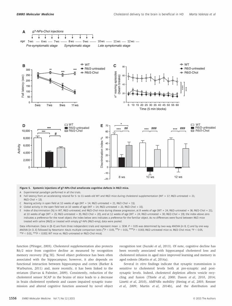
Figure 5. Systemic injections of g7-NPs-Chol ameliorate cognitive defects in R6/2 mice.
A Experimental paradigm performed in all the trials.B Fall latency from an accelerating rotarod for 5- to 11-week-old WT and R6/2 mice during cholesterol supplementation (WT = 17; R6/2-untreated = 21;
R6/2-Chol = 13).C Rearing activity in open field at 10 weeks of age (WT = 14; R6/2-untreated = 21; R6/2-Chol = 15).D Global activity in the open field test at 10 weeks of age (WT = 14; R6/2-untreated = 21; R6/2-Chol = 15).E Index of discrimination (%) in WT, R6/2-untreated, and R6/2-Chol mice during disease progression, at 8 weeks of age (WT = 24; R6/2-untreated = 36; R6/2-Chol = 21),
at 10 weeks of age (WT = 25; R6/2-untreated = 35; R6/2-Chol = 20), and at 12 weeks of age (WT = 24; R6/2-untreated = 30; R6/2-Chol = 19); the index above zeroindicates a preference for the novel object; the index below zero indicates a preference for the familiar object. As no differences were found between R6/2 micetreated with saline (R6/2) or treated with empty g7-NPs (R6/2-emp), data were pooled.
Data information: Data in (B–E) are from three independent trials and represent mean � SEM. P < 0.05 was determined by two-way ANOVA (in B, C) and by one-wayANOVA (in D, E) followed by Newmann–Keuls multiple comparison tests (#P < 0.05, ##P < 0.01, ###P < 0.001 R6/2-untreated mice vs. R6/2-Chol mice; *P < 0.05,**P < 0.01, ***P < 0.001 WT mice vs. R6/2-untreated or R6/2-Chol mice).
EMBO Molecular Medicine Vol 7 | No 12 | 2015 ª 2015 The Authors
EMBO Molecular Medicine Cholesterol delivery to the brain is beneficial in HD Marta Valenza et al
1556

function of NMDAR (Frank et al, 2004, 2008). Accordingly, we
found that cholesterol supplementation increases the levels of
the scaffold synaptic protein PSD95 and NMDARs in synaptic
protein-enriched fractions of HD mice (Fig 6A and B), suggesting
that in vivo delivery of cholesterol contributes to preserve the
structure and integrity of the synaptic machinery. In agreement
with the biochemical findings, the partial but significant increase
of mRNA levels of bdnf, snap25, and complexin II (all involved
in synaptic transmission) in different brain regions of HD mice
after cholesterol supplementation (Fig 6C–E) suggests that choles-
terol may act at different levels in improving synaptic and cogni-
tive functions. In particular, mRNA levels of complexin II, a key
player in the mechanisms underlying cognitive processes (Reim
et al, 2001; Glynn et al, 2003), are reduced in R6/2 mice and in
human HD striatum and cortex (Morton & Edwardson, 2001;
Freeman & Morton, 2004) and complexin II knockout mice show
selective cognitive deficits that reflect those seen in R6/2 mice
(Glynn et al, 2003). The cognitive benefits might be also related to
an effect of exogenous cholesterol on hormone steroids (Hara et al,
2015), but further studies are needed to address this issue.
Cholesterol is also required to establish proper membrane perme-
ability, fluidity, and thickness, and it stabilizes membranes and
provides order to membranes. The partial rescue of membrane
capacitance and the decrease in the decay time constant that we
have observed in striatal MSNs from R6/2 mice treated with g7-NPs-
Chol (Appendix Table S4) suggest that cholesterol supplementation
A
C
F G
D E
B
Figure 6. Systemic injections of g7-NPs-Chol positively influence synaptic protein network but not neuropathology.
A, B Protein levels (A) and relative densitometry quantification (B) of several synaptic proteins in triton-insoluble (synaptic enriched) fractions purified from total brainsfrom WT (n = 5), R6/2-untreated (n = 5) and R6/2-Chol (n = 3). Levels of PSD95 and NMDA receptor subunits GluN1 and GluN2B are rescued in R6/2 mice bycholesterol supplementation.
C–E mRNA levels for Bdnf (C), and Snap25 (D) in cortex and hippocampus; Complexin II (E) in cortex, hippocampus, and striatum from a subset of WT (n = 4), R6/2-untreated (n = 7), and R6/2-chol animals (n = 3). As no differences were found between R6/2 mice treated with saline or treated with empty g7-NPs, data werepooled.
F, G Representative images of Nissl staining (F) and ventricle volume revealed by Neurolucida analysis at 12 weeks of age in WT (n = 7), R6/2 (n = 7), R6/2-emp (n = 6),and R6/2-Chol (n = 8) mice.
Data information: Data in (B–E, G) represent mean � SEM. P < 0.05 was determined by one-way ANOVA followed by Newman–Keuls multiple comparison tests (in B, E)and by Student’s t-test between R6/2-untreated and R6/2-Chol (in C, D) (#P < 0.05, ##P < 0.01, ###P < 0.001 R6/2-untreated mice vs. R6/2-Chol mice; *P < 0.05,**P < 0.01, ***P < 0.001 WT mice vs. R6/2-untreated or R6/2-Chol mice).Source data are available online for this figure.
ª 2015 The Authors EMBO Molecular Medicine Vol 7 | No 12 | 2015
Marta Valenza et al Cholesterol delivery to the brain is beneficial in HD EMBO Molecular Medicine
1557
induces changes in membrane fluidity. However, other than show-
ing its incorporation into the membrane (Appendix Fig S5), we were
not able to establish where exactly the exogenous cholesterol, once
released by g7-NPs, localizes in brain cells, and more refined
methods to visualize exogenous cholesterol at subcellular levels are
needed.
We also reported that cholesterol supplementation does not
rescue motor defects and restores only partially the global activity of
HD mice (Fig 5B and D). The dichotomy that we observed in rescu-
ing cognitive but not motor functions might be associated with
specific roles of cholesterol in neuronal function. Similarly,
neuropathological hallmarks such as striatal volume and MSN
markers (Appendix Figs S9 and S10) do not significantly change
after cholesterol supplementation in R6/2 mice, suggesting that
cholesterol alone, at least within this experimental paradigm, is not
sufficient to prevent brain atrophy or to improve neuropathology.
Of note, both empty g7-NPs and g7-NPs-Chol seem to counteract the
enlargement of ventricle volume observed in R6/2 mice (Fig 6F and G)
likely due to degradation of PLGA in lactic and glycolic acids that
might influence metabolic pathways related to energy. However, the
significance of this effect is unknown and cannot be ascribed to
cholesterol. We should also consider that the content of cholesterol
delivered to the brain through g7-NPs is estimated to be 21 lg (for
the ten injections) and, although we started the treatment at
5 weeks of age, cholesterol was released after 1–2 weeks as shown
by the co-localization studies (Fig 3). Both low dose and slow
timing of cholesterol release might not be sufficient to reverse motor
performance and brain atrophy in HD mice. Further studies, by
A B
C D
E F
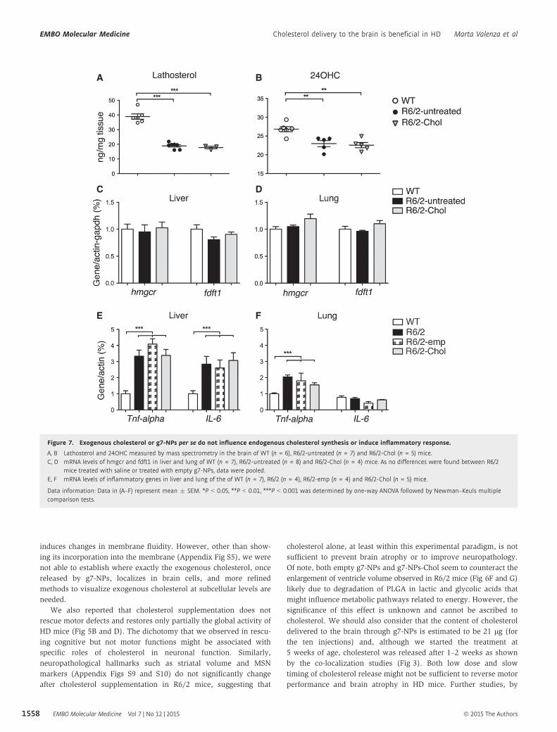
Figure 7. Exogenous cholesterol or g7-NPs per se do not influence endogenous cholesterol synthesis or induce inflammatory response.
A, B Lathosterol and 24OHC measured by mass spectrometry in the brain of WT (n = 6), R6/2-untreated (n = 7) and R6/2-Chol (n = 5) mice.C, D mRNA levels of hmgcr and fdft1 in liver and lung of WT (n = 7), R6/2-untreated (n = 8) and R6/2-Chol (n = 4) mice. As no differences were found between R6/2
mice treated with saline or treated with empty g7-NPs, data were pooled.E, F mRNA levels of inflammatory genes in liver and lung of the of WT (n = 7), R6/2 (n = 4), R6/2-emp (n = 4) and R6/2-Chol (n = 5) mice.
Data information: Data in (A–F) represent mean � SEM. *P < 0.05, **P < 0.01, ***P < 0.001 was determined by one-way ANOVA followed by Newman–Keuls multiplecomparison tests.
EMBO Molecular Medicine Vol 7 | No 12 | 2015 ª 2015 The Authors
EMBO Molecular Medicine Cholesterol delivery to the brain is beneficial in HD Marta Valenza et al
1558
employing mini-pump-based strategies for early and continuous
delivery of well-defined concentrations of cholesterol, will allow to
gain more information about the possible impact of cholesterol
delivery on motor defects.
Recent advances in nanotechnology and growing needs in
biochemical applications have driven the development of multifunc-
tional nanoparticles. Nanodrugs in liposome- or albumin-based
formulations are already used in the clinic for some forms of cancers
(Barenholz, 2012; Sethi et al, 2013; Von Hoff et al, 2013), and
others are being tested in pre-clinical trials (Tasciotti et al, 2008;
Shen et al, 2013). This is the first study in which g7-NPs have been
applied to a disease model for CNS targeting of molecules that are
not able to cross the BBB (such as cholesterol). g7-NPs reach
different brain regions 2–4 h after a single systemic injection and
localize in different brain cells, including striatal neurons (Figs 1
and 2). Previous pharmacological and biodistribution studies esti-
mated that the percentage of g7-NPs that reaches the brain is > 10%
of the injected dose (Tosi et al, 2007) and that multiple non-
receptor-mediated mechanisms are implicated (Tosi et al, 2011b).
Other NPs carrying ligands, antibodies, or peptides for specific
receptors that enter into the brain by receptor-mediated endocytosis
usually reach the brain compartment as maximum level values
ranging from 0.1 to 1% of the injected dose (Gabathuler, 2010; Tosi
et al, 2012; Gosk et al, 2004) owing to a possible saturation of the
receptor or by the competiveness of endogenous ligands. Therefore,
g7-NPs may represent a novel tool that can be used for brain deliv-
ery of several molecules. However, from a therapeutic prospective,
we are conscious that additional quantitative studies of g7-NPs
alone and loaded with cholesterol are needed to increase the knowl-
edge about biodistribution and pharmacokinetics.
The polymeric NPs used in this study are made of PLGA, a
copolymer approved by the FDA as drug delivery system for parent-
eral administration (Danhier et al, 2012). PLGA is considered
biodegradable and biocompatible as it degrades completely into its
original monomers, lactic and glycolic acid, which are easily metab-
olized in the body via the Krebs cycle and then eliminated (Shive &
Anderson, 1997). However, specific studies concerning immune
reactivity of g7-NPs are missing. Similarly, depending on the mole-
cule delivered, specific studies should be performed in order to
exclude any immune reaction or other side effects in different
tissues. Our studies suggest that g7-NPs and cholesterol itself do not
induce inflammatory response in liver and lungs, where almost all
g7-NPs are localized. A more extensive biochemical study to evalu-
ate the impact of g7-NPs degradation (and of the molecule released)
is needed to accelerate preclinical testing and translational develop-
ments of these NPs.
A limitation of g7-NPs in this current study is the low drug
loading (1%) that does not allow the delivery of elevated
amounts of cholesterol. Presumably, a high amount of choles-
terol, its strong affinity for hydrophobic interactions, and the
rigidity of the sterane ring lead to the disruption of the PLGA
organization as observed for NPs-Chol3 formation (Fig 1), with a
marked increase in sample heterogeneity and low quality of
nanoparticles. The identification of strategies aimed at increasing
the amount of cholesterol encapsulated into g7-NPs without
affecting chemical–physical properties of NPs will allow to
reduce the number of injections/week while increasing the
amount of cholesterol that reaches the brain cells. From another
prospective, the low content and the slow release of cholesterol
by g7-NPs might be advantageous as cholesterol accumulation is
dangerous for the brain. Further studies are needed to identify
the threshold of cholesterol increase that is beneficial for HD
brain/neurons and beyond which negative effects may occur.
The very low dose of exogenous cholesterol delivered in the
brain of HD mice in our experimental paradigm (21 lg) does
not allow to discriminate it from the large content of endogenous
cholesterol even by mass spectrometry. However, the demonstra-
tion of a rescue in specific electrophysiological and behavioral
phenotypes support the notion that the exogenous cholesterol
delivered to the adult brain is sufficient to ameliorate neuronal
dysfunction in HD. A similar concentration of cholesterol infused
via osmotic pumps in aged mice has recently been able to
improve learning and memory in aged rodents (Martin et al,
2014a).
In conclusion, these results emphasize the beneficial effects of
cholesterol supplementation in reversing synaptic alterations and
delaying cognitive defects in the HD mouse brain. Additionally, this
study demonstrates the validity of a new technology based on
g7-NPs to administer drugs (besides cholesterol) to the HD brain
and lays the ground for future therapeutic approaches.
Materials and Methods
NPs formulation and characterization
Gly-L-Phe-D-Thr-Gly-L-Phe-L-Leu-L-Ser(O-b-D-Glucose)-CONH2 (g7)
was prepared as previously described (Tosi et al, 2011b) and conju-
gated with PLGA to obtain g7-PLGA. PLGA derivatization yields
were confirmed by nuclear magnetic resonance to be 30–40 lmol
peptide/g of polymer. PLGA conjugated with rhodamine (Sigma-
Aldrich) was prepared as previously described (Costantino et al,
2005; Tosi et al, 2005). In all NPs, a fraction of polyvinyl alcohol
(PVA) (about 12.5 mg PVA/100 mg NPs) remains stably associated
with the NPs despite the repeated purification. The residual PVA
forms a connected network with the PLGA chains becoming a
“secondary” constituent of the NPs and partially masking the
exposed acidic groups of the polymer. This explanation justifies the
less negative values of f-pot with respect to those of the NPs
prepared in the absence of PVA. Details related to the production
and characterization of NPs and related to Fig 1 are listed in the
Appendix Supplementary Methods.
Cell culture and glial and neuronal differentiation
Neural stem (NS) cells carrying normal (Q7/7) or mutant htt
(Q140/7) employed in this study and protocols for their differentia-
tion were previously described (Conforti et al, 2013; Valenza et al,
2015). Primary neuronal cultures were prepared from the cortex of
R6/2 mice embryos (day 18 of gestation) as previously described
(Valenza et al, 2015).
Animals and treatments
Experiments at the University of Milan were carried out in
accordance with the European Communities Council Directive
ª 2015 The Authors EMBO Molecular Medicine Vol 7 | No 12 | 2015
Marta Valenza et al Cholesterol delivery to the brain is beneficial in HD EMBO Molecular Medicine
1559
2010/63/EU revising Directive 86/609/EEC regarding the care and
use of animals for experimental procedures. All procedures at
UCLA were performed in accordance with the U.S. Public Health
Service Guide for Care and Use of Laboratory Animals and were
approved by the Institutional Animal Care and Use Committee at
UCLA. Genotyping of R6/2 mouse colonies (~150 CAG repeats)
was performed by PCR of DNA obtained from tail samples, once
at weaning and again following sacrifice for verification. The
lifespan of this R6/2 mouse colony is approximately 12–14 weeks,
with HD-like phenotypes evident from 8 weeks of age. All the
mice have been randomly assigned to experimental groups, and
the investigators have been blinded to the sample group alloca-
tion during the treatments and experiments. For each injection,
the mice were administered 0.15 mg g7-NPs/g body weight (NPs
stock concentration is 12.5 mg/ml; 0.7 mg in 100 mg of NPs),
which corresponds to 1 lg of cholesterol/g. The chemical–
physical characterization and drug content in the g7-NPs used in
the pre-clinical trials is summarized in Appendix Table S5. The
complete list of WT and R6/2 animals used for each experiment
is described in Appendix Table S6. An initial trial was performed
in WT and R6/2 mice treated with control NPs loaded with
cholesterol (without g7, i.e., not able to cross the BBB). No
changes were found in terms of behavioral tasks and molecular
signature (Appendix Fig S11). Therefore, we decided to not
include these groups in subsequent trials with g7-NPs-Chol.
Immunohistochemistry
The animals were deeply anesthetized and transcardially perfused
with 4% PFA. When only NPs were detected, cells or tissues were
fixed in cold methanol at �20°C for 10 min, since fixation with
paraformaldehyde reduced rhodamine-related NP fluorescence.
Immunohistochemistry was performed on 15–30 lm coronal
sections with the following primary antibodies: rabbit anti-IBA1
(1:500; Wako), rabbit anti-GFAP (1:250; Dako), rabbit anti-
calbindin28 kDa (1:100; Swant), mouse anti-DARPP32 or rabbit
anti-DARPP32 (1:100, Epitomics; S. Cruz), and mouse anti-PMCA
ATPase (clone 5F10, 1:500; Thermo Scientific). Alexa Fluor 488-
conjugated goat secondary antibodies (1:1,000; Invitrogen) were
used for detection. Sections were counterstained with the nuclear
dye Hoechst 33258 or 40,6-diamidino-2-phenylindole (DAPI) (Invitro-
gen). Confocal images were acquired with a ZEISS LSM 510 or a
LEICA SP5 laser scanning confocal microscopes.
NPs quantification
To quantify NPs in different tissues, we used ImageJ software to
measure the fluorescence derived from the rhodamine used to label
the NPs. NPs were counted in 10 images for each tissue taken from
three WT and three R6/2 mice. Images were divided into three color
channels to set a threshold for the red produced by the NPs, and we
calculated the percentage of red signal for each image. Knowing the
total area of the field and the size of the NPs, we calculated the
approximate number of NPs in the selected area. The count of NPs
in the liver and in the brain was made at 20× and 60×, respectively,
and the data were normalized to compare the results. Ten images
for each animal/condition were analyzed. The images were
acquired with a Leica AF6000LX microscope.
Electrophysiology
At 10–11 weeks of age, mice were anesthetized with isoflurane and
decapitated, and the brain was rapidly removed to ice-cold dissec-
tion artificial cerebrospinal fluid (ACSF) containing 130 mM NaCl,
3 mM KCl, 26 mM NaHCO3, 1.25 mM NaHPO4, 10 mM glucose,
5 mM MgCl2, and 1 mM CaCl2 oxygenated with 95% O2/5% CO2
(pH 7.2–7.4, osmolality 290–310 mOsm/l). Coronal slices (300 lm)
of the striatum were cut with a microtome (Model VT 1000S, Leica
Microsystems) and transferred to an incubating chamber containing
oxygenated standard ACSF (with 2 mM CaCl2 and 2 mM MgCl2) for
1 h before electrophysiological recordings.
Whole-cell patch-clamp recordings were obtained from MSNs
visualized in slices with the aid of infrared video microscopy and
identified by somatic size and basic membrane properties (mem-
brane capacitance, input resistance, and time constant). The patch
pipette (3–5 MO) was filled with solution containing 125 mM
Cs-methanesulfonate, 4 mM NaCl, 3 mM KCl, 1 mM MgCl2, 9 mM
EGTA, 8 mM HEPES, 5 mM MgATP, 1 mM Tris-GTP, 10 mM
disodium phosphocreatine, and 0.1 mM leupeptin (pH 7.2, osmolality
270–280 mOsm/l).
Spontaneous postsynaptic currents were recorded in standard
ACSF. The membrane current was filtered at 1 kHz and digitized at
100–200 ls using Clampex 10.2 (gap-free mode). Cells were volt-
age-clamped at �70 mV to assess basic membrane properties.
Membranes were stepped to a holding potential of +10 mV to assess
GABAA receptor-mediated IPSCs. Bicuculline methiodide (10 lM)
was added to block GABAA receptor-mediated currents, and sponta-
neous glutamate receptor-mediated EPSCs were recorded at a hold-
ing potential of �70 mV. Spontaneous synaptic currents and event
kinetics were analyzed offline using the automatic detection proto-
col within the MiniAnalysis Program (Synaptosoft) and checked
manually for accuracy. Event counts were performed blind to geno-
type and treatment. The threshold amplitude for the detection of an
event (5 pA for glutamatergic currents and 10 pA for GABAergic
currents) was set above the root mean square background noise
level (1–2 pA at Vhold = �70 mV and 2–3 pA at Vhold = +10 mV).
Amplitude–frequency and inter-event interval distributions were
constructed to evaluate differences in events at each amplitude and
interval.
Behavioral characterization
Rotarod: Mice were first trained at a fixed speed of 4 rpm on the
apparatus (model 47600, Ugo Basile). After 1 h, the mice were
tested in an accelerating task (from 4 to 40 rpm) over 5 min, for
three trials per day for three consecutive days with an inter-trial
interval of 30 min. Latency to fall was recorded for each trial and
averaged. Open Field: The animals were placed individually into the
center of a transparent, square, activity-cage arena (45 cm × 45 cm)
(2Biological Instrument). Both horizontal and vertical activities
were assessed, monitoring mice allowed to freely move for 60 min
using the Actitrack software (2Biological Instrument) connected to
infrared sensors placed all around the square cage. Novel Object
Recognition Test: The device consisted of a Plexiglass square arena
(dimensions: 40 × 40 × 40 cm). All phases of the test were
conducted in the presence of low-intensity light. Mice were first
habituated to the arena in the absence of objects for 15 min (on one
EMBO Molecular Medicine Vol 7 | No 12 | 2015 ª 2015 The Authors
EMBO Molecular Medicine Cholesterol delivery to the brain is beneficial in HD Marta Valenza et al
1560

day, in the morning). On the same day, in the afternoon, two similar
objects were presented to each mouse for 10 min (A0 and A″), after
which the mice were returned to their home cage. Twenty-four
hours later, the same animals were tested for 10 min in the arena
with a familiar object (A″) and a new object (B). The index of
discrimination was calculated as (time exploring the novel
object � time exploring the familiar object) / (time exploring both
objects) × 100. Object preference was measured as (time exploring
each object) / (time exploring both objects) × 100. All experiments
were done blind to genotypes.
Triton-insoluble protein fraction preparation and Western blot
Triton-insoluble fractions of the brain were prepared as described in
the study by Gardoni et al (2009), separated on SDS–PAGE and
probed with specific antibodies. Antibodies used in these
experiments include anti-PSD-95 (1:1,000; #124011 SySy), NMDAR1
(GluN1) (1:500; #AB9864, Millipore), NMDAR2B (GluN2B) (1:500;
#MAB57578, Millipore), gephyrin (1:1,000; #147111, SySy), synap-
tophysin (1:1,000, S. Cruz), and beta-3-tubulin (1:3,000; #G7121,
Promega). Horseradish peroxidase-conjugated secondary antibodies
were then used (1:3,000; Bio-Rad). Bands were visualized with
enhanced chemoluminescence (Pierce) and imaged with the
ChemiDoc MP Imaging System (Bio-Rad). The bands were densito-
metrically quantified (Image Lab, Bio-Rad) and normalized for
Coomassie staining. Beta-3-tubulin was used as an additional loading
control.
RNA isolation, retrotranscription, and real-time quantitative PCR
Total RNA from tissues was isolated with TRIzol reagent (Life Tech-
nologies). Total RNA (0.25–1 lg) was reverse-transcribed to single-
stranded cDNA using the iScript cDNA synthesis kit (Bio-Rad). For
each reverse-transcribed product, three real-time PCR analyses were
performed in duplicate for each of the analyzed genes. An iCycler
thermal cycler with a Multicolor Real-time PCR Detection System
(Bio-Rad) was used to evaluate gene expressions. Taqman probes
with a FAM dye label (for cholesterol genes) or EVA Green Super-
mix (for inflammatory genes) was used, as previously described
(Valenza et al, 2015).
Nissl staining and Neurolucida analysis
Animals were perfused and brains dissected, frozen and serially cut
(30 lm-thick coronal sections) on the cryostat. One 30-lm-thick
section every five was stained with cresyl violet (Nissl staining).
Briefly, sections were dried overnight. Then, they were dehydrated
with a scale alcoholic of ethanol and xylene, than rehydrated, and
immersed in 1% cresyl violet and 1% glacial acetic acid aqueous
solution for 5 min. The staining was followed by a new dehydration
in ascending ethanol and xylene. Sections were cover-slipped with
Leica CV mounting media (Cat#14046430011). Brain, ventricle, and
striatum perimeters (relative to one hemisphere) were reconstructed
in a cerebral segment included between plates 19 and 39 of the
Franklin K. and Paxinos G. atlas (Paxinos & Franklin, 2008). They
were drawn at 40× at a microscope with a motorized stage inter-
faced to the computer, using the Neurolucida software (Microbright-
field Inc., VT, USA). The obtained volumes were analyzed with the
Neuroexplorer software (Microbrightfield Inc.) using the Cavalieri
formula for volume reconstruction.
Measurement of sterols
Samples were prepared and analyzed by isotopic dilution mass spec-
trometry as previously described (Valenza et al, 2010).
Statistics
SigmaPlot 12.3 (Systat software) or Prism 5 (GraphPad software)
was used to perform all statistical analyses. Data are presented as
means � standard error of the mean (SEM). Grubbs’ test was
applied to identify outliers. For each set of data to be compared, we
determined in Prism whether data were normally distributed or not.
As they were all normally distributed, we used parametric tests.
The paper explained
ProblemHuntington’s disease is a genetic neurodegenerative disorder charac-terized by progressive motor, cognitive, and psychiatric disturbances.Cholesterol biosynthesis and content are reduced in the brain ofmultiple animal models of HD. This dysfunction—of cerebral origin—is measurable in blood of patients with HD since pre-symptomaticstages of disease. However, a link between reduced synthesis/level ofcholesterol and neuronal dysfunction in vivo in HD is missing. Ascirculating or dietary cholesterol is not able to cross the blood–brainbarrier (BBB) and cholesterol in the brain depends largely on endoge-nous biosynthesis, this dysfunction may be detrimental for neuronalfunction especially given that locally synthesized cholesterol is impli-cated in synapses formation, integrity, and remodeling.
ResultsTo address the relationship between cholesterol dysfunction andsynaptic and cognitive deficits in HD mouse models, we deliveredcholesterol into the brain by using a novel technology based oncholesterol-loaded polymeric nanoparticles further modified with apeptide (g7) to cross the BBB after systemic injection in the mice. Weshowed that these nanoparticles (g7-NPs) reach different brainregions and different brain cells and gradually release cholesterol aftertheir degradation. We also showed that repeated systemic administra-tion of cholesterol-loaded g7-NPs in HD mice: (i) rescues synapticcommunication in striatal medium-sized spiny neurons, (ii) preventscognitive decline and partially improves global activity, and (iii)restores the levels of proteins that compose the synaptic machinery.
ImpactNeuronal and synaptic dysfunction is an attractive target for possibleHD therapies because it occurs long before cell death in mousemodels and in humans with HD. An intervention at this stage could,in theory, slow or stop neuron loss before it starts. Our conclusionshighlight the relevance of cholesterol deficits in cognitive impairmentassociated with HD and the benefits of cholesterol supplementationwith a broad impact for other brain disorders.In parallel, the evidence that g7-NPs can be used as vectors for thedelivery of therapeutic molecules (besides cholesterol) to the brainopens new and medically very relevant scenarios for the treatmentof several CNS disorders. Importantly, the nanoparticles employedare made of PLGA, which is approved by FDA in various drugdelivery systems in humans as it is considered biodegradable andbiocompatible.
ª 2015 The Authors EMBO Molecular Medicine Vol 7 | No 12 | 2015
Marta Valenza et al Cholesterol delivery to the brain is beneficial in HD EMBO Molecular Medicine
1561

Indeed, differences between group means were assessed with an
unpaired Student’s t-test, and two-way or one-way ANOVA followed
by Bonferroni or Newman–Keuls post hoc tests, as indicated in the
text. Differences were considered statistically significant if P < 0.05.
No statistical methods were used to pre-determine sample sizes, but
our sample sizes are similar to those reported in the literature. For
details, see also Appendix Table S7 showing statistical analyses and
P-values for the main figures.
Expanded View for this article is available online.
AcknowledgementsWe thank Luca Pignata and Chiara Orciani for technical assistance and Elisa
Battaglia for help with the behavioral tests. We also thank Miriam Ascagni
(CIMA, an advanced microscopy facility established by Università degli Studi
Milano, Milan), Valeria Berno and Silvia Tartari (Imaging Facility in INGM,
Milan), and Centro Grandi Strumenti (University of Modena and Reggio
Emilia) for technical assistance and support with confocal analysis. This work
was partially supported by Neuromics European grant (305121) to E.C. and
by Ministero della Salute under 40 (GR-2008-1145270) to M.V and V.L.. The
electrophysiology studies were supported by USPHS NS41574, HD004612,
and NS081335 to M.S.L. and C.C. The Hereditary Disease Foundation (to M.L.
and G.T.) supported the preparation of g7-NPs for the electrophysiological
studies.
This paper is dedicated to the memory of our dear colleague, devoted
scientist and well known neurologist Stefano Di Donato.
Author contributionsEC and MV developed the study, conceived the experimental plans, and
analyzed the data. JYC, CCe, and MSL developed and performed the electro-
physiological experiments and analyzed the data. GT, BR, DB, MAV, and FF
developed the NP-based strategy, produced, and characterized all NPs used in
this work. MV, EB, and GT performed the immunostaining experiments and
provided most of the confocal images. MV performed biochemical analyses
and with CFB all the molecular analyses; MV, EDP, and CFB performed the
behavioral tests. VL, CCa and SDD performed all the mass spectrometry analy-
ses. MMB and AV performed all reconstruction analyses with Neurolucida. MV,
GT, BR, and EC interpreted the data and wrote the manuscript. All authors
read and edited the manuscript. EC supervised the entire work and gave final
approval of the manuscript version to be published.
Conflict of interestThe authors declare that they have no conflict of interest.
References
Barenholz Y (2012) Doxil(R)–the first FDA-approved nano-drug: lessons
learned. J Control Release 160: 117 – 134
Barker GR, Warburton EC (2011) When is the hippocampus involved in
recognition memory? J Neurosci 31: 10721 – 10731
Belletti D, Tosi G, Forni F, Gamberini MC, Baraldi C, Vandelli MA, Ruozi B
(2012) Chemico-physical investigation of tenofovir loaded polymeric
nanoparticles. Int J Pharm 436: 753 – 763
Cepeda C, Hurst RS, Calvert CR, Hernandez-Echeagaray E, Nguyen OK, Jocoy
E, Christian LJ, Ariano MA, Levine MS (2003) Transient and progressive
electrophysiological alterations in the corticostriatal pathway in a mouse
model of Huntington’s disease. J Neurosci 23: 961 – 969
Cepeda C, Starling AJ, Wu N, Nguyen OK, Uzgil B, Soda T, Andre VM, Ariano
MA, Levine MS (2004) Increased GABAergic function in mouse models of
Huntington’s disease: reversal by BDNF. J Neurosci Res 78: 855 – 867
Chang KH, Wu YR, Chen YC, Chen CM (2015) Plasma inflammatory
biomarkers for Huntington’s disease patients and mouse model. Brain
Behav Immun 44: 121 – 127
Conforti P, Camnasio S, Mutti C, Valenza M, Thompson M, Fossale E, Zeitlin
S, MacDonald ME, Zuccato C, Cattaneo E (2013) Lack of huntingtin
promotes neural stem cells differentiation into glial cells while neurons
expressing huntingtin with expanded polyglutamine tracts undergo cell
death. Neurobiol Dis 50: 160 – 170
Costantino L, Gandolfi F, Tosi G, Rivasi F, Vandelli MA, Forni F (2005) Peptide-
derivatized biodegradable nanoparticles able to cross the blood-brain
barrier. J Control Release 108: 84 – 96
Cummings DM, Milnerwood AJ, Dallerac GM, Waights V, Brown JY, Vatsavayai
SC, Hirst MC, Murphy KP (2006) Aberrant cortical synaptic plasticity and
dopaminergic dysfunction in a mouse model of Huntington’s disease. Hum
Mol Genet 15: 2856 – 2868
Danhier F, Ansorena E, Silva JM, Coco R, Le Breton A, Preat V (2012) PLGA-
based nanoparticles: an overview of biomedical applications. J Control
Release 161: 505 – 522
Darvas M, Palmiter RD (2009) Restriction of dopamine signaling to the
dorsolateral striatum is sufficient for many cognitive behaviors. Proc Natl
Acad Sci USA 106: 14664 – 14669
Dason JS, Smith AJ, Marin L, Charlton MP (2010) Vesicular sterols are
essential for synaptic vesicle cycling. J Neurosci 30: 15856 – 15865
Dason JS, Smith AJ, Marin L, Charlton MP (2014) Cholesterol and F-actin are
required for clustering of recycling synaptic vesicle proteins in the
presynaptic plasma membrane. J Physiol 592: 621 – 633
Dietschy JM, Turley SD (2004) Thematic review series: brain Lipids.
Cholesterol metabolism in the central nervous system during early
development and in the mature animal. J Lipid Res 45: 1375 – 1397
Frank C, Giammarioli AM, Pepponi R, Fiorentini C, Rufini S (2004) Cholesterol
perturbing agents inhibit NMDA-dependent calcium influx in rat
hippocampal primary culture. FEBS Lett 566: 25 – 29
Frank C, Rufini S, Tancredi V, Forcina R, Grossi D, D’Arcangelo G (2008)
Cholesterol depletion inhibits synaptic transmission and synaptic plasticity
in rat hippocampus. Exp Neurol 212: 407 – 414
Freeman W, Morton AJ (2004) Regional and progressive changes in brain
expression of complexin II in a mouse transgenic for the Huntington’s
disease mutation. Brain Res Bull 63: 45 – 55
Gabathuler R (2010) Approaches to transport therapeutic drugs across the
blood-brain barrier to treat brain diseases. Neurobiol Dis 37: 48 – 57
Gardoni F, Mauceri D, Malinverno M, Polli F, Costa C, Tozzi A, Siliquini S,
Picconi B, Cattabeni F, Calabresi P et al (2009) Decreased NR2B subunit
synaptic levels cause impaired long-term potentiation but not long-term
depression. J Neurosci 29: 669 – 677
Gimpl G, Gehrig-Burger K (2007) Cholesterol reporter molecules. Biosci Rep 27:
335 – 358
Glynn D, Bortnick RA, Morton AJ (2003) Complexin II is essential for normal
neurological function in mice. Hum Mol Genet 12: 2431 – 2448
Gosk S, Vermehren C, Storm G, Moos T (2004) Targeting anti-transferrin
receptor antibody (OX26) and OX26-conjugated liposomes to brain
capillary endothelial cells using in situ perfusion. J Cereb Blood Flow Metab
24: 1193 – 1204
Hara Y, Waters EM, McEwen BS, Morrison JH (2015) Estrogen Effects on
Cognitive and Synaptic Health Over the Lifecourse. Physiol Rev 95:
785 – 807
EMBO Molecular Medicine Vol 7 | No 12 | 2015 ª 2015 The Authors
EMBO Molecular Medicine Cholesterol delivery to the brain is beneficial in HD Marta Valenza et al
1562
HDCRG (1993) A novel gene containing a trinucleotide repeat that is
expanded and unstable on Huntington’s disease chromosomes. The
Huntington’s Disease Collaborative Research Group. Cell 72: 971 – 983
Hering H, Lin CC, Sheng M (2003) Lipid rafts in the maintenance of synapses,
dendritic spines, and surface AMPA receptor stability. J Neurosci 23:
3262 – 3271
Huttner WB, Zimmerberg J (2001) Implications of lipid microdomains for
membrane curvature, budding and fission. Curr Opin Cell Biol 13:
478 – 484
Joshi PR, Wu NP, Andre VM, Cummings DM, Cepeda C, Joyce JA, Carroll JB,
Leavitt BR, Hayden MR, Levine MS et al (2009) Age-dependent alterations
of corticostriatal activity in the YAC128 mouse model of Huntington
disease. J Neurosci 29: 2414 – 2427
Lang T, Bruns D, Wenzel D, Riedel D, Holroyd P, Thiele C, Jahn R (2001)
SNAREs are concentrated in cholesterol-dependent clusters that define
docking and fusion sites for exocytosis. EMBO J 20: 2202 – 2213
Lawrence AD, Sahakian BJ, Hodges JR, Rosser AE, Lange KW, Robbins TW
(1996) Executive and mnemonic functions in early Huntington’s disease.
Brain 119(Pt 5): 1633 – 1645
Lawrence AD, Watkins LH, Sahakian BJ, Hodges JR, Robbins TW (2000) Visual
object and visuospatial cognition in Huntington’s disease: implications for
information processing in corticostriatal circuits. Brain 123(Pt 7):
1349 – 1364
Leoni V, Mariotti C, Tabrizi SJ, Valenza M, Wild EJ, Henley SM, Hobbs NZ,
Mandelli ML, Grisoli M, Bjorkhem I et al (2008) Plasma 24S-
hydroxycholesterol and caudate MRI in pre-manifest and early
Huntington’s disease. Brain 131: 2851 – 2859
Leoni V, Long JD, Mills JA, Di Donato S, Paulsen JS, Group P-Hs (2013) Plasma
24S-hydroxycholesterol correlation with markers of Huntington disease
progression. Neurobiol Dis 55: 37 – 43
Levine MS, Cepeda C, Hickey MA, Fleming SM, Chesselet MF (2004) Genetic
mouse models of Huntington’s and Parkinson’s diseases: illuminating but
imperfect. Trends Neurosci 27: 691 – 697
Linetti A, Fratangeli A, Taverna E, Valnegri P, Francolini M, Cappello V,
Matteoli M, Passafaro M, Rosa P (2010) Cholesterol reduction impairs
exocytosis of synaptic vesicles. J Cell Sci 123: 595 – 605
Lund EG, Xie C, Kotti T, Turley SD, Dietschy JM, Russell DW (2003) Knockout
of the cholesterol 24-hydroxylase gene in mice reveals a brain-specific
mechanism of cholesterol turnover. J Biol Chem 278: 22980 – 22988
Mangiarini L, Sathasivam K, Seller M, Cozens B, Harper A, Hetherington C,
Lawton M, Trottier Y, Lehrach H, Davies SW et al (1996) Exon 1 of the HD
gene with an expanded CAG repeat is sufficient to cause a progressive
neurological phenotype in transgenic mice. Cell 87: 493 – 506
Martin MG, Ahmed T, Korovaichuk A, Venero C, Menchon SA, Salas I, Munck
S, Herreras O, Balschun D, Dotti CG (2014a) Constitutive hippocampal
cholesterol loss underlies poor cognition in old rodents. EMBO Mol Med 6:
902 – 917
Martin MG, Pfrieger F, Dotti CG (2014b) Cholesterol in brain disease: sometimes
determinant and frequently implicated. EMBO Rep 15: 1036 – 1052
Marullo M, Valenza M, Leoni V, Caccia C, Scarlatti C, De MA, Zuccato C, Di
Donato S, Carafoli E, Cattaneo E (2012) Pitfalls in the detection of
cholesterol in Huntington’s disease models. PLoS Curr 4:
e505886e505889a501968
Mauch DH, Nagler K, Schumacher S, Goritz C, Muller EC, Otto A, Pfrieger FW
(2001) CNS synaptogenesis promoted by glia-derived cholesterol. Science
294: 1354 – 1357
Milnerwood AJ, Gladding CM, Pouladi MA, Kaufman AM, Hines RM, Boyd JD,
Ko RW, Vasuta OC, Graham RK, Hayden MR et al (2010) Early increase in
extrasynaptic NMDA receptor signaling and expression contributes to
phenotype onset in Huntington’s disease mice. Neuron 65: 178 – 190
Milnerwood AJ, Raymond LA (2010) Early synaptic pathophysiology in
neurodegeneration: insights from Huntington’s disease. Trends Neurosci 33:
513 – 523
Minost A, Delaveau J, Bolzinger MA, Fessi H, Elaissari A (2012) Nanoparticles
via nanoprecipitation process. Recent Pat Drug Deliv Formul 6: 250 – 258
Morton AJ, Edwardson JM (2001) Progressive depletion of complexin II in a
transgenic mouse model of Huntington’s disease. J Neurochem 76:
166 – 172
Mundargi RC, Babu VR, Rangaswamy V, Patel P, Aminabhavi TM (2008)
Nano/micro technologies for delivering macromolecular therapeutics using
poly(D, L-lactide-co-glycolide) and its derivatives. J Control Release 125:
193 – 209
Orth M, Schippling S, Schneider SA, Bhatia KP, Talelli P, Tabrizi SJ, Rothwell JC
(2010) Abnormal motor cortex plasticity in premanifest and very early
manifest Huntington disease. J Neurol Neurosurg Psychiatry 81:
267 – 270
Paratcha G, Ibanez CF (2002) Lipid rafts and the control of neurotrophic
factor signaling in the nervous system: variations on a theme. Curr Opin
Neurobiol 12: 542 – 549
Paulsen JS, Langbehn DR, Stout JC, Aylward E, Ross CA, Nance M, Guttman M,
Johnson S, MacDonald M, Beglinger LJ et al (2008) Detection of
Huntington’s disease decades before diagnosis: the Predict-HD study. J
Neurol Neurosurg Psychiatry 79: 874 – 880
Paulsen JS, Long JD (2014) Onset of Huntington’s disease: can it be purely
cognitive? Mov Disord 29: 1342 – 1350
Paxinos G, Franklin KBJ (2008) Paxinos and Franklin’s the Mouse Brain in
Stereotaxic Coordinates, 3rd edn. Waltham, MA: Academic Press
Pfrieger FW (2003) Role of cholesterol in synapse formation and function.
Biochim Biophys Acta 1610: 271 – 280
Reim K, Mansour M, Varoqueaux F, McMahon HT, Sudhof TC, Brose N,
Rosenmund C (2001) Complexins regulate a late step in Ca2 + -
dependent neurotransmitter release. Cell 104: 71 – 81
Renner M, Choquet D, Triller A (2009) Control of the postsynaptic membrane
viscosity. J Neurosci 29: 2926 – 2937
Ross CA, Aylward EH, Wild EJ, Langbehn DR, Long JD, Warner JH, Scahill RI,
Leavitt BR, Stout JC, Paulsen JS et al (2014) Huntington disease: natural
history, biomarkers and prospects for therapeutics. Nat Rev Neurol 10:
204 – 216
Schippling S, Schneider SA, Bhatia KP, Munchau A, Rothwell JC, Tabrizi SJ,
Orth M (2009) Abnormal motor cortex excitability in preclinical and very
early Huntington’s disease. Biol Psychiatry 65: 959 – 965
Sethi A, Sher M, Akram MR, Karim S, Khiljee S, Sajjad A, Shah SN, Murtaza G
(2013) Albumin as a drug delivery and diagnostic tool and its market
approved products. Acta Pol Pharm 70: 597 – 600
Shen H, Rodriguez-Aguayo C, Xu R, Gonzalez-Villasana V, Mai J, Huang Y,
Zhang G, Guo X, Bai L, Qin G et al (2013) Enhancing chemotherapy
response with sustained EphA2 silencing using multistage vector delivery.
Clin Cancer Res 19: 1806 – 1815
Shive MS, Anderson JM (1997) Biodegradation and biocompatibility of PLA
and PLGA microspheres. Adv Drug Deliv Rev 28: 5 – 24
Suzuki R, Ferris HA, Chee MJ, Maratos-Flier E, Kahn CR (2013) Reduction of
the cholesterol sensor SCAP in the brains of mice causes impaired
synaptic transmission and altered cognitive function. PLoS Biol 11:
e1001532
Tasciotti E, Liu X, Bhavane R, Plant K, Leonard AD, Price BK, Cheng MM,
Decuzzi P, Tour JM, Robertson F et al (2008) Mesoporous silicon particles
ª 2015 The Authors EMBO Molecular Medicine Vol 7 | No 12 | 2015
Marta Valenza et al Cholesterol delivery to the brain is beneficial in HD EMBO Molecular Medicine
1563
as a multistage delivery system for imaging and therapeutic applications.
Nat Nanotechnol 3: 151 – 157
Thiele C, Hannah MJ, Fahrenholz F, Huttner WB (2000) Cholesterol binds to
synaptophysin and is required for biogenesis of synaptic vesicles. Nat Cell
Biol 2: 42 – 49
del Toro D, Xifro X, Pol A, Humbert S, Saudou F, Canals JM, Alberch J (2010)
Altered cholesterol homeostasis contributes to enhanced excitotoxicity in
Huntington’s disease. J Neurochem 115: 153 – 167
Tosi G, Rivasi F, Gandolfi F, Costantino L, Vandelli MA, Forni F (2005)
Conjugated poly(D, L-lactide-co-glycolide) for the preparation of in vivo
detectable nanoparticles. Biomaterials 26: 4189 – 4195
Tosi G, Costantino L, Rivasi F, Ruozi B, Leo E, Vergoni AV, Tacchi R,
Bertolini A, Vandelli MA, Forni F (2007) Targeting the central nervous
system: in vivo experiments with peptide-derivatized nanoparticles
loaded with Loperamide and Rhodamine-123. J Control Release 122:
1 – 9
Tosi G, Costantino L, Ruozi B, Forni F, Vandelli MA (2008) Polymeric
nanoparticles for the drug delivery to the central nervous system. Expert
Opin Drug Deliv 5: 155 – 174
Tosi G, Vergoni AV, Ruozi B, Bondioli L, Badiali L, Rivasi F, Costantino L, Forni F,
Vandelli MA (2010) Sialic acid and glycopeptides conjugated PLGA
nanoparticles for central nervous system targeting: in vivo pharmacological
evidence and biodistribution. J Control Release 145: 49 – 57
Tosi G, Bondioli L, Ruozi B, Badiali L, Severini GM, Biffi S, De Vita A, Bortot B,
Dolcetta D, Forni F et al (2011a) NIR-labeled nanoparticles engineered for
brain targeting: in vivo optical imaging application and fluorescent
microscopy evidences. J Neural Transm 118: 145 – 153
Tosi G, Fano RA, Bondioli L, Badiali L, Benassi R, Rivasi F, Ruozi B, Forni F,
Vandelli MA (2011b) Investigation on mechanisms of glycopeptide
nanoparticles for drug delivery across the blood-brain barrier.
Nanomedicine 6: 423 – 436
Tosi G, Badiali L, Ruozi B, Vergoni AV, Bondioli L, Ferrari A, Rivasi F, Forni F,
Vandelli MA (2012) Can leptin-derived sequence-modified nanoparticles be
suitable tools for brain delivery? Nanomedicine 7: 365 – 382
Tosi G, Vilella A, Chhabra R, Schmeisser MJ, Boeckers TM, Ruozi B, Vandelli
MA, Forni F, Zoli M, Grabrucker AM (2014) Insight on the fate of CNS-
targeted nanoparticles. Part II: intercellular neuronal cell-to-cell transport.
J Control Release 177: 96 – 107
Trager U, Andre R, Magnusson-Lind A, Miller JR, Connolly C, Weiss A,
Grueninger S, Silajdzic E, Smith DL, Leavitt BR et al (2014)
Characterisation of immune cell function in fragment and full-length
Huntington’s disease mouse models. Neurobiol Dis 73C: 388 – 398
Trushina E, Singh RD, Dyer RB, Cao S, Shah VH, Parton RG, Pagano RE,
McMurray CT (2006) Mutant huntingtin inhibits clathrin-independent
endocytosis and causes accumulation of cholesterol in vitro and in vivo.
Hum Mol Genet 15: 3578 – 3591
Trushina E, Canaria CA, Lee DY, McMurray CT (2014) Loss of caveolin-1
expression in knock-in mouse model of Huntington’s disease suppresses
pathophysiology in vivo. Hum Mol Genet 23: 129 – 144
Valenza M, Rigamonti D, Goffredo D, Zuccato C, Fenu S, Jamot L, Strand A,
Tarditi A, Woodman B, Racchi M et al (2005) Dysfunction of the cholesterol
biosynthetic pathway in Huntington’s disease. J Neurosci 25: 9932 – 9939
Valenza M, Cattaneo E (2006) Cholesterol dysfunction in neurodegenerative
diseases: is Huntington’s disease in the list? Prog Neurobiol 80: 165 – 176
Valenza M, Carroll JB, Leoni V, Bertram LN, Bjorkhem I, Singaraja RR, Di
Donato S, Lutjohann D, Hayden MR, Cattaneo E (2007a) Cholesterol
biosynthesis pathway is disturbed in YAC128 mice and is modulated by
huntingtin mutation. Hum Mol Genet 16: 2187 – 2198
Valenza M, Leoni V, Tarditi A, Mariotti C, Bjorkhem I, Di Donato S,
Cattaneo E (2007b) Progressive dysfunction of the cholesterol
biosynthesis pathway in the R6/2 mouse model of Huntington’s disease.
Neurobiol Dis 28: 133 – 142
Valenza M, Leoni V, Karasinska JM, Petricca L, Fan J, Carroll J, Pouladi MA,
Fossale E, Nguyen HP, Riess O et al (2010) Cholesterol defect is marked
across multiple rodent models of Huntington’s disease and is manifest in
astrocytes. J Neurosci 30: 10844 – 10850
Valenza M, Marullo M, Di Paolo E, Cesana E, Zuccato C, Biella G, Cattaneo E
(2015) Disruption of astrocyte-neuron cholesterol cross talk affects
neuronal function in Huntington’s disease. Cell Death Differ 22: 690 – 702
Vergoni AV, Tosi G, Tacchi R, Vandelli MA, Bertolini A, Costantino L (2009)
Nanoparticles as drug delivery agents specific for CNS: in vivo
biodistribution. Nanomedicine 5: 369 – 377
Vilella A, Tosi G, Grabrucker AM, Ruozi B, Belletti D, Vandelli MA, Boeckers TM,
Forni F, Zoli M (2014) Insight on the fate of CNS-targeted nanoparticles.
Part I: Rab5-dependent cell-specific uptake and distribution. J Control
Release 174: 195 – 201
Von Hoff DD, Ervin T, Arena FP, Chiorean EG, Infante J, Moore M, Seay T,
Tjulandin SA, Ma WW, Saleh MN et al (2013) Increased survival in
pancreatic cancer with nab-paclitaxel plus gemcitabine. N Engl J Med 369:
1691 – 1703
Vonsattel JP, DiFiglia M (1998) Huntington disease. J Neuropathol Exp Neurol
57: 369 – 384
Zuccato C, Valenza M, Cattaneo E (2010) Molecular mechanisms and potential
therapeutical targets in Huntington’s disease. Physiol Rev 90: 905 – 981
Zuccato C, Cattaneo E (2014) Huntington’s disease. Handb Exp Pharmacol
220: 357 – 409
License: This is an open access article under the
terms of the Creative Commons Attribution 4.0
License, which permits use, distribution and reproduc-
tion in any medium, provided the original work is
properly cited.
EMBO Molecular Medicine Vol 7 | No 12 | 2015 ª 2015 The Authors
EMBO Molecular Medicine Cholesterol delivery to the brain is beneficial in HD Marta Valenza et al
1564
Related Documents