Cholelithiasis

Welcome message from author
This document is posted to help you gain knowledge. Please leave a comment to let me know what you think about it! Share it to your friends and learn new things together.
Transcript

Cholelithiasis
A Cause For Pain

Background
Presence of gallstones in the gallbladder. Spectrum ranges from asymptomatic, colic,
cholangitis, choledocholithiasis, cholecystitis
Colic is a temporary blockage, cholecystitis is inflammation from obstruction of CBD or cystic duct, cholangitis is infection of the biliary tree.

Anatomy

Pathophysiology
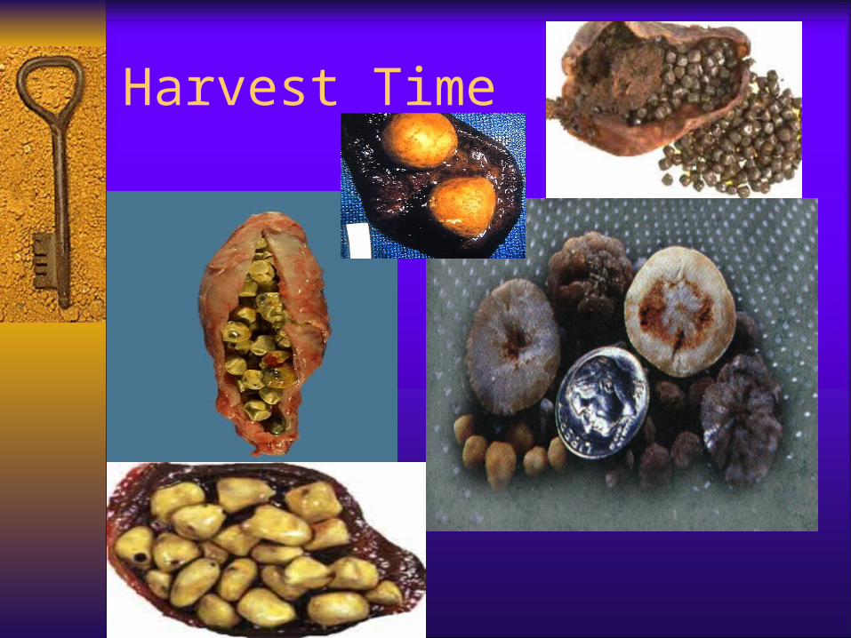
Three types of stones, cholesterol, pigment, mixed.
Formation of each types is caused by crystallization of bile.
Cholesterol stones most common. Bile consists of lethicin, bile acids,
phospholipids in a fine balance. Impaired motility can predispose to stones.

Pathophysiology
Sludge is crystals without stones. It may be a first step in stones, or be independent of it.
Pigment stones (15%) are from calcium bilirubinate. Diseases that increase RBC destruction will cause these. Also in cirrhotic patients, parasitic infections.
Harvest Time

Frequency
Affected by race, ethnicity, sex, medical conditions, fertility.
Every year 1-2% of people develop them. Hispanics are at increased risk.
Internationally: 20% of women, 14% of men. Patients over 60 prevalence was 12.9% for men, 22.4% for women.
Morbidity/Mortality
Every year 1-3% of patients develop symptoms.
Asymptomatic GS are not associated with fatalities.
Morbidity and mortality is associated only with symptomatic stones.
Race
Highest in fair skinned people of northern European descent and in Hispanic populations.
High in Pima Indians (75% of elderly). In addition Asians with stones are more likely to have pigmented stones than other populations.
African descent with Sickle Cell Anemia.
Sex
More common in women. Etiology may be secondary to variations in estrogen causing increased cholesterol secretion, and progesterone causing bile stasis.
Pregnant women more likely to have symptoms. Women with multiple pregnancies at higher risk Oral contraceptives, estrogen replacement tx.
Age
It is uncommon for children to have gallstones. If they do, its more likely that they have congenital anomalies, biliary anomalies, or hemolytic pigment stones.
Incidence of GS increases with age 1-3% per year.

History
3 clinical stages: asymptomatic, symptomatic, and with complications (cholecystitis, cholangitis, CBD stones).
Most (60-80%) are asymptomatic A history of epigastric pain with radiation
to shoulder may suggest it. A detailed history of pattern and
characteristics of symptoms as well as US make the diagnosis.
History
Most patients develop symptoms before complications.
Once symptoms occur, severe symptoms develop in 3-9%, with complications in 1-3% per year, and a cholecystectomy rate of 3-8% per year.
Indigestion, bloating, fatty food intolerance occur in similar frequencies in patients without gallstones, and are not cured with cholecystectomy.
History
Best definition of colic is pain that is severe in epigastrium or RUQ that last 1-5 hrs, often waking patient at night.
In classic cases pain is in the RUQ, however visceral pain and GB wall distension may be only in the epigastric area.
Once peritoneum irritated, localizes to RUQ. Small stones more symptomatic.
Physical
Vital signs and physical findings in asymptomatic cholelithiasis are completely normal.
Fever, tachycardia, hypotension, alert you to more serious infections, including cholangitis, cholecystitis.
Murphy’s sign
Causes
Fair, fat, female, fertile of course. (4F) High fat diet Obesity Rapid weight loss, TPN, Ileal disease, NPO. Increases with age, alcoholism. Diabetics have more complications. Hemolytics
Differentials
AAA Appendicitis Cholangitis, cholelithiasis Diverticulitis Gastroenteritis, hepatitis IBD, MI, SBO Pancreatitis, renal colic, pneumonia
Workup
Labs with asymptomatic cholelithiasis and biliary colic should all be normal.
WBC, elevated LFTS may be helpful in diagnosis of acute cholecystitis, but normal values do not rule it out.
Study by Singer et al examined utility of labs with chole diagnosed with HIDA, and showed no difference in WBC, AST,ALT Bili, and Alk Phos, in patients diagnosed and those without.
Workup
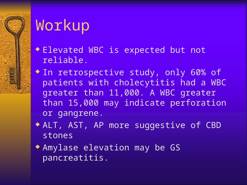
Elevated WBC is expected but not reliable. In retrospective study, only 60% of patients
with cholecytitis had a WBC greater than 11,000. A WBC greater than 15,000 may indicate perforation or gangrene.
ALT, AST, AP more suggestive of CBD stones
Amylase elevation may be GS pancreatitis.

Imaging Studies
US and Hida best. Plain x-rays, CT scans ERCP are adjuncts.
X-rays: 15% stones are radiopaque, porcelain GB may be seen. Air in biliary tree, emphysematous GB wall.
CT: for complications, ductal dilatation, surrounding organs. Misses 20% of GS. Get if diagnosis uncertain.

CT Scan
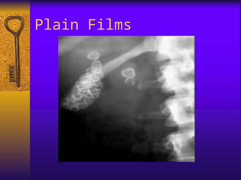
Plain Films
Imaging
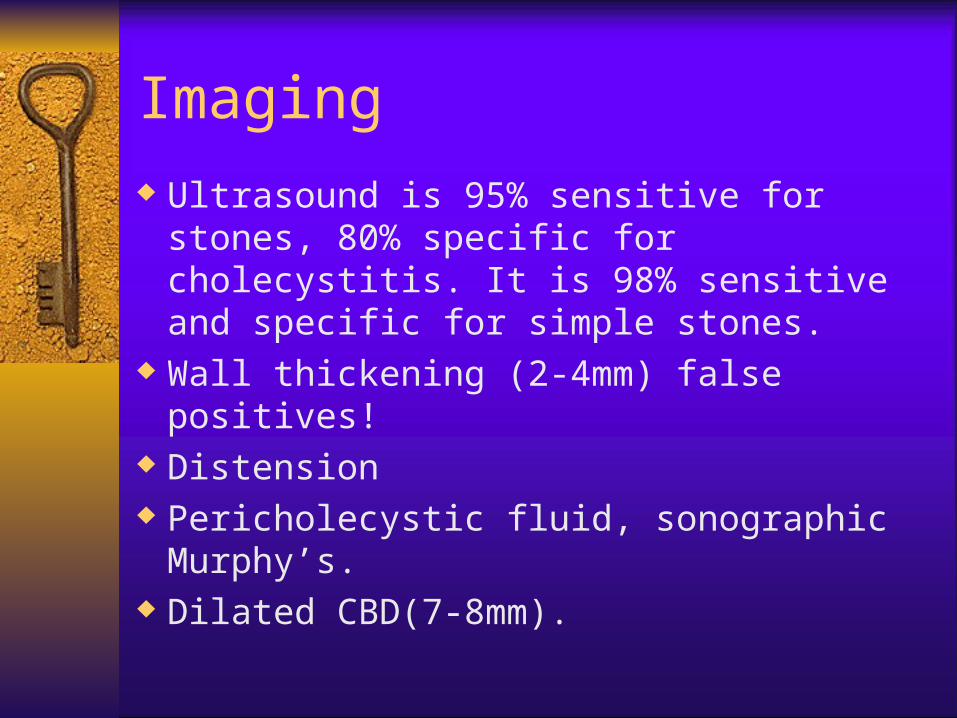
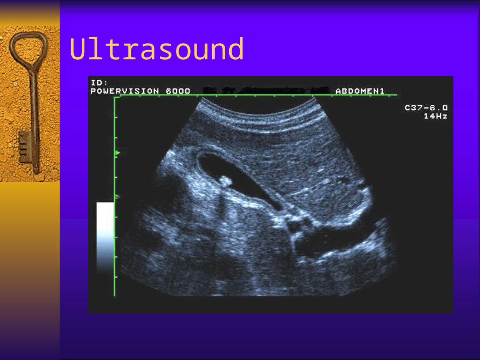
Ultrasound is 95% sensitive for stones, 80% specific for cholecystitis. It is 98% sensitive and specific for simple stones.
Wall thickening (2-4mm) false positives! Distension Pericholecystic fluid, sonographic
Murphy’s. Dilated CBD(7-8mm).

Ultrasound
Ultrasound

Imaging
Hida scan documents cystic duct patency. 94% sensitive, 85% specific GB should be visualized in 30 min. If GB visualized later it may point to
chronic cholecystitis. CBD obstruction appears as non
visualization of small intestine. False positives, high bilirubin.
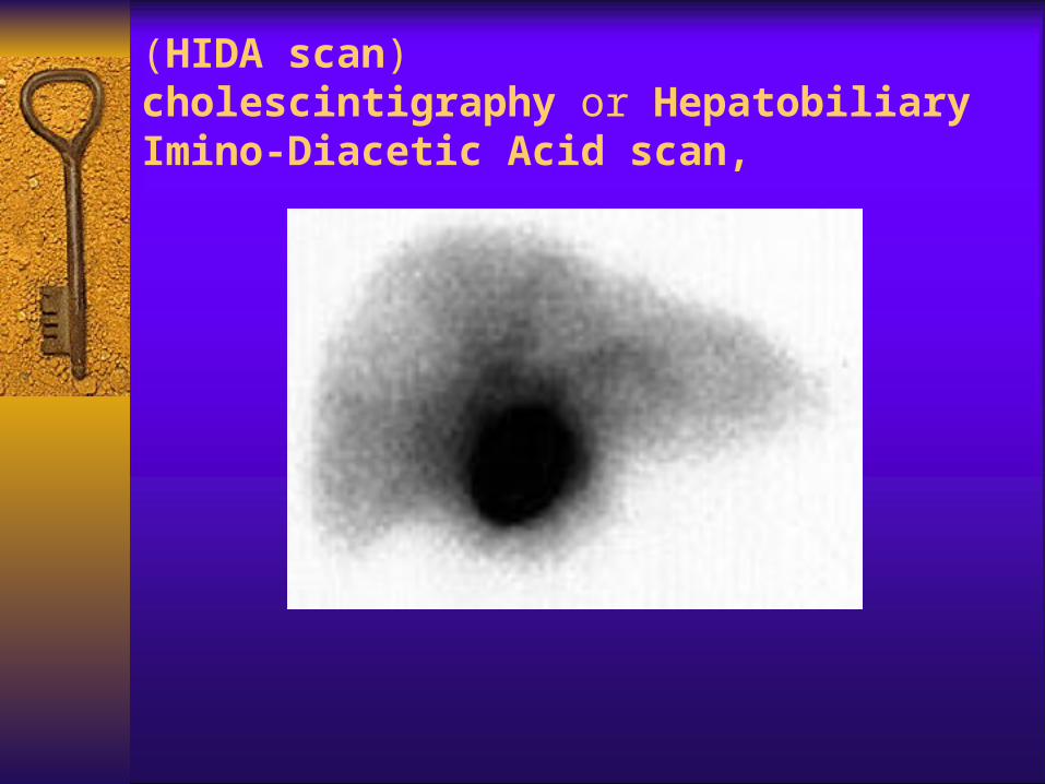
(HIDA scan)cholescintigraphy or Hepatobiliary Imino-Diacetic Acid scan,
Imaging
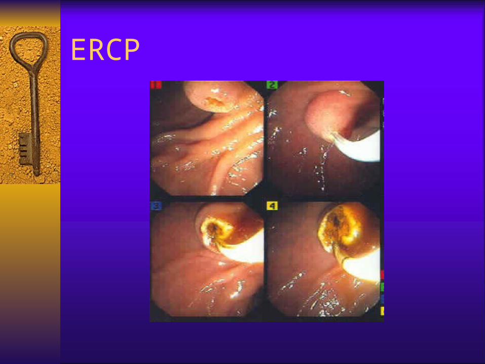
ERCP is diagnostic and therapeutic. Provides radiographic and endoscopic
visualization of biliary tree. Do when CBD dilated and elevated LFTs. Complications include bleeding,
perforation, pancreatitis, cholangitis.
ERCP

Emergency Department Care
Suspect GB colic in patients with RUQ pain of less than 4-6h duration radiating to back.
Consider acute cholecystits in those with longer duration of pain, with or without fever. Elderly and diabetics do not tolerate delay in diagnosis and can proceed to sepsis.
Emergency Department Care
After assessment of ABCs, perform standard IV, pulse oximetry, EKG, and monitoring. Send labs while IV placed, include cultures if febrile.
Primary goal of ED care is diagnosis of acute cholecystitis with labs, US, and or Hida. Once diagnosed, hospitalization usually necessary. Some treated as OP.
Emergency Department Care
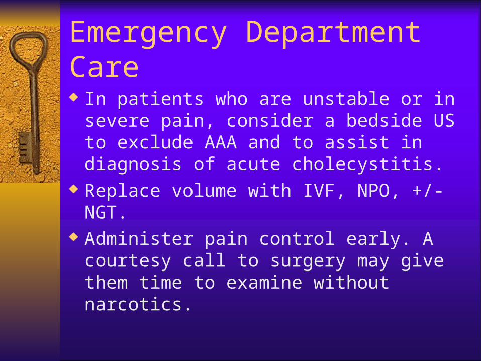
In patients who are unstable or in severe pain, consider a bedside US to exclude AAA and to assist in diagnosis of acute cholecystitis.
Replace volume with IVF, NPO, +/- NGT. Administer pain control early. A courtesy
call to surgery may give them time to examine without narcotics.
Consults
Historically cholecystits was operated on emergently which increased mortality.
Surgical consult is appropriate, and depending on the institution, either medicine or surgery may admit the patients for care.
Get GI involved early if suspect CBD obstruction.
Medications
Anticholinergics such as Bentyl (dicyclomine hydrochloride)to decrease GB and biliary tree tone. (20mg IM q4-6).
Demerol 25-75mg IV/IM q3 Antiemetics (phenergan, compazine). Antibiotics (Zosyn 3.375g IV q6) need to
cover Ecoli(39%), Klebsiella(54%), Enterobacter(34%), enterococci, group D strep.

Further Inpatient Care
Cholecystectomy can be performed after the first 24-48h or after the inflammation has subsided. Unstable patients may need more urgent interventions with ERCP, percutaneous drainage, or cholecystectomy.
Lap chole very effective with few complications (4%). 5% convert to open. In acute setting up to 50% open.
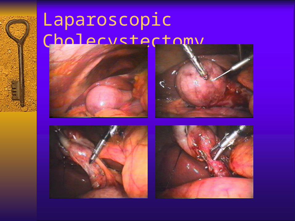
Laparoscopic Cholecystectomy
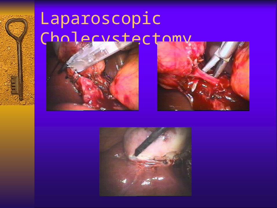
Laparoscopic Cholecystectomy
Further Outpatient Care
Afebrile, normal VS Minimal pain and tenderness. No markedly abnormal labs, normal CBD,
no pericholecystic fluid. No underlying medical problems. Next day follow-up visit. Discharge on oral antibiotics, pain meds.
Complications
Cholangitis, sepsis Pancreatitis Perforation (10%) GS ileus (mortality 20% as diagnosis
difficult). Hepatitis Choledocholithiasis
Prognosis
Uncomplicated cholecystitis as a low mortality.
Emphysematous GB mortality is 15% Perforation of GB occurs in 3-15% with up
to 60% mortality. Gangrenous GB 25% mortality.
Related Documents