CPAF: A Chlamydial Protease in Search of an Authentic Substrate Allan L. Chen 1. , Kirsten A. Johnson 1,2. , Jennifer K. Lee 1,2. , Christine Su ¨ tterlin 2 *, Ming Tan 1,3 * 1 Department of Microbiology and Molecular Genetics, University of California at Irvine, Irvine, California, United States of America, 2 Department of Developmental and Cell Biology, University of California at Irvine, Irvine, California, United States of America, 3 Department of Medicine, University of California at Irvine, Irvine, California, United States of America Abstract Bacteria in the genus Chlamydia are major human pathogens that cause an intracellular infection. A chlamydial protease, CPAF, has been proposed as an important virulence factor that cleaves or degrades at least 16 host proteins, thereby altering multiple cellular processes. We examined 11 published CPAF substrates and found that there was no detectable proteolysis when CPAF activity was inhibited during cell processing. We show that the reported proteolysis of these putative CPAF substrates was due to enzymatic activity in cell lysates rather than in intact cells. Nevertheless, Chlamydia- infecte d cells displayed Chlamydia-host interactions, suc h as Gol gi reorgani zat ion, apoptosis resist anc e, and host cytoskeletal remodeling, that have been attributed to CPAF-dependent proteolysis of host proteins. Our findings suggest that other mechanisms may be responsible for these Chlamydia-host interactions, and raise concerns about all published CPAF substrates and the proposed roles of CPAF in chlamydial pathogenesis. Citation: Chen AL, Johnson KA, Lee JK, Su ¨ tterlin C, Tan M (2012) CPAF: A Chlamydial Protease in Search of an Authentic Substrate. PLoS Pathog 8(8): e1002842 . doi:10.1371/journal.ppat.1002842 Editor: Craig R. Roy, Yale University School of Medicine, United States of America Received March 1, 2012; Accepted June 22, 2012; Published August 2, 2012 Copyright: 2012 Chen et al. This is an open -acce ss article distribu ted under the terms of the Creative Common s Attri butio n Licens e, which permits unrestricted use, distribution, and reproduction in any medium, provided the original author and source are credited. Funding: This work was supported by a grant from the NIH (AI083851) (MT and CS) and a Research Scholar Grant from the American Cancer Society (CS). JKL is supported by a training grant from the National Cancer Institute (T32CA009054). The funders had no role in study design, data collection and analysis, decision to publish, or preparation of the manuscript. Competing Interests: The authors have declared that no competing interests exist. * E-mail: [email protected] (CS); [email protected] (MT) . These authors contributed equally to this work. Introduction Chlamydia are obligate intracellular bacteria that are responsible for more infections reported to the CDC than all other infectious age nts combined [1] . Chlamyd ia trach omati s ca use s the most pre val ent bact eri al sex ual ly transmitt ed dis eas e in the Uni ted States [2] and the most common form of preventable blindness worldwide [3]. Another species, Chlamydia pneumoniae , is a causative agent of community-acquired pneumonia [4]. Despite this wide range of clini cal manif estati ons, Chlamydia spp. dis pla y many similarities at the level of the intracellular infection. Chlamydiae replicate wit hin a membrane-bound compart ment cal led the chlamydial inclusion in which the bacterium converts between two specia lized forms. During this develo pmental cycle, chlamyd iae usurp or subvert a number of processes within the host cell to support the inf ection. For instance, Chlamydia alt ers the hos t sec ret ory path way to acquire lipids from pos t-Golg i ves icl es to support growth of the inclusion and bacterial replication [5–7]. It also blocks host cell apoptosis, which could otherwise be used as a host defense mechanism against this intra cellul ar pathoge n that requires 2–3 days to complete its developmental cycle [8–10]. CPAF ( c hlamydial proteas e or proteas ome-l ike a ctivity f actor) has been proposed to be a major virulence factor in Chlamydia - infected cells [11]. This atypical serine protease [12] is conserved wit hin the Chlamydiales [13], includi ng the distantly relate d env ironme ntal chl amy dia e, whi ch inc lude endosy mbi onts of amoeba [14]. During an infection, CPAF is secreted into the host cytopla sm where it has been reported to cleave or degrade specific host proteins [15]. A rapidly growing number of CPAF substrates has been reporte d [11] , incl udi ng at le ast 16 host pro teins (Table 1). This proteolysis of host proteins by CPAF has been proposed to cause a number of effects on the infected host cell. For example, cleavage of the Golgi matrix protein golgin-84 has been reported to cause fragmentation and reorganization of the Golgi apparatus in Chlamydia -infected cells [16–17]. Similarly, degradation of pro- apoptotic BH3-only proteins, such as Puma, Bik, and Bim, has bee n pro posed to medi at e resi stance to apoptos is [18–19]. Cleavage of several intermediate filaments has been linked to the dynamic remodeling of the host cytoskeleton around the growing chl amydi al incl us ion [20] . In addi ti on, prote ol ysi s of hos t tran scr ipt ion fac tors, such as the MHC transc ription fac tor RFX5 [15,21 ] and the p65/ Rel A subunit of NF kB [22], have been implicated as chlamydial strategies for evading the immune response of the host cell. In general, these associations have been inferred from the known functions of reported CPAF substrates, but the direct effec ts of the se proteol yti c eve nts in Chlamydia - infected cells have not been examined [11]. The evidence for CPAF as a chlamydial protease that targets host proteins has been largely based on the observed cleavage or degr ada tion of spe cific hos t prot eins dur ing an infection, as assaye d by immuno blotti ng lysate s from Chlamydia -infe cted cells [15–16 ,18–20 ,22–2 3]. In additi on, many reports demonstrate d that the timing of this proteolysis correlates with the expression of CPAF during the developmental cycle [24] and can be prevented in vit ro [15,19 ,23] and in viv o [20,23] by the CPAF inhibi tor PLoS Pathogens | www.plospathogens.org 1 August 2012 | Volume 8 | Issue 8 | e1002842

Welcome message from author
This document is posted to help you gain knowledge. Please leave a comment to let me know what you think about it! Share it to your friends and learn new things together.
Transcript

8/13/2019 Chlamydia protein : CPAF
http://slidepdf.com/reader/full/chlamydia-protein-cpaf 1/8
CPAF: A Chlamydial Protease in Search of an AuthenticSubstrate
Allan L. Chen1., Kirsten A. Johnson1,2., Jennifer K. Lee1,2., Christine Su ¨ tterlin2*, Ming Tan1,3*
1 Department of Microbiology and Molecular Genetics, University of California at Irvine, Irvine, California, United States of America, 2 Department of Developmental and
Cell Biology, University of California at Irvine, Irvine, California, United States of America, 3 Department of Medicine, University of California at Irvine, Irvine, California,
United States of America
Abstract
Bacteria in the genus Chlamydia are major human pathogens that cause an intracellular infection. A chlamydial protease,CPAF, has been proposed as an important virulence factor that cleaves or degrades at least 16 host proteins, therebyaltering multiple cellular processes. We examined 11 published CPAF substrates and found that there was no detectableproteolysis when CPAF activity was inhibited during cell processing. We show that the reported proteolysis of theseputative CPAF substrates was due to enzymatic activity in cell lysates rather than in intact cells. Nevertheless, Chlamydia-infected cells displayed Chlamydia-host interactions, such as Golgi reorganization, apoptosis resistance, and hostcytoskeletal remodeling, that have been attributed to CPAF-dependent proteolysis of host proteins. Our findings suggestthat other mechanisms may be responsible for these Chlamydia-host interactions, and raise concerns about all publishedCPAF substrates and the proposed roles of CPAF in chlamydial pathogenesis.
Citation: Chen AL, Johnson KA, Lee JK, Sutterlin C, Tan M (2012) CPAF: A Chlamydial Protease in Search of an Authentic Substrate. PLoS Pathog 8(8): e1002842.
doi:10.1371/journal.ppat.1002842
Editor: Craig R. Roy, Yale University School of Medicine, United States of America
Received March 1, 2012; Accepted June 22, 2012; Published August 2, 2012
Copyright: 2012 Chen et al. This is an open-access article distributed under the terms of the Creative Commons Attribution License, which permitsunrestricted use, distribution, and reproduction in any medium, provided the original author and source are credited.
Funding: This work was supported by a grant from the NIH (AI083851) (MT and CS) and a Research Scholar Grant from the American Cancer Society (CS). JKL issupported by a training grant from the National Cancer Institute (T32CA009054). The funders had no role in study design, data collection and analysis, decision topublish, or preparation of the manuscript.
Competing Interests: The authors have declared that no competing interests exist.
* E-mail: [email protected] (CS); [email protected] (MT)
. These authors contributed equally to this work.
Introduction
Chlamydia are obligate intracellular bacteria that are responsible
for more infections reported to the CDC than all other infectious
agents combined [1]. Chlamydia trachomatis causes the most
prevalent bacterial sexually transmitted disease in the United
States [2] and the most common form of preventable blindness
worldwide [3]. Another species, Chlamydia pneumoniae , is a causative
agent of community-acquired pneumonia [4]. Despite this wide
range of clinical manifestations, Chlamydia spp. display many
similarities at the level of the intracellular infection. Chlamydiae
replicate within a membrane-bound compartment called the
chlamydial inclusion in which the bacterium converts between two
specialized forms. During this developmental cycle, chlamydiae
usurp or subvert a number of processes within the host cell to
support the infection. For instance, Chlamydia alters the host
secretory pathway to acquire lipids from post-Golgi vesicles tosupport growth of the inclusion and bacterial replication [5–7]. It
also blocks host cell apoptosis, which could otherwise be used as a
host defense mechanism against this intracellular pathogen that
requires 2–3 days to complete its developmental cycle [8–10].
CPAF ( c hlamydial protease or proteasome-like a ctivity f actor)
has been proposed to be a major virulence factor in Chlamydia -
infected cells [11]. This atypical serine protease [12] is conserved
within the Chlamydiales [13], including the distantly related
environmental chlamydiae, which include endosymbionts of
amoeba [14]. During an infection, CPAF is secreted into the host
cytoplasm where it has been reported to cleave or degrade specific
host proteins [15]. A rapidly growing number of CPAF substrates
has been reported [11], including at least 16 host proteins(Table 1).
This proteolysis of host proteins by CPAF has been proposed to
cause a number of effects on the infected host cell. For example,
cleavage of the Golgi matrix protein golgin-84 has been reported
to cause fragmentation and reorganization of the Golgi apparatus
in Chlamydia -infected cells [16–17]. Similarly, degradation of pro-
apoptotic BH3-only proteins, such as Puma, Bik, and Bim, has
been proposed to mediate resistance to apoptosis [18–19].
Cleavage of several intermediate filaments has been linked to the
dynamic remodeling of the host cytoskeleton around the growing chlamydial inclusion [20]. In addition, proteolysis of host
transcription factors, such as the MHC transcription factorRFX5 [15,21] and the p65/RelA subunit of NFkB [22], have
been implicated as chlamydial strategies for evading the immune
response of the host cell. In general, these associations have beeninferred from the known functions of reported CPAF substrates,
but the direct effects of these proteolytic events in Chlamydia -
infected cells have not been examined [11].
The evidence for CPAF as a chlamydial protease that targets
host proteins has been largely based on the observed cleavage or
degradation of specific host proteins during an infection, as
assayed by immunoblotting lysates from Chlamydia -infected cells
[15–16,18–20,22–23]. In addition, many reports demonstrated
that the timing of this proteolysis correlates with the expression of
CPAF during the developmental cycle [24] and can be prevented
in vitro [15,19,23] and in vivo [20,23] by the CPAF inhibitor
PLoS Pathogens | www.plospathogens.org 1 August 2012 | Volume 8 | Issue 8 | e1002842

8/13/2019 Chlamydia protein : CPAF
http://slidepdf.com/reader/full/chlamydia-protein-cpaf 2/8
lactacystin [15]. Furthermore, the same cleavage or degradation
patterns have been reproduced when recombinant CPAF was used
in vitro [15,19–20,23] or overexpressed in uninfected cells
[16,18,22].
In this study, we demonstrate that the reported cleavage or
degradation of 11 published CPAF substrates was abrogated when
the enzymatic activity of CPAF was inhibited during cell harvest
and lysate preparation. However, we still observed host-pathogen
interactions, such as Golgi reorganization and resistance to
apoptosis, that are proposed to result from proteolysis of target
proteins by CPAF. Our findings indicate that these host-Chlamydia
interactions are likely to be mediated by mechanisms other thanCPAF-dependent proteolysis of these host proteins. These results
invite a reappraisal of previously identified CPAF substrates and
re-interpretation of models involving the function of this
chlamydial enzyme in the intracellular infection.
Results
Re-examination of Golgin-84 Cleavage during aChlamydial Infection
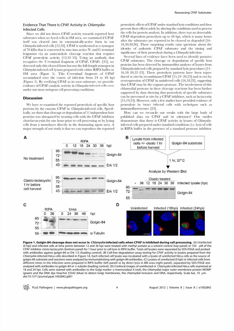
The Golgi protein golgin-84 is reported to be cleaved by CPAF
in Chlamydia -infected cells [16], but we found that cleavage is
dependent on the method of cell harvest and lysis (which we will
hereafter collectively refer to as ‘cell processing’). To reproduce the
published proteolysis, we harvested infected cells under standard
lysis conditions in RIPA buffer [25] and analyzed the cell lysate bySDS-PAGE, followed by immunoblotting with anti-golgin-84
antibodies. Consistent with previous reports, there was progressive
conversion of full-length golgin-84 into two cleavage products of
,78 and ,65 kDa beginning at 18 hours post infection (hpi)
(Figure 1A, top panel), and most of the full-length protein in the
extracts was cleaved by 36 hpi [16–17]. In striking contrast, there
was no golgin-84 cleavage, even as late as 36 hpi, when we treated
Chlamydia -infected cells for one hour prior to cell processing with
150 mM of clasto-lactacystin b-lactone, which is the active form of
the CPAF inhibitor lactacystin [15,26] (Figure 1A, compare top
and bottom panels). This inhibition of golgin-84 proteolysis is
unlikely to be due to the activity of clasto-lactacystin as a
proteasome inhibitor [27] because we did not detect inhibition
of proteasome function in our infected cells from the one hour
treatment (data not shown). These results suggest that the cleavage
of golgin-84 occurred during or after cell processing in standard
lysis buffers and was prevented by inhibiting CPAF activity prior
to these manipulations.
We next developed a cell-free assay to test for CPAF activity in
lysates of Chlamydia -infected cells. When we incubated infected celllysate with a lysate of uninfected HeLa cells as a source of golgin-
84 substrate, there was almost complete cleavage of golgin-84 at
37uC (Figure 1B) and at 0uC (Figure S1A). These in vitro
experiments demonstrate that CPAF remains active in lysates
from Chlamydia -infected cells, even on ice, and thus could cleave a
putative substrate during lysate preparation. In contrast, lysates of
Chlamydia -infected cells pre-treated with clasto-lactacystin, for one
hour before processing at times up to 36 hpi, did not cleave golgin-
84 in this in vitro assay (Figure 1B). These results show that CPAF
activity during lysate preparation can be abolished by treating the
infected cells with clasto-lactacystin for one hour prior to cell
processing.
To determine whether clasto-lactacystin was preventing golgin-
84 cleavage during the one hour treatment before cell lysis or
during cell processing itself, we used an alternative approach toinhibit CPAF activity during cell processing. In these experi-
ments, we lysed cells in urea as a denaturing agent to block
enzymatic activity in our lysates [28]. When we lysed Chlamydia -
infected cells by adding 8M urea directly to the monolayer, no
golgin-84 cleavage was observed even as late as 48 hpi(Figure 1C). These lysates did not contain detectable CPAF
activity as verified with the cell-free degradation assay (data not
shown). This lack of golgin-84 cleavage was observed for
Chlamydia infection of HeLa cells and two other human cell lines
when lysed in urea (Figure S1B). Taken together, these results
lead us to conclude that the reported CPAF-dependent cleavage
of golgin-84 is unlikely to occur in intact cells. Our results are
consistent with an explanation that proteolysis occurred during
cell processing and is due to CPAF activity in the lysates of
Chlamydia -infected cells.
We examined Golgi organization in cells on parallel coverslips
because cleavage of golgin-84 has been proposed to induce Golgi
fragmentation in Chlamydia -infected cells [17]. In uninfected
control cells, Golgi membranes were arranged as an intercon-
nected Golgi ribbon in the pericentriolar region, as detected by
immunofluorescence with antibodies to the Golgi marker
mannosidase II [29]. In contrast, Golgi membranes of infected
cells were reorganized around the growing chlamydial inclusion by
18 and 24 hpi (Figure 1D), which is consistent with previous
reports [17]. Thus, Golgi reorganization in our Chlamydia -infected
cells occurs in the absence of detectable golgin-84 cleavage and is
unlikely to be caused by CPAF-dependent proteolysis of this
structural Golgi protein.
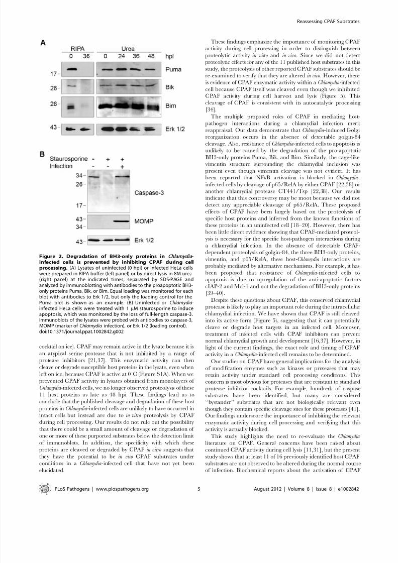
Degradation of Pro-apoptotic BH3-only Proteins Is AlsoDependent on Cell Processing
Our findings with golgin-84 motivated us to investigate other
host proteins reported to be CPAF substrates based on immuno-
blots of lysates prepared with standard buffers. The resistance of
Chlamydia-infected cells to apoptosis [8–10] has been proposed to
be mediated by CPAF-dependent degradation of BH3-only
proteins, including Puma, Bik, and Bim [18–19]. We again
replicated the complete degradation of each of these proteins by
lysing Chlamydia -infected cells in RIPA buffer at 36 hpi (Figure 2A).
However, when infected cells were lysed in 8M urea, Puma, Bik,
Author Summary
Chlamydia are bacteria that invade eukaryotic host cellsand live within a membrane-bound compartment calledthe chlamydial inclusion. Growth and survival of theseimportant human and animal pathogens depends onextensive interactions with the host cell, which allowchlamydiae to acquire critical nutrients and to avoid hostanti-microbial defenses. Chlamydiae are proposed to cause
many of these host-pathogen interactions through thecleavage or degradation of host proteins by the chlamydialprotease CPAF, which is secreted into the host cytoplasm.Here, we raise questions about the proposed roles of thisvirulence factor during infection, as well as its publishedsubstrates. We found that there was no detectablecleavage or degradation of 11 previously reported CPAFsubstrates in Chlamydia-infected cells and that CPAF-mediated proteolysis of these host proteins occurs duringcell harvest and lysis. However, we still observed host-pathogen interactions previously attributed to CPAFproteolysis of these proteins, suggesting that Chlamydiais likely to cause these effects on the host cell throughother mechanisms. Our findings call for a re-evaluation of all published CPAF substrates as well as the proposed roles
of this protease in chlamydial pathogenesis.
Reassessing CPAF Substrates
PLoS Pathogens | www.plospathogens.org 2 August 2012 | Volume 8 | Issue 8 | e1002842
8/13/2019 Chlamydia protein : CPAF
http://slidepdf.com/reader/full/chlamydia-protein-cpaf 3/8
and Bim were unaltered even at 48 hpi (Figure 2A). Thus, as with
cleavage of golgin-84, inhibition of CPAF activity during cell
processing abolished the published degradation of these pro-
apoptotic factors.
Although Puma, Bik, and Bim were not degraded in Chlamydia -
infected cells, these cells were resistant to staurosporine-induced
programmed cell death, as previously reported [8]. Staurosporine
treatment for 3 hours caused the complete loss of full-length
caspase-3 in uninfected cells, but there was no decrease in full-
length caspase-3 levels in our C. trachomatis -infected cells
(Figure 2B). Based on these results, it is doubtful that the anti-
apoptotic effects of a chlamydial infection on the host cell can be
attributed to CPAF-dependent degradation of the BH3-onlyproteins Puma, Bik, and Bim.
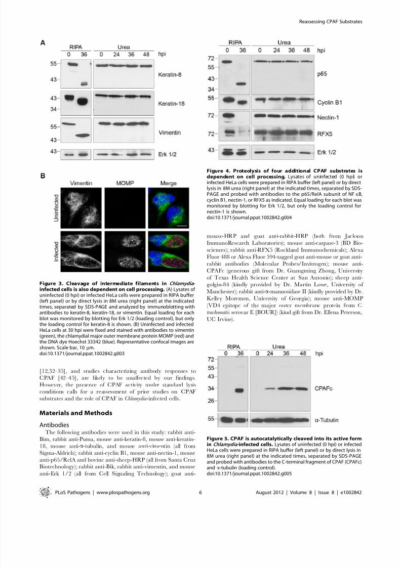
Proteolysis of Seven Additional Host Proteins by CPAF IsDependent on Cell Processing
We next examined the proteolytic effects of CPAF on the three
intermediate filaments keratin-8, keratin-18, and vimentin, whose
cleavage has been implicated in the growth of the chlamydial
inclusion [20,30]. When we lysed Chlamydia -infected cells at 36 hpi
in RIPA buffer (Figure 3A), we observed the conversion of each
full-length protein into smaller fragments, as previously reported
[20,30]. In contrast, when we lysed cells directly in 8M urea, we
did not detect any proteolytic effects on these proteins over the
time course of an infection up to 48 hpi (Figure 3A), indicating that
CPAF was unlikely to cleave these proteins in intact cells. CPAF-
dependent cleavage of vimentin has been proposed to be
important for host cytoskeletal rearrangement into a supportive
cage surrounding the chlamydial inclusion [20]. However, we still
observed a cage-like vimentin structure around the inclusion in our
Chlamydia -infected cells (Figure 3B), which suggests that this
reorganization of the host cytoskeleton during an infection does
not require vimentin cleavage.
We also examined proteolytic effects of CPAF on four
additional host proteins that have been reported to be CPAFsubstrates. These include the NFkB transcription factor subunit
p65/RelA [22], the MHC transcription factor RFX5 [15,21],
the adherens junction protein nectin-1 [23], and the cell cycle
protein cyclin B1 [18,31]. As with the other substrates analyzed,
the apparent proteolysis of these four proteins was only observed
when Chlamydia -infected cells were lysed in RIPA buffer and not
with direct lysis in urea (Figure 4). Taken together, these studies
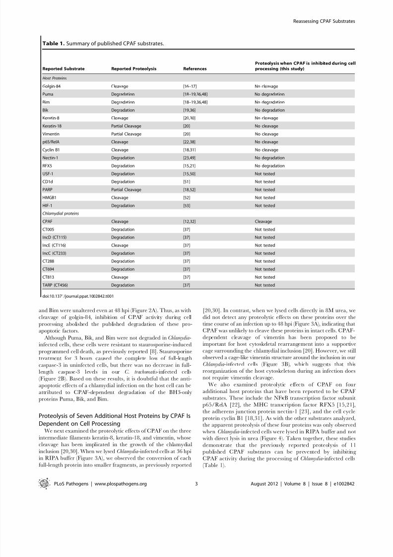
demonstrate that the previously reported proteolysis of 11
published CPAF substrates can be prevented by inhibiting
CPAF activity during the processing of Chlamydia -infected cells
(Table 1).
Table 1. Summary of published CPAF substrates.
Reported Substrate Reported Proteolysis References
Proteolysis when CPAF is inhibited during cell
processing (this study)
Host Proteins
Golgin-84 Cleavage [16–17] No cleavage
Puma Degradation [18–19,36,48] No degradation
Bim Degradation [18–19,36,48] No degradation
Bik Degradation [19,36] No degradation
Keratin-8 Cleavage [20,30] No cleavage
Keratin-18 Partial Cleavage [20] No cleavage
Vimentin Partial Cleavage [20] No cleavage
p65/RelA Cleavage [22,38] No cleavage
Cyclin B1 Cleavage [18,31] No cleavage
Nectin-1 Degradation [23,49] No degradation
RFX5 Degradation [15,21] No degradation
USF-1 Degradation [15,50] Not tested
CD1d Degradation [51] Not tested
PARP Partial Cleavage [18,52] Not tested
HMGB1 Cleavage [52] Not tested
HIF-1 Degradation [53] Not tested
Chlamydial proteins
CPAF Cleavage [12,32] Cleavage
CT005 Degradation [37] Not tested
IncD (CT115) Degradation [37] Not tested
IncE (CT116) Cleavage [37] Not tested
IncC (CT233) Degradation [37] Not tested
CT288 Degradation [37] Not tested
CT694 Degradation [37] Not tested
CT813 Cleavage [37] Not tested
TARP (CT456) Degradation [37] Not tested
doi:10.1371/journal.ppat.1002842.t001
Reassessing CPAF Substrates
PLoS Pathogens | www.plospathogens.org 3 August 2012 | Volume 8 | Issue 8 | e1002842
8/13/2019 Chlamydia protein : CPAF
http://slidepdf.com/reader/full/chlamydia-protein-cpaf 4/8
Evidence That There Is CPAF Activity in Chlamydia-Infected Cells
Since we did not detect CPAF activity towards reported host
substrates when we lysed cells in 8M urea, we examined if CPAF
itself was cleaved into its enzymatically-active form in our
Chlamydia -infected cells [12,32]. CPAF is synthesized as a zymogen
of 70 kDa that is converted in trans into active N- and C-terminal
fragments via an autocatalytic cleavage reaction that requires
CPAF proteolytic activity [12,32–35]. Using an antibody thatrecognizes the C-terminal fragment of CPAF, CPAFc [35], we
detected only this cleaved form but not the full-length zymogen in
Chlamydia -infected cell lysates prepared with either RIPA buffer or
8M urea (Figure 5). This C-terminal fragment of CPAF
accumulated over the course of infection from 24 to 48 hpi
(Figure 5). By verifying CPAF as its own substrate, we provide
evidence of CPAF catalytic activity in Chlamydia -infected cells even
under our most stringent cell processing conditions.
Discussion
We have re-examined the reported proteolysis of specific host
proteins by the enzyme CPAF in Chlamydia -infected cells. Specif-
ically, we show that cleavage or degradation of 11 independent host
proteins was abrogated by treating cells with the CPAF inhibitorclasto-lactacystin for one hour prior to cell processing or by lysing
cells from a monolayer directly in the denaturing agent urea. A
major strength of our study is that we can reproduce the reported
proteolytic effects of CPAF under standard lysis conditions and then
prevent these effects solely by altering the conditions used to process
the cells for protein analysis. In addition, there was no detectable
CPAF-dependent proteolysis up to 48 hpi, which is many hours
after the substrates are reported to be cleaved or degraded [16–
18,20,30,36]. These surprising results raise questions about the
identity of authentic CPAF substrates and the timing and
significance of their proteolysis during a Chlamydia infection.
Several lines of evidence have been used to identify putativeCPAF substrates. The cleavage or degradation of specific host
proteins has been detected by immunoblot analyses of lysates from
Chlamydia -infected cells prepared by standard lysis procedures [15–
16,18–20,22–23]. These proteolysis patterns have been repro-
duced in vitro by recombinant CPAF [15,19–20,23] and in vivo by
overexpression of CPAF in uninfected cells [16,18,22], suggesting
that CPAF may be the cognate protease. The involvement of this
chlamydial protease in these cleavage reactions has been further
supported by data showing that proteolysis of specific substrates
can be prevented in vitro by a CPAF inhibitor, such as lactacystin
[15,19,23]. However, only a few studies have provided evidence of
proteolysis in intact infected cells with techniques such as
immunofluorescence [23].
How can we reconcile our results with the large body of
published data on CPAF and its substrates? Our studiesdemonstrate that there is CPAF activity in lysates of Chlamydia -
infected cells prepared under standard conditions (i.e. lysis of cells
in RIPA buffer in the presence of a standard protease inhibitor
Figure 1. Golgin-84 cleavage does not occur in Chlamydia -infected cells when CPAF is inhibited during cell processing. (A) Uninfected(0 hpi) and infected cells at time points between 12 and 36 hpi were treated with methyl acetate as a solvent control (top panel) or 150 mM of theCPAF inhibitor clasto-lactacystin (bottom panel) for 1 hour prior to cell lysis in RIPA buffer. Total cell lysates were separated by SDS-PAGE and probedwith antibodies against golgin-84 or Erk 1/2 (loading control). (B) Cell-free degradation assay testing for CPAF activity in lysates prepared from theChlamydia-infected HeLa cells described in Figure 1A. Each infected cell lysate was incubated with a lysate of uninfected HeLa cells as the source of golgin-84 substrate and reactions were analyzed by immunoblotting with golgin-84 antibodies. (C) Lysates of uninfected (0 hpi) or infected cells fromdifferent times in the infection were prepared in RIPA buffer (left panel) or by direct lysis in 8M urea (right panel), separated by SDS-PAGE andanalyzed with antibodies to golgin-84 or a-tubulin (loading control). (D) Confocal images of uninfected or Chlamydia-infected HeLa cells examined at18 and 24 hpi. Cells were stained with antibodies to the Golgi marker a-mannosidase II (red), the chlamydial major outer membrane protein MOMP(green) and the DNA dye Hoechst 33342 (blue) to detect Golgi membranes, the chlamydial inclusion and DNA, respectively. Scale bar, 10 mm.doi:10.1371/journal.ppat.1002842.g001
Reassessing CPAF Substrates
PLoS Pathogens | www.plospathogens.org 4 August 2012 | Volume 8 | Issue 8 | e1002842
8/13/2019 Chlamydia protein : CPAF
http://slidepdf.com/reader/full/chlamydia-protein-cpaf 5/8
cocktail on ice). CPAF may remain active in the lysate because it is
an atypical serine protease that is not inhibited by a range of
protease inhibitors [21,37]. This enzymatic activity can then
cleave or degrade susceptible host proteins in the lysate, even when
left on ice, because CPAF is active at 0uC (Figure S1A). When we
prevented CPAF activity in lysates obtained from monolayers of
Chlamydia -infected cells, we no longer observed proteolysis of these
11 host proteins as late as 48 hpi. These findings lead us toconclude that the published cleavage and degradation of these host
proteins in Chlamydia -infected cells are unlikely to have occurred in
intact cells but instead are due to in vitro proteolysis by CPAF
during cell processing. Our results do not rule out the possibility
that there could be a small amount of cleavage or degradation of
one or more of these purported substrates below the detection limit
of immunoblots. In addition, the specificity with which these
proteins are cleaved or degraded by CPAF in vitro suggests that
they have the potential to be in vivo CPAF substrates under
conditions in a Chlamydia -infected cell that have not yet been
elucidated.
These findings emphasize the importance of monitoring CPAF
activity during cell processing in order to distinguish between
proteolytic activity in vitro and in vivo. Since we did not detect
proteolytic effects for any of the 11 published host substrates in this
study, the proteolysis of other reported CPAF substrates should be
re-examined to verify that they are altered in vivo. However, there
is evidence of CPAF enzymatic activity within a Chlamydia -infected
cell because CPAF itself was cleaved even though we inhibited
CPAF activity during cell harvest and lysis (Figure 5). Thiscleavage of CPAF is consistent with its autocatalytic processing
[34].
The multiple proposed roles of CPAF in mediating host-
pathogen interactions during a chlamydial infection merit
reappraisal. Our data demonstrate that Chlamydia -induced Golgi
reorganization occurs in the absence of detectable golgin-84
cleavage. Also, resistance of Chlamydia -infected cells to apoptosis is
unlikely to be caused by the degradation of the pro-apoptotic
BH3-only proteins Puma, Bik, and Bim. Similarly, the cage-like
vimentin structure surrounding the chlamydial inclusion was
present even though vimentin cleavage was not evident. It has
been reported that NFkB activation is blocked in Chlamydia -
infected cells by cleavage of p65/RelA by either CPAF [22,38] or
another chlamydial protease CT441/Tsp [22,38]. Our results
indicate that this controversy may be moot because we did notdetect any appreciable cleavage of p65/RelA. These proposed
effects of CPAF have been largely based on the proteolysis of
specific host proteins and inferred from the known functions of
these proteins in an uninfected cell [18–20]. However, there has
been little direct evidence showing that CPAF-mediated proteol-
ysis is necessary for the specific host-pathogen interactions during
a chlamydial infection. In the absence of detectable CPAF-
dependent proteolysis of golgin-84, the three BH3-only proteins,
vimentin, and p65/RelA, these host-Chlamydia interactions are
probably mediated by alternative mechanisms. For example, it has
been proposed that resistance of Chlamydia -infected cells to
apoptosis is due to upregulation of the anti-apoptotic factors
cIAP-2 and Mcl-1 and not the degradation of BH3-only proteins
[39–40].Despite these questions about CPAF, this conserved chlamydial
protease is likely to play an important role during the intracellular
chlamydial infection. We have shown that CPAF is still cleaved
into its active form (Figure 5), suggesting that it can potentially
cleave or degrade host targets in an infected cell. Moreover,
treatment of infected cells with CPAF inhibitors can prevent
normal chlamydial growth and development [16,37]. However, in
light of the current findings, the exact role and timing of CPAF
activity in a Chlamydia -infected cell remains to be determined.
Our studies on CPAF have general implications for the analysis
of modification enzymes such as kinases or proteases that may
retain activity under standard cell processing conditions. This
concern is most obvious for proteases that are resistant to standard
protease inhibitor cocktails. For example, hundreds of caspase
substrates have been identified, but many are considered‘‘bystander’’ substrates that are not biologically relevant even
though they contain specific cleavage sites for these proteases [41].
Our findings underscore the importance of inhibiting the relevant
enzymatic activity during cell processing and verifying that this
activity is actually blocked.
This study highlights the need to re-evaluate the Chlamydia
literature on CPAF. General concerns have been raised about
continued CPAF activity during cell lysis [11,31], but the present
study shows that at least 11 of 16 previously identified host CPAF
substrates are not observed to be altered during the normal course
of infection. Biochemical reports about the activation of CPAF
Figure 2. Degradation of BH3-only proteins in Chlamydia -infected cells is prevented by inhibiting CPAF during cellprocessing. (A) Lysates of uninfected (0 hpi) or infected HeLa cellswere prepared in RIPA buffer (left panel) or by direct lysis in 8M urea(right panel) at the indicated times, separated by SDS-PAGE andanalyzed by immunoblotting with antibodies to the proapoptotic BH3-only proteins Puma, Bik, or Bim. Equal loading was monitored for each
blot with antibodies to Erk 1/2, but only the loading control for thePuma blot is shown as an example. (B) Uninfected or Chlamydia-infected HeLa cells were treated with 1 mM staurosporine to induceapoptosis, which was monitored by the loss of full-length caspase-3.Immunoblots of the lysates were probed with antibodies to caspase-3,MOMP (marker of Chlamydia infection), or Erk 1/2 (loading control).doi:10.1371/journal.ppat.1002842.g002
Reassessing CPAF Substrates
PLoS Pathogens | www.plospathogens.org 5 August 2012 | Volume 8 | Issue 8 | e1002842
8/13/2019 Chlamydia protein : CPAF
http://slidepdf.com/reader/full/chlamydia-protein-cpaf 6/8
[12,32–35], and studies characterizing antibody responses to
CPAF [42–45], are likely to be unaffected by our findings.
However, the presence of CPAF activity under standard lysis
conditions calls for a reassessment of prior studies on CPAF
substrates and the role of CPAF in Chlamydia -infected cells.
Materials and Methods
AntibodiesThe following antibodies were used in this study: rabbit anti-
Bim, rabbit anti-Puma, mouse anti-keratin-8, mouse anti-keratin-
18, mouse anti-a-tubulin, and mouse anti-vimentin (all from
Sigma-Aldrich); rabbit anti-cyclin B1, mouse anti-nectin-1, mouse
anti-p65/RelA and bovine anti-sheep-HRP (all from Santa Cruz
Biotechnology); rabbit anti-Bik, rabbit anti-vimentin, and mouse
anti-Erk 1/2 (all from Cell Signaling Technology); goat anti-
mouse-HRP and goat anti-rabbit-HRP (both from Jackson
ImmunoResearch Laboratories); mouse anti-caspase-3 (BD Bio-
sciences); rabbit anti-RFX5 (Rockland Immunochemicals); Alexa
Fluor 488 or Alexa Fluor 594-tagged goat anti-mouse or goat anti-
rabbit antibodies (Molecular Probes/Invitrogen); mouse anti-
CPAFc (generous gift from Dr. Guangming Zhong, University
of Texas Health Science Center at San Antonio); sheep anti-
golgin-84 (kindly provided by Dr. Martin Lowe, University of
Manchester); rabbit anti-a-mannosidase II (kindly provided by Dr.
Kelley Moremen, University of Georgia); mouse anti-MOMP(VD4 epitope of the major outer membrane protein from C.
trachomatis serovar E [BOUR]) (kind gift from Dr. Ellena Peterson,
UC Irvine).
Figure 3. Cleavage of intermediate filaments in Chlamydia -infected cells is also dependent on cell processing. (A) Lysates of
uninfected (0 hpi) or infected HeLa cells were prepared in RIPA buffer(left panel) or by direct lysis in 8M urea (right panel) at the indicatedtimes, separated by SDS-PAGE and analyzed by immunoblotting withantibodies to keratin-8, keratin-18, or vimentin. Equal loading for eachblot was monitored by blotting for Erk 1/2 (loading control), but onlythe loading control for keratin-8 is shown. (B) Uninfected and infectedHeLa cells at 30 hpi were fixed and stained with antibodies to vimentin(green), the chlamydial major outer membrane protein MOMP (red) andthe DNA dye Hoechst 33342 (blue). Representative confocal images areshown. Scale bar, 10 mm.doi:10.1371/journal.ppat.1002842.g003
Figure 4. Proteolysis of four additional CPAF substrates isdependent on cell processing. Lysates of uninfected (0 hpi) orinfected HeLa cells were prepared in RIPA buffer (left panel) or by directlysis in 8M urea (right panel) at the indicated times, separated by SDS-PAGE and probed with antibodies to the p65/RelA subunit of NFkB,
cyclin B1, nectin-1, or RFX5 as indicated. Equal loading for each blot wasmonitored by blotting for Erk 1/2, but only the loading control fornectin-1 is shown.doi:10.1371/journal.ppat.1002842.g004
Figure 5. CPAF is autocatalytically cleaved into its active formin Chlamydia- infected cells. Lysates of uninfected (0 hpi) or infectedHeLa cells were prepared in RIPA buffer (left panel) or by direct lysis in8M urea (right panel) at the indicated times, separated by SDS-PAGEand probed with antibodies to the C-terminal fragment of CPAF (CPAFc)and a-tubulin (loading control).doi:10.1371/journal.ppat.1002842.g005
Reassessing CPAF Substrates
PLoS Pathogens | www.plospathogens.org 6 August 2012 | Volume 8 | Issue 8 | e1002842
8/13/2019 Chlamydia protein : CPAF
http://slidepdf.com/reader/full/chlamydia-protein-cpaf 7/8
Cell CultureHeLa cells (ATCC) were grown in 6-well dishes in Advanced
DMEM (4.5 g glucose/L) (Invitrogen) supplemented with 2% fetal
bovine serum (FBS) (Hyclone/Thermo Fisher) and 2 mM
GlutaMAX-I (Invitrogen). HEK 293T cells and retinal pigment
epithelial (hTERT RPE-1) cells (both from ATCC) were cultured
in DMEM (4.5 g glucose/L) (Invitrogen) supplemented with 10%
fetal bovine serum. All cell lines were grown in 5% CO2 at 37uC
and screened for Mycoplasma contamination by PCR [46].
Chlamydia InfectionsCell monolayers were infected with C. trachomatis serovar L2
(L2/434/Bu), LGV biovar, at a multiplicity of infection (MOI) of 3
in sucrose-phosphate-glutamic acid (SPG). In parallel, uninfected
control experiments were performed as mock infections in SPG
alone. Infections were carried out by centrifugation at 7006 g in a
Sorvall Legend Mach 1.6R centrifuge for 1 hr at room temper-
ature. After centrifugation, the inoculum was replaced by fresh cell
culture medium and monolayers were incubated at 37uC and 5%
CO2. Chlamydial EBs (elementary bodies) were verified to be free
of Mycoplasma contamination by PCR [46].
Cell Processing and ImmunoblottingLysis in RIPA buffer: Cells were harvested by trypsinization for
3–5 min at 37uC and the trypsinized cells were transferred to a
15 mL conical tube on ice. The dish was washed twice with
16PBS and the washes were added to the 15 mL conical tube to
collect any remaining cells. The cells were pelleted by centrifu-
gation at 1500 rpm for 3 min at 4uC and lysed on ice for 10 min
in RIPA buffer (50 mM Tris [pH 7.5], 150 mM NaCl, 0.1% SDS,
0.5% sodium deoxycholate, 1% NP-40) supplemented with
protease inhibitors (2 mM pepstatin, 150 mM aprotinin [bothfrom MP Biochemicals], 1 mM leupeptin [Calbiochem], 1 mM
PMSF [Acros]). The cells were resuspended by pipetting up anddown in approximately 1 mL of ice-cold lysis buffer per 56106
cells. Lysates were cleared by centrifugation at 13,0006 g for
10 min at 4uC and protein concentrations were determined by
Bradford assay (BioRad).Lysis in urea: A solution of 8M urea was supplemented with
325 U/mL of Benzonase Nuclease (Sigma-Aldrich) and added
directly to cell monolayers at a volume of 1 mL per 6-well dish for
10 min on ice. Lysates were then pooled and protein concentra-tions were determined by the DC protein assay (BioRad).
Cell lysates were diluted into Laemmli sample buffer (50 mM
Tris-HCl [pH 6.8], 10% glycerol, 2% SDS, 1% 2-mercaptoeth-
anol, 0.1% bromophenol blue). Samples containing equal amounts
of protein were loaded and resolved by SDS-PAGE. Proteins were
transferred onto nitrocellulose membranes and subjected to
immunoblot analysis (Table S1) with enhanced chemilumines-
cence (90 mM p-Courmaric acid, 250 mM 3-Aminophthalhydra-
zide, 100 mM Tris-HCl [pH 8.5]).
Clasto-lactacystin treatment: 150 mM of clasto-lactacystin b-lactone (Cayman Chemical), dissolved in methyl acetate, was
added to the cell culture medium for 1 hr prior to cell processing.
For example, samples of Chlamydia -infected cells at 36 hpi were
treated with clasto-lactacystin at 35 hpi for 1 hr and then processed.
In parallel control experiments, methyl acetate as the solvent was
added to the culture medium.
ImmunofluorescenceCells grown on glass coverslips were fixed in 4% formaldehyde
for 10 min at room temperature and blocked in 5% blocking
buffer (0.1% Triton X-100, 5% FBS in PBS) or TBS-BSA (0.1%
Tween-20, 5% BSA in TBS) for 1 hr. Cells were incubated with
primary antibodies for 1 hr at room temperature or overnight at
4uC followed by Alexa-fluorochrome-conjugated secondary anti-
bodies for 30 min at room temperature. Host and chlamydialDNA were stained with Hoechst 33342 (Molecular Probes/
Invitrogen). Coverslips were mounted onto glass slides withgelvatol [47] and imaged by confocal microscopy on a Nikon
Eclipse Ti-U inverted microscope fitted with a Nikon D-Eclipse
confocal laser assembly and a D-Eclipse C1 controller (Nikon).Images were acquired using the Nikon EZ-C1 program and
analyzed using Nikon NIS Elements and Adobe Photoshop.
Cell-free Degradation AssaysChlamydia -infected HeLa cells at various times in the infection
were either left untreated, mock-treated with methyl acetate, or
treated for 1 hr with 150 mM of clasto-lactacystin, and then lysed in
RIPA buffer as described above. 3.5 mg of Chlamydia -infected
HeLa cell lysate, as the source of CPAF, was incubated with 27 mg
of uninfected HeLa cell lysate, as the source of host protein
substrates, at 37uC for 1 hr in CPAF reaction buffer (25 mM Tris
[pH 8.0], 150 mM NaCl, 3 mM DTT). For reactions performed
at 0uC, 54 mg of Chlamydia -infected HeLa cell lysate from 36 hpi
was incubated with 54 mg of uninfected HeLa cell lysate on ice for
10 or 30 min. Reactions were terminated by adding Laemmlisample buffer and boiling for 5 min.
Apoptosis Induction AssayUninfected or Chlamydia -infected HeLa cells (MOI of 3, at 24
hpi) were incubated with 1 mM staurosporine in tissue culture
medium for 3 hrs. Lysates of the cell monolayers were prepared by
direct lysis in 8M urea as previously described, separated by SDS-
PAGE and analyzed by immunoblotting with antibodies to
caspase-3.
Supporting Information
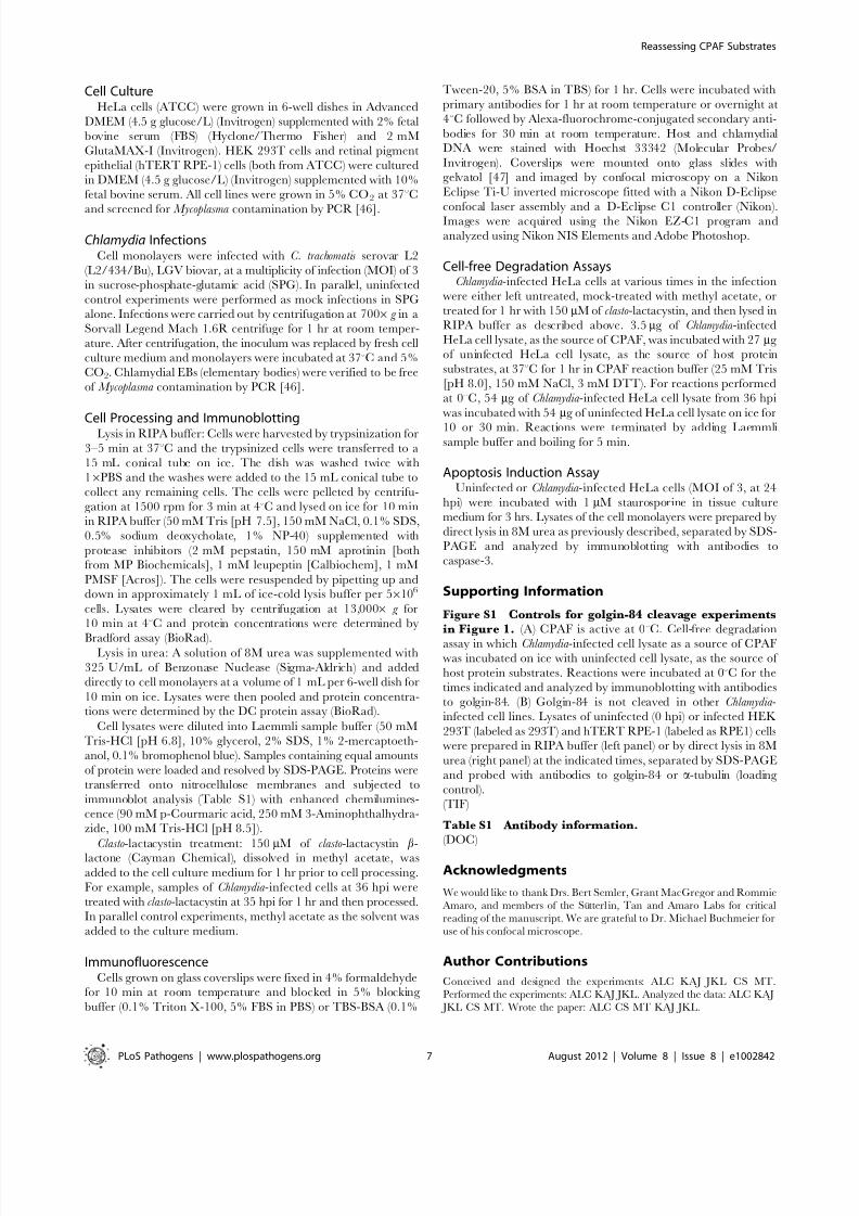
Figure S1 Controls for golgin-84 cleavage experimentsin Figure 1. (A) CPAF is active at 0uC. Cell-free degradation
assay in which Chlamydia -infected cell lysate as a source of CPAFwas incubated on ice with uninfected cell lysate, as the source of
host protein substrates. Reactions were incubated at 0uC for the
times indicated and analyzed by immunoblotting with antibodies
to golgin-84. (B) Golgin-84 is not cleaved in other Chlamydia -
infected cell lines. Lysates of uninfected (0 hpi) or infected HEK
293T (labeled as 293T) and hTERT RPE-1 (labeled as RPE1) cellswere prepared in RIPA buffer (left panel) or by direct lysis in 8M
urea (right panel) at the indicated times, separated by SDS-PAGE
and probed with antibodies to golgin-84 or a-tubulin (loading
control).
(TIF)
Table S1 Antibody information.(DOC)
Acknowledgments
We would like to thank Drs. Bert Semler, Grant MacGregor and Rommie
Amaro, and members of the Sutterl in, Tan and Amaro Labs for critical
reading of the manuscript. We are grateful to Dr. Michael Buchmeier for
use of his confocal microscope.
Author Contributions
Conceived and designed the experiments: ALC KAJ JKL CS MT.
Performed the experiments: ALC KAJ JKL. Analyzed the data: ALC KAJ
JKL CS MT. Wrote the paper: ALC CS MT KAJ JKL.
Reassessing CPAF Substrates
PLoS Pathogens | www.plospathogens.org 7 August 2012 | Volume 8 | Issue 8 | e1002842

8/13/2019 Chlamydia protein : CPAF
http://slidepdf.com/reader/full/chlamydia-protein-cpaf 8/8
References
1. CDC (2011) Summary of notifiable diseases: United States, 2009. MMWRMorb Mortal Wkly Rep 58: 1–100.
2. Schachter J (1999) Infection and disease epidemiology. In: Stephens RS, editor.Chlamydia : Intracellular Biology, Pathogenesis, and Immunity. Washington,D.C.: American Society for Microbiology. pp. 139–169.
3. Burton MJ, Mabey DC (2009) The global burden of trachoma: a review. PLoSNegl Trop Dis 3: e460.
4. Blasi F, Tarsia P, Aliberti S (2009) Chlamydophila pneumoniae . Clin Microbiol Infect15: 29–35.
5. Carabeo RA, Mead DJ, Hackstadt T (2003) Golgi-dependent transport of cholesterol to the Chlamydia trachomatis inclusion. Proc Natl Acad Sci U S A 100:6771–6776.
6. Hackstadt T, Rockey D, Heinzen R, Scidmore M (1996) Chlamydia trachomatis interrupts an exocytic pathway to acquire endogenously synthesized sphingo-myelin in transit from the Golgi apparatus to the plasma membrane. EMBO J15: 964–977.
7. Hackstadt T, Scidmore MA, Rockey DD (1995) Lipid metabolism in Chlamydia trachomatis -infected cells: directed trafficking of Golgi-derived sphingolipids to thechlamydial inclusion. Proc Natl Acad Sci U S A 92: 4877–4881.
8. Fan T, Lu H, Hu H, Shi L, McClarty GA, et al. (1998) Inhibition of apoptosis inChlamydia -infected cells: blockade of mitochondrial cytochrome c release andcaspase activation. J Exp Med 187: 487–496.
9. Fischer SF, Schwarz C, Vier J, Hacker G (2001) Characterization of antiapoptotic activities of Chlamydia pneumoniae in human cells. Infect Immun69: 7121–7129.
10. Rajalingam K, Al-Younes H, Muller A, Meyer TF, Szczepek AJ, et al. (2001)Epithelial cells infected with Chlamydophila pneumoniae (Chlamydia pneumo-niae) are resistant to apoptosis. Infect Immun 69: 7880–7888.
11. Zhong G (2009) Killing me softly: chlamydial use of proteolysis for evading hostdefenses. Trends Microbiol 17: 467–474.
12. Huang Z, Feng Y, Chen D, Wu X, Huang S, et al. (2008) Structural basis foractivation and inhibition of the secreted Chlamydia protease CPAF. Cell HostMicrobe 4: 529–542.
13. Dong F, Zhong Y, Arulanandam B, Zhong G (2005b) Production of aproteolytically active protein, chlamydial protease/proteasome-like activityfactor, by five different Chlamydia species. Infect Immun 73: 1868–1872.
14. Horn M, Collingro A, Schmitz-Esser S, Beier CL, Purkhold U, et al. (2004)Illuminating the evolutionary history of chlamydiae. Science 304: 728–730.
15. Zhong G, Fan P, Ji H, Dong F, Huang Y (2001) Identification of a chlamydialprotease-like activity factor responsible for the degradation of host transcriptionfactors. J Exp Med 193: 935–942.
16. Christian JG, Heymann J, Paschen SA, Vier J, Schauenburg L, et al. (2011)Targeting of a chlamydial protease impedes intracellular bacterial growth. PLoSPathog 7: e1002283.
17. Heuer D, Rejman Lipinski A, Machuy N, Karlas A, Wehrens A, et al. (2009)Chlamydia causes fragmentation of the Golgi compartment to ensure reproduc-tion. Nature 457: 731–735.
18. Paschen SA, Christian JG, Vier J, Schmidt F, Walch A, et al. (2008)Cytopathicity of Chlamydia is largely reproduced by expression of a singlechlamydial protease. J Cell Biol 182: 117–127.
19. Pirbhai M, Dong F, Zhong Y, Pan KZ, Zhong G (2006) The secreted proteasefactor CPAF is responsible for degrading pro-apoptotic BH3-only proteins inChlamydia trachomatis -infected cells. J Biol Chem 281: 31495–31501.
20. Kumar Y, Valdivia RH (2008) Actin and intermediate filaments stabilize theChlamydia trachomatis vacuole by forming dynamic structural scaffolds. Cell HostMicrobe 4: 159–169.
21. Zhong G, Liu L, Fan T, Fan P, Ji H (2000) Degradation of transcription factorRFX5 during the inhibition of both constitutive and interferon gamma-induciblemajor histocompatibility complex class I expression in Chlamydia -infected cells.
J Exp Med 191: 1525–1534.22. Christian J, Vier J, Paschen SA, Hacker G (2010) Cleavage of the NF-kappaB
family protein p65/RelA by the chlamydial protease-like activity factor (CPAF)impairs proinflammatory signaling in cells infected with Chlamydiae. J BiolChem 285: 41320–41327.
23. Sun J, Schoborg RV (2009) The host adherens junction molecule nectin-1 isdegraded by chlamydial protease-like activity factor (CPAF) in Chlamydia trachomatis -infected genital epithelial cells. Microbes Infect 11: 12–19.
24. Belland RJ, Zhong G, Crane DD, Hogan D, Sturdevant D, et al. (2003)Genomic transcriptional profiling of the developmental cycle of Chlamydia trachomatis . Proc Natl Acad Sci U S A 100: 8478–8483.
25. Holden P, Horton WA (2009) Crude subcellular fractionation of culturedmammalian cell lines. BMC Res Notes 2: 243.
26. Dick LR, Cruikshank AA, Destree AT, Grenier L, McCormack TA, et al. (1997)Mechanistic studies on the inactivation of the proteasome by lactacystin incultured cells. J Biol Chem 272: 182–188.
27. Fenteany G, Standaert RF, Lane WS, Choi S, Corey EJ, et al. (1995) Inhibitionof proteasome activities and subunit-specific amino-terminal threonine modifi-cation by lactacystin. Science 268: 726–731.
28. Rajagopalan KV, Fridovich I, Handler P (1961) Competitive inhibition of
enzyme activity by urea. J Biol Chem 236: 1059–1065.
29. Takizawa PA, Yucel JK, Veit B, Faulkner DJ, Deerinck T, et al. (1993)
Complete vesiculation of Golgi membranes and inhibition of protein transport
by a novel sea sponge metabolite, ilimaquinone. Cell 73: 1079–1090.
30. Dong F, Su H, Huang Y, Zhong Y, Zhong G (2004c) Cleavage of host keratin 8
by a Chlamydia -secreted protease. Infect Immun 72: 3863–3868.
31. Balsara ZR, Misaghi S, Lafave JN, Starnbach MN (2006) Chlamydia trachomatis
infection induces cleavage of the mitotic cyclin B1. Infect Immun 74: 5602–
5608.32. Dong F, Pirbhai M, Zhong Y, Zhong G (2004a) Cleavage-dependent activation
of a Chlamydia -secreted protease. Mol Microbiol 52: 1487–1494.
33. Chen D, Chai J, Hart PJ, Zhong G (2009) Identifying catalytic residues in CPAF,
a Chlamydia -secreted protease. Arch Biochem Biophys 485: 16–23.
34. Chen D, Lei L, Flores R, Huang Z, Wu Z, et al. (2010) Autoprocessing and self-activation of the secreted protease CPAF in Chlamydia -infected cells. Microb
Pathog 49: 164–173.
35. Dong F, Sharma J, Xiao Y, Zhong Y, Zhong G (2004b) Intramoleculardimerization is required for the Chlamydia -secreted protease CPAF to degrade
host transcriptional factors. Infect Immun 72: 3869–3875.
36. Dong F, Pirbhai M, Xiao Y, Zhong Y, Wu Y, et al. (2005a) Degradation of the
proapoptotic proteins Bik, Puma, and Bim with Bcl-2 domain 3 homology inChlamydia trachomatis -infected cells. Infect Immun 73: 1861–1864.
37. Jorgensen I, Bednar MM, Amin V, Davis BK, Ting JP, et al. (2011) The
Chlamydia protease CPAF regulates host and bacterial proteins to maintainpathogen vacuole integrity and promote virulence. Cell Host Microbe 10: 21–
32.
38. Lad SP, Li J, da Silva Correia J, Pan Q, Gadwal S, et al. (2007) Cleavage of
p65/RelA of the NF-kappaB pathway by Chlamydia . Proc Natl Acad Sci U S A104: 2933–2938.
39. Rajalingam K, Sharma M, Lohmann C, Oswald M, Thieck O, et al. (2008) Mcl-
1 is a key regulator of apoptosis resistance in Chlamydia trachomatis -infected cells.PLoS One 3: e3102.
40. Rajalingam K, Sharma M, Paland N, Hurwitz R, Thieck O, et al. (2006) IAP-IAP complexes required for apoptosis resistance of C. trachomatis -infected cells.
PLoS Pathog 2: e114.
41. Fischer U, Janicke RU, Schulze-Osthoff K (2003) Many cuts to ruin: a
comprehensive update of caspase substrates. Cell Death Differ 10: 76–100.
42. Cong Y, Jupelli M, Guentzel MN, Zhong G, Murthy AK, et al. (2007) Intranasalimmunization with chlamydial protease-like activity factor and CpG deoxynu-
cleotides enhances protective immunity against genital Chlamydia muridaruminfection. Vaccine 25: 3773–3780.
43. Murthy AK, Cong Y, Murphey C, Guentzel MN, Forsthuber TG, et al. (2006)
Chlamydial protease-like activity factor induces protective immunity againstgenital chlamydial infection in transgenic mice that express the human HLA-
DR4 allele. Infect Immun 74: 6722–6729.
44. Sharma J, Bosnic AM, Piper JM, Zhong G (2004) Human antibody responses toa Chlamydia -secreted protease factor. Infect Immun 72: 7164–7171.
45. Skwor T, Kandel RP, Basravi S, Khan A, Sharma B, et al. (2010)Characterization of humoral immune responses to chlamydial HSP60, CPAF,
and CT795 in inflammatory and severe trachoma. Invest Ophthalmol Vis Sci51: 5128–5136.
46. Ossewaarde J, de Vries A, Bestebroer T, Angulo A (1996) Application of a
Mycoplasma group-specific PCR for monitoring decontamination of Mycoplasma -infected Chlamydia sp. strains. Appl Environ Microbiol 62: 328–331.
47. Harlow E, Lane D (1999) Using antibodies: a laboratory manual. Cold Spring Harbor Laboratory Press. 495 pp.
48. Fischer SF, Vier J, Kirschnek S, Klos A, Hess S, et al. (2004) Chlamydia inhibithost cell apoptosis by degradation of proapoptotic BH3-only proteins. J Exp
Med 200: 905–916.
49. Sun J, Kintner J, Schoborg RV (2008) The host adherens junction moleculenectin-1 is downregulated in Chlamydia trachomatis-infected genital epithelial
cells. Microbiology 154: 1290–1299.
50. Zhong G, Fan T, Liu L (1999) Chlamydia inhibits interferon gamma-inducible
major histocompatibility complex class II expression by degradation of upstream
stimulatory factor 1. J Exp Med 189: 1931–1938.51. Kawana K, Quayle AJ, Ficarra M, Ibana JA, Shen L, et al. (2007) CD1d
degradation in Chlamydia trachomatis-infected epithelial cells is the result of both cellular and chlamydial proteasomal activity. J Biol Chem 282: 7368–7375.
52. Yu H, Schwarzer K, Forster M, Kniemeyer O, Forsbach-Birk V, et al. (2010)Role of high-mobility group box 1 protein and poly(ADP-ribose) polymerase 1
degradation in Chlamydia trachomatis-induced cytopathicity. Infect Immun 78:
3288–3297.
53. Rupp J, Gieffers J, Klinger M, van Zandbergen G, Wrase R, et al. (2007)
Chlamydia pneumoniae directly interferes with HIF-1alpha stabilization inhuman host cells. Cell Microbiol 9: 2181–2191.
Reassessing CPAF Substrates
PLoS Pathogens | www.plospathogens.org 8 August 2012 | Volume 8 | Issue 8 | e1002842
Related Documents