CHK2-independent induction of telomere dysfunction checkpoints in stem and progenitor cells Kodandaramireddy Nalapareddy 1 , Aaheli Roy Choudhury 1 *, Anne Gompf 1 , Zhenyu Ju 1,2 , Satyavani Ravipati 1 , Thomas Leucht 1 , Andre´ Lechel 1 & K. Lenhard Rudolph 1+ 1 Department of Molecular Medicine, and Max Planck Research Group on Stem Cell Aging, University of Ulm, Ulm, Germany, and 2 Institute of Laboratory Animal Sciences, Chinese Academy of Medical Sciences, and Key Laboratory of Human Diseases Comparative Medicine, Ministry of Health, Beijing, China Telomere shortening limits the proliferation of primary human fibroblasts by the induction of senescence, which is mediated by ataxia telangiectasia mutated-dependent activation of p53. Here, we show that CHK2 deletion impairs the induction of senescence in mouse and human fibroblasts. By contrast, CHK2 deletion did not improve the stem-cell function, organ maintenance and lifespan of telomere dysfunctional mice and did not prevent the induction of p53/p21, apoptosis and cell-cycle arrest in telomere dysfunctional progenitor cells. Together, these results indicate that CHK2 mediates the induction of senescence in fibroblasts, but is dispensable for the induction of telomere dysfunction checkpoints at the stem and progenitor cell level in vivo. Keywords: telomeres; senescence; stem cells; CHK2; DNA damage EMBO reports (2010) 11, 619–625. doi:10.1038/embor.2010.83 INTRODUCTION Telomere shortening limits the proliferative capacity of human cells by the induction of senescence or crisis (Wright & Shay, 1992). Studies on telomerase knockout mice (Terc / ) have shown that telomere dysfunction enhances tissue atrophy and thus impairs organ maintenance, resulting in a shortened lifespan of the mice (Rudolph et al, 1999). Telomere shortening and an increase in the expression level of markers of telomere dysfunction are associated with shortened lifespan and the evolution of age- associated diseases in humans (Cawthon et al, 2003; Lansdorp, 2009), indicating that telomere shortening can contribute to ageing and disease in humans. Tissue atrophy in telomere dysfunctional mice correlates with activation of the p53 pathway (Chin et al, 1999; Wong et al, 2003; Choudhury et al, 2007; Schaetzlein et al, 2007). It has been shown that deletion of p21 elongates the lifespan of primary human fibroblast cultures (Brown et al, 1997) and improves stem-cell function and organ maintenance in telomere dysfunctional mice (Choudhury et al, 2007), indicating that checkpoint responses to telomere dysfunction are conserved in cell culture and in vivo models. CHK2 is the downstream target of the ataxia telangiectasia mutated (ATM) kinase, which is the primary mediator of checkpoint induction in response to DNA double-strand breaks. Short hairpin RNA (shRNA)-mediated knockdown of CHK2 was sufficient to abrogate the induction of p53 and senescence in cultured human fibroblasts (Gire et al, 2004). To decipher the in vivo role of CHK2 in telomere-driven ageing, we crossed CHK2 knockout mice (Hirao et al, 2002) with Terc / (Rudolph et al, 1999). This study shows that CHK2 deletion cannot rescue the progenitor cell function, organ maintenance and lifespan of telomere dysfunctional mice, indicating that CHK2-independent checkpoints limit the function of stem and progenitor cells in response to telomere dysfunction. RESULTS AND DISCUSSION Stem-cell function, organ maintenance and lifespan Terc þ / Chk2 þ / double-heterozygous mice were crossed with third-generation Terc / Chk2 þ / mice to generate the experi- mental cohorts (supplementary Fig S1A online). On using this mating scheme, intercross G4 (iG4) Terc / mice show telomere dysfunction, impaired organ maintenance and a shortened lifespan, whereas intercross Terc þ / (iF1 Terc þ / ) littermates show a rescue in telomere function, organ maintenance and lifespan (Hemann et al, 2001; Choudhury et al, 2007). In agreement with previous studies, iG4 Terc / mice from our cohorts showed a shortened lifespan compared with iF1 Terc þ / mice (Fig 1A). Of note, CHK2 deletion did not affect the lifespan of Received 2 December 2009; revised 7 May 2010; accepted 18 May 2010; published online 25 June 2010 *Present address: Department of Gastroenterology, Hepatology and Endocrinology, Hannover Medical School, Hannover 30625, Germany + Corresponding author. Tel: þ 49 731 5036 100; Fax: þ 49 731 5036 102; E-mail: [email protected] 1 Department of Molecular Medicine, and Max Planck Research Group on Stem Cell Aging, Albert Einstein alle 11, University of Ulm, Ulm 89081, Germany 2 Institute of Laboratory Animal Sciences, Chinese Academy of Medical Sciences and Key Laboratory of Human Diseases Comparative Medicine, Ministry of Health, Beijing, China &2010 EUROPEAN MOLECULAR BIOLOGY ORGANIZATION EMBO reports VOL 11 | NO 8 | 2010 scientificreport scientific report 619

Welcome message from author
This document is posted to help you gain knowledge. Please leave a comment to let me know what you think about it! Share it to your friends and learn new things together.
Transcript
CHK2-independent induction of telomere dysfunctioncheckpoints in stem and progenitor cellsKodandaramireddy Nalapareddy1, Aaheli Roy Choudhury1*, Anne Gompf1, Zhenyu Ju1,2, Satyavani Ravipati1,Thomas Leucht1, Andre Lechel1 & K. Lenhard Rudolph1+
1Department of Molecular Medicine, and Max Planck Research Group on Stem Cell Aging, University of Ulm, Ulm, Germany, and2Institute of Laboratory Animal Sciences, Chinese Academy of Medical Sciences, and Key Laboratory of Human Diseases Comparative
Medicine, Ministry of Health, Beijing, China
Telomere shortening limits the proliferation of primary humanfibroblasts by the induction of senescence, which is mediated byataxia telangiectasia mutated-dependent activation of p53. Here,we show that CHK2 deletion impairs the induction of senescencein mouse and human fibroblasts. By contrast, CHK2 deletion didnot improve the stem-cell function, organ maintenance andlifespan of telomere dysfunctional mice and did not prevent theinduction of p53/p21, apoptosis and cell-cycle arrest in telomeredysfunctional progenitor cells. Together, these results indicatethat CHK2 mediates the induction of senescence in fibroblasts,but is dispensable for the induction of telomere dysfunctioncheckpoints at the stem and progenitor cell level in vivo.Keywords: telomeres; senescence; stem cells; CHK2;DNA damageEMBO reports (2010) 11, 619–625. doi:10.1038/embor.2010.83
INTRODUCTIONTelomere shortening limits the proliferative capacity of humancells by the induction of senescence or crisis (Wright & Shay,1992). Studies on telomerase knockout mice (Terc�/�) have shownthat telomere dysfunction enhances tissue atrophy and thusimpairs organ maintenance, resulting in a shortened lifespan ofthe mice (Rudolph et al, 1999). Telomere shortening and anincrease in the expression level of markers of telomere dysfunctionare associated with shortened lifespan and the evolution of age-associated diseases in humans (Cawthon et al, 2003; Lansdorp,
2009), indicating that telomere shortening can contribute toageing and disease in humans.
Tissue atrophy in telomere dysfunctional mice correlates withactivation of the p53 pathway (Chin et al, 1999; Wong et al, 2003;Choudhury et al, 2007; Schaetzlein et al, 2007). It has been shownthat deletion of p21 elongates the lifespan of primary humanfibroblast cultures (Brown et al, 1997) and improves stem-cellfunction and organ maintenance in telomere dysfunctional mice(Choudhury et al, 2007), indicating that checkpoint responsesto telomere dysfunction are conserved in cell culture andin vivo models.
CHK2 is the downstream target of the ataxia telangiectasiamutated (ATM) kinase, which is the primary mediator ofcheckpoint induction in response to DNA double-strand breaks.Short hairpin RNA (shRNA)-mediated knockdown of CHK2 wassufficient to abrogate the induction of p53 and senescence incultured human fibroblasts (Gire et al, 2004). To decipher thein vivo role of CHK2 in telomere-driven ageing, we crossed CHK2knockout mice (Hirao et al, 2002) with Terc�/� (Rudolph et al,1999). This study shows that CHK2 deletion cannot rescue theprogenitor cell function, organ maintenance and lifespan oftelomere dysfunctional mice, indicating that CHK2-independentcheckpoints limit the function of stem and progenitor cells inresponse to telomere dysfunction.
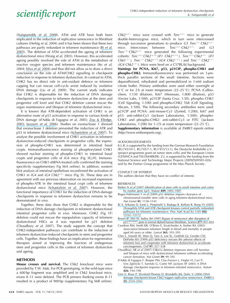
RESULTS AND DISCUSSIONStem-cell function, organ maintenance and lifespanTercþ /�Chk2þ /� double-heterozygous mice were crossed withthird-generation Terc�/�Chk2þ /� mice to generate the experi-mental cohorts (supplementary Fig S1A online). On using thismating scheme, intercross G4 (iG4) Terc�/� mice show telomeredysfunction, impaired organ maintenance and a shortenedlifespan, whereas intercross Tercþ /� (iF1 Tercþ /�) littermatesshow a rescue in telomere function, organ maintenance andlifespan (Hemann et al, 2001; Choudhury et al, 2007). Inagreement with previous studies, iG4 Terc�/� mice from ourcohorts showed a shortened lifespan compared with iF1 Tercþ /�
mice (Fig 1A). Of note, CHK2 deletion did not affect the lifespan ofReceived 2 December 2009; revised 7 May 2010; accepted 18 May 2010; publishedonline 25 June 2010
*Present address: Department of Gastroenterology, Hepatology and Endocrinology,Hannover Medical School, Hannover 30625, Germany+Corresponding author. Tel: þ 49 731 5036 100; Fax: þ 49 731 5036 102;E-mail: [email protected]
1Department of Molecular Medicine, and Max Planck Research Group on Stem CellAging, Albert Einstein alle 11, University of Ulm, Ulm 89081, Germany2Institute of Laboratory Animal Sciences, Chinese Academy of Medical Sciences andKey Laboratory of Human Diseases Comparative Medicine, Ministry of Health,Beijing, China
&2010 EUROPEAN MOLECULAR BIOLOGY ORGANIZATION EMBO reports VOL 11 | NO 8 | 2010
scientificreportscientific report
619

iF1 Tercþ /� or iG4 Terc�/� mice (Fig 1A) and CHK2 deletion didnot lead to an increase in cancer formation (data not shown).
The reduction in the lifespan of iG4 Terc�/�mice was associatedwith impaired maintenance of intestinal epithelium and reduction
in the body weight of 9–12-month-old iG4 compared withage-matched iF1 Tercþ /� mice, but CHK2 deletion did not rescuethese phenotypes (Fig 1B,C; supplementary Fig S1B–D online).CHK2 deletion also did not rescue telomere shortening in iG4
100
80
60
40
20
00 5 10
Age (months)
iF1 Chk2+/+
iF1 Chk2–/–
iG4 Chk2+/+
iG4 Chk2–/–
iF1 Chk2+/+ iF1 Chk2–/– iG4 Chk2–/–iG4 Chk2+/+
n=15
n=24
n=22
n=54
15 20 25
P<0.0001
P=0.941
P= 0.747
P<0.0001
P<0.0001
P<0.0001
P=0.0002
NS NS
n=10n=10
B cells Myeloid cells
n=5n=5
n=6
n=6 n=6 n=6 n=6 n=3
n=4
n=4
100
80
60
40
20
0
Cry
pt
no. p
er c
m
CD
34-L
SK
cel
ls
in B
M (%
)
Leuk
ocyt
es fr
om B
M (%
)
Nor
mal
ized
don
or-d
eriv
edW
BC
s (%
)
500
400
300
200
100
0
80
60
40
20
0
P=0.005
P=0.006
NSNS
0.03
0.02
0.01
0.00
NS NS
NS NS
NS
n=4 n=12 n=7 n=6 n=5n=5 n=4 n=4
Sur
viva
l (%
)
A
B
C D
E F
iF1 Chk2+/+ iF1 Chk2–/– iG4 Chk2+/+ iG4 Chk2–/–
Fig 1 | CHK2 deletion does not rescue progeroid phenotypes of telomere dysfunctional mice. (A) Survival curves for iF1 Tercþ /�Chk2þ /þ mice (n¼ 54),
iF1 Tercþ /�Chk2�/� mice (n¼ 22), iG4 Terc�/�Chk2þ /þ mice (n¼ 24) and iG4 Terc�/�Chk2�/� mice (n¼ 15). (B) Representative photographs of haematoxylin
and eosin-stained longitudinal sections of the small intestine of 9–12-month-old mice of the indicated genotypes. Scale bars, 200mm. (C) Histogram of the
number of basal crypts per vision field (original magnification � 100) in the small intestine of 9–12-month-old mice of the indicated genotypes (n¼ 4–5
mice per group). (D) Histogram of the percentage of haematopoietic stem cells (that is, CD34�/loLSK cells) in total BM cells of 9–12-month-old mice of the
indicated genotypes (n¼ 4–6 mice per group; data are shown as mean; error bars represent s.d.). (E) Histogram showing percentage of B cells and myeloid
cells in WBCs of the BM of 9–12-month-old mice of the indicated genotypes (n¼ 4–6 mice per group; data are shown as mean; error bars represent s.d.).
(F) Histogram showing percentage of donor-derived WBCs, 3 months after competitive transplantation for the indicated genotypes (n¼ 3–6 mice per group;
data are shown as mean; error bars represent s.d.). BM, bone marrow; iF1, intercross; iG4, intercross G4; NS, non-significant; WBC, white blood cell.
CHK2-independent induction of telomere dysfunction checkpoints
K. Nalapareddy et al
EMBO reports VOL 11 | NO 8 | 2010 &2010 EUROPEAN MOLECULAR BIOLOGY ORGANIZATION
scientificreport
620
Terc�/� mice (supplementary Fig S2A online). Similarly, thequantification of anaphase bridges—a tissue marker of telomeredysfunction (Kirk et al, 1997; Rudolph et al, 2001)—showedno rescue by CHK2 deletion (supplementary Fig S2B,C online).Chromosome fluorescence in situ hybridization on basal intestinalcrypts showed that CHK2 deletion did not provoke an increasein chromosomal aberration (ploidy change) in iG4 Terc�/�
mice (supplementary Fig S2D online), as it was detectedin response to intestinal deletion of p53 in previous studies(Begus-Nahrmann et al, 2009).
Impairments in haematopoietic stem-cell (HSC) function andlymphopoiesis are further age-related phenotypes that are inducedby telomere dysfunction in both mice and humans (Ju et al, 2007;Rossi et al, 2007). Previous studies have shown a reduced numberof HSCs (c-Kit-positive, Sca1-positive, Lineage-negative, CD34low/negative¼CD34lo/�LSK cells) in the bone marrow of agedtelomere dysfunctional mice compared with age-matched wild-type mice (Choudhury et al, 2007; Ju et al, 2007; Schaetzlein et al,2007). The deletion of CHK2 did not rescue the maintenance ofHSCs or skewing of haemato-lymphopoiesis (that is, a decrease inB-lymphopoiesis and increase in myelopoiesis) in the bonemarrow of iG4 Terc�/� mice (Fig 1D,E). HSCs from 9–12-month-old iG4 Terc�/� mice had a significantly reducedrepopulation capacity compared with those from iF1 Tercþ /�
mice, which was not rescued by CHK2 deletion (Fig 1F). Together,these data show that CHK2-independent mechanisms impairedthe function of intestinal and haematopoietic stem and progenitorcells in response to telomere dysfunction.
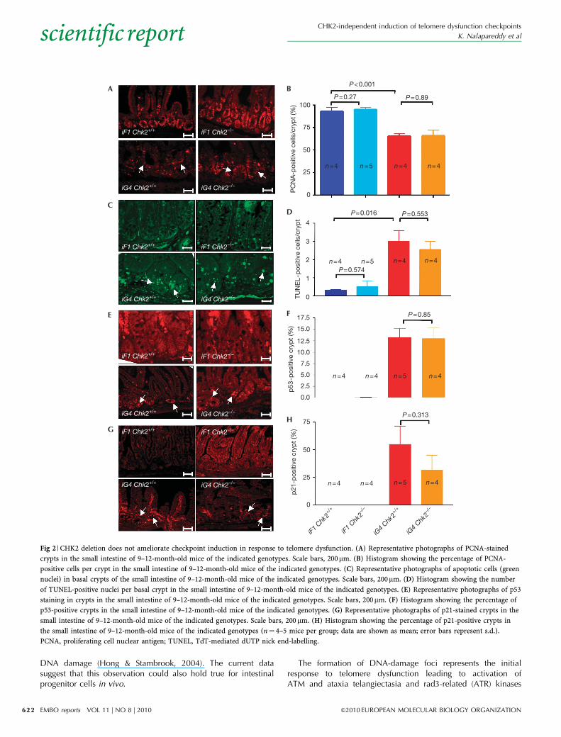
Telomere dysfunction induced checkpointsTo elucidate the failure of CHK2 deletion to rescue organmaintenance in ageing telomere dysfunctional mice, we focusedon the intestinal epithelium, which shows high rates of cellturnover during a lifetime (Barker et al, 2007). Intestinal atrophyin ageing iG4 mice has been associated with impaired cellproliferation in progenitor cells located in the basal crypts of theintestinal epithelium (Choudhury et al, 2007; Schaetzlein et al,2007). An analysis of cell proliferation markers—proliferating cellnuclear antigen (PCNA) and Ki67—in intestinal basal cryptsrevealed that CHK2 deletion did not rescue the induction of cell-cycle arrest in telomere dysfunctional mice (Fig 2A,B; supple-mentary Fig S3 online). CHK2 deletion also did not rescue theelevated rates of apoptosis in 9–12-month-old iG4 Terc�/� mice(Fig 2C,D), or the activation of p53 and p21 in the telomeredysfunctional intestine (Fig 2E–H). Together, these results indicatethat CHK2-independent mechanisms mediate the activation ofp53 and p21 as well as the induction of cell-cycle arrest andapoptosis in response to telomere dysfunction in intestinalprogenitor cells. Owing to the absence of transplantation assaysfor the assessment of intestinal stem-cell function, it was notpossible to prove formally the CHK2-independent induction ofcheckpoints in response to telomere dysfunction at the intestinalstem-cell level.
CHK2-dependent senescence in fibroblastsThe results on CHK2-independent induction of checkpointsin progenitor cells of mice with dysfunctional telomeres stood incontrast to the significant role of CHK2 in the induction ofsenescence in primary human fibroblasts (Takai et al, 2002;
Gire et al, 2004). Infection of late-passage, primary human BJfibroblasts with lentiviral vectors expressing a CHK2-directedshRNA confirmed that CHK2 knockdown abrogated the inductionof senescence, resulting in positive selection of CHK2-knockdowncells and improved proliferation rates of human fibroblast culturesat late passage (supplementary Fig S4A–C online). Similar resultswere obtained for mouse ear fibroblasts. Genetic deletion ofCHK2 rescued proliferation of iG4 Terc�/� fibroblasts (supple-mentary Fig S4D online). These experiments on mouse fibroblastswere carried out under low-stress conditions (3% oxygen),suggesting that CHK2 deletion can rescue telomere-dependentsenescence in cell culture. However, the improvement incell proliferation of Tercþ /þChk2�/� fibroblasts comparedwith Tercþ /þChk2þ /þ fibroblasts indicates that stress-inducedculture conditions (independent of oxygen) can limit fibroblastproliferation in a CHK2-dependent manner. The relativecontribution of CHK2 to the induction of telomere-dependentand telomere-independent senescence in fibroblast senescenceremains to be analysed. A challenge for these experimentsis that oxygen-independent stress factors that contribute toinduction of telomere-independent senescence in fibroblasts arenot well defined.
In contrast to the cell culture data, the deletion of CHK2 didnot rescue organ maintenance in ageing telomere dysfunctionalmice (Figs 1,2). These findings demonstrate that CHK2 isdispensable for the induction of DNA-damage checkpoints instem and progenitor cell compartments in response to telomeredysfunction in vivo.
Localization of CHK2 in response to telomere dysfunctionPrevious studies have shown that ATM is activated in response totelomere dysfunction in fibroblasts (d’Adda di Fagagna et al,2003). In agreement with these data, phosphorylation of ATM wasseen in western blot analysis and immunofluorescence staining ofthe intestinal epithelium of iG4 and iG4 Chk2�/� mice (Fig 3A;supplementary Fig S5A,B online). In line with ATM activation,western blot and immunofluorescence staining showed anupregulation of phospho-CHK2 (T68) in intestinal progenitorcells of 9–12-month-old iG4 Terc�/� mice compared with iF1Tercþ /� mice (Fig 3B–D; supplementary Fig S5C,D online).Interestingly, the expression of activated CHK2 in ageingtelomere dysfunctional intestine was restricted to the cytoplasm,indicating that activated CHK2 did not translocate to thenucleus (Fig 3C; supplementary Fig S5D online). In contrast,intra-nuclear expression of phospho-CHK2 was readily detectablein terminally differentiated epithelial cells of the intestinalvilli of iG4 mice (Fig 3C), as well as in senescent humanfibroblasts (Fig 3E,F).
Together, these data indicate that CHK2 is phosphorylated inresponse to telomere dysfunction. In contrast to senescentfibroblasts, phosphorylated CHK2 did not translocate to thenucleus in telomere dysfunctional progenitor cells of the intestinalbasal crypts. The cytoplasmic localization could be one of thereasons for the failure of activated CHK2 to contribute toactivation of p53-dependent checkpoint responses in intestinalbasal crypt cells. This interpretation is in line with previousfindings showing that the failure of embryonic stem cells to induceG1 cell-cycle arrest in response to g-irradiation is associated withan impaired nuclear localization of activated CHK2 in response to
CHK2-independent induction of telomere dysfunction checkpoints
K. Nalapareddy et al
&2010 EUROPEAN MOLECULAR BIOLOGY ORGANIZATION EMBO reports VOL 11 | NO 8 | 2010
scientificreport
621
DNA damage (Hong & Stambrook, 2004). The current datasuggest that this observation could also hold true for intestinalprogenitor cells in vivo.
The formation of DNA-damage foci represents the initialresponse to telomere dysfunction leading to activation ofATM and ataxia telangiectasia and rad3-related (ATR) kinases
P<0.001
P=0.27100
75
50
n=4
n=4P=0.574
P=0.016 P=0.553
P=0.85
P=0.313
PC
NA
-pos
itive
cel
ls/c
ryp
t (%
)TU
NE
L-p
ositi
ve c
ells
/cry
pt
p53
-pos
itive
cry
pt
(%)
p21
-pos
itive
cry
pt
(%)
n=5
n=5
n=4
n=4
n=4
iF1 C
hk2+/+
iF1 C
hk2–/
–
iG4
Chk2+/+
iG4
Chk2–/
–
n=4
n=4 n=4
n=4n=5
n=5 n=4
n=4
n=425
0
4
3
2
1
0
75
50
25
0
17.5
15.0
12.5
10.0
7.5
5.0
2.5
0.0
P=0.89A B
D
F
H
C
E
G
iF1 Chk2+/+ iF1 Chk2–/–
iG4 Chk2+/+ iG4 Chk2–/–
iF1 Chk2+/+ iF1 Chk2–/–
iG4 Chk2+/+ iG4 Chk2–/–
iF1 Chk2+/+ iF1 Chk2–/–
iG4 Chk2+/+ iG4 Chk2–/–
iF1 Chk2+/+ iF1 Chk2–/–
iG4 Chk2+/+ iG4 Chk2–/–
Fig 2 | CHK2 deletion does not ameliorate checkpoint induction in response to telomere dysfunction. (A) Representative photographs of PCNA-stained
crypts in the small intestine of 9–12-month-old mice of the indicated genotypes. Scale bars, 200mm. (B) Histogram showing the percentage of PCNA-
positive cells per crypt in the small intestine of 9–12-month-old mice of the indicated genotypes. (C) Representative photographs of apoptotic cells (green
nuclei) in basal crypts of the small intestine of 9–12-month-old mice of the indicated genotypes. Scale bars, 200mm. (D) Histogram showing the number
of TUNEL-positive nuclei per basal crypt in the small intestine of 9–12-month-old mice of the indicated genotypes. (E) Representative photographs of p53
staining in crypts in the small intestine of 9–12-month-old mice of the indicated genotypes. Scale bars, 200mm. (F) Histogram showing the percentage of
p53-positive crypts in the small intestine of 9–12-month-old mice of the indicated genotypes. (G) Representative photographs of p21-stained crypts in the
small intestine of 9–12-month-old mice of the indicated genotypes. Scale bars, 200mm. (H) Histogram showing the percentage of p21-positive crypts in
the small intestine of 9–12-month-old mice of the indicated genotypes (n¼ 4–5 mice per group; data are shown as mean; error bars represent s.d.).
PCNA, proliferating cell nuclear antigen; TUNEL, TdT-mediated dUTP nick end-labelling.
CHK2-independent induction of telomere dysfunction checkpoints
K. Nalapareddy et al
EMBO reports VOL 11 | NO 8 | 2010 &2010 EUROPEAN MOLECULAR BIOLOGY ORGANIZATION
scientificreport
622

iG4 Chk2+/+
iF1 C
hk2+/+
iF1 C
hk2–/
–
iG4
Chk2–/
–
iG4
Chk2+/+
pCHK1s345
GAPDH
I
Pho
spho
-CH
K1-
pos
itive
cells
/cry
pt
P<0.001
P=0.172
P=0.023
iF1 C
hk2+/+
iF1 C
hk2–/
–
iG4
Chk2+/+
iG4
Chk2–/
–
12.5
10.0
7.5
n=4 n=4 n=4n=55.0
2.5
0.0
H
G
Cel
ls w
ith n
ucle
ar e
xpre
ssio
nof
pho
spho
-CH
K2
(%)
P<0.0001
BJ36 BJ54
100
75
50
25
0
E F
iF1 C
hk2+/+
iF1 C
hk2–/
–
iG4
Chk2+/+
iG4
Chk2–/
–
50
Pho
spho
-CH
K2-
pos
itive
cryp
t (%
)
40
30
20
10
0
C D
pATMs1981 pCHK2 T68
GAPDH
iF1 C
hk2+/+
iF1 C
hk2+/+
iG4
Chk2+/+
iG4
Chk2–/
–
iF1 C
hk2–/
–
iG4
Chk2–/
–
iG4
Chk2+/+
panATM
A B
BJ36
BJ54
iF1 Chk2+/+ iF1 Chk2–/–
iG4 Chk2+/+ iG4 Chk2–/–
Fig 3 | Localization of activated CHK2 and CHK1 in the intestine of telomere dysfuctional mice. (A) Western blot showing the expression of phospho-
ATM s1981 in intestinal biopsies of 9–12-month-old mice of the indicated genotypes. The lower panel shows panATM expression for the loading
control. (B) Western blot showing activation of phospho-CHK2 T68 in the small intestinal lysates of 9–12-month-old mice of the indicated genotypes.
The lower panel shows GAPDH expression for the loading control. (C) Representative photographs showing cytoplasmic localization of phosphorylated
CHK2 in the intestinal crypts and nuclear localization of phosphorylated CHK2 in villi of the small intestine of 9–12-month-old mice of the indicated
genotypes. Scale bar, 400mm. (D) Histogram showing the percentage of phospho-CHK2-positive cells per crypt in the small intestine of 9–12-month-
old mice of the indicated genotypes. (E) Representative photographs of phosphorylated CHK2 staining in human BJ fibroblasts at the indicated
population doubling. (F) Histogram showing the percentage of phospho-CHK2-positive fibroblasts at the indicated population doublings. (More than
100 cells were counted.) (G) Representative photographs of phospho-CHK1-stained crypts in the small intestine of 9–12-month-old mice of the
indicated genotypes. Scale bars, 400mm. (H) Histogram showing the percentage of phospho-CHK1-positive cells per crypt in the small intestine of
9–12-month-old mice of the indicated genotypes (n¼ 4–5 mice per group; data are shown as mean; error bars represent s.d.). (I) Western blot on
small-intestinal protein lysates from 9–12-month-old mice of the indicated genotypes. The lower panel shows GAPDH expression for the loading
control. ATM, ataxia telangiectasia mutated; GAPDH, glyceraldehyde 3-phosphate dehydrogenase; iF1, intercross; iG4, intercross G4.
CHK2-independent induction of telomere dysfunction checkpoints
K. Nalapareddy et al
&2010 EUROPEAN MOLECULAR BIOLOGY ORGANIZATION EMBO reports VOL 11 | NO 8 | 2010
scientificreport
623
(Nalapareddy et al, 2008). ATM and ATR have both beenimplicated in the induction of replicative senescence in fibroblastcultures (Herbig et al, 2004) and it has been shown that these twopathways are partly redundant in telomere maintenance (Bi et al,2005). The deletion of ATM accelerated the ageing of telomeredysfunctional mice (Wong et al, 2003). However, this acceleratedageing possibly involved the role of ATM in the metabolism ofreactive oxygen species and telomere maintenance (Ito et al,2004; Silva et al, 2004) and thus did not allow us to draw a directconclusion on the role of ATM/CHK2 signalling in checkpointinduction in response to telomere dysfunction. In contrast to ATM,CHK2 has no direct role in anti-oxidant defence or telomerecapping but can rescue cell-cycle arrest induced by oxidativeDNA damage (Liu et al, 2009). The current study indicatesthat CHK2 is dispensable for the induction of DNA damagecheckpoints in response to telomere dysfunction at the stem andprogenitor cell level and that CHK2 deletion cannot rescue theorgan maintenance and lifespan of telomere dysfunctional mice.
It is known that ATR-dependent activation of CHK1 is analternative route of p53 activation in response to various kinds ofDNA damage (d’Adda di Fagagna et al, 2003; Zou & Elledge,2003; Jazayeri et al, 2006). Studies on exonuclease 1 showedthat exonuclease 1 deletion prevented the induction of ATR andp53 in telomere dysfunctional mice (Schaetzlein et al, 2007). Toanalyse the possible involvement of CHK1 activation in telomeredysfunction-induced checkpoints in progenitor cells, the expres-sion of phospho-CHK1 was determined in intestinal basalcrypts. Immunofluorescence staining of phosphorylated CHK1showed nuclear staining of phospho-CHK1 in intestinal basalcrypts and progenitor cells of iG4 mice (Fig 3G,H). Immuno-fluorescence on CHK1-shRNA-treated cells confirmed the stainingspecificity (supplementary Fig S6A online). In addition, westernblot analysis of intestinal epithelium reconfirmed the activation ofCHK1 in iG4 and iG4 Chk2�/� mice (Fig 3I). These data are inagreement with our previous observation on increased expressionof phospho-ATR in the intestinal basal crypt cells of telomeredysfunctional mice (Schaetzlein et al, 2007). However, thefunctional importance of CHK1 for the induction of DNA-damagecheckpoints in response to telomere dysfunction remains to bedemonstrated in vivo.
Together, these data show that CHK2 is dispensable for theinduction of DNA damage checkpoints in telomere dysfunctionalintestinal progenitor cells in vivo. Moreover, CHK2 (Fig 1F)deletion could not rescue the repopulation capacity of telomeredysfunctional HSCs as it was reported for p21 deletion(Choudhury et al, 2007). The study supports the concept thatCHK2-independent pathways can contribute to the induction oftelomere dysfunction-induced checkpoints in stem and progenitorcells. Together, these findings have an implication for regenerativetherapies aimed at improving the function of endogenousstem and progenitor cells in the context of telomere dysfunctionand ageing.
METHODSMouse crosses and survival. The Chk2 knockout mice wereprovided by T.W. Mak. For PCR genotyping, in the wild-type micea 600 bp fragment was amplified and in Chk2 knockout mice,exons 8–11 were deleted. The PCR primers flanking exons 8–11resulted in a product of 900 bp (supplementary Fig S6B online).
Chk2þ /� mice were crossed with Tercþ /� mice to generatedouble-heterozygous mice, which in turn were intercrossedthrough successive generations to produce G3 Terc�/�Chk2þ /�
mice. Intercrosses between Tercþ /�Chk2þ /� and G3Terc�/�Chk2þ /� mice generated the following experimentalcohorts: Tercþ /�Chk2þ /þ (iF1 Chk2þ /þ ), Tercþ /�Chk2�/� (iF1Chk1�/�), Terc�/�Chk2þ /þ (iG4 Chk2þ /þ ) and Terc�/�Chk2�/�
(iG4 Chk2�/�). Mice were bred on a C57BL/6J background.Stainings for PCNA, Ki67, p53, p21CIP, phospho-CHK1 andphospho-CHK2. Immunofluorescence was performed on 3 mm-thick paraffin sections of the small intestine. Sections weredeparaffinized, rehydrated and permeabilized in 1 mM sodiumcitrate buffer. Primary antibodies were used either overnight at4 1C or for 2 h at room temperature (22–25 1C): PCNA (Calbio-chem, 1:150 dilution), Ki67 (Monosan, 1:800 dilution), p53(Vector Labs, 1:500), p21CIP (Santa Cruz, 1:50), phospho CHK1(Cell Signaling, 1:100) and phospho-CHK2 T68 (Cell Signaling,Abcam, 1:100). The following secondary antibodies were used:p21CIP and PCNA: anti-mouse-Cy3 (Zymed, 1:200); ki67 andp53: anti-rabbit-Cy3 (Jackson Laboratories, 1:500); phospho-CHK1 and phospho-CHK2: anti-rabbit-Cy3 or FITC ( JacksonLaboratories, 1:200) for 1–2 h at room temperature (22–25 1C).Supplementary information is available at EMBO reports online(http://www.emboreports.org).
ACKNOWLEDGEMENTSK.L.R. is supported by the funding from the German Research Foundation(RU745/10-1, RU745/7-1, RU7415/13-1), the Deutsche Krebshilfe e.V.(project programme grant on tumor stem cells) and the European Union(GENINCA and TELOMARKER). Z.J. is supported by the funding from theNational Science and Technology Major Projects (2009ZX09501-026)and by the Partner Group programme of the Max Planck Society.
CONFLICT OF INTERESTThe authors declare that they have no conflict of interest.
REFERENCESBarker N et al (2007) Identification of stem cells in small intestine and colon
by marker gene Lgr5. Nature 449: 1003–1007Begus-Nahrmann Y et al (2009) p53 deletion impairs clearance of
chromosomal-instable stem cells in aging telomere-dysfunctional mice.Nat Genet 41: 1138–1143
Bi X, Srikanta D, Fanti L, Pimpinelli S, Badugu R, Kellum R, Rong YS (2005)Drosophila ATM and ATR checkpoint kinases control partially redundantpathways for telomere maintenance. Proc Natl Acad Sci USA 102:15167–15172
Brown JP, Wei W, Sedivy JM (1997) Bypass of senescence after disruption ofp21CIP1/WAF1 gene in normal diploid human fibroblasts. Science 277: 831–834
Cawthon RM, Smith KR, O’Brien E, Sivatchenko A, Kerber RA (2003)Association between telomere length in blood and mortality in peopleaged 60 years or older. Lancet 361: 393–395
Chin L, Artandi SE, Shen Q, Tam A, Lee SL, Gottlieb GJ, Greider CW,DePinho RA (1999) p53 deficiency rescues the adverse effects oftelomere loss and cooperates with telomere dysfunction to acceleratecarcinogenesis. Cell 97: 527–538
Choudhury AR et al (2007) Cdkn1a deletion improves stem cell functionand lifespan of mice with dysfunctional telomeres without acceleratingcancer formation. Nat Genet 39: 99–105
d’Adda di Fagagna F, Reaper PM, Clay-Farrace L, Fiegler H, Carr P,Von Zglinicki T, Saretzki G, Carter NP, Jackson SP (2003) A DNAdamage checkpoint response in telomere-initiated senescence. Nature426: 194–198
Gire V, Roux P, Wynford-Thomas D, Brondello JM, Dulic V (2004) DNAdamage checkpoint kinase Chk2 triggers replicative senescence. EMBO J23: 2554–2563
CHK2-independent induction of telomere dysfunction checkpoints
K. Nalapareddy et al
EMBO reports VOL 11 | NO 8 | 2010 &2010 EUROPEAN MOLECULAR BIOLOGY ORGANIZATION
scientificreport
624
Hemann MT, Strong MA, Hao LY, Greider CW (2001) The shortest telomere,not average telomere length, is critical for cell viability and chromosomestability. Cell 107: 67–77
Herbig U, Jobling WA, Chen BP, Chen DJ, Sedivy JM (2004) Telomereshortening triggers senescence of human cells through a pathwayinvolving ATM, p53, and p21CIP1, but not p16INK4a. Mol Cell 14:501–513
Hirao A et al (2002) Chk2 is a tumor suppressor that regulates apoptosisin both an ataxia telangiectasia mutated (ATM)-dependent and anATM-independent manner. Mol Cell Biol 22: 6521–6532
Hong Y, Stambrook PJ (2004) Restoration of an absent G1 arrest andprotection from apoptosis in embryonic stem cells after ionizingradiation. Proc Natl Acad Sci USA 101: 14443–14448
Ito K et al (2004) Regulation of oxidative stress by ATM is requiredfor self-renewal of haematopoietic stem cells. Nature 431:997–1002
Jazayeri A, Falck J, Lukas C, Bartek J, Smith GC, Lukas J, Jackson SP (2006)ATM- and cell cycle-dependent regulation of ATR in response to DNAdouble-strand breaks. Nat Cell Biol 8: 37–45
Ju Z, Jiang H, Jaworski M, Rathinam C, Gompf A, Klein C, Trumpp A,Rudolph KL (2007) Telomere dysfunction induces environmentalalterations limiting hematopoietic stem cell function and engraftment.Nat Med 13: 742–747
Kirk KE, Harmon BP, Reichardt IK, Sedat JW, Blackburn EH (1997) Blockin anaphase chromosome separation caused by a telomerase templatemutation. Science 275: 1478–1481
Lansdorp PM (2009) Telomeres and disease. EMBO J 28: 2532–2540Liu J et al (2009) Bmi1 regulates mitochondrial function and the DNA damage
response pathway. Nature 459: 387–392
Nalapareddy K, Jiang H, Gutierrez LM, Rudolph KL (2008) Determining theinfluence of telomere dysfunction and DNA damage on stem andprogenitor cell aging—what markers can we use? Exp Gerontol 43:998–1004
Rossi DJ, Bryder D, Seita J, Nussenzweig A, Hoeijmakers J, Weissman IL(2007) Deficiencies in DNA damage repair limit the function ofhaematopoietic stem cells with age. Nature 447: 725–729
Rudolph KL, Chang S, Lee HW, Blasco M, Gottlieb GJ, Greider C,DePinho RA (1999) Longevity, stress response, and cancer in agingtelomerase-deficient mice. Cell 96: 701–712
Rudolph KL, Millard M, Bosenberg MW, DePinho RA (2001) Telomeredysfunction and evolution of intestinal carcinoma in mice and humans.Nat Genet 28: 155–159
Schaetzlein S et al (2007) Exonuclease-1 deletion impairs DNA damagesignaling and prolongs lifespan of telomere-dysfunctional mice. Cell 130:863–877
Silva E, Tiong S, Pedersen M, Homola E, Royou A, Fasulo B, Siriaco G,Campbell SD (2004) ATM is required for telomere maintenance andchromosome stability during Drosophila development. Curr Biol 14:1341–1347
Takai H et al (2002) Chk2-deficient mice exhibit radioresistance and defectivep53-mediated transcription. EMBO J 21: 5195–5205
Wong KK, Maser RS, Bachoo RM, Menon J, Carrasco DR, Gu Y, Alt FW,DePinho RA (2003) Telomere dysfunction and Atm deficiency compromisesorgan homeostasis and accelerates ageing. Nature 421: 643–648
Wright WE, Shay JW (1992) The two-stage mechanism controlling cellularsenescence and immortalization. Exp Gerontol 27: 383–389
Zou L, Elledge SJ (2003) Sensing DNA damage through ATRIP recognitionof RPA-ssDNA complexes. Science 300: 1542–1548
CHK2-independent induction of telomere dysfunction checkpoints
K. Nalapareddy et al
&2010 EUROPEAN MOLECULAR BIOLOGY ORGANIZATION EMBO reports VOL 11 | NO 8 | 2010
scientificreport
625
Related Documents