Alma Mater Studiorum – Università di Bologna DOTTORATO DI RICERCA IN CHIMICA – CHIMICA FARMACEUTICA Ciclo XXVIII Settore Concorsuale di afferenza: 03/D1 Settore Scientifico disciplinare: CHIM/08 NATURALLY INSPIRED PRIVILEGED STRUCTURES IN DRUG DISCOVERY: MULTIFUNCTIONAL COMPOUNDS FOR ALZHEIMER’S DISEASE TREATMENT Presentata da: Rita Maria Concetta Di Martino Coordinatore Dottorato Relatore Prof. Aldo Roda Prof.ssa Alessandra Bisi Correlatore Prof.ssa Federica Belluti Esame finale anno 2016

Welcome message from author
This document is posted to help you gain knowledge. Please leave a comment to let me know what you think about it! Share it to your friends and learn new things together.
Transcript
Alma Mater Studiorum – Università di Bologna
DOTTORATO DI RICERCA IN
CHIMICA – CHIMICA FARMACEUTICA
Ciclo XXVIII
Settore Concorsuale di afferenza: 03/D1
Settore Scientifico disciplinare: CHIM/08
NATURALLY INSPIRED PRIVILEGED STRUCTURES IN
DRUG DISCOVERY: MULTIFUNCTIONAL COMPOUNDS
FOR ALZHEIMER’S DISEASE TREATMENT
Presentata da: Rita Maria Concetta Di Martino
Coordinatore Dottorato Relatore
Prof. Aldo Roda Prof.ssa Alessandra Bisi
Correlatore
Prof.ssa Federica Belluti
Esame finale anno 2016
I
Table of Contents
List of abbreviations 1
1. State of the art 7
1.1. ALZHEIMER’S DISEASE: NEURODEGENERATIVE DISORDER 8
1.2. MULTIFACTORIAL NATURE OF AD 13
1.3. THE AMYLOID CASCADE HYPOTHESIS 14
1.3.1. Aβ-toxicity 15
1.3.2. BACE-1 17
1.3.3. Design of BACE-1 inhibitors: from peptidomimetic to
non-peptidic small molecules 20
1.4. TAU HYPOTHESIS 26
1.4.1. GSK-3 28
1.4.2. GSK-3β inhibitors 32
1.4.3. GSK-3β: molecular linker between Aβ and τ 35
1.5. OXIDATIVE STRESS 36
1.6. NEUROINFLAMMATION 38
1.7. NRF2-KEAP1: A NEUROPROTECTIVE SIGNALING PATHWAY 40
1.8. OTHER PROTEIN KINASES INVOLVED IN AD 46
2. Medicinal Chemistry 51
2.1. MULTITARGET APPROACH: IDEAL STRATEGY FOR AN
EFFECTIVE AD TREATMENT 52
2.2. PRIVILEGED STRUCTURES 54
2.3. NATURAL PRODUCTS 62
II
2.3.1. α,β-unsaturated carbonyl compounds 65
2.3.2. Thiol trapping assay 68
2.4. CURCUMIN: A PROMISING THERANOSTIC TOOL FOR AD 70
2.4.1. Curcumin physical-chemical properties 72
2.4.2. Curcuminoids synthesis 73
2.4.3. Strategies aimed at improving curcumin bioavailability and
pharmacokinetics 76
2.4.4. Structure-activity relationship studies 77
2.4.5. Curcumin neuroprotective potential in AD therapy 78
2.4.6. Curcumin: a fluorescent probe in AD diagnosis 80
2.5. MOLECULAR IMAGING THERANOSTIC PROBES: A PROMISING
FUTURE IN AD TREATMENT 84
2.6. CHITOSAN: A VERY ATTRACTIVE AND USEFUL BIOPOLYMER 86
2.6.1. Physical-chemical properties 88
2.6.2. Applications 89
2.6.3. Chitosan-based bioconjugates 91
3. Aim of the work and Chemistry 97
3.1. DESIGN OF CURCUMIN-BASED COMPOUNDS 98
3.2. DESIGN OF 1,4- AND 1,3-BISCHALCONES [BIS(CINNAMOYL)
BENZENE DERIVATIVES] 106
3.3. DESIGN OF CS-BASED BIOCONJUGATES 107
3.4. DESIGN OF INDOLE-BASED ANALOGUES 108
3.5. SYNTHESIS OF SERIES Ia AND Ib 126
3.5.1. Pabon reaction: synthesis of symmetric compounds 4, 5, 8,
11-14, 16 and 17 126
3.5.2. Pabon reaction: synthesis of asymmetric compounds 2, 3, 6
7, 15 and intermediates 22 and 23 127
III
3.5.3. Williamson reaction: synthesis of symmetric compound
18, and of the tautomeric couples 8 and 19, 9 and 20, 10
and 21 128
3.5.4. Williamson reaction: synthesis of intermediate benzaldehydes
24-26 129
3.6. SYNTHESIS OF SERIES IIa AND IIb 129
3.6.1. Pabon reaction: synthesis of symmetric analogues 27-31 and
asymmetric derivatives 32-38 129
3.6.2. Synthesis of functionalized aldehydes 39-45, azido derivatives
46-50 and intermediate 51 130
3.6.3. Synthesis of tautomeric couples of curcumin-based derivatives
52a,b-54a,b and intermediates 55a,b, 56 and 57a,b 131
3.7. SYNTHESIS OF SERIES III 133
3.7.1. Synthesis of curcumin analogues 58-60, 61a,b and
62 and intermediates 64a,b, 65a,b and 63 133
3.8. SYNTHESIS OF SERIES IVa AND IVb 134
3.8.1. Synthesis of curcumin analogues 66-72 and aldehyde 73 134
3.8.2. Synthesis of curcumin-DF hybrids 74-78 136
3.9. SYNTHESIS OF SERIES Va AND Vb 137
3.9.1. Synthesis of curcumin-coumarin hybrids 79-82 and
85a,b-89a,b and intermediates 92-102 137
3.9.2. Synthesis of curcumin-coumarin hybrids 83a,b, 84a,b
and 90a,b and azido intermediates 103-105 138
3.9.3. Synthesis of amido curcuminoids 91a,b and amine
intermediate 106 139
3.10. SYNTHESIS OF SERIES VIa AND VIb 140
3.10.1. Synthesis of difluoroboron-derivatized curcuminoids
107 and 108 140
3.10.2. Synthesis of pyrazoles 109-113, 116-120, 122, of
dihydropyrazole 115 and of isoxazoles 114 and 121 141
3.10.3. Synthesis of curcumin-based pyrazoles 123-125 143
IV
3.11. SYNTHESIS OF SERIES VII 144
3.11.1. Synthesis of 1,4- and 1,3-bischalcones 126-129 144
3.12. SYNTHESIS OF SERIES VIII 144
3.12.1. Synthesis of CS bioconjugates 130 and 131 and
aldehyde 132 144
3.13. SYNTHESIS OF SERIES IX 146
3.13.1. Synthesis of indole-based derivatives 133-143 146
4. Results and discussion 148
4.1. BACE-1 INHIBITION 149
4.2. GSK-3β INHIBITION 152
4.3. NEUROPROTECTION 158
4.3.1. SH-SY5Y neuroblastoma cell viability 158
4.3.2. Antioxidant activity 158
4.3.3. Total GSH levels enhancement 159
4.4. NEUROINFLAMMATION 160
4.4.1. Neurotoxicity: microglial cell viability 161
4.4.2. Neuroinflammatory potential 162
4.5. THIOL TRAPPING ASSAY AND COVALENT DOCKING
SIMULATION ON GSK-3β 169
4.6. CK1 AND LRRK2 INHIBITION 170
4.6.1. CK1δ and CK1ε inhibition 170
4.6.2. LRRK2 and G2019S-LRRK2 inhibition 172
4.7. BBB PERMEATION 174
5. Conclusions 177

V
6. Experimental section 181
7. Appendix 268
7.1. 1D AND 2D-NMR SAMPLE COMPOUNDS 269
8. Bibliographic references 276
1
List of abbreviations
Aβ amyloid β
ABAD Aβ-binding alcohol dehydrogenase protein
Ac acetyl
ACh acetylcholine
AChE acetylcholinesterase
AChEIs acetylcholinesterase inhibitors
AD Alzheimer’s disease
ALG alginate
ALS amyotrophic lateral sclerosis
APP amyloid β protein precursor
ARE antioxidant-response element
ATP adenosine 5'-triphosphate
BACE-1 β-site APP cleaving enzyme (β-secretase)
BBB blood-brain barrier
BChE butyrylcholinesterase
Bn benzyl
bZip basic region leucine zipper
CAA congophilic amyloid angiopathy
CADD computer-assisted drug discovery
CaMKII calcium and calmodulin-dependent protein kinase II
CC click chemistry (approach)
CCR click chemistry reaction
CDCl3 deuterated chloroform
CDI carbonyldiimidazole
CDK5 cyclin dependent protein kinase 5
2
CK1 casein kinase 1
13C-NMR carbon nuclear magnetic resonance
CNS central nervous system
COSY correlation spectroscopy (NMR)
CS chitosan
CT computed tomography
Cul Cullin
DA degree of acetylation
Da Dalton
DD degree of deacetylation
DDI drug-drug interactions
DDSs drug delivery systems
DF dimethyl fumarate
DMAP 4-(dimethylamino)pyridin
DMF N,N-dimethylformamide
DMSO dimethyl sulfoxide
D2O deuterium oxide
DOS diversity-oriented synthesis
DS degree of substitution
E entgegen (opposite, trans)
EDC N-(3-dimethylaminopropyl)-N'-ethylcarbodiimide hydrochloride
EMA European Medicines Agency
EpRE electrophile-responsive element
equiv equivalent
ERK extracellular receptor kinase
ESI-MS electron spray ionization-mass spectrometry
Et ethyl
3
EtOAc ethyl acetate
FDA Food and Drug Administration
GDP guanosine diphosphate
GPCRs G-protein coupled receptors
GSH glutathione
GSK-3β glycogen synthase kinase-3β
GST glutathione-S-transferase
GTP guanosine triphosphate
HB hydrogen-bonding
HCl hydrochloric acid
HE hydroxyethylene
HMKs halomethylketones
HMBC heteronuclear multiple-bond correlation (NMR)
HMTA hexamethylenetetramine
4HNE 4-hydroxynonenal
1H-NMR proton nuclear magnetic resonance
HO-1 heme oxygenase-1
HPB hydrophobic (interactions)
HPLC high performance liquid chromatography
HSQC heteronuclear single-quantum coherence (NMR)
Hz Hertz
INF-γ interferon gamma
IONs iron oxide nanoparticles
I2PP2A inhibitor-2 of phosphatase protein PP2A
IR infrared (spectroscopy)
JNK Jun-N-terminal kinase
Keap1 Kelch-like ECH-associated protein 1
4
LPS lipopolysaccharide
LRRK2 leucine-rich repeat kinase 2
M molar mol/L
MAOS microwave assisted organic synthesis
MAP microtubule-associated protein
MAPKs mitogen-activated protein kinases
MCM multiple-compound medication
MD molecular dynamic
MI molecular imaging
MMT multiple-medication therapy
MNPs magnetic nanoparticles
MRI magnetic resonance imaging
MTDD multitarget drug design
MTDLs multi-target-directed ligands
MTDs multitarget drugs
MW molecular weight
nAChR nicotinic receptor
NBS N-bromosuccinimide
n-BuNH2 n-butylamine
NDs neurodegenerative diseases
NF-κB nuclear factor-κB
NFTs neurofibrillary tangles
NMDA N-methyl-D-aspartate
NMDAR N-methyl-D-aspartate receptor
NMP N-methyl-2-pyrrolidinone
NPs natural products
NQO1 NAD(P)H: quinone oxidoreductase-1
5
Nrf2 nuclear factor erythroid 2-related factor 2
Nu nucleophile
OMT O-methyltransferase
PAMPA parallel artificial membrane permeability assay
PCL poly(caprolactone)
PD Parkinson’s disease
PE petroleum ether
PEG polyethylene glycol
PERK PKR-like endoplasmic reticulum kinase
PET positron emission tomography
Ph phenyl
PHFs paired helical filaments
PhRMA Pharmaceutical Research and Manufacturers of America
PI3K phosphatidylinositol 3-kinase
PKA protein kinase A
PKC protein kinase C
PKs protein kinases
PLGA poly(lactide-co-glycolide)
ppm parts per million
PPs protein phosphatases
PS1 presenilin-1
RNS reactive nitrogen species
ROC Ras of complex proteins
ROS reactive oxygen species
r.t. room temperature
SAPKs stress-activated protein kinases
SFN sulforaphane
6
SN2 second-order nucleophilic substitution
SOD superoxide dismutase
SPECT single photon emission computed tomography
SPIONs small superparamagnetic iron oxide particles
SPs senile plaques
TBH tert-butyl hydroperoxide
t-Bu tert-buthyl
TDZDs thiadiazolidinones
TEA triethylamine
TFA trifluoroacetic acid
THF tetrahydrofuran
TMC N,N,N-trimethyl CS chloride
TMS tetramethylsilane
TMSA trimethylsilylacetylene
TNF-α tumor necrosis factor-α
TOS target oriented synthesis
UV ultraviolet-visible (spectroscopy)
VDCC voltage-dependent chloride channel
Z zusammen (together, cis)

7
1. State of the art
8
1.1. ALZHEIMER’S DISEASE: NEURODEGENERATIVE DISORDER
Neurodegenerative disease (ND) is an umbrella term to define a range of
conditions characterized by progressive nervous system dysfunction and
recognized as overwhelming health and socio-economic problems. They are
incurable and debilitating disorders that result in progressive degeneration and/or
death of nerve cells with consequent problems with movement (called ataxias), or
mental functioning (called dementias). Among them, dementias are responsible for
the greatest burden of these pathologies with Alzheimer’s disease (AD)
representing the most common form of dementia in industrialized nations among
the elderly. It is the sixth leading cause of death, affecting more than 44 million
people worldwide, and, due to its debilitating nature, causes an enormous financial
and emotional stress on patients and caregivers.
Given that age constitutes the main risk factor for dementia and the population
worldwide is rapidly aging, the number of AD patients is projected to reach 116
million by 2050.1 Thus, this devastating disorder has been identified as one of the
major public health concerns and a real research priority.
AD, described for the first time by the German psychiatrist Alois Alzheimer
in 1906, is a progressive neurological disorder characterized by short-term memory
impairment, at the beginning, and a profound cognitive and physical disability at
the later stage.
The vast majority of AD cases are the late-onset sporadic forms whose greatest
environmental risk factor is represented by aging. However, rare, familial, early-
onset autosomal dominant forms, approximately 5 % only of all AD cases, exist
and are caused by missense mutations in genes encoding amyloid β (Aβ) precursor
protein (APP), presenilin-1 (PS1) and presenilin-2 (PS2).2
Diagnosis of AD is based on a careful analysis of the clinical features,
although it should be confirmed by a depth histopathological examination of
patients’ brains. In general, three different clinical stages of the pathology have
been identified, in which the progressive cognitive and functional decline stretches
over 5-8 years:
9
1) Mild: that usually lasts 2-3 years, characterized by short-term memory
impairment often accompanied by anxiety and depression.
2) Moderate: in which the symptoms of the previous stage decrease and some
neuropsychiatric manifestations such as visual hallucinations, false beliefs
and reversal of sleep patterns emerge.
3) Severe: characterized by motor signs, such as motor rigidity and prominent
cognitive decline.3
Two additional clinical features of the pathology are the deterioration of language
skills,4 and visuospatial deficits.5
The precise onset of clinical AD is very difficult to recognize by both patient
and family because the earliest symptoms are often subtle and sporadic deficits in
the remembrance of minor events of everyday life, referred to as loss of episodic
memory. Although, after many years (a decade or more), a profound dementia
develops and is often accompanied by extrapyramidal motor signs, slowed gait, and
incontinence; death usually comes for minor respiratory complications, such as
aspiration or pneumonia often in the middle of the night.6
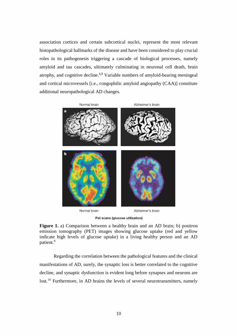
In the final stage of the malady, the brains of AD patients are pathologically
characterized by hippocampal and cerebral cortex atrophy and ventricular
enlargement. Moreover, the brain regions involved in learning and memory
processes, including the temporal and frontal lobes, are reduced in size as
consequence of synaptic degeneration and neuronal death (Fig. 1). Microscopically,
the numbers of neuronal cell bodies in the limbic and association cortices and in
certain subcortical nuclei that project to them are decreased,7 even though this
cellular loss can be difficult to appreciate without performing formal stereological
quantification.
A distinctive feature of AD is an aberrant protein processing, in which
amyloid β (Aβ) peptide and abnormally hyperphosphorylated tau (τ) protein, upon
misfolding and self-assembly, generate neurotoxic aggregates into the brain,
namely amyloid senile plaques (SPs) and neurofibrillary tangles (NFTs),
respectively. These assemblies, usually identified in the hippocampus, amygdala,
10
association cortices and certain subcortical nuclei, represent the most relevant
histopathological hallmarks of the disease and have been considered to play crucial
roles in its pathogenesis triggering a cascade of biological processes, namely
amyloid and tau cascades, ultimately culminating in neuronal cell death, brain
atrophy, and cognitive decline.6,8 Variable numbers of amyloid-bearing meningeal
and cortical microvessels [i.e., congophilic amyloid angiopathy (CAA)] constitute
additional neuropathological AD changes.
Figure 1. a) Comparison between a healthy brain and an AD brain; b) positron
emission tomography (PET) images showing glucose uptake (red and yellow
indicate high levels of glucose uptake) in a living healthy person and an AD
patient.9
Regarding the correlation between the pathological features and the clinical
manifestations of AD, surely, the synaptic loss is better correlated to the cognitive
decline, and synaptic dysfunction is evident long before synapses and neurons are
lost.10 Furthermore, in AD brains the levels of several neurotransmitters, namely
11
noradrenaline, dopamine, serotonin, glutamate, substance P and acetylcholine
(ACh), are very low. In particular, ACh concentration has been found to be
drastically lower compared to that in healthy individuals, and the same has been
seen for cholinergic neurons, mainly in the basal forebrain and in the late stage of
the malady, and nicotinic receptor (nAChR) subtypes in the hippocampus and
cortex.11
Taking into account the pivotal role of ACh in areas of the brain involved in
memory formation, the loss of ACh activity has been closely associated with the
severity of the pathology. In particular, according to the cholinergic hypothesis, the
first working AD hypothesis, formulated about 30 years ago,12 the cholinergic
dysfunction represents the principal AD abnormality and a dynamic imbalance
between ACh and its degrading enzymes, acetylcholinesterase (AChE) and
butyrylcholinesterase (BChE), causes cognitive decline.
From a therapeutic point of view, the inhibition of ACh downregulation
represents a strategy for the treatment of AD because it might increase ACh levels
within synaptic clefts. Furthermore, considering that AChE terminates transmission
at cholinergic synapses by rapidly hydrolyzing ACh, the most pursued approach is
represented by AChE inhibition.13 For this reason, several series of AChE inhibitors
(AChEIs), characterized by different molecular scaffolds and mechanisms of
action, have been designed and synthesized, although only a small number of these
compounds has been approved by the US Food and Drug Administration (FDA)
and the European Medicines Agency (EMA), for moderate to severe AD treatment.
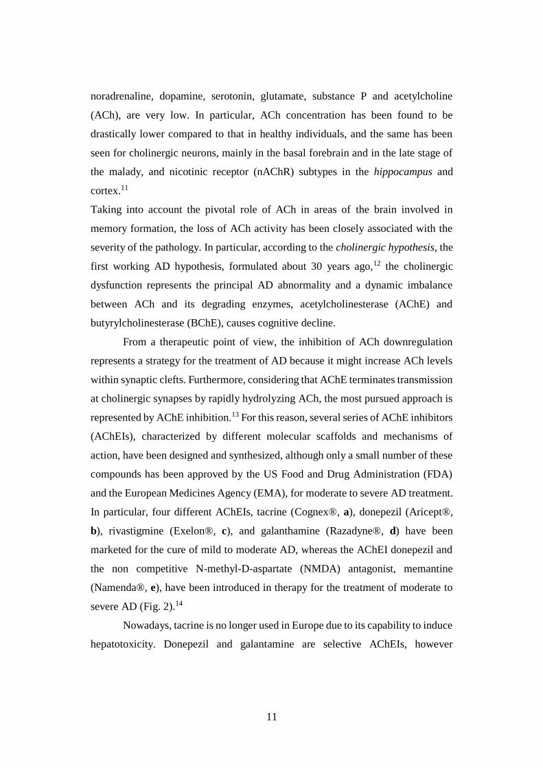
In particular, four different AChEIs, tacrine (Cognex®, a), donepezil (Aricept®,
b), rivastigmine (Exelon®, c), and galanthamine (Razadyne®, d) have been
marketed for the cure of mild to moderate AD, whereas the AChEI donepezil and
the non competitive N-methyl-D-aspartate (NMDA) antagonist, memantine
(Namenda®, e), have been introduced in therapy for the treatment of moderate to
severe AD (Fig. 2).14
Nowadays, tacrine is no longer used in Europe due to its capability to induce
hepatotoxicity. Donepezil and galantamine are selective AChEIs, however
12
galantamine is endowed with an additional mechanism of action based on an
allosteric modulation of the nicotinic receptors responsible of promoting ACh
presynaptic release and postsynaptic neurotransmission.15 Furthermore,
rivastigmine inhibits BChE, which is about 10 % of the total cholinesterases in
normal human brains and is mainly associated with glial cells. Memantine,
approved in Europe in February 2002, is able to protect neurons from glutamate-
mediated excitotoxicity, without preventing the physiological activation of the
NMDA receptor; its introduction in therapy is justified by the fact that in AD
pathogenesis an increment of extracellular glutamate levels is thought to induce an
excessive activation of NMDA receptors, which ultimately leads to neuronal death
for Ca2+ intracellular accumulation.14
Figure 2. Commercially available drugs for AD treatment.
Unfortunately, all the current approved AD therapies offer only temporary
and incomplete symptomatic relief, and represent a palliative tool by which to slow
down the clinical course of the disease.8,16
Despite the considerable advances in the understanding of the molecular and
cellular changes associated with AD pathology and the enormous progresses in the
medicinal chemistry field, no practical treatments have been introduced over the
past quarter of a century. In this context, the Pharmaceutical Research and
13
Manufacturers of America (PhRMA), an industry trade group, reported 123 failures
and only four new medicines approved to treat AD symptoms since 1998.
Thus, the discovery of disease-modifying agents able to control both onset and
progression of the neurodegenerative process is a goal of increasing urgency.17
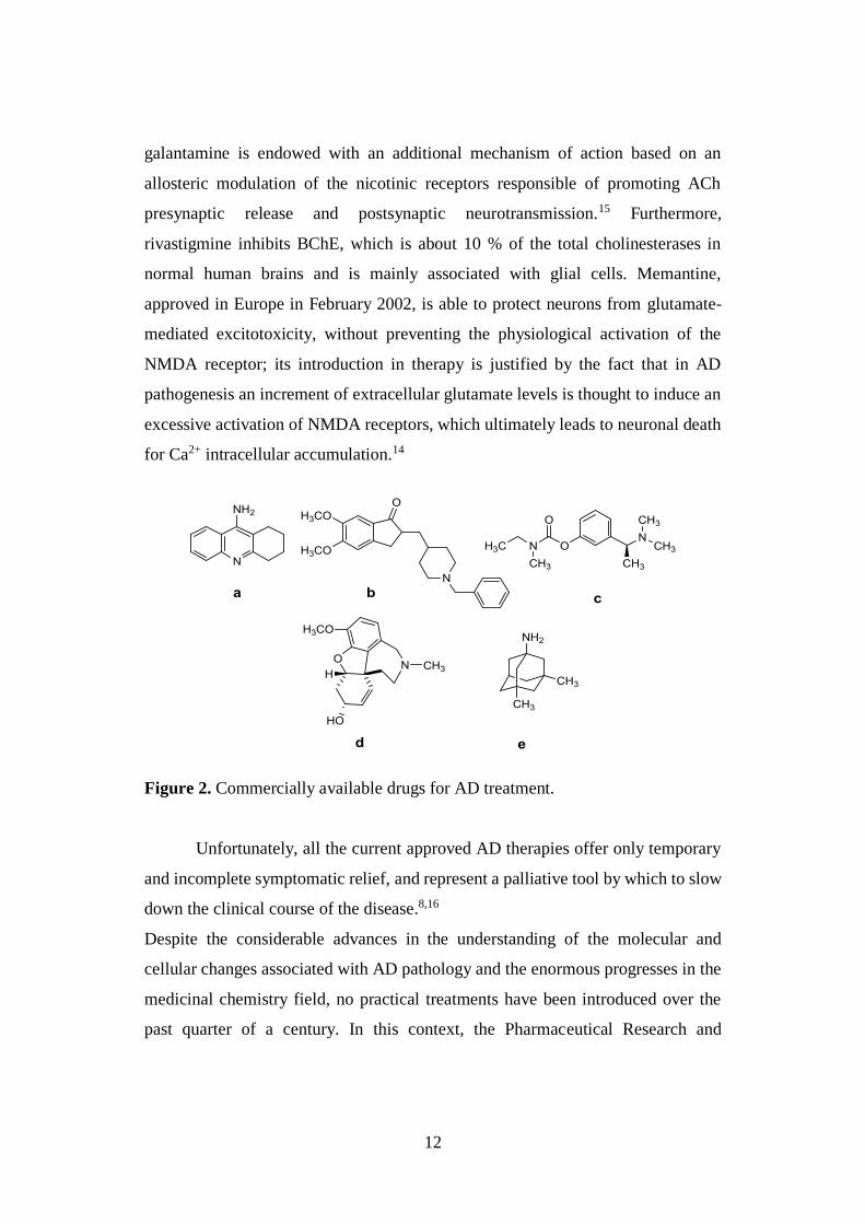
1.2. MULTIFACTORIAL NATURE OF AD
The cause or causes of AD are not yet known, nonetheless available
evidence suggests that both Aβ peptides and hyperphosphorylated τ protein play
crucial roles in its development. Accumulating evidence proposes that compared to
a linear model of AD pathogenesis, which begins with a single factor, such as β-
amyloid (e.g. amyloid hypothesis), AD pathogenesis is better explained by the idea
that a complex network of events, including neuroinflammation, oxidative stress,
reduced energy metabolism, and decreased synaptic function, interact in a feed-
forward loop (Fig. 3).
Figure 3. Complex heterogeneous view of AD pathogenesis.18
Aging is the most significant risk factor for the late onset of AD. Normally,
several feedback homeostatic mechanisms automatically block neuroinflammation
and oxidative stress. During aging, such mechanisms are less robust, resulting in a
14
sustained inflammatory environment in the brain that can trigger oxidative stress
and inhibit synaptic transmission, causing synaptic dysfunction. In this pathological
scenario, neuroinflammation and oxidative stress also result in altered mitochondria
and impaired energy metabolism. Each of these pathological events promote other
pathological features, resulting in the progressive cognitive decline observed in
AD.18
This new heterogeneous view of the malady, emphasizing the important role
of neuroinflammation and oxidative stress, allowed to identify additional pathways
and molecular targets, involved in both onset and progression of the disease,
interplaying with the well known and accredited cholinergic, amyloid and tau
hypothesis.
1.3. THE AMYLOID CASCADE HYPOTHESIS
One of the pathological hallmarks of AD are extracellular deposits of fibrous
protein aggregates, called SPs, in brain regions responsible for cognitive functions,
such as hippocampus and association cortices. In particular, SPs, isolated by
Glenner and Wong in 1984, consist of fibrillary β-pleated structures composed by
Aβ peptides. Among these peptides of 39-43 aminoacids length, Aβ42 is
predominant and presents the highest tendency to aggregate. According to the
amyloid cascade hypothesis, proposed more than 25 years ago,19 Aβ42 itself and its
aggregates are able to trigger a neurotoxic cascade, playing thus an early and crucial
role in the onset and development of AD.
Aβ42 is generated by an anomalous proteolytic processing of the amyloid β
precursor protein (APP),20 a type I transmembrane protein, conserved and
expressed in many tissues. In the central nervous system (CNS), the APP most
abundant isoform consists of 695 aminoacids and is highly concentrated at the
synaptic cleft. The precise physiological role of this protein remains uncertain,
although it is supposed to be involved in several physiological processes, such as
cell growth, neurite outgrowth, cell adhesion, cell signaling, and cell survival.21
15
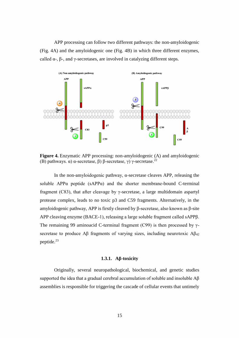
APP processing can follow two different pathways: the non-amyloidogenic
(Fig. 4A) and the amyloidogenic one (Fig. 4B) in which three different enzymes,
called α-, β-, and γ-secretases, are involved in catalyzing different steps.
Figure 4. Enzymatic APP processing: non-amyloidogenic (A) and amyloidogenic
(B) pathways. α) α-secretase, β) β-secretase, γ) γ-secretase.22
In the non-amyloidogenic pathway, α-secretase cleaves APP, releasing the
soluble APPα peptide (sAPPα) and the shorter membrane-bound C-terminal
fragment (C83), that after cleavage by γ-secretase, a large multidomain aspartyl
protease complex, leads to no toxic p3 and C59 fragments. Alternatively, in the
amyloidogenic pathway, APP is firstly cleaved by β-secretase, also known as β-site
APP cleaving enzyme (BACE-1), releasing a large soluble fragment called sAPPβ.
The remaining 99 aminoacid C-terminal fragment (C99) is then processed by γ-
secretase to produce Aβ fragments of varying sizes, including neurotoxic Aβ42
peptide.23
1.3.1. Aβ-toxicity
Originally, several neuropathological, biochemical, and genetic studies
supported the idea that a gradual cerebral accumulation of soluble and insoluble Aβ
assemblies is responsible for triggering the cascade of cellular events that untimely
16
result in the clinical AD phenotype. Actually, it is very difficult to define the nature
of the neurotoxic Aβ species, because studies show that both Aβ monomers, and
their aggregation products namely soluble oligomers, protofibrils, and insoluble
amyloid fibrils can accumulate in the brain. In this context, ever-increased studies
have identified the soluble Aβ oligomers as the most toxic species, capable of
negatively affecting synaptic integrity and inducing memory functional deficits.24
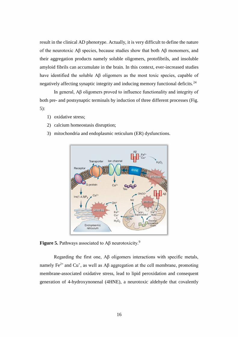
In general, Aβ oligomers proved to influence functionality and integrity of
both pre- and postsynaptic terminals by induction of three different processes (Fig.
5):
1) oxidative stress;
2) calcium homeostasis disruption;
3) mitochondria and endoplasmic reticulum (ER) dysfunctions.
Figure 5. Pathways associated to Aβ neurotoxicity.9
Regarding the first one, Aβ oligomers interactions with specific metals,
namely Fe2+ and Cu+, as well as Aβ aggregation at the cell membrane, promoting
membrane-associated oxidative stress, lead to lipid peroxidation and consequent
generation of 4-hydroxynonenal (4HNE), a neurotoxic aldehyde that covalently
17
modifies several proteins such as membrane transporters (ion-motive ATPases, a
glucose transporter and a glutamate transporter), receptors, GTP-binding proteins
(“G proteins”), ion channels (VDCC, voltage-dependent chloride channel,
NMDAR, N-methyl-D-aspartate receptor) and also τ protein, promoting its
subsequent aggregation in NFTs.
In addition, Aβ oligomers inducing mitochondrial oxidative stress and
dysregulation of Ca2+ homeostasis, cause impairment of the electron transport
chain, decrease of adenosine 5'-triphosphate (ATP) production and increment of
superoxide anion radical (O2•‾) levels. These last reactive oxygen species (ROS)
can in turn produce peroxynitrites interacting with nitric oxide (NO) and/or H2O2
by superoxide dismutase (SOD) activity. The final interaction of H2O2 with Fe2+ or
Cu+ generates the hydroxyl radical (HO•), a highly reactive oxyradical and potent
inducer of membrane-associated oxidative stress, that contributes to ER
dysfunction.
Afterward, neurotoxic forms of Aβ proved to induce neuronal death
triggering the apoptotic cascade through different mechanisms9 and may exert their
neurotoxic effects in a variety of additional ways including the interaction with the
Aβ-binding alcohol dehydrogenase protein (ABAD, molecular linker between Aβ
and mitochondrial toxicity in AD),25 stimulation of the stress-activated protein
kinases (SAPK) pathways,26 and/or activation of the microglial cells with
consequent induction of pro-inflammatory genes expression.
1.3.2. BACE-1
The recognition of Aβ as the main cause of neurodegeneration in AD and
the genetic evidence that links AD pathology with the APP proteolytic processing,
focused the interest of the scientific community for the three enzymes responsible
of APP cleavage: α-, β- and γ-secretases. Among them BACE-1, catalyzing the first
and rate-limiting step of the amyloid APP proteolysis, has aroused particular
interest and become a valuable target supporting the amyloid hypothesis.
18
Originally, five different research groups simultaneously identified this
beta-site APP-cleaving enzyme (BACE-1), also defined as Asp2 and memapsin, as
a type I integral membrane protein, composed by 501 aminoacid residues,
belonging to the pepsin-like A1 family of aspartic proteases. The enzyme is
characterized by a protease domain facing the lumen/extracellular space in the same
orientation as its substrate, APP, it is highly glycosylate and synthesized with a
prosequence rapidly removed during Golgi transit by a furin-like convertase.
Although, a high degree of identity with its homologue BACE-2 was observed, a
number of data concerning substrate specificity, as well as tissue and cellular
distribution, lead to recognize BACE-1 as the main β-secretase in the brain.27
BACE-1 presents the classical bilobal structure of the mammalian aspartyl
proteases, but characterizes a novel subgroup of this family, being the first reported
aspartic protease characterized by a transmembrane domain, a C-terminal
cytoplasmic tail28 and a unique disulphide bridge distribution.29 The crystal
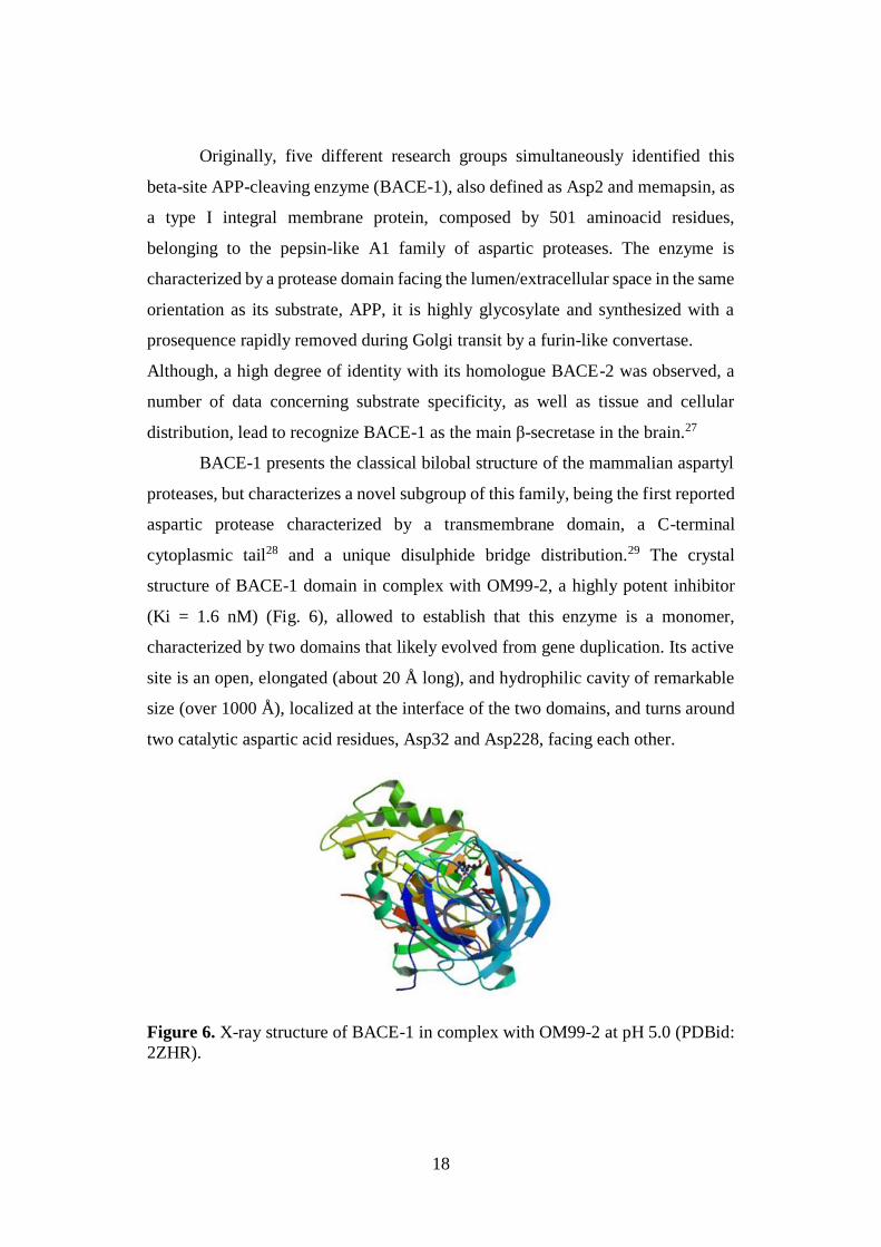
structure of BACE-1 domain in complex with OM99-2, a highly potent inhibitor
(Ki = 1.6 nM) (Fig. 6), allowed to establish that this enzyme is a monomer,
characterized by two domains that likely evolved from gene duplication. Its active
site is an open, elongated (about 20 Å long), and hydrophilic cavity of remarkable
size (over 1000 Å), localized at the interface of the two domains, and turns around
two catalytic aspartic acid residues, Asp32 and Asp228, facing each other.
Figure 6. X-ray structure of BACE-1 in complex with OM99-2 at pH 5.0 (PDBid:
2ZHR).
19
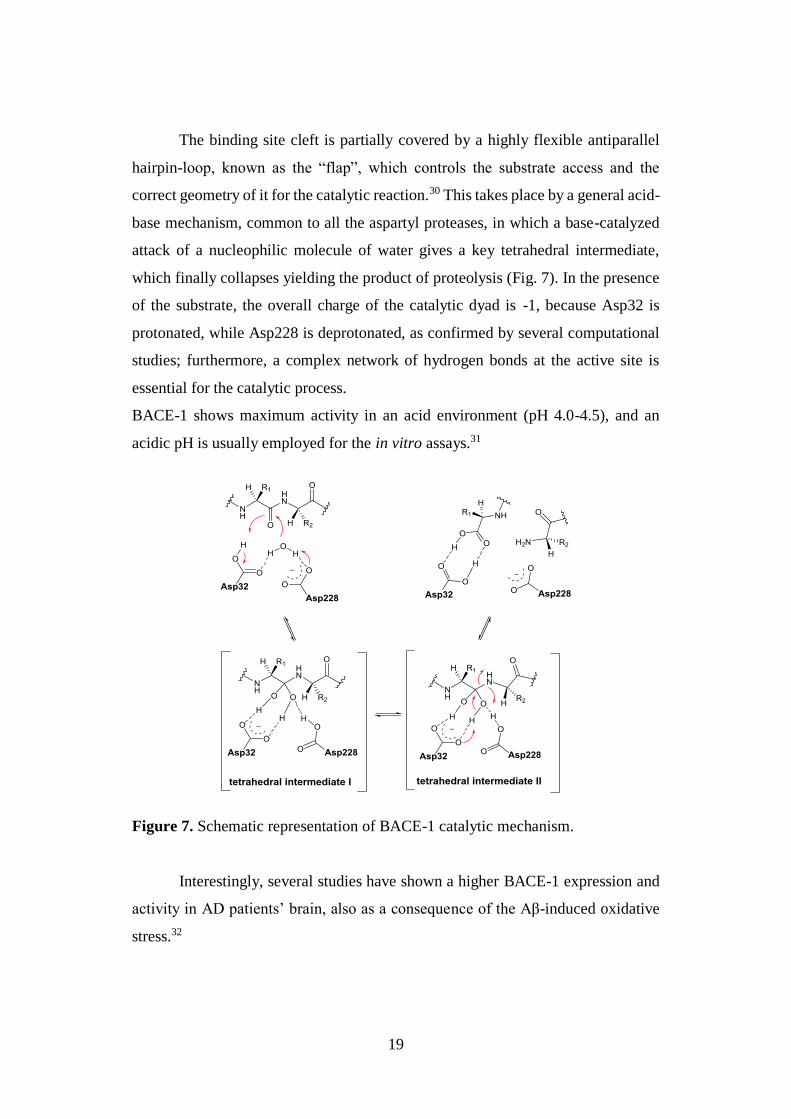
The binding site cleft is partially covered by a highly flexible antiparallel
hairpin-loop, known as the “flap”, which controls the substrate access and the
correct geometry of it for the catalytic reaction.30 This takes place by a general acid-
base mechanism, common to all the aspartyl proteases, in which a base-catalyzed
attack of a nucleophilic molecule of water gives a key tetrahedral intermediate,
which finally collapses yielding the product of proteolysis (Fig. 7). In the presence
of the substrate, the overall charge of the catalytic dyad is -1, because Asp32 is
protonated, while Asp228 is deprotonated, as confirmed by several computational
studies; furthermore, a complex network of hydrogen bonds at the active site is
essential for the catalytic process.
BACE-1 shows maximum activity in an acid environment (pH 4.0-4.5), and an
acidic pH is usually employed for the in vitro assays.31
Figure 7. Schematic representation of BACE-1 catalytic mechanism.
Interestingly, several studies have shown a higher BACE-1 expression and
activity in AD patients’ brain, also as a consequence of the Aβ-induced oxidative
stress.32
20
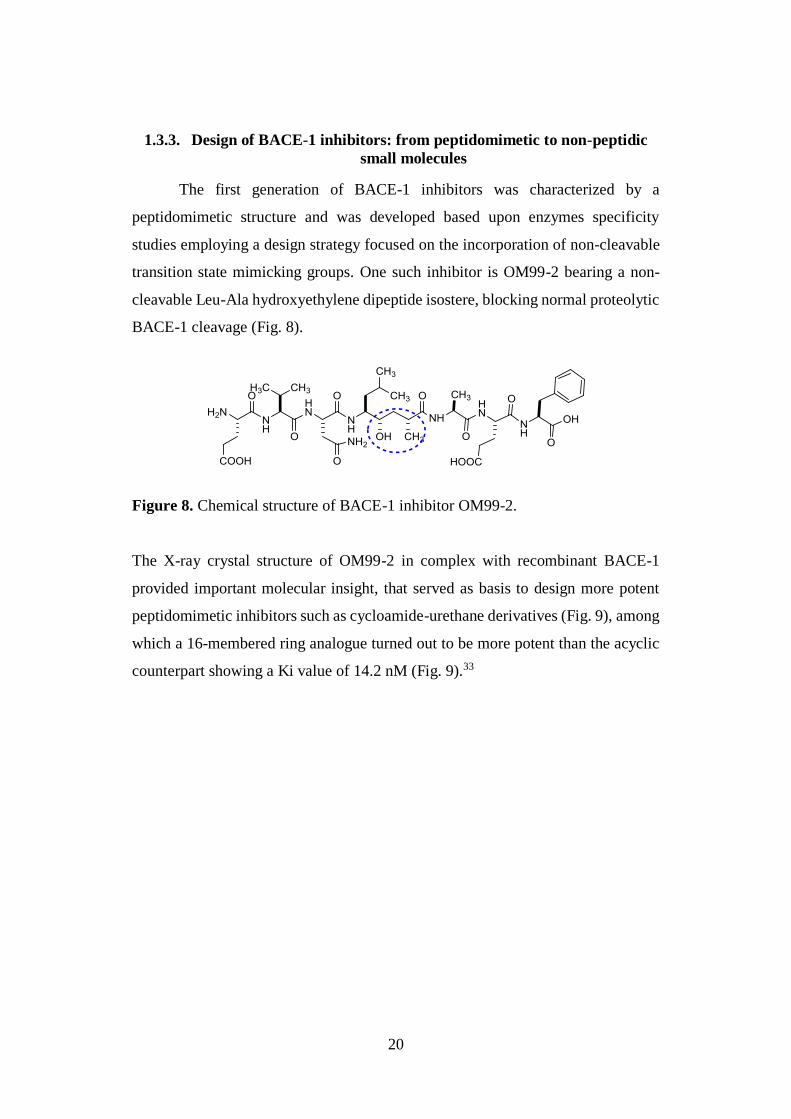
1.3.3. Design of BACE-1 inhibitors: from peptidomimetic to non-peptidic
small molecules
The first generation of BACE-1 inhibitors was characterized by a
peptidomimetic structure and was developed based upon enzymes specificity
studies employing a design strategy focused on the incorporation of non-cleavable
transition state mimicking groups. One such inhibitor is OM99-2 bearing a non-
cleavable Leu-Ala hydroxyethylene dipeptide isostere, blocking normal proteolytic
BACE-1 cleavage (Fig. 8).
Figure 8. Chemical structure of BACE-1 inhibitor OM99-2.
The X-ray crystal structure of OM99-2 in complex with recombinant BACE-1
provided important molecular insight, that served as basis to design more potent
peptidomimetic inhibitors such as cycloamide-urethane derivatives (Fig. 9), among
which a 16-membered ring analogue turned out to be more potent than the acyclic
counterpart showing a Ki value of 14.2 nM (Fig. 9).33
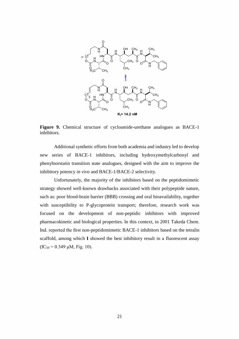
21
Figure 9. Chemical structure of cycloamide-urethane analogues as BACE-1
inhibitors.
Additional synthetic efforts from both academia and industry led to develop
new series of BACE-1 inhibitors, including hydroxymethylcarbonyl and
phenylnorstatin transition state analogues, designed with the aim to improve the
inhibitory potency in vivo and BACE-1/BACE-2 selectivity.
Unfortunately, the majority of the inhibitors based on the peptidomimetic
strategy showed well-known drawbacks associated with their polypeptide nature,
such as: poor blood-brain barrier (BBB) crossing and oral bioavailability, together
with susceptibility to P-glycoprotein transport; therefore, research work was
focused on the development of non-peptidic inhibitors with improved
pharmacokinetic and biological properties. In this context, in 2001 Takeda Chem.
Ind. reported the first non-peptidomimetic BACE-1 inhibitors based on the tetralin
scaffold, among which I showed the best inhibitory result in a fluorescent assay
(IC50 = 0.349 μM, Fig. 10).
22
Figure 10. Chemical structure of the most potent tetralin-based BACE-1 inhibitor.
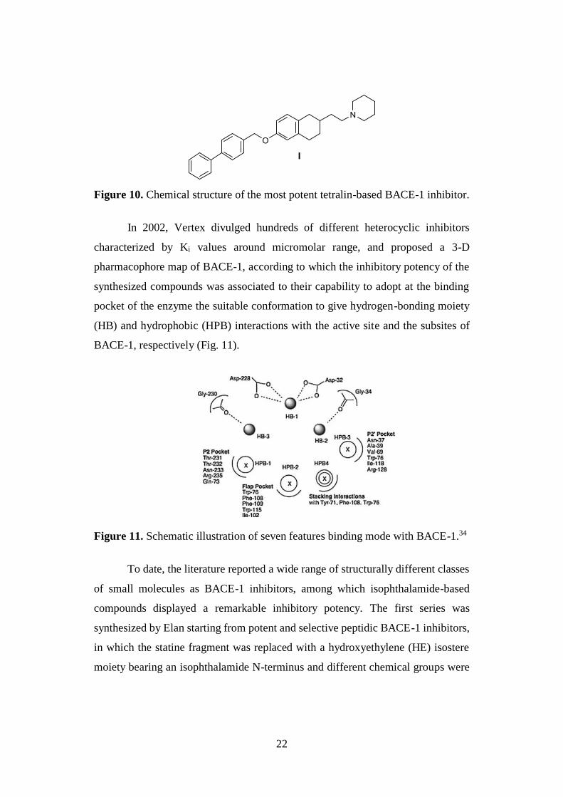
In 2002, Vertex divulged hundreds of different heterocyclic inhibitors
characterized by Ki values around micromolar range, and proposed a 3-D
pharmacophore map of BACE-1, according to which the inhibitory potency of the
synthesized compounds was associated to their capability to adopt at the binding
pocket of the enzyme the suitable conformation to give hydrogen-bonding moiety
(HB) and hydrophobic (HPB) interactions with the active site and the subsites of
BACE-1, respectively (Fig. 11).
Figure 11. Schematic illustration of seven features binding mode with BACE-1.34
To date, the literature reported a wide range of structurally different classes
of small molecules as BACE-1 inhibitors, among which isophthalamide-based
compounds displayed a remarkable inhibitory potency. The first series was
synthesized by Elan starting from potent and selective peptidic BACE-1 inhibitors,
in which the statine fragment was replaced with a hydroxyethylene (HE) isostere
moiety bearing an isophthalamide N-terminus and different chemical groups were
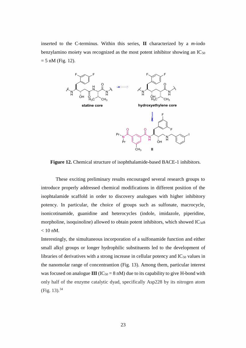
23
inserted to the C-terminus. Within this series, II characterized by a m-iodo
benzylamino moiety was recognized as the most potent inhibitor showing an IC50
= 5 nM (Fig. 12).
Figure 12. Chemical structure of isophthalamide-based BACE-1 inhibitors.
These exciting preliminary results encouraged several research groups to
introduce properly addressed chemical modifications in different position of the
isophtalamide scaffold in order to discovery analogues with higher inhibitory
potency. In particular, the choice of groups such as sulfonate, macrocycle,
isonicotinamide, guanidine and heterocycles (indole, imidazole, piperidine,
morpholine, isoquinoline) allowed to obtain potent inhibitors, which showed IC50s
< 10 nM.
Interestingly, the simultaneous incorporation of a sulfonamide function and either
small alkyl groups or longer hydrophilic substituents led to the development of
libraries of derivatives with a strong increase in cellular potency and IC50 values in
the nanomolar range of concentrantion (Fig. 13). Among them, particular interest
was focused on analogue III (IC50 = 8 nM) due to its capability to give H-bond with
only half of the enzyme catalytic dyad, specifically Asp228 by its nitrogen atom
(Fig. 13).34
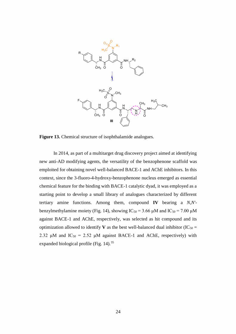
24
Figure 13. Chemical structure of isophthalamide analogues.
In 2014, as part of a multitarget drug discovery project aimed at identifying
new anti-AD modifying agents, the versatility of the benzophenone scaffold was
emploited for obtaining novel well-balanced BACE-1 and AChE inhibitors. In this
context, since the 3-fluoro-4-hydroxy-benzophenone nucleus emerged as essential
chemical feature for the binding with BACE-1 catalytic dyad, it was employed as a
starting point to develop a small library of analogues characterized by different
tertiary amine functions. Among them, compound IV bearing a N,N'-
benzylmethylamine moiety (Fig. 14), showing IC50 = 3.66 μM and IC50 = 7.00 μM
against BACE-1 and AChE, respectively, was selected as hit compound and its
optimization allowed to identify V as the best well-balanced dual inhibitor (IC50 =
2.32 μM and IC50 = 2.52 μM against BACE-1 and AChE, respectively) with
expanded biological profile (Fig. 14).35
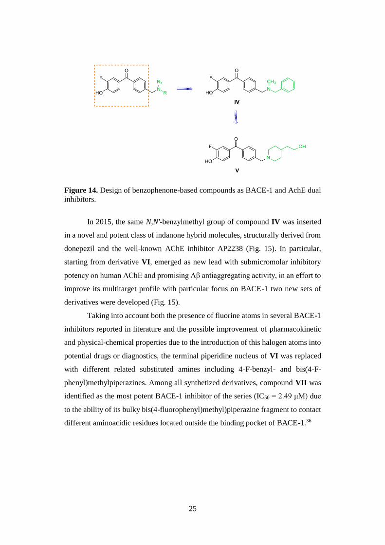
25
Figure 14. Design of benzophenone-based compounds as BACE-1 and AchE dual
inhibitors.
In 2015, the same N,N'-benzylmethyl group of compound IV was inserted
in a novel and potent class of indanone hybrid molecules, structurally derived from
donepezil and the well-known AChE inhibitor AP2238 (Fig. 15). In particular,
starting from derivative VI, emerged as new lead with submicromolar inhibitory
potency on human AChE and promising Aβ antiaggregating activity, in an effort to
improve its multitarget profile with particular focus on BACE-1 two new sets of
derivatives were developed (Fig. 15).
Taking into account both the presence of fluorine atoms in several BACE-1
inhibitors reported in literature and the possible improvement of pharmacokinetic
and physical-chemical properties due to the introduction of this halogen atoms into
potential drugs or diagnostics, the terminal piperidine nucleus of VI was replaced
with different related substituted amines including 4-F-benzyl- and bis(4-F-
phenyl)methylpiperazines. Among all synthetized derivatives, compound VII was
identified as the most potent BACE-1 inhibitor of the series (IC50 = 2.49 μM) due
to the ability of its bulky bis(4-fluorophenyl)methyl)piperazine fragment to contact
different aminoacidic residues located outside the binding pocket of BACE-1.36
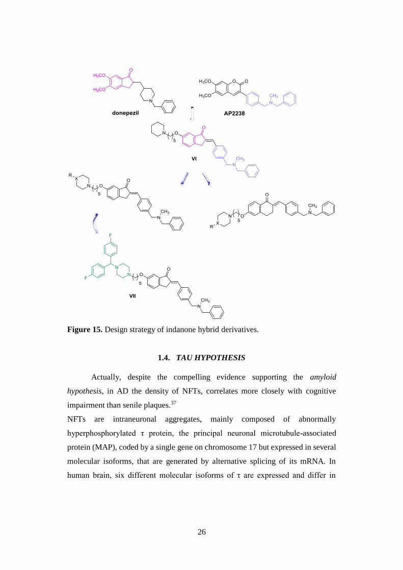
26
Figure 15. Design strategy of indanone hybrid derivatives.
1.4. TAU HYPOTHESIS
Actually, despite the compelling evidence supporting the amyloid
hypothesis, in AD the density of NFTs, correlates more closely with cognitive
impairment than senile plaques.37
NFTs are intraneuronal aggregates, mainly composed of abnormally
hyperphosphorylated τ protein, the principal neuronal microtubule-associated
protein (MAP), coded by a single gene on chromosome 17 but expressed in several
molecular isoforms, that are generated by alternative splicing of its mRNA. In
human brain, six different molecular isoforms of τ are expressed and differ in
27
containing three or four microtubule binding repeats of 31-32 aminoacids in the C-
terminal half and one, two, or zero N-terminal inserts of 29 aminoacids. Among
them, the isoform with a total of 441 aminoacids (tau441) in length is the largest size
human brain τ protein predominantly expressed in neuronal axons, although recent
studies reported that it is also expressed in glia and astrocytes.38
Under physiological conditions, stabilizes microtubules, promotes their
assembly, and affects their dynamics by interaction with tubulin. These biological
activities are finely modulated through phosphorylation in correspondence of a
specific number of Ser/Thr sites. In the longest isoform of protein, 79 potential
Ser and Thr phosphate acceptor residues have been identified, although only about
30 of them seem to act as real sites of physiological phosphorylation.
In contrast, an abnormal phosphorylation, leading to its dissociation from
microtubules and resulting in cellular cytoarchitecture disruption, and further
accumulation in paired helical filaments (PHFs), is a characteristic AD feature.
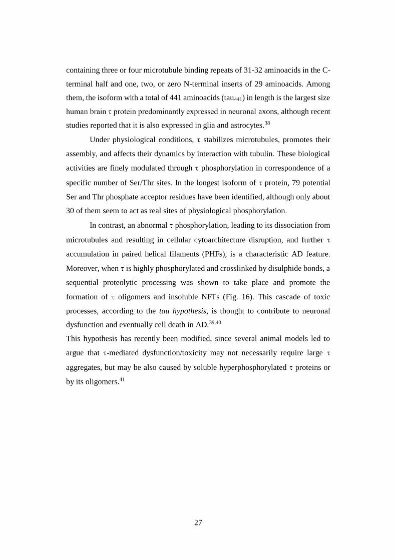
Moreover, when is highly phosphorylated and crosslinked by disulphide bonds, a
sequential proteolytic processing was shown to take place and promote the
formation of oligomers and insoluble NFTs (Fig. 16). This cascade of toxic
processes, according to the tau hypothesis, is thought to contribute to neuronal
dysfunction and eventually cell death in AD.39,40
This hypothesis has recently been modified, since several animal models led to
argue that -mediated dysfunction/toxicity may not necessarily require large
aggregates, but may be also caused by soluble hyperphosphorylated proteins or
by its oligomers.41
28
Figure 16. Intracellular neuronal aggregation of hyperphosphorylated τ protein.39
Nowadays, causal factors affecting phosphorylation and sequential
formation of NFTs are not fully understood, however, a large number of studies
revealed the critical role of Aβ and/or chronic oxidative stress in τ
hyperphosphorylation and aggregation.40 In particular, several evidence supports
that different products of oxidative stress, such as 4-HNE, together with a large
number of oxidative stress-activated kinases, namely glycogen synthase kinase-3
(GSK-3), mitogen-activated protein kinases (MAPKs), extracellular receptor
kinase (ERK), p38 MAPK and Jun-N-terminal kinase (JNK), are involved in
intracellular NFTs deposition.42
1.4.1. GSK-3
The state of τ phosphorylation is the result of the balance between protein
kinases (PKs) and phosphatases (PPs) activities. Several Ser/Thr protein kinases
have been associated to the abnormal hyperphosphorylation of protein, among
which GSK-3, cyclin dependent protein kinase 5 (CDK5), protein kinase A (PKA),
29
calcium and calmodulin-dependent protein kinase II (CaMKII), casein kinase 1
(CK1), mitogen activated protein (MAP) kinase ERK1/2, and SAPKs.
There are 518 genes that encode for more than 2000 different protein
kinases. These proteins are specific Ser, Thr or Tyr kinases and are responsible for
the regulation of several physiological processes, including cellular death and
division, transport and secretion of molecules, as well as, modulation of some brain
functions, blood pressure, metabolism and protein synthesis.43
Structurally, PKs have a highly conserved catalytic domain, nevertheless
they differ in the way in which their catalytic function is regulated. In particular,
Ser/Thr kinases can be classified as proline-directed or non-proline-directed
proteins. Within the first class, GSK-3β has been recognized as validated AD
target38,39 due to the observed link between its overactivity and/or overexpression
and the neuropathological hallmarks described for the disorder (Aβ deposition,
hyperphosphorylation, gliosis, and neuronal cells death).44
GSK-3 is involved in the regulation of several cellular processes, including
cellular division, proliferation, differentiation and adhesion. In 1980, it was isolated
from skeletal muscle and recognized as one of the five enzymes involved in
glycogen synthase phosphorylation.45 Two different isoforms of this enzyme exist,
namely GSK-3α and GSK-3β, which are similarly regulated although encoded by
different genes. GSK-3α (51 kDa) differs from isoform β (47 kDa) for the presence
of a glycine-rich extension at the N-terminal end. Both isoforms are ubiquitously
expressed in the brain, with high levels of expression in the hippocampus, cerebral
cortex, and the Purkinje cells of the cerebellum, the expression ratio of these two
isoforms favors GSK-3β (Fig. 17).
In vitro and in cell culture models both GSK-3 isoforms have shown their capacity
to phosphorylate τ at various sites, consistent with the epitopes found to be
hyperphosphorylated in AD brains. However, in several animal models
overexpression of GSK-3β proved to both induce hyperphosphorylation mainly
at Ser199, Ser396, and Ser413 and accelerate neurodegeneration, whereas an
inhibition of this enzyme led to a decrement of toxicity.46
30
Interestingly, τ overexpression promotes GSK-3β activation and mediates GSK-3β
toxicity; in τ absence, indeed, the neurodegenerative and cognitive phenotypes
observed in GSK-3-overexpressing mice is ameliorated.47
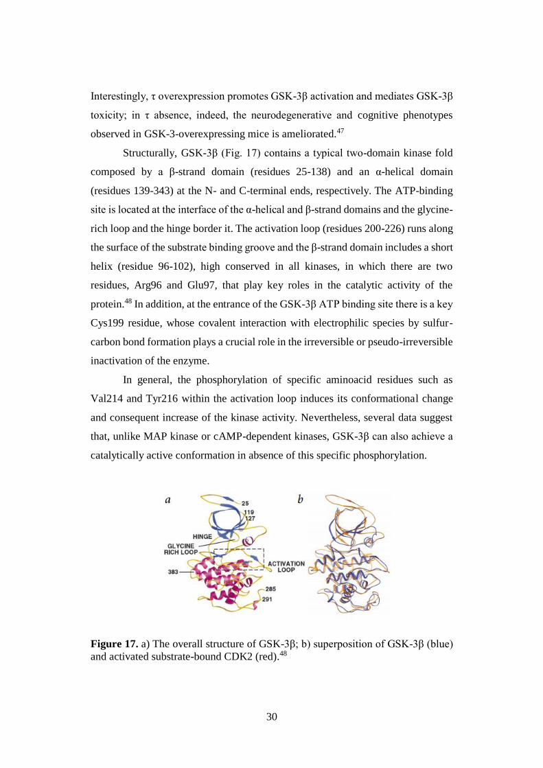
Structurally, GSK-3β (Fig. 17) contains a typical two-domain kinase fold
composed by a β-strand domain (residues 25-138) and an α-helical domain
(residues 139-343) at the N- and C-terminal ends, respectively. The ATP-binding
site is located at the interface of the α-helical and β-strand domains and the glycine-
rich loop and the hinge border it. The activation loop (residues 200-226) runs along
the surface of the substrate binding groove and the β-strand domain includes a short
helix (residue 96-102), high conserved in all kinases, in which there are two
residues, Arg96 and Glu97, that play key roles in the catalytic activity of the
protein.48 In addition, at the entrance of the GSK-3β ATP binding site there is a key
Cys199 residue, whose covalent interaction with electrophilic species by sulfur-
carbon bond formation plays a crucial role in the irreversible or pseudo-irreversible
inactivation of the enzyme.
In general, the phosphorylation of specific aminoacid residues such as
Val214 and Tyr216 within the activation loop induces its conformational change
and consequent increase of the kinase activity. Nevertheless, several data suggest
that, unlike MAP kinase or cAMP-dependent kinases, GSK-3β can also achieve a
catalytically active conformation in absence of this specific phosphorylation.
Figure 17. a) The overall structure of GSK-3β; b) superposition of GSK-3β (blue)
and activated substrate-bound CDK2 (red).48
31
Generally, GSK-3 is constitutively active, and the activation sites can
undergo autophosphorylation; furthermore, different PKs can regulate its activity
in different ways, depending on the particular site of phosphorylation. Concerning
GSK-3β, phosphorylation at Ser9 decreases the activity, while at Tyr216 leads to
enzyme overactivation. Currently, a large variety of GSK-3 regulatory pathways
are known, and their underlying molecular basis have been elucidated. Among
these, the most studied is based on Akt (protein kinase B) activation, in which
insulin stimulation, activates phosphatidylinositol 3-kinase (PI3K) and leads to Akt
(protein kinase B) phosphorylation and consequent GSK-3β inhibition. However, a
brief exposure to insulin can also transiently activate GSK-3β through
phosphorylation of Tyr216 promoted by the non-receptor tyrosine kinase Fyn.
Besides PI3K, even other kinases, including protein kinase C (PKC), are able to
inhibit GSK-3 by phosphorylation at Ser9 and, within the brain, p38 mitogen-
activated protein kinase (MAPK) proved to inactivate this enzyme by direct
phosphorylation at its C-terminus end.
An additional mechanism associated to GSK-3 activation consists in its
dephosphorylation at specific inhibitory sites by means of different phosphatases,
such as protein phosphatase 1 (PP1) for GSK-3β, protein phosphatase 2A (PP2A)
which favors GSK-3α, and protein phosphatase 2B (PP2B, calcineurin).47,49
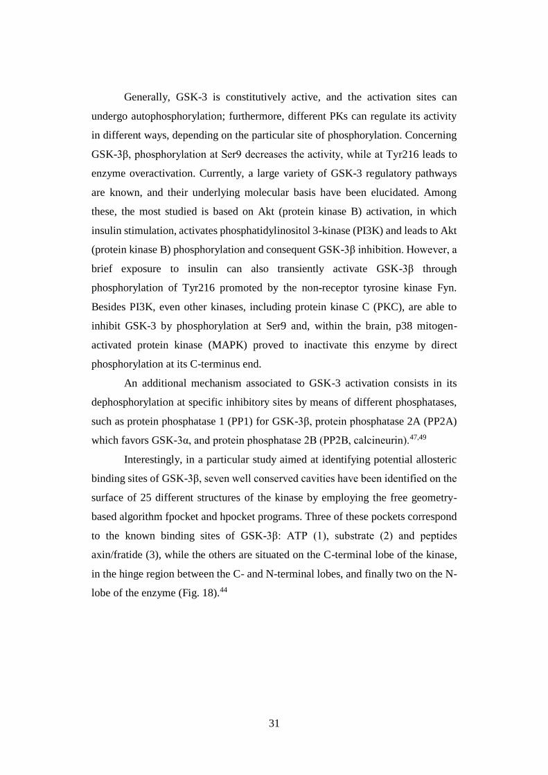
Interestingly, in a particular study aimed at identifying potential allosteric
binding sites of GSK-3β, seven well conserved cavities have been identified on the
surface of 25 different structures of the kinase by employing the free geometry-
based algorithm fpocket and hpocket programs. Three of these pockets correspond
to the known binding sites of GSK-3β: ATP (1), substrate (2) and peptides
axin/fratide (3), while the others are situated on the C-terminal lobe of the kinase,
in the hinge region between the C- and N-terminal lobes, and finally two on the N-
lobe of the enzyme (Fig. 18).44
32
Figure 18. Seven cavities found by hpocket in the 25 PDB structures of GSK-3β
analyzed independently.44
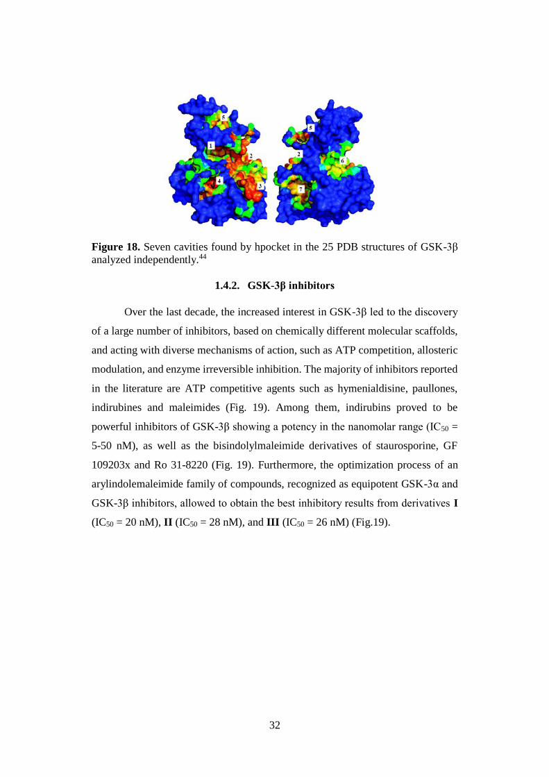
1.4.2. GSK-3β inhibitors
Over the last decade, the increased interest in GSK-3β led to the discovery
of a large number of inhibitors, based on chemically different molecular scaffolds,
and acting with diverse mechanisms of action, such as ATP competition, allosteric
modulation, and enzyme irreversible inhibition. The majority of inhibitors reported
in the literature are ATP competitive agents such as hymenialdisine, paullones,
indirubines and maleimides (Fig. 19). Among them, indirubins proved to be
powerful inhibitors of GSK-3β showing a potency in the nanomolar range (IC50 =
5-50 nM), as well as the bisindolylmaleimide derivatives of staurosporine, GF
109203x and Ro 31-8220 (Fig. 19). Furthermore, the optimization process of an
arylindolemaleimide family of compounds, recognized as equipotent GSK-3α and
GSK-3β inhibitors, allowed to obtain the best inhibitory results from derivatives I
(IC50 = 20 nM), II (IC50 = 28 nM), and III (IC50 = 26 nM) (Fig.19).
33
Figure 19. Chemical structure of ATP competitive GSK-3β inhibitors.
Generally, one of the main limitations for the therapeutic use of ATP
competitive inhibitors consists in the lack of kinase selectively, as consequence of
the high degree of PKs identity in the catalytic site. Therefore, in a potential chronic
treatment these compounds could offer adverse secondary effects.50
In contrast, non-ATP competitive inhibitors, showing better cellular and in vivo
potency in comparison with competitive inhibitors together with better kinase
selectivity, have recently attracted ever-increased attention as promising candidates
to achieve an effective treatment of chronic disorders including AD.
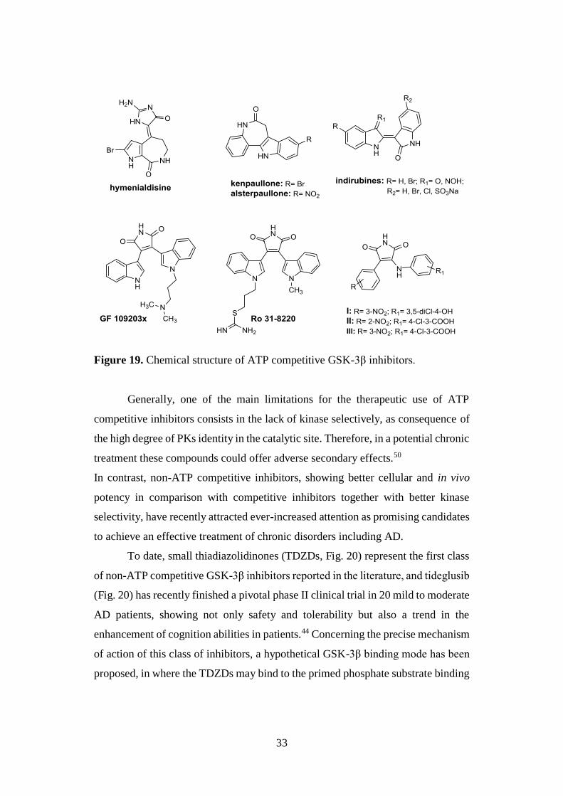
To date, small thiadiazolidinones (TDZDs, Fig. 20) represent the first class
of non-ATP competitive GSK-3β inhibitors reported in the literature, and tideglusib
(Fig. 20) has recently finished a pivotal phase II clinical trial in 20 mild to moderate
AD patients, showing not only safety and tolerability but also a trend in the
enhancement of cognition abilities in patients.44 Concerning the precise mechanism
of action of this class of inhibitors, a hypothetical GSK-3β binding mode has been
proposed, in where the TDZDs may bind to the primed phosphate substrate binding
34
site of the enzyme. In addition to them, two marine natural compounds, the alkaloid
manzamine and the sesquiterpene palinurin (Fig. 20), have been reported as cell
permeable non-ATP competitive inhibitors, able to reduce tau phosphorylation in
cell cultures. In particular, the binding site of manzamine has been recently
postulated as an allosteric site at the back of the ATP site51 and results derived from
both experimental data and molecular dynamic (MD) simulations suggested that
the palinurin allosteric site is located at the N-terminal lobe of GSK-3β (pocket 5,
Fig. 18).52 These allosteric inhibitors are likely to be more selective and may be
useful in overcoming resistance developed to ATP competitive drugs.
Furthermore, they provide more subtle modulation of kinase activity than simply
blocking ATP entrance.44
Figure 20. Chemical structure of non-ATP competitive GSK-3β inhibitors.
The irreversible GSK-3β inhibition, by selective targeting of Cys199 in the
ATP-binding site, has recently been reported as a promising strategy to minimize
the undesirable off-target effects associated with enzyme inhibition obtaining useful
pharmacological tools.8 In this context, halomethylketones (HMKs, Fig. 20),
35
irreversible inhibitors with IC50 values in the low micromolar range, have just been
reported representing valid alternative for the future design of specific and potent
inhibitors, due to their ability to decrease tau phosphorylation in cell cultures, to
cross the BBB, together with their good kinase selectivity.51
1.4.3. GSK-3β: molecular link between Aβ and τ
Although Aβ and τ exert toxic effects through separate mechanisms, several
lines of evidence, from both in vitro and in vivo models, confirm the existence of a
molecular interplay between these two proteins in causing synergic toxicity and a
cross-talk between Aβ and GSK-3β has been reported. Indeed, several studies
confirmed both the pivotal role of Aβ in driving τ pathology by a general induction
of τ hyperphosphorylation and NFTs formation, and τ aptitude to mediate Aβ-
toxicity. The interaction of these two proteins, together with their ability to amplify
each other’s toxic effects by synergistically targeting cellular processes or
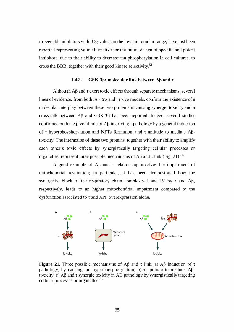
organelles, represent three possible mechanisms of Aβ and τ link (Fig. 21).53
A good example of Aβ and τ relationship involves the impairment of
mitochondrial respiration; in particular, it has been demonstrated how the
synergistic block of the respiratory chain complexes I and IV by τ and Aβ,
respectively, leads to an higher mitochondrial impairment compared to the
dysfunction associated to τ and APP overexpression alone.
Figure 21. Three possible mechanisms of Aβ and τ link; a) Aβ induction of τ
pathology, by causing tau hyperphosphorylation; b) τ aptitude to mediate Aβ-
toxicity; c) Aβ and τ synergic toxicity in AD pathology by synergistically targeting
cellular processes or organelles.53
36
Interestingly, GSK-3β represents the molecular link between Aβ and τ
neurotoxic cascades. In vitro studies and transgenic animal models of AD have been
shown as the pathologic activation of GSK-3β by Aβ, prevents inhibitory
phosphorylation of the enzyme, and leads to an increment of τ phosphorylation. On
the contrary, GSK-3β inhibition decreases Aβ production and Aβ-induced
neurotoxicity by reducing BACE-1 cleavage of APP.8 Additional investigations
also confirmed GSK-3β capability to promote the amyloidogenic APP processing
by inhibition of the α-secretase complex and interfering with APP cleavage at the
γ-secretase complex step.49
1.5. OXIDATIVE STRESS
According to the multifactorial view of AD, oxidative stress has been
recognized as a common pathological feature of the disorder. Recently,
experimental evidence indicates that a dysregulation of the redox state contributes
to the onset of the neurodegenerative process.
Commonly, oxidative stress is caused by an imbalance between reactive radical
species, among others ROS, and a loss of function of many antioxidant defense
enzymes, resulting in a disequilibrium between the formation of cellular oxidants
and the antioxidative processes.54 This impairment of the cellular redox balance
leads to oxidative alterations of biological macromolecules, such as proteins, lipids,
and nucleic acids, identified as biochemical markers in AD brains. In addition,
modifications in the activities or expression of antioxidant enzymes such as SOD
and catalase have been observed in both CNS and peripheral tissues of AD
patients.55
ROS, namely O2•‾, H2O2 and •OH, are small biological molecules, which are
continuously produced in aerobic organisms such as a natural by-product of oxygen
metabolism. Although they play an important physiological role in cell signaling,
the long-term exposure of cells to higher levels of ROS leads to toxic effects. ROS
production occurs largely in mitochondria, starting from O2•‾, the product of one-
electron reduction of oxygen that is converted, spontaneously or enzymatically by
37
SOD, into H2O2 and oxygen (Scheme 1b). H2O2 can easily diffuse across the
biological membranes and damage essential macromolecules.56 Alternatively, O2•‾
can react with nitric oxide producing highly toxic peroxynitrite.
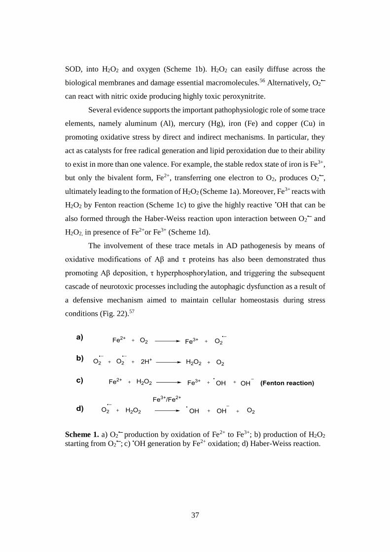
Several evidence supports the important pathophysiologic role of some trace
elements, namely aluminum (Al), mercury (Hg), iron (Fe) and copper (Cu) in
promoting oxidative stress by direct and indirect mechanisms. In particular, they
act as catalysts for free radical generation and lipid peroxidation due to their ability
to exist in more than one valence. For example, the stable redox state of iron is Fe3+,
but only the bivalent form, Fe2+, transferring one electron to O2, produces O2•‾,
ultimately leading to the formation of H2O2 (Scheme 1a). Moreover, Fe3+ reacts with
H2O2 by Fenton reaction (Scheme 1c) to give the highly reactive •OH that can be
also formed through the Haber-Weiss reaction upon interaction between O2•‾ and
H2O2, in presence of Fe2+or Fe3+ (Scheme 1d).
The involvement of these trace metals in AD pathogenesis by means of
oxidative modifications of Aβ and τ proteins has also been demonstrated thus
promoting Aβ deposition, τ hyperphosphorylation, and triggering the subsequent
cascade of neurotoxic processes including the autophagic dysfunction as a result of
a defensive mechanism aimed to maintain cellular homeostasis during stress
conditions (Fig. 22).57
Scheme 1. a) O2•‾ production by oxidation of Fe2+ to Fe3+; b) production of H2O2
starting from O2•‾; c) •OH generation by Fe2+ oxidation; d) Haber-Weiss reaction.
38
Concerning Aβ oxidative changes, the Butterfield group demonstrated in
1994 the crucial role of the metal-induced oxidation of the sulphur atom of Met35
residue in Aβ42, in imparting pro-oxidant and neurotoxic properties in vitro, while
Greenough et al. alternatively proposed a direct binding of metal ions (Cu and Fe)
to Aβ peptide as possible mechanism of Aβ pro-oxidant activity.58
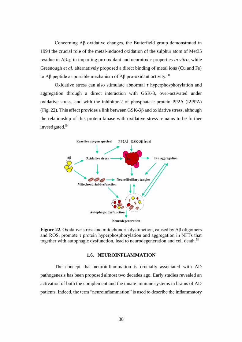
Oxidative stress can also stimulate abnormal τ hyperphosphorylation and
aggregation through a direct interaction with GSK-3, over-activated under
oxidative stress, and with the inhibitor-2 of phosphatase protein PP2A (I2PPA)
(Fig. 22). This effect provides a link between GSK-3β and oxidative stress, although
the relationship of this protein kinase with oxidative stress remains to be further
investigated.54
Figure 22. Oxidative stress and mitochondria dysfunction, caused by Aβ oligomers
and ROS, promote τ protein hyperphosphorylation and aggregation in NFTs that
together with autophagic dysfunction, lead to neurodegeneration and cell death.54
1.6. NEUROINFLAMMATION
The concept that neuroinflammation is crucially associated with AD
pathogenesis has been proposed almost two decades ago. Early studies revealed an
activation of both the complement and the innate immune systems in brains of AD
patients. Indeed, the term “neuroinflammation” is used to describe the inflammatory
39
response originated in the injured CNS, characterized by an accumulation of glial
cells, i.e., microglia and astrocytes, aimed at repairing damaged area. Nevertheless,
in presence of a persistent stimulus, a chronic inflammatory condition develops
causing cumulative damages. All these events generate complex interactions and
feedback loops between glial and neuronal cells, and lead to neurodegeneration.59
Microglia can be readily activated producing beneficial functions, essential
to neuron survival, through the release of trophic and antiinflammatory factors.
However, under particular circumstances, including chronic inflammation, it turn
out to be overactivated and it can induce neurotoxicity through the production of
pro-inflammatory cytokines, namely IL-1β, IL-6, IL-12, interferon gamma (INF-γ)
and tumor necrosis factor-α (TNF-α), and the synthesis and the release of several
cytotoxic factors such as O2•‾, nitric oxide (NO) and ROS.18
In AD, microglial cells are able to respond to various stimuli, including Aβ
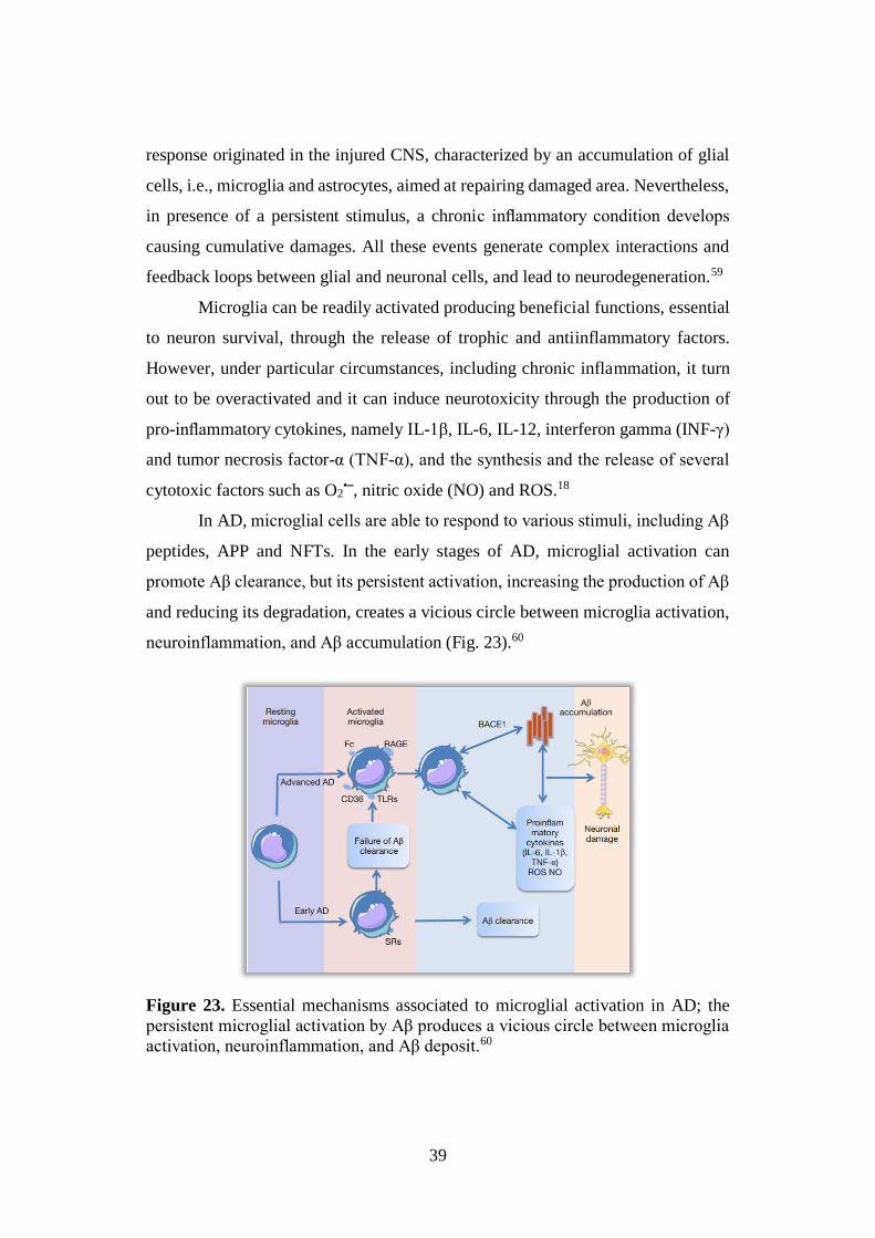
peptides, APP and NFTs. In the early stages of AD, microglial activation can
promote Aβ clearance, but its persistent activation, increasing the production of Aβ
and reducing its degradation, creates a vicious circle between microglia activation,
neuroinflammation, and Aβ accumulation (Fig. 23).60
Figure 23. Essential mechanisms associated to microglial activation in AD; the
persistent microglial activation by Aβ produces a vicious circle between microglia
activation, neuroinflammation, and Aβ deposit.60
40
Interestingly, small diffusible Aβ oligomers activate microglia, leading to a
more potent induction of inflammation, whereas fibrillary Aβ or SPs maintain the
chronic inflammation characteristic of the terminal stage of AD pathogenesis. In
fact, both pro-inflammatory cytokines and oxidative damage are observed early in
AD progression and can be identified prior to fibrillary Aβ deposition in AD brain.
Furthermore, Aβ oligomers proved to be able to damage microglial phagocytic
function, in particular the terminal Aβ fibrils clearance, providing a probable
explanation for the inability of “activated” microglia surrounding plaques to
phagocytize Aβ deposits during the terminal stage of AD.
Taking into account these intriguingly findings, it has been proposed that,
in AD, Aβ oligomers first trigger an acute inflammation, and subsequently Aβ
fibrils sustain a chronic inflammatory environment, which inhibits the activation of
the phagocytic machinery, inducing a secondary immune response and worsening
brain inflammation. In this context, cytokines, as mediators of the so-called
secondary damage, may in turn promote Aβ production through β- and γ-secretases
stimulation and/or reduce Aβ clearance.61
Pathological tau aggregates are also able to induce microglial activation
triggering the events of the neuroinflammatory cascade. In particular, after neuronal
death, these aggregates are released into the extracellular medium causing
activation of microglia and generation of a cascade of toxic signals. Moreover,
several evidence suggests that a peripheral sustained inflammation can cause
breakdown of the BBB, exposure of brain parenchyma to serum proteins, microglia
activation and consequent release of inflammatory mediators that can contribute to
cognitive decline in AD patients.18
1.7. NRF2-KEAP1: A NEUROPROTECTIVE SIGNALING PATHWAY
In the early stage of AD pathology, as a result of a defensive mechanism, a
battery of genes with detoxificant, antioxidant and antiinflammatory capacities
have been found to be remarkably increased, while they decrease at a later stage.
41
In particular, several postmortem studies of AD patients’ temporal cortex and
hippocampus showed that the percentage of astrocytes expressing heme oxygenase-
1 (HO-1), a cytoprotective microsomial enzyme that catalyses the degradation of
the heme group to yield biliverdin, iron and carbon monoxide,62 was significantly
higher than in non-demented individuals. In additional studies, the activity and
expression of NAD(P)H: quinone oxidoreductase 1 (NQO1), a detoxifying phase II
enzyme that catalyzes the reduction of quinones to hydroquinones and scavenges
superoxide molecules, were increased in neurons and astrocytes to reduce the
highly oxidant environment typical of AD brains.63 These intriguing findings lead
to propose the NQO1 upregulation as a first indicator of the pathology.64
Furthermore, most of the postmortem analyses of AD brains report depleted levels
of gluthatione (GSH: 1 glutamyl-cysteinyl-glycine), the main endogenous
antioxidant enzyme responsible for ROS detoxification and regulation of the
intracellular redox environment, suggesting a correlation between AD pathology
and reduced GSH levels.65
Although the mechanisms underlying the fluctuations in these enzymes
contents in AD have not been yet clarified, impairment of some pathways involved
in their expression might be associated with the progression of the disease. In this
context, particular attention was focused on the Nrf2-Keap1-ARE pathway (Fig.
24) recognized as the major regulator of cytoprotective responses to endogenous
and exogenous stresses caused by ROS, electrophiles, and inflammation and the
main determinant of phase II gene induction, including NQO1, HO-1 and
glutathione-S-transferase (GST), a key enzyme that catalyzes the reaction between
GSH and nucleophilic compounds.
Nuclear factor erythroid 2-related factor 2 (Nrf2) is a redox-sensitive transcription
factor belonging to a protein family characterized by a conserved basic region
leucine zipper (bZip) dimerization domain, and ubiquitously expressed in the body,
including the CNS. It regulates the basal expression of some cytoprotective genes
through interaction with a cis-acting enhancer sequence, namely antioxidant-
42
response element (ARE) or electrophile-responsive element (EpRE), located in
their promoters.66
Under basal conditions, Nrf2 remains in the cytoplasm, associated with the
actin cytoskeleton through the repressor Kelch-like ECH-associated protein 1
(Keap1), an adaptor protein that, connecting Nrf2 to Cullin (Cul) 3, promotes Nrf2
degradation by the proteasome (Fig. 24). Nrf2 contains two Keap1 binding sites,
DLG and ETGE, within the Neh2 regulatory N-terminus domain, suitable for the
formation of a complex with two molecules of Keap1 (Fig. 24). This, in turn, is
characterized by two known protein-interacting domains: the BTB (bric-a-brac,
tramtrack, broad-complex) domain in the N-terminal region and the Kelch repeats
in the C-terminal region [Kelch repeat, double glycine repeat (DGR) domain]. The
BTB domain mediates Keap1 homodimerization and binding to Cul3, while DGR
favors the binding of the repressor with Nrf2 Neh2 domain. Between BTB and DGR
is the intervening region (IVR) or linker region (LR) rich in cysteine residues (27
in human Keap1), some of which reactive and critical for Nrf2 modulation.
Originally, a study by Dinkova-Kostova et al. identified Cys257, Cys273, Cys288,
and Cys297 as the cysteine residues of Keap1 mediating Nrf2 activation.
Subsequent independent studies confirmed the central role of Cys273, Cys288, and
Cys151 in Nrf2 modulation and defined Keap1 as a sensor protein responding to
oxidative and environmental stresses through dynamic changes in the reducing
status of these cysteine residues.67
Under stress conditions and in the presence of different chemicals, including
phytochemicals and derivatives (sulforaphane, SFN), therapeutics (oltipraz,
auranofin), environmental agents (paraquat, arsenic), endogenous chemicals (NO,
nitro-fatty acids, and 4-HNE) and electrophilic compounds, the Nrf2-Keap1
pathway is activated with consequent Keap1 detachment from Nrf2, translocation
of the latter in the nucleus and induction of cytoprotective gene expression through
Nrf2 binding to DNA assisted by small Maf proteins (Fig. 24).68
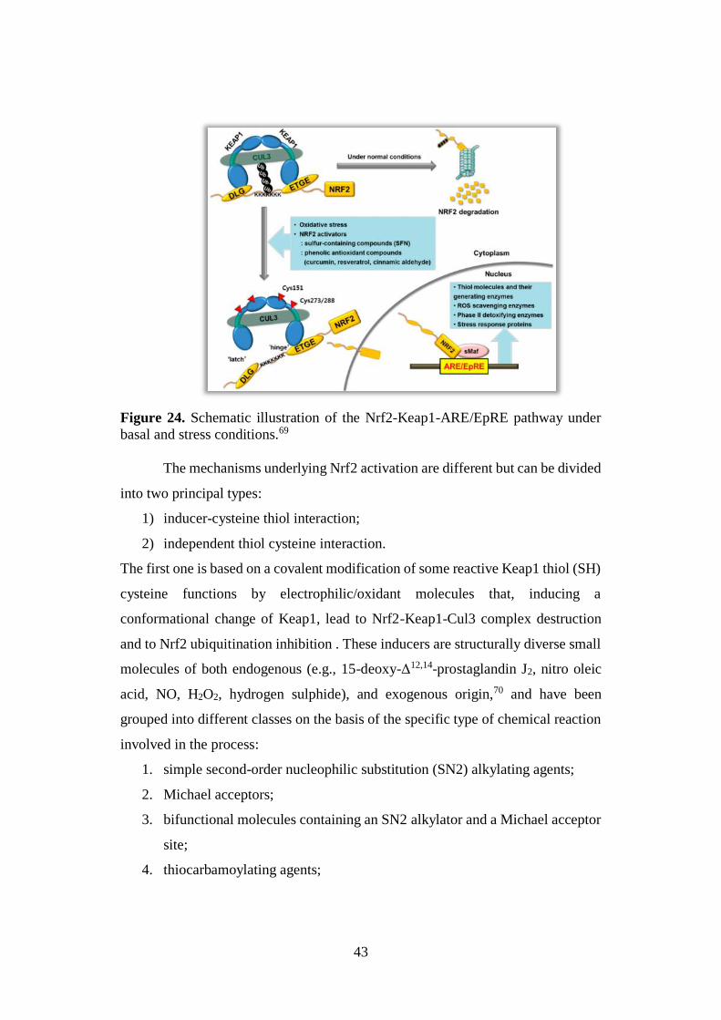
43
Figure 24. Schematic illustration of the Nrf2-Keap1-ARE/EpRE pathway under
basal and stress conditions.69
The mechanisms underlying Nrf2 activation are different but can be divided
into two principal types:
1) inducer-cysteine thiol interaction;
2) independent thiol cysteine interaction.
The first one is based on a covalent modification of some reactive Keap1 thiol (SH)
cysteine functions by electrophilic/oxidant molecules that, inducing a
conformational change of Keap1, lead to Nrf2-Keap1-Cul3 complex destruction
and to Nrf2 ubiquitination inhibition . These inducers are structurally diverse small
molecules of both endogenous (e.g., 15-deoxy-Δ12,14-prostaglandin J2, nitro oleic
acid, NO, H2O2, hydrogen sulphide), and exogenous origin,70 and have been
grouped into different classes on the basis of the specific type of chemical reaction
involved in the process:
1. simple second-order nucleophilic substitution (SN2) alkylating agents;
2. Michael acceptors;
3. bifunctional molecules containing an SN2 alkylator and a Michael acceptor
site;
4. thiocarbamoylating agents;
44
5. oxidant agents.71
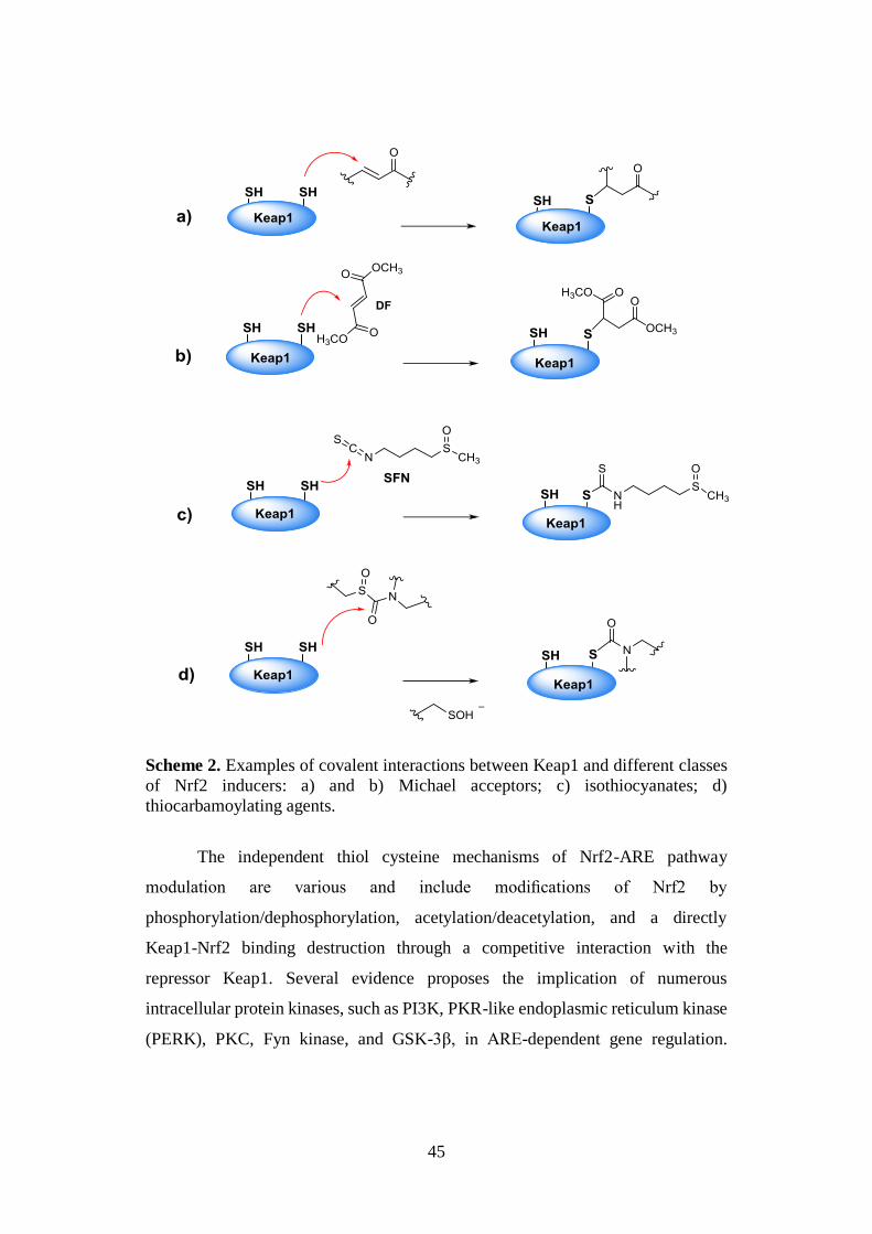
Among them, the second class, including many synthetic, semisynthetic and
naturally-inspired electrophilic compounds, mainly bearing an essential α,β-
unsaturated carbonyl system, have been investigated. In particular, the discovery
that their inducer potency correlates with the capability to react with the reactive
cysteine sulphydryl (SH) fuctionts of Keap1 via Michael reaction (Scheme 2a) was
a critical milestone in the understanding of the mechanism of Nrf2 activation.72 The
Michael addition is properly the reaction by which dimethyl fumarate (DF), a well
known Nrf2 inducer, activates several cytoprotective phase II enzymes (Scheme
2b).73 Furthermore, additional studies allowed to elucidate the mechanism
underlying the cytoprotective action of some thiocarbamoylating agents, namely
isothiocyanates [e.g. SFN, (Scheme 2c)] and sulfoxythiocarbamates, based on
reversible and irreversible interactions between their electrophilic moieties and
Keap1 cysteine thiol functions, respectively (Scheme 2c and 2d).70
45
Scheme 2. Examples of covalent interactions between Keap1 and different classes
of Nrf2 inducers: a) and b) Michael acceptors; c) isothiocyanates; d)
thiocarbamoylating agents.
The independent thiol cysteine mechanisms of Nrf2-ARE pathway
modulation are various and include modifications of Nrf2 by
phosphorylation/dephosphorylation, acetylation/deacetylation, and a directly
Keap1-Nrf2 binding destruction through a competitive interaction with the
repressor Keap1. Several evidence proposes the implication of numerous
intracellular protein kinases, such as PI3K, PKR-like endoplasmic reticulum kinase
(PERK), PKC, Fyn kinase, and GSK-3β, in ARE-dependent gene regulation.
46
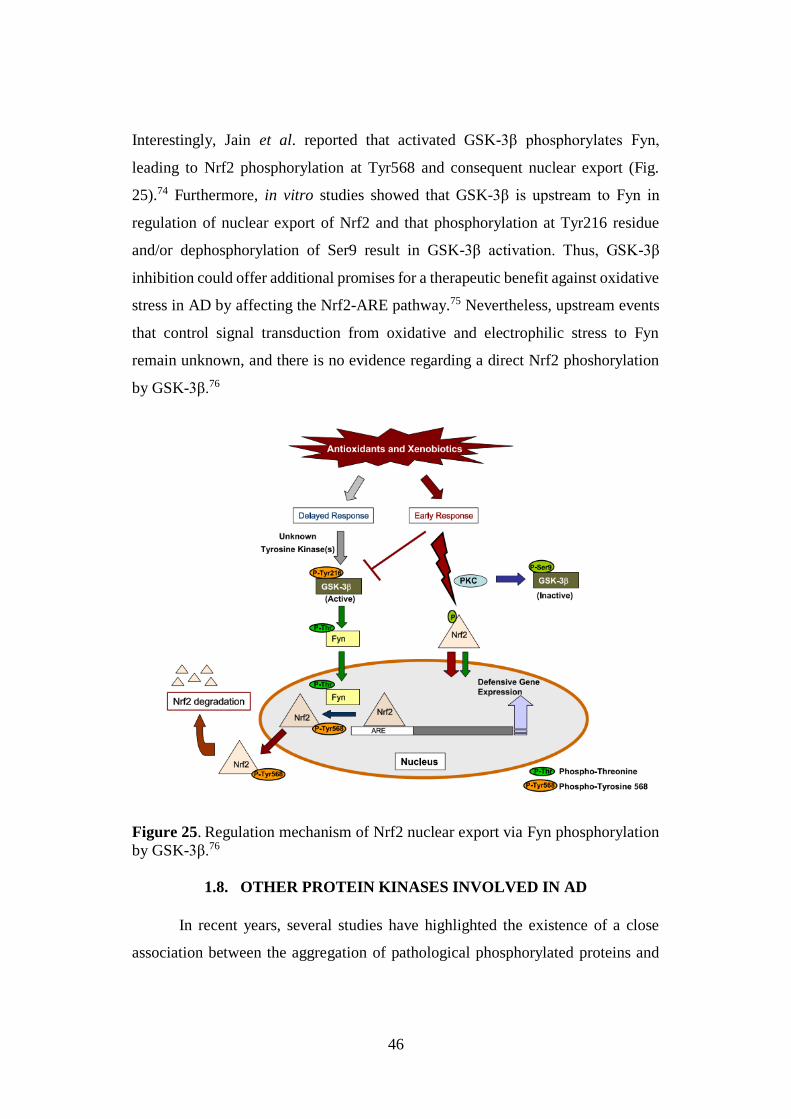
Interestingly, Jain et al. reported that activated GSK-3β phosphorylates Fyn,
leading to Nrf2 phosphorylation at Tyr568 and consequent nuclear export (Fig.
25).74 Furthermore, in vitro studies showed that GSK-3β is upstream to Fyn in
regulation of nuclear export of Nrf2 and that phosphorylation at Tyr216 residue
and/or dephosphorylation of Ser9 result in GSK-3β activation. Thus, GSK-3β
inhibition could offer additional promises for a therapeutic benefit against oxidative
stress in AD by affecting the Nrf2-ARE pathway.75 Nevertheless, upstream events
that control signal transduction from oxidative and electrophilic stress to Fyn
remain unknown, and there is no evidence regarding a direct Nrf2 phoshorylation
by GSK-3β.76
Figure 25. Regulation mechanism of Nrf2 nuclear export via Fyn phosphorylation
by GSK-3β.76
1.8. OTHER PROTEIN KINASES INVOLVED IN AD
In recent years, several studies have highlighted the existence of a close
association between the aggregation of pathological phosphorylated proteins and
47
the dysregulation of specific PKs in AD and in other neurodegenerative disorders,
including tauopathies, Parkinson’s disease (PD) and amyotrophic lateral sclerosis
(ALS). In this context, next to GSK-3β, a validated AD target, CK1 and leucine-
rich repeat kinase 2 (LRRK2) are gaining ever-increased attention as intriguing
kinases also involved in Aβ and τ cascades.
CK1 is a Ser/Thr protein kinase, found in plants and animals, whose activity
has been detected in various subcellular compartments including cell membranes,
cytosol, and nuclei. It has been associated to various biological functions, such as
DNA repair, cell morphology, modulation of the metabolic Wnt/β-catenin pathway,
Hedgehog pathway, circadian rhythms and sleep disorders, cancer, inflammation as
well as different neurodegenerative diseases including AD, PD, ALS. With the first
cloning of CK1 cDNAs, it became evident that CK1 constituted a subfamily of PKs
composed of seven isoforms (α, β, γ1-3, δ and ɛ) characterized by a catalytic domain
of about 300 aminoacids which shares more than 50 % sequence identity in the
different isoforms. In contrast, CK1 enzymes have highly variable C-terminal
domains, that have been implicated in both subcellular targeting and activity
regulation.77 All the different isoforms act as monomeric and constitutive enzymes,
although autophosphorylation of C-terminal residues inhibits the activity of CK1α,
CK1δ, and CK1ε and helps regulating their catalytic activity. Furthermore, they
exclusively use ATP as a phosphate donor, and are cofactor independent proteins.78
Several evidence confirms that all isoforms are overexpressed in AD
hippocampus, nevertheless only CK1δ and CK1ε proved to be implicated in AD
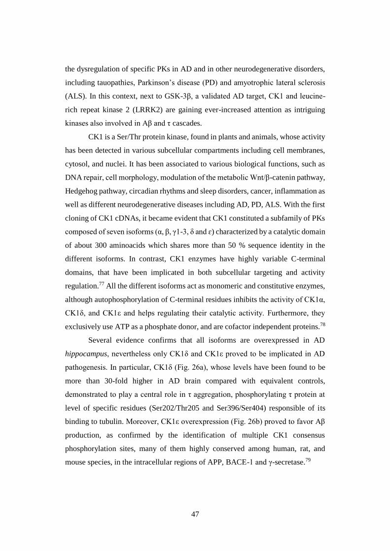
pathogenesis. In particular, CK1δ (Fig. 26a), whose levels have been found to be
more than 30-fold higher in AD brain compared with equivalent controls,
demonstrated to play a central role in τ aggregation, phosphorylating τ protein at
level of specific residues (Ser202/Thr205 and Ser396/Ser404) responsible of its
binding to tubulin. Moreover, CK1ε overexpression (Fig. 26b) proved to favor Aβ
production, as confirmed by the identification of multiple CK1 consensus
phosphorylation sites, many of them highly conserved among human, rat, and
mouse species, in the intracellular regions of APP, BACE-1 and γ-secretase.79
48
Figure 26. X-ray structure of a) CK1δ (PDBib: 4TN6) and b) CK1ε (PDBid:
4HNI), each of them in complex with a specific inhibitor.
Taking into account all these exciting findings, CK1, in particular CK1δ and
CK1ε isoforms, appear new promising AD targets, whose simultaneous inhibition
could represent an additional therapeutic strategy to affect both Aβ and τ proteins
misfolding and consequent aggregation.
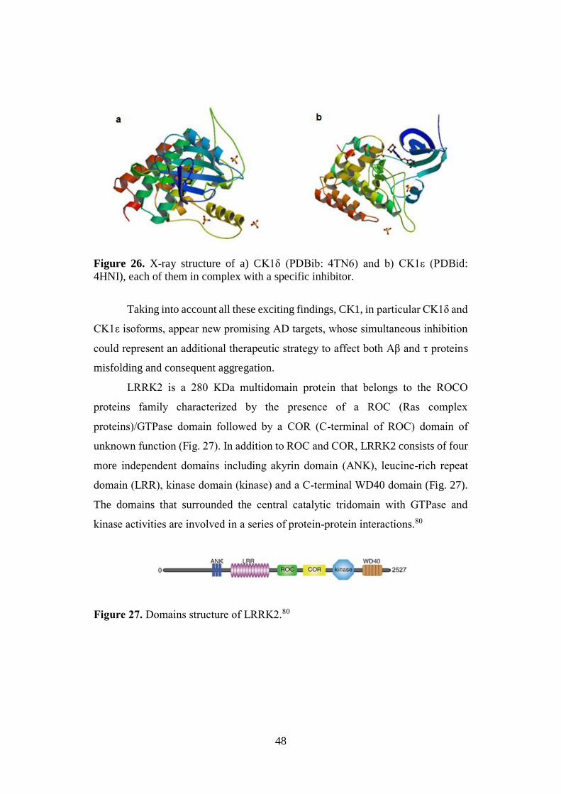
LRRK2 is a 280 KDa multidomain protein that belongs to the ROCO
proteins family characterized by the presence of a ROC (Ras complex
proteins)/GTPase domain followed by a COR (C-terminal of ROC) domain of
unknown function (Fig. 27). In addition to ROC and COR, LRRK2 consists of four
more independent domains including akyrin domain (ANK), leucine-rich repeat
domain (LRR), kinase domain (kinase) and a C-terminal WD40 domain (Fig. 27).
The domains that surrounded the central catalytic tridomain with GTPase and
kinase activities are involved in a series of protein-protein interactions.80
Figure 27. Domains structure of LRRK2.80
49
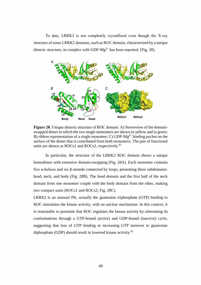
To date, LRRK2 is not completely crystallized even though the X-ray
structure of some LRRK2 domains, such as ROC domain, characterized by a unique
dimeric structure, in complex with GDP-Mg2+ has been reported. (Fig. 28).
Figure 28. Unique dimeric structure of ROC domain. A) Stereoview of the domain-
swapped dimer in which the two single monomers are shown in yellow and in green;
B) ribbon representation of a single monomer; C) GDP-Mg2+ binding pocket on the
surface of the dimer that is contributed from both monomers. The pair of functional
units are shown as ROCs1 and ROCs2, respectively.81
In particular, the structure of the LRRK2 ROC domain shows a unique
homodimer with extensive domain-swapping (Fig. 28A). Each monomer contains
five α-helices and six β-strands connected by loops, presenting three subdomains:
head, neck, and body (Fig. 28B). The head domain and the first half of the neck
domain from one monomer couple with the body domain from the other, making
two compact units (ROCs1 and ROCs2; Fig. 28C).
LRRK2 is an unusual PK, actually the guanosine triphosphate (GTP) binding to
ROC stimulates the kinase activity, with an unclear mechanism. In this context, it
is reasonable to postulate that ROC regulates the kinase activity by alternating its
conformations through a GTP-bound (active) and GDP-bound (inactive) cycle,
suggesting that loss of GTP binding or increasing GTP turnover to guanosine
diphosphate (GDP) should result in lowered kinase activity.81
50
LRRK2 has been identified as the causal molecule for autosomal-dominant
PD and some of its mutations, such as R1441, I1371 and principally G2019S, have
been recognized as the most common genetic cause of the malady.
Recently, several evidence suggests the important role of LRRK2 in the pathologies
induced by abnormal τ phosphorylation, including AD. In particular, in SH-SY5Y
cells, a direct interaction between LRRK2 kinase domain and GSK-3β, enhancing
the kinase activity of this latter, proved to induce τ phosporylation at Ser396.82
Interestingly, additional studies showed the higher binding affinity of G2019S-
LRRK2 mutant for GSK-3β and suggest LRRK2 as a positive modulator of
neuroinflammation in murine microglial cells, due to its capability to increase the
expression of pro-inflammatory mediators, namely TNF-α, IL-1β, IL-6 and NO, as
well as the ability of its mutations to alter the brain microenvironment inducing
oxidative stress.83
Taking into account these stimulating results, LRRK2 emerges as a novel
GSK-3β enhancer and a PK able to stimulate microglial inflammatory responses;
its inhibition could therefore offer promises to decrease τ phosphorylation and
neuroinflammation.
51
2. Medicinal Chemistry
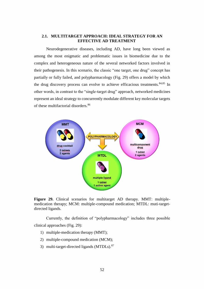
52
2.1. MULTITARGET APPROACH: IDEAL STRATEGY FOR AN
EFFECTIVE AD TREATMENT
Neurodegenerative diseases, including AD, have long been viewed as
among the most enigmatic and problematic issues in biomedicine due to the
complex and heterogeneous nature of the several networked factors involved in
their pathogenesis. In this scenario, the classic “one target, one drug” concept has
partially or fully failed, and polypharmacology (Fig. 29) offers a model by which
the drug discovery process can evolve to achieve efficacious treatments.84,85 In
other words, in contrast to the “single-target drug” approach, networked medicines
represent an ideal strategy to concurrently modulate different key molecular targets
of these multifactorial disorders.86
Figure 29. Clinical scenarios for multitarget AD therapy. MMT: multiple-
medication therapy; MCM: multiple-compound medication; MTDL: muti-target-
directed ligands.
Currently, the definition of “polypharmacology” includes three possible
clinical approaches (Fig. 29):
1) multiple-medication therapy (MMT);
2) multiple-compound medication (MCM);
3) multi-target-directed ligands (MTDLs).87
53
The first one, also referred to as a “cocktail” or “combination of drugs”, is
based on the administration of two or three different drugs, in the form of two or
more individual tablets, which combine different therapeutic mechanisms.
However, the benefits of this approach are often compromised by poor patient
compliance and possibility of drug-drug interactions (DDI).
A second approach involves the use of MCM, also known as a “single-pill
drug combination” or “multicomponent drugs”, in which two or more agents are
coformulated in a single tablet with the aim to simplify dosing regimens and
improve patient compliance. Although several multicomponent drugs have recently
been launched from pharmaceutical industry, there are significant risks involved in
their development because clinicians might still prefer prescribing combinations of
existing monotherapies that may offer greater dose flexibility and lower cost
treatment. Furthermore, DDI are possible and the fixed-dose combination is not
practicable in cases in which the routes of administration of two or more starting
drugs are different.
An alternative strategy consists in the administration of MTDL, a single
chemical entity able to simultaneously modulate several targets involved in the
disease.87 In general, this approach consists in the combination of various
pharmacophores, of different known drugs, in order to obtain a new hybrid
molecule able to interact with the selected targets.
In 2005, Morphy and Rankovic introduced the definition “designed multiple
ligands” to highlight how the multitarget profile of these compounds was rationally
designed and not discovered in retrospect. Afterwards, additional terms to illustrate
the same concept were introduced including “multimodal” or “multi-functional” by
Moussa Youdim, “dual- and triple-acting agents” by Mark Millan, “hybrid drugs”
and “multi-target-direct ligands”. Nowadays, “multitarget drugs” (MTDs) appears
a standardized terminology that could also improve the accuracy of future
bibliographic searches.84
Compared to multicomponent drugs, the multiple ligand approach has a
profoundly different risk-benefit profile. Although it is significantly more difficult
54
to regulate the ratio of activities at the different targets, the clinical development of
multiple drugs, in terms of the risks and costs involved, is no different from that of
any other single chemical entity. Furthermore, the possible DDI associated to
multiple agents administration are lower than cocktails or multicomponent drugs.87
Currently, the multitarget drug discovery represents a hot topic within the
medicinal chemistry community, as confirmed by the variety of design strategies
that has been proposed and successfully employed. In this context, fragment-based
and computational strategies are showing promising potential in accelerating and
optimizing the MTDs development. In particular, the fragment-based approach, that
consists in the step-wise addition of functional groups to simpler low-molecular-
weight chemical entities, has gained ever-increased interest both in academia and
industry. On the contrary, the application of computer-assisted drug discovery
(CADD) to the multitarget drug design (MTDD) is very recent and episodic,
although it has been an attractive idea for some time.88
In addition to these strategies, the ligand-based approach is still the method
of choice in which two starting structures and/or their pharmacophore elements are
combined for obtaining a new single molecule with both activities. Alternatively,
single fragments, even though interacting with modest affinity with the
corresponding targets, can be used as core scaffolds to be properly functionalized
in order to generate higher affinity derivatives.84
In conclusion, despite resistance of some research groups within the
pharmaceutical community, polypharmacology-based strategies are gaining ever-
increasing consideration as innovative approach to obtain potentially effective
disease-modifying drug candidates for the treatment of multifactorial diseases.88
2.2. PRIVILEGED STRUCTURES
Over the past 15 years, the “privileged structure” concept gained particular
attention in the discovery and optimization of novel biologically active molecules.
In particular, it emerged as a possible way to accelerate the drug discovery process,
especially for targets with unknown 3D structure, including G-protein coupled
55
receptors (GPCRs). Similarly, in the MTDD, the use of “privileged structures” has
been identified as a fruitful approach to develop multipotent drug candidates and
chemical probes for the treatment of several neurodegenerative disorders, including
AD.
Nowadays, a “privileged structure” is defined as a molecular fragment able
to interact with more than one biological macromolecule and to exhibit a number
of properties associated with drug-like molecules. However, this definition has
undergone some refinements since its introduction by Evans and coworkers in
1988, as a single molecular framework able to provide ligands for diverse receptors,
with refer to the 1,4-benzodiazepine-2-one heterocycle.89
The term “structure” is technically incorrect because it is only a
substructure, the essential core of the molecule, to be considered as “privileged”.
Although there are no rigorous rules to classify a subunit as “privileged”, it must
constitute a significant part of the molecular size to give a big contribution to the
molecular interactions. For instance, in a large number of bioactive molecules there
are some small groups, such as halogens or amides, that can hardly be considered
as “privileged structures” due to their ubiquitous nature and small overall
contribution to the binding activities. On the contrary, molecular scaffolds
characterized by relatively rigid frameworks, that present optimal functionalities
for molecular recognition of the target, could be defined as “privileged structures”.
In some cases, these molecular arrangements, probably as a result of the ubiquitous
nature of their aromatic ring systems (connected by single bonds or ring fusion)
showed versatile binding properties. Thus, the selectivity could represent a potential
issue; nevertheless, the functionalization of these rigid frameworks, can allow to
confer selectivity to them.
Several “privileged structures” have been identified simply by empirical
observations, although, computational techniques helped to recognize them in
existing compounds sets.
These “privileged structures” can serve as useful starting points for the rational
design of focused libraries of bioactive compounds. To this aim, the selection of the
56
appropriate molecular scaffold for a biological target is of paramount importance
because it can increase the hit rates. In this context, a scaffold with a molecular
weight within the 100-300 range would be preferred, as it is well known that only
a substantial portion of the final molecule represents a “privileged structure”.
Furthermore, to obtain analogues with improved pharmacokinetic properties and
the desired diversity, i.e. different functionalities hanging from the same central
core, the selected scaffold should have a well-balanced polarity profile and 1-3
molecular points suitable for a rapid functionalization. Indeed, the synthetic
accessibility of the employed scaffolds is of pivotal importance.90
Literature reports several sets of privileged scaffolds that have been
identified from molecules synthesized de novo, from marketed drugs, as well as
from natural products (NPs) that served as inspiration for medicinal chemistry
programs (Tables 1 and 2).
Among them, indoles, coumarins, chalcones and carbohydrates (Tables 1 and 2)
have attracted particular attention, from both academia and pharmaceutical
industry, and have been appropriately used as starting points to develop collections
of biologically active analogues.
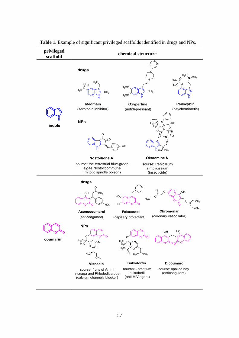
57
Table 1. Example of significant privileged scaffolds identified in drugs and NPs.
privileged
scaffold chemical structure
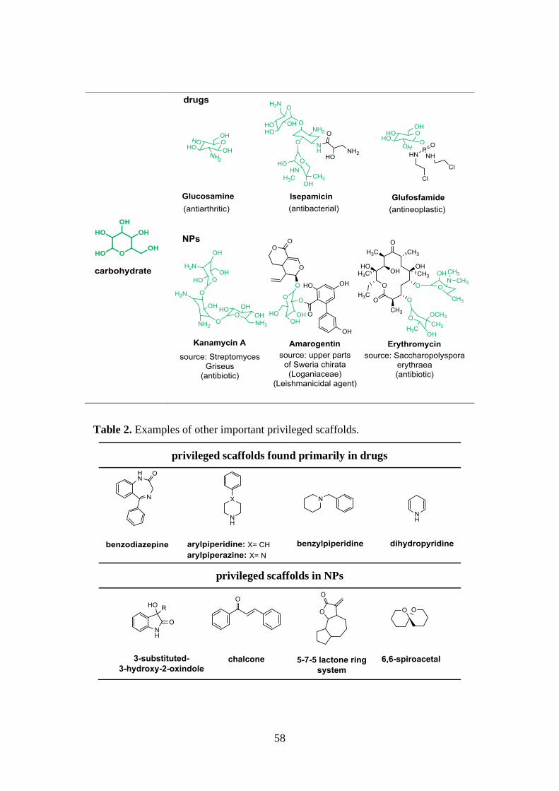
58
Table 2. Examples of other important privileged scaffolds.
privileged scaffolds found primarily in drugs
privileged scaffolds in NPs
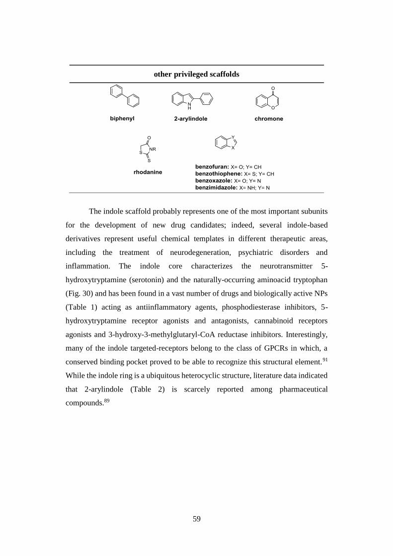
59
other privileged scaffolds
The indole scaffold probably represents one of the most important subunits
for the development of new drug candidates; indeed, several indole-based
derivatives represent useful chemical templates in different therapeutic areas,
including the treatment of neurodegeneration, psychiatric disorders and
inflammation. The indole core characterizes the neurotransmitter 5-
hydroxytryptamine (serotonin) and the naturally-occurring aminoacid tryptophan
(Fig. 30) and has been found in a vast number of drugs and biologically active NPs
(Table 1) acting as antiinflammatory agents, phosphodiesterase inhibitors, 5-
hydroxytryptamine receptor agonists and antagonists, cannabinoid receptors
agonists and 3-hydroxy-3-methylglutaryl-CoA reductase inhibitors. Interestingly,
many of the indole targeted-receptors belong to the class of GPCRs in which, a
conserved binding pocket proved to be able to recognize this structural element.91
While the indole ring is a ubiquitous heterocyclic structure, literature data indicated
that 2-arylindole (Table 2) is scarcely reported among pharmaceutical
compounds.89
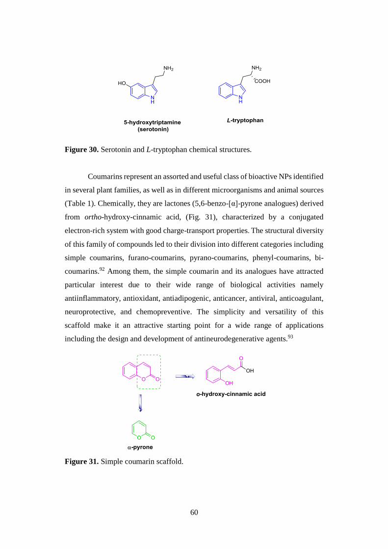
60
Figure 30. Serotonin and L-tryptophan chemical structures.
Coumarins represent an assorted and useful class of bioactive NPs identified
in several plant families, as well as in different microorganisms and animal sources
(Table 1). Chemically, they are lactones (5,6-benzo-[α]-pyrone analogues) derived
from ortho-hydroxy-cinnamic acid, (Fig. 31), characterized by a conjugated
electron-rich system with good charge-transport properties. The structural diversity
of this family of compounds led to their division into different categories including
simple coumarins, furano-coumarins, pyrano-coumarins, phenyl-coumarins, bi-
coumarins.92 Among them, the simple coumarin and its analogues have attracted
particular interest due to their wide range of biological activities namely
antiinflammatory, antioxidant, antiadipogenic, anticancer, antiviral, anticoagulant,
neuroprotective, and chemopreventive. The simplicity and versatility of this
scaffold make it an attractive starting point for a wide range of applications
including the design and development of antineurodegenerative agents.93
Figure 31. Simple coumarin scaffold.
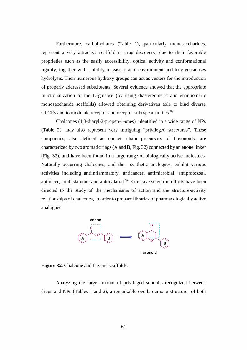
61
Furthermore, carbohydrates (Table 1), particularly monosaccharides,
represent a very attractive scaffold in drug discovery, due to their favorable
proprieties such as the easily accessibility, optical activity and conformational
rigidity, together with stability in gastric acid environment and to glycosidases
hydrolysis. Their numerous hydroxy groups can act as vectors for the introduction
of properly addressed substituents. Several evidence showed that the appropriate
functionalization of the D-glucose (by using diastereomeric and enantiomeric
monosaccharide scaffolds) allowed obtaining derivatives able to bind diverse
GPCRs and to modulate receptor and receptor subtype affinities.89
Chalcones (1,3-diaryl-2-propen-1-ones), identified in a wide range of NPs
(Table 2), may also represent very intriguing “privileged structures”. These
compounds, also defined as opened chain precursors of flavonoids, are
characterized by two aromatic rings (A and B, Fig. 32) connected by an enone linker
(Fig. 32), and have been found in a large range of biologically active molecules.
Naturally occurring chalcones, and their synthetic analogues, exhibit various
activities including antiinflammatory, anticancer, antimicrobial, antiprotozoal,
antiulcer, antihistaminic and antimalarial.94 Extensive scientific efforts have been
directed to the study of the mechanisms of action and the structure-activity
relationships of chalcones, in order to prepare libraries of pharmacologically active
analogues.
Figure 32. Chalcone and flavone scaffolds.
Analyzing the large amount of privileged subunits recognized between
drugs and NPs (Tables 1 and 2), a remarkable overlap among structures of both
62
classes can be detected, since that the vast majority of the reported scaffolds have
members in both groups. This result should not be so surprising because nature
frequently repeats itself after the identification of an appropriate solution to a
particular biochemical problem, and, obviously, the macromolecular structures in
living systems have a high level of non-random patterning. However, there are few
examples of molecules synthesized by chemists whose essential cores are not
typically obtained from a natural source (Table 2).95
2.3. NATURAL PRODUCTS
Despite competition from other “privileged structures”, we have recently
seen a real explosion of interest in NPs as excellent source of lead scaffolds for new
clinical candidates and drugs. Review of NPs over 30 years, from 1981 to 2010,
revealed that approximately 40 % of the developed therapeutic agents approved by
FDA were NPs, their derivatives, or synthetic mimetics related to NPs.96 Between
2005 and 2010, 19 NPs-based drugs were approved worldwide for the treatment of
infectious, immunological, cardiovascular, neurological, inflammatory and related
diseases, as well as cancer.97
NPs have produced a profound impact on both chemical biology and drug
development, as the consequence of the large diversity in their chemical space. In
particular, through the natural selection process, they have developed a unique and
vast chemical diversity and have been evolved for an optimal interaction with
biological macromolecules (targets). In general, these compounds proved to have
an enormous potential as:
1) essential source for drug discovery;
2) inspiration for combinatorial chemistry libraries;
3) source of chemical probes for biomolecular functions.
Over the last decades, collections of NPs-inspired small molecules have
been designed applying innovative combinatorial techniques, among which the
diversity-oriented synthesis (DOS) emerged as valuable strategy to explore the
chemical space. In contrast to the traditional target oriented synthesis (TOS), based
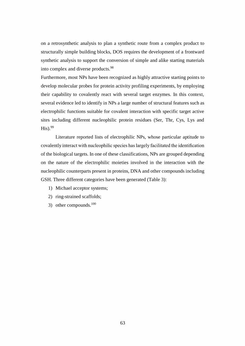
63
on a retrosynthetic analysis to plan a synthetic route from a complex product to
structurally simple building blocks, DOS requires the development of a frontward
synthetic analysis to support the conversion of simple and alike starting materials
into complex and diverse products.98
Furthermore, most NPs have been recognized as highly attractive starting points to
develop molecular probes for protein activity profiling experiments, by employing
their capability to covalently react with several target enzymes. In this context,
several evidence led to identify in NPs a large number of structural features such as
electrophilic functions suitable for covalent interaction with specific target active
sites including different nucleophilic protein residues (Ser, Thr, Cys, Lys and
His).99
Literature reported lists of electrophilic NPs, whose particular aptitude to
covalently interact with nucleophilic species has largely facilitated the identification
of the biological targets. In one of these classifications, NPs are grouped depending
on the nature of the electrophilic moieties involved in the interaction with the
nucleophilic counterparts present in proteins, DNA and other compounds including
GSH. Three different categories have been generated (Table 3):
1) Michael acceptor systems;
2) ring-strained scaffolds;
3) other compounds.100
64
Table 3. Three different classes of electrophilic NPs.
1. Michael acceptor systems
2. Ring-strained scaffolds
3. Other electrophilic NPs
65
The first category comprises carbonyl α,β-unsaturated-based compounds,
among which caffeic acid, chalcones, curcumin, etc., whose beneficial biological
activities proved to be strictly related to their electrophilic nature as Michael-
acceptor systems.101
The second one encompasses all ring-strained electrophiles, from three to
five membered rings, namely: epoxides/spiroepoxides, monocyclic and bicyclic
aziridines, cyclopropyl derivatives, monocyclic and bicyclic β-lactones and β-
lactams. They undergo a ring-opening and, in some cases, a subsequent molecular
arrangement after the nucleophilic target attack.100
The last class includes electrophilic compounds such as: 4-
hydroxycoumarins, isothiocyanates, disulfides, carbamates etc., able to form
covalent target-adducts by means of different chemical reactions.102
In this classification, the interest was focused on well-studied electrophilic
compounds, for which, the covalent attachment to target proteins or to binding site
residues are known and proceed via nucleophilic attack (either via substitution or
addition); nevertheless, the literature reports several additional examples of NPs
able to form covalent adducts via alternative mechanisms including radical-based
or redox-based processes.
2.3.1. α,β-unsaturated carbonyl compounds
The α,β-unsaturated carbonyl function of certain electrophilic NPs proved
to be essential for their biological activity.
For instance, several phytochemicals, especially polyphenols abundant in food
plants such as tea leaves, parsley, dill, peppers, onions, kale, cocoa, and cranberries,
act as cancer chemopreventive and chemoprotective agents due to their capability
to add to the electrophilic β-position of their unsaturated carbonyl system
nucleophiles (activity a, Scheme 3). These last consist in a large variety of species
characterized by heteroatomic groups containing critical Lewis basic sulfur, oxygen
and nitrogen atoms. Among these reactive moieties, the sulfhydryl function of
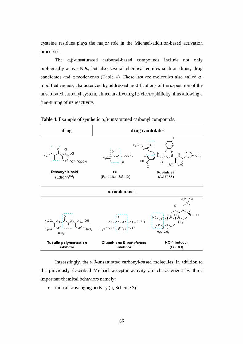
66
cysteine residues plays the major role in the Michael-addition-based activation
processes.
The α,β-unsaturated carbonyl-based compounds include not only
biologically active NPs, but also several chemical entities such as drugs, drug
candidates and α-modenones (Table 4). These last are molecules also called α-
modified enones, characterized by addressed modifications of the α-position of the
unsaturated carbonyl system, aimed at affecting its electrophilicity, thus allowing a
fine-tuning of its reactivity.
Table 4. Example of synthetic α,β-unsaturated carbonyl compounds.
drug drug candidates
α-modenones
Interestingly, the α,β-unsaturated carbonyl-based molecules, in addition to
the previously described Michael acceptor activity are characterized by three
important chemical behaviors namely:
radical scavenging activity (b, Scheme 3);
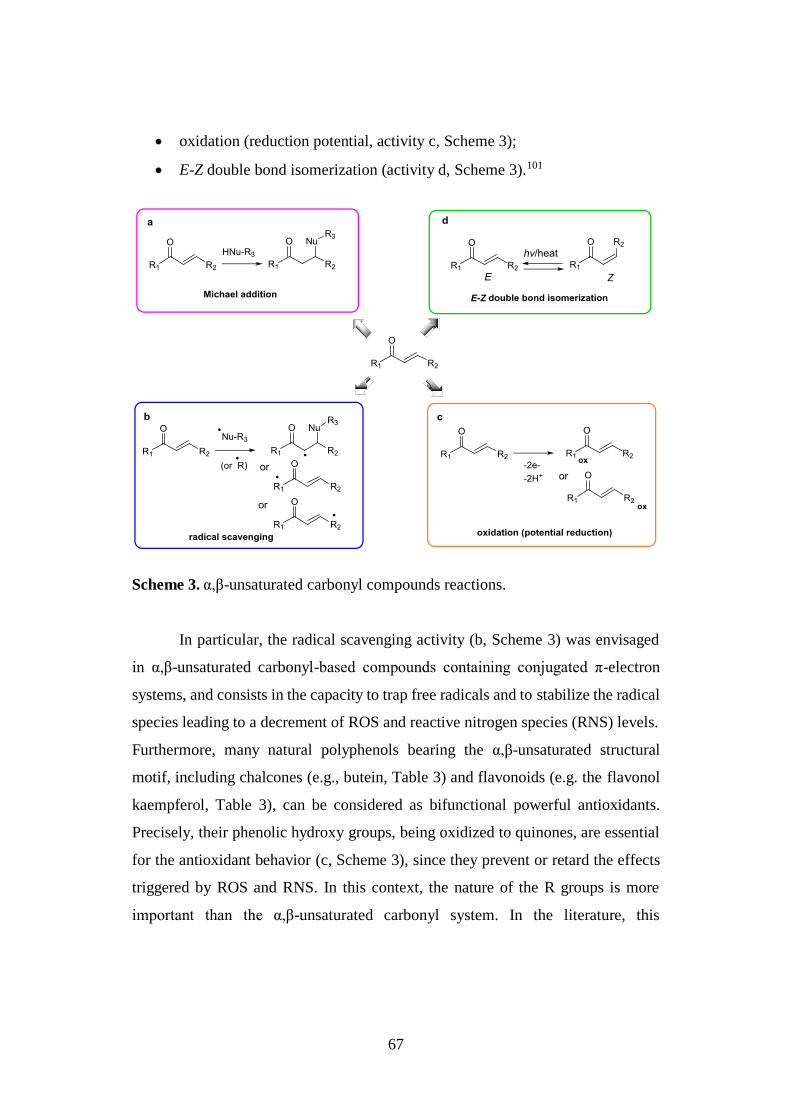
67
oxidation (reduction potential, activity c, Scheme 3);
E-Z double bond isomerization (activity d, Scheme 3).101
Scheme 3. α,β-unsaturated carbonyl compounds reactions.
In particular, the radical scavenging activity (b, Scheme 3) was envisaged
in α,β-unsaturated carbonyl-based compounds containing conjugated π-electron
systems, and consists in the capacity to trap free radicals and to stabilize the radical
species leading to a decrement of ROS and reactive nitrogen species (RNS) levels.
Furthermore, many natural polyphenols bearing the α,β-unsaturated structural
motif, including chalcones (e.g., butein, Table 3) and flavonoids (e.g. the flavonol
kaempferol, Table 3), can be considered as bifunctional powerful antioxidants.
Precisely, their phenolic hydroxy groups, being oxidized to quinones, are essential
for the antioxidant behavior (c, Scheme 3), since they prevent or retard the effects
triggered by ROS and RNS. In this context, the nature of the R groups is more
important than the α,β-unsaturated carbonyl system. In the literature, this
68
antioxidant action is often referred to as “radical scavenging activity” since the
radicals are being reduced to, or “trapped”, as non radical species.
The last reactivity, maybe less frequently addressed, is the unsaturated carbonyl
compounds’ potential to show thermal and photoisomerization of their double bond
(activity d, Scheme 3). There are different examples of this kind of reactivity in
biology, such as the photoisomerization of 11-cis-retinal to all-trans-retinal when
one photon is absorbed in the initial phase of the signal cascade occurring in vision.
From a medicinal chemistry standpoint, the exploitation of α,β-unsaturated
carbonyl compounds as potential drug candidates is a controversial topic
considering that these electrophiles, as highly reactive and low selective species,
can lead to cell damage and cytotoxicity as a result of concomitant covalent
modifications of essential biological macromolecules. To avoid this drawback,
several efforts have been directed to a fine-tuning of the α,β-unsaturated carbonyl
system reactivity, aimed at the improvement of the specificity of action. In this
context, different approaches have been applied to predict the reactivity of the α,β-
unsaturated carbonyl compounds. Among them, 13C-NMR studies gained particular
attention and allowed to correlate Michael acceptor reactivity, reduction potential,
and double bond photoisomerization with decreasing values of the α,β-unsaturated
carbonyl system electrophilicity. Interestingly, these studies demonstrated that
Michael acceptor reactivity correlates to lower ELUMO and EHOMO values, and that
the presence of a substituent in the α-position of the α,β-unsaturated carbonyl unit
has a strong influence on its reactivity.
In the light of these intriguing results, a recent drug design approach was focused
on an appropriate choice of the α-substituent, allowing to manipulate the reactivity
of the electrophilic centre, obtaining α-modenones (Table 4) endowed with several
promising activities often obtained by modifying inactive NPs.101
2.3.2. Thiol trapping assay
Although Michael acceptors have been avoided in modern drug discovery
as thought to exert toxic effects due to their tendency to irreversibly inhibit enzymes
69
through covalent modification (i.e. trapping of peripheral reactive cysteine thiol
functions), the α,β-unsaturated carbonyl group has proven to be essential for
disease-fighting activity. Indeed, for this class of compounds the capability to affect
many biologically relevant pathways such as Nrf2-Keap1-ARE and the pro-
inflammatory nuclear factor-κB (NF-κB) has been largely reported (or
documented).101
Taking into account the strong correlation between α,β-unsaturated carbonyl
compound’s bioactivity and reactivity as Michael acceptors, the assessment of their
tendency to react with thiols at the β-position of the α,β-unsaturated carbonyl
framework, is a very helpful tool to predict their biological activity.
In this context, different methods aimed to identify Michael acceptors and to sort
them into reversible and irreversible thiol sinks have been developed, among which
the spectroscopic 1H-NMR-based thiol trapping assay, certainly, plays a major role.
The concept involves the reaction, in a NMR test tube, between the studied
electrophile and a reactive thiol-based compound such as cysteamine, β-
mercaptoethanol or thiophenol, using DMSO-d6, as solvent. Experimentally,
cysteamine (2-aminoethanethiol), 2.0 molar equivalent per each potential
electrophilic site, is added to a solution of the α,β-unsaturated carbonyl compound,
in DMSO-d6, and the Michael reaction is assessed by 1H-NMR spectroscopic
analysis. The positivity of the assay is evidenced by the disappearance, in the
spectra, of the olefin system signals, and the irreversible nature of the addition is
also demonstrated by the failure of the reappearance of the characteristic olefin
signals upon appropriate dilution with CDCl3.103
This procedure has been described for several electrophilic compounds,
such as dienones, α,β-unsaturated esters, amides, lactones, and enones. Among the
members of this last class, the symmetric diketo tautomer of curcumin 1b (see Table
3 for the β-keto-enol counterpart 1a), one of the most thoroughly investigated and
promising dietary NPs, was reported to irreversibly and rapidly react with
cysteamine in DMSO-d6 affording the corresponding bis-1,7-thia-Michael adduct
(Scheme 4).104
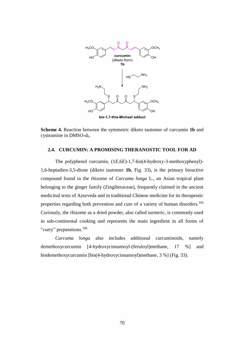
70
Scheme 4. Reaction between the symmetric diketo tautomer of curcumin 1b and
cysteamine in DMSO-d6.
2.4. CURCUMIN: A PROMISING THERANOSTIC TOOL FOR AD
The polyphenol curcumin, (1E,6E)-1,7-bis(4-hydroxy-3-methoxyphenyl)-
1,6-heptadien-3,5-dione (diketo tautomer 1b, Fig. 33), is the primary bioactive
compound found in the rhizome of Curcuma longa L., an Asian tropical plant
belonging to the ginger family (Zingiberaceae), frequently claimed in the ancient
medicinal texts of Ayurveda and in traditional Chinese medicine for its therapeutic
properties regarding both prevention and cure of a variety of human disorders.105
Curiously, the rhizome as a dried powder, also called turmeric, is commonly used
in sub-continental cooking and represents the main ingredient in all forms of
“curry” preparations.106
Curcuma longa also includes additional curcuminoids, namely
demethoxycurcumin [4-hydroxycinnamoyl-(feruloyl)methane, 17 %] and
bisdemethoxycurcumin [bis(4-hydroxycinnamoyl)methane, 3 %] (Fig. 33).
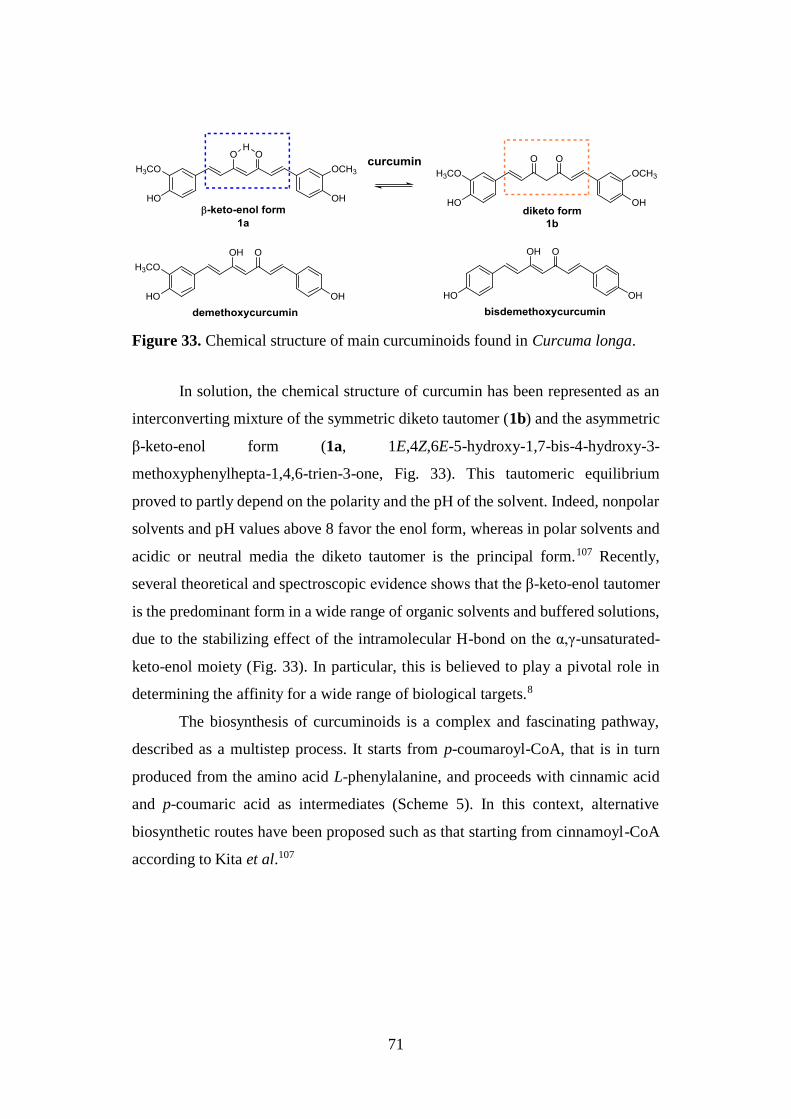
71
Figure 33. Chemical structure of main curcuminoids found in Curcuma longa.
In solution, the chemical structure of curcumin has been represented as an
interconverting mixture of the symmetric diketo tautomer (1b) and the asymmetric
β-keto-enol form (1a, 1E,4Z,6E-5-hydroxy-1,7-bis-4-hydroxy-3-
methoxyphenylhepta-1,4,6-trien-3-one, Fig. 33). This tautomeric equilibrium
proved to partly depend on the polarity and the pH of the solvent. Indeed, nonpolar
solvents and pH values above 8 favor the enol form, whereas in polar solvents and
acidic or neutral media the diketo tautomer is the principal form.107 Recently,
several theoretical and spectroscopic evidence shows that the β-keto-enol tautomer
is the predominant form in a wide range of organic solvents and buffered solutions,
due to the stabilizing effect of the intramolecular H-bond on the α,γ-unsaturated-
keto-enol moiety (Fig. 33). In particular, this is believed to play a pivotal role in
determining the affinity for a wide range of biological targets.8
The biosynthesis of curcuminoids is a complex and fascinating pathway,
described as a multistep process. It starts from p-coumaroyl-CoA, that is in turn
produced from the amino acid L-phenylalanine, and proceeds with cinnamic acid
and p-coumaric acid as intermediates (Scheme 5). In this context, alternative
biosynthetic routes have been proposed such as that starting from cinnamoyl-CoA
according to Kita et al.107
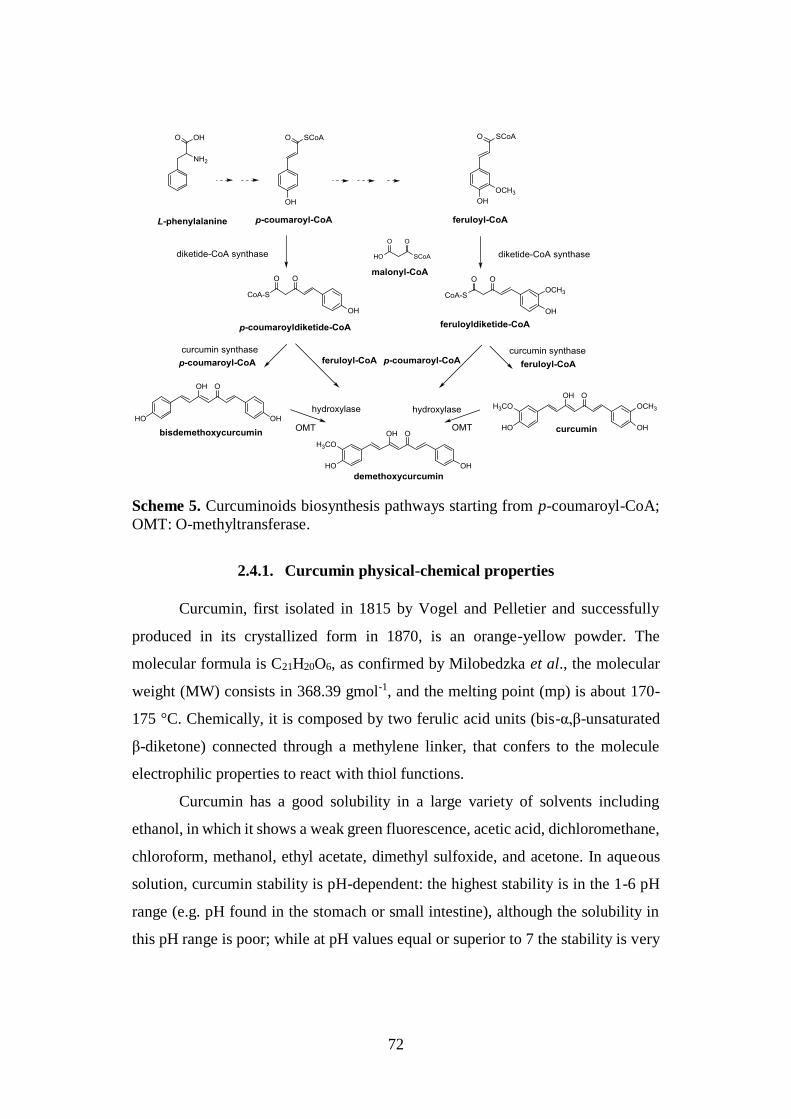
72
Scheme 5. Curcuminoids biosynthesis pathways starting from p-coumaroyl-CoA;
OMT: O-methyltransferase.
2.4.1. Curcumin physical-chemical properties
Curcumin, first isolated in 1815 by Vogel and Pelletier and successfully
produced in its crystallized form in 1870, is an orange-yellow powder. The
molecular formula is C21H20O6, as confirmed by Milobedzka et al., the molecular
weight (MW) consists in 368.39 gmol-1, and the melting point (mp) is about 170-
175 °C. Chemically, it is composed by two ferulic acid units (bis-α,β-unsaturated
β-diketone) connected through a methylene linker, that confers to the molecule
electrophilic properties to react with thiol functions.
Curcumin has a good solubility in a large variety of solvents including
ethanol, in which it shows a weak green fluorescence, acetic acid, dichloromethane,
chloroform, methanol, ethyl acetate, dimethyl sulfoxide, and acetone. In aqueous
solution, curcumin stability is pH-dependent: the highest stability is in the 1-6 pH
range (e.g. pH found in the stomach or small intestine), although the solubility in
this pH range is poor; while at pH values equal or superior to 7 the stability is very
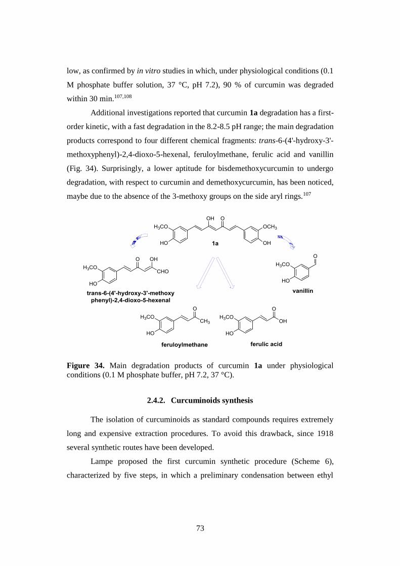
73
low, as confirmed by in vitro studies in which, under physiological conditions (0.1
M phosphate buffer solution, 37 °C, pH 7.2), 90 % of curcumin was degraded
within 30 min.107,108
Additional investigations reported that curcumin 1a degradation has a first-
order kinetic, with a fast degradation in the 8.2-8.5 pH range; the main degradation
products correspond to four different chemical fragments: trans-6-(4'-hydroxy-3'-
methoxyphenyl)-2,4-dioxo-5-hexenal, feruloylmethane, ferulic acid and vanillin
(Fig. 34). Surprisingly, a lower aptitude for bisdemethoxycurcumin to undergo
degradation, with respect to curcumin and demethoxycurcumin, has been noticed,
maybe due to the absence of the 3-methoxy groups on the side aryl rings.107
Figure 34. Main degradation products of curcumin 1a under physiological
conditions (0.1 M phosphate buffer, pH 7.2, 37 °C).
2.4.2. Curcuminoids synthesis
The isolation of curcuminoids as standard compounds requires extremely
long and expensive extraction procedures. To avoid this drawback, since 1918
several synthetic routes have been developed.
Lampe proposed the first curcumin synthetic procedure (Scheme 6),
characterized by five steps, in which a preliminary condensation between ethyl
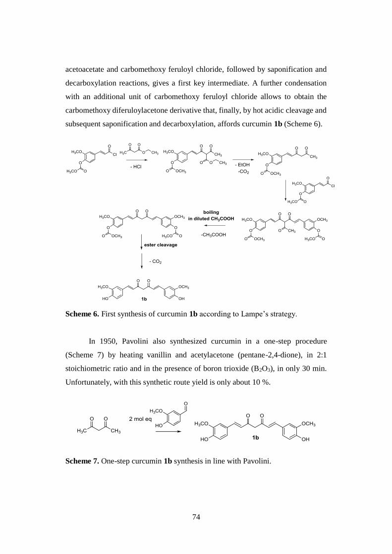
74
acetoacetate and carbomethoxy feruloyl chloride, followed by saponification and
decarboxylation reactions, gives a first key intermediate. A further condensation
with an additional unit of carbomethoxy feruloyl chloride allows to obtain the
carbomethoxy diferuloylacetone derivative that, finally, by hot acidic cleavage and
subsequent saponification and decarboxylation, affords curcumin 1b (Scheme 6).
Scheme 6. First synthesis of curcumin 1b according to Lampe’s strategy.
In 1950, Pavolini also synthesized curcumin in a one-step procedure
(Scheme 7) by heating vanillin and acetylacetone (pentane-2,4-dione), in 2:1
stoichiometric ratio and in the presence of boron trioxide (B2O3), in only 30 min.
Unfortunately, with this synthetic route yield is only about 10 %.
Scheme 7. One-step curcumin 1b synthesis in line with Pavolini.
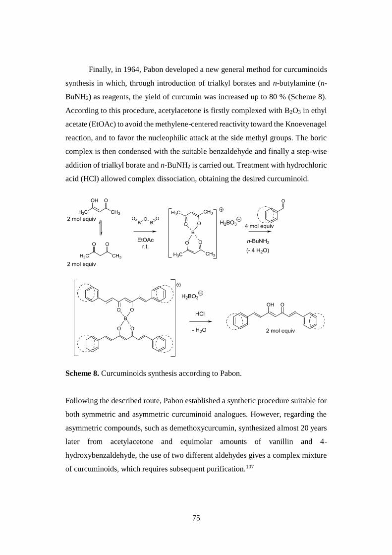
75
Finally, in 1964, Pabon developed a new general method for curcuminoids
synthesis in which, through introduction of trialkyl borates and n-butylamine (n-
BuNH2) as reagents, the yield of curcumin was increased up to 80 % (Scheme 8).
According to this procedure, acetylacetone is firstly complexed with B2O3 in ethyl
acetate (EtOAc) to avoid the methylene-centered reactivity toward the Knoevenagel
reaction, and to favor the nucleophilic attack at the side methyl groups. The boric
complex is then condensed with the suitable benzaldehyde and finally a step-wise
addition of trialkyl borate and n-BuNH2 is carried out. Treatment with hydrochloric
acid (HCl) allowed complex dissociation, obtaining the desired curcuminoid.
Scheme 8. Curcuminoids synthesis according to Pabon.
Following the described route, Pabon established a synthetic procedure suitable for
both symmetric and asymmetric curcuminoid analogues. However, regarding the
asymmetric compounds, such as demethoxycurcumin, synthesized almost 20 years
later from acetylacetone and equimolar amounts of vanillin and 4-
hydroxybenzaldehyde, the use of two different aldehydes gives a complex mixture
of curcuminoids, which requires subsequent purification.107
76
Currently, the Pabon reaction represents the most followed procedure for
the synthesis of curcuminoids and the corresponding derivatives.8
2.4.3. Strategies aimed at improving curcumin bioavailability and
pharmacokinetics
Curcumin is now recognized as multifunctional compound and “privileged
structure”, due to its capability to simultaneously modulate several molecular
targets or pathways involved in complex diseases including cancer, inflammation,
arthritis, diabetes, nephropathy, acquired immunodeficiency syndrome, chronic
bacterial prostatitis, disorders of cardiovascular, gastric, pulmonary and renal
systems and those affecting CNS such as AD and PD.109,110
Nowadays, curcumin is used in several countries as a dietary supplement,
due to its proven efficacy and safety; nevertheless, its therapeutic use is limited by
different drawbacks including the color, the lack of water solubility and the fast
degradation, together with a relatively poor bioavailability. Among them, the latter
represents the major concern and several strategies, including curcumin conjugation
and structural modifications, have been developed in an effort to improve its
pharmacokinetic and pharmacological profiles.109 In this context, apart from the
synthesis of curcumin analogues, different adjuvants (e.g. piperine, quercetin,
genistein, etc.) have been selected to prevent the rapid metabolism of curcumin by
interfering with the enzymes that catalyze its degradation. Furthermore, a large
variety of pharmaceutical formulations such as nanoparticles, liposomes, micelles,
and phospholipid complexes have been developed to overcome challenges and to
ease the translation of curcumin from bench to clinical application.110,111
In particular, several drug carrier-curcumin systems, namely nanocurcumin,
polylactic-co-glycolic acid (PLGA) encapsulated curcumin, liposome-encapsulated
curcumin (LEC), silica-coated and uncoated flexible liposomes loaded curcumin
(CUR-SLs), N-trimethyl chitosan chloride (TMC)-coated curcumin liposomes and
cyclodextrin encapsulated curcumin (CDC), proved to effectively increase the
curcumin bioavailability in animals and in humans.109 Interestingly, some of these
77
nanoformulations or nanomedicines have been developed as advanced drug
delivery systems in cancer therapy with the aim to increase curcumin’s anticancer
therapeutic benefits by improving its binding, internalization and targeting of
cancer cells and minimizing systemic toxicity. In this scenario, particular attention
has been focused on PLGA or poly(caprolactone) (PCL) nanoparticles, liposomal
and self-assembly formulations due to their attractive biocompatibility and the
intrinsic curcumin fluorescence property has been exploited to study the effective
internalization process of curcumin nanoformulations by fluorescence or confocal
microscope.111
Moreover, in additional investigations aimed at improving the curcumin low
water solubility, it has been demonstrated its increase of 12-fold by heat tratment.112
2.4.4. Structure-activity relationship studies
Nowadays, in addition to the different health benefits that have already been
attributed to curcumin, further effects including anticarcinogenic and
neuroprotective properties are also being investigated.113
To date, the exact role of curcumin’s functionalities in affecting its physico-
chemical properties and pharmacological pleiotropic behavior is far from
elucidated. Nevertheless, several structure-activity relationship (SAR) studies
revealed some pharmacophore elements strictly associated to its ability to
simultaneously interact with different biological macromolecules. In particular, the
relatively flexible chain connecting the two hydrophobic phenyl rings has been
proposed to be an essential fragment by which the molecule can adopt the suitable
conformations for optimal π–π and van der Waals interactions with both the
aromatic and the hydrophobic amino acid residues of the protein targets.
Furthermore, the hydroxy and the methoxy groups on the side aryl rings, as well as
the β-keto-enol central framework, have been identified as the key structural
elements involved in strong hydrogen-bonding interactions. Additionally, the α,β-
unsaturated ketone moiety acts as high reactive Michael acceptor for the
nucleophilic attack by thiol moieties of cysteine residues (Fig. 35).114
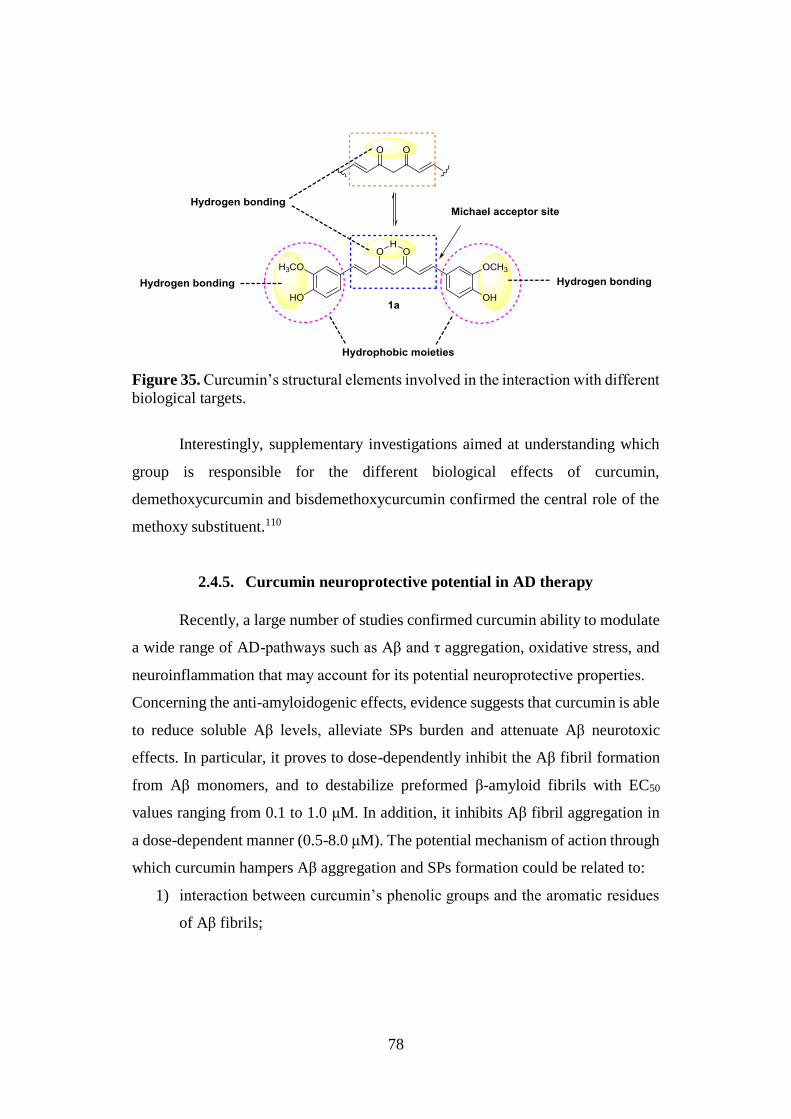
78
Figure 35. Curcumin’s structural elements involved in the interaction with different
biological targets.
Interestingly, supplementary investigations aimed at understanding which
group is responsible for the different biological effects of curcumin,
demethoxycurcumin and bisdemethoxycurcumin confirmed the central role of the
methoxy substituent.110
2.4.5. Curcumin neuroprotective potential in AD therapy
Recently, a large number of studies confirmed curcumin ability to modulate
a wide range of AD-pathways such as Aβ and τ aggregation, oxidative stress, and
neuroinflammation that may account for its potential neuroprotective properties.
Concerning the anti-amyloidogenic effects, evidence suggests that curcumin is able
to reduce soluble Aβ levels, alleviate SPs burden and attenuate Aβ neurotoxic
effects. In particular, it proves to dose-dependently inhibit the Aβ fibril formation
from Aβ monomers, and to destabilize preformed β-amyloid fibrils with EC50
values ranging from 0.1 to 1.0 μM. In addition, it inhibits Aβ fibril aggregation in
a dose-dependent manner (0.5-8.0 μM). The potential mechanism of action through
which curcumin hampers Aβ aggregation and SPs formation could be related to:
1) interaction between curcumin’s phenolic groups and the aromatic residues
of Aβ fibrils;
79
2) creation of competitive hydrogen bonds between the hydroxy groups
present in the curcumin’s β-diketone unit and the β-sheet of Aβ.
Furthermore, curcumin proves to reduce Aβ levels via two possible
mechanisms, i.e. by delaying the APP maturation and through the suppression of
Aβ42-induced BACE-1 expression.107,115
On the other hand, the potential effects of this NP on the τ protein pathways
have not been so extensively studied: curcumin treatment proves to reduce both
mRNA and protein levels of GSK-3β, and to inhibit the PK activity inducing the
enzyme phosphorylation at Ser9 with consequent prevention of NFTs formation
and neuroprotection against Aβ-induced mitochondrial dysfunction.115,116
A large amount of studies also confirmed the antiinflammatory and
antioxidant effects of curcumin both in vitro and in vivo. Regarding the protection
against inflammation, several evidence proposed curcumin ability to inhibit
(nuclear factor-κB) NF-κB, a ubiquitously expressed transcription factor involved
in the regulation of numerous pro-inflammatory genes including TNF-α, IL-1β,
iNOS and COX2. Under normal conditions, NF-κB cannot translocate from cytosol
to the nucleus and activate genes transcription, being bound to the inhibitory protein
IκB; the repressor phosphorylation promotes NF-κB release and its nuclear
translocation. In this context, it has been demonstrated that curcumin prevents NF-
κB activation through inhibition of IκB phosphorylation.107
Several investigations were also focused on elucidation of the molecular
mechanisms associated to curcumin antioxidant effects. Remarkably, it has been
shown how curcumin protection includes not only a free radical scavenging activity
but also an endogenous induction of the antioxidant defenses.117 Indeed, as a result
of its polyphenolic nature, curcumin proved to be endowed with antioxidative
properties; for instance, its free radical scavenging activity has been attributed to
the phenolic groups, even if recent evidence also supported involvement of the
methoxy groups, as well as of the β-diketone framework and its β-keto-enol form.118
Furthermore, several investigations demonstrated indirect antioxidative
properties for curcumin, due to its capability to modulate the cytoprotective gene
80
expression by activation of the Nrf2-Keap1-ARE pathway. The exact mechanism
through which curcumin activates Nrf2 is still unclear, but a possible involvement
of a Michael addition with some critical cysteine residues of the repressor Keap1
could account for the induction of additional phase II detoxifying enzymes, among
which HO-1 and GSH.107 Moreover, curcumin prevents Nrf2 nuclear export
through GSK-3β inhibition in order to avoid phosphorylation of Fyn protein.119
Interestingly, the cytoprotective effects of curcumin have also been related
to its metal ions chelating capacity, in particular Fe2+, Cu2+, Zn2+, largely found in
Aβ plaques and responsible for ROS generation, oxidative stress, and neuronal cell
death. 1H-NMR study showed a directly participation of the curcumin β-keto-enol
fragment in metal ion chelation, and the high stability of the formed complexes at
physiological pH.115,120
Taking into account the large variety of curcumin neuroprotective effects,
its versatile scaffold clearly emerged as promising pharmacological tool for a
mutitarget AD-modifying treatment. Moreover, from a medicinal chemistry
standpoint, the curcumin pharmacophore proved to be an excellent lead for the
design of analogues with expanded biological profile.8
2.4.6. Curcumin: a fluorescent probe in AD diagnosis
Curcumin absorbs in the visible region and gives fluorescence with low
quantum yield121 due to its unique structural features such as the two symmetric
chromophores of the structural motif C=O-C=C and the conjugated double bond
that are involved in its characteristic yellow pigmentation.
Recently curcumin, due to its fluorescent properties, emerged as an
attractive molecular tool for the diagnosis of AD, and thus it has been employed for
molecular imaging (MI).
MI is a fascinating modality that has largely been applied in biomedical
research, not only with the aim of characterizing and quantifying biological
processes at the cellular and subcellular level in intact living subjects, but also for
the early detection of diseases including cancer, inflammation and
81
neurodegeneration. To achieve these ambitious goals, chemical probes, small
molecules able to specifically interact with cellular and sub-cellular endogenous
components and to generate a detectable signal, have been exploited.
Generally, imaging modalities can be divided into two categories:
1. primarily morphological/anatomical imaging technologies: such as
computed tomography (CT) and magnetic resonance imaging (MRI)
characterized by high spatial resolution but with low ability to detect
diseases.
2. Primarily molecular imaging modalities: including optical imaging,
positron emission tomography (PET) and single photon emission computed
tomography (SPECT), which offer the potential to detect molecular and
cellular changes associated to pathological conditions, but suffer from a
poor spatial resolution.
Combining the strengths of the two classes allows the detection of
pathophysiological changes in early disease phases at high structural resolution (for
example, PET-MRI technology). Furthermore, each MI modality is based on the
use of a particular kind of chemical probe.122
Within the first group, MRI represents one of the most reliable diagnostic
imaging modalities, coupling high spatial resolution with exquisite dynamic
information and anatomical contrast. Therefore, it is now extensively used to map
human brain activity. Similarly to nuclear magnetic resonance (NMR), MRI depend
on the relaxation efficiency of protons placed in a fixed magnetic field. In this case,
the magnetically active protons are those of the water present in different tissues
and the contrast obtained in images is commonly due to their different relaxation
rates. So-called contrast agents are need to achieve a contrast enhancement and they
include complexes of paramagnetic ions such as gadolinium (Gd3+) and other
lanthanide ions, as well as ferromagnetic iron oxide particles and nanoparticles.123
Regarding the MI modalities of the second class, with the advent of laser
technology and sophisticated optical devices characterized by high sensitivity,
selectivity and simplicity, optical imaging methods have gained ever-increased
82
attention. In particular, they can be broadly divided into fluorescence and
bioluminescence imaging modalities, both of them characterized by easy
accessibility, high sensitivity, together with quick and easy performance in vivo. In
bioluminescence imaging, a chemiluminescent reaction between an enzyme (for
example, luciferase) and its substrate (for example, D-Luciferin) generates light.
Hence, the externally detected light is an indicator of a biological/molecular
process. The bioluminescent reactions are exergonic, with molecular oxygen
reacting with luciferase and the substrate to form a luciferase-bound peroxy-
intermediate, which, in turn, releases photons over an emission spectrum of 400-
620 nm.
Fluorescence imaging exploits the ability of a fluorochrome to absorb
energy from an external excitation light of one wavelength and re-emit photons at
a longer wavelength of lower energy.
In detail, the absorption of light by a fluorescent molecule yields a singlet excited-
state and the key parameters used to describe this process are the absorption maxima
(λmax) and the extinction coefficient at λmax (ε). Fluorescence occurs when this
excited state relaxes to the ground state (S0) through emission of photons. Typically,
the emission wavelength is longer than the absorption maximum due to the energy
loss stemming from relaxation to S1 and solvent reorganization around the excited
molecule. Interestingly, each fluorophore has a characteristic emission maximum
(λem) and “Stokes shift” consisting in the difference between λmax and λem.
Fluorophores characterized by small “Stokes shifts” being susceptible to self-
quenching via energy transfer are hardly used as labels for biomolecules. Another
important fluorophore feature is the quantum yield or quantum efficiency (Φ) that
represents the ratio of emitted photons to those absorbed.124
Each fluorophore presents specific chemical (e.g., reactivity, lipophilicity, pKa,
stability) and photophysical properties (e.g., λmax, λem, Φ, etc) based on which it is
possible to identify the most suitable probes for fluorescence imaging.
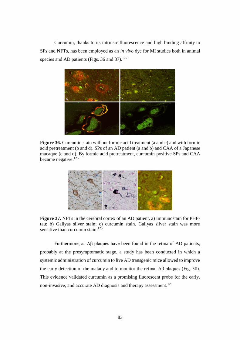
83
Curcumin, thanks to its intrinsic fluorescence and high binding affinity to
SPs and NFTs, has been employed as an in vivo dye for MI studies both in animal
species and AD patients (Figs. 36 and 37).125
Figure 36. Curcumin stain without formic acid treatment (a and c) and with formic
acid pretreatment (b and d). SPs of an AD patient (a and b) and CAA of a Japanese
macaque (c and d). By formic acid pretreatment, curcumin-positive SPs and CAA
became negative.125
Figure 37. NFTs in the cerebral cortex of an AD patient. a) Immunostain for PHF-
tau; b) Gallyas silver stain; c) curcumin stain. Gallyas silver stain was more
sensitive than curcumin stain.125
Furthermore, as Aβ plaques have been found in the retina of AD patients,
probably at the presymptomatic stage, a study has been conducted in which a
systemic administration of curcumin to live AD transgenic mice allowed to improve
the early detection of the malady and to monitor the retinal Aβ plaques (Fig. 38).
This evidence validated curcumin as a promising fluorescent probe for the early,
non-invasive, and accurate AD diagnosis and therapy assessment.126
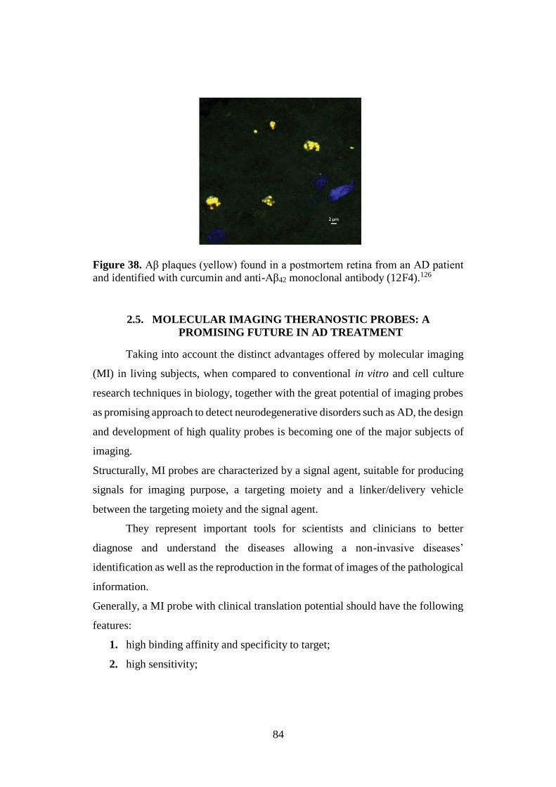
84
Figure 38. Aβ plaques (yellow) found in a postmortem retina from an AD patient
and identified with curcumin and anti-Aβ42 monoclonal antibody (12F4).126
2.5. MOLECULAR IMAGING THERANOSTIC PROBES: A
PROMISING FUTURE IN AD TREATMENT
Taking into account the distinct advantages offered by molecular imaging
(MI) in living subjects, when compared to conventional in vitro and cell culture
research techniques in biology, together with the great potential of imaging probes
as promising approach to detect neurodegenerative disorders such as AD, the design
and development of high quality probes is becoming one of the major subjects of
imaging.
Structurally, MI probes are characterized by a signal agent, suitable for producing
signals for imaging purpose, a targeting moiety and a linker/delivery vehicle
between the targeting moiety and the signal agent.
They represent important tools for scientists and clinicians to better
diagnose and understand the diseases allowing a non-invasive diseases’
identification as well as the reproduction in the format of images of the pathological
information.
Generally, a MI probe with clinical translation potential should have the following
features:
1. high binding affinity and specificity to target;
2. high sensitivity;
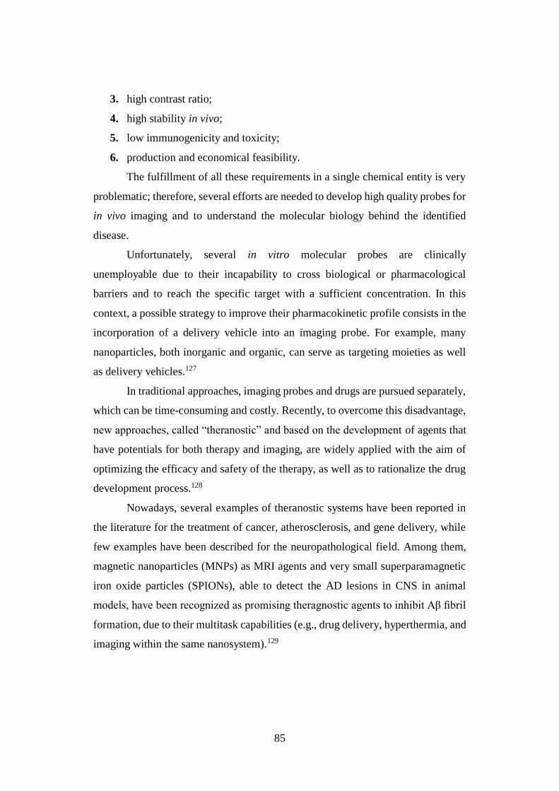
85
3. high contrast ratio;
4. high stability in vivo;
5. low immunogenicity and toxicity;
6. production and economical feasibility.
The fulfillment of all these requirements in a single chemical entity is very
problematic; therefore, several efforts are needed to develop high quality probes for
in vivo imaging and to understand the molecular biology behind the identified
disease.
Unfortunately, several in vitro molecular probes are clinically
unemployable due to their incapability to cross biological or pharmacological
barriers and to reach the specific target with a sufficient concentration. In this
context, a possible strategy to improve their pharmacokinetic profile consists in the
incorporation of a delivery vehicle into an imaging probe. For example, many
nanoparticles, both inorganic and organic, can serve as targeting moieties as well
as delivery vehicles.127
In traditional approaches, imaging probes and drugs are pursued separately,
which can be time-consuming and costly. Recently, to overcome this disadvantage,
new approaches, called “theranostic” and based on the development of agents that
have potentials for both therapy and imaging, are widely applied with the aim of
optimizing the efficacy and safety of the therapy, as well as to rationalize the drug
development process.128
Nowadays, several examples of theranostic systems have been reported in
the literature for the treatment of cancer, atherosclerosis, and gene delivery, while
few examples have been described for the neuropathological field. Among them,
magnetic nanoparticles (MNPs) as MRI agents and very small superparamagnetic
iron oxide particles (SPIONs), able to detect the AD lesions in CNS in animal
models, have been recognized as promising theragnostic agents to inhibit Aβ fibril
formation, due to their multitask capabilities (e.g., drug delivery, hyperthermia, and
imaging within the same nanosystem).129
86
MNPs consist in a magnetic core (e.g. maghemite) and a biocompatible
coating [e.g. polyethylene glycol (PEG)] and can be used as target-specific agents
if functionalized, for instance by antibodies incorporation. For example, in a study
by Paduslo et al., Aβ plaques were targeted using a Gadolinium-loaded molecular
probe, that after intravenous injection in animals, due to its small size, was able to
cross the BBB and specifically bind to the SPs, resulting in their selective
enhancement in MRI. Furthermore, an additional investigation of Mahmoudi et al.
showed “dual” SPIONs’ effects on the fibrillation kinetics, depending on the size,
surface area and charge of these small particles. Specifically, lower concentrations
of SPIONs proved to inhibit Aβ fibrillation rate, while higher concentrations
enhanced it and positively charged SPIONs promoting the fibrillation process at
significantly lower particle concentrations than both negatively charged and
essentially uncharged (plain) SPIONs.130
Taking into account the great potential of purposely-designed theranostics
as promising future to combat neurodegenerative disorders including AD, further
investigations and efforts are still urgently needed to access in vivo studies.
2.6. CHITOSAN: A VERY ATTRACTIVE AND USEFUL BIOPOLYMER
Chitosan (CS) is a natural non toxic biopolymer, widely recognized as one
of the most popular and applied materials for drug delivery and biomedical
applications, due to its interesting structural and biological properties, including the
cationic character and the solubility in aqueous acidic medium on one side and,
most important, the biodegradability and mucoadhesivity on the other. These
properties are the result of its peculiar structure, which is composed of repeating
alternated units of N-acetylglucosamine and D-glucosamine, linked by β-(1-4)
glycosidic bonds (Fig. 39).131 Similarly to cellulose (Fig. 39), CS is composed by
linear β-(1-4)-linked monosaccharides, but is characterized by 2-amino-2-deoxy-β-
D-glucan units, whose primary amino groups confer special features, such as the
chemical reactivity, that make it a very attractive polysaccharide.
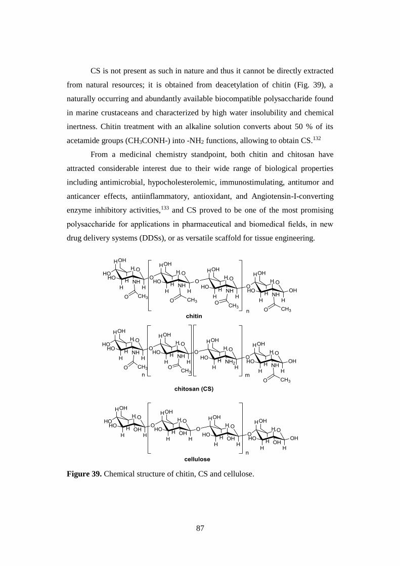
87
CS is not present as such in nature and thus it cannot be directly extracted
from natural resources; it is obtained from deacetylation of chitin (Fig. 39), a
naturally occurring and abundantly available biocompatible polysaccharide found
in marine crustaceans and characterized by high water insolubility and chemical
inertness. Chitin treatment with an alkaline solution converts about 50 % of its
acetamide groups (CH3CONH-) into -NH2 functions, allowing to obtain CS.132
From a medicinal chemistry standpoint, both chitin and chitosan have
attracted considerable interest due to their wide range of biological properties
including antimicrobial, hypocholesterolemic, immunostimulating, antitumor and
anticancer effects, antiinflammatory, antioxidant, and Angiotensin-I-converting
enzyme inhibitory activities,133 and CS proved to be one of the most promising
polysaccharide for applications in pharmaceutical and biomedical fields, in new
drug delivery systems (DDSs), or as versatile scaffold for tissue engineering.
Figure 39. Chemical structure of chitin, CS and cellulose.
88
2.6.1. Physical-chemical properties
CS is commercially available in various forms including powder, paste,
film, fiber, etc.; it is characterized by a MW ranging between 3800 and 20000
Daltons and a degree of deacetylation (% DD) from 66 % to 95 %.134
Within these large varieties of species, strong differences in water solubility and
mucoadhesivity have been detected, mainly associated to changes in MW and
degree of acetylation (% DA) values.
CS is soluble in acidic aqueous media, such as diluted CH3COOH and
HCOOH, due to the protonation of its -NH2 functions at C-2 positions; on the
contrary, it is slightly soluble in organic solvents such as DMSO and p-toluene
sulfonic acid. Accordingly, aqueous 1% CH3COOH solution has been selected to
test the solubility of different CS samples. In this context, the concentration of
acidic solution needed for obtaining CS dissolution depends on the amount of -NH2
units involved in the ionization process.132 Therefore, water solubility is closely
dependent on chitosan % DA: highly deacetylated samples (85 %) show a good
solubility in media with pH up to 6.5, a more difficult solubilization is observed
with the progressive decrement of the % DD, until getting a complete insolubility
for degree of acetylation greater than 60 %.131
CS solubility is a very difficult parameter to regulate, being influenced by
additional properties including pH and pKa of the acid selected for protonation,
distribution of acetyl groups along the polymeric chain, the intra-chain H-bonds
between the hydroxyl functions of the N-acetylglucosamine and D-glucosamine
units and, finally, the ionic concentration of the solution. Regarding this last issue,
an interesting salting-out effect in excess of HCl (1 M HCl) has been observed, and
was exploited to prepare the hydrochloride form of the polymer.132
Reganding mucoadhesivity, several studies confirmed its increment with the
increase of the % DD, as this characteristic provides a large number of positively
charged -NH2 groups thus available for the interaction with the negatively charged
residues of the mucus, such as the sialic acid units.131
89
Currently, various techniques such as IR and UV spectroscopies, elemental
analysis, 13C-NMR and 1H-NMR are employed to characterize the different CS
samples, mainly based on the determination of their % DA. In particular, 1H-NMR
in D2O is largely used for calculating the acetyl content and viscometry and HPLC
are employed for the determination of the MW and the molecular weight
distributions, respectively.
2.6.2. Applications
During the past 20 years, a substantial amount of works has been published
on CS and its uses in the fields of agriculture, industry, and medicine. In particular,
it has been described as plant antivirus, additive in liquid multicomponent
fertilizers, metal trap in wastewater due to its capability to adsorb several toxic
metals including Cu2+, Hg2+, Ni2+ and Zn2+.135
In the biomedical field, CS has largely been used as biomaterial to prepare
hydrogels, films, fibers or sponges owing to its biocompatibility, antibacterial,
antifungal and anticoagulant activities, together with its hydrophilicity, introduced
by addition of polar groups able to form secondary interactions (-OH and -NH2
groups involved in H bonds with other polymers). Furthermore, CS proved to be an
ideal polymer for developing substratum for skin replacement and for making
contact lenses, by exploiting its optical clarity, mechanical stability, oxygen
permeability, wettability and immunological compatibility.132,136
At present, the most promising developments are in the pharmaceutical area,
particularly in the drug delivery field. Interesting applications may also be seen in
cosmetics, where organic acids are usually selected as good solvents and CS is used
in the formulation of creams, lotions, and permanent waving lotions, due to its
fungicidal and fungistatic properties and to its ability to modulate viscousity.136
In the field of drug delivery, CS is extensively used to form matrixes or
membranes able to control drug release, thus avoiding repetitive dosing, to produce
carriers for the delivery of poorly soluble and/or instable drugs and for
biotechnology-based active molecules.
90
Within this large variety of DDSs, nano and microparticles represent the most usual
form of carriers with a chitosan-based composition, mainly aimed at improving the
encapsulated molecules performance and effectiveness.131,137 Importantly, many
works also report the use of chitosan as a coating material of several different core
structures, including solid lipid nanoparticles, polymeric nano and microparticles
and liposomes.
CS proved to be an ideal polysaccharide for drug delivery due to its high
biocompatibility, defined as capability to perform the desired function without
eliciting any undesirable local or systemic effects, and biodegradability, as
consequence of the fact that, under physiological conditions, its molecular chains
can be digested by lysozyme and chitinase. In addition, when oral delivery is under
consideration, the hydrolytic activity of the acidic gastric medium can be
considered as an extra mean of polymer degradation.131
In the literature, several chitosan nanoparticles are reported as delivery
system for drugs or drug candidates characterized by different biological activities
and administration modalities. Among them, chitosan nanoparticle for tacrine and
rivastigmine delivery to CNS and for curcumin encapsulation in cancer therapy
represent three very attractive examples.
In particular, tacrine-loaded chitosan nanoparticles coated by Polysorbate
80 were prepared and investigated in a pre-clinical study with the aim of improving
the bioavailability of the drug in the brain, by promoting its release in a sustained
manner, considering the tacrine ability to freely crosses the BBB, as well as
prolonging its residence time in blood after intravenous injection.138
Similarly, in an additional study rivastigmine-loaded chitosan (CS-RHT)
nanoparticles were exploited to improve the bioavailability of the drug and enhance
its uptake to the brain via intranasal (i.n.) delivery.139
Furthermore, composite nanoparticles prepared by three biocompatible
polymers: alginate (ALG), CS, and pluronic F127 were investigated as hydrophobic
DDS to cancer cells. Interestingly, in this study curcumin was used as model drug
91
and the curcumin-loaded composite nanoparticles cellular internalization was
confirmed from green fluorescence inside the cancer cells.140
There are several additional examples of CS-based nanoparticles formulated
with the aim of improving curcumin bioavailability by its encapsulation and
delivery to cancer cells. Amon them, cationic curcumin-CS poly (butyl
cyanoacrylate) nanoparticles proved to suppress hepatocellular carcinoma growth
and to inhibit tumor angiogenesis efficiently in vitro and in vivo. Furthermore,
biodegradable thermoresponsive CS-g-poly (N-vinylcaprolactam) (TRC)
nanoparticles containing curcumin have also been recognized as promising
candidates for cancer drug delivery.141
2.6.3. Chitosan-based bioconjugates
CS, due to its -NH2 and -OH functions, is an excellent starting point for the
design of polysaccharide-based conjugates very useful for drug, gene,
macromolecule delivery and tissue engineering, together with additional uses in the
biomedical field. In particular, different CS-based bioconjugates have been
developed by functionalization of the primary -NH2 groups through the classical
amine reactions such as (Scheme 9):
a) reductive amination;
b) N-alkylation;
c) N-acylation;
d) azide formation;
e) synthesis of urea or thiourea derivatives using isocyanates or thiocyanates.
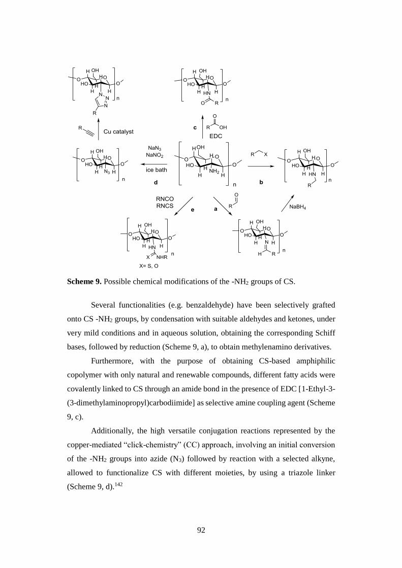
92
Scheme 9. Possible chemical modifications of the -NH2 groups of CS.
Several functionalities (e.g. benzaldehyde) have been selectively grafted
onto CS -NH2 groups, by condensation with suitable aldehydes and ketones, under
very mild conditions and in aqueous solution, obtaining the corresponding Schiff
bases, followed by reduction (Scheme 9, a), to obtain methylenamino derivatives.
Furthermore, with the purpose of obtaining CS-based amphiphilic
copolymer with only natural and renewable compounds, different fatty acids were
covalently linked to CS through an amide bond in the presence of EDC [1-Ethyl-3-
(3-dimethylaminopropyl)carbodiimide] as selective amine coupling agent (Scheme
9, c).
Additionally, the high versatile conjugation reactions represented by the
copper-mediated “click-chemistry” (CC) approach, involving an initial conversion
of the -NH2 groups into azide (N3) followed by reaction with a selected alkyne,
allowed to functionalize CS with different moieties, by using a triazole linker
(Scheme 9, d).142
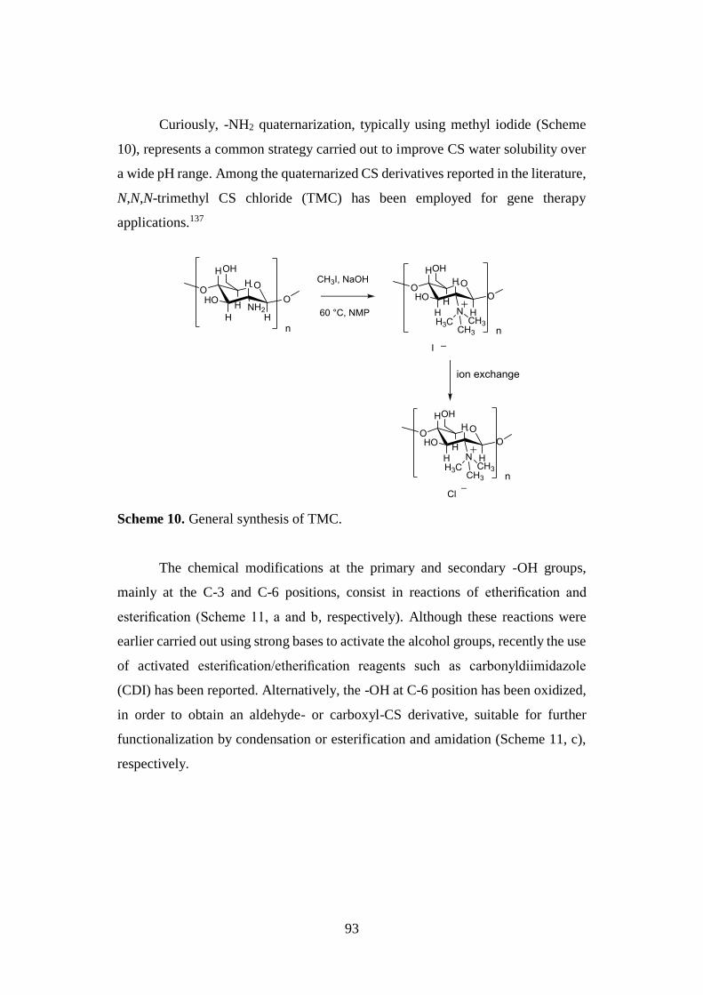
93
Curiously, -NH2 quaternarization, typically using methyl iodide (Scheme
10), represents a common strategy carried out to improve CS water solubility over
a wide pH range. Among the quaternarized CS derivatives reported in the literature,
N,N,N-trimethyl CS chloride (TMC) has been employed for gene therapy
applications.137
Scheme 10. General synthesis of TMC.
The chemical modifications at the primary and secondary -OH groups,
mainly at the C-3 and C-6 positions, consist in reactions of etherification and
esterification (Scheme 11, a and b, respectively). Although these reactions were
earlier carried out using strong bases to activate the alcohol groups, recently the use
of activated esterification/etherification reagents such as carbonyldiimidazole
(CDI) has been reported. Alternatively, the -OH at C-6 position has been oxidized,
in order to obtain an aldehyde- or carboxyl-CS derivative, suitable for further
functionalization by condensation or esterification and amidation (Scheme 11, c),
respectively.
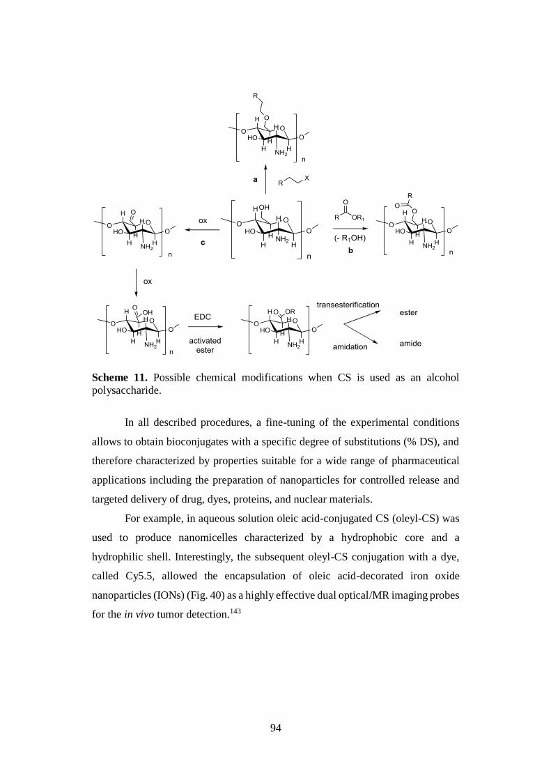
94
Scheme 11. Possible chemical modifications when CS is used as an alcohol
polysaccharide.
In all described procedures, a fine-tuning of the experimental conditions
allows to obtain bioconjugates with a specific degree of substitutions (% DS), and
therefore characterized by properties suitable for a wide range of pharmaceutical
applications including the preparation of nanoparticles for controlled release and
targeted delivery of drug, dyes, proteins, and nuclear materials.
For example, in aqueous solution oleic acid-conjugated CS (oleyl-CS) was
used to produce nanomicelles characterized by a hydrophobic core and a
hydrophilic shell. Interestingly, the subsequent oleyl-CS conjugation with a dye,
called Cy5.5, allowed the encapsulation of oleic acid-decorated iron oxide
nanoparticles (IONs) (Fig. 40) as a highly effective dual optical/MR imaging probes
for the in vivo tumor detection.143
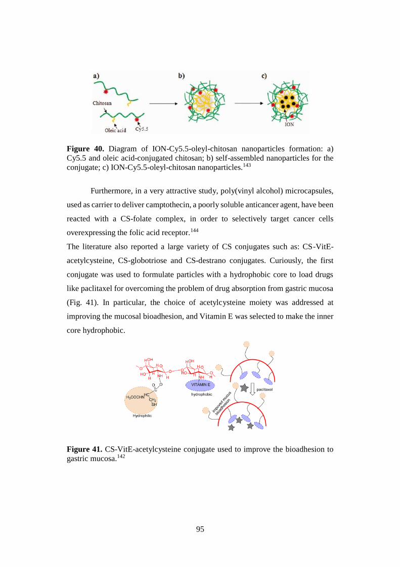
95
Figure 40. Diagram of ION-Cy5.5-oleyl-chitosan nanoparticles formation: a)
Cy5.5 and oleic acid-conjugated chitosan; b) self-assembled nanoparticles for the
conjugate; c) ION-Cy5.5-oleyl-chitosan nanoparticles.143
Furthermore, in a very attractive study, poly(vinyl alcohol) microcapsules,
used as carrier to deliver camptothecin, a poorly soluble anticancer agent, have been
reacted with a CS-folate complex, in order to selectively target cancer cells
overexpressing the folic acid receptor.144
The literature also reported a large variety of CS conjugates such as: CS-VitE-
acetylcysteine, CS-globotriose and CS-destrano conjugates. Curiously, the first
conjugate was used to formulate particles with a hydrophobic core to load drugs
like paclitaxel for overcoming the problem of drug absorption from gastric mucosa
(Fig. 41). In particular, the choice of acetylcysteine moiety was addressed at
improving the mucosal bioadhesion, and Vitamin E was selected to make the inner
core hydrophobic.
Figure 41. CS-VitE-acetylcysteine conjugate used to improve the bioadhesion to
gastric mucosa.142
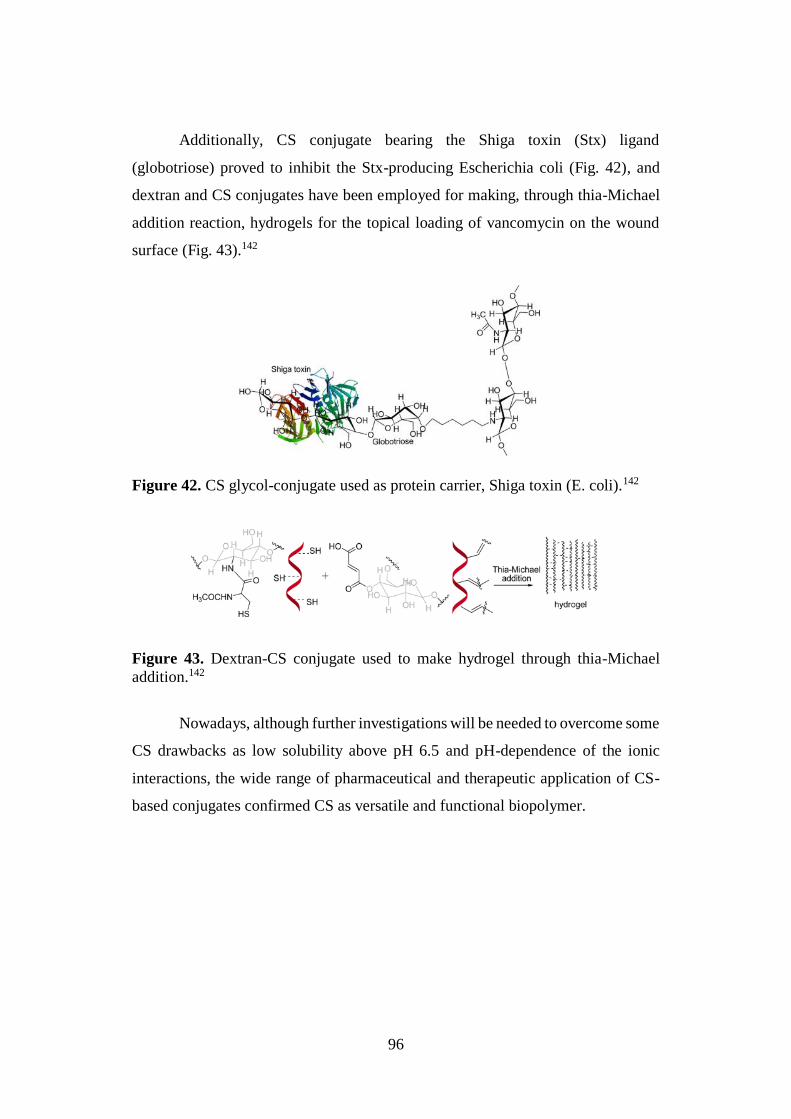
96
Additionally, CS conjugate bearing the Shiga toxin (Stx) ligand
(globotriose) proved to inhibit the Stx-producing Escherichia coli (Fig. 42), and
dextran and CS conjugates have been employed for making, through thia-Michael
addition reaction, hydrogels for the topical loading of vancomycin on the wound
surface (Fig. 43).142
Figure 42. CS glycol-conjugate used as protein carrier, Shiga toxin (E. coli).142
Figure 43. Dextran-CS conjugate used to make hydrogel through thia-Michael
addition.142
Nowadays, although further investigations will be needed to overcome some
CS drawbacks as low solubility above pH 6.5 and pH-dependence of the ionic
interactions, the wide range of pharmaceutical and therapeutic application of CS-
based conjugates confirmed CS as versatile and functional biopolymer.
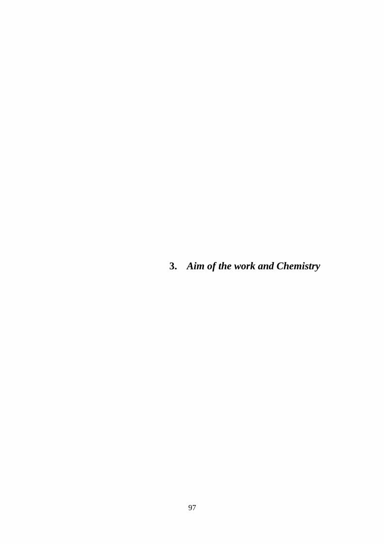
97
3. Aim of the work and Chemistry
98
3.1. DESIGN OF CURCUMIN-BASED COMPOUNDS
The heterogeneous nature of AD directed attention to polypharmacological
strategies and multitarget drugs that, acting on several targets involved in the
pathology, could offer promises to achieve a successful treatment. In this scenario,
the central roles of Aβ aggregation and τ hyperphosphorylation in AD pathogenesis,
together with the molecular interplay between Aβ and τ proteins in causing synergic
toxicity, led to recognize BACE-1 and GSK-3β as two validated targets. The
simultaneous inhibition of these enzymes represents a promising therapeutic
modifying strategy, able to halt the disease progression.
Although BACE-1 and GSK-3β are structurally unrelated enzymes and
greatly differ in both three-dimensional structures and binding site topologies, we
rationally envisaged the 5-hydroxy-1,7-diarylhepta-1,4,6-trien-3-one framework of
curcumin 1a (Fig. 33), as essential structural element for their concurrent
modulation. In particular, the curcumin β-keto-enol central core looked suitable for
the contact with BACE-1 catalytic dyad (Asp32 and Asp228), whereas the highly
electrophilic α,β-unsaturated carbonyl system, a well-known Michael acceptor
system, seemed appropriate to react with the crucial Cys199 residue of GSK-3β
hinge region.
To validate our hypothesis, the interactions of curcumin 1a with the binding
pocket of both targets (Figs. 44a and 44b) were investigated by docking simulations
from which clearly emerged how:
1) in the BACE-1 catalytic pocket (Fig. 44a), the central curcumin core was
hydrogen bonded to the crucial Asp32 and Asp228 residues, while the side
aryl rings established contacts with Tyr198, Lys224, and Tyr71.
2) In the GSK-3β binding site (Fig. 44b), the central β-keto-enol function
participated in a H-bond with Tyr134 and Val135, and the two aryl functions
contacted Lys85, Glu97, and Arg141, Glu173, respectively.
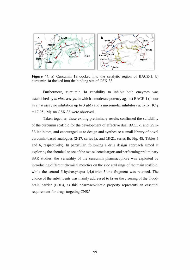
99
Figure 44. a) Curcumin 1a docked into the catalytic region of BACE-1; b)
curcumin 1a docked into the binding site of GSK-3β.
Furthermore, curcumin 1a capability to inhibit both enzymes was
established by in vitro assays, in which a moderate potency against BACE-1 (in our
in vitro assay no inhibition up to 3 μM) and a micromolar inhibitory activity (IC50
= 17.95 μM) on GSK-3β were observed.
Taken together, these exiting preliminary results confirmed the suitability
of the curcumin scaffold for the development of effective dual BACE-1 and GSK-
3β inhibitors, and encouraged us to design and synthesize a small library of novel
curcumin-based analogues (2-17, series Ia, and 18-21, series Ib, Fig. 45, Tables 5
and 6, respectively). In particular, following a drug design approach aimed at
exploring the chemical space of the two selected targets and performing preliminary
SAR studies, the versatility of the curcumin pharmacophore was exploited by
introducing different chemical moieties on the side aryl rings of the main scaffold,
while the central 5-hydroxyhepta-1,4,6-trien-3-one fragment was retained. The
choice of the substituents was mainly addressed to favor the crossing of the blood-
brain barrier (BBB), as this pharmacokinetic property represents an essential
requirement for drugs targeting CNS.8
a b
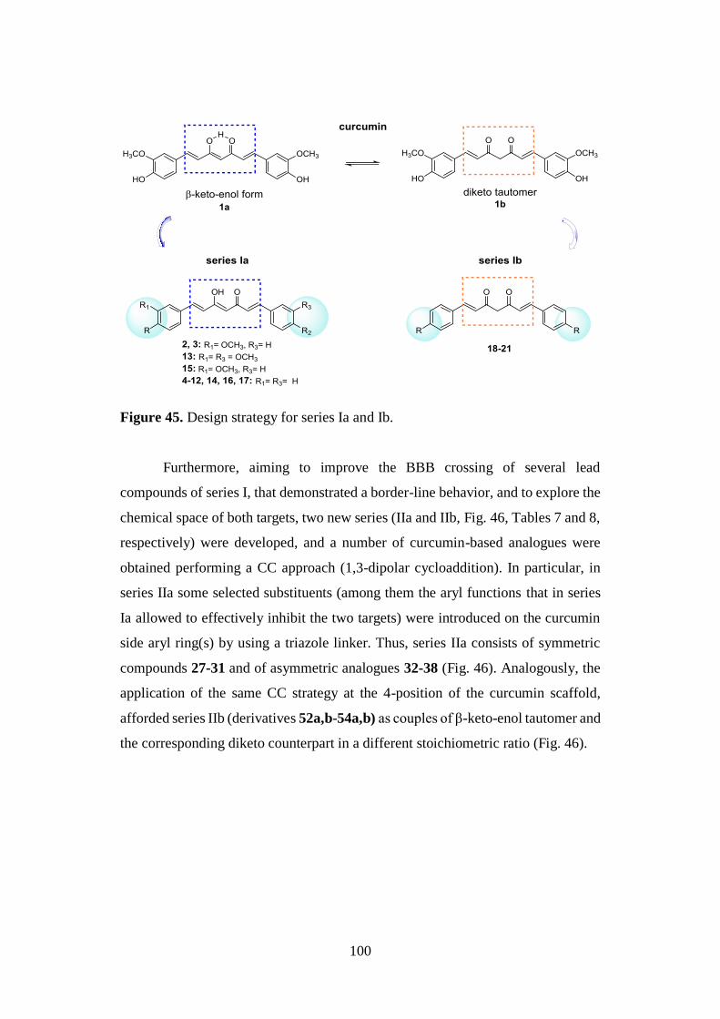
100
Figure 45. Design strategy for series Ia and Ib.
Furthermore, aiming to improve the BBB crossing of several lead
compounds of series I, that demonstrated a border-line behavior, and to explore the
chemical space of both targets, two new series (IIa and IIb, Fig. 46, Tables 7 and 8,
respectively) were developed, and a number of curcumin-based analogues were
obtained performing a CC approach (1,3-dipolar cycloaddition). In particular, in
series IIa some selected substituents (among them the aryl functions that in series
Ia allowed to effectively inhibit the two targets) were introduced on the curcumin
side aryl ring(s) by using a triazole linker. Thus, series IIa consists of symmetric
compounds 27-31 and of asymmetric analogues 32-38 (Fig. 46). Analogously, the
application of the same CC strategy at the 4-position of the curcumin scaffold,
afforded series IIb (derivatives 52a,b-54a,b) as couples of β-keto-enol tautomer and
the corresponding diketo counterpart in a different stoichiometric ratio (Fig. 46).
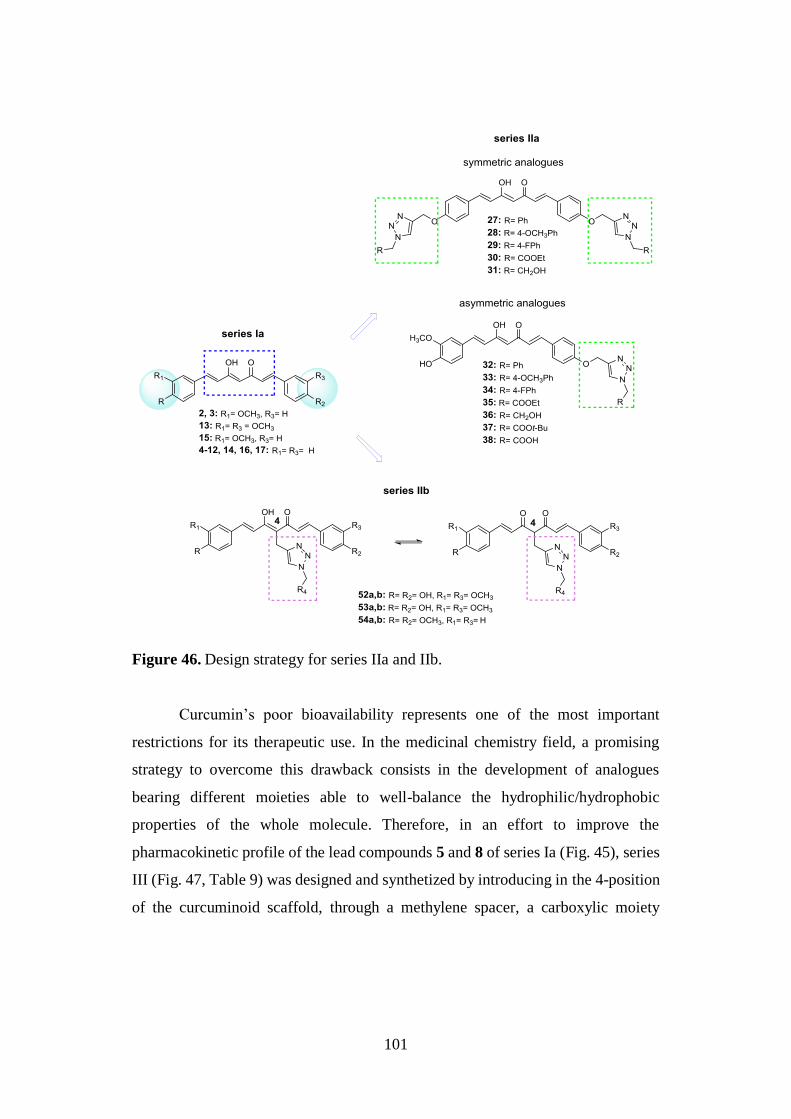
101
Figure 46. Design strategy for series IIa and IIb.
Curcumin’s poor bioavailability represents one of the most important
restrictions for its therapeutic use. In the medicinal chemistry field, a promising
strategy to overcome this drawback consists in the development of analogues
bearing different moieties able to well-balance the hydrophilic/hydrophobic
properties of the whole molecule. Therefore, in an effort to improve the
pharmacokinetic profile of the lead compounds 5 and 8 of series Ia (Fig. 45), series
III (Fig. 47, Table 9) was designed and synthetized by introducing in the 4-position
of the curcuminoid scaffold, through a methylene spacer, a carboxylic moiety
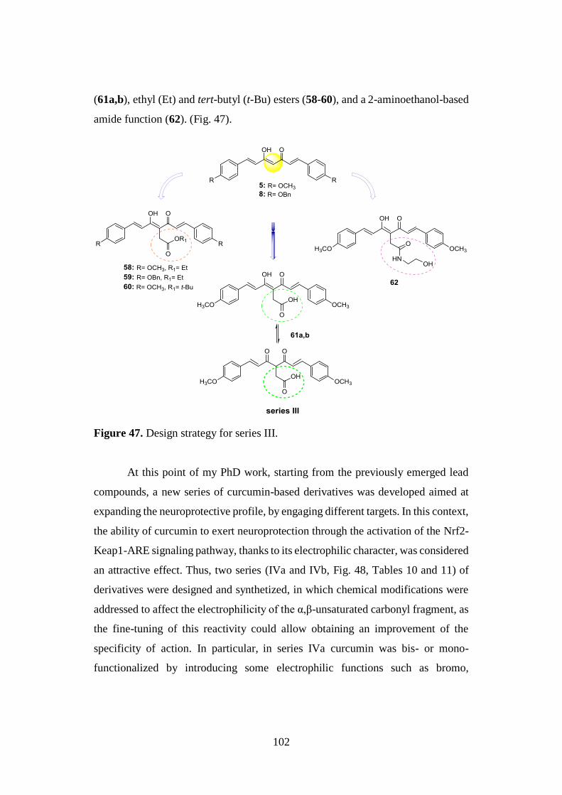
102
(61a,b), ethyl (Et) and tert-butyl (t-Bu) esters (58-60), and a 2-aminoethanol-based
amide function (62). (Fig. 47).
Figure 47. Design strategy for series III.
At this point of my PhD work, starting from the previously emerged lead
compounds, a new series of curcumin-based derivatives was developed aimed at
expanding the neuroprotective profile, by engaging different targets. In this context,
the ability of curcumin to exert neuroprotection through the activation of the Nrf2-
Keap1-ARE signaling pathway, thanks to its electrophilic character, was considered
an attractive effect. Thus, two series (IVa and IVb, Fig. 48, Tables 10 and 11) of
derivatives were designed and synthetized, in which chemical modifications were
addressed to affect the electrophilicity of the α,β-unsaturated carbonyl fragment, as
the fine-tuning of this reactivity could allow obtaining an improvement of the
specificity of action. In particular, in series IVa curcumin was bis- or mono-
functionalized by introducing some electrophilic functions such as bromo,
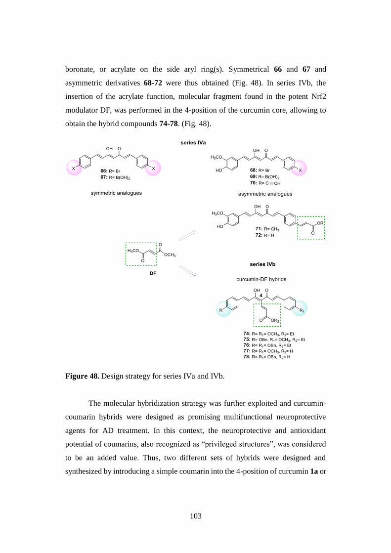
103
boronate, or acrylate on the side aryl ring(s). Symmetrical 66 and 67 and
asymmetric derivatives 68-72 were thus obtained (Fig. 48). In series IVb, the
insertion of the acrylate function, molecular fragment found in the potent Nrf2
modulator DF, was performed in the 4-position of the curcumin core, allowing to
obtain the hybrid compounds 74-78. (Fig. 48).
Figure 48. Design strategy for series IVa and IVb.
The molecular hybridization strategy was further exploited and curcumin-
coumarin hybrids were designed as promising multifunctional neuroprotective
agents for AD treatment. In this context, the neuroprotective and antioxidant
potential of coumarins, also recognized as “privileged structures”, was considered
to be an added value. Thus, two different sets of hybrids were designed and
synthesized by introducing a simple coumarin into the 4-position of curcumin 1a or
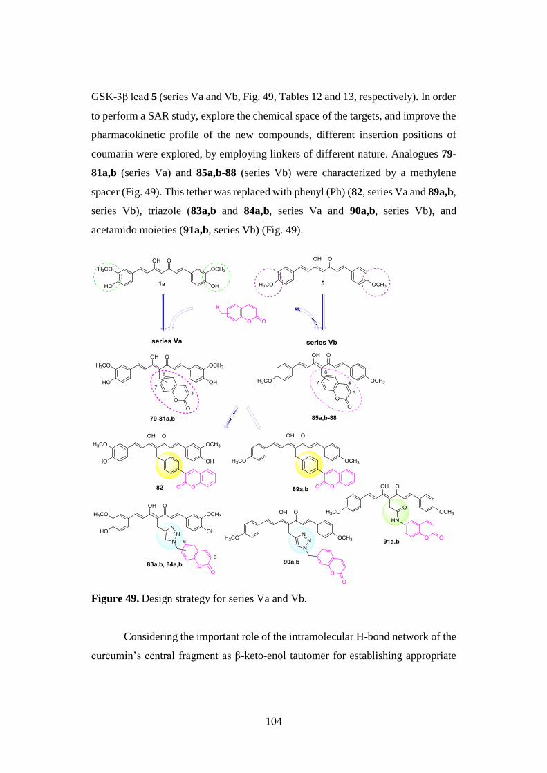
104
GSK-3β lead 5 (series Va and Vb, Fig. 49, Tables 12 and 13, respectively). In order
to perform a SAR study, explore the chemical space of the targets, and improve the
pharmacokinetic profile of the new compounds, different insertion positions of
coumarin were explored, by employing linkers of different nature. Analogues 79-
81a,b (series Va) and 85a,b-88 (series Vb) were characterized by a methylene
spacer (Fig. 49). This tether was replaced with phenyl (Ph) (82, series Va and 89a,b,
series Vb), triazole (83a,b and 84a,b, series Va and 90a,b, series Vb), and
acetamido moieties (91a,b, series Vb) (Fig. 49).
Figure 49. Design strategy for series Va and Vb.
Considering the important role of the intramolecular H-bond network of the
curcumin’s central fragment as β-keto-enol tautomer for establishing appropriate
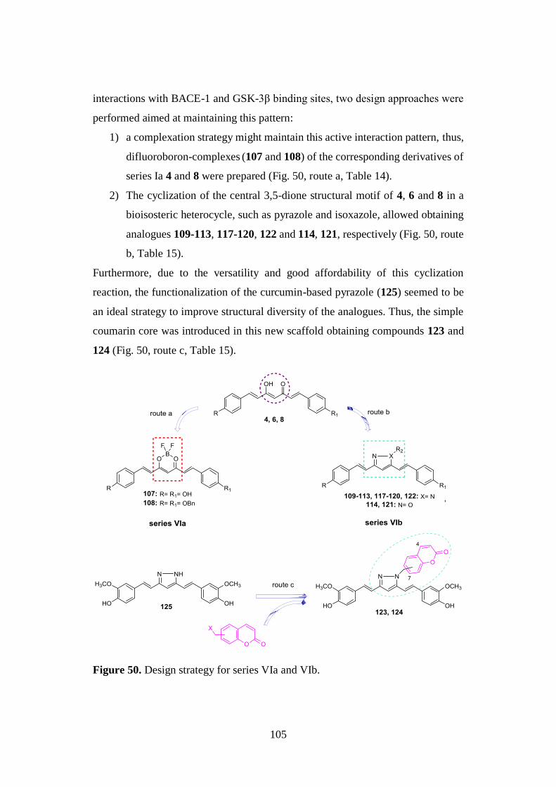
105
interactions with BACE-1 and GSK-3β binding sites, two design approaches were
performed aimed at maintaining this pattern:
1) a complexation strategy might maintain this active interaction pattern, thus,
difluoroboron-complexes (107 and 108) of the corresponding derivatives of
series Ia 4 and 8 were prepared (Fig. 50, route a, Table 14).
2) The cyclization of the central 3,5-dione structural motif of 4, 6 and 8 in a
bioisosteric heterocycle, such as pyrazole and isoxazole, allowed obtaining
analogues 109-113, 117-120, 122 and 114, 121, respectively (Fig. 50, route
b, Table 15).
Furthermore, due to the versatility and good affordability of this cyclization
reaction, the functionalization of the curcumin-based pyrazole (125) seemed to be
an ideal strategy to improve structural diversity of the analogues. Thus, the simple
coumarin core was introduced in this new scaffold obtaining compounds 123 and
124 (Fig. 50, route c, Table 15).
Figure 50. Design strategy for series VIa and VIb.
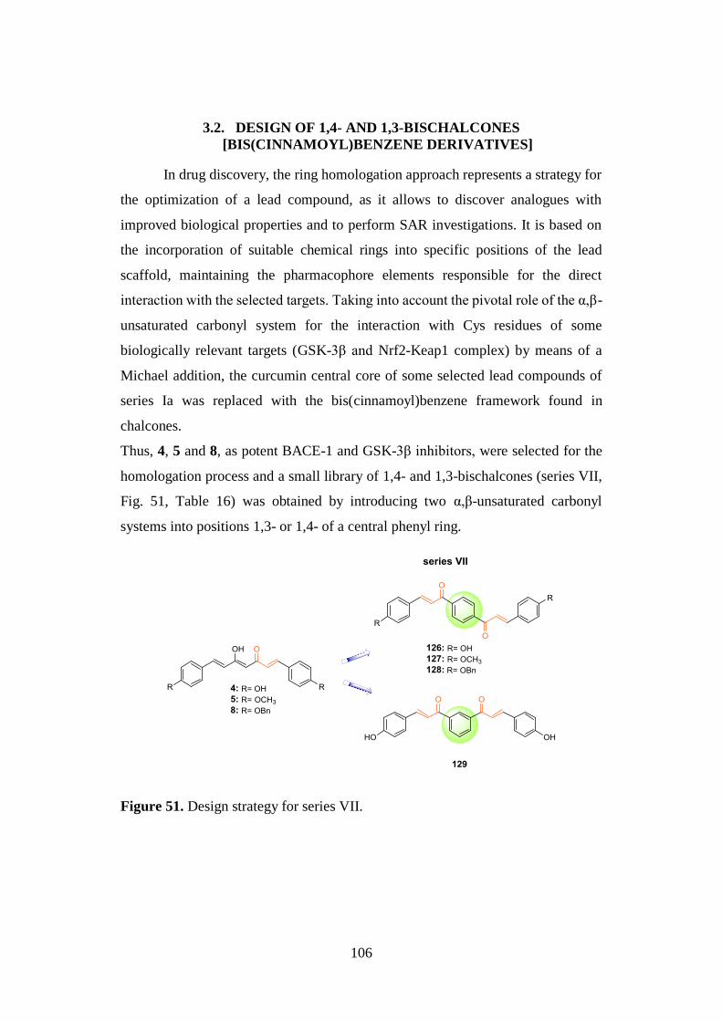
106
3.2. DESIGN OF 1,4- AND 1,3-BISCHALCONES
[BIS(CINNAMOYL)BENZENE DERIVATIVES]
In drug discovery, the ring homologation approach represents a strategy for
the optimization of a lead compound, as it allows to discover analogues with
improved biological properties and to perform SAR investigations. It is based on
the incorporation of suitable chemical rings into specific positions of the lead
scaffold, maintaining the pharmacophore elements responsible for the direct
interaction with the selected targets. Taking into account the pivotal role of the α,β-
unsaturated carbonyl system for the interaction with Cys residues of some
biologically relevant targets (GSK-3β and Nrf2-Keap1 complex) by means of a
Michael addition, the curcumin central core of some selected lead compounds of
series Ia was replaced with the bis(cinnamoyl)benzene framework found in
chalcones.
Thus, 4, 5 and 8, as potent BACE-1 and GSK-3β inhibitors, were selected for the
homologation process and a small library of 1,4- and 1,3-bischalcones (series VII,
Fig. 51, Table 16) was obtained by introducing two α,β-unsaturated carbonyl
systems into positions 1,3- or 1,4- of a central phenyl ring.
Figure 51. Design strategy for series VII.
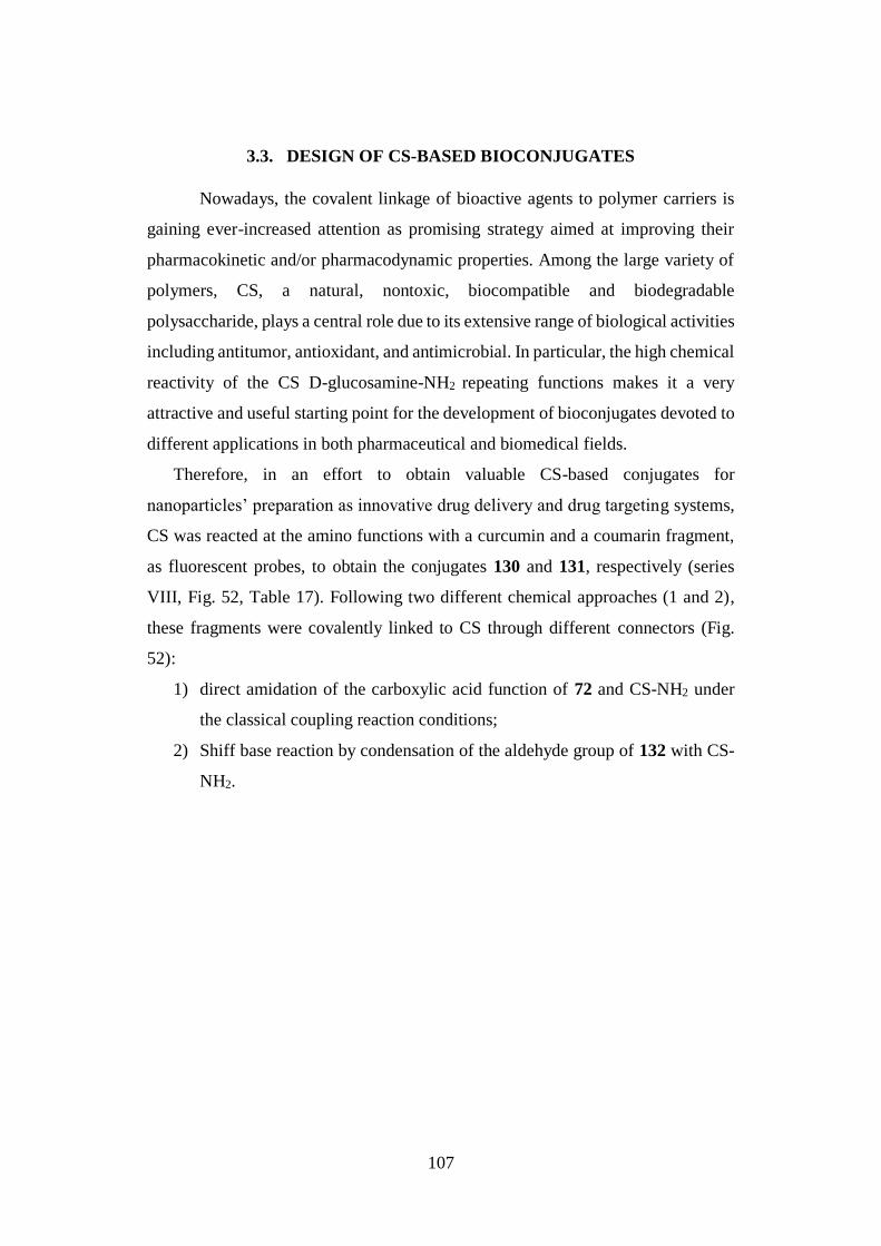
107
3.3. DESIGN OF CS-BASED BIOCONJUGATES
Nowadays, the covalent linkage of bioactive agents to polymer carriers is
gaining ever-increased attention as promising strategy aimed at improving their
pharmacokinetic and/or pharmacodynamic properties. Among the large variety of
polymers, CS, a natural, nontoxic, biocompatible and biodegradable
polysaccharide, plays a central role due to its extensive range of biological activities
including antitumor, antioxidant, and antimicrobial. In particular, the high chemical
reactivity of the CS D-glucosamine-NH2 repeating functions makes it a very
attractive and useful starting point for the development of bioconjugates devoted to
different applications in both pharmaceutical and biomedical fields.
Therefore, in an effort to obtain valuable CS-based conjugates for
nanoparticles’ preparation as innovative drug delivery and drug targeting systems,
CS was reacted at the amino functions with a curcumin and a coumarin fragment,
as fluorescent probes, to obtain the conjugates 130 and 131, respectively (series
VIII, Fig. 52, Table 17). Following two different chemical approaches (1 and 2),
these fragments were covalently linked to CS through different connectors (Fig.
52):
1) direct amidation of the carboxylic acid function of 72 and CS-NH2 under
the classical coupling reaction conditions;
2) Shiff base reaction by condensation of the aldehyde group of 132 with CS-
NH2.
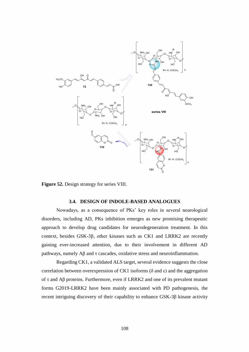
108
Figure 52. Design strategy for series VIII.
3.4. DESIGN OF INDOLE-BASED ANALOGUES
Nowadays, as a consequence of PKs’ key roles in several neurological
disorders, including AD, PKs inhibition emerges as new promising therapeutic
approach to develop drug candidates for neurodegeneration treatment. In this
context, besides GSK-3β, other kinases such as CK1 and LRRK2 are recently
gaining ever-increased attention, due to their involvement in different AD
pathways, namely Aβ and τ cascades, oxidative stress and neuroinflammation.
Regarding CK1, a validated ALS target, several evidence suggests the close
correlation between overexpression of CK1 isoforms (δ and ε) and the aggregation
of τ and Aβ proteins. Furthermore, even if LRRK2 and one of its prevalent mutant
forms G2019-LRRK2 have been mainly associated with PD pathogenesis, the
recent intriguing discovery of their capability to enhance GSK-3β kinase activity
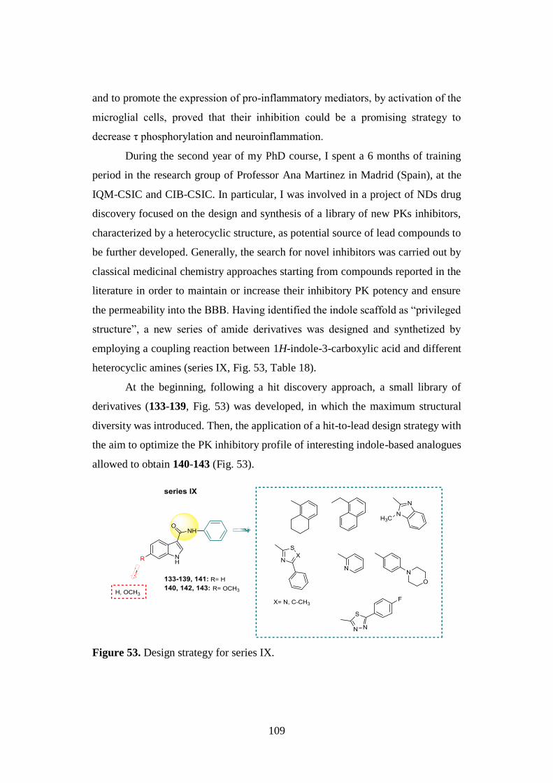
109
and to promote the expression of pro-inflammatory mediators, by activation of the
microglial cells, proved that their inhibition could be a promising strategy to
decrease τ phosphorylation and neuroinflammation.
During the second year of my PhD course, I spent a 6 months of training
period in the research group of Professor Ana Martinez in Madrid (Spain), at the
IQM-CSIC and CIB-CSIC. In particular, I was involved in a project of NDs drug
discovery focused on the design and synthesis of a library of new PKs inhibitors,
characterized by a heterocyclic structure, as potential source of lead compounds to
be further developed. Generally, the search for novel inhibitors was carried out by
classical medicinal chemistry approaches starting from compounds reported in the
literature in order to maintain or increase their inhibitory PK potency and ensure
the permeability into the BBB. Having identified the indole scaffold as “privileged
structure”, a new series of amide derivatives was designed and synthetized by
employing a coupling reaction between 1H-indole-3-carboxylic acid and different
heterocyclic amines (series IX, Fig. 53, Table 18).
At the beginning, following a hit discovery approach, a small library of
derivatives (133-139, Fig. 53) was developed, in which the maximum structural
diversity was introduced. Then, the application of a hit-to-lead design strategy with
the aim to optimize the PK inhibitory profile of interesting indole-based analogues
allowed to obtain 140-143 (Fig. 53).
Figure 53. Design strategy for series IX.
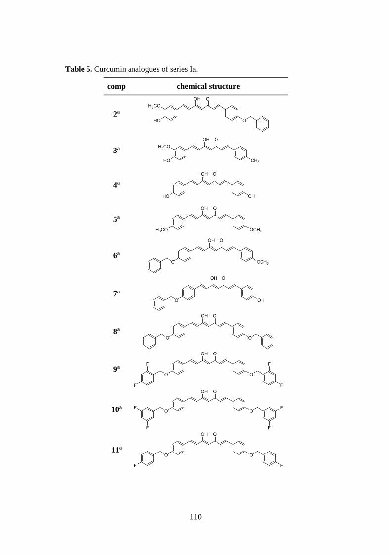
110
Table 5. Curcumin analogues of series Ia.
comp chemical structure
2a
3a
4a
5a
6a
7a
8a
9a
10a
11a
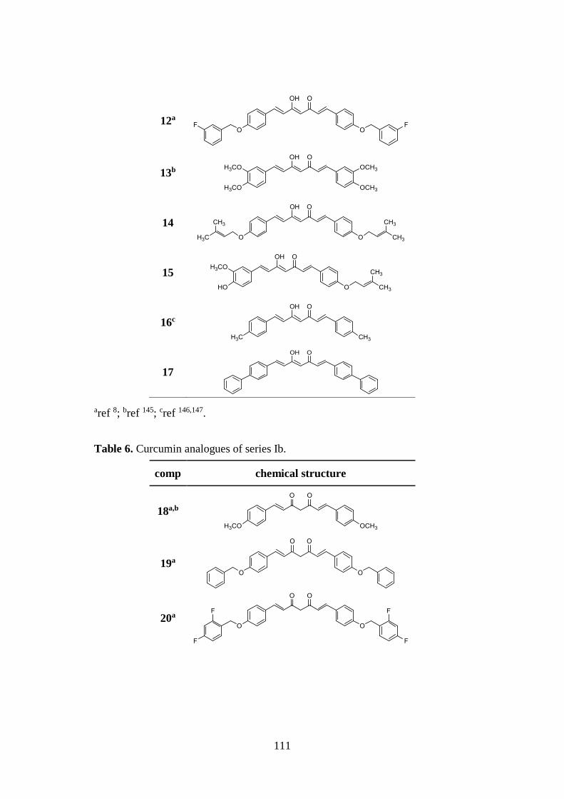
111
12a
13b
14
15
16c
17
aref 8; bref 145; cref 146,147.
Table 6. Curcumin analogues of series Ib.
comp chemical structure
18a,b
19a
20a
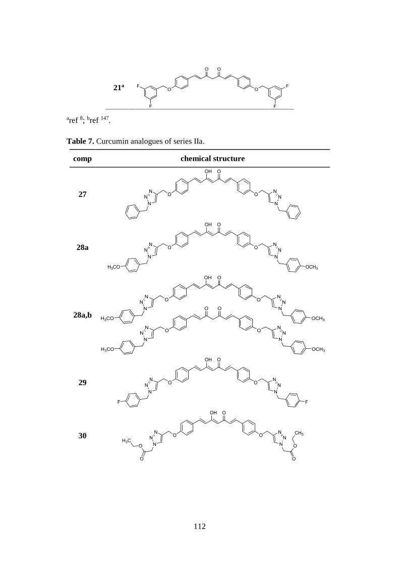
112
21a
aref 8; bref 147.
Table 7. Curcumin analogues of series IIa.
comp chemical structure
27
28a
28a,b
29
30
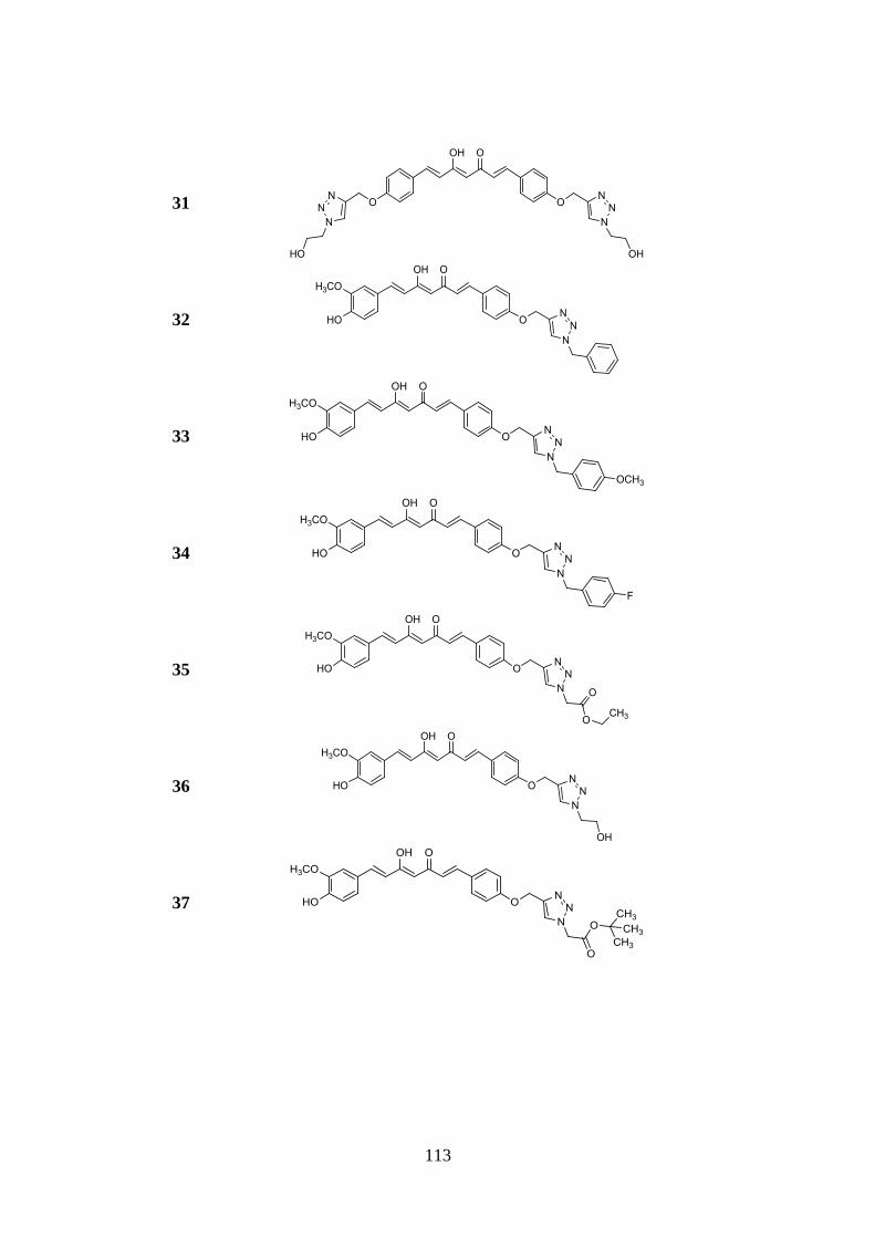
113
31
32
33
34
35
36
37
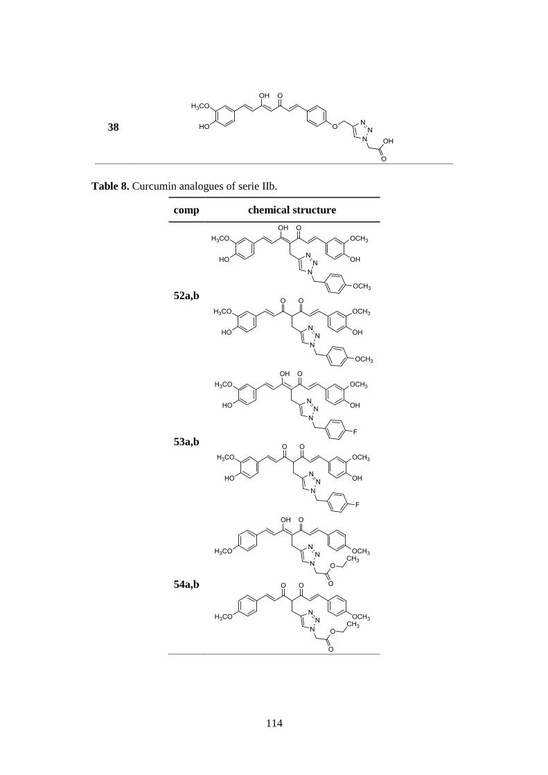
114
38
Table 8. Curcumin analogues of serie IIb.
comp chemical structure
52a,b
53a,b
54a,b
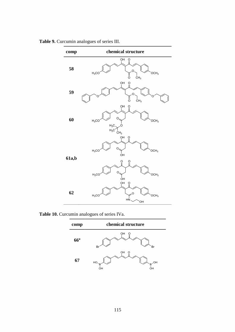
115
Table 9. Curcumin analogues of series III.
comp chemical structure
58
59
60
61a,b
62
Table 10. Curcumin analogues of series IVa.
comp chemical structure
66a
67
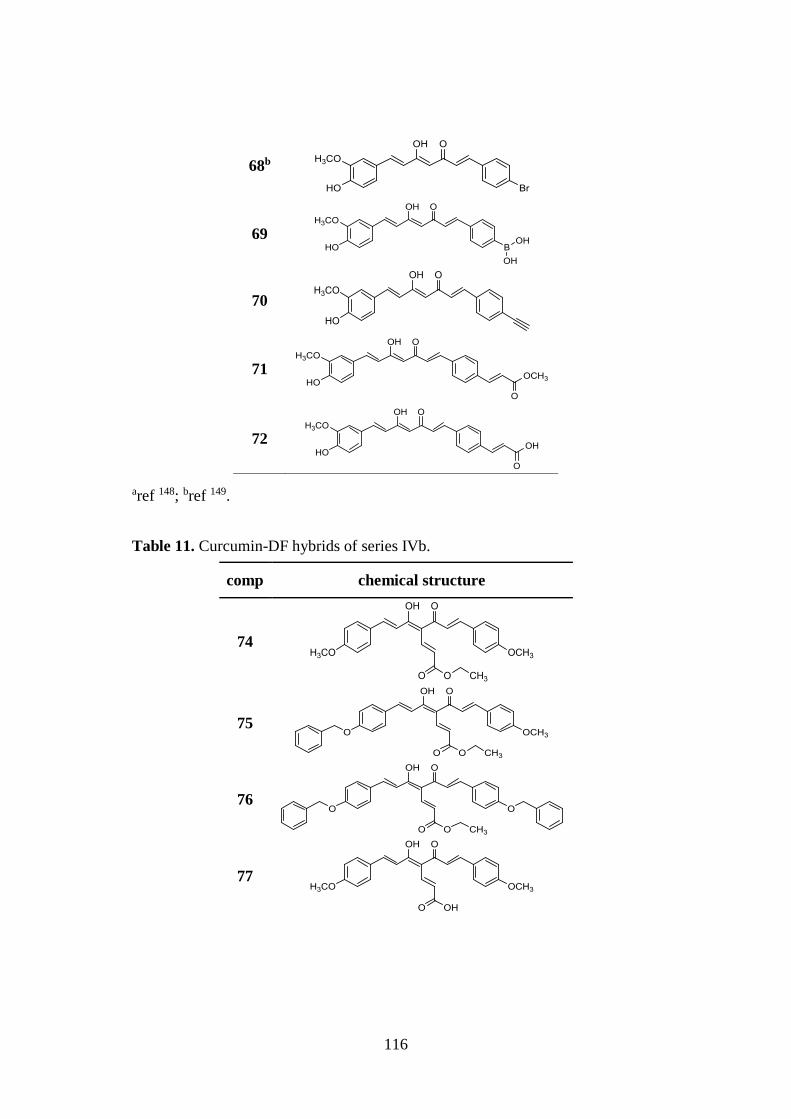
116
68b
69
70
71
72
aref 148; bref 149.
Table 11. Curcumin-DF hybrids of series IVb.
comp chemical structure
74
75
76
77
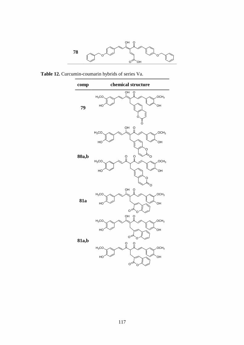
117
78
Table 12. Curcumin-coumarin hybrids of series Va.
comp chemical structure
79
80a,b
81a
81a,b
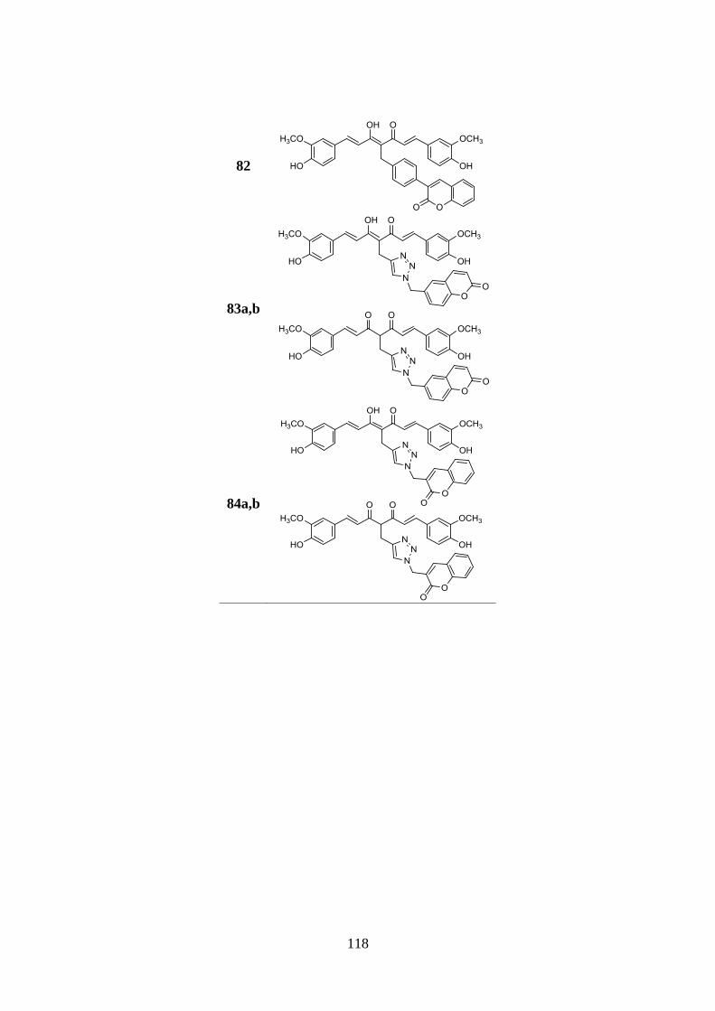
118
82
83a,b
84a,b
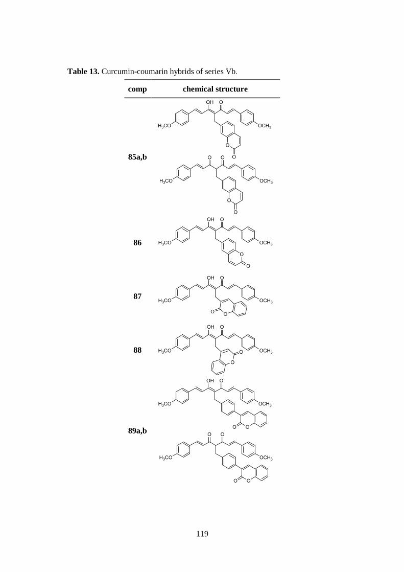
119
Table 13. Curcumin-coumarin hybrids of series Vb.
comp chemical structure
85a,b
86
87
88
89a,b
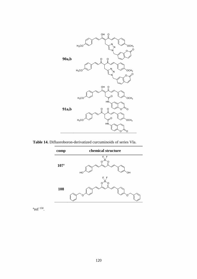
120
90a,b
91a,b
Table 14. Difluoroboron-derivatized curcuminoids of series VIa.
comp chemical structure
107a
108
aref 150.
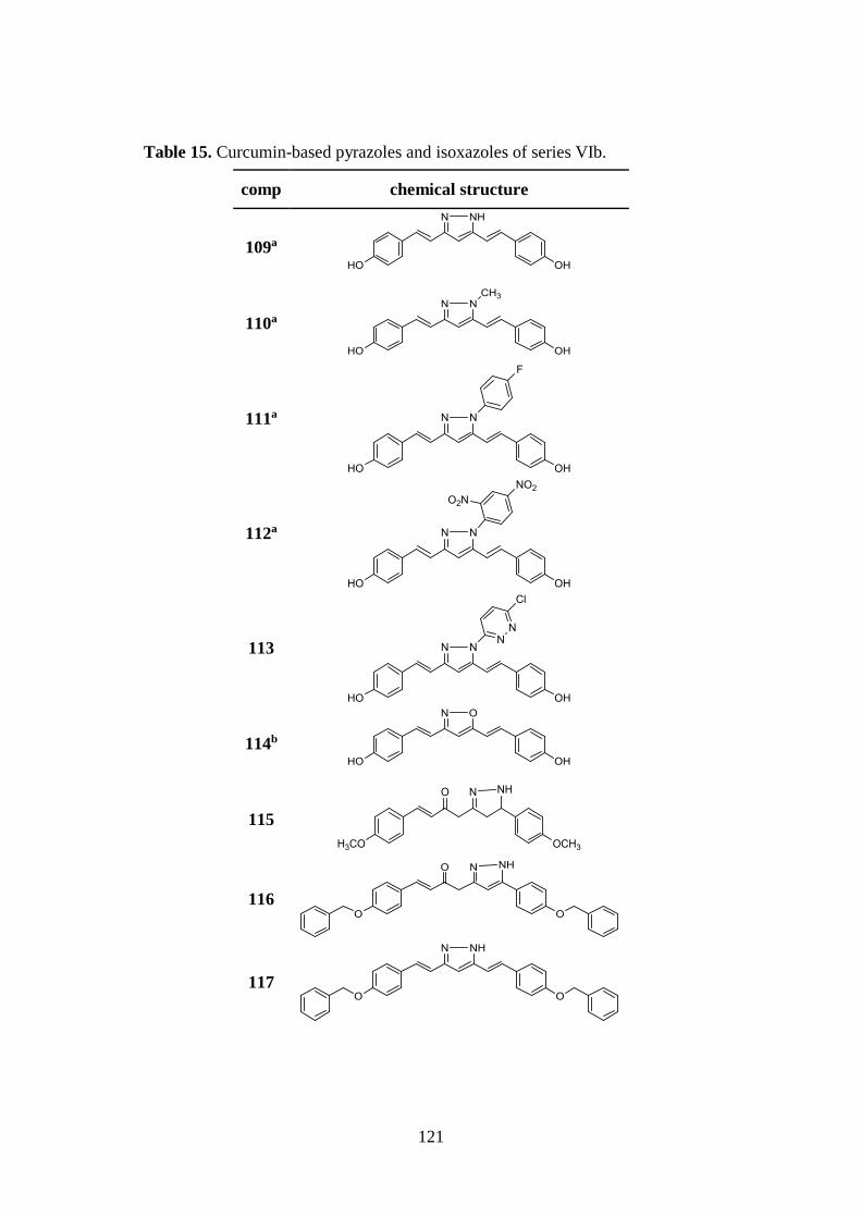
121
Table 15. Curcumin-based pyrazoles and isoxazoles of series VIb.
comp chemical structure
109a
110a
111a
112a
113
114b
115
116
117
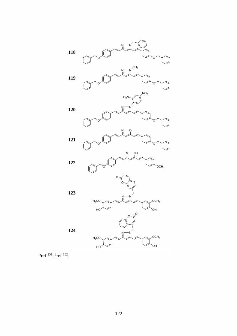
122
118
119
120
121
122
123
124
aref 151; bref 152.
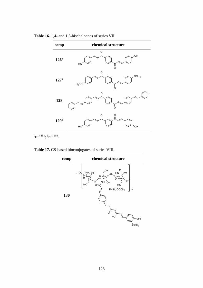
123
Table 16. 1,4- and 1,3-bischalcones of series VII.
comp chemical structure
126a
127a
128
129b
aref 153; bref 154.
Table 17. CS-based bioconjugates of series VIII.
comp chemical structure
130
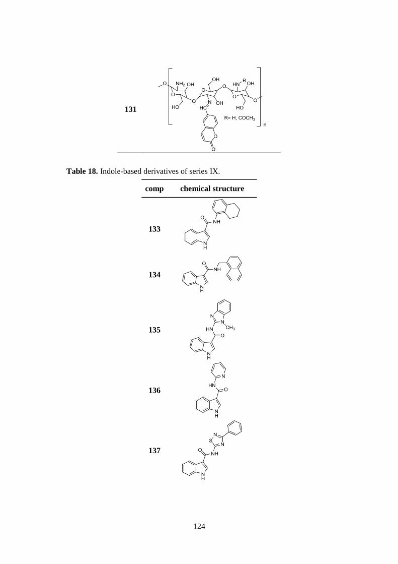
124
131
Table 18. Indole-based derivatives of series IX.
comp chemical structure
133
134
135
136
137
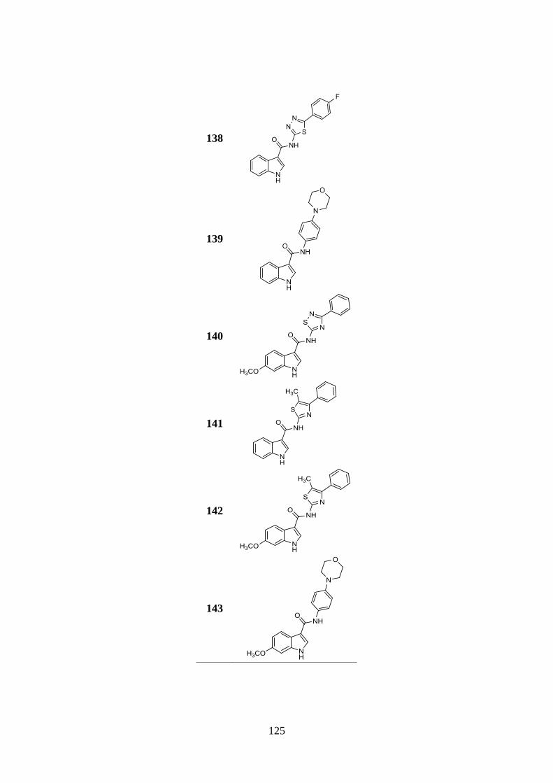
125
138
139
140
141
142
143
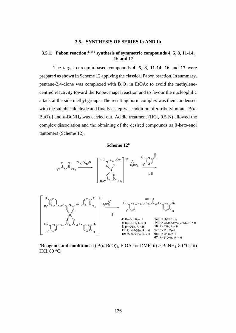
126
3.5. SYNTHESIS OF SERIES Ia AND Ib
3.5.1. Pabon reaction:8,155 synthesis of symmetric compounds 4, 5, 8, 11-14,
16 and 17
The target curcumin-based compounds 4, 5, 8, 11-14, 16 and 17 were
prepared as shown in Scheme 12 applying the classical Pabon reaction. In summary,
pentane-2,4-dione was complexed with B2O3 in EtOAc to avoid the methylene-
centred reactivity toward the Knoevenagel reaction and to favour the nucleophilic
attack at the side methyl groups. The resulting boric complex was then condensed
with the suitable aldehyde and finally a step-wise addition of n-tributylborate [B(n-
BuO)3] and n-BuNH2 was carried out. Acidic treatment (HCl, 0.5 N) allowed the
complex dissociation and the obtaining of the desired compounds as β-keto-enol
tautomers (Scheme 12).
Scheme 12a
aReagents and conditions: i) B(n-BuO)3, EtOAc or DMF; ii) n-BuNH2, 80 °C; iii)
HCl, 80 °C.
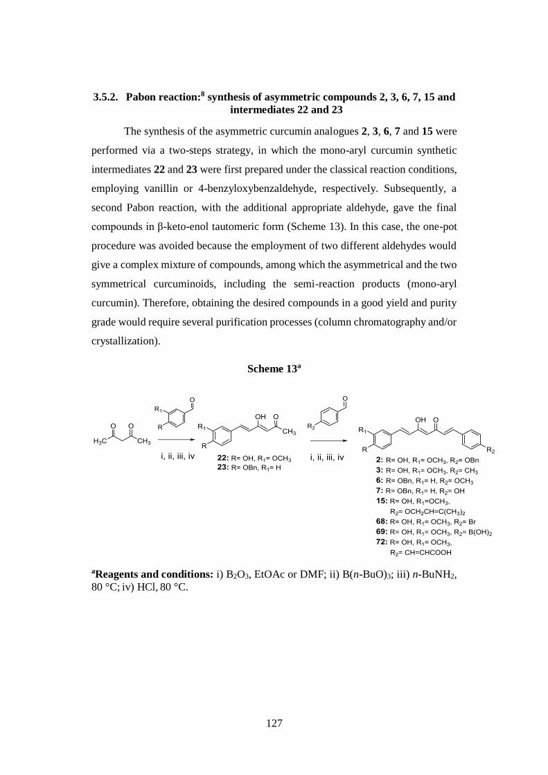
127
3.5.2. Pabon reaction:8 synthesis of asymmetric compounds 2, 3, 6, 7, 15 and
intermediates 22 and 23
The synthesis of the asymmetric curcumin analogues 2, 3, 6, 7 and 15 were
performed via a two-steps strategy, in which the mono-aryl curcumin synthetic
intermediates 22 and 23 were first prepared under the classical reaction conditions,
employing vanillin or 4-benzyloxybenzaldehyde, respectively. Subsequently, a
second Pabon reaction, with the additional appropriate aldehyde, gave the final
compounds in β-keto-enol tautomeric form (Scheme 13). In this case, the one-pot
procedure was avoided because the employment of two different aldehydes would
give a complex mixture of compounds, among which the asymmetrical and the two
symmetrical curcuminoids, including the semi-reaction products (mono-aryl
curcumin). Therefore, obtaining the desired compounds in a good yield and purity
grade would require several purification processes (column chromatography and/or
crystallization).
Scheme 13a
aReagents and conditions: i) B2O3, EtOAc or DMF; ii) B(n-BuO)3; iii) n-BuNH2, 80 °C; iv) HCl, 80 °C.
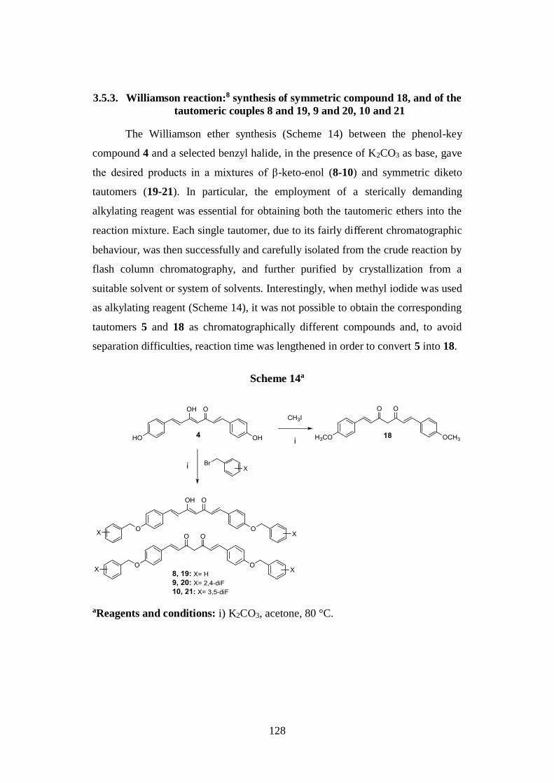
128
3.5.3. Williamson reaction:8 synthesis of symmetric compound 18, and of the
tautomeric couples 8 and 19, 9 and 20, 10 and 21
The Williamson ether synthesis (Scheme 14) between the phenol-key
compound 4 and a selected benzyl halide, in the presence of K2CO3 as base, gave
the desired products in a mixtures of β-keto-enol (8-10) and symmetric diketo
tautomers (19-21). In particular, the employment of a sterically demanding
alkylating reagent was essential for obtaining both the tautomeric ethers into the
reaction mixture. Each single tautomer, due to its fairly different chromatographic
behaviour, was then successfully and carefully isolated from the crude reaction by
flash column chromatography, and further purified by crystallization from a
suitable solvent or system of solvents. Interestingly, when methyl iodide was used
as alkylating reagent (Scheme 14), it was not possible to obtain the corresponding
tautomers 5 and 18 as chromatographically different compounds and, to avoid
separation difficulties, reaction time was lengthened in order to convert 5 into 18.
Scheme 14a
aReagents and conditions: i) K2CO3, acetone, 80 °C.
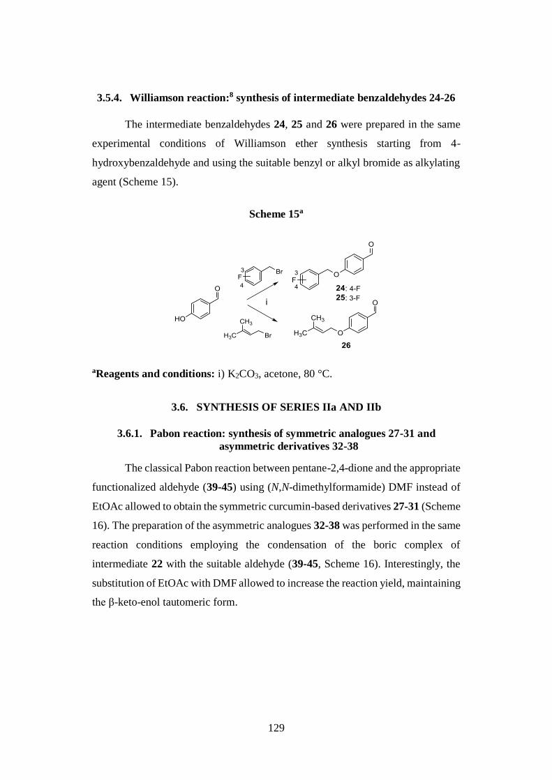
129
3.5.4. Williamson reaction:8 synthesis of intermediate benzaldehydes 24-26
The intermediate benzaldehydes 24, 25 and 26 were prepared in the same
experimental conditions of Williamson ether synthesis starting from 4-
hydroxybenzaldehyde and using the suitable benzyl or alkyl bromide as alkylating
agent (Scheme 15).
Scheme 15a
aReagents and conditions: i) K2CO3, acetone, 80 °C.
3.6. SYNTHESIS OF SERIES IIa AND IIb
3.6.1. Pabon reaction: synthesis of symmetric analogues 27-31 and
asymmetric derivatives 32-38
The classical Pabon reaction between pentane-2,4-dione and the appropriate
functionalized aldehyde (39-45) using (N,N-dimethylformamide) DMF instead of
EtOAc allowed to obtain the symmetric curcumin-based derivatives 27-31 (Scheme
16). The preparation of the asymmetric analogues 32-38 was performed in the same
reaction conditions employing the condensation of the boric complex of
intermediate 22 with the suitable aldehyde (39-45, Scheme 16). Interestingly, the
substitution of EtOAc with DMF allowed to increase the reaction yield, maintaining
the β-keto-enol tautomeric form.
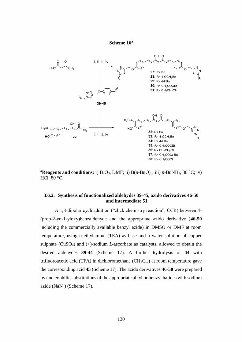
130
Scheme 16a
aReagents and conditions: i) B2O3, DMF; ii) B(n-BuO)3; iii) n-BuNH2, 80 °C; iv)
HCl, 80 °C.
3.6.2. Synthesis of functionalized aldehydes 39-45, azido derivatives 46-50
and intermediate 51
A 1,3-dipolar cycloaddition (“click chemistry reaction”, CCR) between 4-
(prop-2-yn-1-yloxy)benzaldehyde and the appropriate azido derivative (46-50
including the commercially available benzyl azide) in DMSO or DMF at room
temperature, using triethylamine (TEA) as base and a water solution of copper
sulphate (CuSO4) and (+)-sodium L-ascorbate as catalysts, allowed to obtain the
desired aldehydes 39-44 (Scheme 17). A further hydrolysis of 44 with
trifluoroacetic acid (TFA) in dichloromethane (CH2Cl2) at room temperature gave
the corresponding acid 45 (Scheme 17). The azido derivatives 46-50 were prepared
by nucleophilic substitutions of the appropriate alkyl or benzyl halides with sodium
azide (NaN3) (Scheme 17).
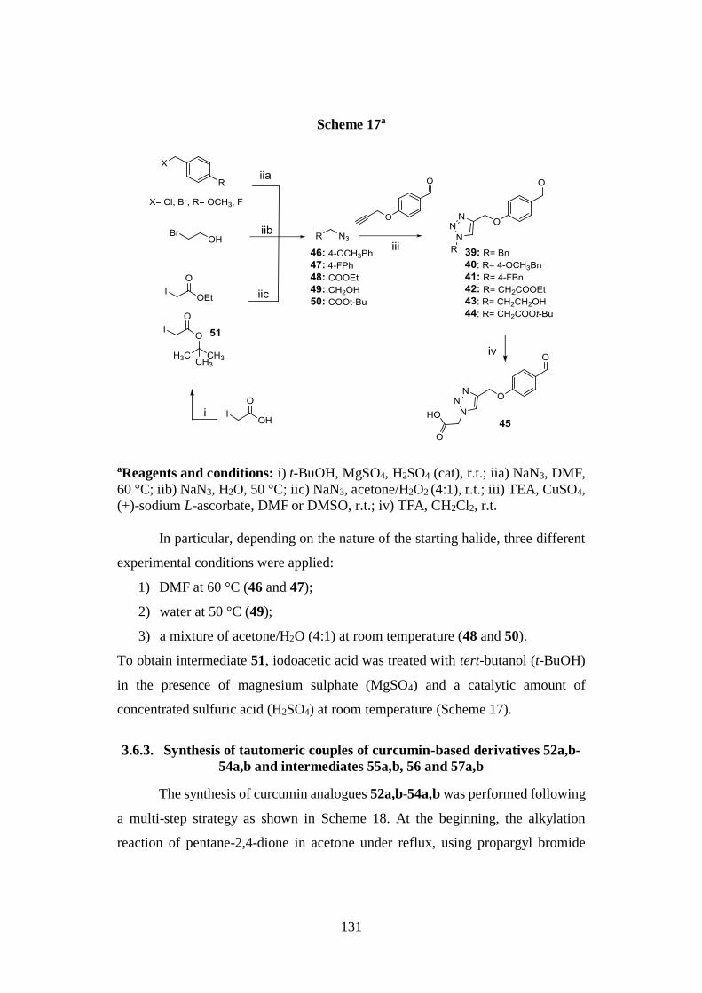
131
Scheme 17a
aReagents and conditions: i) t-BuOH, MgSO4, H2SO4 (cat), r.t.; iia) NaN3, DMF,
60 °C; iib) NaN3, H2O, 50 °C; iic) NaN3, acetone/H2O2 (4:1), r.t.; iii) TEA, CuSO4,
(+)-sodium L-ascorbate, DMF or DMSO, r.t.; iv) TFA, CH2Cl2, r.t.
In particular, depending on the nature of the starting halide, three different
experimental conditions were applied:
1) DMF at 60 °C (46 and 47);
2) water at 50 °C (49);
3) a mixture of acetone/H2O (4:1) at room temperature (48 and 50).
To obtain intermediate 51, iodoacetic acid was treated with tert-butanol (t-BuOH)
in the presence of magnesium sulphate (MgSO4) and a catalytic amount of
concentrated sulfuric acid (H2SO4) at room temperature (Scheme 17).
3.6.3. Synthesis of tautomeric couples of curcumin-based derivatives 52a,b-
54a,b and intermediates 55a,b, 56 and 57a,b
The synthesis of curcumin analogues 52a,b-54a,b was performed following
a multi-step strategy as shown in Scheme 18. At the beginning, the alkylation
reaction of pentane-2,4-dione in acetone under reflux, using propargyl bromide
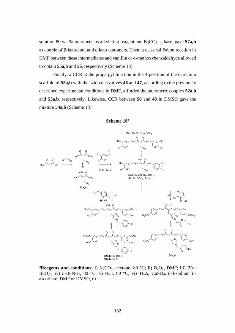
132
solution 80 wt. % in toluene as alkylating reagent and K2CO3 as base, gave 57a,b
as couple of β-keto-enol and diketo tautomers. Then, a classical Pabon reaction in
DMF between these intermediates and vanillin or 4-methoxybenzaldehyde allowed
to obtain 55a,b and 56, respectively (Scheme 18).
Finally, a CCR at the propargyl function in the 4-position of the curcumin
scaffold of 55a,b with the azido derivatives 46 and 47, according to the previously
described experimental conditions in DMF, afforded the tautomeric couples 52a,b
and 53a,b, respectively. Likewise, CCR between 56 and 48 in DMSO gave the
mixture 54a,b (Scheme 18).
Scheme 18a
aReagents and conditions: i) K2CO3, acetone, 80 °C; ii) B2O3, DMF; iii) B(n-
BuO)3; iv) n-BuNH2, 80 °C; v) HCl, 80 °C; vi) TEA, CuSO4, (+)-sodium L-
ascorbate, DMF or DMSO, r.t.
133
Interestingly, in the synthetic route for series IIb, the addressed modification
of the central curcumin framework promoted the partial conversion of its β-keto-
enol form into the corresponding diketo counterpart and it was not possible to
isolate each single tautomer as chromatographically different compound.
3.7. SYNTHESIS OF SERIES III
3.7.1. Synthesis of curcumin analogues 58-60, 61a,b and 62 and
intermediates 64a,b, 65a,b and 63
The curcumin-based compounds 58-60 were synthesized by the two-step
strategy depicted in Scheme 19. In particular, treatment of pentane-2,4-dione with
the suitable alkyl halides, in presence of sodium hydride (NaH) as base, in
tetrahydrofuran (THF) and under nitrogen (N2) atmosphere, gave the key
intermediates 64a,b and 65a,b, whose condensation with 4-methoxybenzaldehyde
and 4-benzyloxybenzaldehyde under classical Pabon reaction conditions in DMF
allowed to obtain 58-60 (Scheme 19).
Additionally, the acid 61a,b was prepared by saponification of the
corresponding ethyl ester derivative 58 using a solution of sodium hydroxide
(NaOH, 0.2 N) in methanol (CH3OH, Scheme 19). The synthesis of the amide
analogue 62 was carried out by a preliminary conversion of 61a,b into the acyl
chloride 63, using thionyl chloride (SOCl2) under reflux, and a further N-acylation
with ethanolamine in acetonitrile (CH3CN) at 60 °C (Scheme 19).
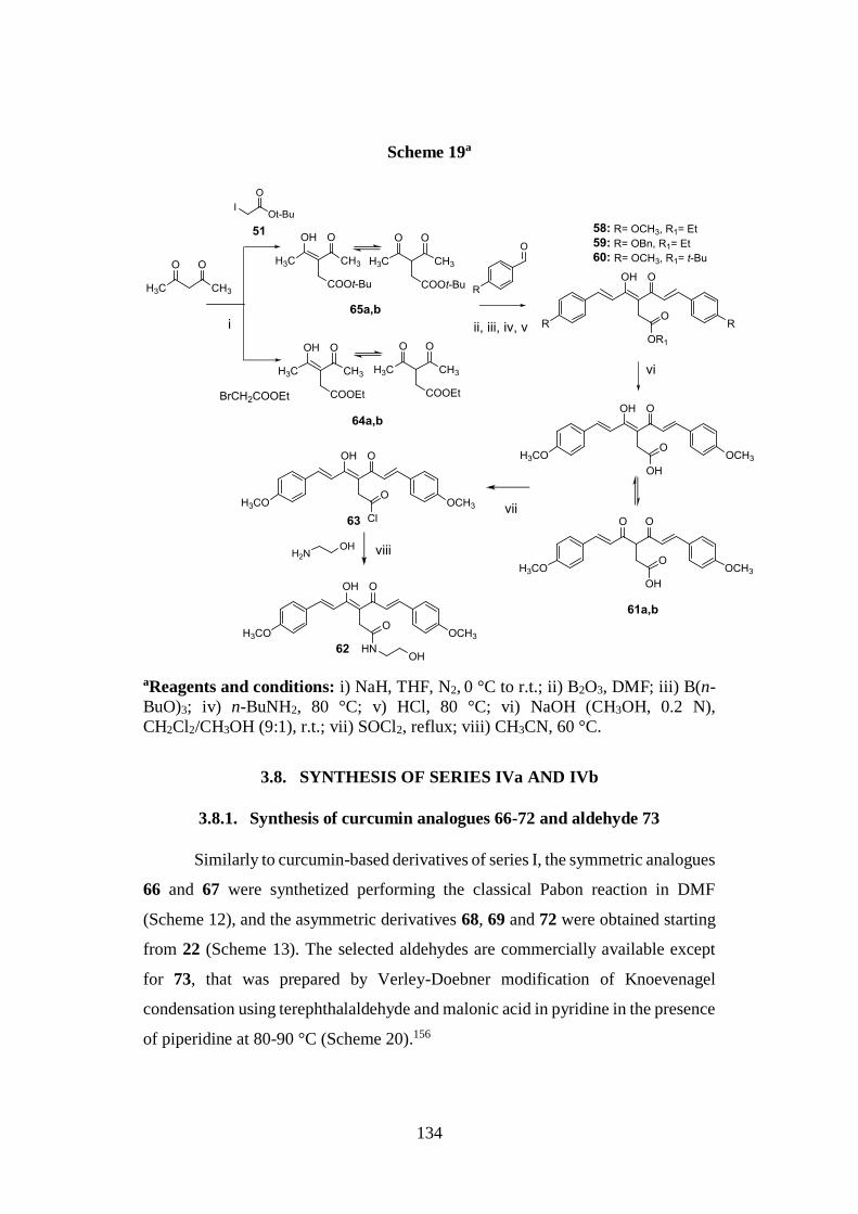
134
Scheme 19a
aReagents and conditions: i) NaH, THF, N2, 0 °C to r.t.; ii) B2O3, DMF; iii) B(n-
BuO)3; iv) n-BuNH2, 80 °C; v) HCl, 80 °C; vi) NaOH (CH3OH, 0.2 N),
CH2Cl2/CH3OH (9:1), r.t.; vii) SOCl2, reflux; viii) CH3CN, 60 °C.
3.8. SYNTHESIS OF SERIES IVa AND IVb
3.8.1. Synthesis of curcumin analogues 66-72 and aldehyde 73
Similarly to curcumin-based derivatives of series I, the symmetric analogues
66 and 67 were synthetized performing the classical Pabon reaction in DMF
(Scheme 12), and the asymmetric derivatives 68, 69 and 72 were obtained starting
from 22 (Scheme 13). The selected aldehydes are commercially available except
for 73, that was prepared by Verley-Doebner modification of Knoevenagel
condensation using terephthalaldehyde and malonic acid in pyridine in the presence
of piperidine at 80-90 °C (Scheme 20).156
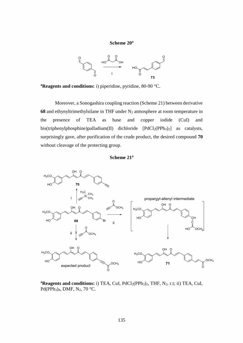
135
Scheme 20a
aReagents and conditions: i) piperidine, pyridine, 80-90 °C.
Moreover, a Sonogashira coupling reaction (Scheme 21) between derivative
68 and ethynyltrimethylsilane in THF under N2 atmosphere at room temperature in
the presence of TEA as base and copper iodide (CuI) and
bis(triphenylphosphine)palladium(II) dichloride [PdCl2(PPh3)2] as catalysts,
surprisingly gave, after purification of the crude product, the desired compound 70
without cleavage of the protecting group.
Scheme 21a
aReagents and conditions: i) TEA, CuI, PdCl2(PPh3)2, THF, N2, r.t; ii) TEA, CuI,
Pd(PPh3)4, DMF, N2, 70 °C.
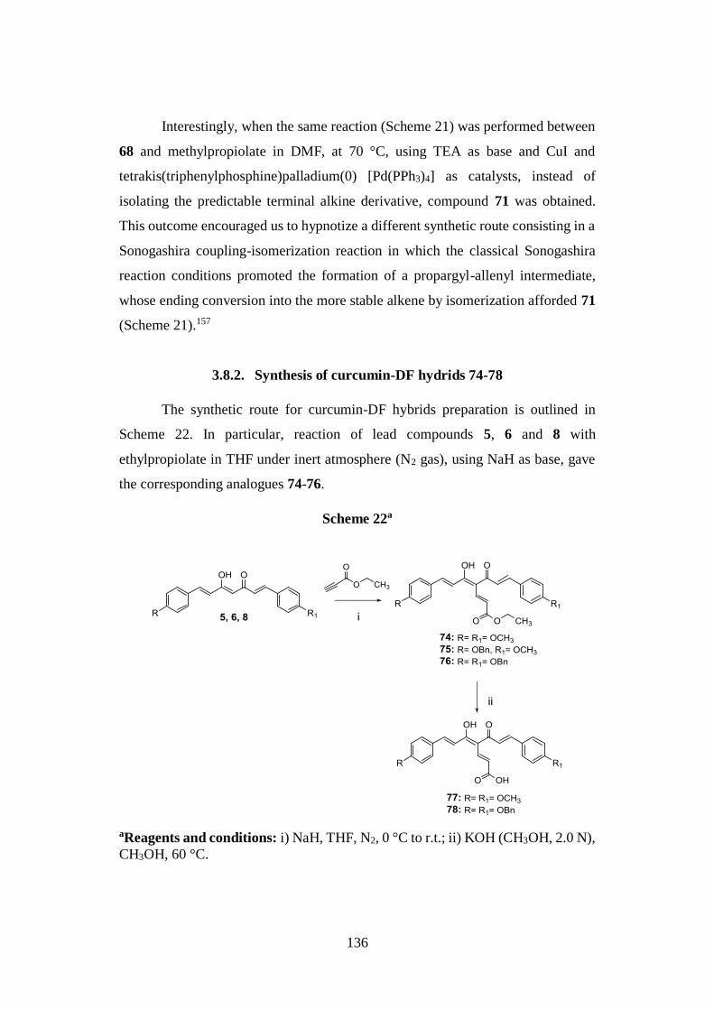
136
Interestingly, when the same reaction (Scheme 21) was performed between
68 and methylpropiolate in DMF, at 70 °C, using TEA as base and CuI and
tetrakis(triphenylphosphine)palladium(0) [Pd(PPh3)4] as catalysts, instead of
isolating the predictable terminal alkine derivative, compound 71 was obtained.
This outcome encouraged us to hypnotize a different synthetic route consisting in a
Sonogashira coupling-isomerization reaction in which the classical Sonogashira
reaction conditions promoted the formation of a propargyl-allenyl intermediate,
whose ending conversion into the more stable alkene by isomerization afforded 71
(Scheme 21).157
3.8.2. Synthesis of curcumin-DF hydrids 74-78
The synthetic route for curcumin-DF hybrids preparation is outlined in
Scheme 22. In particular, reaction of lead compounds 5, 6 and 8 with
ethylpropiolate in THF under inert atmosphere (N2 gas), using NaH as base, gave
the corresponding analogues 74-76.
Scheme 22a
aReagents and conditions: i) NaH, THF, N2, 0 °C to r.t.; ii) KOH (CH3OH, 2.0 N),
CH3OH, 60 °C.
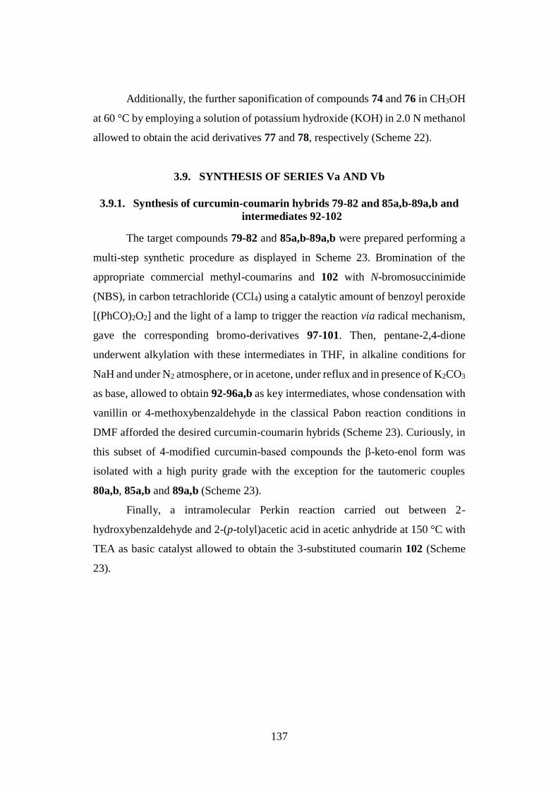
137
Additionally, the further saponification of compounds 74 and 76 in CH3OH
at 60 °C by employing a solution of potassium hydroxide (KOH) in 2.0 N methanol
allowed to obtain the acid derivatives 77 and 78, respectively (Scheme 22).
3.9. SYNTHESIS OF SERIES Va AND Vb
3.9.1. Synthesis of curcumin-coumarin hybrids 79-82 and 85a,b-89a,b and
intermediates 92-102
The target compounds 79-82 and 85a,b-89a,b were prepared performing a
multi-step synthetic procedure as displayed in Scheme 23. Bromination of the
appropriate commercial methyl-coumarins and 102 with N-bromosuccinimide
(NBS), in carbon tetrachloride (CCl4) using a catalytic amount of benzoyl peroxide
[(PhCO)2O2] and the light of a lamp to trigger the reaction via radical mechanism,
gave the corresponding bromo-derivatives 97-101. Then, pentane-2,4-dione
underwent alkylation with these intermediates in THF, in alkaline conditions for
NaH and under N2 atmosphere, or in acetone, under reflux and in presence of K2CO3
as base, allowed to obtain 92-96a,b as key intermediates, whose condensation with
vanillin or 4-methoxybenzaldehyde in the classical Pabon reaction conditions in
DMF afforded the desired curcumin-coumarin hybrids (Scheme 23). Curiously, in
this subset of 4-modified curcumin-based compounds the β-keto-enol form was
isolated with a high purity grade with the exception for the tautomeric couples
80a,b, 85a,b and 89a,b (Scheme 23).
Finally, a intramolecular Perkin reaction carried out between 2-
hydroxybenzaldehyde and 2-(p-tolyl)acetic acid in acetic anhydride at 150 °C with
TEA as basic catalyst allowed to obtain the 3-substituted coumarin 102 (Scheme
23).
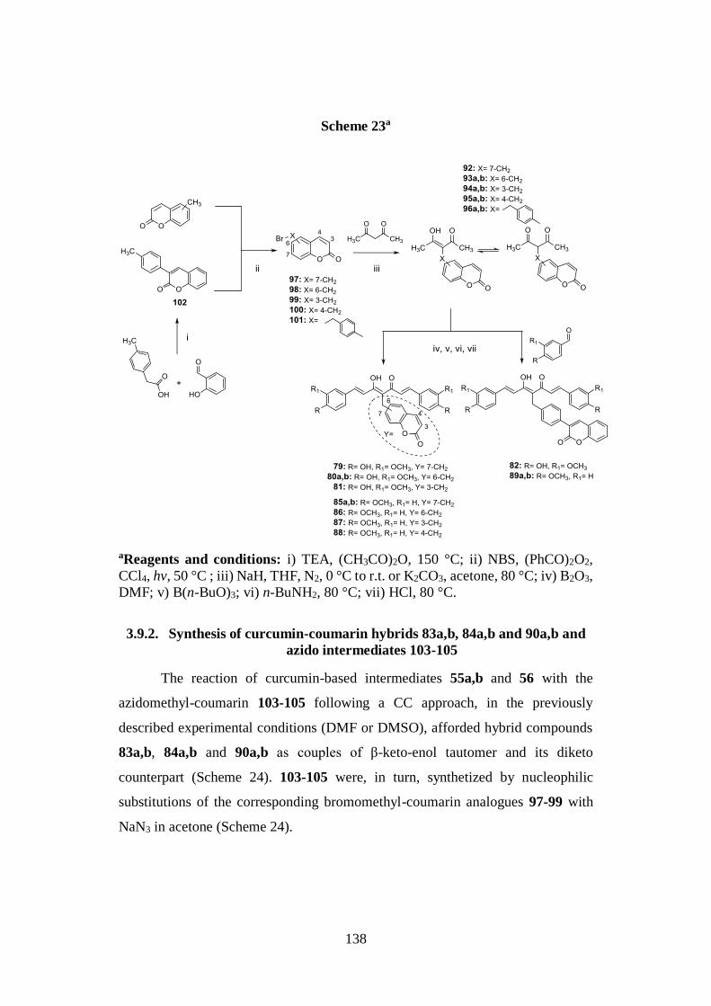
138
Scheme 23a
aReagents and conditions: i) TEA, (CH3CO)2O, 150 °C; ii) NBS, (PhCO)2O2,
CCl4, hv, 50 °C ; iii) NaH, THF, N2, 0 °C to r.t. or K2CO3, acetone, 80 °C; iv) B2O3,
DMF; v) B(n-BuO)3; vi) n-BuNH2, 80 °C; vii) HCl, 80 °C.
3.9.2. Synthesis of curcumin-coumarin hybrids 83a,b, 84a,b and 90a,b and
azido intermediates 103-105
The reaction of curcumin-based intermediates 55a,b and 56 with the
azidomethyl-coumarin 103-105 following a CC approach, in the previously
described experimental conditions (DMF or DMSO), afforded hybrid compounds
83a,b, 84a,b and 90a,b as couples of β-keto-enol tautomer and its diketo
counterpart (Scheme 24). 103-105 were, in turn, synthetized by nucleophilic
substitutions of the corresponding bromomethyl-coumarin analogues 97-99 with
NaN3 in acetone (Scheme 24).
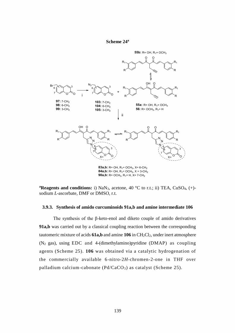
139
Scheme 24a
aReagents and conditions: i) NaN3, acetone, 40 °C to r.t.; ii) TEA, CuSO4, (+)-
sodium L-ascorbate, DMF or DMSO, r.t.
3.9.3. Synthesis of amido curcuminoids 91a,b and amine intermediate 106
The synthesis of the β-keto-enol and diketo couple of amido derivatives
91a,b was carried out by a classical coupling reaction between the corresponding
tautomeric mixture of acids 61a,b and amine 106 in CH2Cl2, under inert atmosphere
(N2 gas), using EDC and 4-(dimethylamino)pyridine (DMAP) as coupling
agents (Scheme 25). 106 was obtained via a catalytic hydrogenation of
the commercially available 6-nitro-2H-chromen-2-one in THF over
palladium calcium-cabonate (Pd/CaCO3) as catalyst (Scheme 25).
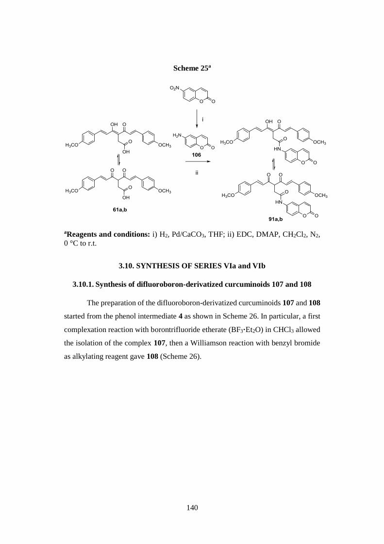
140
Scheme 25a
aReagents and conditions: i) H2, Pd/CaCO3, THF; ii) EDC, DMAP, CH2Cl2, N2,
0 °C to r.t.
3.10. SYNTHESIS OF SERIES VIa and VIb
3.10.1. Synthesis of difluoroboron-derivatized curcuminoids 107 and 108
The preparation of the difluoroboron-derivatized curcuminoids 107 and 108
started from the phenol intermediate 4 as shown in Scheme 26. In particular, a first
complexation reaction with borontrifluoride etherate (BF3·Et2O) in CHCl3 allowed
the isolation of the complex 107, then a Williamson reaction with benzyl bromide
as alkylating reagent gave 108 (Scheme 26).
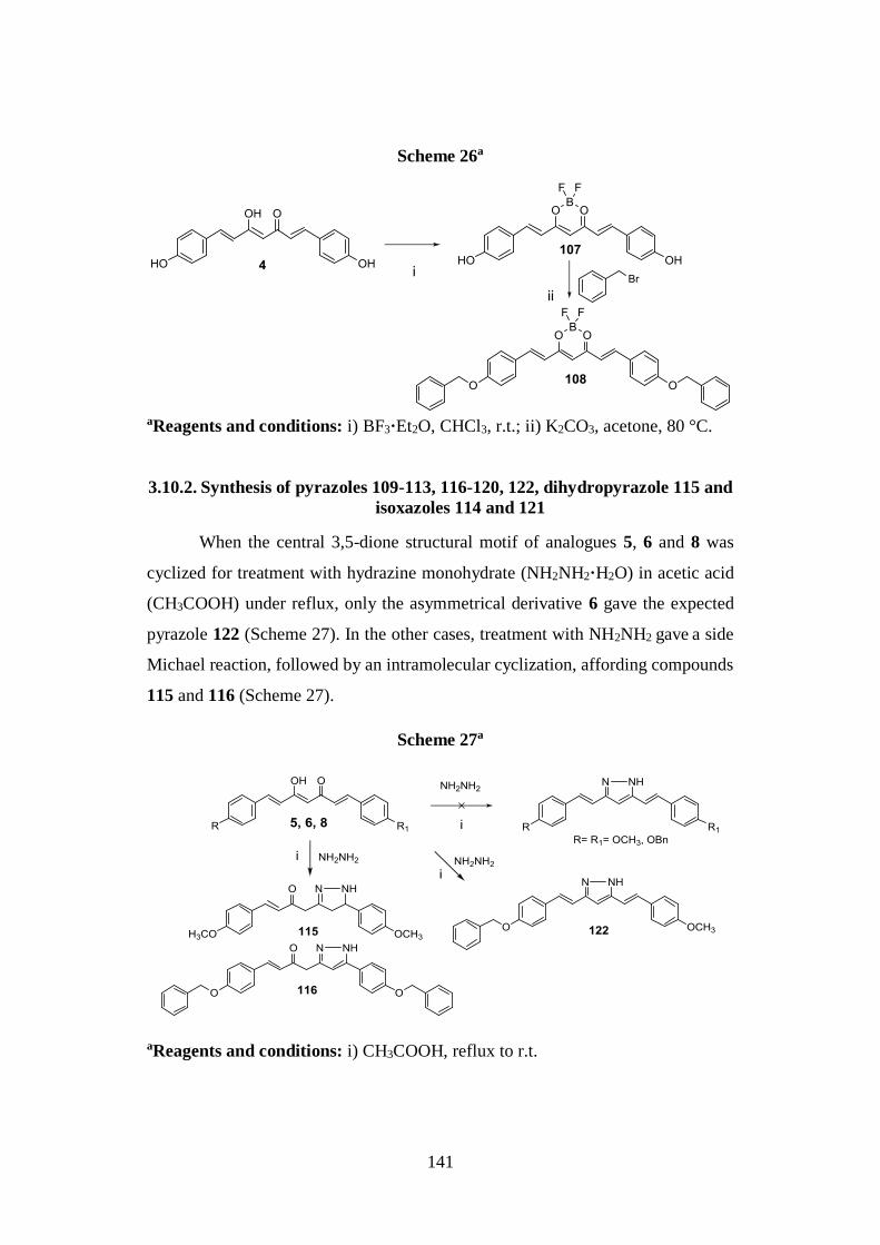
141
Scheme 26a
aReagents and conditions: i) BF3·Et2O, CHCl3, r.t.; ii) K2CO3, acetone, 80 °C.
3.10.2. Synthesis of pyrazoles 109-113, 116-120, 122, dihydropyrazole 115 and
isoxazoles 114 and 121
When the central 3,5-dione structural motif of analogues 5, 6 and 8 was
cyclized for treatment with hydrazine monohydrate (NH2NH2·H2O) in acetic acid
(CH3COOH) under reflux, only the asymmetrical derivative 6 gave the expected
pyrazole 122 (Scheme 27). In the other cases, treatment with NH2NH2 gave a side
Michael reaction, followed by an intramolecular cyclization, affording compounds
115 and 116 (Scheme 27).
Scheme 27a
aReagents and conditions: i) CH3COOH, reflux to r.t.
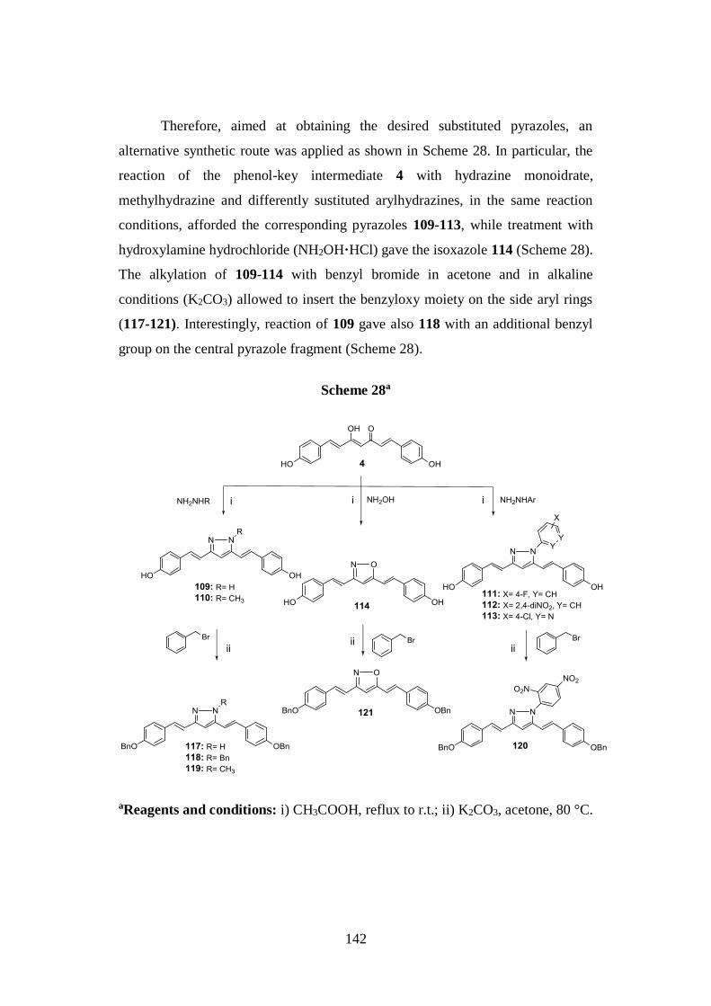
142
Therefore, aimed at obtaining the desired substituted pyrazoles, an
alternative synthetic route was applied as shown in Scheme 28. In particular, the
reaction of the phenol-key intermediate 4 with hydrazine monoidrate,
methylhydrazine and differently sustituted arylhydrazines, in the same reaction
conditions, afforded the corresponding pyrazoles 109-113, while treatment with
hydroxylamine hydrochloride (NH2OH·HCl) gave the isoxazole 114 (Scheme 28).
The alkylation of 109-114 with benzyl bromide in acetone and in alkaline
conditions (K2CO3) allowed to insert the benzyloxy moiety on the side aryl rings
(117-121). Interestingly, reaction of 109 gave also 118 with an additional benzyl
group on the central pyrazole fragment (Scheme 28).
Scheme 28a
aReagents and conditions: i) CH3COOH, reflux to r.t.; ii) K2CO3, acetone, 80 °C.
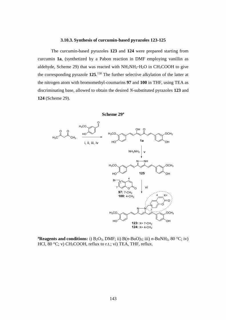
143
3.10.3. Synthesis of curcumin-based pyrazoles 123-125
The curcumin-based pyrazoles 123 and 124 were prepared starting from
curcumin 1a, (synthetized by a Pabon reaction in DMF employing vanillin as
aldehyde, Scheme 29) that was reacted with NH2NH2·H2O in CH3COOH to give
the corresponding pyrazole 125.158 The further selective alkylation of the latter at
the nitrogen atom with bromomethyl-coumarins 97 and 100 in THF, using TEA as
discriminating base, allowed to obtain the desired N-substituted pyrazoles 123 and
124 (Scheme 29).
Scheme 29a
aReagents and conditions: i) B2O3, DMF; ii) B(n-BuO)3; iii) n-BuNH2, 80 °C; iv)
HCl, 80 °C; v) CH3COOH, reflux to r.t.; vi) TEA, THF, reflux.
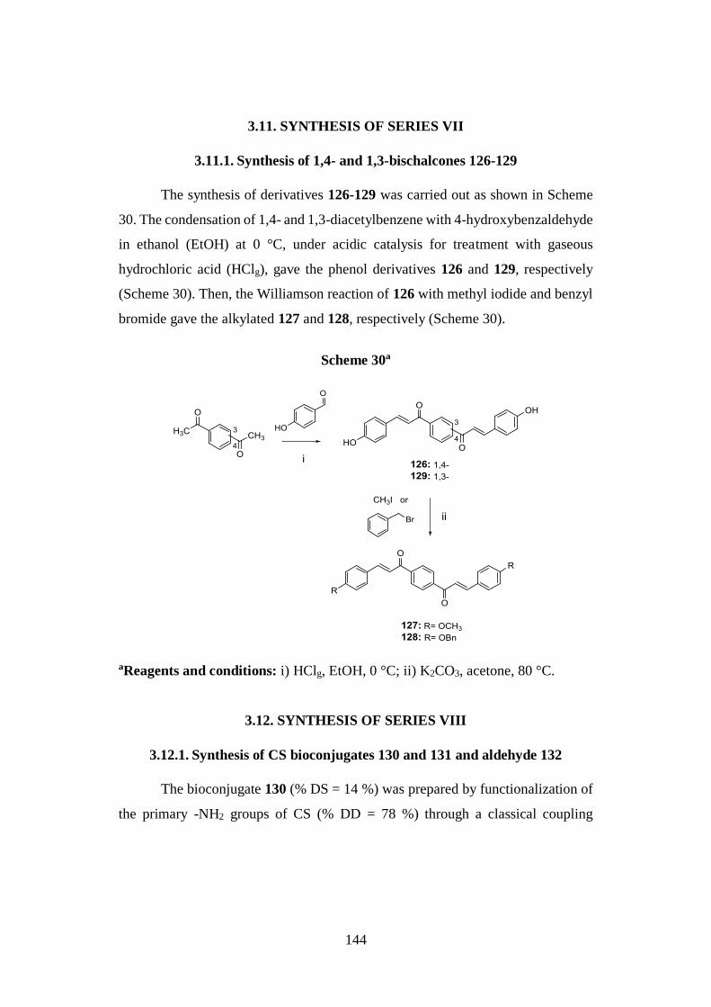
144
3.11. SYNTHESIS OF SERIES VII
3.11.1. Synthesis of 1,4- and 1,3-bischalcones 126-129
The synthesis of derivatives 126-129 was carried out as shown in Scheme
30. The condensation of 1,4- and 1,3-diacetylbenzene with 4-hydroxybenzaldehyde
in ethanol (EtOH) at 0 °C, under acidic catalysis for treatment with gaseous
hydrochloric acid (HClg), gave the phenol derivatives 126 and 129, respectively
(Scheme 30). Then, the Williamson reaction of 126 with methyl iodide and benzyl
bromide gave the alkylated 127 and 128, respectively (Scheme 30).
Scheme 30a
aReagents and conditions: i) HClg, EtOH, 0 °C; ii) K2CO3, acetone, 80 °C.
3.12. SYNTHESIS OF SERIES VIII
3.12.1. Synthesis of CS bioconjugates 130 and 131 and aldehyde 132
The bioconjugate 130 (% DS = 14 %) was prepared by functionalization of
the primary -NH2 groups of CS (% DD = 78 %) through a classical coupling
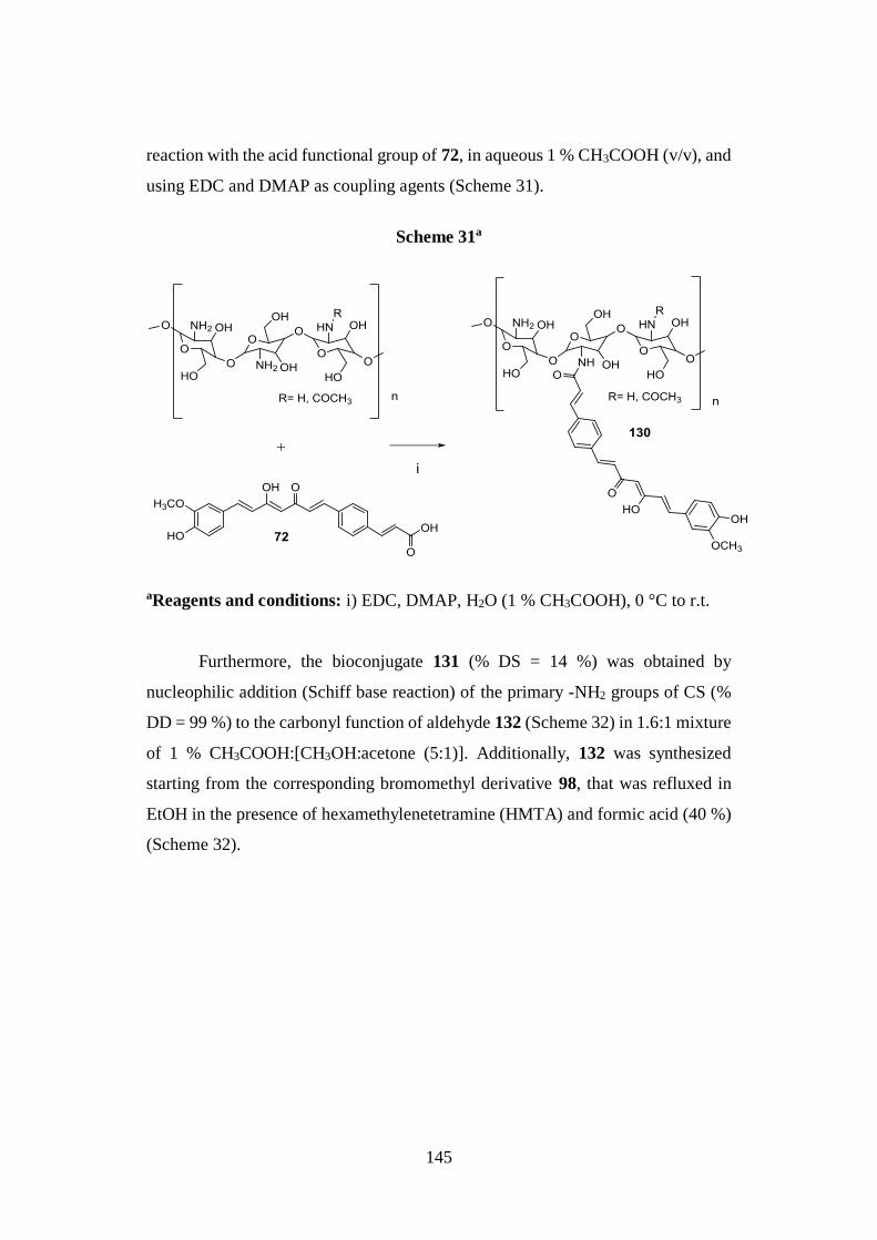
145
reaction with the acid functional group of 72, in aqueous 1 % CH3COOH (v/v), and
using EDC and DMAP as coupling agents (Scheme 31).
Scheme 31a
aReagents and conditions: i) EDC, DMAP, H2O (1 % CH3COOH), 0 °C to r.t.
Furthermore, the bioconjugate 131 (% DS = 14 %) was obtained by
nucleophilic addition (Schiff base reaction) of the primary -NH2 groups of CS (%
DD = 99 %) to the carbonyl function of aldehyde 132 (Scheme 32) in 1.6:1 mixture
of 1 % CH3COOH:[CH3OH:acetone (5:1)]. Additionally, 132 was synthesized
starting from the corresponding bromomethyl derivative 98, that was refluxed in
EtOH in the presence of hexamethylenetetramine (HMTA) and formic acid (40 %)
(Scheme 32).
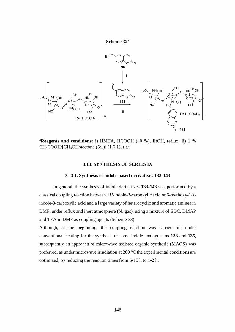
146
Scheme 32a
aReagents and conditions: i) HMTA, HCOOH (40 %), EtOH, reflux; ii) 1 %
CH3COOH:[CH3OH/acetone (5:1)] (1.6:1), r.t.;
3.13. SYNTHESIS OF SERIES IX
3.13.1. Synthesis of indole-based derivatives 133-143
In general, the synthesis of indole derivatives 133-143 was performed by a
classical coupling reaction between 1H-indole-3-carboxylic acid or 6-methoxy-1H-
indole-3-carboxylic acid and a large variety of heterocyclic and aromatic amines in
DMF, under reflux and inert atmosphere (N2 gas), using a mixture of EDC, DMAP
and TEA in DMF as coupling agents (Scheme 33).
Although, at the beginning, the coupling reaction was carried out under
conventional heating for the synthesis of some indole analogues as 133 and 135,
subsequently an approach of microwave assisted organic synthesis (MAOS) was
preferred, as under microwave irradiation at 200 °C the experimental conditions are
optimized, by reducing the reaction times from 6-15 h to 1-2 h.
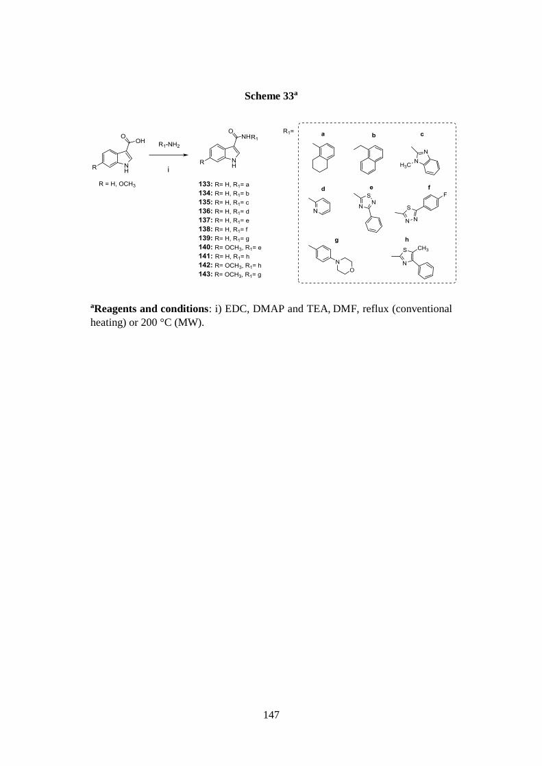
147
Scheme 33a
aReagents and conditions: i) EDC, DMAP and TEA, DMF, reflux (conventional
heating) or 200 °C (MW).

148
4. Results and discussion
149
4.1. BACE-1 INHIBITION
Subset of curcumin-based analogues 2-17 (β-keto-enol forms) and 18-21 (diketo
tautomers) and reference compound 1a.
The fluorescence resonance energy transfer (FRET) methodology31 was
employed to evaluate the BACE-1 in vitro inhibitory potency of a set of curcumin-
based derivatives (2-21), using curcumin 1a as reference compound (Tables 19 and
20). The compounds, except for 14, were able to inhibit BACE-1, with IC50 values
ranging from low micromolar to nanomolar, being more potent than curcumin 1a,
that in our test did not inhibit the enzyme up to 3 µM. Moreover, referring to the
tautomeric couples 8, 19; 5, 18; 9, 20; and 10, 21 the β-keto-enol forms showed a
more favorable trend of inhibition than the corresponding diketo counterparts. The
data confirmed the suitability of the β-keto-enol curcumin main scaffold for
inhibition of this target, and underlined the potential intense effect on activity
exerted by the substituents on the side aryl rings.8
Indeed, for the bis-p-prenyloxyphenyl symmetrical analogue 14 no enzyme
inhibition was observed up to 3 μM. On the contrary, the bis-p-benzyloxyphenyl
analogue 8 showed an IC50 value of 40 nM, and was thus identified as the most
active of the series, whereas the corresponding diketo tautomer 19 displayed a
notable drop in potency. A decrease in activity was observed when the benzyloxy
functions were replaced with the less hindering methoxy groups (5, IC50 = 1.65
µM); again, the corresponding diketo analogue (18) was less active than 5. Low-
micromolar potencies were noticed for the other symmetrical derivatives bearing
hydroxy, methyl, phenyl groups in position 4 (4, 16, and 17, respectively), and for
the bis-3,4-dimethoxy-substituted 13.
The symmetrical fluorinated analogues of 8 showed a decrease in potency,
that was one order of magnitude lower for the 2,4- and 3,5-di-fluorinated 9 and 10,
respectively (IC50 = 0.40 and 0.39 μM), while their corresponding diketo
counterparts 20 and 21, respectively, showed low-micromolar IC50 values. This
level of potency was similarly observed for the 4- and 3- mono-fluorinated
analogues of 8 (11 and 12, respectively).
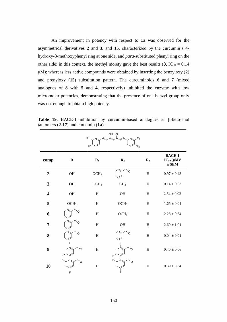
150
An improvement in potency with respect to 1a was observed for the
asymmetrical derivatives 2 and 3, and 15, characterized by the curcumin’s 4-
hydroxy-3-methoxyphenyl ring at one side, and para-substituted phenyl ring on the
other side; in this context, the methyl moiety gave the best results (3, IC50 = 0.14
μM); whereas less active compounds were obtained by inserting the benzyloxy (2)
and prenyloxy (15) substitution pattern. The curcuminoids 6 and 7 (mixed
analogues of 8 with 5 and 4, respectively) inhibited the enzyme with low
micromolar potencies, demonstrating that the presence of one benzyl group only
was not enough to obtain high potency.
Table 19. BACE-1 inhibition by curcumin-based analogues as β-keto-enol
tautomers (2-17) and curcumin (1a).
comp R R1 R2 R3
BACE-1
IC50 (μM)a
± SEM
2 OH OCH3
H 0.97 ± 0.43
3 OH OCH3 CH3 H 0.14 ± 0.03
4 OH H OH H 2.54 ± 0.02
5 OCH3 H OCH3 H 1.65 ± 0.01
6
H OCH3 H 2.28 ± 0.64
7
H OH H 2.69 ± 1.01
8
H
H 0.04 ± 0.01
9
H
H 0.40 ± 0.06
10
H
H 0.39 ± 0.34
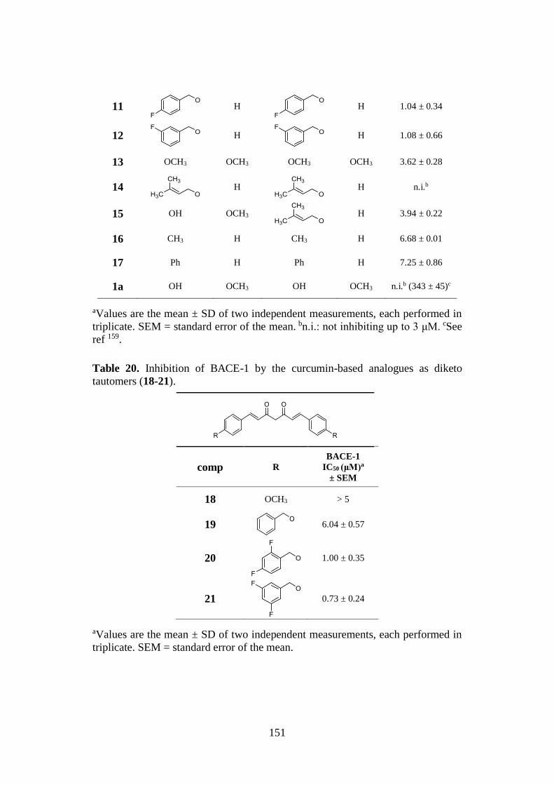
151
11
H
H 1.04 ± 0.34
12
H
H 1.08 ± 0.66
13 OCH3 OCH3 OCH3 OCH3 3.62 ± 0.28
14
H
H n.i.b
15 OH OCH3
H 3.94 ± 0.22
16 CH3 H CH3 H 6.68 ± 0.01
17 Ph H Ph H 7.25 ± 0.86
1a OH OCH3 OH OCH3 n.i.b (343 ± 45)c
aValues are the mean ± SD of two independent measurements, each performed in
triplicate. SEM = standard error of the mean. bn.i.: not inhibiting up to 3 μM. cSee
ref 159.
Table 20. Inhibition of BACE-1 by the curcumin-based analogues as diketo
tautomers (18-21).
comp R
BACE-1
IC50 (μM)a
± SEM
18 OCH3 > 5
19
6.04 ± 0.57
20
1.00 ± 0.35
21
0.73 ± 0.24
aValues are the mean ± SD of two independent measurements, each performed in
triplicate. SEM = standard error of the mean.
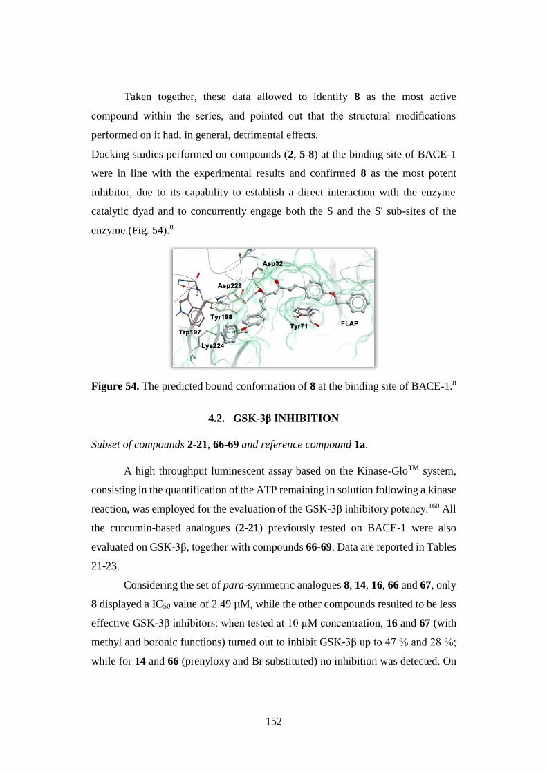
152
Taken together, these data allowed to identify 8 as the most active
compound within the series, and pointed out that the structural modifications
performed on it had, in general, detrimental effects.
Docking studies performed on compounds (2, 5-8) at the binding site of BACE-1
were in line with the experimental results and confirmed 8 as the most potent
inhibitor, due to its capability to establish a direct interaction with the enzyme
catalytic dyad and to concurrently engage both the S and the S' sub-sites of the
enzyme (Fig. 54).8
Figure 54. The predicted bound conformation of 8 at the binding site of BACE-1.8
4.2. GSK-3β INHIBITION
Subset of compounds 2-21, 66-69 and reference compound 1a.
A high throughput luminescent assay based on the Kinase-GloTM system,
consisting in the quantification of the ATP remaining in solution following a kinase
reaction, was employed for the evaluation of the GSK-3β inhibitory potency.160 All
the curcumin-based analogues (2-21) previously tested on BACE-1 were also
evaluated on GSK-3β, together with compounds 66-69. Data are reported in Tables
21-23.
Considering the set of para-symmetric analogues 8, 14, 16, 66 and 67, only
8 displayed a IC50 value of 2.49 µM, while the other compounds resulted to be less
effective GSK-3β inhibitors: when tested at 10 µM concentration, 16 and 67 (with
methyl and boronic functions) turned out to inhibit GSK-3β up to 47 % and 28 %;
while for 14 and 66 (prenyloxy and Br substituted) no inhibition was detected. On
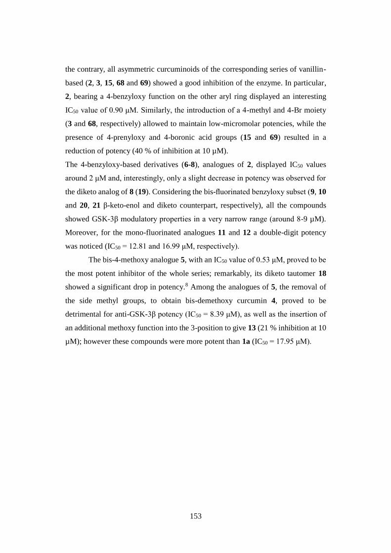
153
the contrary, all asymmetric curcuminoids of the corresponding series of vanillin-
based (2, 3, 15, 68 and 69) showed a good inhibition of the enzyme. In particular,
2, bearing a 4-benzyloxy function on the other aryl ring displayed an interesting
IC50 value of 0.90 μM. Similarly, the introduction of a 4-methyl and 4-Br moiety
(3 and 68, respectively) allowed to maintain low-micromolar potencies, while the
presence of 4-prenyloxy and 4-boronic acid groups (15 and 69) resulted in a
reduction of potency (40 % of inhibition at 10 µM).
The 4-benzyloxy-based derivatives (6-8), analogues of 2, displayed IC50 values
around 2 μM and, interestingly, only a slight decrease in potency was observed for
the diketo analog of 8 (19). Considering the bis-fluorinated benzyloxy subset (9, 10
and 20, 21 β-keto-enol and diketo counterpart, respectively), all the compounds
showed GSK-3β modulatory properties in a very narrow range (around 8-9 µM).
Moreover, for the mono-fluorinated analogues 11 and 12 a double-digit potency
was noticed (IC50 = 12.81 and 16.99 μM, respectively).
The bis-4-methoxy analogue 5, with an IC50 value of 0.53 μM, proved to be
the most potent inhibitor of the whole series; remarkably, its diketo tautomer 18
showed a significant drop in potency.8 Among the analogues of 5, the removal of
the side methyl groups, to obtain bis-demethoxy curcumin 4, proved to be
detrimental for anti-GSK-3β potency (IC50 = 8.39 μM), as well as the insertion of
an additional methoxy function into the 3-position to give 13 (21 % inhibition at 10
µM); however these compounds were more potent than 1a (IC50 = 17.95 μM).
154
Table 21. GSK-3β inhibitory potencies of 2-17 and curcumin (1a) as keto-enol
tautomers.
comp R R1 R2 R3
GSK-3β
IC50 (μM)a
± SEM
or
% inh @ 10 μM
2 OH OCH3
H 0.90 ± 0.38
3 OH OCH3 CH3 H 2.09 ± 0.51
4 OH H OH H 8.39 ± 1.59
5 OCH3 H OCH3 H 0.53 ± 0.27
6
H OCH3 H 2.78 ± 0.44
7
H OH H 2.01 ± 0.71
8
H
H 2.49 ± 0.82
9
H
H 9.63 ± 0.21
10
H
H 8.30 ± 0.54
11
H
H 12.81 ± 0.14
12
H
H 16.99 ± 2.68
13 OCH3 OCH3 OCH3 OCH3 21 %
14
H
H n.i.b
15 OH OCH3
H 40 %
16 CH3 H CH3 H 47 %
17 Ph H Ph H 7.74 ± 0.59
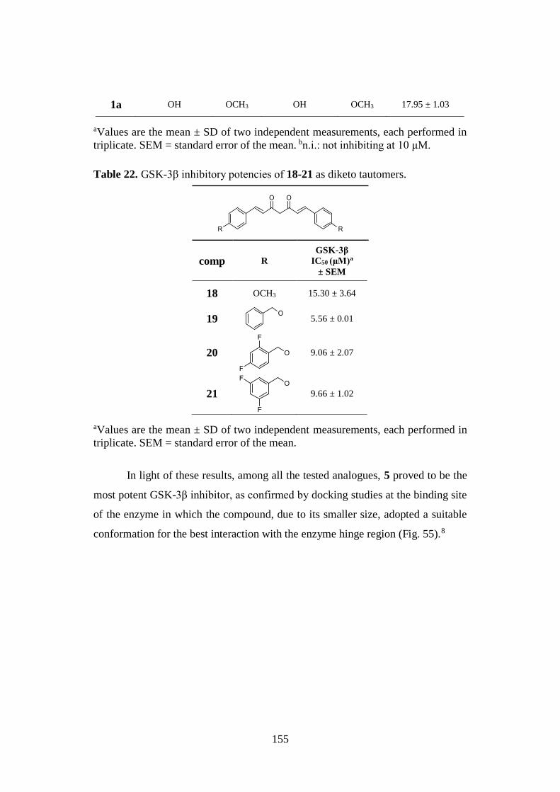
155
1a OH OCH3 OH OCH3 17.95 ± 1.03
aValues are the mean ± SD of two independent measurements, each performed in
triplicate. SEM = standard error of the mean. bn.i.: not inhibiting at 10 μM.
Table 22. GSK-3β inhibitory potencies of 18-21 as diketo tautomers.
comp R
GSK-3β
IC50 (μM)a
± SEM
18 OCH3 15.30 ± 3.64
19
5.56 ± 0.01
20
9.06 ± 2.07
21
9.66 ± 1.02
aValues are the mean ± SD of two independent measurements, each performed in
triplicate. SEM = standard error of the mean.
In light of these results, among all the tested analogues, 5 proved to be the
most potent GSK-3β inhibitor, as confirmed by docking studies at the binding site
of the enzyme in which the compound, due to its smaller size, adopted a suitable
conformation for the best interaction with the enzyme hinge region (Fig. 55).8

156
Figure 55. The predicted bound conformation of 5 at the binding site of GSK-3β.8
Table 23. Inhibition of GSK-3β enzymatic activities by the curcumin-based
analogues 66-69.
comp R R1 R2
GSK-3β
IC50 (μM)a
± SEM
or
% inh @ 10 μM
66 Br H Br n.i.b
67 B(OH)2 H B(OH)2 28 %
68 OH OCH3 Br 7.67 ± 0.18
69 OH OCH3 B(OH)2 41 %
aValues are the mean ± SD of two independent measurements, each performed in
triplicate. SEM = standard error of the mean. n.i.b not inhibiting at 10 μM.
Subset of curcuminoid-DF hybrids (74-76 and 78).
In an effort to obtain derivatives with an expanded biological profile, some
of the most effective BACE-1 and GSK-3β inhibitors (5, 6, and 8), were selected to
be subjected to hybridization strategy, by introducing into the 4-position of the main
scaffold a molecular fragment of the potent Nrf2 inducer dimethyl fumarate (DF),
to give the curcumin-DF series of compounds 74, 75 and 76, respectively (Table
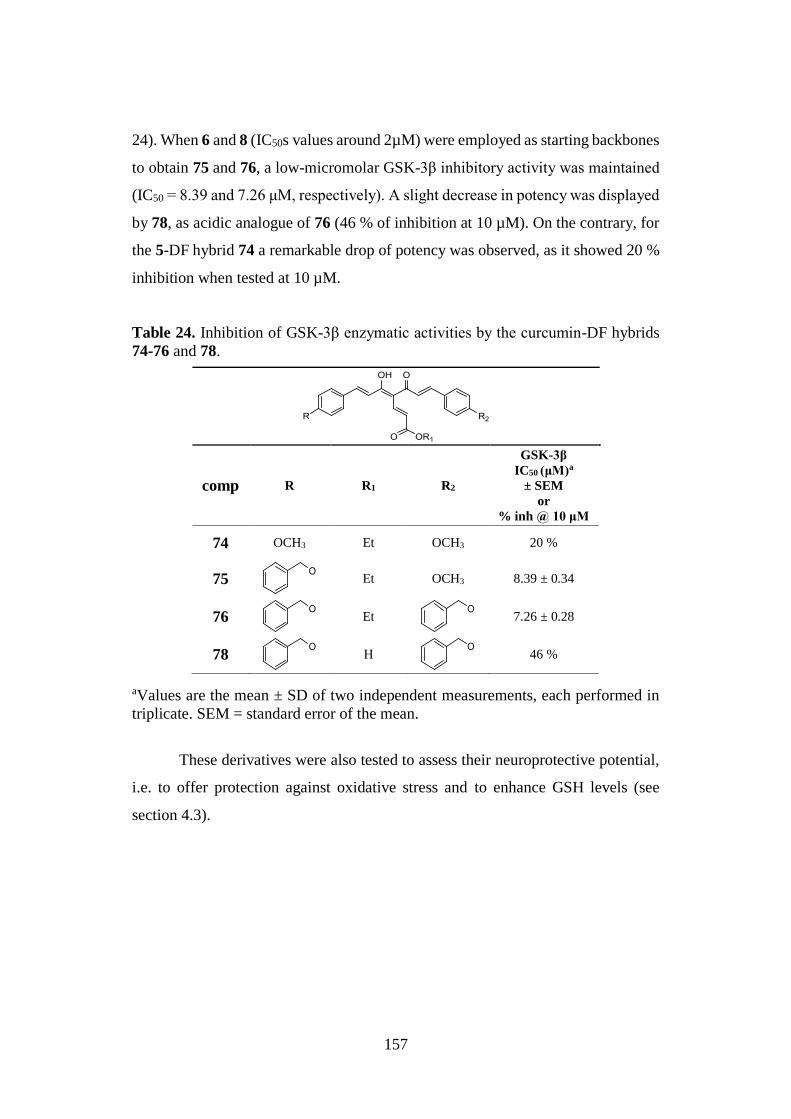
157
24). When 6 and 8 (IC50s values around 2µM) were employed as starting backbones
to obtain 75 and 76, a low-micromolar GSK-3β inhibitory activity was maintained
(IC50 = 8.39 and 7.26 μM, respectively). A slight decrease in potency was displayed
by 78, as acidic analogue of 76 (46 % of inhibition at 10 µM). On the contrary, for
the 5-DF hybrid 74 a remarkable drop of potency was observed, as it showed 20 %
inhibition when tested at 10 µM.
Table 24. Inhibition of GSK-3β enzymatic activities by the curcumin-DF hybrids
74-76 and 78.
comp R R1 R2
GSK-3β
IC50 (μM)a
± SEM
or
% inh @ 10 μM
74 OCH3 Et OCH3 20 %
75
Et OCH3 8.39 ± 0.34
76
Et
7.26 ± 0.28
78
H
46 %
aValues are the mean ± SD of two independent measurements, each performed in
triplicate. SEM = standard error of the mean.
These derivatives were also tested to assess their neuroprotective potential,
i.e. to offer protection against oxidative stress and to enhance GSH levels (see
section 4.3).
158
4.3. NEUROPROTECTION
The neuroprotective potential of derivatives 5, 8, and the corresponding DF-
hybrids 74-76 and 78 was investigated through in vitro evaluation of their neuronal
effects on:
1. cell viability;
2. protection against oxidative stress;
3. enhancement of GSH levels.
In each investigation, DF was used as reference compound.
4.3.1. SH-SY5Y neuroblastoma cell viability
The cellular toxicity of the selected curcumin-based derivatives was
examined on SH-SY5Y neuroblastoma cells, which were exposed for 24 h to the
tested compounds at 0-10 μM range of concentrations. Cell viability was measured
by MTT assay. Similarly to DF, no remarkable decrease in cell viability was
observed at the tested concentrations.
4.3.2. Antioxidant activity
Oxidative stress has been recognized as a common pathological AD feature,
and it is substantially produced by reactive radical species, among others ROS.
Thus, 74-76 and 78 and the parent compounds 5 and 8 were assayed for their
scavenging potential, in the absence or presence of pre-treatment, on SH-SY5Y
cells exposed to high levels of tert-butyl hydroperoxide (TBH, 100 μM).
In particular, SH-SY5Y cells were first incubated for 24 h with 5 µM of the
tested compounds and then exposed to TBH for 30 min. At the end of the
incubation, ROS formation was detected (using the fluorescent probe DCFH-DA).
None of the tested compounds could significantly affect ROS scavenging activity
(Fig. 56); interestingly, this effect was increased in cells pre-treated with 74-76,
suggesting an indirect antioxidant mechanism of action. In particular, at 5 μM,
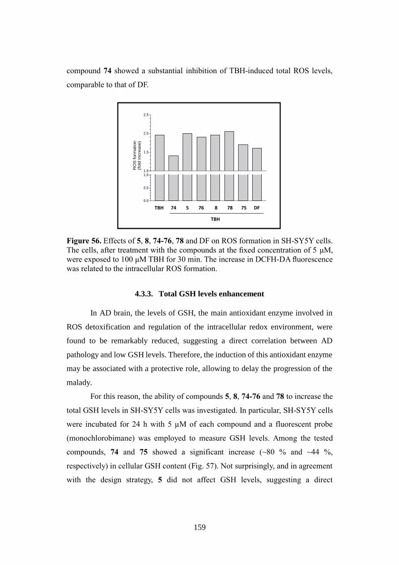
159
compound 74 showed a substantial inhibition of TBH-induced total ROS levels,
comparable to that of DF.
Figure 56. Effects of 5, 8, 74-76, 78 and DF on ROS formation in SH-SY5Y cells.
The cells, after treatment with the compounds at the fixed concentration of 5 μM,
were exposed to 100 μM TBH for 30 min. The increase in DCFH-DA fluorescence
was related to the intracellular ROS formation.
4.3.3. Total GSH levels enhancement
In AD brain, the levels of GSH, the main antioxidant enzyme involved in
ROS detoxification and regulation of the intracellular redox environment, were
found to be remarkably reduced, suggesting a direct correlation between AD
pathology and low GSH levels. Therefore, the induction of this antioxidant enzyme
may be associated with a protective role, allowing to delay the progression of the
malady.
For this reason, the ability of compounds 5, 8, 74-76 and 78 to increase the
total GSH levels in SH-SY5Y cells was investigated. In particular, SH-SY5Y cells
were incubated for 24 h with 5 µM of each compound and a fluorescent probe
(monochlorobimane) was employed to measure GSH levels. Among the tested
compounds, 74 and 75 showed a significant increase (~80 % and ~44 %,
respectively) in cellular GSH content (Fig. 57). Not surprisingly, and in agreement
with the design strategy, 5 did not affect GSH levels, suggesting a direct
t-BuOOHCur11-1 Cur11 Cur3-1 Cur3 Cur3-1A Cur14-1 DF0.0
0.5
1.01.0
1.5
2.0
2.5
t-BuOOH
RO
S f
orm
ation
(fold
incre
ase)
TBH 74 5 76 8 78 75 DF
TBH
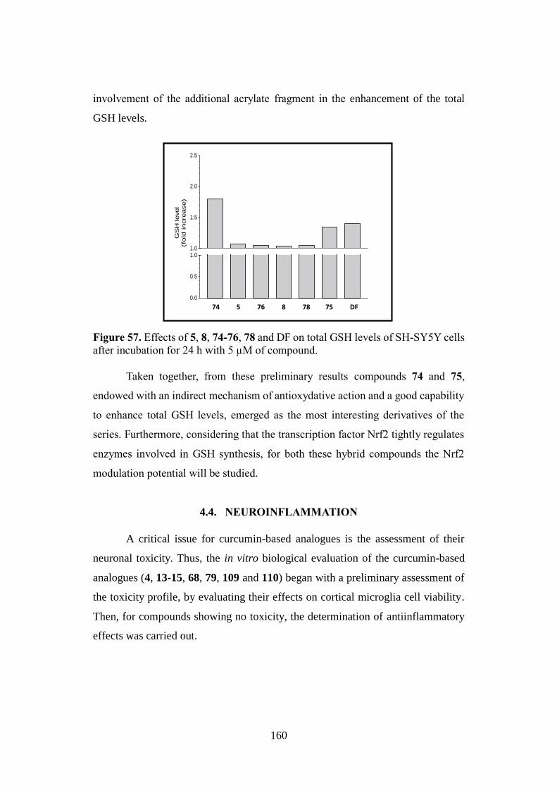
160
involvement of the additional acrylate fragment in the enhancement of the total
GSH levels.
Figure 57. Effects of 5, 8, 74-76, 78 and DF on total GSH levels of SH-SY5Y cells
after incubation for 24 h with 5 µM of compound.
Taken together, from these preliminary results compounds 74 and 75,
endowed with an indirect mechanism of antioxydative action and a good capability
to enhance total GSH levels, emerged as the most interesting derivatives of the
series. Furthermore, considering that the transcription factor Nrf2 tightly regulates
enzymes involved in GSH synthesis, for both these hybrid compounds the Nrf2
modulation potential will be studied.
4.4. NEUROINFLAMMATION
A critical issue for curcumin-based analogues is the assessment of their
neuronal toxicity. Thus, the in vitro biological evaluation of the curcumin-based
analogues (4, 13-15, 68, 79, 109 and 110) began with a preliminary assessment of
the toxicity profile, by evaluating their effects on cortical microglia cell viability.
Then, for compounds showing no toxicity, the determination of antiinflammatory
effects was carried out.
Cur11-1 Cur11 Cur3-1 Cur3 Cur3-1A Cur14-1 DF0.0
0.5
1.01.0
1.5
2.0
2.5
GS
H level
(fold
incre
ase)
74 5 76 8 78 75 DF
161
1. Subset of compounds 1a, 4, 79, 109 and 110.
4.4.1. Neurotoxicity: microglial cell viability
Generally, the compounds were tested in both primary and
lipopolysaccharide (LPS)-activated microglial cells in a 1-40 μM range of
concentrations, and cell viability was determined by MTT assay. Results were
expresses as percentage of viability relative to the control or LPS treated culture.
Morphological examination of the culture were also performed to corroborate the
data.
Treatment of 1a, 4, 79, 109 and 110 for 16 h at the fixed concentrations of
1, 5, and 10 µM, in both cellular conditions, did not significantly affect neither cell
viability nor cell morphology compared to untreated control (Fig. 58A).
Conversely, in the 20-40 µM range of concentrations, 1a, 4, 79, and 109
demonstrated toxic effects, particularly evident for 1a, and the high amount of cell
debris observed in the cell cultures also confirmed these data. On the contrary, 110
did not show any toxic behaviour (Fig. 58A).
When LPS-stimulated microglia (100 ng/mL) was treated for 16 h with the
selected set of derivatives (1-10 µM, Fig. 58B), an activated phenotype was
observed, in which cells presented a macrophage-like morphology, that was not
substantially different from that of control (LPS-activated cells). At 10 µM, 79
proved to significantly reduce cell viability. In the same conditions, an increase in
cell death was observed upon treatment with higher concentrations (20-40 µM) of
derivatives (Fig. 58B).
These preliminary studies allowed identifying the range of concentrations to be
employed in the next cytokine release evaluation assay: 1-5 µM for 79, 1-10 µM
for 1a, 4, and 109.
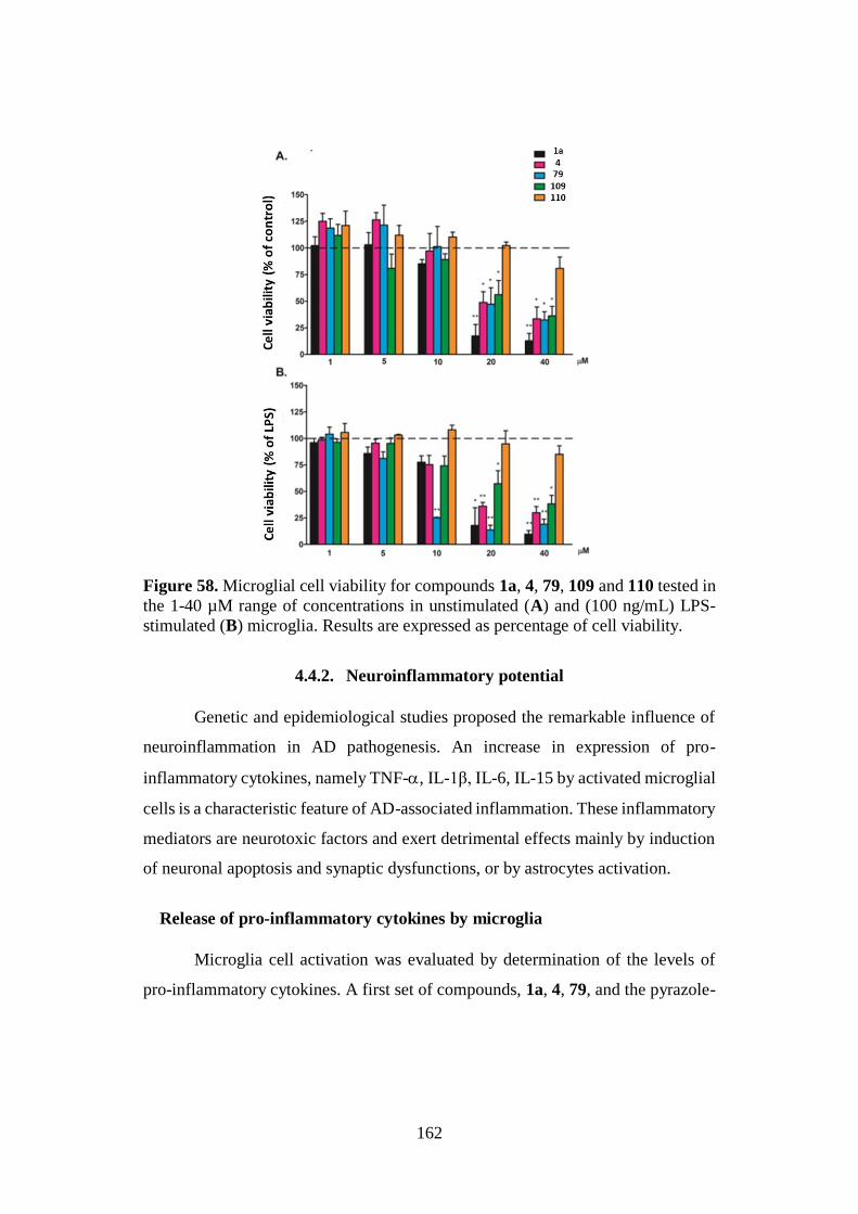
162
Figure 58. Microglial cell viability for compounds 1a, 4, 79, 109 and 110 tested in
the 1-40 µM range of concentrations in unstimulated (A) and (100 ng/mL) LPS-
stimulated (B) microglia. Results are expressed as percentage of cell viability.
4.4.2. Neuroinflammatory potential
Genetic and epidemiological studies proposed the remarkable influence of
neuroinflammation in AD pathogenesis. An increase in expression of pro-
inflammatory cytokines, namely TNF-, IL-1β, IL-6, IL-15 by activated microglial
cells is a characteristic feature of AD-associated inflammation. These inflammatory
mediators are neurotoxic factors and exert detrimental effects mainly by induction
of neuronal apoptosis and synaptic dysfunctions, or by astrocytes activation.
Release of pro-inflammatory cytokines by microglia
Microglia cell activation was evaluated by determination of the levels of
pro-inflammatory cytokines. A first set of compounds, 1a, 4, 79, and the pyrazole-
163
based analogues 109 and 110, was tested for the release of the pro-inflammatory
cytokines (IL-1β, IL-6 and TNF-α) by LPS-stimulated microglia.
Microglial cells were first exposed (1 h) to increasing and not-toxic
concentrations (1-10 μM) of the tested compounds, and then to LPS (100 ng/mL
for 6 h). The levels of the released IL-1β, IL-6, and TNF-α were then determined
into the culture medium by means of an enzyme-linked immunosorbent assay
(ELISA). Unstimulated cells, characterized by a low or undetectable release of
cytokines, were employed as reference; these basal levels were unchanged upon
treatment with the tested compounds (data not shown). LPS proved to promote the
release of IL-1β, IL-6, and TNF-α (404 pg/mL, 307 pg/mL, and 1035 pg/mL,
respectively).161
In general, treatment with 1a, 4 and 79 significantly suppressed the release
of IL-1β, IL-6 and TNF-α in a concentration-dependent manner. Conversely, and
not surprisingly, this behavior was not shown for 109 and 110 that, in agreement
with the design rationale, were ineffective. These data confirmed the pivotal role of
the α,β-unsaturated carbonyl moiety in influencing the extent of the inflammatory
response elicited by LPS.
IL-1β release
At the concentration of 5 µM, a 50 % reduction in IL-1β release was
observed. Among the tested compounds, at the higher concentrations of 7.5 and 10
µM only 1a achieved a maximum inhibitory effect, in which the IL-1β levels did
not differ from the basal levels (Fig. 59A).
IL-6 release
Interestingly, a very strong inhibitory effect was observed for IL-6 release,
that was maximally inhibited (reaching the basal levels) by 1a and 4 at 5 µM and
by 79 at 2.5 µM (Fig. 59B).
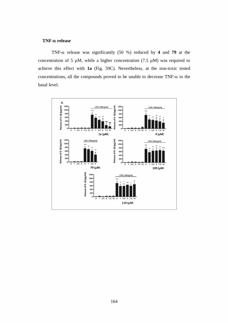
164
TNF- release
TNF- release was significantly (50 %) reduced by 4 and 79 at the
concentration of 5 µM, while a higher concentration (7.5 µM) was required to
achieve this effect with 1a (Fig. 59C). Nevertheless, at the non-toxic tested
concentrations, all the compounds proved to be unable to decrease TNF- to the
basal level.
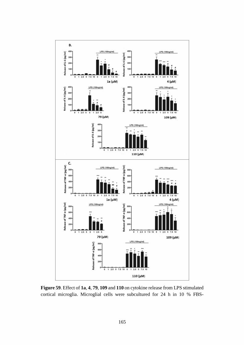
165
Figure 59. Effect of 1a, 4, 79, 109 and 110 on cytokine release from LPS stimulated
cortical microglia. Microglial cells were subcultured for 24 h in 10 % FBS-
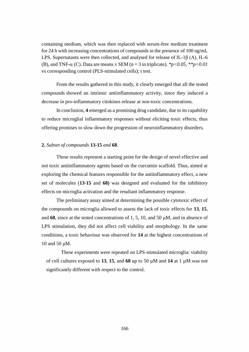
166
containing medium, which was then replaced with serum-free medium treatment
for 24 h with increasing concentrations of compounds in the presence of 100 ng/mL
LPS. Supernatants were then collected, and analysed for release of IL-1β (A), IL-6
(B), and TNF- (C). Data are means ± SEM (n = 3 in triplicate). *p<0.05, **p<0.01
vs corresponding control (PLS-stimulated cells); t test.
From the results gathered in this study, it clearly emerged that all the tested
compounds showed an intrinsic antiinflammatory activity, since they induced a
decrease in pro-inflammatory citokines release at non-toxic concentrations.
In conclusion, 4 emerged as a promising drug candidate, due to its capability
to reduce microglial inflammatory responses without eliciting toxic effects, thus
offering promises to slow down the progression of neuroinflammatory disorders.
2. Subset of compounds 13-15 and 68.
These results represent a starting point for the design of novel effective and
not toxic antiinflammatory agents based on the curcumin scaffold. Thus, aimed at
exploring the chemical features responsible for the antiinflammatory effect, a new
set of molecules (13-15 and 68) was designed and evaluated for the inhibitory
effects on microglia activation and the resultant inflammatory response.
The preliminary assay aimed at determining the possible cytotoxic effect of
the compounds on microglia allowed to assess the lack of toxic effects for 13, 15,
and 68, since at the tested concentrations of 1, 5, 10, and 50 μM, and in absence of
LPS stimulation, they did not affect cell viability and morphology. In the same
conditions, a toxic behaviour was observed for 14 at the highest concentrations of
10 and 50 μM.
These experiments were repeated on LPS-stimulated microglia: viability
of cell cultures exposed to 13, 15, and 68 up to 50 µM and 14 at 1 µM was not
significantly different with respect to the control.
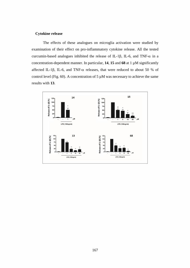
167
Cytokine release
The effects of these analogues on microglia activation were studied by
examination of their effect on pro-inflammatory cytokine release. All the tested
curcumin-based analogues inhibited the release of IL-1β, IL-6, and TNF- in a
concentration-dependent manner. In particular, 14, 15 and 68 at 1 µM significantly
affected IL-1β, IL-6, and TNF- releases, that were reduced to about 50 % of
control level (Fig. 60). A concentration of 5 µM was necessary to achieve the same
results with 13.
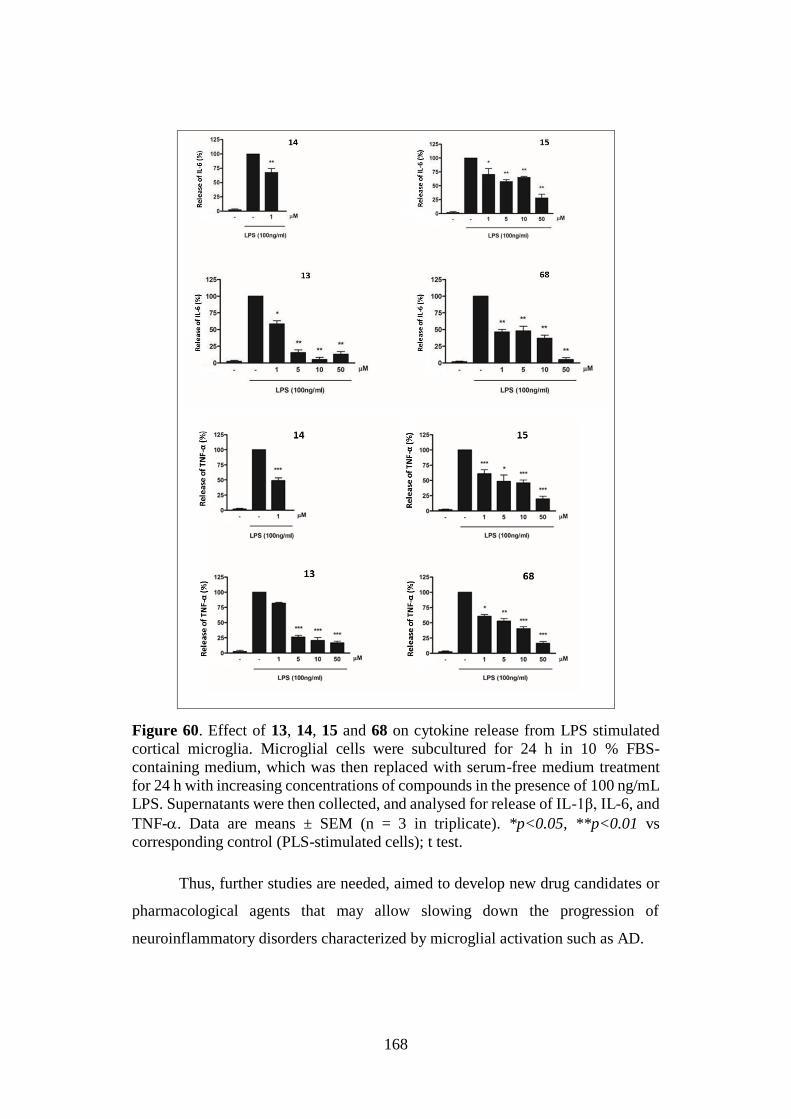
168
Figure 60. Effect of 13, 14, 15 and 68 on cytokine release from LPS stimulated
cortical microglia. Microglial cells were subcultured for 24 h in 10 % FBS-
containing medium, which was then replaced with serum-free medium treatment
for 24 h with increasing concentrations of compounds in the presence of 100 ng/mL
LPS. Supernatants were then collected, and analysed for release of IL-1β, IL-6, and
TNF-. Data are means ± SEM (n = 3 in triplicate). *p<0.05, **p<0.01 vs
corresponding control (PLS-stimulated cells); t test.
Thus, further studies are needed, aimed to develop new drug candidates or
pharmacological agents that may allow slowing down the progression of
neuroinflammatory disorders characterized by microglial activation such as AD.
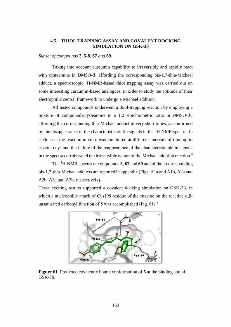
169
4.5. THIOL TRAPPING ASSAY AND COVALENT DOCKING
SIMULATION ON GSK-3β
Subset of compounds 2, 5-8, 67 and 69.
Taking into account curcumin capability to irreversibly and rapidly react
with cysteamine in DMSO-d6 affording the corresponding bis-1,7-thia-Michael
adduct, a spectroscopic 1H-NMR-based thiol trapping assay was carried out on
some interesting curcumin-based analogues, in order to study the aptitude of their
electrophilic central framework to undergo a Michael addition.
All tested compounds underwent a thiol-trapping reaction by employing a
mixture of compounds/cysteamine in a 1:2 stoichiometric ratio in DMSO-d6,
affording the corresponding thia-Michael adduct in very short times, as confirmed
by the disappearance of the characteristic olefin signals in the 1H-NMR spectra. In
each case, the reaction mixture was monitored at different intervals of time up to
several days and the failure of the reappearance of the characteristic olefin signals
in the spectra corroborated the irreversible nature of the Michael addition reaction.8
The 1H-NMR spectra of compounds 5, 67 and 69 and of their corresponding
bis-1,7-thia-Michael adducts are reported in appendix (Figs. A1a and A1b, A2a and
A2b, A3a and A3b, respectively).
These exciting results supported a covalent docking simulation on GSK-3β, in
which a nucleophilic attack of Cys199 residue of the enzyme on the reactive α,β-
unsaturated carbonyl function of 5 was accomplished (Fig. 61).8
Figure 61. Predicted covalently bound conformation of 5 at the binding site of
GSK-3β.
170
4.6. CK1 AND LRRK2 INHIBITION
The indole-based derivatives 133-142 were investigated to assess their
inhibitory activity on CK1 and LRRK2, two additional PKs involved in AD
pathogenesis. The compounds were tested at a fixed concentration of 10 μM to
determine the percentage of inhibition (% inh) and, when this was higher than 50
%, IC50 was calculated.
4.6.1. CK1δ and CK1ε inhibition
Subset of compounds 133-142.
The Kinase-Glo™ methodology,160 was employed to evaluate the CK1δ and
CK1ε inhibitory potency. At the beginning, the biological evaluation was carried
out on the CK1δ isoform, then for analogues that showed an interesting biological
profile, the inhibitory activity on CK1ε was also investigated (Table 25).
The preliminary results on CK1δ allowed to identify 137 and 140, bearing
a 3-phenyl-1,2,4-thiadiazolyl heterocycle, as active compounds with micromolar
values of potency. In particular, derivative 140, characterized by a methoxy group
into the 6-position of the indole scaffold, showing an IC50 value of 1.03 μM, proved
to be more potent than 137 (IC50 = 19.49 μM) and was recognized as the most
interesting compound of the series to be subjected to further investigation.
Therefore, 140 was tested on CK1ε, confirming a low micromolar inhibitory
profile (IC50 = 7.31 μM), although lower than that on CK1δ.
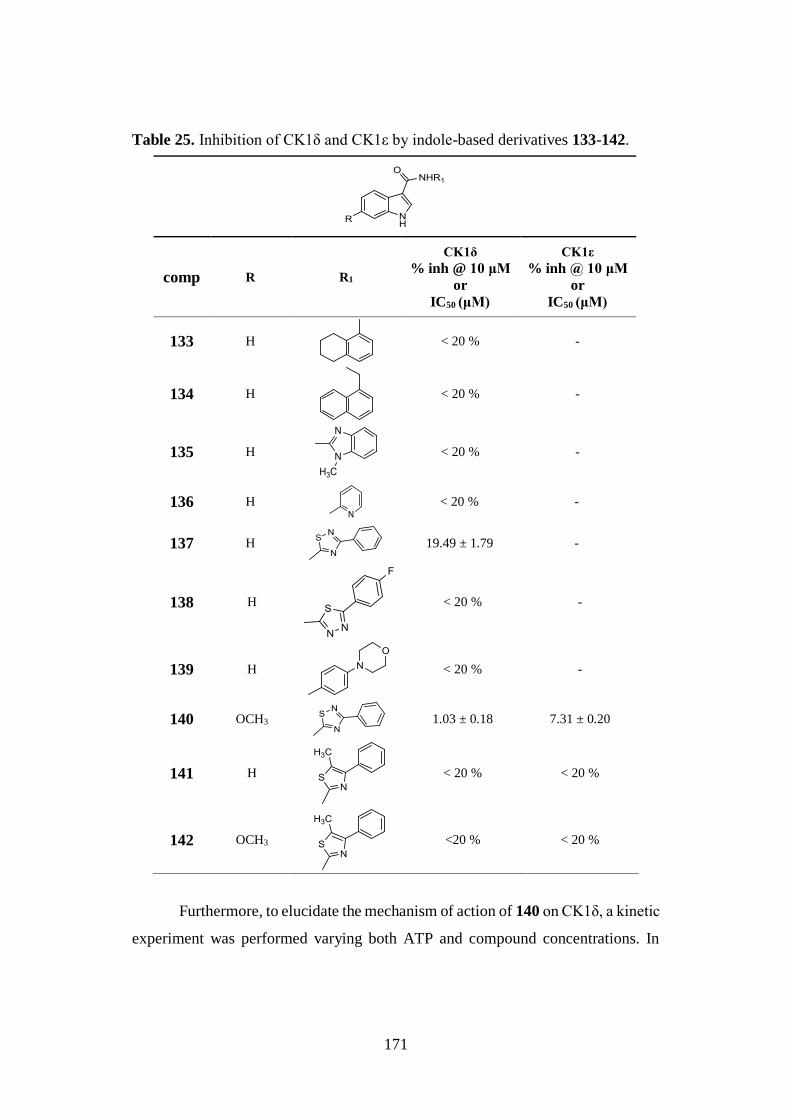
171
Table 25. Inhibition of CK1δ and CK1ε by indole-based derivatives 133-142.
comp R R1
CK1δ
% inh @ 10 μM
or
IC50 (μM)
CK1ε
% inh @ 10 μM
or
IC50 (μM)
133 H
< 20 % -
134 H
< 20 % -
135 H
< 20 % -
136 H
< 20 % -
137 H
19.49 ± 1.79 -
138 H
< 20 % -
139 H
< 20 % -
140 OCH3
1.03 ± 0.18 7.31 ± 0.20
141 H
< 20 % < 20 %
142 OCH3
<20 % < 20 %
Furthermore, to elucidate the mechanism of action of 140 on CK1δ, a kinetic
experiment was performed varying both ATP and compound concentrations. In
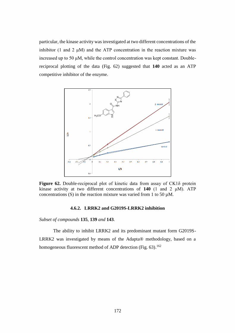
172
particular, the kinase activity was investigated at two different concentrations of the
inhibitor (1 and 2 μM) and the ATP concentration in the reaction mixture was
increased up to 50 μM, while the control concentration was kept constant. Double-
reciprocal plotting of the data (Fig. 62) suggested that 140 acted as an ATP
competitive inhibitor of the enzyme.
Figure 62. Double-reciprocal plot of kinetic data from assay of CK1δ protein
kinase activity at two different concentrations of 140 (1 and 2 μM). ATP
concentrations (S) in the reaction mixture was varied from 1 to 50 µM.
4.6.2. LRRK2 and G2019S-LRRK2 inhibition
Subset of compounds 135, 139 and 143.
The ability to inhibit LRRK2 and its predominant mutant form G2019S-
LRRK2 was investigated by means of the Adapta® methodology, based on a
homogeneous fluorescent method of ADP detection (Fig. 63).162
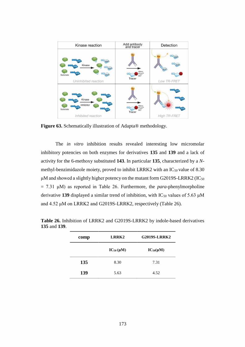
173
Figure 63. Schematically illustration of Adapta® methodology.
The in vitro inhibition results revealed interesting low micromolar
inhibitory potencies on both enzymes for derivatives 135 and 139 and a lack of
activity for the 6-methoxy substituted 143. In particular 135, characterized by a N-
methyl-benzimidazole moiety, proved to inhibit LRRK2 with an IC50 value of 8.30
μM and showed a slightly higher potency on the mutant form G2019S-LRRK2 (IC50
= 7.31 μM) as reported in Table 26. Furthermore, the para-phenylmorpholine
derivative 139 displayed a similar trend of inhibition, with IC50 values of 5.63 μM
and 4.52 μM on LRRK2 and G2019S-LRRK2, respectively (Table 26).
Table 26. Inhibition of LRRK2 and G2019S-LRRK2 by indole-based derivatives
135 and 139.
comp LRRK2 G2019S-LRRK2
IC50 (μM)
IC50(μM)
135 8.30 7.31
139 5.63 4.52
174
Taken together, these preliminary results suggests 135 and 139 as promising
lead compounds to be optimized for the development of more potent LRRK2 and
G2019S-LKKR2inhibitors.
4.7. BBB PERMEATION
Subset of compounds 2-6, 8, 135, 137, 139, and 140.
An essential requirement for a CNS targeting drug is its capability to
penetrate into the BBB at therapeutic concentrations. For this reason, and taking
into account that more than 95 % of compounds cross the BBB by passive transport,
an in vitro methodology developed by Prof. Martinez research group and based on
the high throughput technique PAMPA (Parallel Artificial Membrane Permeability
Assay)163 was employed to predict the CNS passive permeation of some interesting
curcumin- and indole-based derivatives. In this assay, the human BBB was
emulated by using a porcine lipid membrane and UV spectroscopy was exploited
to perform compounds quantification; additionally, for curcumin analogues 2-6 and
8, curcumin 1a was used as standard.
At the outset, an assay validation was made comparing the experimental
data with the permeability (Pe) values of ten commercial drugs of known Pe (Fig.
64). Two different experiments were carried out: analogues 137 and 140 were
evaluated in the first test, whereas 135, 139, 2-6 and 8, together with 1a were tested
later.
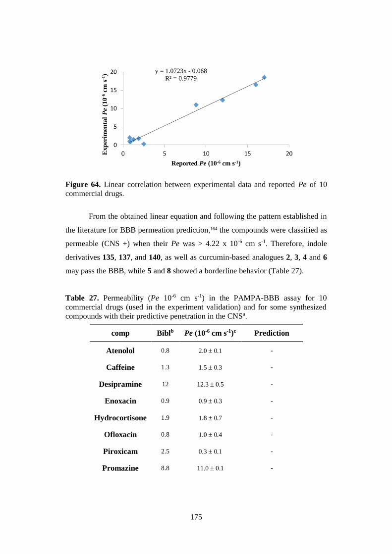
175
Figure 64. Linear correlation between experimental data and reported Pe of 10
commercial drugs.
From the obtained linear equation and following the pattern established in
the literature for BBB permeation prediction,164 the compounds were classified as
permeable (CNS +) when their Pe was > 4.22 x 10⁻⁶ cm s-1. Therefore, indole
derivatives 135, 137, and 140, as well as curcumin-based analogues 2, 3, 4 and 6
may pass the BBB, while 5 and 8 showed a borderline behavior (Table 27).
Table 27. Permeability (Pe 10-6 cm s-1) in the PAMPA-BBB assay for 10
commercial drugs (used in the experiment validation) and for some synthesized
compounds with their predictive penetration in the CNSa.
comp Biblb Pe (10-6 cm s-1)c Prediction
Atenolol 0.8 2.0 0.1 -
Caffeine 1.3 1.5 0.3 -
Desipramine 12 12.3 0.5 -
Enoxacin 0.9 0.9 0.3 -
Hydrocortisone 1.9 1.8 0.7 -
Ofloxacin 0.8 1.0 0.4 -
Piroxicam 2.5 0.3 0.1 -
Promazine 8.8 11.0 0.1 -
y = 1.0723x - 0.068
R² = 0.9779
0
5
10
15
20
0 5 10 15 20Exp
erim
enta
lP
e(1
0-6
cm s
-1)
Reported Pe (10-6 cm s-1)
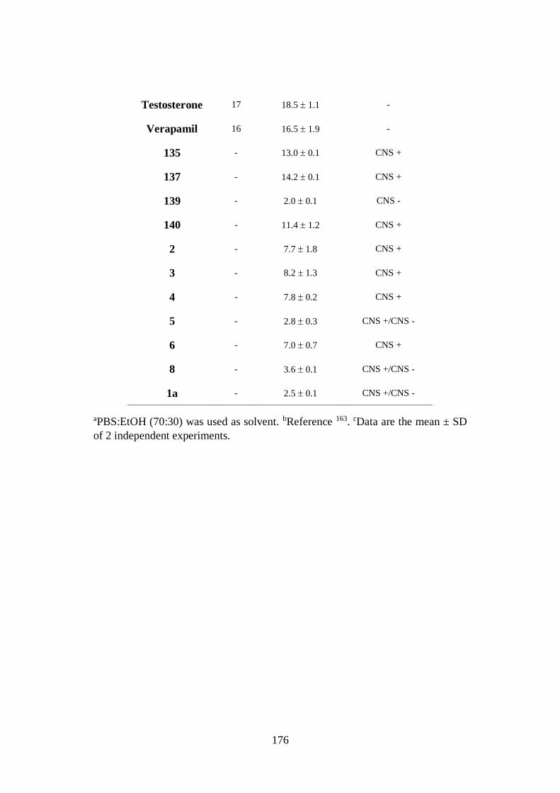
176
Testosterone 17 18.5 1.1 -
Verapamil 16 16.5 1.9 -
135 - 13.0 0.1 CNS +
137 - 14.2 0.1 CNS +
139 - 2.0 0.1 CNS -
140 - 11.4 1.2 CNS +
2 - 7.7 1.8 CNS +
3 - 8.2 ± 1.3 CNS +
4 - 7.8 0.2 CNS +
5 - 2.8 0.3 CNS +/CNS -
6 - 7.0 0.7 CNS +
8 - 3.6 0.1 CNS +/CNS -
1a - 2.5 0.1 CNS +/CNS -
aPBS:EtOH (70:30) was used as solvent. bReference 163. cData are the mean ± SD
of 2 independent experiments.

177
5. Conclusions
178
Main goal of my PhD thesis was the identification of naturally inspired
privileged scaffolds as starting point for the development of novel small molecules
as multifunctional lead candidates for AD treatment or valuable probes to explore
the chemical space of AD validated targets.
In view of the complex pathological mechanisms of AD, the multitarget
approach has gained increasing acceptance as a useful tool to discover drug
candidates that could promise improved efficacy compared to single-target drugs.
In this scenario, BACE-1 and GSK-3β emerged as AD validated targets and their
concurrent inhibition has been identified as a promising therapeutic strategy.
From a medicinal chemistry standpoint, the curcumin pharmacophore, in its
β-keto-enol form, incorporating some structural elements suitable for the
modulation of both enzymes, was rationally envisaged as potential dualistic BACE-
1/GSK-3β inhibitor and was selected as lead compound for the design and synthesis
of well-balanced dual modulators with good pharmacokinetic properties, mainly
referred to the BBB crossing ability. Therefore, several synthetic efforts were
dedicated at obtaining different series of novel curcumin-based derivatives, among
which compounds of series Ia (β-keto-enol forms) and Ib (diketo tautomers) were
mainly investigated for their capability to inhibit both structurally unrelated
enzymes.
Generally, the biological in vitro results confirmed the enol tautomeric form
as an important feature for achieving good inhibitory potency and chemical
stability, and highlighted the intense effect of the nature of the substituents on the
side aryl rings on BACE-1 and GSK-3β inhibition. Remarkably, a number of
derivatives proved to modulate BACE-1 and GSK-3 β enzymes with quite
comparable potencies in the micromolar range. In particular, while a mild dualistic
profile was shown for analogues 4 and 5, a crucial achievement in the multitarget
drug discovery context was represented by the subset of analogues 2, 6, and 7, that
proved to be well-balanced low-micromolar inhibitors of both enzymes. Among
them, derivative 2, thanks to the absence of evident neurotoxic effects and the
179
potential BBB permeability, clearly emerged as the most promising AD-modifying
drug candidate to be further developed.
On the other hand compound 8, showing a remarkable gap of potency (2
orders of magnitude) between its inhibition of BACE-1 and GSK-3β, proved to be
a single-target inhibitor of BACE-1, and it could be considered as a starting point
for the development of optimized dual modulators. Furthermore, compounds 3, 9,
and 10, also characterized by higher potencies on BACE-1 with respect to GSK-3β
(submicromolar and low-micromolar IC50 values, respectively), showed reduced
deviation from the dualistic profile.
Beside Aβ and τ cascades, neuroinflammation has been recognized to play
a crucial role in AD pathogenesis. In particular, an increase in expression of pro-
inflammatory cytokines by activated microglia is a characteristic AD feature.
Therefore, the capability to reduce microglial inflammatory responses without
eliciting toxic effects could be considered an added value to realize a successful AD
treatment. In this context, among the curcumin-based analogues that proved to
decrease the pro-inflammatory cytokines release at non-toxic concentrations, 4, 15
and 68 deserve particular attention due to their additional inhibitory activity on
BACE-1 and GSK-3β. In particular, 4 and 15, displaying micromolar dualistic
potencies on both enzymes accompanied by lack of cytotoxic effects and intrinsic
antiinflammatory activity, could represent valuable lead compounds for the
development of novel well-balanced dual BACE-1/GSK-3β modulators, able to
slow down the progression of neuroinflammatory diseases. Furthermore, compound
68, due to a comparable aptitude to decrease the release of pro-inflammatory
mediators by activated microglial cells, without affecting neither cell viability nor
morphology, and a promising low-micromolar potency on GSK-3β, could be
considered an interesting hit for the design of GSK-3β inhibitors endowed with
good antiinflammatory activity.
In AD, a decrease in the levels of many antioxidant defense enzymes such
as GSH cause oxidative stress, as a result of an imbalance between ROS generation
and antioxidant processes. Therefore, compounds able to induce GSH could offer
180
substantial therapeutic efficacy as neuroprotective agents to delay the progression
of the malady. In this respect, the curcumin-DF hybrids 74 and 75 showed a
significant enhancement of GSH total levels, that could be correlated to the Michael
acceptor reactivity of their additional α,β-unsaturated carbonyl function. Among
them 75, thanks to its low micromolar inhibitory activity on GSK-3β and the lack
of neurotoxic effects, could offer promises for the discovery of optimized GSK-3β
inhibitors with a neuroprotective potential.
Additional investigations are still in progress to further assess the great
potential of the most interesting curcumin analogues as promising multifunctional
lead compounds or drug candidates for an effective AD treatment.
Finally, concerning the indole-based derivatives designed and synthetized
as a part of a drug discovery project aimed at discovering PKs inhibitors as BBB
permeable promising pharmacological agents, the full satisfaction of the planned
goals occurred with analogues 140 and 135. In particular, 140 proved to be a low-
micromolar ATP competitive CK1δ inhibitor, able also to inhibit CK1ε; likewise,
135 showed a comparable low-micromolar activity on LRRK2 and its mutant form
G2019S-LRRK2. Although the results are only preliminary and further
investigations are required, both indole analogues could be considered as valuable
lead compounds to be further developed.

181
6. Experimental section
182
General Chemical Methods
Starting materials, unless otherwise specified in the Experimental Section,
were used as high-grade commercial products. Solvents were of analytical grade.
Melting points were determined in open glass capillaries, using a Büchi apparatus
and are uncorrected. 1H-NMR and 13C-NMR spectra were recorded on Varian
Gemini 400 MHz, unless diversely indicated, and chemical shifts are reported as
parts per million (ppm value) relative to the peak for tetramethylsilane (TMS) as
internal standard. Standard abbreviations indicating spin multiplicities are given as
follows: s (singlet), d (doublet), t (triplet), br (broad), q (quartet), dd (doublet of
doublet) or m (multiplet). Mass spectra were recorded on a Waters ZQ 4000
apparatus operating in electrospray mode (ES). Chromatographic separations were
performed on silica gel columns using the flash method (Kieselgel 40, 0.040-0.063
mm, Merck). Reactions were followed by thin layer chromatography (TLC) on
precoated silica gel plates (Merck Silica Gel 60 F254) and then visualized with a
UV lamp. Compounds were named following IUPAC rules as applied by Beilstein-
Institute AutoNom (version 2.1), a PC-integrated software package for systematic
names in organic chemistry.
Pabon reaction: general procedure A (synthesis of compounds 2-8, 11-17
and intermediates 22 and 23).
To a stirred solution of pentane-2,4-dione or intermediates 22 and 23 (1.00
mmol) in EtOAc (1.0 mL), B2O3 (1.0 molar equiv) was added, and the suspension
was stirred for 30 min at 80 °C before addition of a solution of the appropriate
aldehyde/s, (0.9 molar equiv for monoaryl or 1.8 molar equiv for bi-aryl curcumin
derivatives), and tri-n-butyl borate [B(n-BuO)3] (2.0 molar equiv for monoaryl or
4.0 molar equiv for bi-aryl curcumin derivatives) in EtOAc (0.5 mL). The reaction
mixture was stirred at 80 °C for 30 min, then a solution of n-BuNH2 (0.2 molar
equiv for monoaryl or 0.4 molar equiv for bi-aryl curcumin derivatives) in EtOAc
(1.0 mL) was added over a period of 15 min. The mixture was heated to 80 °C for
6-8 h and, after cooling to room temperature, it was acidified with HCl (0.5 N, 30
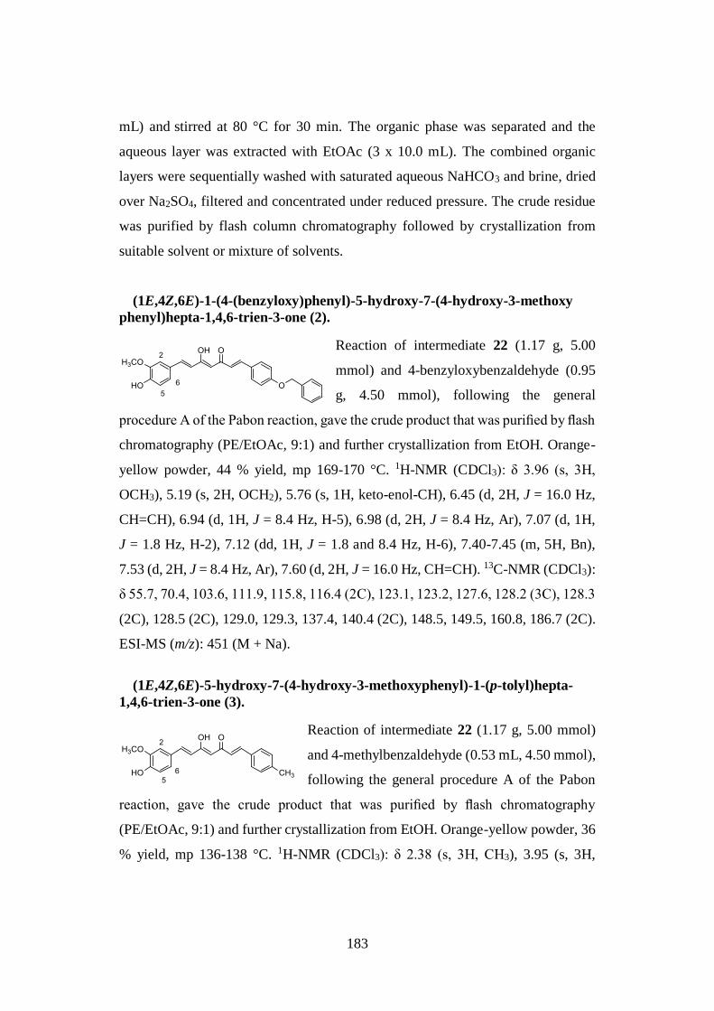
183
mL) and stirred at 80 °C for 30 min. The organic phase was separated and the
aqueous layer was extracted with EtOAc (3 x 10.0 mL). The combined organic
layers were sequentially washed with saturated aqueous NaHCO3 and brine, dried
over Na2SO4, filtered and concentrated under reduced pressure. The crude residue
was purified by flash column chromatography followed by crystallization from
suitable solvent or mixture of solvents.
(1E,4Z,6E)-1-(4-(benzyloxy)phenyl)-5-hydroxy-7-(4-hydroxy-3-methoxy
phenyl)hepta-1,4,6-trien-3-one (2).
Reaction of intermediate 22 (1.17 g, 5.00
mmol) and 4-benzyloxybenzaldehyde (0.95
g, 4.50 mmol), following the general
procedure A of the Pabon reaction, gave the crude product that was purified by flash
chromatography (PE/EtOAc, 9:1) and further crystallization from EtOH. Orange-
yellow powder, 44 % yield, mp 169-170 °C. 1H-NMR (CDCl3): δ 3.96 (s, 3H,
OCH3), 5.19 (s, 2H, OCH2), 5.76 (s, 1H, keto-enol-CH), 6.45 (d, 2H, J = 16.0 Hz,
CH=CH), 6.94 (d, 1H, J = 8.4 Hz, H-5), 6.98 (d, 2H, J = 8.4 Hz, Ar), 7.07 (d, 1H,
J = 1.8 Hz, H-2), 7.12 (dd, 1H, J = 1.8 and 8.4 Hz, H-6), 7.40-7.45 (m, 5H, Bn),
7.53 (d, 2H, J = 8.4 Hz, Ar), 7.60 (d, 2H, J = 16.0 Hz, CH=CH). 13C-NMR (CDCl3):
δ 55.7, 70.4, 103.6, 111.9, 115.8, 116.4 (2C), 123.1, 123.2, 127.6, 128.2 (3C), 128.3
(2C), 128.5 (2C), 129.0, 129.3, 137.4, 140.4 (2C), 148.5, 149.5, 160.8, 186.7 (2C).
ESI-MS (m/z): 451 (M + Na).
(1E,4Z,6E)-5-hydroxy-7-(4-hydroxy-3-methoxyphenyl)-1-(p-tolyl)hepta-
1,4,6-trien-3-one (3).
Reaction of intermediate 22 (1.17 g, 5.00 mmol)
and 4-methylbenzaldehyde (0.53 mL, 4.50 mmol),
following the general procedure A of the Pabon
reaction, gave the crude product that was purified by flash chromatography
(PE/EtOAc, 9:1) and further crystallization from EtOH. Orange-yellow powder, 36
% yield, mp 136-138 °C. 1H-NMR (CDCl3): δ 2.38 (s, 3H, CH3), 3.95 (s, 3H,

184
OCH3), 5.81 (s, 1H, keto-enol-CH), 6.49 (d, 1H J = 15.8 Hz, CH=CH), 6.60 (d, 1H,
J = 15.8 Hz, CH=CH), 6.94 (d, 1H, J = 8.2 Hz, H-5), 7.06 (d, 1H, J = 1.8 Hz, H-2),
7.13 (dd, 1H, J = 1.8 and 8.2 Hz, H-6), 7.20 (d, 2H, J = 8.0 Hz, Ar), 7.46 (d, 2H, J
= 8.0 Hz, Ar), 7.59 (d, 1H, J = 15.8 Hz, CH=CH), 7.63 (d, 1H, J = 15.8 Hz,
CH=CH). 13C-NMR (CDCl3): δ 21.0, 55.7, 107.6, 112.9, 116.0, 121.5, 121.9, 126.7,
127.4 (2C), 129.3 (2C), 130.9, 134.1, 141.2 (2C), 137.9, 148.5, 150.2, 183.7 (2C).
ESI-MS (m/z): 359 (M + Na).
(1E,4Z,6E)-5-hydroxy-1,7-bis(4-hydroxyphenyl)hepta-1,4,6-trien-3-one (4).
Reaction of pentane-2,4-dione (1.03 mL, 10.00
mmol) and 4-hydroxybenzaldehyde (2.24 g, 18.00
mmol), following the general procedure A of the
Pabon reaction, gave the crude product that was purified by flash chromatography
(PE/EtOAc, 7:3) and further crystallization from EtOH. Red-orange powder, 88 %
yield, mp 228-230 °C. 1H-NMR (acetone-d6): δ 5.99 (s, 1H, keto-enol-CH), 6.67
(d, 2H, J = 15.8 Hz, CH=CH), 6.91 (d, 4H, J = 8.6 Hz, Ar), 7.57 (d, 4H, J = 8.6 Hz,
Ar), 7.61 (d, 2H, J = 15.8 Hz, CH=CH). 1H-NMR (DMSO-d6): δ 6.22 (s, 1H, keto-
enol-CH), 6.82 (d, 2H, J = 16.4 Hz, CH=CH), 6.98 (d, 4H, J = 8.4 Hz, Ar), 7.25 (d,
4H, J = 8.4 Hz, Ar), 7.35 (br s, 1H, OH), 7.62 (d, 2H, J = 16.4 Hz, CH=CH), 9.70
(br s, 2H, OH). 13C-NMR (acetone-d6): δ 114.0, 116.5 (4C), 122.9, 127.9 (2C),
129.7, 130.0 (4C), 141.1 (2C), 161.1 (2C), 183.5 (2C). ESI-MS (m/z): 331 (M +
Na).
(1E,4Z,6E)-5-hydroxy-1,7-bis(4-methoxyphenyl)hepta-1,4,6-trien-3-one (5).
Reaction of pentane-2,4-dione (1.03 mL, 10.00
mmol) and 4-methoxybenzaldehyde (2.19 mL,
18.00 mmol), following the general procedure
A of the Pabon reaction, gave the crude product purified by flash chromatography
(PE/EtOAc, 9:1) and further crystallization from CH2Cl2/PE. Yellow powder, 87 %
yield, mp 110-112 °C. 1H-NMR (CDCl3): δ 3.86 (s, 6H, OCH3), 5.79 (s, 1H, keto-
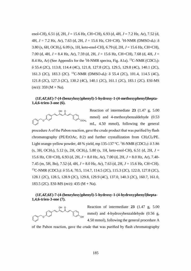
185
enol-CH), 6.51 (d, 2H, J = 15.6 Hz, CH=CH), 6.93 (d, 4H, J = 7.2 Hz, Ar), 7.52 (d,
4H, J = 7.2 Hz, Ar), 7.63 (d, 2H, J = 15.6 Hz, CH=CH). 1H-NMR (DMSO-d6): δ
3.80 (s, 6H, OCH3), 6.09 (s, 1H, keto-enol-CH), 6.79 (d, 2H, J = 15.6 Hz, CH=CH),
7.00 (d, 4H, J = 8.4 Hz, Ar), 7.59 (d, 2H, J = 15.6 Hz, CH=CH), 7.68 (d, 4H, J =
8.4 Hz, Ar) (See Appendix for the 1H-NMR spectra, Fig. A1a). 13C-NMR (CDCl3):
δ 55.4 (2C), 113.8, 114.4 (4C), 121.8, 127.8 (2C), 129.5, 129.8 (4C), 140.1 (2C),
161.3 (2C), 183.3 (2C). 13C-NMR (DMSO-d6): δ 55.4 (2C), 101.4, 114.5 (4C),
121.8 (2C), 127.3 (2C), 130.2 (4C), 140.1 (2C), 161.1 (2C), 183.1 (2C). ESI-MS
(m/z): 359 (M + Na).
(1E,4Z,6E)-7-(4-(benzyloxy)phenyl)-5-hydroxy-1-(4-methoxyphenyl)hepta-
1,4,6-trien-3-one (6).
Reaction of intermediate 23 (1.47 g, 5.00
mmol) and 4-methoxybenzaldehyde (0.53
mL, 4.50 mmol), following the general
procedure A of the Pabon reaction, gave the crude product that was purified by flash
chromatography (PE/EtOAc, 8:2) and further crystallization from CH2Cl2/PE.
Light orange-yellow powder, 48 % yield, mp 135-137 °C. 1H-NMR (CDCl3): δ 3.86
(s, 3H, OCH3), 5.12 (s, 2H, OCH2), 5.80 (s, 1H, keto-enol-CH), 6.51 (d, 2H, J =
15.6 Hz, CH=CH), 6.93 (d, 2H, J = 8.0 Hz, Ar), 7.00 (d, 2H, J = 8.0 Hz, Ar), 7.40-
7.45 (m, 5H, Bn), 7.52 (d, 4H, J = 8.0 Hz, Ar), 7.63 (d, 2H, J = 15.6 Hz, CH=CH).
13C-NMR (CDCl3): δ 55.4, 70.5, 114.7, 114.5 (2C), 115.3 (2C), 122.0, 127.8 (2C),
128.1 (2C), 128.5, 128.9 (2C), 129.8, 129.9 (4C), 137.0, 140.3 (2C), 160.7, 161.0,
183.5 (2C). ESI-MS (m/z): 435 (M + Na).
(1E,4Z,6E)-7-(4-(benzyloxy)phenyl)-5-hydroxy-1-(4-hydroxyphenyl)hepta-
1,4,6-trien-3-one (7).
Reaction of intermediate 23 (1.47 g, 5.00
mmol) and 4-hydroxybenzaldehyde (0.56 g,
4.50 mmol), following the general procedure A
of the Pabon reaction, gave the crude that was purified by flash chromatography
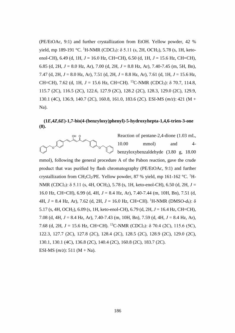
186
(PE/EtOAc, 9:1) and further crystallization from EtOH. Yellow powder, 42 %
yield, mp 189-191 °C. 1H-NMR (CDCl3): δ 5.11 (s, 2H, OCH2), 5.78 (s, 1H, keto-
enol-CH), 6.49 (d, 1H, J = 16.0 Hz, CH=CH), 6.50 (d, 1H, J = 15.6 Hz, CH=CH),
6.85 (d, 2H, J = 8.0 Hz, Ar), 7.00 (d, 2H, J = 8.8 Hz, Ar), 7.40-7.45 (m, 5H, Bn),
7.47 (d, 2H, J = 8.0 Hz, Ar), 7.51 (d, 2H, J = 8.8 Hz, Ar), 7.61 (d, 1H, J = 15.6 Hz,
CH=CH), 7.62 (d, 1H, J = 15.6 Hz, CH=CH). 13C-NMR (CDCl3): δ 70.7, 114.8,
115.7 (2C), 116.5 (2C), 122.6, 127.9 (2C), 128.2 (2C), 128.3, 129.0 (2C), 129.9,
130.1 (4C), 136.9, 140.7 (2C), 160.8, 161.0, 183.6 (2C). ESI-MS (m/z): 421 (M +
Na).
(1E,4Z,6E)-1,7-bis(4-(benzyloxy)phenyl)-5-hydroxyhepta-1,4,6-trien-3-one
(8).
Reaction of pentane-2,4-dione (1.03 mL,
10.00 mmol) and 4-
benzyloxybenzaldehyde (3.80 g, 18.00
mmol), following the general procedure A of the Pabon reaction, gave the crude
product that was purified by flash chromatography (PE/EtOAc, 9:1) and further
crystallization from CH2Cl2/PE. Yellow powder, 87 % yield, mp 161-162 °C. 1H-
NMR (CDCl3): δ 5.11 (s, 4H, OCH2), 5.78 (s, 1H, keto-enol-CH), 6.50 (d, 2H, J =
16.0 Hz, CH=CH), 6.99 (d, 4H, J = 8.4 Hz, Ar), 7.40-7.44 (m, 10H, Bn), 7.51 (d,
4H, J = 8.4 Hz, Ar), 7.62 (d, 2H, J = 16.0 Hz, CH=CH). 1H-NMR (DMSO-d6): δ
5.17 (s, 4H, OCH2), 6.09 (s, 1H, keto-enol-CH), 6.79 (d, 2H, J = 16.4 Hz, CH=CH),
7.08 (d, 4H, J = 8.4 Hz, Ar), 7.40-7.43 (m, 10H, Bn), 7.59 (d, 4H, J = 8.4 Hz, Ar),
7.68 (d, 2H, J = 15.6 Hz, CH=CH). 13C-NMR (CDCl3): δ 70.4 (2C), 115.6 (5C),
122.3, 127.7 (2C), 127.8 (2C), 128.4 (2C), 128.5 (2C), 128.9 (2C), 129.0 (2C),
130.1, 130.1 (4C), 136.8 (2C), 140.4 (2C), 160.8 (2C), 183.7 (2C).
ESI-MS (m/z): 511 (M + Na).
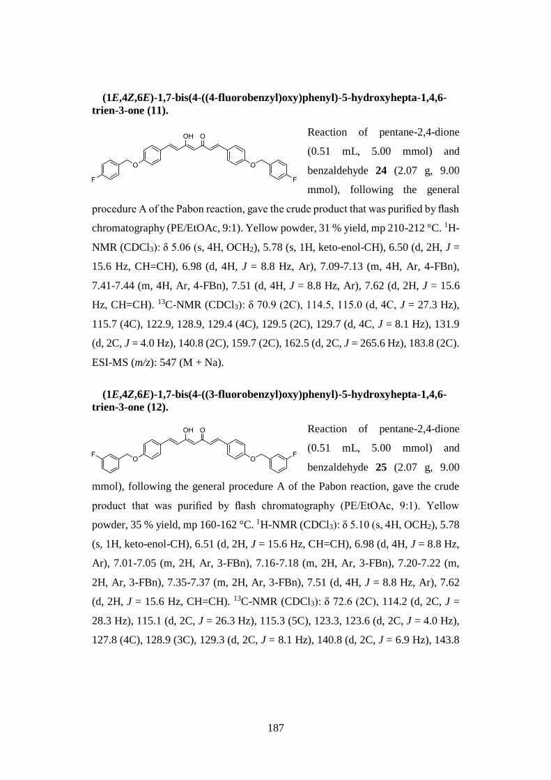
187
(1E,4Z,6E)-1,7-bis(4-((4-fluorobenzyl)oxy)phenyl)-5-hydroxyhepta-1,4,6-
trien-3-one (11).
Reaction of pentane-2,4-dione
(0.51 mL, 5.00 mmol) and
benzaldehyde 24 (2.07 g, 9.00
mmol), following the general
procedure A of the Pabon reaction, gave the crude product that was purified by flash
chromatography (PE/EtOAc, 9:1). Yellow powder, 31 % yield, mp 210-212 °C. 1H-
NMR (CDCl3): δ 5.06 (s, 4H, OCH2), 5.78 (s, 1H, keto-enol-CH), 6.50 (d, 2H, J =
15.6 Hz, CH=CH), 6.98 (d, 4H, J = 8.8 Hz, Ar), 7.09-7.13 (m, 4H, Ar, 4-FBn),
7.41-7.44 (m, 4H, Ar, 4-FBn), 7.51 (d, 4H, J = 8.8 Hz, Ar), 7.62 (d, 2H, J = 15.6
Hz, CH=CH). 13C-NMR (CDCl3): δ 70.9 (2C), 114.5, 115.0 (d, 4C, J = 27.3 Hz),
115.7 (4C), 122.9, 128.9, 129.4 (4C), 129.5 (2C), 129.7 (d, 4C, J = 8.1 Hz), 131.9
(d, 2C, J = 4.0 Hz), 140.8 (2C), 159.7 (2C), 162.5 (d, 2C, J = 265.6 Hz), 183.8 (2C).
ESI-MS (m/z): 547 (M + Na).
(1E,4Z,6E)-1,7-bis(4-((3-fluorobenzyl)oxy)phenyl)-5-hydroxyhepta-1,4,6-
trien-3-one (12).
Reaction of pentane-2,4-dione
(0.51 mL, 5.00 mmol) and
benzaldehyde 25 (2.07 g, 9.00
mmol), following the general procedure A of the Pabon reaction, gave the crude
product that was purified by flash chromatography (PE/EtOAc, 9:1). Yellow
powder, 35 % yield, mp 160-162 °C. 1H-NMR (CDCl3): δ 5.10 (s, 4H, OCH2), 5.78
(s, 1H, keto-enol-CH), 6.51 (d, 2H, J = 15.6 Hz, CH=CH), 6.98 (d, 4H, J = 8.8 Hz,
Ar), 7.01-7.05 (m, 2H, Ar, 3-FBn), 7.16-7.18 (m, 2H, Ar, 3-FBn), 7.20-7.22 (m,
2H, Ar, 3-FBn), 7.35-7.37 (m, 2H, Ar, 3-FBn), 7.51 (d, 4H, J = 8.8 Hz, Ar), 7.62
(d, 2H, J = 15.6 Hz, CH=CH). 13C-NMR (CDCl3): δ 72.6 (2C), 114.2 (d, 2C, J =
28.3 Hz), 115.1 (d, 2C, J = 26.3 Hz), 115.3 (5C), 123.3, 123.6 (d, 2C, J = 4.0 Hz),
127.8 (4C), 128.9 (3C), 129.3 (d, 2C, J = 8.1 Hz), 140.8 (d, 2C, J = 6.9 Hz), 143.8
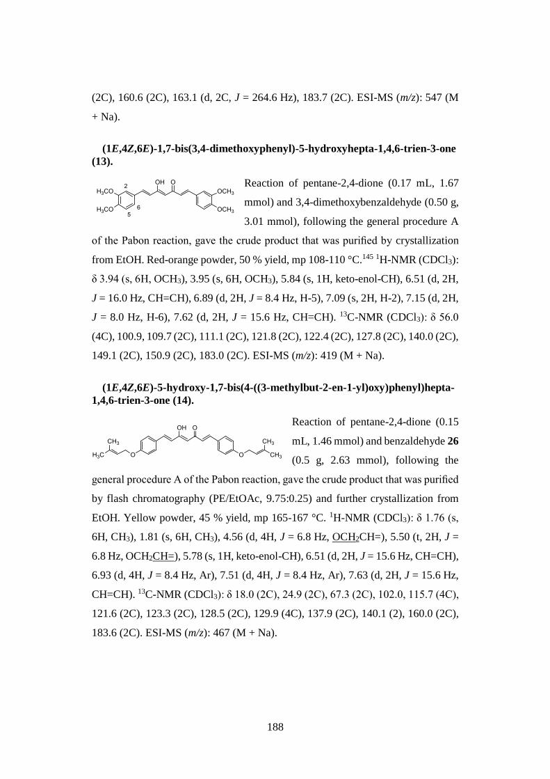
188
(2C), 160.6 (2C), 163.1 (d, 2C, J = 264.6 Hz), 183.7 (2C). ESI-MS (m/z): 547 (M
+ Na).
(1E,4Z,6E)-1,7-bis(3,4-dimethoxyphenyl)-5-hydroxyhepta-1,4,6-trien-3-one
(13).
Reaction of pentane-2,4-dione (0.17 mL, 1.67
mmol) and 3,4-dimethoxybenzaldehyde (0.50 g,
3.01 mmol), following the general procedure A
of the Pabon reaction, gave the crude product that was purified by crystallization
from EtOH. Red-orange powder, 50 % yield, mp 108-110 °C.145 1H-NMR (CDCl3):
δ 3.94 (s, 6H, OCH3), 3.95 (s, 6H, OCH3), 5.84 (s, 1H, keto-enol-CH), 6.51 (d, 2H,
J = 16.0 Hz, CH=CH), 6.89 (d, 2H, J = 8.4 Hz, H-5), 7.09 (s, 2H, H-2), 7.15 (d, 2H,
J = 8.0 Hz, H-6), 7.62 (d, 2H, J = 15.6 Hz, CH=CH). 13C-NMR (CDCl3): δ 56.0
(4C), 100.9, 109.7 (2C), 111.1 (2C), 121.8 (2C), 122.4 (2C), 127.8 (2C), 140.0 (2C),
149.1 (2C), 150.9 (2C), 183.0 (2C). ESI-MS (m/z): 419 (M + Na).
(1E,4Z,6E)-5-hydroxy-1,7-bis(4-((3-methylbut-2-en-1-yl)oxy)phenyl)hepta-
1,4,6-trien-3-one (14).
Reaction of pentane-2,4-dione (0.15
mL, 1.46 mmol) and benzaldehyde 26
(0.5 g, 2.63 mmol), following the
general procedure A of the Pabon reaction, gave the crude product that was purified
by flash chromatography (PE/EtOAc, 9.75:0.25) and further crystallization from
EtOH. Yellow powder, 45 % yield, mp 165-167 °C. 1H-NMR (CDCl3): δ 1.76 (s,
6H, CH3), 1.81 (s, 6H, CH3), 4.56 (d, 4H, J = 6.8 Hz, OCH2CH=), 5.50 (t, 2H, J =
6.8 Hz, OCH2CH=), 5.78 (s, 1H, keto-enol-CH), 6.51 (d, 2H, J = 15.6 Hz, CH=CH),
6.93 (d, 4H, J = 8.4 Hz, Ar), 7.51 (d, 4H, J = 8.4 Hz, Ar), 7.63 (d, 2H, J = 15.6 Hz,
CH=CH). 13C-NMR (CDCl3): δ 18.0 (2C), 24.9 (2C), 67.3 (2C), 102.0, 115.7 (4C),
121.6 (2C), 123.3 (2C), 128.5 (2C), 129.9 (4C), 137.9 (2C), 140.1 (2), 160.0 (2C),
183.6 (2C). ESI-MS (m/z): 467 (M + Na).
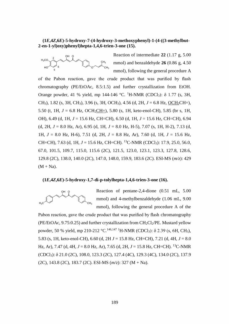
189
(1E,4Z,6E)-5-hydroxy-7-(4-hydroxy-3-methoxyphenyl)-1-(4-((3-methylbut-
2-en-1-yl)oxy)phenyl)hepta-1,4,6-trien-3-one (15).
Reaction of intermediate 22 (1.17 g, 5.00
mmol) and benzaldehyde 26 (0.86 g, 4.50
mmol), following the general procedure A
of the Pabon reaction, gave the crude product that was purified by flash
chromatography (PE/EtOAc, 8.5:1.5) and further crystallization from EtOH.
Orange powder, 41 % yield, mp 144-146 °C. 1H-NMR (CDCl3): δ 1.77 (s, 3H,
CH3), 1.82 (s, 3H, CH3), 3.96 (s, 3H, OCH3), 4.56 (d, 2H, J = 6.8 Hz, OCH2CH=),
5.50 (t, 1H, J = 6.8 Hz, OCH2CH=), 5.80 (s, 1H, keto-enol-CH), 5.85 (br s, 1H,
OH), 6.49 (d, 1H, J = 15.6 Hz, CH=CH), 6.50 (d, 1H, J = 15.6 Hz, CH=CH), 6.94
(d, 2H, J = 8.0 Hz, Ar), 6.95 (d, 1H, J = 8.0 Hz, H-5), 7.07 (s, 1H, H-2), 7.13 (d,
1H, J = 8.0 Hz, H-6), 7.51 (d, 2H, J = 8.8 Hz, Ar), 7.60 (d, 1H, J = 15.6 Hz,
CH=CH), 7.63 (d, 1H, J = 15.6 Hz, CH=CH). 13C-NMR (CDCl3): 17.9, 25.0, 56.0,
67.0, 101.5, 109.7, 115.0, 115.6 (2C), 121.5, 123.0, 123.1, 123.3, 127.8, 128.6,
129.8 (2C), 138.0, 140.0 (2C), 147.0, 148.0, 159.9, 183.6 (2C). ESI-MS (m/z): 429
(M + Na).
(1E,4Z,6E)-5-hydroxy-1,7-di-p-tolylhepta-1,4,6-trien-3-one (16).
Reaction of pentane-2,4-dione (0.51 mL, 5.00
mmol) and 4-methylbenzaldehyde (1.06 mL, 9.00
mmol), following the general procedure A of the
Pabon reaction, gave the crude product that was purified by flash chromatography
(PE/EtOAc, 9.75:0.25) and further crystallization from CH2Cl2/PE. Mustard yellow
powder, 50 % yield, mp 210-212 °C.146,147 1H-NMR (CDCl3): δ 2.39 (s, 6H, CH3),
5.83 (s, 1H, keto-enol-CH), 6.60 (d, 2H J = 15.8 Hz, CH=CH), 7.21 (d, 4H, J = 8.0
Hz, Ar), 7.47 (d, 4H, J = 8.0 Hz, Ar), 7.65 (d, 2H, J = 15.8 Hz, CH=CH). 13C-NMR
(CDCl3): δ 21.0 (2C), 108.0, 123.3 (2C), 127.4 (4C), 129.3 (4C), 134.0 (2C), 137.9
(2C), 143.8 (2C), 183.7 (2C). ESI-MS (m/z): 327 (M + Na).
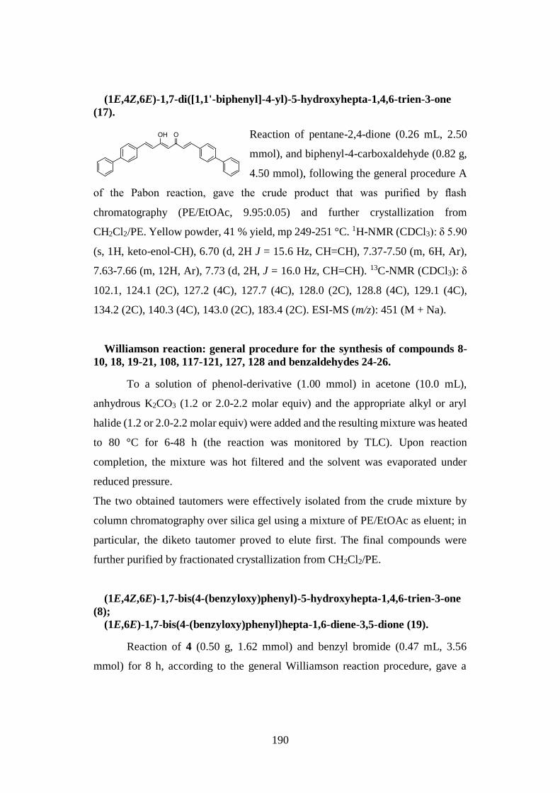
190
(1E,4Z,6E)-1,7-di([1,1'-biphenyl]-4-yl)-5-hydroxyhepta-1,4,6-trien-3-one
(17).
Reaction of pentane-2,4-dione (0.26 mL, 2.50
mmol), and biphenyl-4-carboxaldehyde (0.82 g,
4.50 mmol), following the general procedure A
of the Pabon reaction, gave the crude product that was purified by flash
chromatography (PE/EtOAc, 9.95:0.05) and further crystallization from
CH2Cl2/PE. Yellow powder, 41 % yield, mp 249-251 °C. 1H-NMR (CDCl3): δ 5.90
(s, 1H, keto-enol-CH), 6.70 (d, 2H J = 15.6 Hz, CH=CH), 7.37-7.50 (m, 6H, Ar),
7.63-7.66 (m, 12H, Ar), 7.73 (d, 2H, J = 16.0 Hz, CH=CH). 13C-NMR (CDCl3): δ
102.1, 124.1 (2C), 127.2 (4C), 127.7 (4C), 128.0 (2C), 128.8 (4C), 129.1 (4C),
134.2 (2C), 140.3 (4C), 143.0 (2C), 183.4 (2C). ESI-MS (m/z): 451 (M + Na).
Williamson reaction: general procedure for the synthesis of compounds 8-
10, 18, 19-21, 108, 117-121, 127, 128 and benzaldehydes 24-26.
To a solution of phenol-derivative (1.00 mmol) in acetone (10.0 mL),
anhydrous K2CO3 (1.2 or 2.0-2.2 molar equiv) and the appropriate alkyl or aryl
halide (1.2 or 2.0-2.2 molar equiv) were added and the resulting mixture was heated
to 80 °C for 6-48 h (the reaction was monitored by TLC). Upon reaction
completion, the mixture was hot filtered and the solvent was evaporated under
reduced pressure.
The two obtained tautomers were effectively isolated from the crude mixture by
column chromatography over silica gel using a mixture of PE/EtOAc as eluent; in
particular, the diketo tautomer proved to elute first. The final compounds were
further purified by fractionated crystallization from CH2Cl2/PE.
(1E,4Z,6E)-1,7-bis(4-(benzyloxy)phenyl)-5-hydroxyhepta-1,4,6-trien-3-one
(8);
(1E,6E)-1,7-bis(4-(benzyloxy)phenyl)hepta-1,6-diene-3,5-dione (19).
Reaction of 4 (0.50 g, 1.62 mmol) and benzyl bromide (0.47 mL, 3.56
mmol) for 8 h, according to the general Williamson reaction procedure, gave a
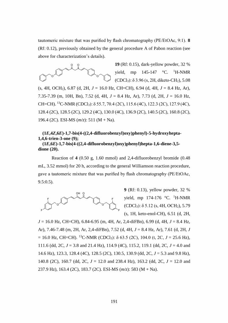
191
tautomeric mixture that was purified by flash chromatography (PE/EtOAc, 9:1). 8
(Rf: 0.12), previously obtained by the general procedure A of Pabon reaction (see
above for characterization’s details).
19 (Rf: 0.15), dark-yellow powder, 32 %
yield, mp 145-147 °C. 1H-NMR
(CDCl3): δ 3.96 (s, 2H, diketo-CH2), 5.08
(s, 4H, OCH2), 6.87 (d, 2H, J = 16.0 Hz, CH=CH), 6.94 (d, 4H, J = 8.4 Hz, Ar),
7.35-7.39 (m, 10H, Bn), 7.52 (d, 4H, J = 8.4 Hz, Ar), 7.73 (d, 2H, J = 16.0 Hz,
CH=CH). 13C-NMR (CDCl3): δ 55.7, 70.4 (2C), 115.6 (4C), 122.3 (2C), 127.9 (4C),
128.4 (2C), 128.5 (2C), 129.2 (4C), 130.0 (4C), 136.9 (2C), 140.5 (2C), 160.8 (2C),
196.4 (2C). ESI-MS (m/z): 511 (M + Na).
(1E,4Z,6E)-1,7-bis(4-((2,4-difluorobenzyl)oxy)phenyl)-5-hydroxyhepta-
1,4,6-trien-3-one (9);
(1E,6E)-1,7-bis(4-((2,4-difluorobenzyl)oxy)phenyl)hepta-1,6-diene-3,5-
dione (20).
Reaction of 4 (0.50 g, 1.60 mmol) and 2,4-difluorobenzyl bromide (0.48
mL, 3.52 mmol) for 20 h, according to the general Williamson reaction procedure,
gave a tautomeric mixture that was purified by flash chromatography (PE/EtOAc,
9.5:0.5).
9 (Rf: 0.13), yellow powder, 32 %
yield, mp 174-176 °C. 1H-NMR
(CDCl3): δ 5.12 (s, 4H, OCH2), 5.79
(s, 1H, keto-enol-CH), 6.51 (d, 2H,
J = 16.0 Hz, CH=CH), 6.84-6.95 (m, 4H, Ar, 2,4-diFBn), 6.99 (d, 4H, J = 8.4 Hz,
Ar), 7.46-7.48 (m, 2H, Ar, 2,4-diFBn), 7.52 (d, 4H, J = 8.4 Hz, Ar), 7.61 (d, 2H, J
= 16.0 Hz, CH=CH). 13C-NMR (CDCl3): δ 63.5 (2C), 104.0 (t, 2C, J = 25.6 Hz),
111.6 (dd, 2C, J = 3.8 and 21.4 Hz), 114.9 (4C), 115.2, 119.1 (dd, 2C, J = 4.0 and
14.6 Hz), 123.3, 128.4 (4C), 128.5 (2C), 130.5, 130.9 (dd, 2C, J = 5.3 and 9.8 Hz),
140.8 (2C), 160.7 (dd, 2C, J = 12.0 and 238.4 Hz), 163.2 (dd, 2C, J = 12.0 and
237.9 Hz), 163.4 (2C), 183.7 (2C). ESI-MS (m/z): 583 (M + Na).
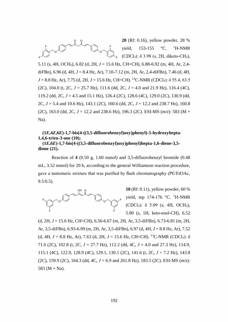
192
20 (Rf: 0.16), yellow powder, 28 %
yield, 153-155 °C. 1H-NMR
(CDCl3): δ 3.98 (s, 2H, diketo-CH2),
5.11 (s, 4H, OCH2), 6.82 (d, 2H, J = 15.6 Hz, CH=CH), 6.88-6.92 (m, 4H, Ar, 2,4-
diFBn), 6.96 (d, 4H, J = 8.4 Hz, Ar), 7.10-7.12 (m, 2H, Ar, 2,4-diFBn), 7.46 (d, 4H,
J = 8.8 Hz, Ar), 7.75 (d, 2H, J = 15.6 Hz, CH=CH). 13C-NMR (CDCl3): δ 55.4, 63.5
(2C), 104.0 (t, 2C, J = 25.7 Hz), 111.6 (dd, 2C, J = 4.0 and 21.9 Hz), 116.4 (4C),
119.2 (dd, 2C, J = 4.5 and 15.1 Hz), 126.4 (2C), 128.6 (4C), 129.0 (2C), 130.9 (dd,
2C, J = 5.4 and 10.6 Hz), 143.1 (2C), 160.6 (dd, 2C, J = 12.2 and 238.7 Hz), 160.8
(2C), 163.0 (dd, 2C, J = 12.2 and 238.6 Hz), 196.3 (2C). ESI-MS (m/z): 583 (M +
Na).
(1E,4Z,6E)-1,7-bis(4-((3,5-difluorobenzyl)oxy)phenyl)-5-hydroxyhepta-
1,4,6-trien-3-one (10);
(1E,6E)-1,7-bis(4-((3,5-difluorobenzyl)oxy)phenyl)hepta-1,6-diene-3,5-
dione (21).
Reaction of 4 (0.50 g, 1.60 mmol) and 3,5-difluorobenzyl bromide (0.48
mL, 3.52 mmol) for 20 h, according to the general Williamson reaction procedure,
gave a tautomeric mixture that was purified by flash chromatography (PE/EtOAc,
9.5:0.5).
10 (Rf: 0.11), yellow powder, 60 %
yield, mp 174-176 °C. 1H-NMR
(CDCl3): δ 5.09 (s, 4H, OCH2),
5.80 (s, 1H, keto-enol-CH), 6.52
(d, 2H, J = 15.6 Hz, CH=CH), 6.56-6.67 (m, 2H, Ar, 3,5-diFBn), 6.73-6.81 (m, 2H,
Ar, 3,5-diFBn), 6.93-6.99 (m, 2H, Ar, 3,5-diFBn), 6.97 (d, 4H, J = 8.8 Hz, Ar), 7.52
(d, 4H, J = 8.8 Hz, Ar), 7.63 (d, 2H, J = 15.6 Hz, CH=CH). 13C-NMR (CDCl3): δ
71.6 (2C), 102.8 (t, 2C, J = 27.7 Hz), 112.2 (dd, 4C, J = 4.0 and 27.3 Hz), 114.9,
115.1 (4C), 122.9, 128.9 (4C), 129.1, 130.1 (2C), 141.6 (t, 2C, J = 7.2 Hz), 143.8
(2C), 159.9 (2C), 164.3 (dd, 4C, J = 6.9 and 261.8 Hz), 183.5 (2C). ESI-MS (m/z):
583 (M + Na).
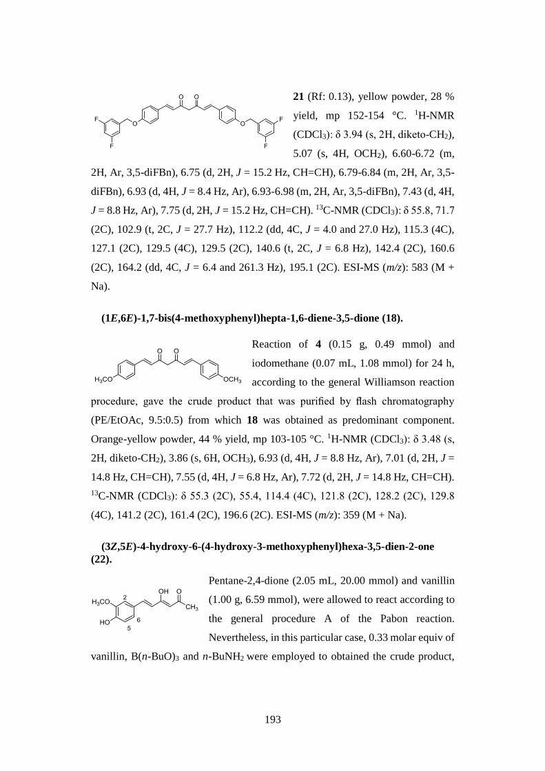
193
21 (Rf: 0.13), yellow powder, 28 %
yield, mp 152-154 °C. 1H-NMR
(CDCl3): δ 3.94 (s, 2H, diketo-CH2),
5.07 (s, 4H, OCH2), 6.60-6.72 (m,
2H, Ar, 3,5-diFBn), 6.75 (d, 2H, J = 15.2 Hz, CH=CH), 6.79-6.84 (m, 2H, Ar, 3,5-
diFBn), 6.93 (d, 4H, J = 8.4 Hz, Ar), 6.93-6.98 (m, 2H, Ar, 3,5-diFBn), 7.43 (d, 4H,
J = 8.8 Hz, Ar), 7.75 (d, 2H, J = 15.2 Hz, CH=CH). 13C-NMR (CDCl3): δ 55.8, 71.7
(2C), 102.9 (t, 2C, J = 27.7 Hz), 112.2 (dd, 4C, J = 4.0 and 27.0 Hz), 115.3 (4C),
127.1 (2C), 129.5 (4C), 129.5 (2C), 140.6 (t, 2C, J = 6.8 Hz), 142.4 (2C), 160.6
(2C), 164.2 (dd, 4C, J = 6.4 and 261.3 Hz), 195.1 (2C). ESI-MS (m/z): 583 (M +
Na).
(1E,6E)-1,7-bis(4-methoxyphenyl)hepta-1,6-diene-3,5-dione (18).
Reaction of 4 (0.15 g, 0.49 mmol) and
iodomethane (0.07 mL, 1.08 mmol) for 24 h,
according to the general Williamson reaction
procedure, gave the crude product that was purified by flash chromatography
(PE/EtOAc, 9.5:0.5) from which 18 was obtained as predominant component.
Orange-yellow powder, 44 % yield, mp 103-105 °C. 1H-NMR (CDCl3): δ 3.48 (s,
2H, diketo-CH2), 3.86 (s, 6H, OCH3), 6.93 (d, 4H, J = 8.8 Hz, Ar), 7.01 (d, 2H, J =
14.8 Hz, CH=CH), 7.55 (d, 4H, J = 6.8 Hz, Ar), 7.72 (d, 2H, J = 14.8 Hz, CH=CH).
13C-NMR (CDCl3): δ 55.3 (2C), 55.4, 114.4 (4C), 121.8 (2C), 128.2 (2C), 129.8
(4C), 141.2 (2C), 161.4 (2C), 196.6 (2C). ESI-MS (m/z): 359 (M + Na).
(3Z,5E)-4-hydroxy-6-(4-hydroxy-3-methoxyphenyl)hexa-3,5-dien-2-one
(22).
Pentane-2,4-dione (2.05 mL, 20.00 mmol) and vanillin
(1.00 g, 6.59 mmol), were allowed to react according to
the general procedure A of the Pabon reaction.
Nevertheless, in this particular case, 0.33 molar equiv of
vanillin, B(n-BuO)3 and n-BuNH2 were employed to obtained the crude product,
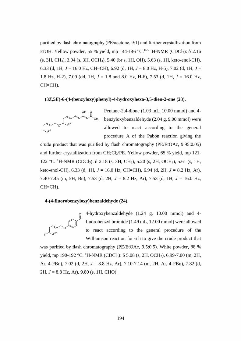
194
purified by flash chromatography (PE/acetone, 9:1) and further crystallization from
EtOH. Yellow powder, 55 % yield, mp 144-146 °C.165 1H-NMR (CDCl3): 2.16
(s, 3H, CH3), 3.94 (s, 3H, OCH3), 5.40 (br s, 1H, OH), 5.63 (s, 1H, keto-enol-CH),
6.33 (d, 1H, J = 16.0 Hz, CH=CH), 6.92 (d, 1H, J = 8.0 Hz, H-5), 7.02 (d, 1H, J =
1.8 Hz, H-2), 7.09 (dd, 1H, J = 1.8 and 8.0 Hz, H-6), 7.53 (d, 1H, J = 16.0 Hz,
CH=CH).
(3Z,5E)-6-(4-(benzyloxy)phenyl)-4-hydroxyhexa-3,5-dien-2-one (23).
Pentane-2,4-dione (1.03 mL, 10.00 mmol) and 4-
benzyloxybenzaldehyde (2.04 g, 9.00 mmol) were
allowed to react according to the general
procedure A of the Pabon reaction giving the
crude product that was purified by flash chromatography (PE/EtOAc, 9.95:0.05)
and further crystallization from CH2Cl2/PE. Yellow powder, 65 % yield, mp 121-
122 °C. 1H-NMR (CDCl3): 2.18 (s, 3H, CH3), 5.20 (s, 2H, OCH2), 5.61 (s, 1H,
keto-enol-CH), 6.33 (d, 1H, J = 16.0 Hz, CH=CH), 6.94 (d, 2H, J = 8.2 Hz, Ar),
7.40-7.45 (m, 5H, Bn), 7.53 (d, 2H, J = 8.2 Hz, Ar), 7.53 (d, 1H, J = 16.0 Hz,
CH=CH).
4-(4-fluorobenzyloxy)benzaldehyde (24).
4-hydroxybenzaldehyde (1.24 g, 10.00 mmol) and 4-
fluorobenzyl bromide (1.49 mL, 12.00 mmol) were allowed
to react according to the general procedure of the
Williamson reaction for 6 h to give the crude product that
was purified by flash chromatography (PE/EtOAc, 9.5:0.5). White powder, 88 %
yield, mp 190-192 °C. 1H-NMR (CDCl3): δ 5.08 (s, 2H, OCH2), 6.99-7.00 (m, 2H,
Ar, 4-FBn), 7.02 (d, 2H, J = 8.8 Hz, Ar), 7.10-7.14 (m, 2H, Ar, 4-FBn), 7.82 (d,
2H, J = 8.8 Hz, Ar), 9.80 (s, 1H, CHO).
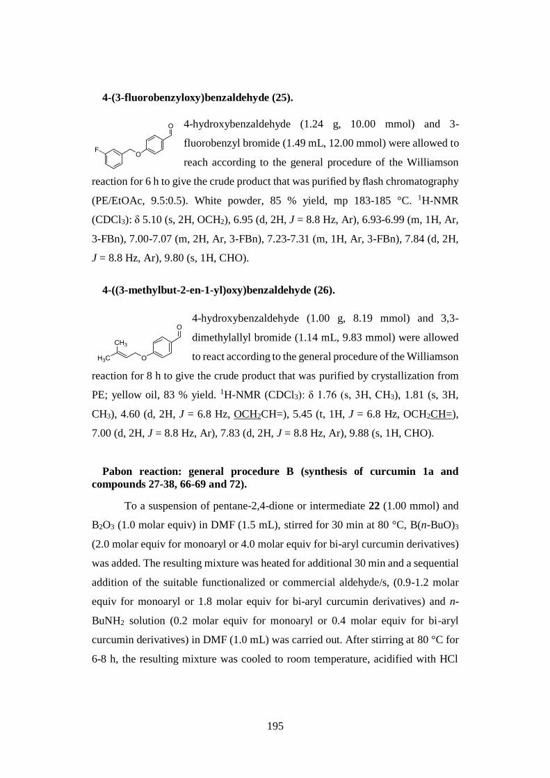
195
4-(3-fluorobenzyloxy)benzaldehyde (25).
4-hydroxybenzaldehyde (1.24 g, 10.00 mmol) and 3-
fluorobenzyl bromide (1.49 mL, 12.00 mmol) were allowed to
reach according to the general procedure of the Williamson
reaction for 6 h to give the crude product that was purified by flash chromatography
(PE/EtOAc, 9.5:0.5). White powder, 85 % yield, mp 183-185 °C. 1H-NMR
(CDCl3): δ 5.10 (s, 2H, OCH2), 6.95 (d, 2H, J = 8.8 Hz, Ar), 6.93-6.99 (m, 1H, Ar,
3-FBn), 7.00-7.07 (m, 2H, Ar, 3-FBn), 7.23-7.31 (m, 1H, Ar, 3-FBn), 7.84 (d, 2H,
J = 8.8 Hz, Ar), 9.80 (s, 1H, CHO).
4-((3-methylbut-2-en-1-yl)oxy)benzaldehyde (26).
4-hydroxybenzaldehyde (1.00 g, 8.19 mmol) and 3,3-
dimethylallyl bromide (1.14 mL, 9.83 mmol) were allowed
to react according to the general procedure of the Williamson
reaction for 8 h to give the crude product that was purified by crystallization from
PE; yellow oil, 83 % yield. 1H-NMR (CDCl3): δ 1.76 (s, 3H, CH3), 1.81 (s, 3H,
CH3), 4.60 (d, 2H, J = 6.8 Hz, OCH2CH=), 5.45 (t, 1H, J = 6.8 Hz, OCH2CH=),
7.00 (d, 2H, J = 8.8 Hz, Ar), 7.83 (d, 2H, J = 8.8 Hz, Ar), 9.88 (s, 1H, CHO).
Pabon reaction: general procedure B (synthesis of curcumin 1a and
compounds 27-38, 66-69 and 72).
To a suspension of pentane-2,4-dione or intermediate 22 (1.00 mmol) and
B2O3 (1.0 molar equiv) in DMF (1.5 mL), stirred for 30 min at 80 °C, B(n-BuO)3
(2.0 molar equiv for monoaryl or 4.0 molar equiv for bi-aryl curcumin derivatives)
was added. The resulting mixture was heated for additional 30 min and a sequential
addition of the suitable functionalized or commercial aldehyde/s, (0.9-1.2 molar
equiv for monoaryl or 1.8 molar equiv for bi-aryl curcumin derivatives) and n-
BuNH2 solution (0.2 molar equiv for monoaryl or 0.4 molar equiv for bi-aryl
curcumin derivatives) in DMF (1.0 mL) was carried out. After stirring at 80 °C for
6-8 h, the resulting mixture was cooled to room temperature, acidified with HCl
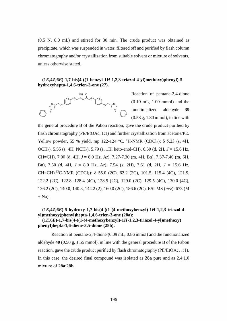
196
(0.5 N, 8.0 mL) and stirred for 30 min. The crude product was obtained as
precipitate, which was suspended in water, filtered off and purified by flash column
chromatography and/or crystallization from suitable solvent or mixture of solvents,
unless otherwise stated.
(1E,4Z,6E)-1,7-bis(4-((1-benzyl-1H-1,2,3-triazol-4-yl)methoxy)phenyl)-5-
hydroxyhepta-1,4,6-trien-3-one (27).
Reaction of pentane-2,4-dione
(0.10 mL, 1.00 mmol) and the
functionalized aldehyde 39
(0.53 g, 1.80 mmol), in line with
the general procedure B of the Pabon reaction, gave the crude product purified by
flash chromatography (PE/EtOAc, 1:1) and further crystallization from acetone/PE.
Yellow powder, 55 % yield, mp 122-124 °C. 1H-NMR (CDCl3): δ 5.23 (s, 4H,
OCH2), 5.55 (s, 4H, NCH2), 5.79 (s, 1H, keto-enol-CH), 6.50 (d, 2H, J = 15.6 Hz,
CH=CH), 7.00 (d, 4H, J = 8.0 Hz, Ar), 7.27-7.30 (m, 4H, Bn), 7.37-7.40 (m, 6H,
Bn), 7.50 (d, 4H, J = 8.0 Hz, Ar), 7.54 (s, 2H), 7.61 (d, 2H, J = 15.6 Hz,
CH=CH).13C-NMR (CDCl3): δ 55.0 (2C), 62.2 (2C), 101.5, 115.4 (4C), 121.9,
122.2 (2C), 122.8, 128.4 (4C), 128.5 (2C), 129.0 (2C), 129.5 (4C), 130.0 (4C),
136.2 (2C), 140.0, 140.8, 144.2 (2), 160.0 (2C), 186.6 (2C). ESI-MS (m/z): 673 (M
+ Na).
(1E,4Z,6E)-5-hydroxy-1,7-bis(4-((1-(4-methoxybenzyl)-1H-1,2,3-triazol-4-
yl)methoxy)phenyl)hepta-1,4,6-trien-3-one (28a);
(1E,6E)-1,7-bis(4-((1-(4-methoxybenzyl)-1H-1,2,3-triazol-4-yl)methoxy)
phenyl)hepta-1,6-diene-3,5-dione (28b).
Reaction of pentane-2,4-dione (0.09 mL, 0.86 mmol) and the functionalized
aldehyde 40 (0.50 g, 1.55 mmol), in line with the general procedure B of the Pabon
reaction, gave the crude product purified by flash chromatography (PE/EtOAc, 1:1).
In this case, the desired final compound was isolated as 28a pure and as 2.4:1.0
mixture of 28a:28b.
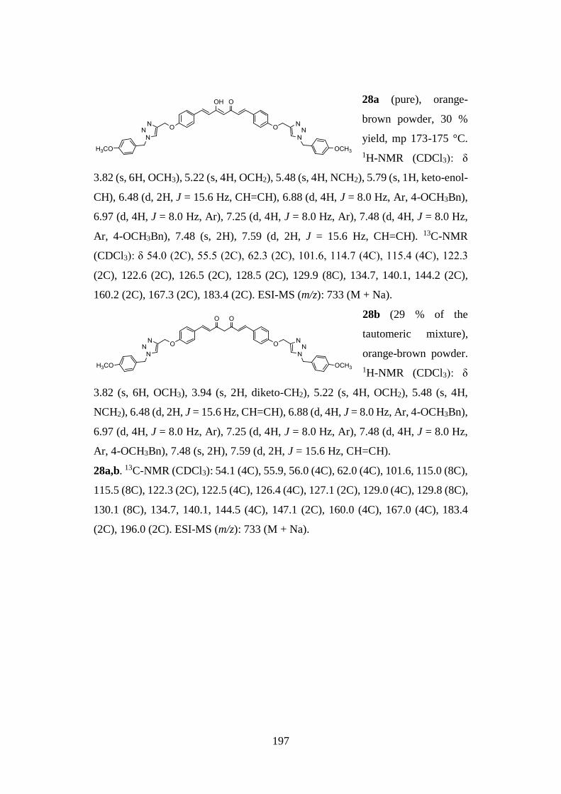
197
28a (pure), orange-
brown powder, 30 %
yield, mp 173-175 °C.
1H-NMR (CDCl3): δ
3.82 (s, 6H, OCH3), 5.22 (s, 4H, OCH2), 5.48 (s, 4H, NCH2), 5.79 (s, 1H, keto-enol-
CH), 6.48 (d, 2H, J = 15.6 Hz, CH=CH), 6.88 (d, 4H, J = 8.0 Hz, Ar, 4-OCH3Bn),
6.97 (d, 4H, J = 8.0 Hz, Ar), 7.25 (d, 4H, J = 8.0 Hz, Ar), 7.48 (d, 4H, J = 8.0 Hz,
Ar, 4-OCH3Bn), 7.48 (s, 2H), 7.59 (d, 2H, J = 15.6 Hz, CH=CH). 13C-NMR
(CDCl3): δ 54.0 (2C), 55.5 (2C), 62.3 (2C), 101.6, 114.7 (4C), 115.4 (4C), 122.3
(2C), 122.6 (2C), 126.5 (2C), 128.5 (2C), 129.9 (8C), 134.7, 140.1, 144.2 (2C),
160.2 (2C), 167.3 (2C), 183.4 (2C). ESI-MS (m/z): 733 (M + Na).
28b (29 % of the
tautomeric mixture),
orange-brown powder.
1H-NMR (CDCl3): δ
3.82 (s, 6H, OCH3), 3.94 (s, 2H, diketo-CH2), 5.22 (s, 4H, OCH2), 5.48 (s, 4H,
NCH2), 6.48 (d, 2H, J = 15.6 Hz, CH=CH), 6.88 (d, 4H, J = 8.0 Hz, Ar, 4-OCH3Bn),
6.97 (d, 4H, J = 8.0 Hz, Ar), 7.25 (d, 4H, J = 8.0 Hz, Ar), 7.48 (d, 4H, J = 8.0 Hz,
Ar, 4-OCH3Bn), 7.48 (s, 2H), 7.59 (d, 2H, J = 15.6 Hz, CH=CH).
28a,b. 13C-NMR (CDCl3): 54.1 (4C), 55.9, 56.0 (4C), 62.0 (4C), 101.6, 115.0 (8C),
115.5 (8C), 122.3 (2C), 122.5 (4C), 126.4 (4C), 127.1 (2C), 129.0 (4C), 129.8 (8C),
130.1 (8C), 134.7, 140.1, 144.5 (4C), 147.1 (2C), 160.0 (4C), 167.0 (4C), 183.4
(2C), 196.0 (2C). ESI-MS (m/z): 733 (M + Na).
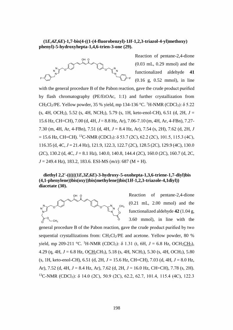
198
(1E,4Z,6E)-1,7-bis(4-((1-(4-fluorobenzyl)-1H-1,2,3-triazol-4-yl)methoxy)
phenyl)-5-hydroxyhepta-1,4,6-trien-3-one (29).
Reaction of pentane-2,4-dione
(0.03 mL, 0.29 mmol) and the
functionalized aldehyde 41
(0.16 g, 0.52 mmol), in line
with the general procedure B of the Pabon reaction, gave the crude product purified
by flash chromatography (PE/EtOAc, 1:1) and further crystallization from
CH2Cl2/PE. Yellow powder, 35 % yield, mp 134-136 °C. 1H-NMR (CDCl3): δ 5.22
(s, 4H, OCH2), 5.52 (s, 4H, NCH2), 5.79 (s, 1H, keto-enol-CH), 6.51 (d, 2H, J =
15.6 Hz, CH=CH), 7.00 (d, 4H, J = 8.8 Hz, Ar), 7.06-7.10 (m, 4H, Ar, 4-FBn), 7.27-
7.30 (m, 4H, Ar, 4-FBn), 7.51 (d, 4H, J = 8.4 Hz, Ar), 7.54 (s, 2H), 7.62 (d, 2H, J
= 15.6 Hz, CH=CH). 13C-NMR (CDCl3): δ 53.7 (2C), 62.2 (2C), 101.5, 115.3 (4C),
116.35 (d, 4C, J = 21.4 Hz), 121.9, 122.3, 122.7 (2C), 128.5 (2C), 129.9 (4C), 130.0
(2C), 130.2 (d, 4C, J = 8.1 Hz), 140.0, 140.8, 144.4 (2C), 160.0 (2C), 160.7 (d, 2C,
J = 249.4 Hz), 183.2, 183.6. ESI-MS (m/z): 687 (M + H).
diethyl 2,2'-((((((1E,3Z,6E)-3-hydroxy-5-oxohepta-1,3,6-triene-1,7-diyl)bis
(4,1-phenylene))bis(oxy))bis(methylene))bis(1H-1,2,3-triazole-4,1diyl))
diacetate (30).
Reaction of pentane-2,4-dione
(0.21 mL, 2.00 mmol) and the
functionalized aldehyde 42 (1.04 g,
3.60 mmol), in line with the
general procedure B of the Pabon reaction, gave the crude product purified by two
sequential crystallizations from: CH2Cl2/PE and acetone. Yellow powder, 80 %
yield, mp 209-211 °C. 1H-NMR (CDCl3): δ 1.31 (t, 6H, J = 6.8 Hz, OCH2CH3),
4.29 (q, 4H, J = 6.8 Hz, OCH2CH3), 5.18 (s, 4H, NCH2), 5.30 (s, 4H, OCH2), 5.80
(s, 1H, keto-enol-CH), 6.51 (d, 2H, J = 15.6 Hz, CH=CH), 7.03 (d, 4H, J = 8.0 Hz,
Ar), 7.52 (d, 4H, J = 8.4 Hz, Ar), 7.62 (d, 2H, J = 16.0 Hz, CH=CH), 7.78 (s, 2H).
13C-NMR (CDCl3): δ 14.0 (2C), 50.9 (2C), 62.2, 62.7, 101.4, 115.4 (4C), 122.3
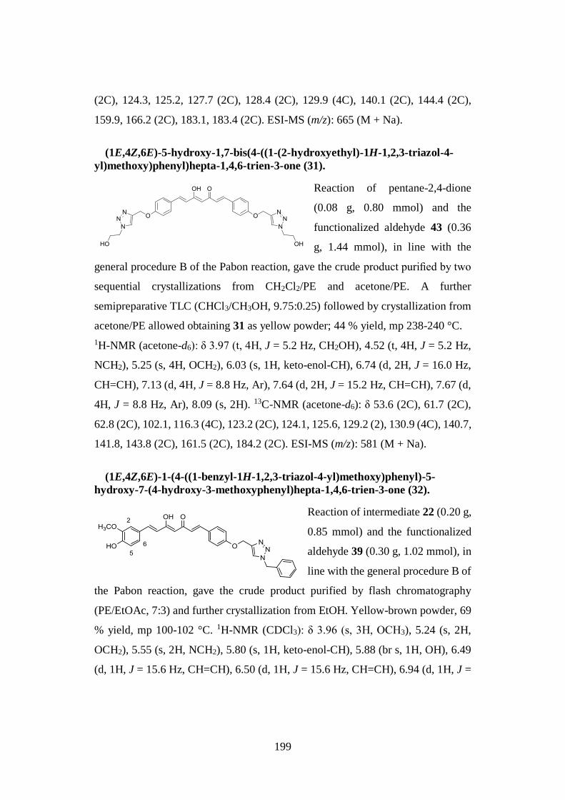
199
(2C), 124.3, 125.2, 127.7 (2C), 128.4 (2C), 129.9 (4C), 140.1 (2C), 144.4 (2C),
159.9, 166.2 (2C), 183.1, 183.4 (2C). ESI-MS (m/z): 665 (M + Na).
(1E,4Z,6E)-5-hydroxy-1,7-bis(4-((1-(2-hydroxyethyl)-1H-1,2,3-triazol-4-
yl)methoxy)phenyl)hepta-1,4,6-trien-3-one (31).
Reaction of pentane-2,4-dione
(0.08 g, 0.80 mmol) and the
functionalized aldehyde 43 (0.36
g, 1.44 mmol), in line with the
general procedure B of the Pabon reaction, gave the crude product purified by two
sequential crystallizations from CH2Cl2/PE and acetone/PE. A further
semipreparative TLC (CHCl3/CH3OH, 9.75:0.25) followed by crystallization from
acetone/PE allowed obtaining 31 as yellow powder; 44 % yield, mp 238-240 °C.
1H-NMR (acetone-d6): δ 3.97 (t, 4H, J = 5.2 Hz, CH2OH), 4.52 (t, 4H, J = 5.2 Hz,
NCH2), 5.25 (s, 4H, OCH2), 6.03 (s, 1H, keto-enol-CH), 6.74 (d, 2H, J = 16.0 Hz,
CH=CH), 7.13 (d, 4H, J = 8.8 Hz, Ar), 7.64 (d, 2H, J = 15.2 Hz, CH=CH), 7.67 (d,
4H, J = 8.8 Hz, Ar), 8.09 (s, 2H). 13C-NMR (acetone-d6): δ 53.6 (2C), 61.7 (2C),
62.8 (2C), 102.1, 116.3 (4C), 123.2 (2C), 124.1, 125.6, 129.2 (2), 130.9 (4C), 140.7,
141.8, 143.8 (2C), 161.5 (2C), 184.2 (2C). ESI-MS (m/z): 581 (M + Na).
(1E,4Z,6E)-1-(4-((1-benzyl-1H-1,2,3-triazol-4-yl)methoxy)phenyl)-5-
hydroxy-7-(4-hydroxy-3-methoxyphenyl)hepta-1,4,6-trien-3-one (32).
Reaction of intermediate 22 (0.20 g,
0.85 mmol) and the functionalized
aldehyde 39 (0.30 g, 1.02 mmol), in
line with the general procedure B of
the Pabon reaction, gave the crude product purified by flash chromatography
(PE/EtOAc, 7:3) and further crystallization from EtOH. Yellow-brown powder, 69
% yield, mp 100-102 °C. 1H-NMR (CDCl3): δ 3.96 (s, 3H, OCH3), 5.24 (s, 2H,
OCH2), 5.55 (s, 2H, NCH2), 5.80 (s, 1H, keto-enol-CH), 5.88 (br s, 1H, OH), 6.49
(d, 1H, J = 15.6 Hz, CH=CH), 6.50 (d, 1H, J = 15.6 Hz, CH=CH), 6.94 (d, 1H, J =
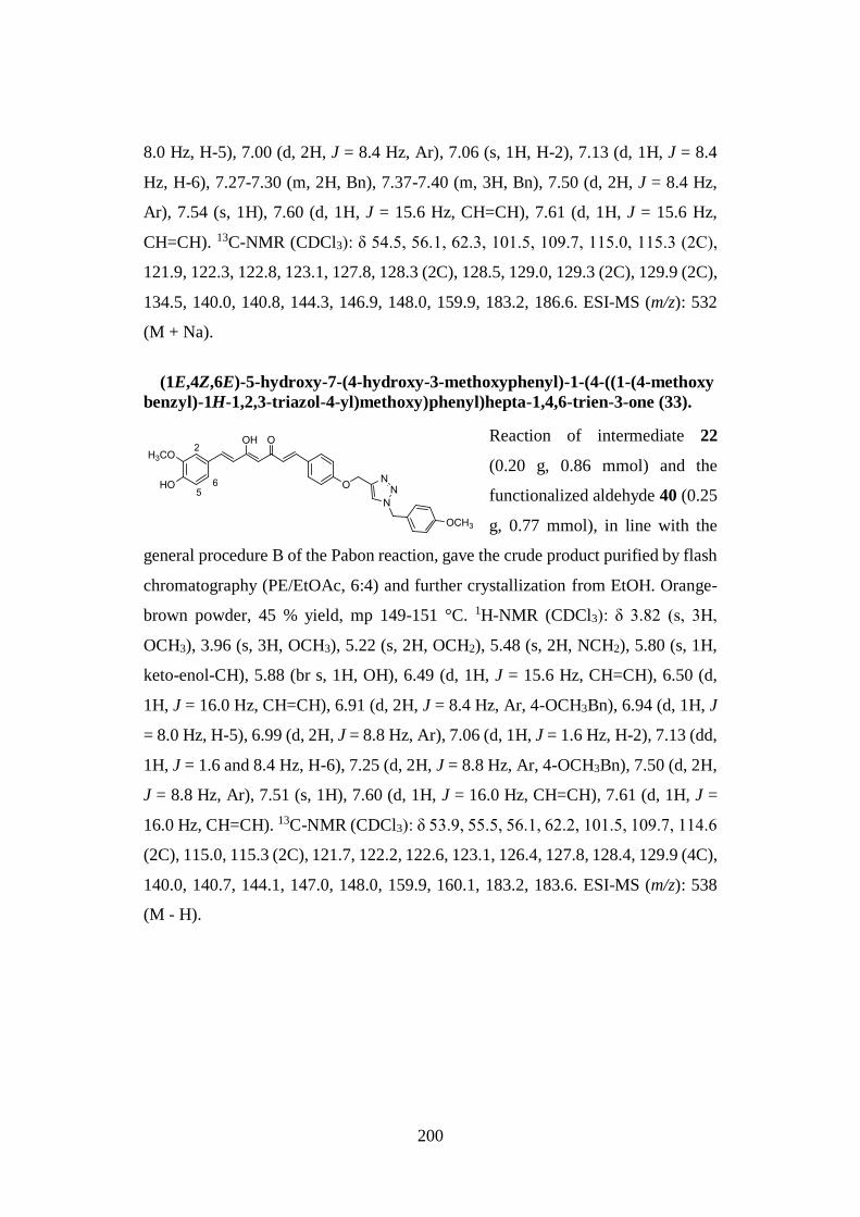
200
8.0 Hz, H-5), 7.00 (d, 2H, J = 8.4 Hz, Ar), 7.06 (s, 1H, H-2), 7.13 (d, 1H, J = 8.4
Hz, H-6), 7.27-7.30 (m, 2H, Bn), 7.37-7.40 (m, 3H, Bn), 7.50 (d, 2H, J = 8.4 Hz,
Ar), 7.54 (s, 1H), 7.60 (d, 1H, J = 15.6 Hz, CH=CH), 7.61 (d, 1H, J = 15.6 Hz,
CH=CH). 13C-NMR (CDCl3): δ 54.5, 56.1, 62.3, 101.5, 109.7, 115.0, 115.3 (2C),
121.9, 122.3, 122.8, 123.1, 127.8, 128.3 (2C), 128.5, 129.0, 129.3 (2C), 129.9 (2C),
134.5, 140.0, 140.8, 144.3, 146.9, 148.0, 159.9, 183.2, 186.6. ESI-MS (m/z): 532
(M + Na).
(1E,4Z,6E)-5-hydroxy-7-(4-hydroxy-3-methoxyphenyl)-1-(4-((1-(4-methoxy
benzyl)-1H-1,2,3-triazol-4-yl)methoxy)phenyl)hepta-1,4,6-trien-3-one (33).
Reaction of intermediate 22
(0.20 g, 0.86 mmol) and the
functionalized aldehyde 40 (0.25
g, 0.77 mmol), in line with the
general procedure B of the Pabon reaction, gave the crude product purified by flash
chromatography (PE/EtOAc, 6:4) and further crystallization from EtOH. Orange-
brown powder, 45 % yield, mp 149-151 °C. 1H-NMR (CDCl3): δ 3.82 (s, 3H,
OCH3), 3.96 (s, 3H, OCH3), 5.22 (s, 2H, OCH2), 5.48 (s, 2H, NCH2), 5.80 (s, 1H,
keto-enol-CH), 5.88 (br s, 1H, OH), 6.49 (d, 1H, J = 15.6 Hz, CH=CH), 6.50 (d,
1H, J = 16.0 Hz, CH=CH), 6.91 (d, 2H, J = 8.4 Hz, Ar, 4-OCH3Bn), 6.94 (d, 1H, J
= 8.0 Hz, H-5), 6.99 (d, 2H, J = 8.8 Hz, Ar), 7.06 (d, 1H, J = 1.6 Hz, H-2), 7.13 (dd,
1H, J = 1.6 and 8.4 Hz, H-6), 7.25 (d, 2H, J = 8.8 Hz, Ar, 4-OCH3Bn), 7.50 (d, 2H,
J = 8.8 Hz, Ar), 7.51 (s, 1H), 7.60 (d, 1H, J = 16.0 Hz, CH=CH), 7.61 (d, 1H, J =
16.0 Hz, CH=CH). 13C-NMR (CDCl3): δ 53.9, 55.5, 56.1, 62.2, 101.5, 109.7, 114.6
(2C), 115.0, 115.3 (2C), 121.7, 122.2, 122.6, 123.1, 126.4, 127.8, 128.4, 129.9 (4C),
140.0, 140.7, 144.1, 147.0, 148.0, 159.9, 160.1, 183.2, 183.6. ESI-MS (m/z): 538
(M - H).
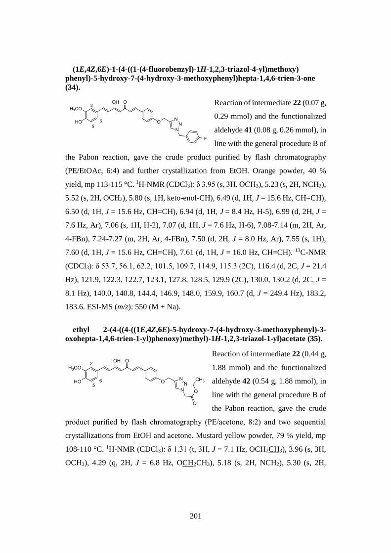
201
(1E,4Z,6E)-1-(4-((1-(4-fluorobenzyl)-1H-1,2,3-triazol-4-yl)methoxy)
phenyl)-5-hydroxy-7-(4-hydroxy-3-methoxyphenyl)hepta-1,4,6-trien-3-one
(34).
Reaction of intermediate 22 (0.07 g,
0.29 mmol) and the functionalized
aldehyde 41 (0.08 g, 0.26 mmol), in
line with the general procedure B of
the Pabon reaction, gave the crude product purified by flash chromatography
(PE/EtOAc, 6:4) and further crystallization from EtOH. Orange powder, 40 %
yield, mp 113-115 °C. 1H-NMR (CDCl3): δ 3.95 (s, 3H, OCH3), 5.23 (s, 2H, NCH2),
5.52 (s, 2H, OCH2), 5.80 (s, 1H, keto-enol-CH), 6.49 (d, 1H, J = 15.6 Hz, CH=CH),
6.50 (d, 1H, J = 15.6 Hz, CH=CH), 6.94 (d, 1H, J = 8.4 Hz, H-5), 6.99 (d, 2H, J =
7.6 Hz, Ar), 7.06 (s, 1H, H-2), 7.07 (d, 1H, J = 7.6 Hz, H-6), 7.08-7.14 (m, 2H, Ar,
4-FBn), 7.24-7.27 (m, 2H, Ar, 4-FBn), 7.50 (d, 2H, J = 8.0 Hz, Ar), 7.55 (s, 1H),
7.60 (d, 1H, J = 15.6 Hz, CH=CH), 7.61 (d, 1H, J = 16.0 Hz, CH=CH). 13C-NMR
(CDCl3): δ 53.7, 56.1, 62.2, 101.5, 109.7, 114.9, 115.3 (2C), 116.4 (d, 2C, J = 21.4
Hz), 121.9, 122.3, 122.7, 123.1, 127.8, 128.5, 129.9 (2C), 130.0, 130.2 (d, 2C, J =
8.1 Hz), 140.0, 140.8, 144.4, 146.9, 148.0, 159.9, 160.7 (d, J = 249.4 Hz), 183.2,
183.6. ESI-MS (m/z): 550 (M + Na).
ethyl 2-(4-((4-((1E,4Z,6E)-5-hydroxy-7-(4-hydroxy-3-methoxyphenyl)-3-
oxohepta-1,4,6-trien-1-yl)phenoxy)methyl)-1H-1,2,3-triazol-1-yl)acetate (35).
Reaction of intermediate 22 (0.44 g,
1.88 mmol) and the functionalized
aldehyde 42 (0.54 g, 1.88 mmol), in
line with the general procedure B of
the Pabon reaction, gave the crude
product purified by flash chromatography (PE/acetone, 8:2) and two sequential
crystallizations from EtOH and acetone. Mustard yellow powder, 79 % yield, mp
108-110 °C. 1H-NMR (CDCl3): δ 1.31 (t, 3H, J = 7.1 Hz, OCH2CH3), 3.96 (s, 3H,
OCH3), 4.29 (q, 2H, J = 6.8 Hz, OCH2CH3), 5.18 (s, 2H, NCH2), 5.30 (s, 2H,
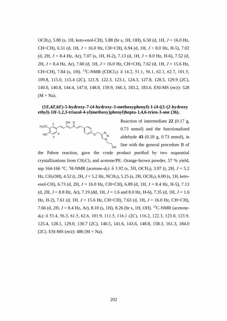
202
OCH2), 5.80 (s, 1H, keto-enol-CH), 5.88 (br s, 1H, OH), 6.50 (d, 1H, J = 16.0 Hz,
CH=CH), 6.51 (d, 1H, J = 16.0 Hz, CH=CH), 6.94 (d, 1H, J = 8.0 Hz, H-5), 7.02
(d, 2H, J = 8.4 Hz, Ar), 7.07 (s, 1H, H-2), 7.13 (d, 1H, J = 8.0 Hz, H-6), 7.52 (d,
2H, J = 8.4 Hz, Ar), 7.60 (d, 1H, J = 16.0 Hz, CH=CH), 7.62 (d, 1H, J = 15.6 Hz,
CH=CH), 7.84 (s, 1H). 13C-NMR (CDCl3): δ 14.2, 51.1, 56.1, 62.1, 62.7, 101.5,
109.8, 115.0, 115.4 (2C), 121.9, 122.3, 123.1, 124.3, 127.8, 128.5, 129.9 (2C),
140.0, 140.8, 144.4, 147.0, 148.0, 159.9, 166.3, 183.2, 183.6. ESI-MS (m/z): 528
(M + Na).
(1E,4Z,6E)-5-hydroxy-7-(4-hydroxy-3-methoxyphenyl)-1-(4-((1-(2-hydroxy
ethyl)-1H-1,2,3-triazol-4-yl)methoxy)phenyl)hepta-1,4,6-trien-3-one (36).
Reaction of intermediate 22 (0.17 g,
0.73 mmol) and the functionalized
aldehyde 43 (0.18 g, 0.73 mmol), in
line with the general procedure B of
the Pabon reaction, gave the crude product purified by two sequential
crystallizations from CH2Cl2 and acetone/PE. Orange-brown powder, 57 % yield,
mp 164-166 °C. 1H-NMR (acetone-d6): δ 3.92 (s, 3H, OCH3), 3.97 (t, 2H, J = 5.2
Hz, CH2OH), 4.52 (t, 2H, J = 5.2 Hz, NCH2), 5.25 (s, 2H, OCH2), 6.00 (s, 1H, keto-
enol-CH), 6.73 (d, 2H, J = 16.0 Hz, CH=CH), 6.89 (d, 1H, J = 8.4 Hz, H-5), 7.13
(d, 2H, J = 8.8 Hz, Ar), 7.19 (dd, 1H, J = 1.6 and 8.0 Hz, H-6), 7.35 (d, 1H, J = 1.6
Hz, H-2), 7.61 (d, 1H, J = 15.6 Hz, CH=CH), 7.63 (d, 1H, J = 16.0 Hz, CH=CH),
7.66 (d, 2H, J = 8.4 Hz, Ar), 8.10 (s, 1H), 8.26 (br s, 1H, OH). 13C-NMR (acetone-
d6): δ 53.4, 56.3, 61.5, 62.6, 101.9, 111.5, 116.1 (2C), 116.2, 122.3, 123.0, 123.9,
125.4, 128.1, 129.0, 130.7 (2C), 140.5, 141.6, 143.6, 148.8, 158.3, 161.3, 184.0
(2C). ESI-MS (m/z): 486 (M + Na).
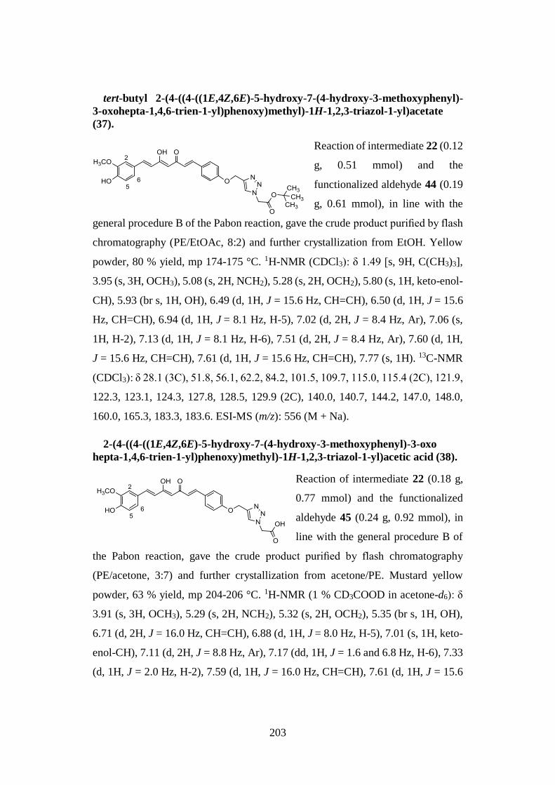
203
tert-butyl 2-(4-((4-((1E,4Z,6E)-5-hydroxy-7-(4-hydroxy-3-methoxyphenyl)-
3-oxohepta-1,4,6-trien-1-yl)phenoxy)methyl)-1H-1,2,3-triazol-1-yl)acetate
(37).
Reaction of intermediate 22 (0.12
g, 0.51 mmol) and the
functionalized aldehyde 44 (0.19
g, 0.61 mmol), in line with the
general procedure B of the Pabon reaction, gave the crude product purified by flash
chromatography (PE/EtOAc, 8:2) and further crystallization from EtOH. Yellow
powder, 80 % yield, mp 174-175 °C. 1H-NMR (CDCl3): δ 1.49 [s, 9H, C(CH3)3],
3.95 (s, 3H, OCH3), 5.08 (s, 2H, NCH2), 5.28 (s, 2H, OCH2), 5.80 (s, 1H, keto-enol-
CH), 5.93 (br s, 1H, OH), 6.49 (d, 1H, J = 15.6 Hz, CH=CH), 6.50 (d, 1H, J = 15.6
Hz, CH=CH), 6.94 (d, 1H, J = 8.1 Hz, H-5), 7.02 (d, 2H, J = 8.4 Hz, Ar), 7.06 (s,
1H, H-2), 7.13 (d, 1H, J = 8.1 Hz, H-6), 7.51 (d, 2H, J = 8.4 Hz, Ar), 7.60 (d, 1H,
J = 15.6 Hz, CH=CH), 7.61 (d, 1H, J = 15.6 Hz, CH=CH), 7.77 (s, 1H). 13C-NMR
(CDCl3): δ 28.1 (3C), 51.8, 56.1, 62.2, 84.2, 101.5, 109.7, 115.0, 115.4 (2C), 121.9,
122.3, 123.1, 124.3, 127.8, 128.5, 129.9 (2C), 140.0, 140.7, 144.2, 147.0, 148.0,
160.0, 165.3, 183.3, 183.6. ESI-MS (m/z): 556 (M + Na).
2-(4-((4-((1E,4Z,6E)-5-hydroxy-7-(4-hydroxy-3-methoxyphenyl)-3-oxo
hepta-1,4,6-trien-1-yl)phenoxy)methyl)-1H-1,2,3-triazol-1-yl)acetic acid (38).
Reaction of intermediate 22 (0.18 g,
0.77 mmol) and the functionalized
aldehyde 45 (0.24 g, 0.92 mmol), in
line with the general procedure B of
the Pabon reaction, gave the crude product purified by flash chromatography
(PE/acetone, 3:7) and further crystallization from acetone/PE. Mustard yellow
powder, 63 % yield, mp 204-206 °C. 1H-NMR (1 % CD3COOD in acetone-d6): δ
3.91 (s, 3H, OCH3), 5.29 (s, 2H, NCH2), 5.32 (s, 2H, OCH2), 5.35 (br s, 1H, OH),
6.71 (d, 2H, J = 16.0 Hz, CH=CH), 6.88 (d, 1H, J = 8.0 Hz, H-5), 7.01 (s, 1H, keto-
enol-CH), 7.11 (d, 2H, J = 8.8 Hz, Ar), 7.17 (dd, 1H, J = 1.6 and 6.8 Hz, H-6), 7.33
(d, 1H, J = 2.0 Hz, H-2), 7.59 (d, 1H, J = 16.0 Hz, CH=CH), 7.61 (d, 1H, J = 15.6
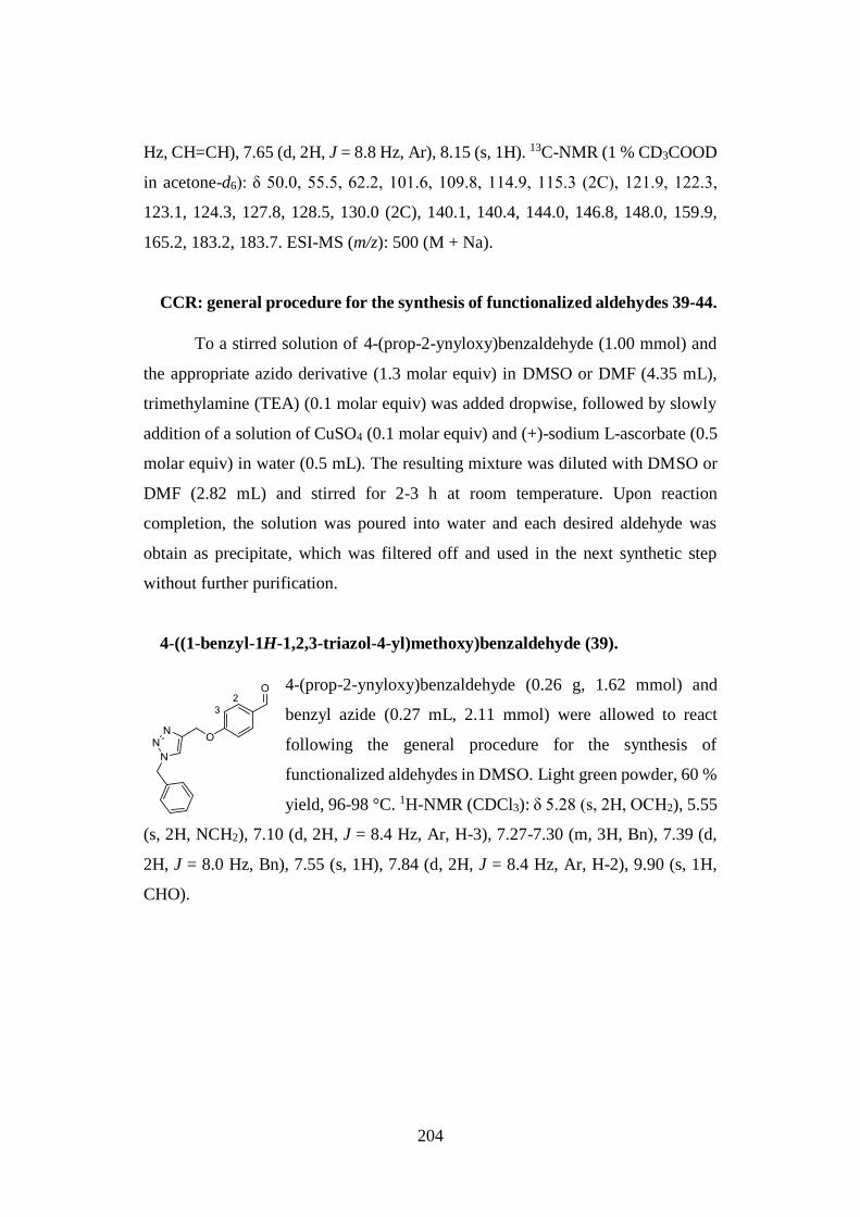
204
Hz, CH=CH), 7.65 (d, 2H, J = 8.8 Hz, Ar), 8.15 (s, 1H). 13C-NMR (1 % CD3COOD
in acetone-d6): δ 50.0, 55.5, 62.2, 101.6, 109.8, 114.9, 115.3 (2C), 121.9, 122.3,
123.1, 124.3, 127.8, 128.5, 130.0 (2C), 140.1, 140.4, 144.0, 146.8, 148.0, 159.9,
165.2, 183.2, 183.7. ESI-MS (m/z): 500 (M + Na).
CCR: general procedure for the synthesis of functionalized aldehydes 39-44.
To a stirred solution of 4-(prop-2-ynyloxy)benzaldehyde (1.00 mmol) and
the appropriate azido derivative (1.3 molar equiv) in DMSO or DMF (4.35 mL),
trimethylamine (TEA) (0.1 molar equiv) was added dropwise, followed by slowly
addition of a solution of CuSO4 (0.1 molar equiv) and (+)-sodium L-ascorbate (0.5
molar equiv) in water (0.5 mL). The resulting mixture was diluted with DMSO or
DMF (2.82 mL) and stirred for 2-3 h at room temperature. Upon reaction
completion, the solution was poured into water and each desired aldehyde was
obtain as precipitate, which was filtered off and used in the next synthetic step
without further purification.
4-((1-benzyl-1H-1,2,3-triazol-4-yl)methoxy)benzaldehyde (39).
4-(prop-2-ynyloxy)benzaldehyde (0.26 g, 1.62 mmol) and
benzyl azide (0.27 mL, 2.11 mmol) were allowed to react
following the general procedure for the synthesis of
functionalized aldehydes in DMSO. Light green powder, 60 %
yield, 96-98 °C. 1H-NMR (CDCl3): δ 5.28 (s, 2H, OCH2), 5.55
(s, 2H, NCH2), 7.10 (d, 2H, J = 8.4 Hz, Ar, H-3), 7.27-7.30 (m, 3H, Bn), 7.39 (d,
2H, J = 8.0 Hz, Bn), 7.55 (s, 1H), 7.84 (d, 2H, J = 8.4 Hz, Ar, H-2), 9.90 (s, 1H,
CHO).
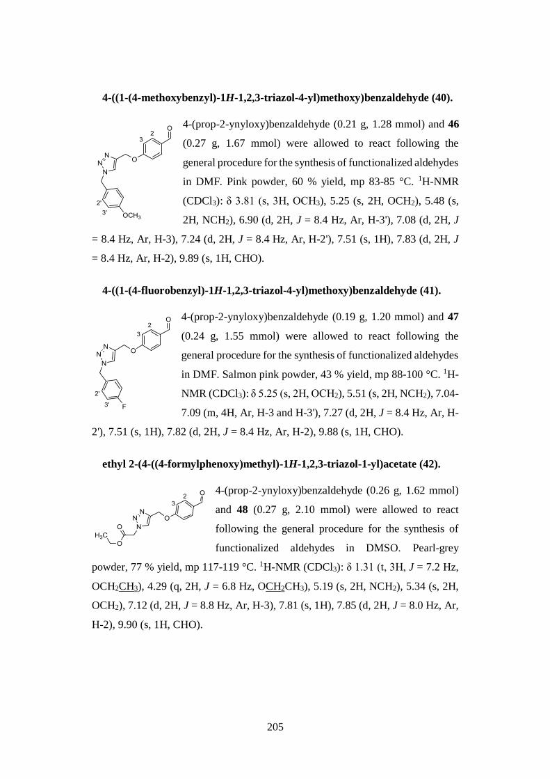
205
4-((1-(4-methoxybenzyl)-1H-1,2,3-triazol-4-yl)methoxy)benzaldehyde (40).
4-(prop-2-ynyloxy)benzaldehyde (0.21 g, 1.28 mmol) and 46
(0.27 g, 1.67 mmol) were allowed to react following the
general procedure for the synthesis of functionalized aldehydes
in DMF. Pink powder, 60 % yield, mp 83-85 °C. 1H-NMR
(CDCl3): δ 3.81 (s, 3H, OCH3), 5.25 (s, 2H, OCH2), 5.48 (s,
2H, NCH2), 6.90 (d, 2H, J = 8.4 Hz, Ar, H-3'), 7.08 (d, 2H, J
= 8.4 Hz, Ar, H-3), 7.24 (d, 2H, J = 8.4 Hz, Ar, H-2'), 7.51 (s, 1H), 7.83 (d, 2H, J
= 8.4 Hz, Ar, H-2), 9.89 (s, 1H, CHO).
4-((1-(4-fluorobenzyl)-1H-1,2,3-triazol-4-yl)methoxy)benzaldehyde (41).
4-(prop-2-ynyloxy)benzaldehyde (0.19 g, 1.20 mmol) and 47
(0.24 g, 1.55 mmol) were allowed to react following the
general procedure for the synthesis of functionalized aldehydes
in DMF. Salmon pink powder, 43 % yield, mp 88-100 °C. 1H-
NMR (CDCl3): δ 5.25 (s, 2H, OCH2), 5.51 (s, 2H, NCH2), 7.04-
7.09 (m, 4H, Ar, H-3 and H-3'), 7.27 (d, 2H, J = 8.4 Hz, Ar, H-
2'), 7.51 (s, 1H), 7.82 (d, 2H, J = 8.4 Hz, Ar, H-2), 9.88 (s, 1H, CHO).
ethyl 2-(4-((4-formylphenoxy)methyl)-1H-1,2,3-triazol-1-yl)acetate (42).
4-(prop-2-ynyloxy)benzaldehyde (0.26 g, 1.62 mmol)
and 48 (0.27 g, 2.10 mmol) were allowed to react
following the general procedure for the synthesis of
functionalized aldehydes in DMSO. Pearl-grey
powder, 77 % yield, mp 117-119 °C. 1H-NMR (CDCl3): δ 1.31 (t, 3H, J = 7.2 Hz,
OCH2CH3), 4.29 (q, 2H, J = 6.8 Hz, OCH2CH3), 5.19 (s, 2H, NCH2), 5.34 (s, 2H,
OCH2), 7.12 (d, 2H, J = 8.8 Hz, Ar, H-3), 7.81 (s, 1H), 7.85 (d, 2H, J = 8.0 Hz, Ar,
H-2), 9.90 (s, 1H, CHO).
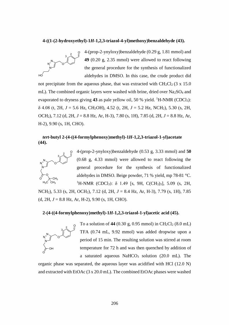
206
4-((1-(2-hydroxyethyl)-1H-1,2,3-triazol-4-yl)methoxy)benzaldehyde (43).
4-(prop-2-ynyloxy)benzaldehyde (0.29 g, 1.81 mmol) and
49 (0.20 g, 2.35 mmol) were allowed to react following
the general procedure for the synthesis of functionalized
aldehydes in DMSO. In this case, the crude product did
not precipitate from the aqueous phase, that was extracted with CH2Cl2 (3 x 15.0
mL). The combined organic layers were washed with brine, dried over Na2SO4 and
evaporated to dryness giving 43 as pale yellow oil, 50 % yield. 1H-NMR (CDCl3):
δ 4.08 (t, 2H, J = 5.6 Hz, CH2OH), 4.52 (t, 2H, J = 5.2 Hz, NCH2), 5.30 (s, 2H,
OCH2), 7.12 (d, 2H, J = 8.8 Hz, Ar, H-3), 7.80 (s, 1H), 7.85 (d, 2H, J = 8.8 Hz, Ar,
H-2), 9.90 (s, 1H, CHO).
tert-butyl 2-(4-((4-formylphenoxy)methyl)-1H-1,2,3-triazol-1-yl)acetate
(44).
4-(prop-2-ynyloxy)benzaldehyde (0.53 g, 3.33 mmol) and 50
(0.68 g, 4.33 mmol) were allowed to react following the
general procedure for the synthesis of functionalized
aldehydes in DMSO. Beige powder, 71 % yield, mp 78-81 °C.
1H-NMR (CDCl3): δ 1.49 [s, 9H, C(CH3)3], 5.09 (s, 2H,
NCH2), 5.33 (s, 2H, OCH2), 7.12 (d, 2H, J = 8.4 Hz, Ar, H-3), 7.79 (s, 1H), 7.85
(d, 2H, J = 8.8 Hz, Ar, H-2), 9.90 (s, 1H, CHO).
2-(4-((4-formylphenoxy)methyl)-1H-1,2,3-triazol-1-yl)acetic acid (45).
To a solution of 44 (0.30 g, 0.95 mmol) in CH2Cl2 (8.0 mL)
TFA (0.74 mL, 9.92 mmol) was added dropwise upon a
period of 15 min. The resulting solution was stirred at room
temperature for 72 h and was then quenched by addition of
a saturated aqueous NaHCO3 solution (20.0 mL). The
organic phase was separated, the aqueous layer was acidified with HCl (12.0 N)
and extracted with EtOAc (3 x 20.0 mL). The combined EtOAc phases were washed
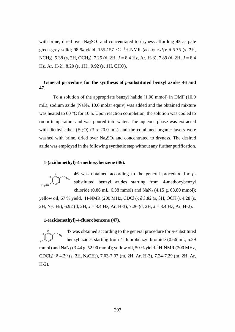
207
with brine, dried over Na2SO4 and concentrated to dryness affording 45 as pale
green-grey solid; 98 % yield, 155-157 °C. 1H-NMR (acetone-d6): δ 5.35 (s, 2H,
NCH2), 5.38 (s, 2H, OCH2), 7.25 (d, 2H, J = 8.4 Hz, Ar, H-3), 7.89 (d, 2H, J = 8.4
Hz, Ar, H-2), 8.20 (s, 1H), 9.92 (s, 1H, CHO).
General procedure for the synthesis of p-substituted benzyl azides 46 and
47.
To a solution of the appropriate benzyl halide (1.00 mmol) in DMF (10.0
mL), sodium azide (NaN3, 10.0 molar equiv) was added and the obtained mixture
was heated to 60 °C for 10 h. Upon reaction completion, the solution was cooled to
room temperature and was poured into water. The aqueous phase was extracted
with diethyl ether (Et2O) (3 x 20.0 mL) and the combined organic layers were
washed with brine, dried over Na2SO4 and concentrated to dryness. The desired
azide was employed in the following synthetic step without any further purification.
1-(azidomethyl)-4-methoxybenzene (46).
46 was obtained according to the general procedure for p-
substituted benzyl azides starting from 4-methoxybenzyl
chloride (0.86 mL, 6.38 mmol) and NaN3 (4.15 g, 63.80 mmol);
yellow oil, 67 % yield. 1H-NMR (200 MHz, CDCl3): δ 3.82 (s, 3H, OCH3), 4.28 (s,
2H, N3CH2), 6.92 (d, 2H, J = 8.4 Hz, Ar, H-3), 7.26 (d, 2H, J = 8.4 Hz, Ar, H-2).
1-(azidomethyl)-4-fluorobenzene (47).
47 was obtained according to the general procedure for p-substituted
benzyl azides starting from 4-fluorobenzyl bromide (0.66 mL, 5.29
mmol) and NaN3 (3.44 g, 52.90 mmol); yellow oil, 50 % yield. 1H-NMR (200 MHz,
CDCl3): δ 4.29 (s, 2H, N3CH2), 7.03-7.07 (m, 2H, Ar, H-3), 7.24-7.29 (m, 2H, Ar,
H-2).
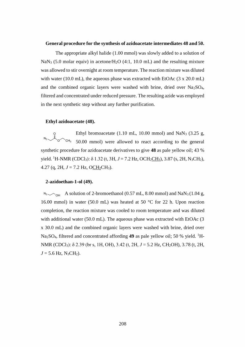
208
General procedure for the synthesis of azidoacetate intermediates 48 and 50.
The appropriate alkyl halide (1.00 mmol) was slowly added to a solution of
NaN3 (5.0 molar equiv) in acetone/H2O (4:1, 10.0 mL) and the resulting mixture
was allowed to stir overnight at room temperature. The reaction mixture was diluted
with water (10.0 mL), the aqueous phase was extracted with EtOAc (3 x 20.0 mL)
and the combined organic layers were washed with brine, dried over Na2SO4,
filtered and concentrated under reduced pressure. The resulting azide was employed
in the next synthetic step without any further purification.
Ethyl azidoacetate (48).
Ethyl bromoacetate (1.10 mL, 10.00 mmol) and NaN3 (3.25 g,
50.00 mmol) were allowed to react according to the general
synthetic procedure for azidoacetate derivatives to give 48 as pale yellow oil; 43 %
yield. 1H-NMR (CDCl3): δ 1.32 (t, 3H, J = 7.2 Hz, OCH2CH3), 3.87 (s, 2H, N3CH2),
4.27 (q, 2H, J = 7.2 Hz, OCH2CH3).
2-azidoethan-1-ol (49).
A solution of 2-bromoethanol (0.57 mL, 8.00 mmol) and NaN3 (1.04 g,
16.00 mmol) in water (50.0 mL) was heated at 50 °C for 22 h. Upon reaction
completion, the reaction mixture was cooled to room temperature and was diluted
with additional water (50.0 mL). The aqueous phase was extracted with EtOAc (3
x 30.0 mL) and the combined organic layers were washed with brine, dried over
Na2SO4, filtered and concentrated affording 49 as pale yellow oil; 50 % yield. 1H-
NMR (CDCl3): δ 2.39 (br s, 1H, OH), 3.42 (t, 2H, J = 5.2 Hz, CH2OH), 3.78 (t, 2H,
J = 5.6 Hz, N3CH2).
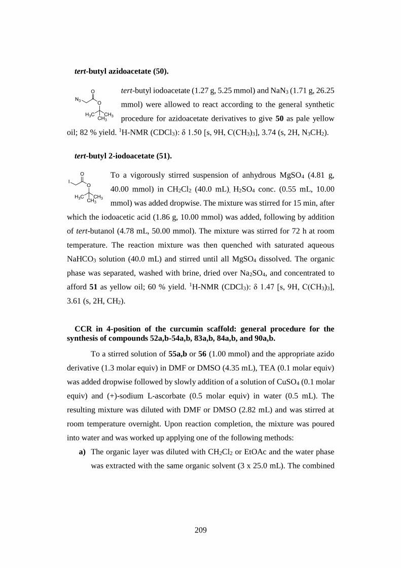
209
tert-butyl azidoacetate (50).
tert-butyl iodoacetate (1.27 g, 5.25 mmol) and NaN3 (1.71 g, 26.25
mmol) were allowed to react according to the general synthetic
procedure for azidoacetate derivatives to give 50 as pale yellow
oil; 82 % yield. 1H-NMR (CDCl3): δ 1.50 [s, 9H, C(CH3)3], 3.74 (s, 2H, N3CH2).
tert-butyl 2-iodoacetate (51).
To a vigorously stirred suspension of anhydrous MgSO4 (4.81 g,
40.00 mmol) in CH2Cl2 (40.0 mL), H2SO4 conc. (0.55 mL, 10.00
mmol) was added dropwise. The mixture was stirred for 15 min, after
which the iodoacetic acid (1.86 g, 10.00 mmol) was added, following by addition
of tert-butanol (4.78 mL, 50.00 mmol). The mixture was stirred for 72 h at room
temperature. The reaction mixture was then quenched with saturated aqueous
NaHCO3 solution (40.0 mL) and stirred until all MgSO4 dissolved. The organic
phase was separated, washed with brine, dried over Na2SO4, and concentrated to
afford 51 as yellow oil; 60 % yield. 1H-NMR (CDCl3): δ 1.47 [s, 9H, C(CH3)3],
3.61 (s, 2H, CH2).
CCR in 4-position of the curcumin scaffold: general procedure for the
synthesis of compounds 52a,b-54a,b, 83a,b, 84a,b, and 90a,b.
To a stirred solution of 55a,b or 56 (1.00 mmol) and the appropriate azido
derivative (1.3 molar equiv) in DMF or DMSO (4.35 mL), TEA (0.1 molar equiv)
was added dropwise followed by slowly addition of a solution of CuSO4 (0.1 molar
equiv) and (+)-sodium L-ascorbate (0.5 molar equiv) in water (0.5 mL). The
resulting mixture was diluted with DMF or DMSO (2.82 mL) and was stirred at
room temperature overnight. Upon reaction completion, the mixture was poured
into water and was worked up applying one of the following methods:
a) The organic layer was diluted with CH2Cl2 or EtOAc and the water phase
was extracted with the same organic solvent (3 x 25.0 mL). The combined
210
organic layers were dried over Na2SO4 and evaporated under reduced
pressure.
b) The obtained precipitate was filtered under vacuum to dryness.
In both cases, purification of the crude product by flash chromatography and further
crystallization from suitable solvent or mixture of solvents, afforded the desired
final compound as tautomeric mixture.
(1E,4Z,6E)-5-hydroxy-1,7-bis(4-hydroxy-3-methoxyphenyl)-4-((1-(4-
methoxybenzyl)-1H-1,2,3-triazol-4-yl)methyl)hepta-1,4,6-trien-3-one (52a);
(1E,6E)-1,7-bis(4-hydroxy-3-methoxyphenyl)-4-((1-(4-methoxybenzyl)-
1H-1,2,3-triazol-4-yl)methyl)hepta-1,6-diene-3,5-dione (52b).
55a,b (0.36 g, 0.89 mmol) and azido 46 (0.19 g, 1.15 mmol) were allowed
to react according to the general procedure of the CCR in 4-position of the curcumin
scaffold in DMF (method b). The crude product was purified by flash
cromatography (EP/EtOAc, 7:3) and further crystallization from CH2Cl2/PE. Red
powder, 43 % yield (isolated as 1.0:1.2 mixture of 52a:52b), mp 98-100°C.
52a (45 % of the tautomeric mixture). 1H-
NMR (CDCl3): δ 3.68 (s, 3H, OCH3), 3.92 (s,
6H, OCH3), 4.06 (s, 2H, CH2), 5.37 (s, 2H,
NCH2), 5.92 (br s, 2H, OH), 6.67 (d, 2H, J =
6.8 Hz, H-5), 6.70 (s, 2H, H-2), 6.81 (d, 2H,
J = 8.8 Hz, H-6), 6.91 (d, 2H, J = 8.0 Hz, Ar),
6.98 (s, 1H), 7.07 (d, 2H, J = 8.4 Hz, Ar), 7.10
(d, 1H, J = 15.2 Hz, CH=CH), 7.12 (d, 1H, J
= 15.6 Hz, CH=CH), 7.67 (d, 2H, J = 15.6 Hz, CH=CH).
52b (55 % of the tautomeric mixture). 1H-NMR (CDCl3): δ 3.27 (d, 2H, J = 7.2 Hz,
CH2), 3.72 (s, 3H, OCH3), 3.92 (s, 6H, OCH3), 4.77 (t, 1H, J = 7.2 Hz, diketo-CH),
5.37 (s, 2H, NCH2), 5.88 (br s, 2H, OH), 6.67 (d, 2H, J = 6.8 Hz, H-5), 6.70 (s, 2H,
H-2), 6.81 (d, 2H, J = 8.8 Hz, H-6), 6.83 (d, 2H, J = 15.2 Hz, CH=CH), 6.91 (d,
2H, J = 8.0 Hz, Ar), 7.02 (s, 1H), 7.07 (d, 2H, J = 8.4 Hz, Ar), 7.59 (d, 2H, J = 15.6
Hz, CH=CH). 52a,b. 13C-NMR (CDCl3): δ 24.9, 29.8, 53.7, 53.9, 55.3, 55.4, 56.1
211
(2C), 56.2 (2C), 62.9, 107.8, 109.8 (2C), 109.9 (2C), 114.4 (2C), 114.5 (2C), 114.9
(2C), 115.0 (2C), 117.9, 121.9, 122.1 (2C), 123.5 (2C), 124.3 (2C), 127.6 (2C),
127.9 (4C), 129.3 (2C), 129.6 (2C), 129.7, 142.5, 145.1 (2C), 145.3 (2C), 147.0
(2C), 148.3 (4C), 149.0 (2C), 149.2 (2C), 159.9 (2C), 183.2 (2C), 194.6 (2C).
ESI-MS (m/z): 592 (M + Na).
(1E,4Z,6E)-4-((1-(4-fluorobenzyl)-1H-1,2,3-triazol-4-yl)methyl)-5-hydroxy-
1,7-bis(4-hydroxy-3-methoxyphenyl)hepta-1,4,6-trien-3-one (53a);
(1E,6E)-4-((1-(4-fluorobenzyl)-1H-1,2,3-triazol-4-yl)methyl)-1,7-bis(4-
hydroxy-3-methoxyphenyl)hepta-1,6-diene-3,5-dione (53b).
55a,b (0.24 g, 0.60 mmol) and azido 47 (0.12 g, 0.78 mmol) were allowed
to reach according to the general procedure of the CCR in 4-position of the
curcumin scaffold in DMF (method a). The crude product was purified by flash
cromatography (EP/EtOAc, 1:1) and further crystallization from CH2Cl2/PE. Dark
red powder, 41 % yield (isolated as 1.3:1.0 mixture of 53a:53b), mp 95-97 °C.
53a (57 % of the tautomeric mixture). 1H-
NMR (acetone-d6): δ 3.89 (s, 6H, OCH3), 4.05
(s, 2H, CH2), 5.54 (s, 2H, NCH2), 5.58 (br s,
2H, OH), 6.87 (d, 2H, J = 8.4 Hz, H-5), 6.88
(s, 2H, H-2), 6.93-6.98 (m, 2H, Ar), 7.18 (d,
2H, J = 8.0 Hz, H-6), 7.27 (d, 2H, J = 16.0 Hz,
CH=CH), 7.27-7.34 (m, 2H, Ar), 7.62 (d, 2H,
J = 15.6 Hz, CH=CH), 7.66 (s, 1H).
53b (43 % of the tautomeric mixture). 1H-NMR (acetone-d6): δ 3.27 (d, 2H, J = 6.8
Hz, CH2), 3.89 (s, 6H, OCH3), 4.93 (t, 1H, J = 6.8 Hz, diketo-CH), 5.54 (s, 2H,
NCH2), 5.58 (br s, 2H, OH), 6.87 (d, 2H, J = 8.4 Hz, H-5), 6.93 (s, 2H, H-2), 7.01-
7.05 (m, 2H, Ar), 7.18 (d, 2H, J = 8.0 Hz, H-6), 7.27-7.34 (m, 2H, Ar), 7.29 (d, 2H,
J = 15.6 Hz, CH=CH), 7.64 (d, 2H, J = 15.6 Hz, CH=CH), 7.75 (s, 1H).
53a,b. 13C-NMR (CDCl3): δ 24.8, 29.8, 53.5, 53.6, 56.1 (4C), 62.9, 109.9 (4C),
115.0 (4C), 116.1 (d, 4C, J = 22.2 Hz), 117.8, 122.1, 122.3, 123.4 (4C), 124.3 (2C),
126.7 (2C), 127.9 (4C), 129.7 (2C), 129.9 (d, 4C, J = 8.4 Hz), 130.6, 142.6, 145.4
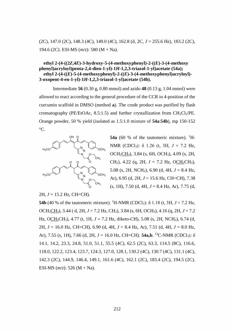
212
(2C), 147.0 (2C), 148.3 (4C), 149.0 (4C), 162.8 (d, 2C, J = 255.6 Hz), 183.2 (2C),
194.6 (2C). ESI-MS (m/z): 580 (M + Na).
ethyl 2-(4-((2Z,4E)-3-hydroxy-5-(4-methoxyphenyl)-2-((E)-3-(4-methoxy
phenyl)acryloyl)penta-2,4-dien-1-yl)-1H-1,2,3-triazol-1-yl)acetate (54a);
ethyl 2-(4-((E)-5-(4-methoxyphenyl)-2-((E)-3-(4-methoxyphenyl)acryloyl)-
3-oxopent-4-en-1-yl)-1H-1,2,3-triazol-1-yl)acetate (54b).
Intermediate 56 (0.30 g, 0.80 mmol) and azido 48 (0.13 g, 1.04 mmol) were
allowed to react according to the general procedure of the CCR in 4-position of the
curcumin scaffold in DMSO (method a). The crude product was purified by flash
cromatography (PE/EtOAc, 8.5:1.5) and further crystallization from CH2Cl2/PE.
Orange powder, 50 % yield (isolated as 1.5:1.0 mixture of 54a:54b), mp 150-152
°C.
54a (60 % of the tautomeric mixture). 1H-
NMR (CDCl3): δ 1.26 (t, 3H, J = 7.2 Hz,
OCH2CH3), 3.84 (s, 6H, OCH3), 4.09 (s, 2H,
CH2), 4.22 (q, 2H, J = 7.2 Hz, OCH2CH3),
5.08 (s, 2H, NCH2), 6.90 (d, 4H, J = 8.4 Hz,
Ar), 6.95 (d, 2H, J = 15.6 Hz, CH=CH), 7.38
(s, 1H), 7.50 (d, 4H, J = 8.4 Hz, Ar), 7.75 (d,
2H, J = 15.2 Hz, CH=CH).
54b (40 % of the tautomeric mixture). 1H-NMR (CDCl3): δ 1.18 (t, 3H, J = 7.2 Hz,
OCH2CH3), 3.44 ( d, 2H, J = 7.2 Hz, CH2), 3.84 (s, 6H, OCH3), 4.16 (q, 2H, J = 7.2
Hz, OCH2CH3), 4.77 (t, 1H, J = 7.2 Hz, diketo-CH), 5.08 (s, 2H, NCH2), 6.74 (d,
2H, J = 16.0 Hz, CH=CH), 6.90 (d, 4H, J = 8.4 Hz, Ar), 7.51 (d, 4H, J = 8.0 Hz,
Ar), 7.55 (s, 1H), 7.66 (d, 2H, J = 16.0 Hz, CH=CH). 54a,b. 13C-NMR (CDCl3): δ
14.1, 14.2, 23.3, 24.8, 51.0, 51.1, 55.5 (4C), 62.5 (2C), 63.3, 114.5 (8C), 116.6,
118.0, 122.2, 123.4, 123.7, 124.3, 127.0, 128.1, 130.2 (4C), 130.7 (4C), 131.1 (4C),
142.3 (2C), 144.9, 146.4, 149.1, 161.6 (4C), 162.1 (2C), 183.4 (2C), 194.5 (2C).
ESI-MS (m/z): 526 (M + Na).
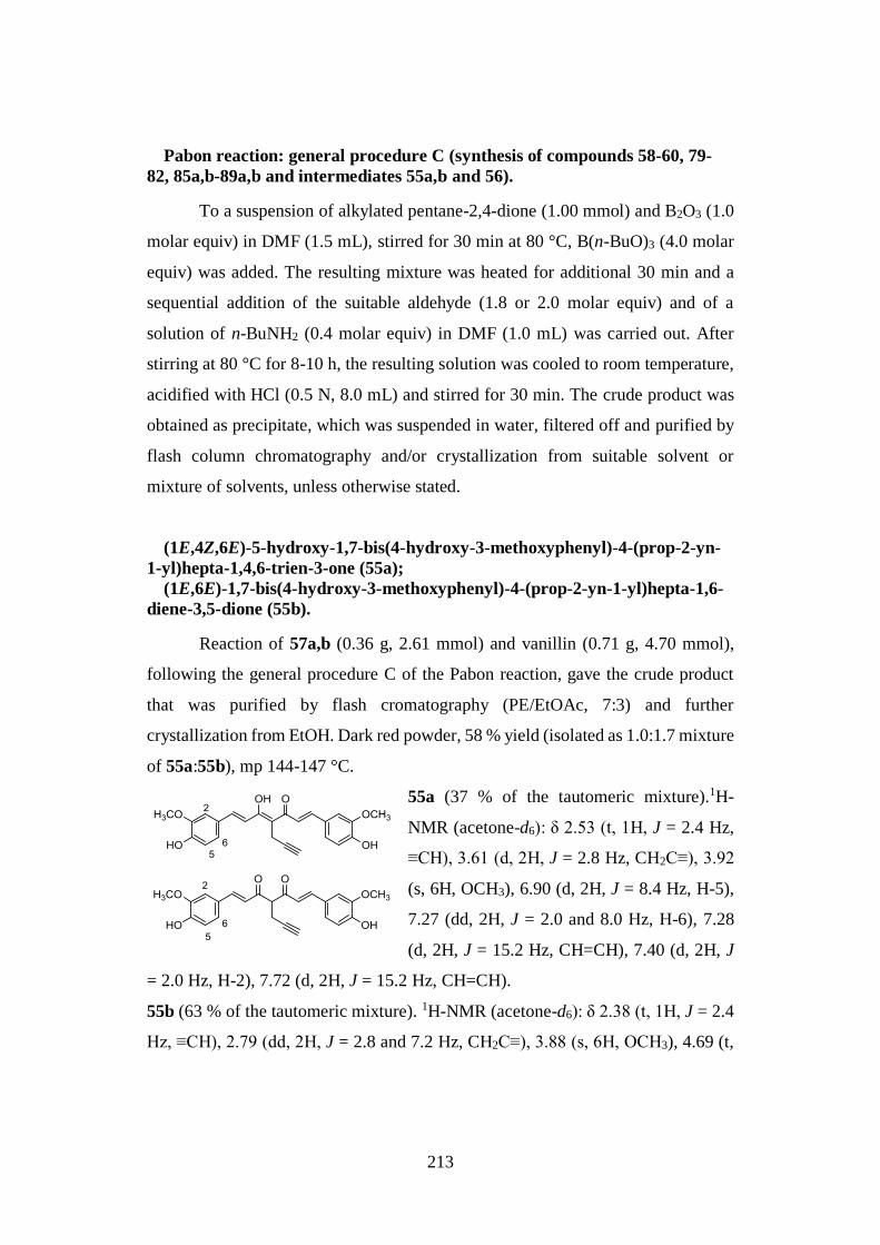
213
Pabon reaction: general procedure C (synthesis of compounds 58-60, 79-
82, 85a,b-89a,b and intermediates 55a,b and 56).
To a suspension of alkylated pentane-2,4-dione (1.00 mmol) and B2O3 (1.0
molar equiv) in DMF (1.5 mL), stirred for 30 min at 80 °C, B(n-BuO)3 (4.0 molar
equiv) was added. The resulting mixture was heated for additional 30 min and a
sequential addition of the suitable aldehyde (1.8 or 2.0 molar equiv) and of a
solution of n-BuNH2 (0.4 molar equiv) in DMF (1.0 mL) was carried out. After
stirring at 80 °C for 8-10 h, the resulting solution was cooled to room temperature,
acidified with HCl (0.5 N, 8.0 mL) and stirred for 30 min. The crude product was
obtained as precipitate, which was suspended in water, filtered off and purified by
flash column chromatography and/or crystallization from suitable solvent or
mixture of solvents, unless otherwise stated.
(1E,4Z,6E)-5-hydroxy-1,7-bis(4-hydroxy-3-methoxyphenyl)-4-(prop-2-yn-
1-yl)hepta-1,4,6-trien-3-one (55a);
(1E,6E)-1,7-bis(4-hydroxy-3-methoxyphenyl)-4-(prop-2-yn-1-yl)hepta-1,6-
diene-3,5-dione (55b).
Reaction of 57a,b (0.36 g, 2.61 mmol) and vanillin (0.71 g, 4.70 mmol),
following the general procedure C of the Pabon reaction, gave the crude product
that was purified by flash cromatography (PE/EtOAc, 7:3) and further
crystallization from EtOH. Dark red powder, 58 % yield (isolated as 1.0:1.7 mixture
of 55a:55b), mp 144-147 °C.
55a (37 % of the tautomeric mixture).1H-
NMR (acetone-d6): δ 2.53 (t, 1H, J = 2.4 Hz,
≡CH), 3.61 (d, 2H, J = 2.8 Hz, CH2C≡), 3.92
(s, 6H, OCH3), 6.90 (d, 2H, J = 8.4 Hz, H-5),
7.27 (dd, 2H, J = 2.0 and 8.0 Hz, H-6), 7.28
(d, 2H, J = 15.2 Hz, CH=CH), 7.40 (d, 2H, J
= 2.0 Hz, H-2), 7.72 (d, 2H, J = 15.2 Hz, CH=CH).
55b (63 % of the tautomeric mixture). 1H-NMR (acetone-d6): δ 2.38 (t, 1H, J = 2.4
Hz, ≡CH), 2.79 (dd, 2H, J = 2.8 and 7.2 Hz, CH2C≡), 3.88 (s, 6H, OCH3), 4.69 (t,
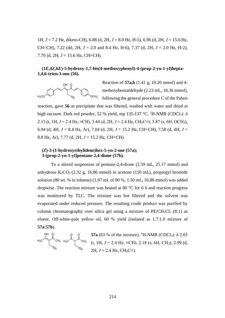
214
1H, J = 7.2 Hz, diketo-CH), 6.88 (d, 2H, J = 8.0 Hz, H-5), 6.96 (d, 2H, J = 15.6 Hz,
CH=CH), 7.22 (dd, 2H, J = 2.0 and 8.4 Hz, H-6), 7.37 (d, 2H, J = 2.0 Hz, H-2),
7.70 (d, 2H, J = 15.6 Hz, CH=CH).
(1E,4Z,6E)-5-hydroxy-1,7-bis(4-methoxyphenyl)-4-(prop-2-yn-1-yl)hepta-
1,4,6-trien-3-one (56).
Reaction of 57a,b (1.41 g, 10.20 mmol) and 4-
methoxybenzaldehyde (2.23 mL, 18.36 mmol),
following the general procedure C of the Pabon
reaction, gave 56 as precipitate that was filtered, washed with water and dried at
high vacuum. Dark red powder, 52 % yield, mp 135-137 °C. 1H-NMR (CDCl3): δ
2.15 (t, 1H, J = 2.4 Hz, ≡CH), 3.44 (d, 2H, J = 2.4 Hz, CH2C≡), 3.87 (s, 6H, OCH3),
6.94 (d, 4H, J = 8.4 Hz, Ar), 7.04 (d, 2H, J = 15.2 Hz, CH=CH), 7.58 (d, 4H, J =
8.8 Hz, Ar), 7.77 (d, 2H, J = 15.2 Hz, CH=CH).
(Z)-3-(1-hydroxyethylidene)hex-5-yn-2-one (57a);
3-(prop-2-yn-1-yl)pentane-2,4-dione (57b).
To a stirred suspension of pentane-2,4-dione (2.59 mL, 25.17 mmol) and
anhydrous Κ2CO3 (2.32 g, 16.86 mmol) in acetone (150 mL), propargyl bromide
solution (80 wt. % in toluene) (1.87 mL of 80 %, 1.50 mL, 16.86 mmol) was added
dropwise. The reaction mixture was heated at 80 °C for 6 h and reaction progress
was monitored by TLC. The mixture was hot filtered and the solvent was
evaporated under reduced pressure. The resulting crude product was purified by
column chromatography over silica gel using a mixture of PE/CH2Cl2 (8:1) as
eluent. Off-white-pale yellow oil, 60 % yield (isolated as 1.7:1.0 mixture of
57a:57b).
57a (63 % of the mixture). 1H-NMR (CDCl3): δ 2.03
(t, 1H, J = 2.4 Hz, ≡CH), 2.18 (s, 6H, CH3), 2.99 (d,
2H, J = 2.4 Hz, CH2C≡).
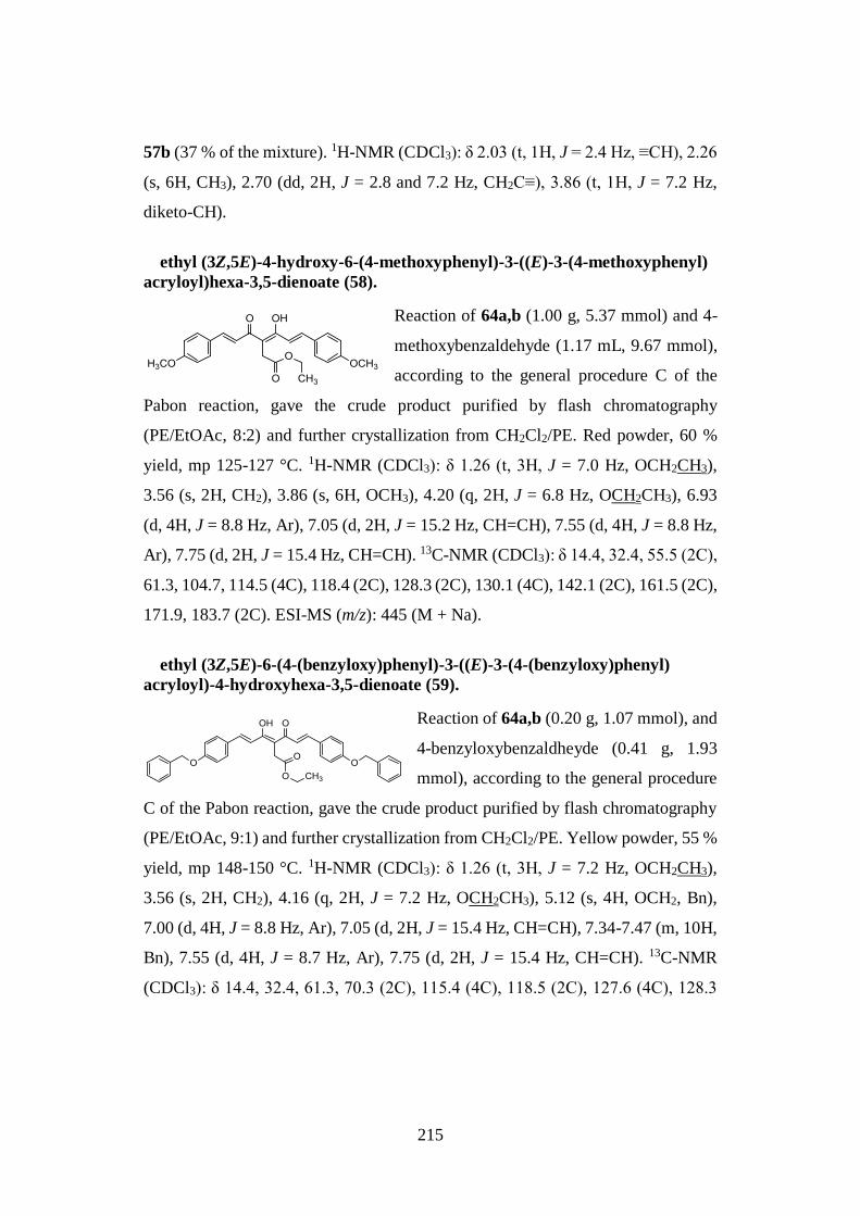
215
57b (37 % of the mixture). 1H-NMR (CDCl3): δ 2.03 (t, 1H, J = 2.4 Hz, ≡CH), 2.26
(s, 6H, CH3), 2.70 (dd, 2H, J = 2.8 and 7.2 Hz, CH2C≡), 3.86 (t, 1H, J = 7.2 Hz,
diketo-CH).
ethyl (3Z,5E)-4-hydroxy-6-(4-methoxyphenyl)-3-((E)-3-(4-methoxyphenyl)
acryloyl)hexa-3,5-dienoate (58).
Reaction of 64a,b (1.00 g, 5.37 mmol) and 4-
methoxybenzaldehyde (1.17 mL, 9.67 mmol),
according to the general procedure C of the
Pabon reaction, gave the crude product purified by flash chromatography
(PE/EtOAc, 8:2) and further crystallization from CH2Cl2/PE. Red powder, 60 %
yield, mp 125-127 °C. 1H-NMR (CDCl3): δ 1.26 (t, 3H, J = 7.0 Hz, OCH2CH3),
3.56 (s, 2H, CH2), 3.86 (s, 6H, OCH3), 4.20 (q, 2H, J = 6.8 Hz, OCH2CH3), 6.93
(d, 4H, J = 8.8 Hz, Ar), 7.05 (d, 2H, J = 15.2 Hz, CH=CH), 7.55 (d, 4H, J = 8.8 Hz,
Ar), 7.75 (d, 2H, J = 15.4 Hz, CH=CH). 13C-NMR (CDCl3): δ 14.4, 32.4, 55.5 (2C),
61.3, 104.7, 114.5 (4C), 118.4 (2C), 128.3 (2C), 130.1 (4C), 142.1 (2C), 161.5 (2C),
171.9, 183.7 (2C). ESI-MS (m/z): 445 (M + Na).
ethyl (3Z,5E)-6-(4-(benzyloxy)phenyl)-3-((E)-3-(4-(benzyloxy)phenyl)
acryloyl)-4-hydroxyhexa-3,5-dienoate (59).
Reaction of 64a,b (0.20 g, 1.07 mmol), and
4-benzyloxybenzaldheyde (0.41 g, 1.93
mmol), according to the general procedure
C of the Pabon reaction, gave the crude product purified by flash chromatography
(PE/EtOAc, 9:1) and further crystallization from CH2Cl2/PE. Yellow powder, 55 %
yield, mp 148-150 °C. 1H-NMR (CDCl3): δ 1.26 (t, 3H, J = 7.2 Hz, OCH2CH3),
3.56 (s, 2H, CH2), 4.16 (q, 2H, J = 7.2 Hz, OCH2CH3), 5.12 (s, 4H, OCH2, Bn),
7.00 (d, 4H, J = 8.8 Hz, Ar), 7.05 (d, 2H, J = 15.4 Hz, CH=CH), 7.34-7.47 (m, 10H,
Bn), 7.55 (d, 4H, J = 8.7 Hz, Ar), 7.75 (d, 2H, J = 15.4 Hz, CH=CH). 13C-NMR
(CDCl3): δ 14.4, 32.4, 61.3, 70.3 (2C), 115.4 (4C), 118.5 (2C), 127.6 (4C), 128.3
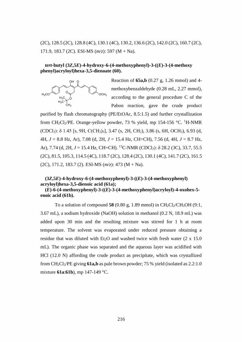
216
(2C), 128.5 (2C), 128.8 (4C), 130.1 (4C), 130.2, 136.6 (2C), 142.0 (2C), 160.7 (2C),
171.9, 183.7 (2C). ESI-MS (m/z): 597 (M + Na).
tert-butyl (3Z,5E)-4-hydroxy-6-(4-methoxyphenyl)-3-((E)-3-(4-methoxy
phenyl)acryloyl)hexa-3,5-dienoate (60).
Reaction of 65a,b (0.27 g, 1.26 mmol) and 4-
methoxybenzaldehyde (0.28 mL, 2.27 mmol),
according to the general procedure C of the
Pabon reaction, gave the crude product
purified by flash chromatography (PE/EtOAc, 8.5:1.5) and further crystallization
from CH2Cl2/PE. Orange-yellow powder, 73 % yield, mp 154-156 °C. 1H-NMR
(CDCl3): δ 1.45 [s, 9H, C(CH3)3], 3.47 (s, 2H, CH2), 3.86 (s, 6H, OCH3), 6.93 (d,
4H, J = 8.8 Hz, Ar), 7.08 (d, 2H, J = 15.4 Hz, CH=CH), 7.56 (d, 4H, J = 8.7 Hz,
Ar), 7.74 (d, 2H, J = 15.4 Hz, CH=CH). 13C-NMR (CDCl3): δ 28.2 (3C), 33.7, 55.5
(2C), 81.5, 105.3, 114.5 (4C), 118.7 (2C), 128.4 (2C), 130.1 (4C), 141.7 (2C), 161.5
(2C), 171.2, 183.7 (2). ESI-MS (m/z): 473 (M + Na).
(3Z,5E)-4-hydroxy-6-(4-methoxyphenyl)-3-((E)-3-(4-methoxyphenyl)
acryloyl)hexa-3,5-dienoic acid (61a);
(E)-6-(4-methoxyphenyl)-3-((E)-3-(4-methoxyphenyl)acryloyl)-4-oxohex-5-
enoic acid (61b).
To a solution of compound 58 (0.80 g, 1.89 mmol) in CH2Cl2/CH3OH (9:1,
3.67 mL), a sodium hydroxide (NaOH) solution in methanol (0.2 N, 18.9 mL) was
added upon 30 min and the resulting mixture was stirred for 1 h at room
temperature. The solvent was evaporated under reduced pressure obtaining a
residue that was diluted with Et2O and washed twice with fresh water (2 x 15.0
mL). The organic phase was separated and the aqueous layer was acidified with
HCl (12.0 N) affording the crude product as precipitate, which was crystallized
from CH2Cl2/PE giving 61a,b as pale brown powder; 75 % yield (isolated as 2.2:1.0
mixture 61a:61b), mp 147-149 °C.
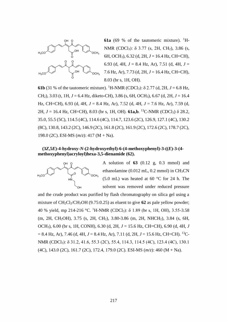
217
61a (69 % of the tautomeric mixture). 1H-
NMR (CDCl3): δ 3.77 (s, 2H, CH2), 3.86 (s,
6H, OCH3), 6.32 (d, 2H, J = 16.4 Hz, CH=CH),
6.93 (d, 4H, J = 8.4 Hz, Ar), 7.51 (d, 4H, J =
7.6 Hz, Ar), 7.73 (d, 2H, J = 16.4 Hz, CH=CH),
8.03 (br s, 1H, OH).
61b (31 % of the tautomeric mixture). 1H-NMR (CDCl3): δ 2.77 (d, 2H, J = 6.8 Hz,
CH2), 3.03 (t, 1H, J = 6.4 Hz, diketo-CH), 3.86 (s, 6H, OCH3), 6.67 (d, 2H, J = 16.4
Hz, CH=CH), 6.93 (d, 4H, J = 8.4 Hz, Ar), 7.52 (d, 4H, J = 7.6 Hz, Ar), 7.59 (d,
2H, J = 16.4 Hz, CH=CH), 8.03 (br s, 1H, OH). 61a,b. 13C-NMR (CDCl3) δ 28.2,
35.0, 55.5 (5C), 114.5 (4C), 114.6 (4C), 114.7, 123.6 (2C), 126.9, 127.1 (4C), 130.2
(8C), 130.8, 143.2 (2C), 146.9 (2C), 161.8 (2C), 161.9 (2C), 172.6 (2C), 178.7 (2C),
198.0 (2C). ESI-MS (m/z): 417 (M + Na).
(3Z,5E)-4-hydroxy-N-(2-hydroxyethyl)-6-(4-methoxyphenyl)-3-((E)-3-(4-
methoxyphenyl)acryloyl)hexa-3,5-dienamide (62).
A solution of 63 (0.12 g, 0.3 mmol) and
ethanolamine (0.012 mL, 0.2 mmol) in CH3CN
(5.0 mL) was heated at 60 °C for 24 h. The
solvent was removed under reduced pressure
and the crude product was purified by flash chromatography on silica gel using a
mixture of CH2Cl2/CH3OH (9.75:0.25) as eluent to give 62 as pale yellow powder;
40 % yield, mp 214-216 °C. 1H-NMR (CDCl3): δ 1.89 (br s, 1H, OH), 3.55-3.58
(m, 2H, CH2OH), 3.75 (s, 2H, CH2), 3.80-3.86 (m, 2H, NHCH2), 3.84 (s, 6H,
OCH3), 6.00 (br s, 1H, CONH), 6.30 (d, 2H, J = 15.6 Hz, CH=CH), 6.90 (d, 4H, J
= 8.4 Hz, Ar), 7.46 (d, 4H, J = 8.4 Hz, Ar), 7.11 (d, 2H, J = 15.6 Hz, CH=CH). 13C-
NMR (CDCl3): δ 31.2, 41.6, 55.3 (2C), 55.4, 114.3, 114.5 (4C), 123.4 (4C), 130.1
(4C), 143.0 (2C), 161.7 (2C), 172.4, 179.0 (2C). ESI-MS (m/z): 460 (M + Na).
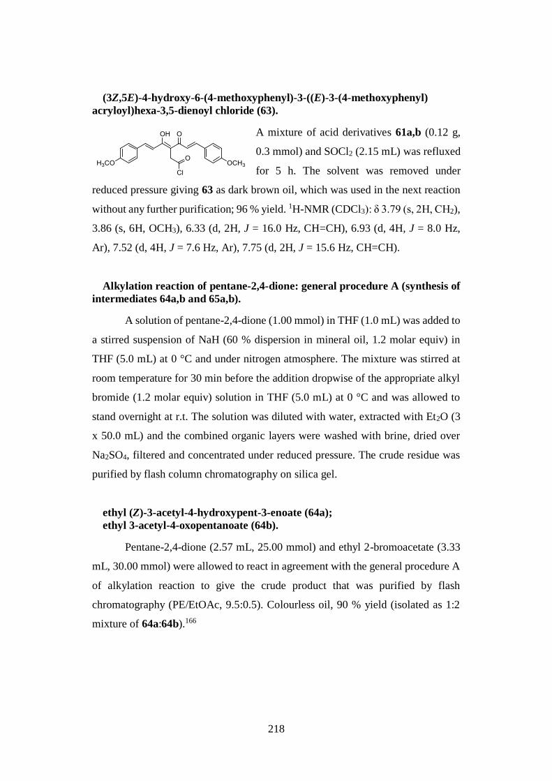
218
(3Z,5E)-4-hydroxy-6-(4-methoxyphenyl)-3-((E)-3-(4-methoxyphenyl)
acryloyl)hexa-3,5-dienoyl chloride (63).
A mixture of acid derivatives 61a,b (0.12 g,
0.3 mmol) and SOCl2 (2.15 mL) was refluxed
for 5 h. The solvent was removed under
reduced pressure giving 63 as dark brown oil, which was used in the next reaction
without any further purification; 96 % yield. 1H-NMR (CDCl3): δ 3.79 (s, 2H, CH2),
3.86 (s, 6H, OCH3), 6.33 (d, 2H, J = 16.0 Hz, CH=CH), 6.93 (d, 4H, J = 8.0 Hz,
Ar), 7.52 (d, 4H, J = 7.6 Hz, Ar), 7.75 (d, 2H, J = 15.6 Hz, CH=CH).
Alkylation reaction of pentane-2,4-dione: general procedure A (synthesis of
intermediates 64a,b and 65a,b).
A solution of pentane-2,4-dione (1.00 mmol) in THF (1.0 mL) was added to
a stirred suspension of NaH (60 % dispersion in mineral oil, 1.2 molar equiv) in
THF (5.0 mL) at 0 °C and under nitrogen atmosphere. The mixture was stirred at
room temperature for 30 min before the addition dropwise of the appropriate alkyl
bromide (1.2 molar equiv) solution in THF (5.0 mL) at 0 °C and was allowed to
stand overnight at r.t. The solution was diluted with water, extracted with Et2O (3
x 50.0 mL) and the combined organic layers were washed with brine, dried over
Na2SO4, filtered and concentrated under reduced pressure. The crude residue was
purified by flash column chromatography on silica gel.
ethyl (Z)-3-acetyl-4-hydroxypent-3-enoate (64a);
ethyl 3-acetyl-4-oxopentanoate (64b).
Pentane-2,4-dione (2.57 mL, 25.00 mmol) and ethyl 2-bromoacetate (3.33
mL, 30.00 mmol) were allowed to react in agreement with the general procedure A
of alkylation reaction to give the crude product that was purified by flash
chromatography (PE/EtOAc, 9.5:0.5). Colourless oil, 90 % yield (isolated as 1:2
mixture of 64a:64b).166
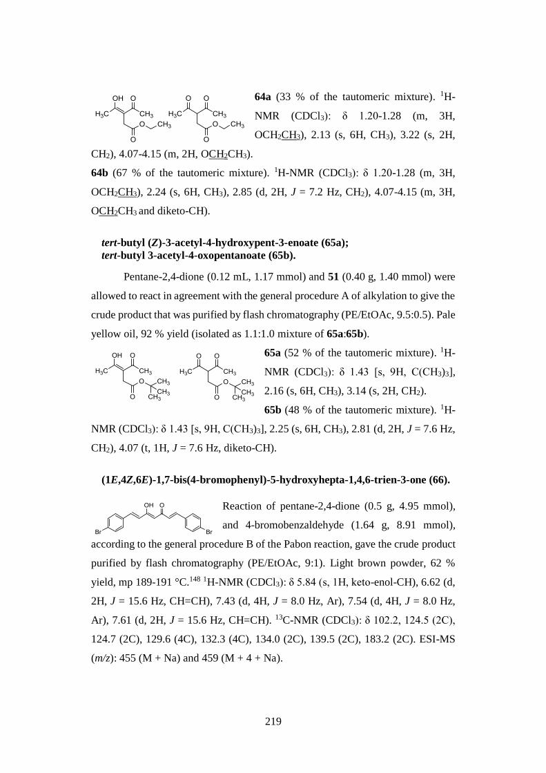
219
64a (33 % of the tautomeric mixture). 1H-
NMR (CDCl3): δ 1.20-1.28 (m, 3H,
OCH2CH3), 2.13 (s, 6H, CH3), 3.22 (s, 2H,
CH2), 4.07-4.15 (m, 2H, OCH2CH3).
64b (67 % of the tautomeric mixture). 1H-NMR (CDCl3): δ 1.20-1.28 (m, 3H,
OCH2CH3), 2.24 (s, 6H, CH3), 2.85 (d, 2H, J = 7.2 Hz, CH2), 4.07-4.15 (m, 3H,
OCH2CH3 and diketo-CH).
tert-butyl (Z)-3-acetyl-4-hydroxypent-3-enoate (65a);
tert-butyl 3-acetyl-4-oxopentanoate (65b).
Pentane-2,4-dione (0.12 mL, 1.17 mmol) and 51 (0.40 g, 1.40 mmol) were
allowed to react in agreement with the general procedure A of alkylation to give the
crude product that was purified by flash chromatography (PE/EtOAc, 9.5:0.5). Pale
yellow oil, 92 % yield (isolated as 1.1:1.0 mixture of 65a:65b).
65a (52 % of the tautomeric mixture). 1H-
NMR (CDCl3): δ 1.43 [s, 9H, C(CH3)3],
2.16 (s, 6H, CH3), 3.14 (s, 2H, CH2).
65b (48 % of the tautomeric mixture). 1H-
NMR (CDCl3): δ 1.43 [s, 9H, C(CH3)3], 2.25 (s, 6H, CH3), 2.81 (d, 2H, J = 7.6 Hz,
CH2), 4.07 (t, 1H, J = 7.6 Hz, diketo-CH).
(1E,4Z,6E)-1,7-bis(4-bromophenyl)-5-hydroxyhepta-1,4,6-trien-3-one (66).
Reaction of pentane-2,4-dione (0.5 g, 4.95 mmol),
and 4-bromobenzaldehyde (1.64 g, 8.91 mmol),
according to the general procedure B of the Pabon reaction, gave the crude product
purified by flash chromatography (PE/EtOAc, 9:1). Light brown powder, 62 %
yield, mp 189-191 °C.148 1H-NMR (CDCl3): δ 5.84 (s, 1H, keto-enol-CH), 6.62 (d,
2H, J = 15.6 Hz, CH=CH), 7.43 (d, 4H, J = 8.0 Hz, Ar), 7.54 (d, 4H, J = 8.0 Hz,
Ar), 7.61 (d, 2H, J = 15.6 Hz, CH=CH). 13C-NMR (CDCl3): δ 102.2, 124.5 (2C),
124.7 (2C), 129.6 (4C), 132.3 (4C), 134.0 (2C), 139.5 (2C), 183.2 (2C). ESI-MS
(m/z): 455 (M + Na) and 459 (M + 4 + Na).
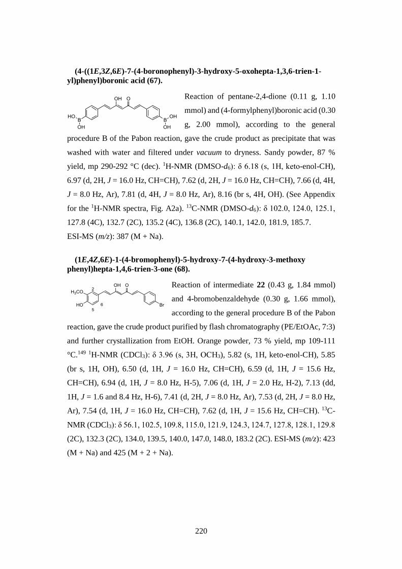
220
(4-((1E,3Z,6E)-7-(4-boronophenyl)-3-hydroxy-5-oxohepta-1,3,6-trien-1-
yl)phenyl)boronic acid (67).
Reaction of pentane-2,4-dione (0.11 g, 1.10
mmol) and (4-formylphenyl)boronic acid (0.30
g, 2.00 mmol), according to the general
procedure B of the Pabon reaction, gave the crude product as precipitate that was
washed with water and filtered under vacuum to dryness. Sandy powder, 87 %
yield, mp 290-292 °C (dec). 1H-NMR (DMSO-d6): δ 6.18 (s, 1H, keto-enol-CH),
6.97 (d, 2H, J = 16.0 Hz, CH=CH), 7.62 (d, 2H, J = 16.0 Hz, CH=CH), 7.66 (d, 4H,
J = 8.0 Hz, Ar), 7.81 (d, 4H, J = 8.0 Hz, Ar), 8.16 (br s, 4H, OH). (See Appendix
for the 1H-NMR spectra, Fig. A2a). 13C-NMR (DMSO-d6): δ 102.0, 124.0, 125.1,
127.8 (4C), 132.7 (2C), 135.2 (4C), 136.8 (2C), 140.1, 142.0, 181.9, 185.7.
ESI-MS (m/z): 387 (M + Na).
(1E,4Z,6E)-1-(4-bromophenyl)-5-hydroxy-7-(4-hydroxy-3-methoxy
phenyl)hepta-1,4,6-trien-3-one (68).
Reaction of intermediate 22 (0.43 g, 1.84 mmol)
and 4-bromobenzaldehyde (0.30 g, 1.66 mmol),
according to the general procedure B of the Pabon
reaction, gave the crude product purified by flash chromatography (PE/EtOAc, 7:3)
and further crystallization from EtOH. Orange powder, 73 % yield, mp 109-111
°C.149 1H-NMR (CDCl3): δ 3.96 (s, 3H, OCH3), 5.82 (s, 1H, keto-enol-CH), 5.85
(br s, 1H, OH), 6.50 (d, 1H, J = 16.0 Hz, CH=CH), 6.59 (d, 1H, J = 15.6 Hz,
CH=CH), 6.94 (d, 1H, J = 8.0 Hz, H-5), 7.06 (d, 1H, J = 2.0 Hz, H-2), 7.13 (dd,
1H, J = 1.6 and 8.4 Hz, H-6), 7.41 (d, 2H, J = 8.0 Hz, Ar), 7.53 (d, 2H, J = 8.0 Hz,
Ar), 7.54 (d, 1H, J = 16.0 Hz, CH=CH), 7.62 (d, 1H, J = 15.6 Hz, CH=CH). 13C-
NMR (CDCl3): δ 56.1, 102.5, 109.8, 115.0, 121.9, 124.3, 124.7, 127.8, 128.1, 129.8
(2C), 132.3 (2C), 134.0, 139.5, 140.0, 147.0, 148.0, 183.2 (2C). ESI-MS (m/z): 423
(M + Na) and 425 (M + 2 + Na).
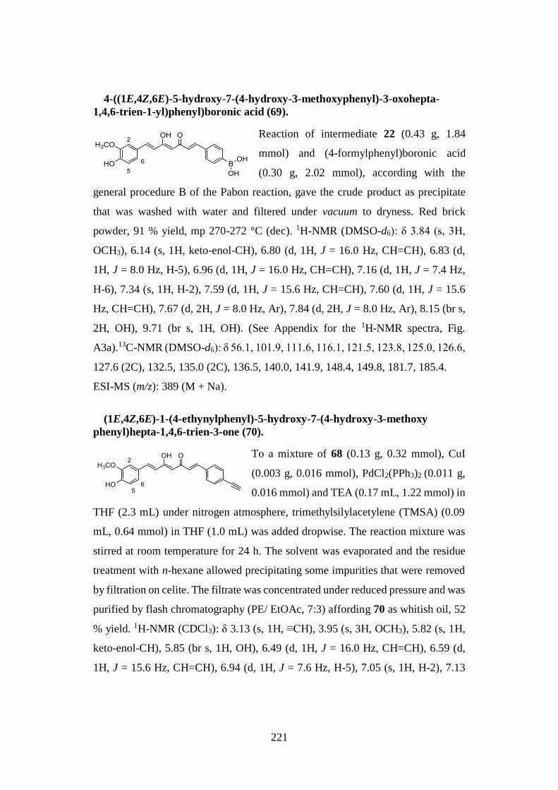
221
4-((1E,4Z,6E)-5-hydroxy-7-(4-hydroxy-3-methoxyphenyl)-3-oxohepta-
1,4,6-trien-1-yl)phenyl)boronic acid (69).
Reaction of intermediate 22 (0.43 g, 1.84
mmol) and (4-formylphenyl)boronic acid
(0.30 g, 2.02 mmol), according with the
general procedure B of the Pabon reaction, gave the crude product as precipitate
that was washed with water and filtered under vacuum to dryness. Red brick
powder, 91 % yield, mp 270-272 °C (dec). 1H-NMR (DMSO-d6): δ 3.84 (s, 3H,
OCH3), 6.14 (s, 1H, keto-enol-CH), 6.80 (d, 1H, J = 16.0 Hz, CH=CH), 6.83 (d,
1H, J = 8.0 Hz, H-5), 6.96 (d, 1H, J = 16.0 Hz, CH=CH), 7.16 (d, 1H, J = 7.4 Hz,
H-6), 7.34 (s, 1H, H-2), 7.59 (d, 1H, J = 15.6 Hz, CH=CH), 7.60 (d, 1H, J = 15.6
Hz, CH=CH), 7.67 (d, 2H, J = 8.0 Hz, Ar), 7.84 (d, 2H, J = 8.0 Hz, Ar), 8.15 (br s,
2H, OH), 9.71 (br s, 1H, OH). (See Appendix for the 1H-NMR spectra, Fig.
A3a).13C-NMR (DMSO-d6): δ 56.1, 101.9, 111.6, 116.1, 121.5, 123.8, 125.0, 126.6,
127.6 (2C), 132.5, 135.0 (2C), 136.5, 140.0, 141.9, 148.4, 149.8, 181.7, 185.4.
ESI-MS (m/z): 389 (M + Na).
(1E,4Z,6E)-1-(4-ethynylphenyl)-5-hydroxy-7-(4-hydroxy-3-methoxy
phenyl)hepta-1,4,6-trien-3-one (70).
To a mixture of 68 (0.13 g, 0.32 mmol), CuI
(0.003 g, 0.016 mmol), PdCl2(PPh3)2 (0.011 g,
0.016 mmol) and TEA (0.17 mL, 1.22 mmol) in
THF (2.3 mL) under nitrogen atmosphere, trimethylsilylacetylene (TMSA) (0.09
mL, 0.64 mmol) in THF (1.0 mL) was added dropwise. The reaction mixture was
stirred at room temperature for 24 h. The solvent was evaporated and the residue
treatment with n-hexane allowed precipitating some impurities that were removed
by filtration on celite. The filtrate was concentrated under reduced pressure and was
purified by flash chromatography (PE/ EtOAc, 7:3) affording 70 as whitish oil, 52
% yield. 1H-NMR (CDCl3): δ 3.13 (s, 1H, ≡CH), 3.95 (s, 3H, OCH3), 5.82 (s, 1H,
keto-enol-CH), 5.85 (br s, 1H, OH), 6.49 (d, 1H, J = 16.0 Hz, CH=CH), 6.59 (d,
1H, J = 15.6 Hz, CH=CH), 6.94 (d, 1H, J = 7.6 Hz, H-5), 7.05 (s, 1H, H-2), 7.13
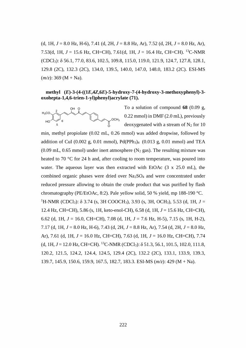
222
(d, 1H, J = 8.0 Hz, H-6), 7.41 (d, 2H, J = 8.8 Hz, Ar), 7.52 (d, 2H, J = 8.0 Hz, Ar),
7.53(d, 1H, J = 15.6 Hz, CH=CH), 7.61(d, 1H, J = 16.4 Hz, CH=CH). 13C-NMR
(CDCl3): δ 56.1, 77.0, 83.6, 102.5, 109.8, 115.0, 119.0, 121.9, 124.7, 127.8, 128.1,
129.8 (2C), 132.3 (2C), 134.0, 139.5, 140.0, 147.0, 148.0, 183.2 (2C). ESI-MS
(m/z): 369 (M + Na).
methyl (E)-3-(4-((1E,4Z,6E)-5-hydroxy-7-(4-hydroxy-3-methoxyphenyl)-3-
oxohepta-1,4,6-trien-1-yl)phenyl)acrylate (71).
To a solution of compound 68 (0.09 g,
0.22 mmol) in DMF (2.0 mL), previously
deoxygenated with a stream of N2 for 10
min, methyl propiolate (0.02 mL, 0.26 mmol) was added dropwise, followed by
addition of CuI (0.002 g, 0.01 mmol), Pd(PPh3)4 (0.013 g, 0.01 mmol) and TEA
(0.09 mL, 0.65 mmol) under inert atmosphere (N2 gas). The resulting mixture was
heated to 70 °C for 24 h and, after cooling to room temperature, was poured into
water. The aqueous layer was then extracted with EtOAc (3 x 25.0 mL), the
combined organic phases were dried over Na2SO4 and were concentrated under
reduced pressure allowing to obtain the crude product that was purified by flash
chromatography (PE/EtOAc, 8:2). Pale yellow solid, 50 % yield, mp 188-190 °C.
1H-NMR (CDCl3): δ 3.74 (s, 3H COOCH3), 3.93 (s, 3H, OCH3), 5.53 (d, 1H, J =
12.4 Hz, CH=CH), 5.86 (s, 1H, keto-enol-CH), 6.58 (d, 1H, J = 15.6 Hz, CH=CH),
6.62 (d, 1H, J = 16.0, CH=CH), 7.08 (d, 1H, J = 7.6 Hz, H-5), 7.15 (s, 1H, H-2),
7.17 (d, 1H, J = 8.0 Hz, H-6), 7.43 (d, 2H, J = 8.8 Hz, Ar), 7.54 (d, 2H, J = 8.0 Hz,
Ar), 7.61 (d, 1H, J = 16.0 Hz, CH=CH), 7.63 (d, 1H, J = 16.0 Hz, CH=CH), 7.74
(d, 1H, J = 12.0 Hz, CH=CH). 13C-NMR (CDCl3): δ 51.3, 56.1, 101.5, 102.0, 111.8,
120.2, 121.5, 124.2, 124.4, 124.5, 129.4 (2C), 132.2 (2C), 133.1, 133.9, 139.3,
139.7, 145.9, 150.6, 159.9, 167.5, 182.7, 183.3. ESI-MS (m/z): 429 (M + Na).
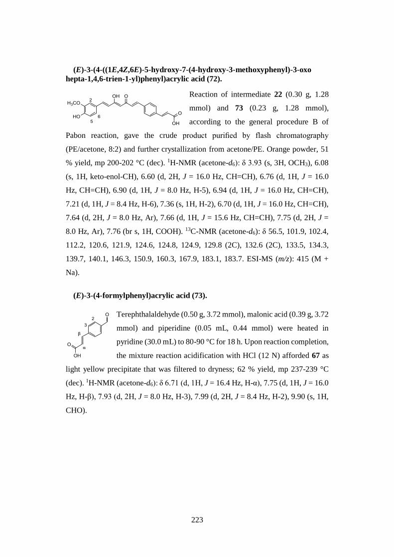
223
(E)-3-(4-((1E,4Z,6E)-5-hydroxy-7-(4-hydroxy-3-methoxyphenyl)-3-oxo
hepta-1,4,6-trien-1-yl)phenyl)acrylic acid (72).
Reaction of intermediate 22 (0.30 g, 1.28
mmol) and 73 (0.23 g, 1.28 mmol),
according to the general procedure B of
Pabon reaction, gave the crude product purified by flash chromatography
(PE/acetone, 8:2) and further crystallization from acetone/PE. Orange powder, 51
% yield, mp 200-202 °C (dec). 1H-NMR (acetone-d6): δ 3.93 (s, 3H, OCH3), 6.08
(s, 1H, keto-enol-CH), 6.60 (d, 2H, J = 16.0 Hz, CH=CH), 6.76 (d, 1H, J = 16.0
Hz, CH=CH), 6.90 (d, 1H, J = 8.0 Hz, H-5), 6.94 (d, 1H, J = 16.0 Hz, CH=CH),
7.21 (d, 1H, J = 8.4 Hz, H-6), 7.36 (s, 1H, H-2), 6.70 (d, 1H, J = 16.0 Hz, CH=CH),
7.64 (d, 2H, J = 8.0 Hz, Ar), 7.66 (d, 1H, J = 15.6 Hz, CH=CH), 7.75 (d, 2H, J =
8.0 Hz, Ar), 7.76 (br s, 1H, COOH). 13C-NMR (acetone-d6): δ 56.5, 101.9, 102.4,
112.2, 120.6, 121.9, 124.6, 124.8, 124.9, 129.8 (2C), 132.6 (2C), 133.5, 134.3,
139.7, 140.1, 146.3, 150.9, 160.3, 167.9, 183.1, 183.7. ESI-MS (m/z): 415 (M +
Na).
(E)-3-(4-formylphenyl)acrylic acid (73).
Terephthalaldehyde (0.50 g, 3.72 mmol), malonic acid (0.39 g, 3.72
mmol) and piperidine (0.05 mL, 0.44 mmol) were heated in
pyridine (30.0 mL) to 80-90 °C for 18 h. Upon reaction completion,
the mixture reaction acidification with HCl (12 N) afforded 67 as
light yellow precipitate that was filtered to dryness; 62 % yield, mp 237-239 °C
(dec). 1H-NMR (acetone-d6): δ 6.71 (d, 1H, J = 16.4 Hz, H-α), 7.75 (d, 1H, J = 16.0
Hz, H-β), 7.93 (d, 2H, J = 8.0 Hz, H-3), 7.99 (d, 2H, J = 8.4 Hz, H-2), 9.90 (s, 1H,
CHO).
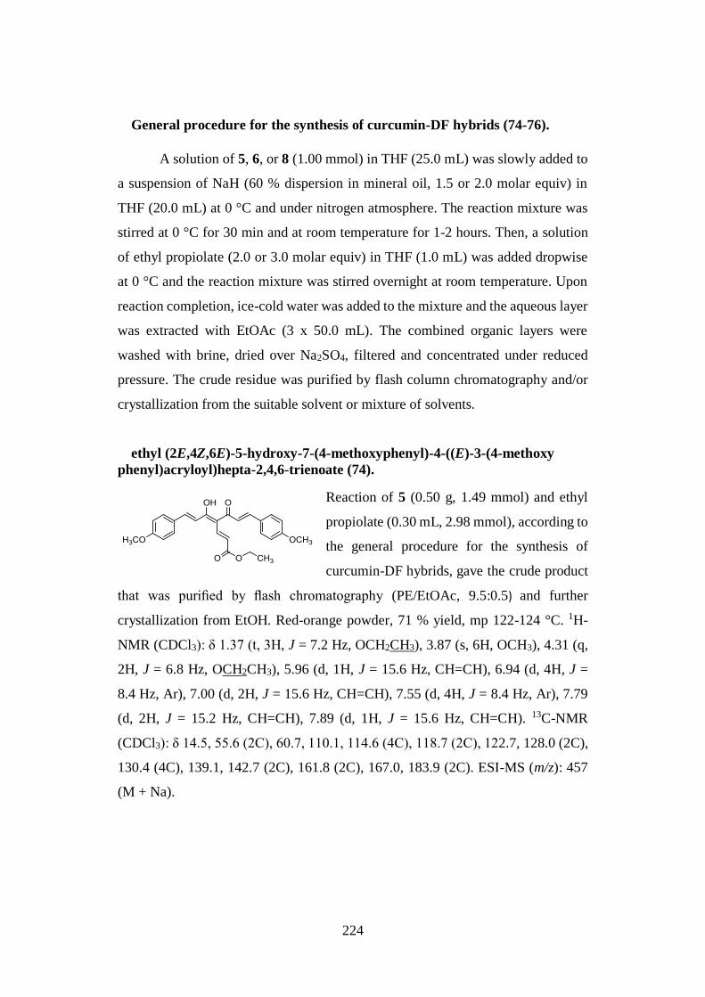
224
General procedure for the synthesis of curcumin-DF hybrids (74-76).
A solution of 5, 6, or 8 (1.00 mmol) in THF (25.0 mL) was slowly added to
a suspension of NaH (60 % dispersion in mineral oil, 1.5 or 2.0 molar equiv) in
THF (20.0 mL) at 0 °C and under nitrogen atmosphere. The reaction mixture was
stirred at 0 °C for 30 min and at room temperature for 1-2 hours. Then, a solution
of ethyl propiolate (2.0 or 3.0 molar equiv) in THF (1.0 mL) was added dropwise
at 0 °C and the reaction mixture was stirred overnight at room temperature. Upon
reaction completion, ice-cold water was added to the mixture and the aqueous layer
was extracted with EtOAc (3 x 50.0 mL). The combined organic layers were
washed with brine, dried over Na2SO4, filtered and concentrated under reduced
pressure. The crude residue was purified by flash column chromatography and/or
crystallization from the suitable solvent or mixture of solvents.
ethyl (2E,4Z,6E)-5-hydroxy-7-(4-methoxyphenyl)-4-((E)-3-(4-methoxy
phenyl)acryloyl)hepta-2,4,6-trienoate (74).
Reaction of 5 (0.50 g, 1.49 mmol) and ethyl
propiolate (0.30 mL, 2.98 mmol), according to
the general procedure for the synthesis of
curcumin-DF hybrids, gave the crude product
that was purified by flash chromatography (PE/EtOAc, 9.5:0.5) and further
crystallization from EtOH. Red-orange powder, 71 % yield, mp 122-124 °C. 1H-
NMR (CDCl3): δ 1.37 (t, 3H, J = 7.2 Hz, OCH2CH3), 3.87 (s, 6H, OCH3), 4.31 (q,
2H, J = 6.8 Hz, OCH2CH3), 5.96 (d, 1H, J = 15.6 Hz, CH=CH), 6.94 (d, 4H, J =
8.4 Hz, Ar), 7.00 (d, 2H, J = 15.6 Hz, CH=CH), 7.55 (d, 4H, J = 8.4 Hz, Ar), 7.79
(d, 2H, J = 15.2 Hz, CH=CH), 7.89 (d, 1H, J = 15.6 Hz, CH=CH). 13C-NMR
(CDCl3): δ 14.5, 55.6 (2C), 60.7, 110.1, 114.6 (4C), 118.7 (2C), 122.7, 128.0 (2C),
130.4 (4C), 139.1, 142.7 (2C), 161.8 (2C), 167.0, 183.9 (2C). ESI-MS (m/z): 457
(M + Na).
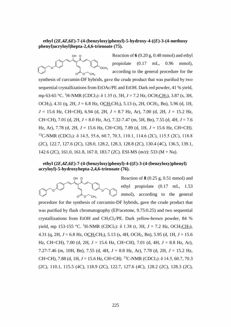
225
ethyl (2E,4Z,6E)-7-(4-(benzyloxy)phenyl)-5-hydroxy-4-((E)-3-(4-methoxy
phenyl)acryloyl)hepta-2,4,6-trienoate (75).
Reaction of 6 (0.20 g, 0.48 mmol) and ethyl
propiolate (0.17 mL, 0.96 mmol),
according to the general procedure for the
synthesis of curcumin-DF hybrids, gave the crude product that was purified by two
sequential crystallizations from EtOAc/PE and EtOH. Dark red powder, 41 % yield,
mp 63-65 °C. 1H-NMR (CDCl3): δ 1.35 (t, 3H, J = 7.2 Hz, OCH2CH3), 3.87 (s, 3H,
OCH3), 4.31 (q, 2H, J = 6.8 Hz, OCH2CH3), 5.13 (s, 2H, OCH2, Bn), 5.96 (d, 1H,
J = 15.6 Hz, CH=CH), 6.94 (d, 2H, J = 8.7 Hz, Ar), 7.00 (d, 2H, J = 15.2 Hz,
CH=CH), 7.01 (d, 2H, J = 8.0 Hz, Ar), 7.32-7.47 (m, 5H, Bn), 7.55 (d, 4H, J = 7.6
Hz, Ar), 7.78 (d, 2H, J = 15.6 Hz, CH=CH), 7.89 (d, 1H, J = 15.6 Hz, CH=CH).
13C-NMR (CDCl3): δ 14.5, 55.6, 60.7, 70.3, 110.1, 114.6 (2C), 115.5 (2C), 118.8
(2C), 122.7, 127.6 (2C), 128.0, 128.2, 128.3, 128.8 (2C), 130.4 (4C), 136.5, 139.1,
142.6 (2C), 161.0, 161.8, 167.0, 183.7 (2C). ESI-MS (m/z): 533 (M + Na).
ethyl (2E,4Z,6E)-7-(4-(benzyloxy)phenyl)-4-((E)-3-(4-(benzyloxy)phenyl)
acryloyl)-5-hydroxyhepta-2,4,6-trienoate (76).
Reaction of 8 (0.25 g, 0.51 mmol) and
ethyl propiolate (0.17 mL, 1.53
mmol), according to the general
procedure for the synthesis of curcumin-DF hybrids, gave the crude product that
was purified by flash chromatography (EP/acetone, 9.75:0.25) and two sequential
crystallizations from EtOH and CH2Cl2/PE. Dark yellow-brown powder, 84 %
yield, mp 153-155 °C. 1H-NMR (CDCl3): δ 1.38 (t, 3H, J = 7.2 Hz, OCH2CH3),
4.31 (q, 2H, J = 6.8 Hz, OCH2CH3), 5.13 (s, 4H, OCH2, Bn), 5.95 (d, 1H, J = 15.6
Hz, CH=CH), 7.00 (d, 2H, J = 15.6 Hz, CH=CH), 7.01 (d, 4H, J = 8.8 Hz, Ar),
7.27-7.46 (m, 10H, Bn), 7.55 (d, 4H, J = 8.8 Hz, Ar), 7.78 (d, 2H, J = 15.2 Hz,
CH=CH), 7.88 (d, 1H, J = 15.6 Hz, CH=CH). 13C-NMR (CDCl3): δ 14.5, 60.7, 70.3
(2C), 110.1, 115.5 (4C), 118.9 (2C), 122.7, 127.6 (4C), 128.2 (2C), 128.3 (2C),
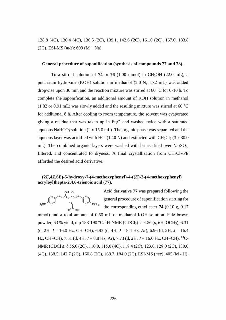
226
128.8 (4C), 130.4 (4C), 136.5 (2C), 139.1, 142.6 (2C), 161.0 (2C), 167.0, 183.8
(2C). ESI-MS (m/z): 609 (M + Na).
General procedure of saponification (synthesis of compounds 77 and 78).
To a stirred solution of 74 or 76 (1.00 mmol) in CH3OH (22.0 mL), a
potassium hydroxide (ΚOH) solution in methanol (2.0 N, 1.82 mL) was added
dropwise upon 30 min and the reaction mixture was stirred at 60 °C for 6-10 h. To
complete the saponification, an additional amount of KOH solution in methanol
(1.82 or 0.91 mL) was slowly added and the resulting mixture was stirred at 60 °C
for additional 8 h. After cooling to room temperature, the solvent was evaporated
giving a residue that was taken up in Et2O and washed twice with a saturated
aqueous NaHCO3 solution (2 x 15.0 mL). The organic phase was separated and the
aqueous layer was acidified with HCl (12.0 N) and extracted with CH2Cl2 (3 x 30.0
mL). The combined organic layers were washed with brine, dried over Na2SO4,
filtered, and concentrated to dryness. A final crystallization from CH2Cl2/PE
afforded the desired acid derivative.
(2E,4Z,6E)-5-hydroxy-7-(4-methoxyphenyl)-4-((E)-3-(4-methoxyphenyl)
acryloyl)hepta-2,4,6-trienoic acid (77).
Acid derivative 77 was prepared following the
general procedure of saponification starting for
the corresponding ethyl ester 74 (0.10 g, 0.17
mmol) and a total amount of 0.50 mL of methanol ΚOH solution. Pale brown
powder, 63 % yield, mp 188-190 °C. 1H-NMR (CDCl3): δ 3.86 (s, 6H, OCH3), 6.31
(d, 2H, J = 16.0 Hz, CH=CH), 6.93 (d, 4H, J = 8.4 Hz, Ar), 6.96 (d, 2H, J = 16.4
Hz, CH=CH), 7.51 (d, 4H, J = 8.8 Hz, Ar), 7.73 (d, 2H, J = 16.0 Hz, CH=CH). 13C-
NMR (CDCl3): δ 56.0 (2C), 110.0, 115.0 (4C), 118.4 (2C), 123.0, 128.0 (2C), 130.0
(4C), 138.5, 142.7 (2C), 160.8 (2C), 168.7, 184.0 (2C). ESI-MS (m/z): 405 (M - H).
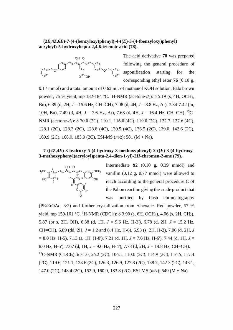
227
(2E,4Z,6E)-7-(4-(benzyloxy)phenyl)-4-((E)-3-(4-(benzyloxy)phenyl)
acryloyl)-5-hydroxyhepta-2,4,6-trienoic acid (78).
The acid derivative 78 was prepared
following the general procedure of
saponification starting for the
corresponding ethyl ester 76 (0.10 g,
0.17 mmol) and a total amount of 0.62 mL of methanol ΚOH solution. Pale brown
powder, 75 % yield, mp 182-184 °C. 1H-NMR (acetone-d6): δ 5.19 (s, 4H, OCH2,
Bn), 6.39 (d, 2H, J = 15.6 Hz, CH=CH), 7.08 (d, 4H, J = 8.8 Hz, Ar), 7.34-7.42 (m,
10H, Bn), 7.49 (d, 4H, J = 7.6 Hz, Ar), 7.63 (d, 4H, J = 16.4 Hz, CH=CH). 13C-
NMR (acetone-d6): δ 70.0 (2C), 110.1, 116.0 (4C), 119.0 (2C), 122.7, 127.6 (4C),
128.1 (2C), 128.3 (2C), 128.8 (4C), 130.5 (4C), 136.5 (2C), 139.0, 142.6 (2C),
160.9 (2C), 168.0, 183.9 (2C). ESI-MS (m/z): 581 (M + Na).
7-((2Z,4E)-3-hydroxy-5-(4-hydroxy-3-methoxyphenyl)-2-((E)-3-(4-hydroxy-
3-methoxyphenyl)acryloyl)penta-2,4-dien-1-yl)-2H-chromen-2-one (79).
Intermediate 92 (0.10 g, 0.39 mmol) and
vanillin (0.12 g, 0.77 mmol) were allowed to
reach according to the general procedure C of
the Pabon reaction giving the crude product that
was purified by flash chromatography
(PE/EtOAc, 8:2) and further crystallization from n-hexane. Red powder, 57 %
yield, mp 159-161 °C. 1H-NMR (CDCl3): δ 3.90 (s, 6H, OCH3), 4.06 (s, 2H, CH2),
5.87 (br s, 2H, OH), 6.38 (d, 1H, J = 9.6 Hz, H-3'), 6.78 (d, 2H, J = 15.2 Hz,
CH=CH), 6.89 (dd, 2H, J = 1.2 and 8.4 Hz, H-6), 6.93 (s, 2H, H-2), 7.06 (d, 2H, J
= 8.0 Hz, H-5), 7.13 (s, 1H, H-8'), 7.21 (d, 1H, J = 7.6 Hz, H-6'), 7.44 (d, 1H, J =
8.0 Hz, H-5'), 7.67 (d, 1H, J = 9.6 Hz, H-4'), 7.73 (d, 2H, J = 14.8 Hz, CH=CH).
13C-NMR (CDCl3): δ 31.0, 56.2 (2C), 106.1, 110.0 (2C), 114.9 (2C), 116.5, 117.4
(2C), 119.6, 121.1, 123.6 (2C), 126.3, 126.9, 127.8 (2C), 138.7, 142.3 (2C), 143.1,
147.0 (2C), 148.4 (2C), 152.9, 160.9, 183.8 (2C). ESI-MS (m/z): 549 (M + Na).
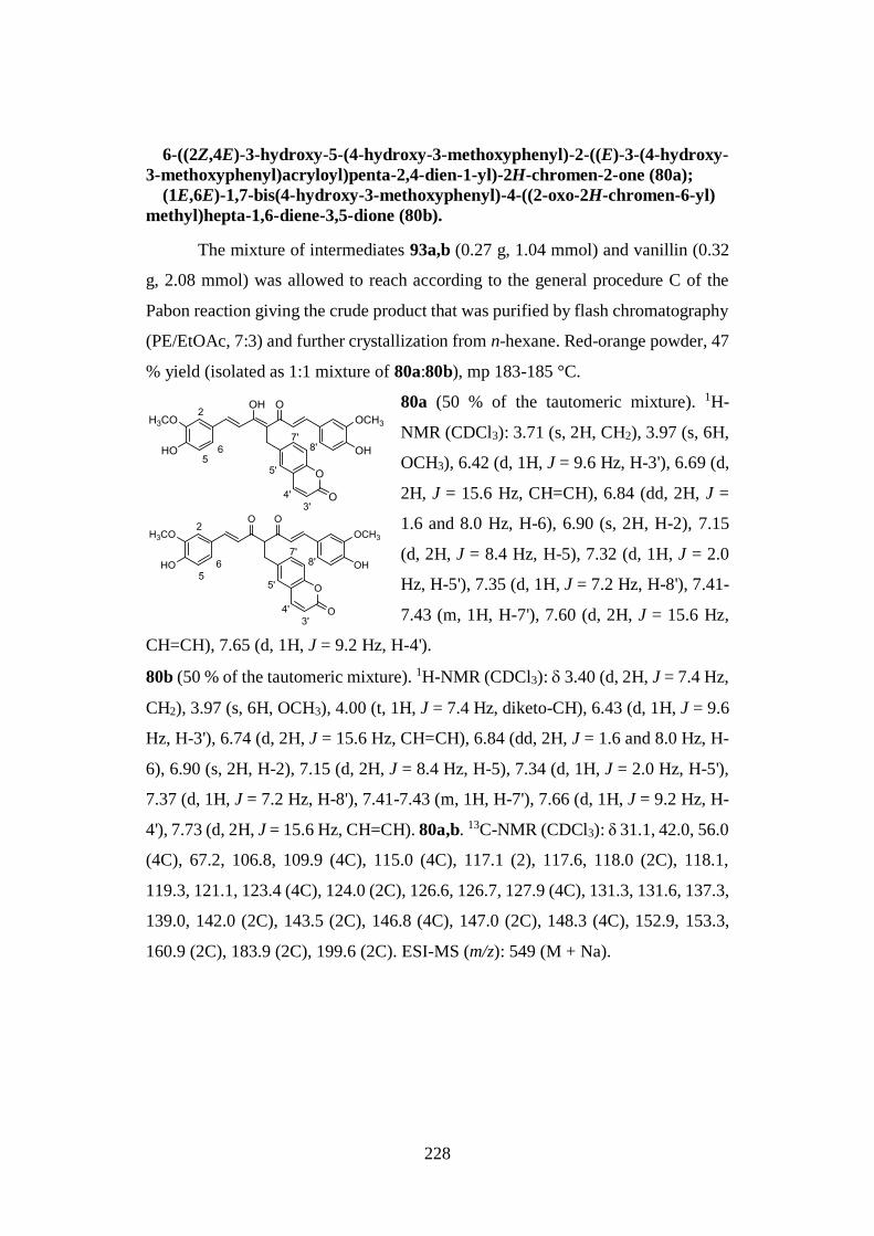
228
6-((2Z,4E)-3-hydroxy-5-(4-hydroxy-3-methoxyphenyl)-2-((E)-3-(4-hydroxy-
3-methoxyphenyl)acryloyl)penta-2,4-dien-1-yl)-2H-chromen-2-one (80a);
(1E,6E)-1,7-bis(4-hydroxy-3-methoxyphenyl)-4-((2-oxo-2H-chromen-6-yl)
methyl)hepta-1,6-diene-3,5-dione (80b).
The mixture of intermediates 93a,b (0.27 g, 1.04 mmol) and vanillin (0.32
g, 2.08 mmol) was allowed to reach according to the general procedure C of the
Pabon reaction giving the crude product that was purified by flash chromatography
(PE/EtOAc, 7:3) and further crystallization from n-hexane. Red-orange powder, 47
% yield (isolated as 1:1 mixture of 80a:80b), mp 183-185 °C.
80a (50 % of the tautomeric mixture). 1H-
NMR (CDCl3): 3.71 (s, 2H, CH2), 3.97 (s, 6H,
OCH3), 6.42 (d, 1H, J = 9.6 Hz, H-3'), 6.69 (d,
2H, J = 15.6 Hz, CH=CH), 6.84 (dd, 2H, J =
1.6 and 8.0 Hz, H-6), 6.90 (s, 2H, H-2), 7.15
(d, 2H, J = 8.4 Hz, H-5), 7.32 (d, 1H, J = 2.0
Hz, H-5'), 7.35 (d, 1H, J = 7.2 Hz, H-8'), 7.41-
7.43 (m, 1H, H-7'), 7.60 (d, 2H, J = 15.6 Hz,
CH=CH), 7.65 (d, 1H, J = 9.2 Hz, H-4').
80b (50 % of the tautomeric mixture). 1H-NMR (CDCl3): 3.40 (d, 2H, J = 7.4 Hz,
CH2), 3.97 (s, 6H, OCH3), 4.00 (t, 1H, J = 7.4 Hz, diketo-CH), 6.43 (d, 1H, J = 9.6
Hz, H-3'), 6.74 (d, 2H, J = 15.6 Hz, CH=CH), 6.84 (dd, 2H, J = 1.6 and 8.0 Hz, H-
6), 6.90 (s, 2H, H-2), 7.15 (d, 2H, J = 8.4 Hz, H-5), 7.34 (d, 1H, J = 2.0 Hz, H-5'),
7.37 (d, 1H, J = 7.2 Hz, H-8'), 7.41-7.43 (m, 1H, H-7'), 7.66 (d, 1H, J = 9.2 Hz, H-
4'), 7.73 (d, 2H, J = 15.6 Hz, CH=CH). 80a,b. 13C-NMR (CDCl3): δ 31.1, 42.0, 56.0
(4C), 67.2, 106.8, 109.9 (4C), 115.0 (4C), 117.1 (2), 117.6, 118.0 (2C), 118.1,
119.3, 121.1, 123.4 (4C), 124.0 (2C), 126.6, 126.7, 127.9 (4C), 131.3, 131.6, 137.3,
139.0, 142.0 (2C), 143.5 (2C), 146.8 (4C), 147.0 (2C), 148.3 (4C), 152.9, 153.3,
160.9 (2C), 183.9 (2C), 199.6 (2C). ESI-MS (m/z): 549 (M + Na).
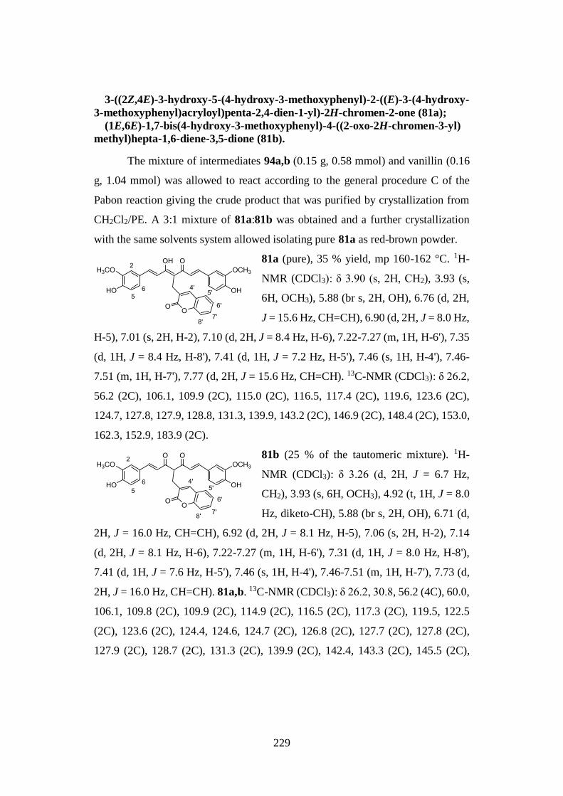
229
3-((2Z,4E)-3-hydroxy-5-(4-hydroxy-3-methoxyphenyl)-2-((E)-3-(4-hydroxy-
3-methoxyphenyl)acryloyl)penta-2,4-dien-1-yl)-2H-chromen-2-one (81a);
(1E,6E)-1,7-bis(4-hydroxy-3-methoxyphenyl)-4-((2-oxo-2H-chromen-3-yl)
methyl)hepta-1,6-diene-3,5-dione (81b).
The mixture of intermediates 94a,b (0.15 g, 0.58 mmol) and vanillin (0.16
g, 1.04 mmol) was allowed to react according to the general procedure C of the
Pabon reaction giving the crude product that was purified by crystallization from
CH2Cl2/PE. A 3:1 mixture of 81a:81b was obtained and a further crystallization
with the same solvents system allowed isolating pure 81a as red-brown powder.
81a (pure), 35 % yield, mp 160-162 °C. 1H-
NMR (CDCl3): δ 3.90 (s, 2H, CH2), 3.93 (s,
6H, OCH3), 5.88 (br s, 2H, OH), 6.76 (d, 2H,
J = 15.6 Hz, CH=CH), 6.90 (d, 2H, J = 8.0 Hz,
H-5), 7.01 (s, 2H, H-2), 7.10 (d, 2H, J = 8.4 Hz, H-6), 7.22-7.27 (m, 1H, H-6'), 7.35
(d, 1H, J = 8.4 Hz, H-8'), 7.41 (d, 1H, J = 7.2 Hz, H-5'), 7.46 (s, 1H, H-4'), 7.46-
7.51 (m, 1H, H-7'), 7.77 (d, 2H, J = 15.6 Hz, CH=CH). 13C-NMR (CDCl3): δ 26.2,
56.2 (2C), 106.1, 109.9 (2C), 115.0 (2C), 116.5, 117.4 (2C), 119.6, 123.6 (2C),
124.7, 127.8, 127.9, 128.8, 131.3, 139.9, 143.2 (2C), 146.9 (2C), 148.4 (2C), 153.0,
162.3, 152.9, 183.9 (2C).
81b (25 % of the tautomeric mixture). 1H-
NMR (CDCl3): δ 3.26 (d, 2H, J = 6.7 Hz,
CH2), 3.93 (s, 6H, OCH3), 4.92 (t, 1H, J = 8.0
Hz, diketo-CH), 5.88 (br s, 2H, OH), 6.71 (d,
2H, J = 16.0 Hz, CH=CH), 6.92 (d, 2H, J = 8.1 Hz, H-5), 7.06 (s, 2H, H-2), 7.14
(d, 2H, J = 8.1 Hz, H-6), 7.22-7.27 (m, 1H, H-6'), 7.31 (d, 1H, J = 8.0 Hz, H-8'),
7.41 (d, 1H, J = 7.6 Hz, H-5'), 7.46 (s, 1H, H-4'), 7.46-7.51 (m, 1H, H-7'), 7.73 (d,
2H, J = 16.0 Hz, CH=CH). 81a,b. 13C-NMR (CDCl3): δ 26.2, 30.8, 56.2 (4C), 60.0,
106.1, 109.8 (2C), 109.9 (2C), 114.9 (2C), 116.5 (2C), 117.3 (2C), 119.5, 122.5
(2C), 123.6 (2C), 124.4, 124.6, 124.7 (2C), 126.8 (2C), 127.7 (2C), 127.8 (2C),
127.9 (2C), 128.7 (2C), 131.3 (2C), 139.9 (2C), 142.4, 143.3 (2C), 145.5 (2C),
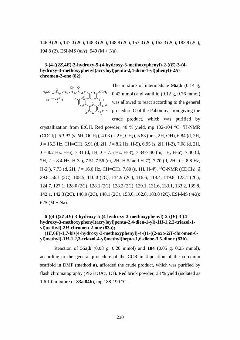
230
146.9 (2C), 147.0 (2C), 148.3 (2C), 148.8 (2C), 153.0 (2C), 162.3 (2C), 183.9 (2C),
194.8 (2). ESI-MS (m/z): 549 (M + Na).
3-(4-((2Z,4E)-3-hydroxy-5-(4-hydroxy-3-methoxyphenyl)-2-((E)-3-(4-
hydroxy-3-methoxyphenyl)acryloyl)penta-2,4-dien-1-yl)phenyl)-2H-
chromen-2-one (82).
The mixture of intermediate 96a,b (0.14 g,
0.42 mmol) and vanillin (0.12 g, 0.76 mmol)
was allowed to react according to the general
procedure C of the Pabon reaction giving the
crude product, which was purified by
crystallization from EtOH. Red powder, 40 % yield, mp 102-104 °C. 1H-NMR
(CDCl3): δ 3.92 (s, 6H, OCH3), 4.03 (s, 2H, CH2), 5.83 (br s, 2H, OH), 6.84 (d, 2H,
J = 15.3 Hz, CH=CH), 6.91 (d, 2H, J = 8.2 Hz, H-5), 6.95 (s, 2H, H-2), 7.08 (d, 2H,
J = 8.2 Hz, H-6), 7.31 (d, 1H, J = 7.5 Hz, H-8'), 7.34-7.40 (m, 1H, H-6'), 7.40 (d,
2H, J = 8.4 Hz, H-3''), 7.51-7.56 (m, 2H, H-5' and H-7'), 7.70 (d, 2H, J = 8.8 Hz,
H-2''), 7.73 (d, 2H, J = 16.0 Hz, CH=CH), 7.80 (s, 1H, H-4'). 13C-NMR (CDCl3): δ
29.8, 56.1 (2C), 108.5, 110.0 (2C), 114.9 (2C), 116.6, 118.4, 119.8, 123.1 (2C),
124.7, 127.1, 128.0 (2C), 128.1 (2C), 128.2 (2C), 129.1, 131.6, 133.1, 133.2, 139.8,
142.1, 142.3 (2C), 146.9 (2C), 148.1 (2C), 153.6, 162.8, 183.8 (2C). ESI-MS (m/z):
625 (M + Na).
6-((4-((2Z,4E)-3-hydroxy-5-(4-hydroxy-3-methoxyphenyl)-2-((E)-3-(4-
hydroxy-3-methoxyphenyl)acryloyl)penta-2,4-dien-1-yl)-1H-1,2,3-triazol-1-
yl)methyl)-2H-chromen-2-one (83a);
(1E,6E)-1,7-bis(4-hydroxy-3-methoxyphenyl)-4-((1-((2-oxo-2H-chromen-6-
yl)methyl)-1H-1,2,3-triazol-4-yl)methyl)hepta-1,6-diene-3,5-dione (83b).
Reaction of 55a,b (0.08 g, 0.20 mmol) and 104 (0.05 g, 0.25 mmol),
according to the general procedure of the CCR in 4-position of the curcumin
scaffold in DMF (method a), afforded the crude product, which was purified by
flash chromatography (PE/EtOAc, 1:1). Red brick powder, 33 % yield (isolated as
1.6:1.0 mixture of 83a:84b), mp 188-190 °C.
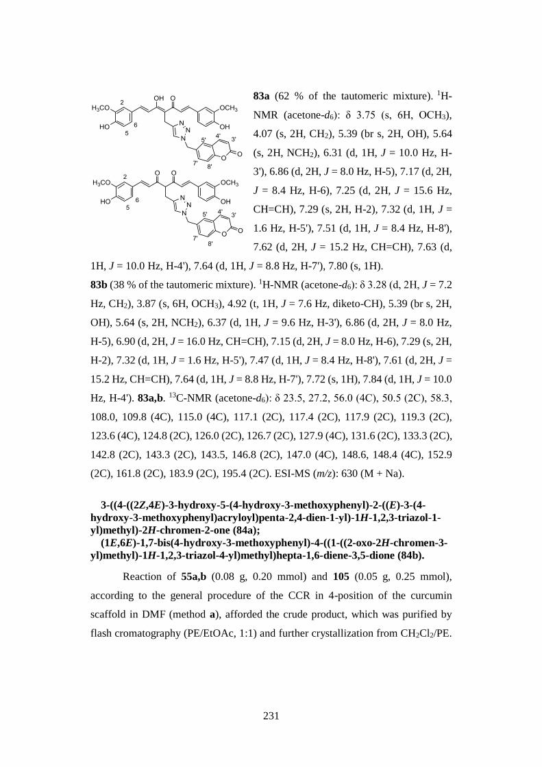
231
83a (62 % of the tautomeric mixture). 1H-
NMR (acetone-d6): δ 3.75 (s, 6H, OCH3),
4.07 (s, 2H, CH2), 5.39 (br s, 2H, OH), 5.64
(s, 2H, NCH2), 6.31 (d, 1H, J = 10.0 Hz, H-
3'), 6.86 (d, 2H, J = 8.0 Hz, H-5), 7.17 (d, 2H,
J = 8.4 Hz, H-6), 7.25 (d, 2H, J = 15.6 Hz,
CH=CH), 7.29 (s, 2H, H-2), 7.32 (d, 1H, J =
1.6 Hz, H-5'), 7.51 (d, 1H, J = 8.4 Hz, H-8'),
7.62 (d, 2H, J = 15.2 Hz, CH=CH), 7.63 (d,
1H, J = 10.0 Hz, H-4'), 7.64 (d, 1H, J = 8.8 Hz, H-7'), 7.80 (s, 1H).
83b (38 % of the tautomeric mixture). 1H-NMR (acetone-d6): δ 3.28 (d, 2H, J = 7.2
Hz, CH2), 3.87 (s, 6H, OCH3), 4.92 (t, 1H, J = 7.6 Hz, diketo-CH), 5.39 (br s, 2H,
OH), 5.64 (s, 2H, NCH2), 6.37 (d, 1H, J = 9.6 Hz, H-3'), 6.86 (d, 2H, J = 8.0 Hz,
H-5), 6.90 (d, 2H, J = 16.0 Hz, CH=CH), 7.15 (d, 2H, J = 8.0 Hz, H-6), 7.29 (s, 2H,
H-2), 7.32 (d, 1H, J = 1.6 Hz, H-5'), 7.47 (d, 1H, J = 8.4 Hz, H-8'), 7.61 (d, 2H, J =
15.2 Hz, CH=CH), 7.64 (d, 1H, J = 8.8 Hz, H-7'), 7.72 (s, 1H), 7.84 (d, 1H, J = 10.0
Hz, H-4'). 83a,b. 13C-NMR (acetone-d6): δ 23.5, 27.2, 56.0 (4C), 50.5 (2C), 58.3,
108.0, 109.8 (4C), 115.0 (4C), 117.1 (2C), 117.4 (2C), 117.9 (2C), 119.3 (2C),
123.6 (4C), 124.8 (2C), 126.0 (2C), 126.7 (2C), 127.9 (4C), 131.6 (2C), 133.3 (2C),
142.8 (2C), 143.3 (2C), 143.5, 146.8 (2C), 147.0 (4C), 148.6, 148.4 (4C), 152.9
(2C), 161.8 (2C), 183.9 (2C), 195.4 (2C). ESI-MS (m/z): 630 (M + Na).
3-((4-((2Z,4E)-3-hydroxy-5-(4-hydroxy-3-methoxyphenyl)-2-((E)-3-(4-
hydroxy-3-methoxyphenyl)acryloyl)penta-2,4-dien-1-yl)-1H-1,2,3-triazol-1-
yl)methyl)-2H-chromen-2-one (84a);
(1E,6E)-1,7-bis(4-hydroxy-3-methoxyphenyl)-4-((1-((2-oxo-2H-chromen-3-
yl)methyl)-1H-1,2,3-triazol-4-yl)methyl)hepta-1,6-diene-3,5-dione (84b).
Reaction of 55a,b (0.08 g, 0.20 mmol) and 105 (0.05 g, 0.25 mmol),
according to the general procedure of the CCR in 4-position of the curcumin
scaffold in DMF (method a), afforded the crude product, which was purified by
flash cromatography (PE/EtOAc, 1:1) and further crystallization from CH2Cl2/PE.
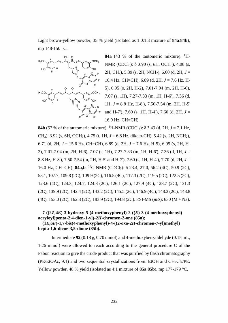
232
Light brown-yellow powder, 35 % yield (isolated as 1.0:1.3 mixture of 84a:84b),
mp 148-150 °C.
84a (43 % of the tautomeric mixture). 1H-
NMR (CDCl3): δ 3.90 (s, 6H, OCH3), 4.08 (s,
2H, CH2), 5.39 (s, 2H, NCH2), 6.60 (d, 2H, J =
16.4 Hz, CH=CH), 6.89 (d, 2H, J = 7.6 Hz, H-
5), 6.95 (s, 2H, H-2), 7.01-7.04 (m, 2H, H-6),
7.07 (s, 1H), 7.27-7.33 (m, 1H, H-6'), 7.36 (d,
1H, J = 8.8 Hz, H-8'), 7.50-7.54 (m, 2H, H-5'
and H-7'), 7.60 (s, 1H, H-4'), 7.60 (d, 2H, J =
16.0 Hz, CH=CH).
84b (57 % of the tautomeric mixture). 1H-NMR (CDCl3): δ 3.43 (d, 2H, J = 7.1 Hz,
CH2), 3.92 (s, 6H, OCH3), 4.75 (t, 1H, J = 6.8 Hz, diketo-CH), 5.42 (s, 2H, NCH2),
6.71 (d, 2H, J = 15.6 Hz, CH=CH), 6.89 (d, 2H, J = 7.6 Hz, H-5), 6.95 (s, 2H, H-
2), 7.01-7.04 (m, 2H, H-6), 7.07 (s, 1H), 7.27-7.33 (m, 1H, H-6'), 7.36 (d, 1H, J =
8.8 Hz, H-8'), 7.50-7.54 (m, 2H, H-5' and H-7'), 7.60 (s, 1H, H-4'), 7.70 (d, 2H, J =
16.0 Hz, CH=CH). 84a,b. 13C-NMR (CDCl3): δ 23.4, 27.0, 56.2 (4C), 50.9 (2C),
58.1, 107.7, 109.8 (2C), 109.9 (2C), 116.5 (4C), 117.3 (2C), 119.5 (2C), 122.5 (2C),
123.6 (4C), 124.3, 124.7, 124.8 (2C), 126.1 (2C), 127.9 (4C), 128.7 (2C), 131.3
(2C), 139.9 (2C), 142.4 (2C), 143.2 (2C), 145.5 (2C), 146.9 (4C), 148.3 (2C), 148.8
(4C), 153.0 (2C), 162.3 (2C), 183.9 (2C), 194.8 (2C). ESI-MS (m/z): 630 (M + Na).
7-((2Z,4E)-3-hydroxy-5-(4-methoxyphenyl)-2-((E)-3-(4-methoxyphenyl)
acryloyl)penta-2,4-dien-1-yl)-2H-chromen-2-one (85a);
(1E,6E)-1,7-bis(4-methoxyphenyl)-4-((2-oxo-2H-chromen-7-yl)methyl)
hepta-1,6-diene-3,5-dione (85b).
Intermediate 92 (0.18 g, 0.70 mmol) and 4-methoxybenzaldehyde (0.15 mL,
1.26 mmol) were allowed to reach according to the general procedure C of the
Pabon reaction to give the crude product that was purified by flash chromatography
(PE/EtOAc, 9:1) and two sequential crystallizations from: EtOH and CH2Cl2/PE.
Yellow powder, 48 % yield (isolated as 4:1 mixture of 85a:85b), mp 177-179 °C.
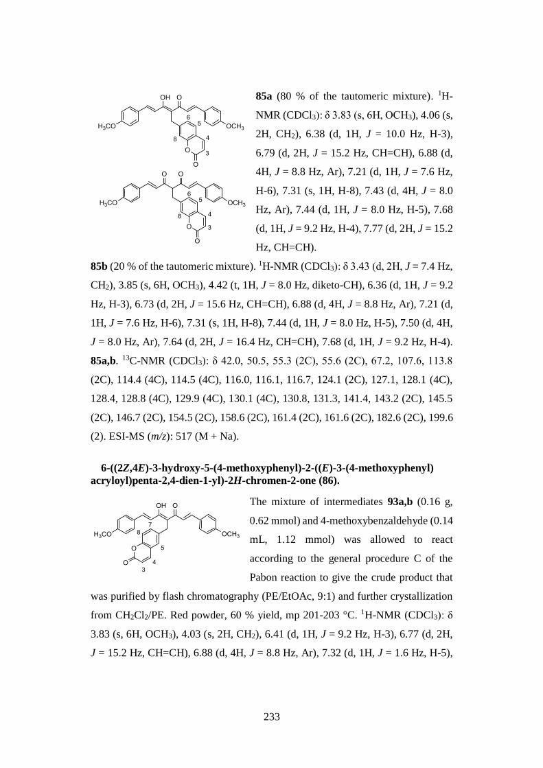
233
85a (80 % of the tautomeric mixture). 1H-
NMR (CDCl3): δ 3.83 (s, 6H, OCH3), 4.06 (s,
2H, CH2), 6.38 (d, 1H, J = 10.0 Hz, H-3),
6.79 (d, 2H, J = 15.2 Hz, CH=CH), 6.88 (d,
4H, J = 8.8 Hz, Ar), 7.21 (d, 1H, J = 7.6 Hz,
H-6), 7.31 (s, 1H, H-8), 7.43 (d, 4H, J = 8.0
Hz, Ar), 7.44 (d, 1H, J = 8.0 Hz, H-5), 7.68
(d, 1H, J = 9.2 Hz, H-4), 7.77 (d, 2H, J = 15.2
Hz, CH=CH).
85b (20 % of the tautomeric mixture). 1H-NMR (CDCl3): δ 3.43 (d, 2H, J = 7.4 Hz,
CH2), 3.85 (s, 6H, OCH3), 4.42 (t, 1H, J = 8.0 Hz, diketo-CH), 6.36 (d, 1H, J = 9.2
Hz, H-3), 6.73 (d, 2H, J = 15.6 Hz, CH=CH), 6.88 (d, 4H, J = 8.8 Hz, Ar), 7.21 (d,
1H, J = 7.6 Hz, H-6), 7.31 (s, 1H, H-8), 7.44 (d, 1H, J = 8.0 Hz, H-5), 7.50 (d, 4H,
J = 8.0 Hz, Ar), 7.64 (d, 2H, J = 16.4 Hz, CH=CH), 7.68 (d, 1H, J = 9.2 Hz, H-4).
85a,b. 13C-NMR (CDCl3): δ 42.0, 50.5, 55.3 (2C), 55.6 (2C), 67.2, 107.6, 113.8
(2C), 114.4 (4C), 114.5 (4C), 116.0, 116.1, 116.7, 124.1 (2C), 127.1, 128.1 (4C),
128.4, 128.8 (4C), 129.9 (4C), 130.1 (4C), 130.8, 131.3, 141.4, 143.2 (2C), 145.5
(2C), 146.7 (2C), 154.5 (2C), 158.6 (2C), 161.4 (2C), 161.6 (2C), 182.6 (2C), 199.6
(2). ESI-MS (m/z): 517 (M + Na).
6-((2Z,4E)-3-hydroxy-5-(4-methoxyphenyl)-2-((E)-3-(4-methoxyphenyl)
acryloyl)penta-2,4-dien-1-yl)-2H-chromen-2-one (86).
The mixture of intermediates 93a,b (0.16 g,
0.62 mmol) and 4-methoxybenzaldehyde (0.14
mL, 1.12 mmol) was allowed to react
according to the general procedure C of the
Pabon reaction to give the crude product that
was purified by flash chromatography (PE/EtOAc, 9:1) and further crystallization
from CH2Cl2/PE. Red powder, 60 % yield, mp 201-203 °C. 1H-NMR (CDCl3): δ
3.83 (s, 6H, OCH3), 4.03 (s, 2H, CH2), 6.41 (d, 1H, J = 9.2 Hz, H-3), 6.77 (d, 2H,
J = 15.2 Hz, CH=CH), 6.88 (d, 4H, J = 8.8 Hz, Ar), 7.32 (d, 1H, J = 1.6 Hz, H-5),
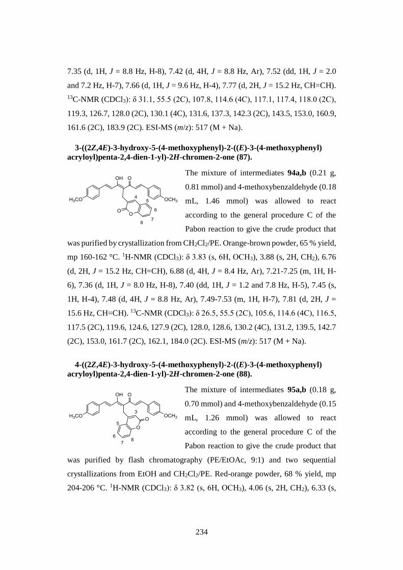
234
7.35 (d, 1H, J = 8.8 Hz, H-8), 7.42 (d, 4H, J = 8.8 Hz, Ar), 7.52 (dd, 1H, J = 2.0
and 7.2 Hz, H-7), 7.66 (d, 1H, J = 9.6 Hz, H-4), 7.77 (d, 2H, J = 15.2 Hz, CH=CH).
13C-NMR (CDCl3): δ 31.1, 55.5 (2C), 107.8, 114.6 (4C), 117.1, 117.4, 118.0 (2C),
119.3, 126.7, 128.0 (2C), 130.1 (4C), 131.6, 137.3, 142.3 (2C), 143.5, 153.0, 160.9,
161.6 (2C), 183.9 (2C). ESI-MS (m/z): 517 (M + Na).
3-((2Z,4E)-3-hydroxy-5-(4-methoxyphenyl)-2-((E)-3-(4-methoxyphenyl)
acryloyl)penta-2,4-dien-1-yl)-2H-chromen-2-one (87).
The mixture of intermediates 94a,b (0.21 g,
0.81 mmol) and 4-methoxybenzaldehyde (0.18
mL, 1.46 mmol) was allowed to react
according to the general procedure C of the
Pabon reaction to give the crude product that
was purified by crystallization from CH2Cl2/PE. Orange-brown powder, 65 % yield,
mp 160-162 °C. 1H-NMR (CDCl3): δ 3.83 (s, 6H, OCH3), 3.88 (s, 2H, CH2), 6.76
(d, 2H, J = 15.2 Hz, CH=CH), 6.88 (d, 4H, J = 8.4 Hz, Ar), 7.21-7.25 (m, 1H, H-
6), 7.36 (d, 1H, J = 8.0 Hz, H-8), 7.40 (dd, 1H, J = 1.2 and 7.8 Hz, H-5), 7.45 (s,
1H, H-4), 7.48 (d, 4H, J = 8.8 Hz, Ar), 7.49-7.53 (m, 1H, H-7), 7.81 (d, 2H, J =
15.6 Hz, CH=CH). 13C-NMR (CDCl3): δ 26.5, 55.5 (2C), 105.6, 114.6 (4C), 116.5,
117.5 (2C), 119.6, 124.6, 127.9 (2C), 128.0, 128.6, 130.2 (4C), 131.2, 139.5, 142.7
(2C), 153.0, 161.7 (2C), 162.1, 184.0 (2C). ESI-MS (m/z): 517 (M + Na).
4-((2Z,4E)-3-hydroxy-5-(4-methoxyphenyl)-2-((E)-3-(4-methoxyphenyl)
acryloyl)penta-2,4-dien-1-yl)-2H-chromen-2-one (88).
The mixture of intermediates 95a,b (0.18 g,
0.70 mmol) and 4-methoxybenzaldehyde (0.15
mL, 1.26 mmol) was allowed to react
according to the general procedure C of the
Pabon reaction to give the crude product that
was purified by flash chromatography (PE/EtOAc, 9:1) and two sequential
crystallizations from EtOH and CH2Cl2/PE. Red-orange powder, 68 % yield, mp
204-206 °C. 1H-NMR (CDCl3): δ 3.82 (s, 6H, OCH3), 4.06 (s, 2H, CH2), 6.33 (s,
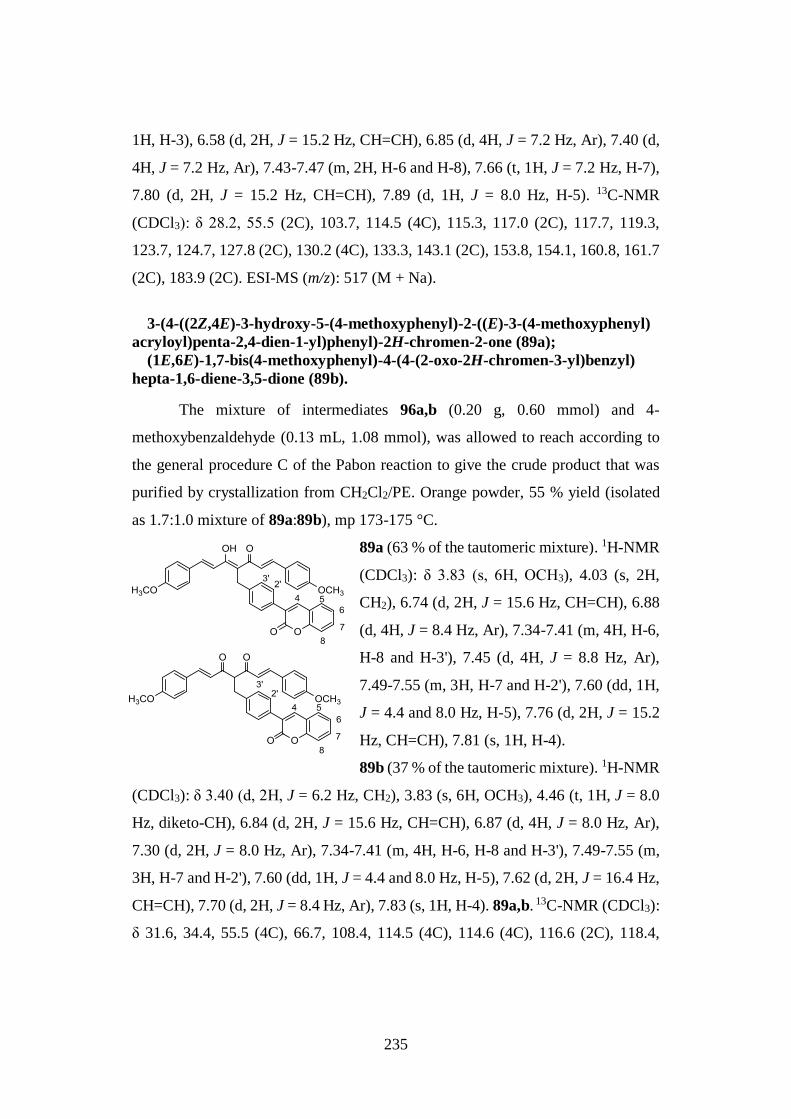
235
1H, H-3), 6.58 (d, 2H, J = 15.2 Hz, CH=CH), 6.85 (d, 4H, J = 7.2 Hz, Ar), 7.40 (d,
4H, J = 7.2 Hz, Ar), 7.43-7.47 (m, 2H, H-6 and H-8), 7.66 (t, 1H, J = 7.2 Hz, H-7),
7.80 (d, 2H, J = 15.2 Hz, CH=CH), 7.89 (d, 1H, J = 8.0 Hz, H-5). 13C-NMR
(CDCl3): δ 28.2, 55.5 (2C), 103.7, 114.5 (4C), 115.3, 117.0 (2C), 117.7, 119.3,
123.7, 124.7, 127.8 (2C), 130.2 (4C), 133.3, 143.1 (2C), 153.8, 154.1, 160.8, 161.7
(2C), 183.9 (2C). ESI-MS (m/z): 517 (M + Na).
3-(4-((2Z,4E)-3-hydroxy-5-(4-methoxyphenyl)-2-((E)-3-(4-methoxyphenyl)
acryloyl)penta-2,4-dien-1-yl)phenyl)-2H-chromen-2-one (89a);
(1E,6E)-1,7-bis(4-methoxyphenyl)-4-(4-(2-oxo-2H-chromen-3-yl)benzyl)
hepta-1,6-diene-3,5-dione (89b).
The mixture of intermediates 96a,b (0.20 g, 0.60 mmol) and 4-
methoxybenzaldehyde (0.13 mL, 1.08 mmol), was allowed to reach according to
the general procedure C of the Pabon reaction to give the crude product that was
purified by crystallization from CH2Cl2/PE. Orange powder, 55 % yield (isolated
as 1.7:1.0 mixture of 89a:89b), mp 173-175 °C.
89a (63 % of the tautomeric mixture). 1H-NMR
(CDCl3): δ 3.83 (s, 6H, OCH3), 4.03 (s, 2H,
CH2), 6.74 (d, 2H, J = 15.6 Hz, CH=CH), 6.88
(d, 4H, J = 8.4 Hz, Ar), 7.34-7.41 (m, 4H, H-6,
H-8 and H-3'), 7.45 (d, 4H, J = 8.8 Hz, Ar),
7.49-7.55 (m, 3H, H-7 and H-2'), 7.60 (dd, 1H,
J = 4.4 and 8.0 Hz, H-5), 7.76 (d, 2H, J = 15.2
Hz, CH=CH), 7.81 (s, 1H, H-4).
89b (37 % of the tautomeric mixture). 1H-NMR
(CDCl3): δ 3.40 (d, 2H, J = 6.2 Hz, CH2), 3.83 (s, 6H, OCH3), 4.46 (t, 1H, J = 8.0
Hz, diketo-CH), 6.84 (d, 2H, J = 15.6 Hz, CH=CH), 6.87 (d, 4H, J = 8.0 Hz, Ar),
7.30 (d, 2H, J = 8.0 Hz, Ar), 7.34-7.41 (m, 4H, H-6, H-8 and H-3'), 7.49-7.55 (m,
3H, H-7 and H-2'), 7.60 (dd, 1H, J = 4.4 and 8.0 Hz, H-5), 7.62 (d, 2H, J = 16.4 Hz,
CH=CH), 7.70 (d, 2H, J = 8.4 Hz, Ar), 7.83 (s, 1H, H-4). 89a,b. 13C-NMR (CDCl3):
δ 31.6, 34.4, 55.5 (4C), 66.7, 108.4, 114.5 (4C), 114.6 (4C), 116.6 (2C), 118.4,
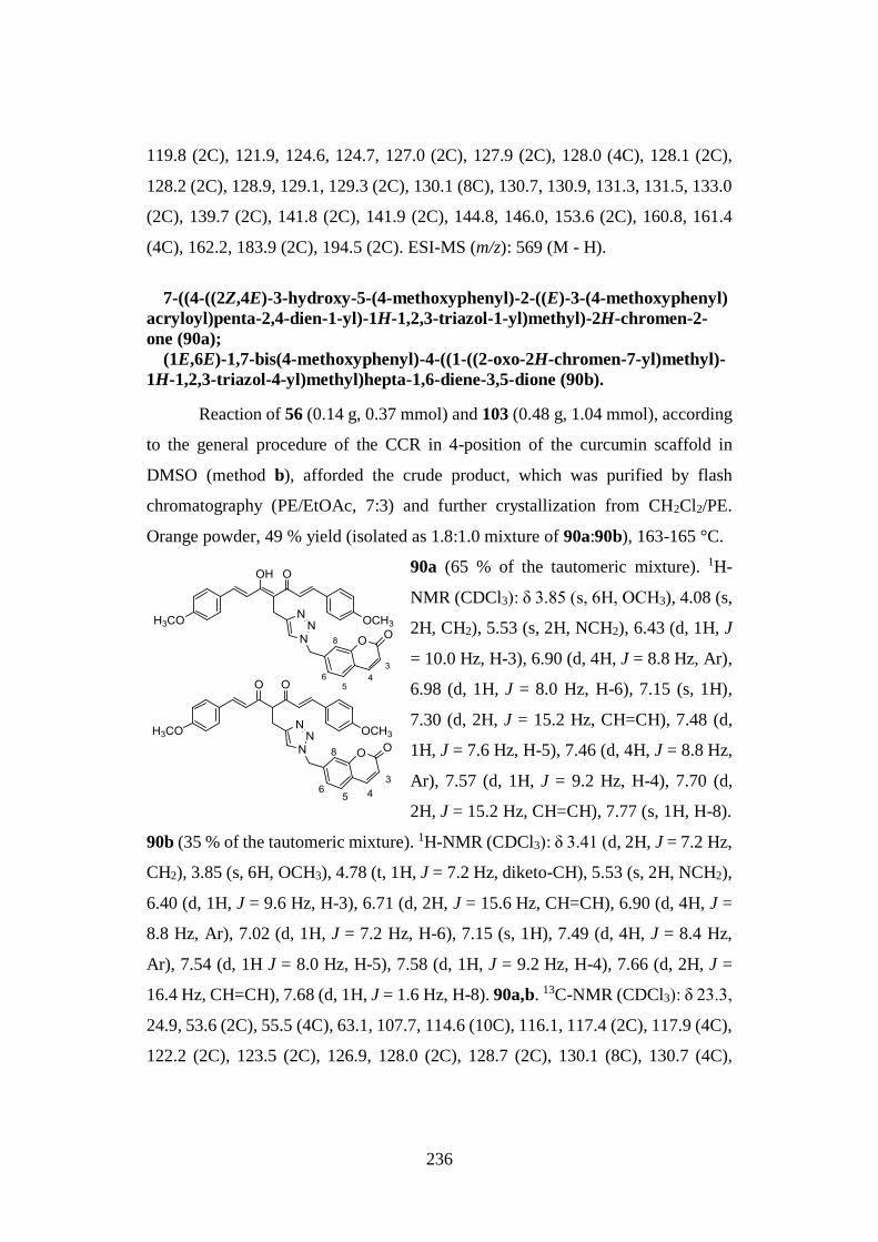
236
119.8 (2C), 121.9, 124.6, 124.7, 127.0 (2C), 127.9 (2C), 128.0 (4C), 128.1 (2C),
128.2 (2C), 128.9, 129.1, 129.3 (2C), 130.1 (8C), 130.7, 130.9, 131.3, 131.5, 133.0
(2C), 139.7 (2C), 141.8 (2C), 141.9 (2C), 144.8, 146.0, 153.6 (2C), 160.8, 161.4
(4C), 162.2, 183.9 (2C), 194.5 (2C). ESI-MS (m/z): 569 (M - H).
7-((4-((2Z,4E)-3-hydroxy-5-(4-methoxyphenyl)-2-((E)-3-(4-methoxyphenyl)
acryloyl)penta-2,4-dien-1-yl)-1H-1,2,3-triazol-1-yl)methyl)-2H-chromen-2-
one (90a);
(1E,6E)-1,7-bis(4-methoxyphenyl)-4-((1-((2-oxo-2H-chromen-7-yl)methyl)-
1H-1,2,3-triazol-4-yl)methyl)hepta-1,6-diene-3,5-dione (90b).
Reaction of 56 (0.14 g, 0.37 mmol) and 103 (0.48 g, 1.04 mmol), according
to the general procedure of the CCR in 4-position of the curcumin scaffold in
DMSO (method b), afforded the crude product, which was purified by flash
chromatography (PE/EtOAc, 7:3) and further crystallization from CH2Cl2/PE.
Orange powder, 49 % yield (isolated as 1.8:1.0 mixture of 90a:90b), 163-165 °C.
90a (65 % of the tautomeric mixture). 1H-
NMR (CDCl3): δ 3.85 (s, 6H, OCH3), 4.08 (s,
2H, CH2), 5.53 (s, 2H, NCH2), 6.43 (d, 1H, J
= 10.0 Hz, H-3), 6.90 (d, 4H, J = 8.8 Hz, Ar),
6.98 (d, 1H, J = 8.0 Hz, H-6), 7.15 (s, 1H),
7.30 (d, 2H, J = 15.2 Hz, CH=CH), 7.48 (d,
1H, J = 7.6 Hz, H-5), 7.46 (d, 4H, J = 8.8 Hz,
Ar), 7.57 (d, 1H, J = 9.2 Hz, H-4), 7.70 (d,
2H, J = 15.2 Hz, CH=CH), 7.77 (s, 1H, H-8).
90b (35 % of the tautomeric mixture). 1H-NMR (CDCl3): δ 3.41 (d, 2H, J = 7.2 Hz,
CH2), 3.85 (s, 6H, OCH3), 4.78 (t, 1H, J = 7.2 Hz, diketo-CH), 5.53 (s, 2H, NCH2),
6.40 (d, 1H, J = 9.6 Hz, H-3), 6.71 (d, 2H, J = 15.6 Hz, CH=CH), 6.90 (d, 4H, J =
8.8 Hz, Ar), 7.02 (d, 1H, J = 7.2 Hz, H-6), 7.15 (s, 1H), 7.49 (d, 4H, J = 8.4 Hz,
Ar), 7.54 (d, 1H J = 8.0 Hz, H-5), 7.58 (d, 1H, J = 9.2 Hz, H-4), 7.66 (d, 2H, J =
16.4 Hz, CH=CH), 7.68 (d, 1H, J = 1.6 Hz, H-8). 90a,b. 13C-NMR (CDCl3): δ 23.3,
24.9, 53.6 (2C), 55.5 (4C), 63.1, 107.7, 114.6 (10C), 116.1, 117.4 (2C), 117.9 (4C),
122.2 (2C), 123.5 (2C), 126.9, 128.0 (2C), 128.7 (2C), 130.1 (8C), 130.7 (4C),

237
139.0 (2C), 142.2 (2C), 142.7 (2C), 144.9 (2C), 149.5 (2C), 160.2 (2C), 161.6 (2C),
162.2 (2C), 183.3 (2C), 195.6 (2C). ESI-MS (m/z): 598 (M + Na).
(3Z,5E)-4-hydroxy-6-(4-methoxyphenyl)-3-((E)-3-(4-methoxyphenyl)
acryloyl)-N-(2-oxo-2H-chromen-6-yl)hexa-3,5-dienamide (91a);
(E)-6-(4-methoxyphenyl)-3-((E)-3-(4-methoxyphenyl)acryloyl)-4-oxo-N-(2-
oxo-2H-chromen-6-yl)hex-5-enamide (91b).
To a solution of 61a,b (0.29 g, 0.34 mmol) in CH2Cl2 (15.0 mL) cooled to
0 °C, under nitrogen atmosphere, addition of EDC (0.07 g, 0.34 mmol) and
DMAP (0.008 g, 0.068 mmol) was carried out. After 15 min, a solution
of the amine 106 (0.07 g, 0.41 mmol) in CH2Cl2 (2.0 mL) was added
dropwise and the resulting mixture was stirred at 0 °C for 2 h and
overnight at room temperature. Upon reaction completion, the mixture
was diluted with additional CH2Cl2 and sequentially washed with water
and brine. The organic phase was dried over Na 2SO4, filtered and
evaporated under vacuum affording the crude product that was purified
by flash chromatography (PE/EtOAc, 8:2), semipreparative TLC
(CHCl3) and further crystallization from CH2Cl2/PE. Pale yellow
powder, 41 % yield (isolated as 1.0:3.3 mixture of 91a:91b), mp 228-
230 °C.
91a (23 % of the tautomeric mixture). 1H-
NMR (CDCl3): δ 3.87 (s, 6H, OCH3), 3.95 (s,
2H, CH2), 6.42 (d, 2H, J = 15.6 Hz, CH=CH),
6.46 (d, 1H, J = 9.2 Hz, H-3), 6.94 (d, 4H, J =
8.8 Hz, Ar), 7.32 (d, 1H, J = 8.8 Hz, H-8), 7.36
(br s, 1H, CONH), 7.47 (dd, 1H, J = 2.4 and
8.8 Hz, H-7), 7.52 (d, 4H, J = 8.8 Hz, Ar), 7.72
(d, 1H, J = 9.2 Hz, H-4), 7.75 (d, 2H, J = 14.4
Hz, CH=CH), 8.15 (s, 1H, H-5).
91b (77 % of the tautomeric mixture). 1H-NMR (CDCl3): δ 3.46-3.53 (m, 2H, CH2),
3.87 (s, 6H, OCH3), 4.01 (t, 1H, J = 6.4 Hz, diketo-CH), 6.42 (d, 2H, J = 15.6 Hz,
238
CH=CH), 6.46 (d, 1H, J = 9.2 Hz, H-3), 6.94 (d, 4H, J = 8.8 Hz, Ar), 7.32 (d, 1H,
J = 8.8 Hz, H-8), 7.36 (br s, 1H, CONH), 7.47 (dd, 1H, J = 2.4 and 8.8 Hz, H-7),
7.52 (d, 4H, J = 8.8 Hz, Ar), 7.72 (d, 1H, J = 9.2 Hz, H-4), 7.75 (d, 2H, J = 14.4
Hz, CH=CH), 8.15 (s, 1H, H-5). 91a,b. 13C-NMR (CDCl3): δ 28.0, 34.9, 55.3 (4C),
55.5, 114.3 (4C), 114.4 (4C), 114.5, 117.0 (2C), 118.0 (2C), 119.3 (2C), 123.4 (2C),
126.7 (2C), 126.9 (2C), 128.7 (2C), 128.9 (2C), 129.9 (4C), 130.0 (4C), 133.6 (2C),
137.0 (2C), 143.0 (2C), 143.8 (2C), 146.8 (2C), 154.0 (2C), 160.9 (2C), 161.6 (2C),
161.7 (2C), 173.2, 174.3, 178.5 (2C), 197.9 (2C). ESI-MS (m/z): 560 (M + Na).
Alkylation reaction of pentane-2,4-dione: general procedure B (synthesis of
intermediates 92-96a,b).
1) A solution of pentane-2,4-dione (1.00 mmol) in THF (1.0 mL) was added to a
stirred suspension of NaH (60 % dispersion in mineral oil, 1.2 molar equiv) in
THF (5.0 mL) at 0 °C and under nitrogen atmosphere. The suspension was
stirred at room temperature for 30 min before the addition dropwise of the
appropriate bromomethyl-coumarin/bromotolyl-coumarin solution (1.2 molar
equiv) in THF (5.0 mL) at 0 °C. The resulting mixture was stirred overnight at
room temperature and the reaction was quenched with water. The aqueous phase
was extracted with Et2O (3 x 50.0 mL) and the combined organic layers were
washed with brine, dried over Na2SO4, filtered and concentrated in vacuo. The
crude residue was purified by flash column chromatography on silica gel.
2) To a solution of pentane-2,4-dione (1.00 mmol) in acetone (10.0 mL),
anhydrous Κ2CO3 (0.5 molar equiv) and the appropriate bromomethyl-coumarin
(0.5 molar equiv) were added. The reaction mixture was heated to 80 °C for 6
h and TLC monitored the reaction progress. Upon reaction completion, the
mixture was hot filtered, the solvent was evaporated and the crude product was
purified by column chromatography over silica gel.
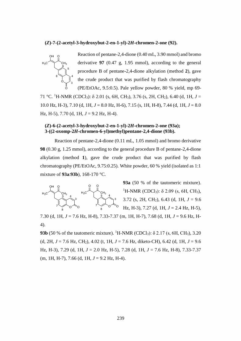
239
(Z)-7-(2-acetyl-3-hydroxybut-2-en-1-yl)-2H-chromen-2-one (92).
Reaction of pentane-2,4-dione (0.40 mL, 3.90 mmol) and bromo
derivative 97 (0.47 g, 1.95 mmol), according to the general
procedure B of pentane-2,4-dione alkylation (method 2), gave
the crude product that was purified by flash chromatography
(PE/EtOAc, 9.5:0.5). Pale yellow powder, 80 % yield, mp 69-
71 °C. 1H-NMR (CDCl3): 2.01 (s, 6H, CH3), 3.76 (s, 2H, CH2), 6.40 (d, 1H, J =
10.0 Hz, H-3), 7.10 (d, 1H, J = 8.0 Hz, H-6), 7.15 (s, 1H, H-8), 7.44 (d, 1H, J = 8.0
Hz, H-5), 7.70 (d, 1H, J = 9.2 Hz, H-4).
(Z)-6-(2-acetyl-3-hydroxybut-2-en-1-yl)-2H-chromen-2-one (93a);
3-((2-oxomp-2H-chromen-6-yl)methyl)pentane-2,4-dione (93b).
Reaction of pentane-2,4-dione (0.11 mL, 1.05 mmol) and bromo derivative
98 (0.30 g, 1.25 mmol), according to the general procedure B of pentane-2,4-dione
alkylation (method 1), gave the crude product that was purified by flash
chromatography (PE/EtOAc, 9.75:0.25). White powder, 60 % yield (isolated as 1:1
mixture of 93a:93b), 168-170 °C.
93a (50 % of the tautomeric mixture).
1H-NMR (CDCl3): δ 2.09 (s, 6H, CH3),
3.72 (s, 2H, CH2), 6.43 (d, 1H, J = 9.6
Hz, H-3), 7.27 (d, 1H, J = 2.4 Hz, H-5),
7.30 (d, 1H, J = 7.6 Hz, H-8), 7.33-7.37 (m, 1H, H-7), 7.68 (d, 1H, J = 9.6 Hz, H-
4).
93b (50 % of the tautomeric mixture). 1H-NMR (CDCl3): δ 2.17 (s, 6H, CH3), 3.20
(d, 2H, J = 7.6 Hz, CH2), 4.02 (t, 1H, J = 7.6 Hz, diketo-CH), 6.42 (d, 1H, J = 9.6
Hz, H-3), 7.29 (d, 1H, J = 2.0 Hz, H-5), 7.28 (d, 1H, J = 7.6 Hz, H-8), 7.33-7.37
(m, 1H, H-7), 7.66 (d, 1H, J = 9.2 Hz, H-4).
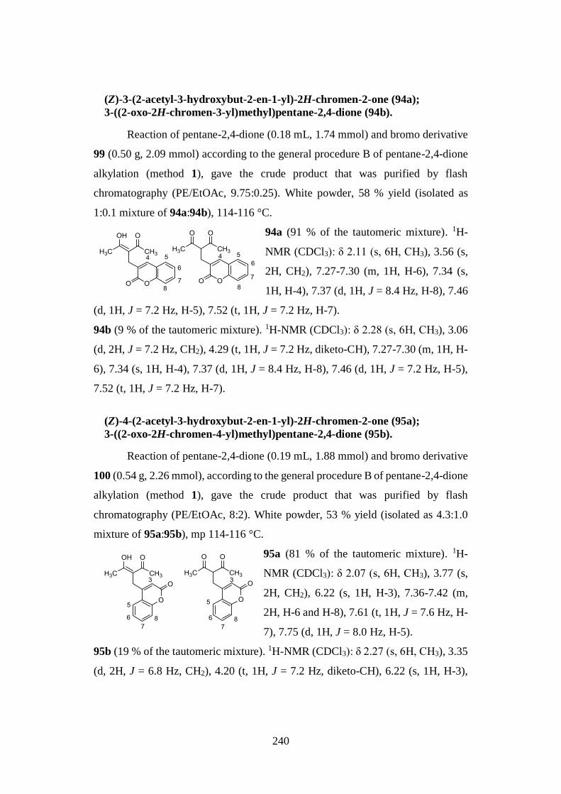
240
(Z)-3-(2-acetyl-3-hydroxybut-2-en-1-yl)-2H-chromen-2-one (94a);
3-((2-oxo-2H-chromen-3-yl)methyl)pentane-2,4-dione (94b).
Reaction of pentane-2,4-dione (0.18 mL, 1.74 mmol) and bromo derivative
99 (0.50 g, 2.09 mmol) according to the general procedure B of pentane-2,4-dione
alkylation (method 1), gave the crude product that was purified by flash
chromatography (PE/EtOAc, 9.75:0.25). White powder, 58 % yield (isolated as
1:0.1 mixture of 94a:94b), 114-116 °C.
94a (91 % of the tautomeric mixture). 1H-
NMR (CDCl3): δ 2.11 (s, 6H, CH3), 3.56 (s,
2H, CH2), 7.27-7.30 (m, 1H, H-6), 7.34 (s,
1H, H-4), 7.37 (d, 1H, J = 8.4 Hz, H-8), 7.46
(d, 1H, J = 7.2 Hz, H-5), 7.52 (t, 1H, J = 7.2 Hz, H-7).
94b (9 % of the tautomeric mixture). 1H-NMR (CDCl3): δ 2.28 (s, 6H, CH3), 3.06
(d, 2H, J = 7.2 Hz, CH2), 4.29 (t, 1H, J = 7.2 Hz, diketo-CH), 7.27-7.30 (m, 1H, H-
6), 7.34 (s, 1H, H-4), 7.37 (d, 1H, J = 8.4 Hz, H-8), 7.46 (d, 1H, J = 7.2 Hz, H-5),
7.52 (t, 1H, J = 7.2 Hz, H-7).
(Z)-4-(2-acetyl-3-hydroxybut-2-en-1-yl)-2H-chromen-2-one (95a);
3-((2-oxo-2H-chromen-4-yl)methyl)pentane-2,4-dione (95b).
Reaction of pentane-2,4-dione (0.19 mL, 1.88 mmol) and bromo derivative
100 (0.54 g, 2.26 mmol), according to the general procedure B of pentane-2,4-dione
alkylation (method 1), gave the crude product that was purified by flash
chromatography (PE/EtOAc, 8:2). White powder, 53 % yield (isolated as 4.3:1.0
mixture of 95a:95b), mp 114-116 °C.
95a (81 % of the tautomeric mixture). 1H-
NMR (CDCl3): δ 2.07 (s, 6H, CH3), 3.77 (s,
2H, CH2), 6.22 (s, 1H, H-3), 7.36-7.42 (m,
2H, H-6 and H-8), 7.61 (t, 1H, J = 7.6 Hz, H-
7), 7.75 (d, 1H, J = 8.0 Hz, H-5).
95b (19 % of the tautomeric mixture). 1H-NMR (CDCl3): δ 2.27 (s, 6H, CH3), 3.35
(d, 2H, J = 6.8 Hz, CH2), 4.20 (t, 1H, J = 7.2 Hz, diketo-CH), 6.22 (s, 1H, H-3),
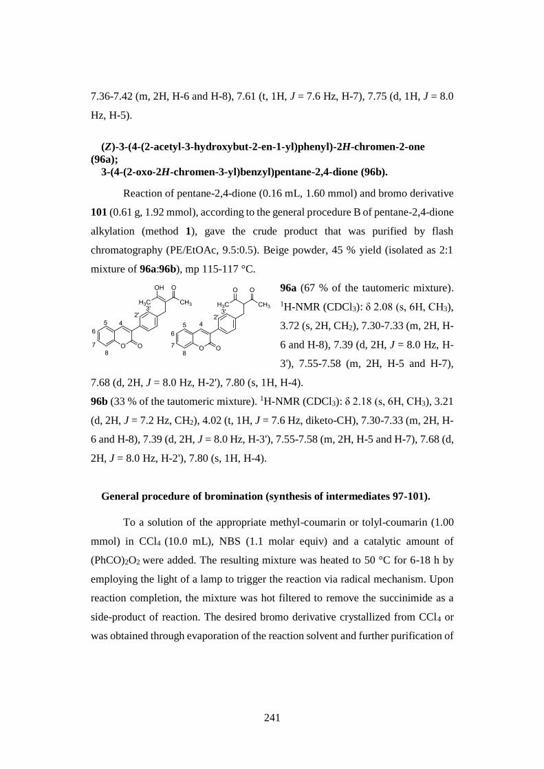
241
7.36-7.42 (m, 2H, H-6 and H-8), 7.61 (t, 1H, J = 7.6 Hz, H-7), 7.75 (d, 1H, J = 8.0
Hz, H-5).
(Z)-3-(4-(2-acetyl-3-hydroxybut-2-en-1-yl)phenyl)-2H-chromen-2-one
(96a);
3-(4-(2-oxo-2H-chromen-3-yl)benzyl)pentane-2,4-dione (96b).
Reaction of pentane-2,4-dione (0.16 mL, 1.60 mmol) and bromo derivative
101 (0.61 g, 1.92 mmol), according to the general procedure B of pentane-2,4-dione
alkylation (method 1), gave the crude product that was purified by flash
chromatography (PE/EtOAc, 9.5:0.5). Beige powder, 45 % yield (isolated as 2:1
mixture of 96a:96b), mp 115-117 °C.
96a (67 % of the tautomeric mixture).
1H-NMR (CDCl3): δ 2.08 (s, 6H, CH3),
3.72 (s, 2H, CH2), 7.30-7.33 (m, 2H, H-
6 and H-8), 7.39 (d, 2H, J = 8.0 Hz, H-
3'), 7.55-7.58 (m, 2H, H-5 and H-7),
7.68 (d, 2H, J = 8.0 Hz, H-2'), 7.80 (s, 1H, H-4).
96b (33 % of the tautomeric mixture). 1H-NMR (CDCl3): δ 2.18 (s, 6H, CH3), 3.21
(d, 2H, J = 7.2 Hz, CH2), 4.02 (t, 1H, J = 7.6 Hz, diketo-CH), 7.30-7.33 (m, 2H, H-
6 and H-8), 7.39 (d, 2H, J = 8.0 Hz, H-3'), 7.55-7.58 (m, 2H, H-5 and H-7), 7.68 (d,
2H, J = 8.0 Hz, H-2'), 7.80 (s, 1H, H-4).
General procedure of bromination (synthesis of intermediates 97-101).
To a solution of the appropriate methyl-coumarin or tolyl-coumarin (1.00
mmol) in CCl4 (10.0 mL), NBS (1.1 molar equiv) and a catalytic amount of
(PhCO)2O2 were added. The resulting mixture was heated to 50 °C for 6-18 h by
employing the light of a lamp to trigger the reaction via radical mechanism. Upon
reaction completion, the mixture was hot filtered to remove the succinimide as a
side-product of reaction. The desired bromo derivative crystallized from CCl4 or
was obtained through evaporation of the reaction solvent and further purification of
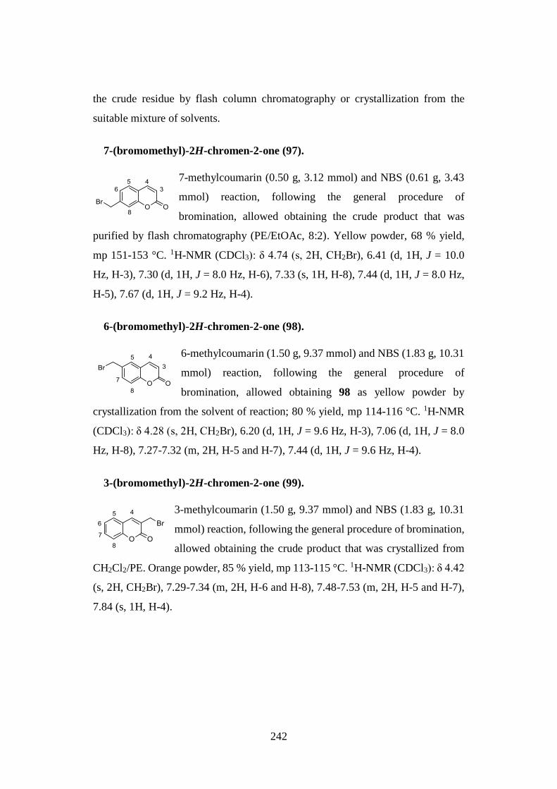
242
the crude residue by flash column chromatography or crystallization from the
suitable mixture of solvents.
7-(bromomethyl)-2H-chromen-2-one (97).
7-methylcoumarin (0.50 g, 3.12 mmol) and NBS (0.61 g, 3.43
mmol) reaction, following the general procedure of
bromination, allowed obtaining the crude product that was
purified by flash chromatography (PE/EtOAc, 8:2). Yellow powder, 68 % yield,
mp 151-153 °C. 1H-NMR (CDCl3): δ 4.74 (s, 2H, CH2Br), 6.41 (d, 1H, J = 10.0
Hz, H-3), 7.30 (d, 1H, J = 8.0 Hz, H-6), 7.33 (s, 1H, H-8), 7.44 (d, 1H, J = 8.0 Hz,
H-5), 7.67 (d, 1H, J = 9.2 Hz, H-4).
6-(bromomethyl)-2H-chromen-2-one (98).
6-methylcoumarin (1.50 g, 9.37 mmol) and NBS (1.83 g, 10.31
mmol) reaction, following the general procedure of
bromination, allowed obtaining 98 as yellow powder by
crystallization from the solvent of reaction; 80 % yield, mp 114-116 °C. 1H-NMR
(CDCl3): δ 4.28 (s, 2H, CH2Br), 6.20 (d, 1H, J = 9.6 Hz, H-3), 7.06 (d, 1H, J = 8.0
Hz, H-8), 7.27-7.32 (m, 2H, H-5 and H-7), 7.44 (d, 1H, J = 9.6 Hz, H-4).
3-(bromomethyl)-2H-chromen-2-one (99).
3-methylcoumarin (1.50 g, 9.37 mmol) and NBS (1.83 g, 10.31
mmol) reaction, following the general procedure of bromination,
allowed obtaining the crude product that was crystallized from
CH2Cl2/PE. Orange powder, 85 % yield, mp 113-115 °C. 1H-NMR (CDCl3): δ 4.42
(s, 2H, CH2Br), 7.29-7.34 (m, 2H, H-6 and H-8), 7.48-7.53 (m, 2H, H-5 and H-7),
7.84 (s, 1H, H-4).
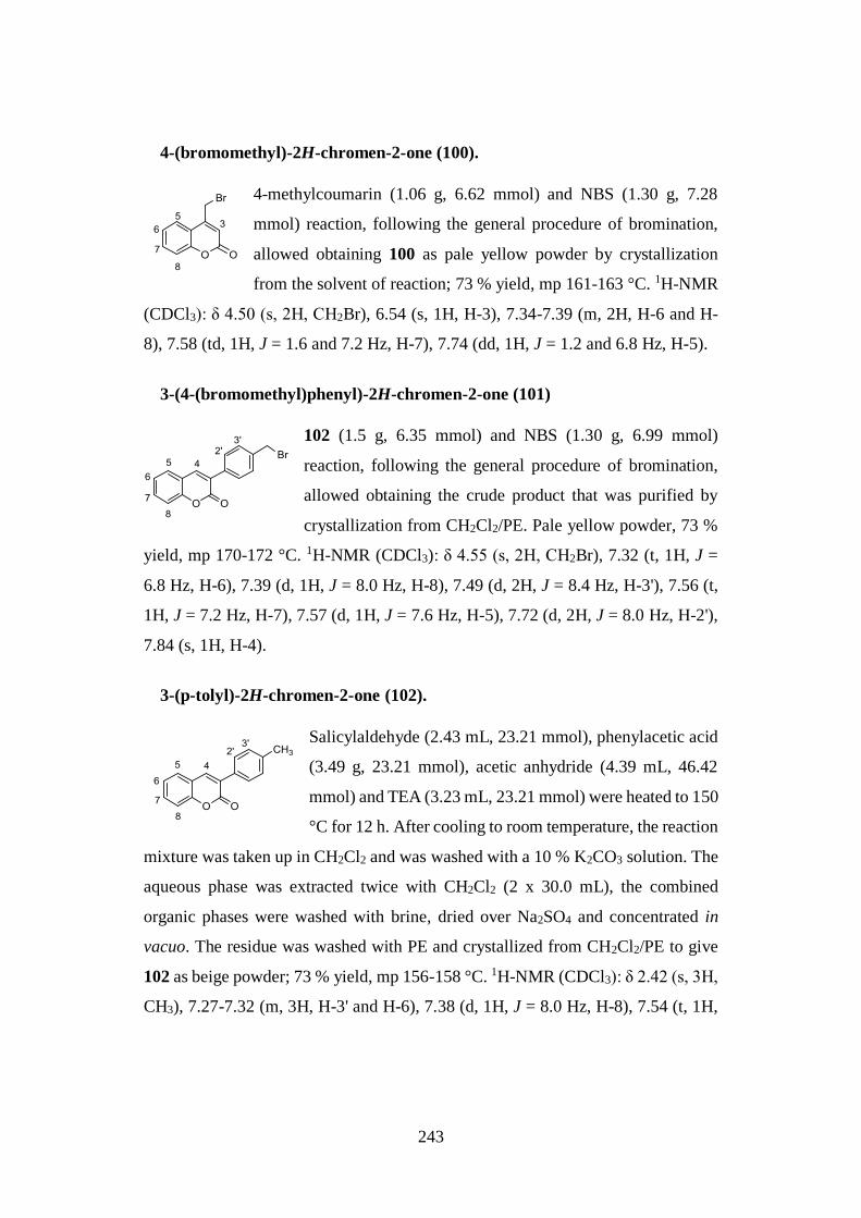
243
4-(bromomethyl)-2H-chromen-2-one (100).
4-methylcoumarin (1.06 g, 6.62 mmol) and NBS (1.30 g, 7.28
mmol) reaction, following the general procedure of bromination,
allowed obtaining 100 as pale yellow powder by crystallization
from the solvent of reaction; 73 % yield, mp 161-163 °C. 1H-NMR
(CDCl3): δ 4.50 (s, 2H, CH2Br), 6.54 (s, 1H, H-3), 7.34-7.39 (m, 2H, H-6 and H-
8), 7.58 (td, 1H, J = 1.6 and 7.2 Hz, H-7), 7.74 (dd, 1H, J = 1.2 and 6.8 Hz, H-5).
3-(4-(bromomethyl)phenyl)-2H-chromen-2-one (101)
102 (1.5 g, 6.35 mmol) and NBS (1.30 g, 6.99 mmol)
reaction, following the general procedure of bromination,
allowed obtaining the crude product that was purified by
crystallization from CH2Cl2/PE. Pale yellow powder, 73 %
yield, mp 170-172 °C. 1H-NMR (CDCl3): δ 4.55 (s, 2H, CH2Br), 7.32 (t, 1H, J =
6.8 Hz, H-6), 7.39 (d, 1H, J = 8.0 Hz, H-8), 7.49 (d, 2H, J = 8.4 Hz, H-3'), 7.56 (t,
1H, J = 7.2 Hz, H-7), 7.57 (d, 1H, J = 7.6 Hz, H-5), 7.72 (d, 2H, J = 8.0 Hz, H-2'),
7.84 (s, 1H, H-4).
3-(p-tolyl)-2H-chromen-2-one (102).
Salicylaldehyde (2.43 mL, 23.21 mmol), phenylacetic acid
(3.49 g, 23.21 mmol), acetic anhydride (4.39 mL, 46.42
mmol) and TEA (3.23 mL, 23.21 mmol) were heated to 150
°C for 12 h. After cooling to room temperature, the reaction
mixture was taken up in CH2Cl2 and was washed with a 10 % K2CO3 solution. The
aqueous phase was extracted twice with CH2Cl2 (2 x 30.0 mL), the combined
organic phases were washed with brine, dried over Na2SO4 and concentrated in
vacuo. The residue was washed with PE and crystallized from CH2Cl2/PE to give
102 as beige powder; 73 % yield, mp 156-158 °C. 1H-NMR (CDCl3): δ 2.42 (s, 3H,
CH3), 7.27-7.32 (m, 3H, H-3' and H-6), 7.38 (d, 1H, J = 8.0 Hz, H-8), 7.54 (t, 1H,
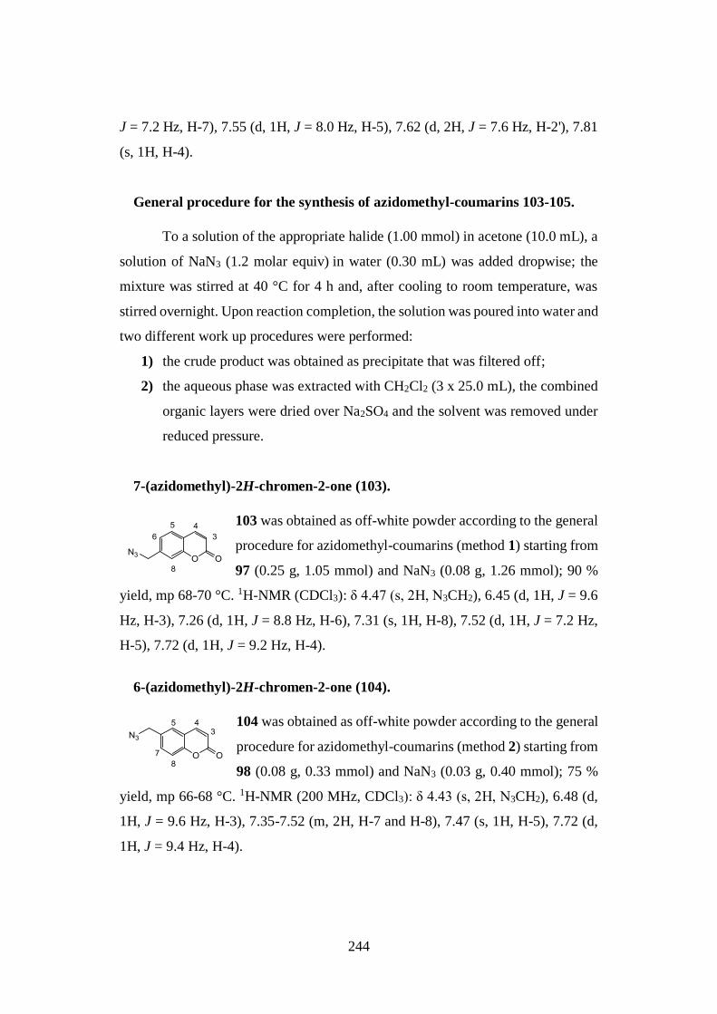
244
J = 7.2 Hz, H-7), 7.55 (d, 1H, J = 8.0 Hz, H-5), 7.62 (d, 2H, J = 7.6 Hz, H-2'), 7.81
(s, 1H, H-4).
General procedure for the synthesis of azidomethyl-coumarins 103-105.
To a solution of the appropriate halide (1.00 mmol) in acetone (10.0 mL), a
solution of NaN3 (1.2 molar equiv) in water (0.30 mL) was added dropwise; the
mixture was stirred at 40 °C for 4 h and, after cooling to room temperature, was
stirred overnight. Upon reaction completion, the solution was poured into water and
two different work up procedures were performed:
1) the crude product was obtained as precipitate that was filtered off;
2) the aqueous phase was extracted with CH2Cl2 (3 x 25.0 mL), the combined
organic layers were dried over Na2SO4 and the solvent was removed under
reduced pressure.
7-(azidomethyl)-2H-chromen-2-one (103).
103 was obtained as off-white powder according to the general
procedure for azidomethyl-coumarins (method 1) starting from
97 (0.25 g, 1.05 mmol) and NaN3 (0.08 g, 1.26 mmol); 90 %
yield, mp 68-70 °C. 1H-NMR (CDCl3): δ 4.47 (s, 2H, N3CH2), 6.45 (d, 1H, J = 9.6
Hz, H-3), 7.26 (d, 1H, J = 8.8 Hz, H-6), 7.31 (s, 1H, H-8), 7.52 (d, 1H, J = 7.2 Hz,
H-5), 7.72 (d, 1H, J = 9.2 Hz, H-4).
6-(azidomethyl)-2H-chromen-2-one (104).
104 was obtained as off-white powder according to the general
procedure for azidomethyl-coumarins (method 2) starting from
98 (0.08 g, 0.33 mmol) and NaN3 (0.03 g, 0.40 mmol); 75 %
yield, mp 66-68 °C. 1H-NMR (200 MHz, CDCl3): δ 4.43 (s, 2H, N3CH2), 6.48 (d,
1H, J = 9.6 Hz, H-3), 7.35-7.52 (m, 2H, H-7 and H-8), 7.47 (s, 1H, H-5), 7.72 (d,
1H, J = 9.4 Hz, H-4).
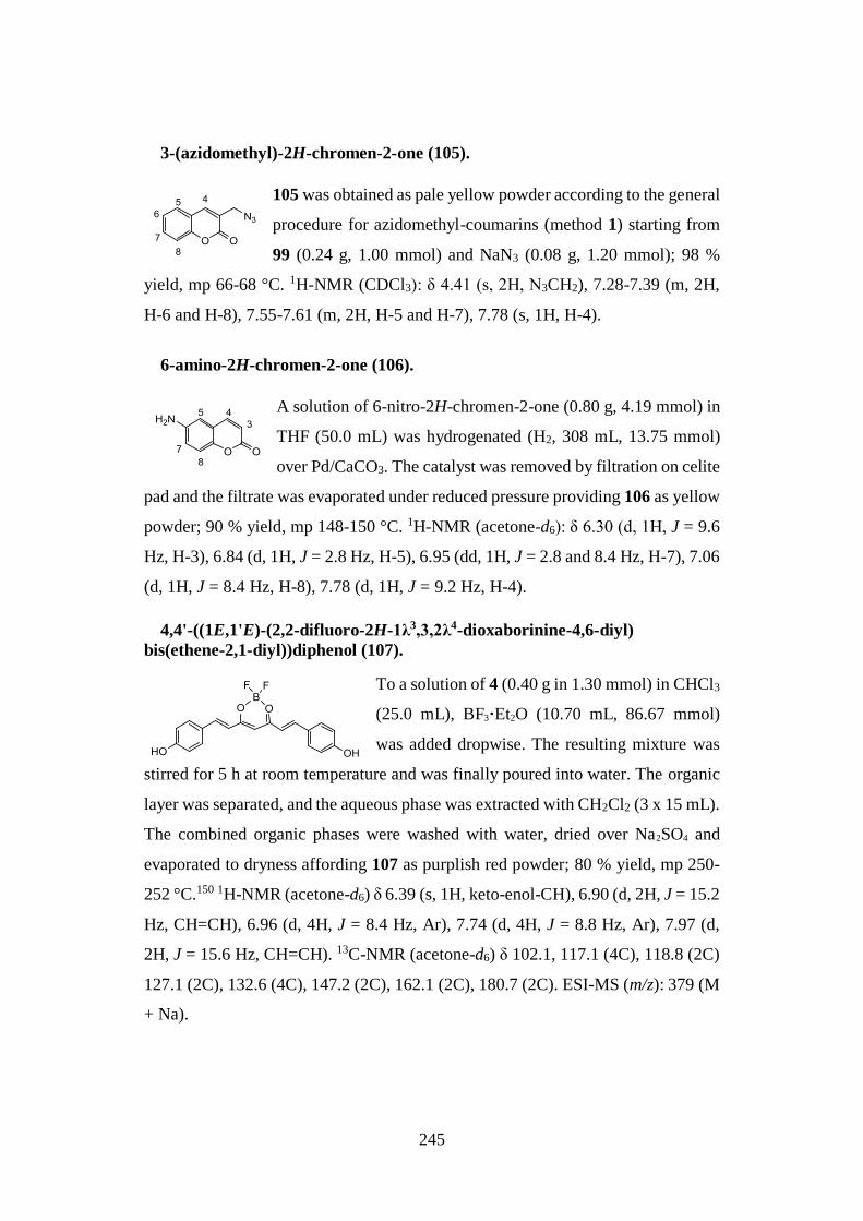
245
3-(azidomethyl)-2H-chromen-2-one (105).
105 was obtained as pale yellow powder according to the general
procedure for azidomethyl-coumarins (method 1) starting from
99 (0.24 g, 1.00 mmol) and NaN3 (0.08 g, 1.20 mmol); 98 %
yield, mp 66-68 °C. 1H-NMR (CDCl3): δ 4.41 (s, 2H, N3CH2), 7.28-7.39 (m, 2H,
H-6 and H-8), 7.55-7.61 (m, 2H, H-5 and H-7), 7.78 (s, 1H, H-4).
6-amino-2H-chromen-2-one (106).
A solution of 6-nitro-2H-chromen-2-one (0.80 g, 4.19 mmol) in
THF (50.0 mL) was hydrogenated (H2, 308 mL, 13.75 mmol)
over Pd/CaCO3. The catalyst was removed by filtration on celite
pad and the filtrate was evaporated under reduced pressure providing 106 as yellow
powder; 90 % yield, mp 148-150 °C. 1H-NMR (acetone-d6): δ 6.30 (d, 1H, J = 9.6
Hz, H-3), 6.84 (d, 1H, J = 2.8 Hz, H-5), 6.95 (dd, 1H, J = 2.8 and 8.4 Hz, H-7), 7.06
(d, 1H, J = 8.4 Hz, H-8), 7.78 (d, 1H, J = 9.2 Hz, H-4).
4,4'-((1E,1'E)-(2,2-difluoro-2H-1λ3,3,2λ4-dioxaborinine-4,6-diyl)
bis(ethene-2,1-diyl))diphenol (107).
To a solution of 4 (0.40 g in 1.30 mmol) in CHCl3
(25.0 mL), BF3·Et2O (10.70 mL, 86.67 mmol)
was added dropwise. The resulting mixture was
stirred for 5 h at room temperature and was finally poured into water. The organic
layer was separated, and the aqueous phase was extracted with CH2Cl2 (3 x 15 mL).
The combined organic phases were washed with water, dried over Na2SO4 and
evaporated to dryness affording 107 as purplish red powder; 80 % yield, mp 250-
252 °C.150 1H-NMR (acetone-d6) δ 6.39 (s, 1H, keto-enol-CH), 6.90 (d, 2H, J = 15.2
Hz, CH=CH), 6.96 (d, 4H, J = 8.4 Hz, Ar), 7.74 (d, 4H, J = 8.8 Hz, Ar), 7.97 (d,
2H, J = 15.6 Hz, CH=CH). 13C-NMR (acetone-d6) δ 102.1, 117.1 (4C), 118.8 (2C)
127.1 (2C), 132.6 (4C), 147.2 (2C), 162.1 (2C), 180.7 (2C). ESI-MS (m/z): 379 (M
+ Na).
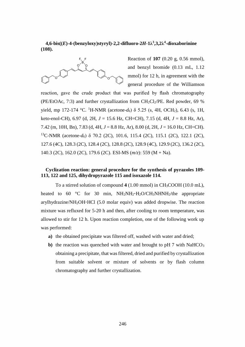
246
4,6-bis((E)-4-(benzyloxy)styryl)-2,2-difluoro-2H-1λ3,3,2λ4-dioxaborinine
(108).
Reaction of 107 (0.20 g, 0.56 mmol),
and benzyl bromide (0.13 mL, 1.12
mmol) for 12 h, in agreement with the
general procedure of the Williamson
reaction, gave the crude product that was purified by flash chromatography
(PE/EtOAc, 7:3) and further crystallization from CH2Cl2/PE. Red powder, 69 %
yield, mp 172-174 °C. 1H-NMR (acetone-d6) δ 5.25 (s, 4H, OCH2), 6.43 (s, 1H,
keto-enol-CH), 6.97 (d, 2H, J = 15.6 Hz, CH=CH), 7.15 (d, 4H, J = 8.8 Hz, Ar),
7.42 (m, 10H, Bn), 7.83 (d, 4H, J = 8.8 Hz, Ar), 8.00 (d, 2H, J = 16.0 Hz, CH=CH).
13C-NMR (acetone-d6) δ 70.2 (2C), 101.6, 115.4 (2C), 115.1 (2C), 122.1 (2C),
127.6 (4C), 128.3 (2C), 128.4 (2C), 128.8 (2C), 128.9 (4C), 129.9 (2C), 136.2 (2C),
140.3 (2C), 162.0 (2C), 179.6 (2C). ESI-MS (m/z): 559 (M + Na).
Cyclization reaction: general procedure for the synthesis of pyrazoles 109-
113, 122 and 125, dihydropyrazole 115 and isoxazole 114.
To a stirred solution of compound 4 (1.00 mmol) in CH3COOH (10.0 mL),
heated to 60 °C for 30 min, NH2NH2·H2O/CH3NHNH2/the appropriate
arylhydrazine/NH2OH·HCl (5.0 molar equiv) was added dropwise. The reaction
mixture was refluxed for 5-20 h and then, after cooling to room temperature, was
allowed to stir for 12 h. Upon reaction completion, one of the following work up
was performed:
a) the obtained precipitate was filtered off, washed with water and dried;
b) the reaction was quenched with water and brought to pH 7 with NaHCO3
obtaining a precipitate, that was filtered, dried and purified by crystallization
from suitable solvent or mixture of solvents or by flash column
chromatography and further crystallization.
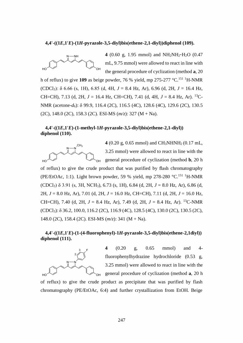
247
4,4'-((1E,1'E)-(1H-pyrazole-3,5-diyl)bis(ethene-2,1-diyl))diphenol (109).
4 (0.60 g, 1.95 mmol) and NH2NH2·H2O (0.47
mL, 9.75 mmol) were allowed to react in line with
the general procedure of cyclization (method a, 20
h of reflux) to give 109 as beige powder, 76 % yield, mp 275-277 °C.151 1H-NMR
(CDCl3): δ 6.66 (s, 1H), 6.85 (d, 4H, J = 8.4 Hz, Ar), 6.96 (d, 2H, J = 16.4 Hz,
CH=CH), 7.13 (d, 2H, J = 16.4 Hz, CH=CH), 7.41 (d, 4H, J = 8.4 Hz, Ar). 13C-
NMR (acetone-d6): δ 99.9, 116.4 (2C), 116.5 (4C), 128.6 (4C), 129.6 (2C), 130.5
(2C), 148.0 (2C), 158.3 (2C). ESI-MS (m/z): 327 (M + Na).
4,4'-((1E,1'E)-(1-methyl-1H-pyrazole-3,5-diyl)bis(ethene-2,1-diyl))
diphenol (110).
4 (0.20 g, 0.65 mmol) and CH3NHNH2 (0.17 mL,
3.25 mmol) were allowed to react in line with the
general procedure of cyclization (method b, 20 h
of reflux) to give the crude product that was purified by flash chromatography
(PE/EtOAc, 1:1). Light brown powder, 59 % yield, mp 278-280 °C.151 1H-NMR
(CDCl3) δ 3.91 (s, 3H, NCH3), 6.73 (s, 1H), 6.84 (d, 2H, J = 8.0 Hz, Ar), 6.86 (d,
2H, J = 8.0 Hz, Ar), 7.01 (d, 2H, J = 16.0 Hz, CH=CH), 7.11 (d, 2H, J = 16.0 Hz,
CH=CH), 7.40 (d, 2H, J = 8.4 Hz, Ar), 7.49 (d, 2H, J = 8.4 Hz, Ar). 13C-NMR
(CDCl3): δ 36.2, 100.0, 116.2 (2C), 116.9 (4C), 128.5 (4C), 130.0 (2C), 130.5 (2C),
148.0 (2C), 158.4 (2C). ESI-MS (m/z): 341 (M + Na).
4,4'-((1E,1'E)-(1-(4-fluorophenyl)-1H-pyrazole-3,5-diyl)bis(ethene-2,1diyl))
diphenol (111).
4 (0.20 g, 0.65 mmol) and 4-
fluorophenylhydrazine hydrochloride (0.53 g,
3.25 mmol) were allowed to react in line with the
general procedure of cyclization (method a, 20 h
of reflux) to give the crude product as precipitate that was purified by flash
chromatography (PE/EtOAc, 6:4) and further crystallization from EtOH. Beige
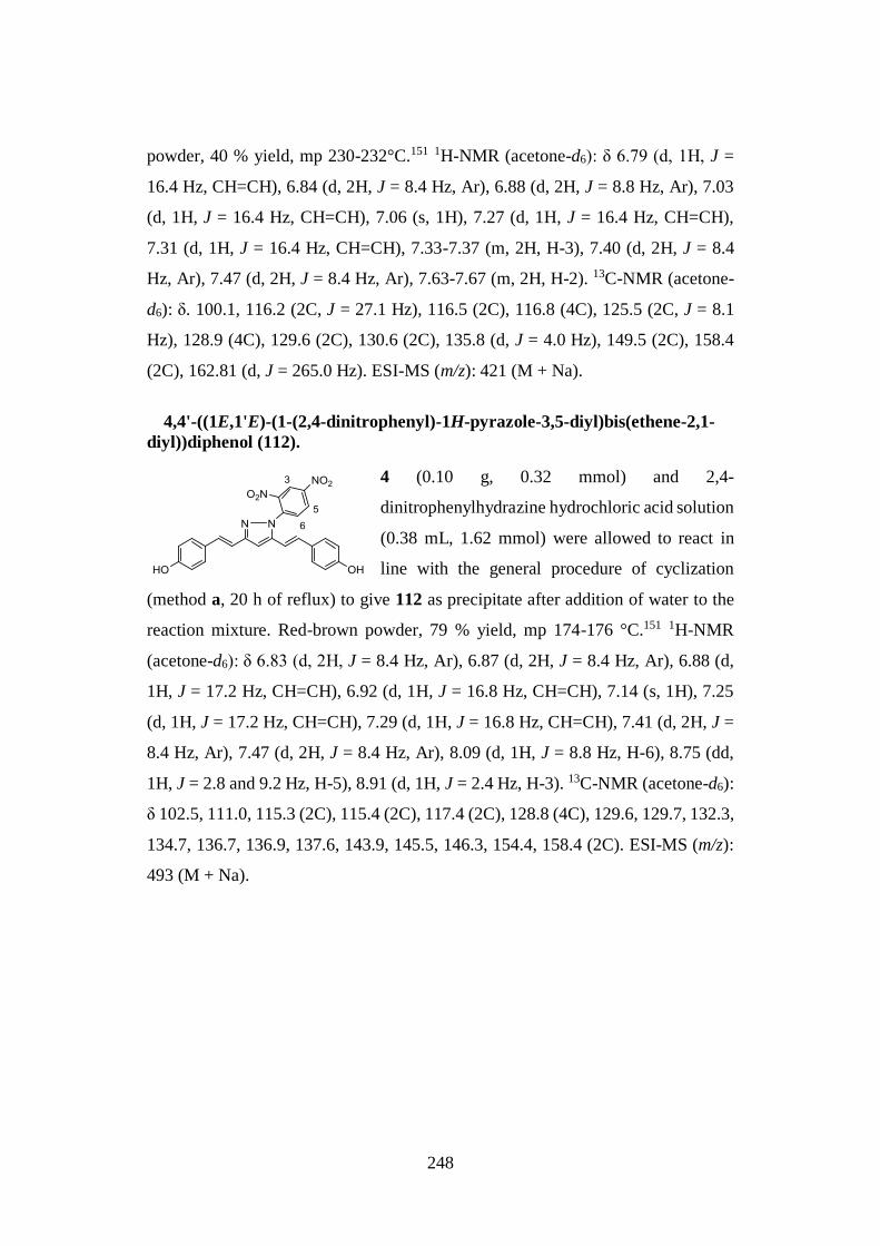
248
powder, 40 % yield, mp 230-232°C.151 1H-NMR (acetone-d6): δ 6.79 (d, 1H, J =
16.4 Hz, CH=CH), 6.84 (d, 2H, J = 8.4 Hz, Ar), 6.88 (d, 2H, J = 8.8 Hz, Ar), 7.03
(d, 1H, J = 16.4 Hz, CH=CH), 7.06 (s, 1H), 7.27 (d, 1H, J = 16.4 Hz, CH=CH),
7.31 (d, 1H, J = 16.4 Hz, CH=CH), 7.33-7.37 (m, 2H, H-3), 7.40 (d, 2H, J = 8.4
Hz, Ar), 7.47 (d, 2H, J = 8.4 Hz, Ar), 7.63-7.67 (m, 2H, H-2). 13C-NMR (acetone-
d6): δ. 100.1, 116.2 (2C, J = 27.1 Hz), 116.5 (2C), 116.8 (4C), 125.5 (2C, J = 8.1
Hz), 128.9 (4C), 129.6 (2C), 130.6 (2C), 135.8 (d, J = 4.0 Hz), 149.5 (2C), 158.4
(2C), 162.81 (d, J = 265.0 Hz). ESI-MS (m/z): 421 (M + Na).
4,4'-((1E,1'E)-(1-(2,4-dinitrophenyl)-1H-pyrazole-3,5-diyl)bis(ethene-2,1-
diyl))diphenol (112).
4 (0.10 g, 0.32 mmol) and 2,4-
dinitrophenylhydrazine hydrochloric acid solution
(0.38 mL, 1.62 mmol) were allowed to react in
line with the general procedure of cyclization
(method a, 20 h of reflux) to give 112 as precipitate after addition of water to the
reaction mixture. Red-brown powder, 79 % yield, mp 174-176 °C.151 1H-NMR
(acetone-d6): δ 6.83 (d, 2H, J = 8.4 Hz, Ar), 6.87 (d, 2H, J = 8.4 Hz, Ar), 6.88 (d,
1H, J = 17.2 Hz, CH=CH), 6.92 (d, 1H, J = 16.8 Hz, CH=CH), 7.14 (s, 1H), 7.25
(d, 1H, J = 17.2 Hz, CH=CH), 7.29 (d, 1H, J = 16.8 Hz, CH=CH), 7.41 (d, 2H, J =
8.4 Hz, Ar), 7.47 (d, 2H, J = 8.4 Hz, Ar), 8.09 (d, 1H, J = 8.8 Hz, H-6), 8.75 (dd,
1H, J = 2.8 and 9.2 Hz, H-5), 8.91 (d, 1H, J = 2.4 Hz, H-3). 13C-NMR (acetone-d6):
δ 102.5, 111.0, 115.3 (2C), 115.4 (2C), 117.4 (2C), 128.8 (4C), 129.6, 129.7, 132.3,
134.7, 136.7, 136.9, 137.6, 143.9, 145.5, 146.3, 154.4, 158.4 (2C). ESI-MS (m/z):
493 (M + Na).
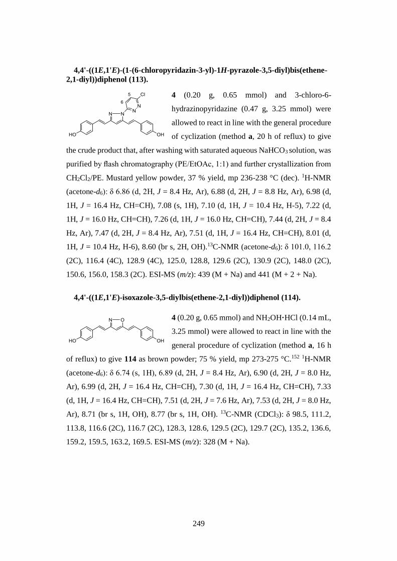
249
4,4'-((1E,1'E)-(1-(6-chloropyridazin-3-yl)-1H-pyrazole-3,5-diyl)bis(ethene-
2,1-diyl))diphenol (113).
4 (0.20 g, 0.65 mmol) and 3-chloro-6-
hydrazinopyridazine (0.47 g, 3.25 mmol) were
allowed to react in line with the general procedure
of cyclization (method a, 20 h of reflux) to give
the crude product that, after washing with saturated aqueous NaHCO3 solution, was
purified by flash chromatography (PE/EtOAc, 1:1) and further crystallization from
CH2Cl2/PE. Mustard yellow powder, 37 % yield, mp 236-238 °C (dec). 1H-NMR
(acetone-d6): δ 6.86 (d, 2H, J = 8.4 Hz, Ar), 6.88 (d, 2H, J = 8.8 Hz, Ar), 6.98 (d,
1H, J = 16.4 Hz, CH=CH), 7.08 (s, 1H), 7.10 (d, 1H, J = 10.4 Hz, H-5), 7.22 (d,
1H, J = 16.0 Hz, CH=CH), 7.26 (d, 1H, J = 16.0 Hz, CH=CH), 7.44 (d, 2H, J = 8.4
Hz, Ar), 7.47 (d, 2H, J = 8.4 Hz, Ar), 7.51 (d, 1H, J = 16.4 Hz, CH=CH), 8.01 (d,
1H, J = 10.4 Hz, H-6), 8.60 (br s, 2H, OH).13C-NMR (acetone-d6): δ 101.0, 116.2
(2C), 116.4 (4C), 128.9 (4C), 125.0, 128.8, 129.6 (2C), 130.9 (2C), 148.0 (2C),
150.6, 156.0, 158.3 (2C). ESI-MS (m/z): 439 (M + Na) and 441 (M + 2 + Na).
4,4'-((1E,1'E)-isoxazole-3,5-diylbis(ethene-2,1-diyl))diphenol (114).
4 (0.20 g, 0.65 mmol) and NH2OH·HCl (0.14 mL,
3.25 mmol) were allowed to react in line with the
general procedure of cyclization (method a, 16 h
of reflux) to give 114 as brown powder; 75 % yield, mp 273-275 °C.152 1H-NMR
(acetone-d6): δ 6.74 (s, 1H), 6.89 (d, 2H, J = 8.4 Hz, Ar), 6.90 (d, 2H, J = 8.0 Hz,
Ar), 6.99 (d, 2H, J = 16.4 Hz, CH=CH), 7.30 (d, 1H, J = 16.4 Hz, CH=CH), 7.33
(d, 1H, J = 16.4 Hz, CH=CH), 7.51 (d, 2H, J = 7.6 Hz, Ar), 7.53 (d, 2H, J = 8.0 Hz,
Ar), 8.71 (br s, 1H, OH), 8.77 (br s, 1H, OH). 13C-NMR (CDCl3): δ 98.5, 111.2,
113.8, 116.6 (2C), 116.7 (2C), 128.3, 128.6, 129.5 (2C), 129.7 (2C), 135.2, 136.6,
159.2, 159.5, 163.2, 169.5. ESI-MS (m/z): 328 (M + Na).
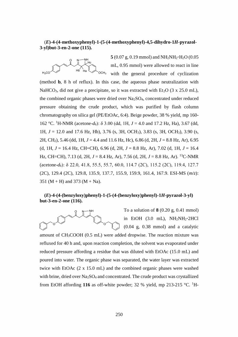
250
(E)-4-(4-methoxyphenyl)-1-(5-(4-methoxyphenyl)-4,5-dihydro-1H-pyrazol-
3-yl)but-3-en-2-one (115).
5 (0.07 g, 0.19 mmol) and NH2NH2·H2O (0.05
mL, 0.95 mmol) were allowed to react in line
with the general procedure of cyclization
(method b, 8 h of reflux). In this case, the aqueous phase neutralization with
NaHCO3, did not give a precipitate, so it was extracted with Et2O (3 x 25.0 mL),
the combined organic phases were dried over Na2SO4, concentrated under reduced
pressure obtaining the crude product, which was purified by flash column
chromatography on silica gel (PE/EtOAc, 6:4). Beige powder, 38 % yield, mp 160-
162 °C. 1H-NMR (acetone-d6): δ 3.00 (dd, 1H, J = 4.0 and 17.2 Hz, Ha), 3.67 (dd,
1H, J = 12.0 and 17.6 Hz, Hb), 3.76 (s, 3H, OCH3), 3.83 (s, 3H, OCH3), 3.90 (s,
2H, CH2), 5.46 (dd, 1H, J = 4.4 and 11.6 Hz, Hc), 6.86 (d, 2H, J = 8.8 Hz, Ar), 6.95
(d, 1H, J = 16.4 Hz, CH=CH), 6.96 (d, 2H, J = 8.8 Hz, Ar), 7.02 (d, 1H, J = 16.4
Hz, CH=CH), 7.13 (d, 2H, J = 8.4 Hz, Ar), 7.56 (d, 2H, J = 8.8 Hz, Ar). 13C-NMR
(acetone-d6): δ 22.0, 41.8, 55.5, 55.7, 60.0, 114.7 (2C), 115.2 (2C), 119.4, 127.7
(2C), 129.4 (2C), 129.8, 135.9, 137.7, 155.9, 159.9, 161.4, 167.9. ESI-MS (m/z):
351 (M + H) and 373 (M + Na).
(E)-4-(4-(benzyloxy)phenyl)-1-(5-(4-(benzyloxy)phenyl)-1H-pyrazol-3-yl)
but-3-en-2-one (116).
To a solution of 8 (0.20 g, 0.41 mmol)
in EtOH (3.0 mL), NH2NH2·2HCl
(0.04 g, 0.38 mmol) and a catalytic
amount of CH3COOH (0.5 mL) were added dropwise. The reaction mixture was
refluxed for 40 h and, upon reaction completion, the solvent was evaporated under
reduced pressure affording a residue that was diluted with EtOAc (15.0 mL) and
poured into water. The organic phase was separated, the water layer was extracted
twice with EtOAc (2 x 15.0 mL) and the combined organic phases were washed
with brine, dried over Na2SO4 and concentrated. The crude product was crystallized
from EtOH affording 116 as off-white powder; 32 % yield, mp 213-215 °C. 1H-
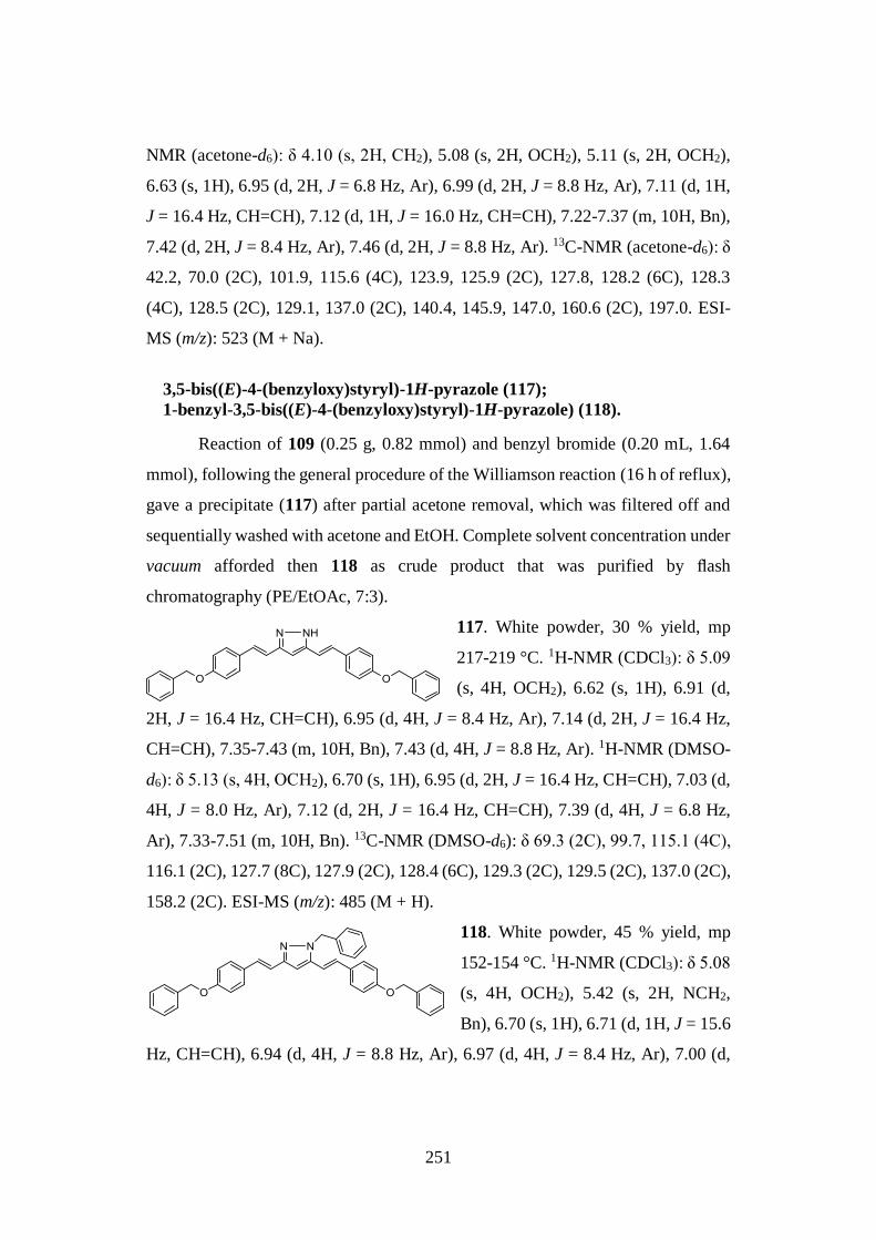
251
NMR (acetone-d6): δ 4.10 (s, 2H, CH2), 5.08 (s, 2H, OCH2), 5.11 (s, 2H, OCH2),
6.63 (s, 1H), 6.95 (d, 2H, J = 6.8 Hz, Ar), 6.99 (d, 2H, J = 8.8 Hz, Ar), 7.11 (d, 1H,
J = 16.4 Hz, CH=CH), 7.12 (d, 1H, J = 16.0 Hz, CH=CH), 7.22-7.37 (m, 10H, Bn),
7.42 (d, 2H, J = 8.4 Hz, Ar), 7.46 (d, 2H, J = 8.8 Hz, Ar). 13C-NMR (acetone-d6): δ
42.2, 70.0 (2C), 101.9, 115.6 (4C), 123.9, 125.9 (2C), 127.8, 128.2 (6C), 128.3
(4C), 128.5 (2C), 129.1, 137.0 (2C), 140.4, 145.9, 147.0, 160.6 (2C), 197.0. ESI-
MS (m/z): 523 (M + Na).
3,5-bis((E)-4-(benzyloxy)styryl)-1H-pyrazole (117);
1-benzyl-3,5-bis((E)-4-(benzyloxy)styryl)-1H-pyrazole) (118).
Reaction of 109 (0.25 g, 0.82 mmol) and benzyl bromide (0.20 mL, 1.64
mmol), following the general procedure of the Williamson reaction (16 h of reflux),
gave a precipitate (117) after partial acetone removal, which was filtered off and
sequentially washed with acetone and EtOH. Complete solvent concentration under
vacuum afforded then 118 as crude product that was purified by flash
chromatography (PE/EtOAc, 7:3).
117. White powder, 30 % yield, mp
217-219 °C. 1H-NMR (CDCl3): δ 5.09
(s, 4H, OCH2), 6.62 (s, 1H), 6.91 (d,
2H, J = 16.4 Hz, CH=CH), 6.95 (d, 4H, J = 8.4 Hz, Ar), 7.14 (d, 2H, J = 16.4 Hz,
CH=CH), 7.35-7.43 (m, 10H, Bn), 7.43 (d, 4H, J = 8.8 Hz, Ar). 1H-NMR (DMSO-
d6): δ 5.13 (s, 4H, OCH2), 6.70 (s, 1H), 6.95 (d, 2H, J = 16.4 Hz, CH=CH), 7.03 (d,
4H, J = 8.0 Hz, Ar), 7.12 (d, 2H, J = 16.4 Hz, CH=CH), 7.39 (d, 4H, J = 6.8 Hz,
Ar), 7.33-7.51 (m, 10H, Bn). 13C-NMR (DMSO-d6): δ 69.3 (2C), 99.7, 115.1 (4C),
116.1 (2C), 127.7 (8C), 127.9 (2C), 128.4 (6C), 129.3 (2C), 129.5 (2C), 137.0 (2C),
158.2 (2C). ESI-MS (m/z): 485 (M + H).
118. White powder, 45 % yield, mp
152-154 °C. 1H-NMR (CDCl3): δ 5.08
(s, 4H, OCH2), 5.42 (s, 2H, NCH2,
Bn), 6.70 (s, 1H), 6.71 (d, 1H, J = 15.6
Hz, CH=CH), 6.94 (d, 4H, J = 8.8 Hz, Ar), 6.97 (d, 4H, J = 8.4 Hz, Ar), 7.00 (d,
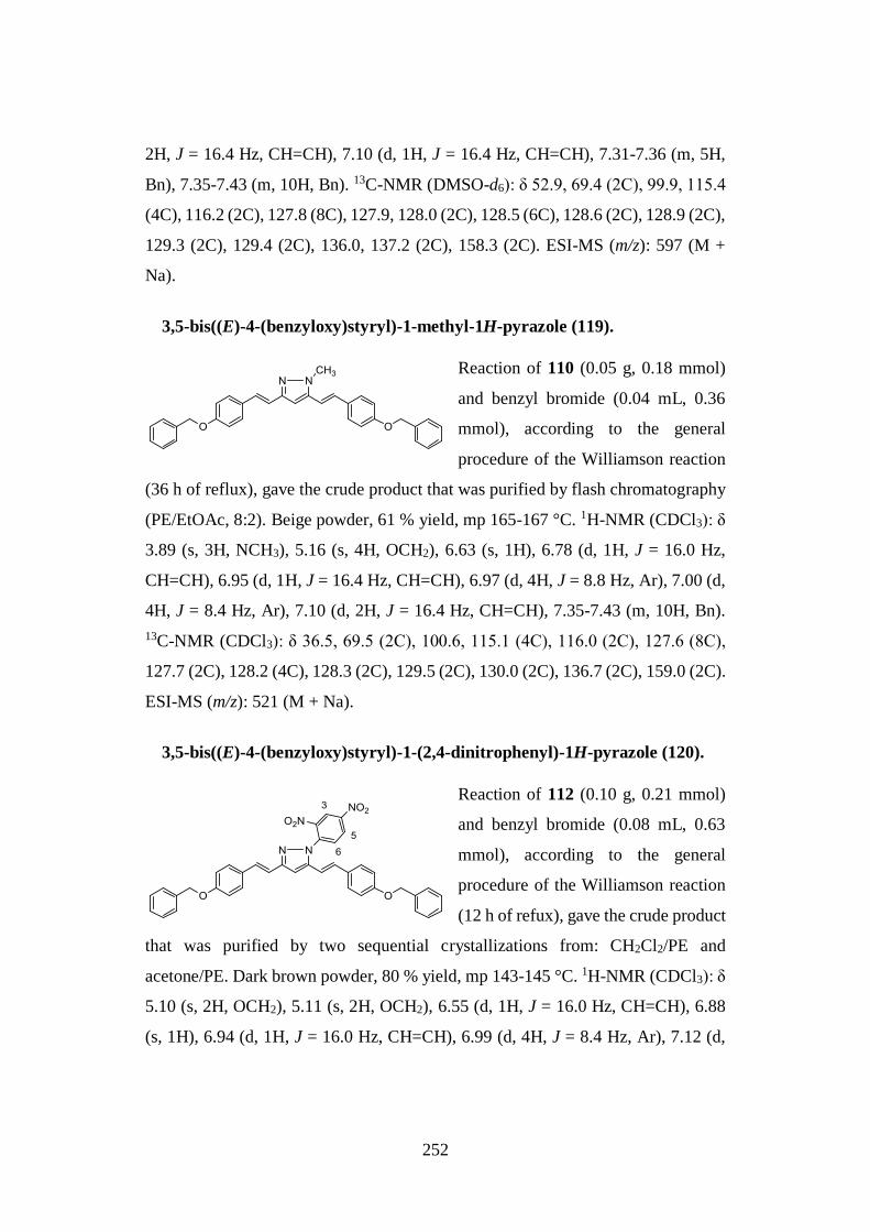
252
2H, J = 16.4 Hz, CH=CH), 7.10 (d, 1H, J = 16.4 Hz, CH=CH), 7.31-7.36 (m, 5H,
Bn), 7.35-7.43 (m, 10H, Bn). 13C-NMR (DMSO-d6): δ 52.9, 69.4 (2C), 99.9, 115.4
(4C), 116.2 (2C), 127.8 (8C), 127.9, 128.0 (2C), 128.5 (6C), 128.6 (2C), 128.9 (2C),
129.3 (2C), 129.4 (2C), 136.0, 137.2 (2C), 158.3 (2C). ESI-MS (m/z): 597 (M +
Na).
3,5-bis((E)-4-(benzyloxy)styryl)-1-methyl-1H-pyrazole (119).
Reaction of 110 (0.05 g, 0.18 mmol)
and benzyl bromide (0.04 mL, 0.36
mmol), according to the general
procedure of the Williamson reaction
(36 h of reflux), gave the crude product that was purified by flash chromatography
(PE/EtOAc, 8:2). Beige powder, 61 % yield, mp 165-167 °C. 1H-NMR (CDCl3): δ
3.89 (s, 3H, NCH3), 5.16 (s, 4H, OCH2), 6.63 (s, 1H), 6.78 (d, 1H, J = 16.0 Hz,
CH=CH), 6.95 (d, 1H, J = 16.4 Hz, CH=CH), 6.97 (d, 4H, J = 8.8 Hz, Ar), 7.00 (d,
4H, J = 8.4 Hz, Ar), 7.10 (d, 2H, J = 16.4 Hz, CH=CH), 7.35-7.43 (m, 10H, Bn).
13C-NMR (CDCl3): δ 36.5, 69.5 (2C), 100.6, 115.1 (4C), 116.0 (2C), 127.6 (8C),
127.7 (2C), 128.2 (4C), 128.3 (2C), 129.5 (2C), 130.0 (2C), 136.7 (2C), 159.0 (2C).
ESI-MS (m/z): 521 (M + Na).
3,5-bis((E)-4-(benzyloxy)styryl)-1-(2,4-dinitrophenyl)-1H-pyrazole (120).
Reaction of 112 (0.10 g, 0.21 mmol)
and benzyl bromide (0.08 mL, 0.63
mmol), according to the general
procedure of the Williamson reaction
(12 h of refux), gave the crude product
that was purified by two sequential crystallizations from: CH2Cl2/PE and
acetone/PE. Dark brown powder, 80 % yield, mp 143-145 °C. 1H-NMR (CDCl3): δ
5.10 (s, 2H, OCH2), 5.11 (s, 2H, OCH2), 6.55 (d, 1H, J = 16.0 Hz, CH=CH), 6.88
(s, 1H), 6.94 (d, 1H, J = 16.0 Hz, CH=CH), 6.99 (d, 4H, J = 8.4 Hz, Ar), 7.12 (d,
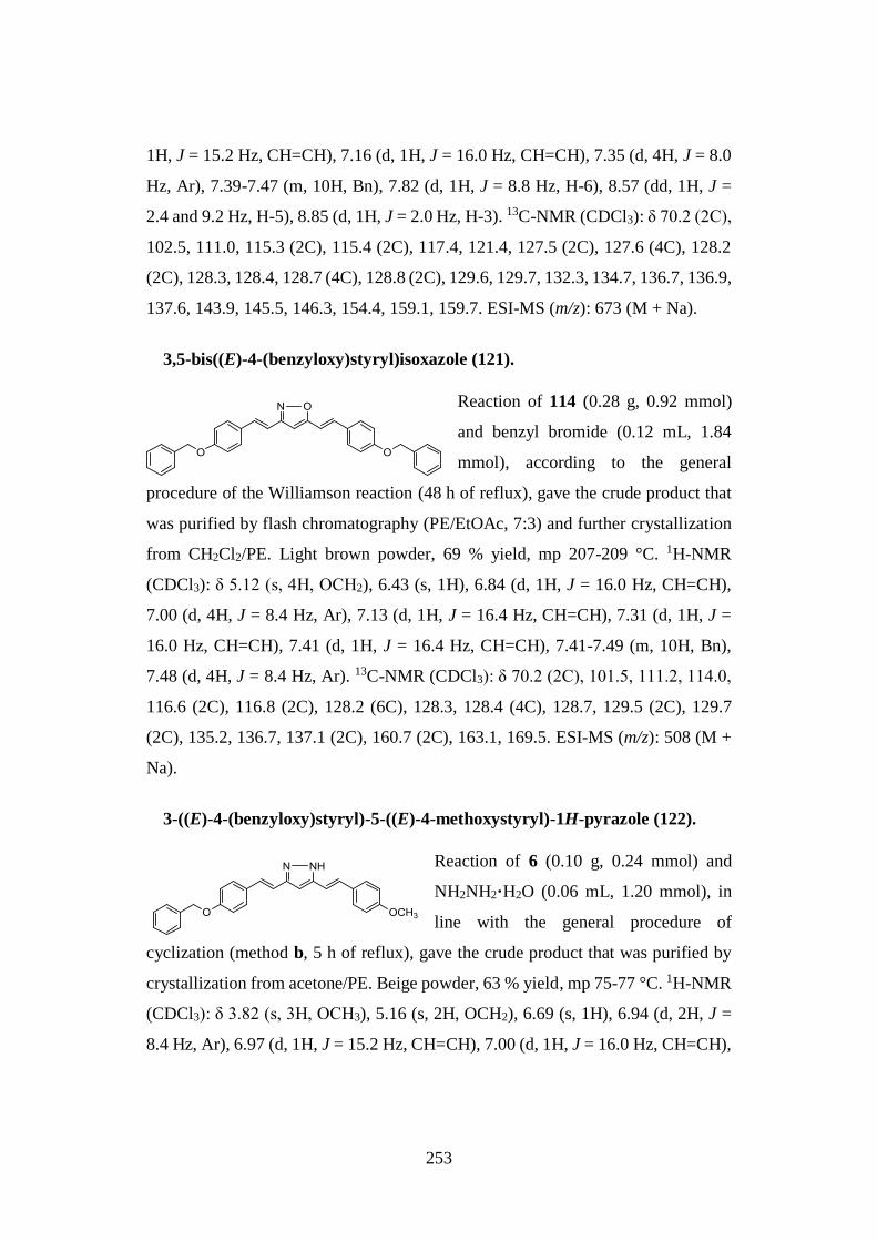
253
1H, J = 15.2 Hz, CH=CH), 7.16 (d, 1H, J = 16.0 Hz, CH=CH), 7.35 (d, 4H, J = 8.0
Hz, Ar), 7.39-7.47 (m, 10H, Bn), 7.82 (d, 1H, J = 8.8 Hz, H-6), 8.57 (dd, 1H, J =
2.4 and 9.2 Hz, H-5), 8.85 (d, 1H, J = 2.0 Hz, H-3). 13C-NMR (CDCl3): δ 70.2 (2C),
102.5, 111.0, 115.3 (2C), 115.4 (2C), 117.4, 121.4, 127.5 (2C), 127.6 (4C), 128.2
(2C), 128.3, 128.4, 128.7 (4C), 128.8 (2C), 129.6, 129.7, 132.3, 134.7, 136.7, 136.9,
137.6, 143.9, 145.5, 146.3, 154.4, 159.1, 159.7. ESI-MS (m/z): 673 (M + Na).
3,5-bis((E)-4-(benzyloxy)styryl)isoxazole (121).
Reaction of 114 (0.28 g, 0.92 mmol)
and benzyl bromide (0.12 mL, 1.84
mmol), according to the general
procedure of the Williamson reaction (48 h of reflux), gave the crude product that
was purified by flash chromatography (PE/EtOAc, 7:3) and further crystallization
from CH2Cl2/PE. Light brown powder, 69 % yield, mp 207-209 °C. 1H-NMR
(CDCl3): δ 5.12 (s, 4H, OCH2), 6.43 (s, 1H), 6.84 (d, 1H, J = 16.0 Hz, CH=CH),
7.00 (d, 4H, J = 8.4 Hz, Ar), 7.13 (d, 1H, J = 16.4 Hz, CH=CH), 7.31 (d, 1H, J =
16.0 Hz, CH=CH), 7.41 (d, 1H, J = 16.4 Hz, CH=CH), 7.41-7.49 (m, 10H, Bn),
7.48 (d, 4H, J = 8.4 Hz, Ar). 13C-NMR (CDCl3): δ 70.2 (2C), 101.5, 111.2, 114.0,
116.6 (2C), 116.8 (2C), 128.2 (6C), 128.3, 128.4 (4C), 128.7, 129.5 (2C), 129.7
(2C), 135.2, 136.7, 137.1 (2C), 160.7 (2C), 163.1, 169.5. ESI-MS (m/z): 508 (M +
Na).
3-((E)-4-(benzyloxy)styryl)-5-((E)-4-methoxystyryl)-1H-pyrazole (122).
Reaction of 6 (0.10 g, 0.24 mmol) and
NH2NH2·H2O (0.06 mL, 1.20 mmol), in
line with the general procedure of
cyclization (method b, 5 h of reflux), gave the crude product that was purified by
crystallization from acetone/PE. Beige powder, 63 % yield, mp 75-77 °C. 1H-NMR
(CDCl3): δ 3.82 (s, 3H, OCH3), 5.16 (s, 2H, OCH2), 6.69 (s, 1H), 6.94 (d, 2H, J =
8.4 Hz, Ar), 6.97 (d, 1H, J = 15.2 Hz, CH=CH), 7.00 (d, 1H, J = 16.0 Hz, CH=CH),
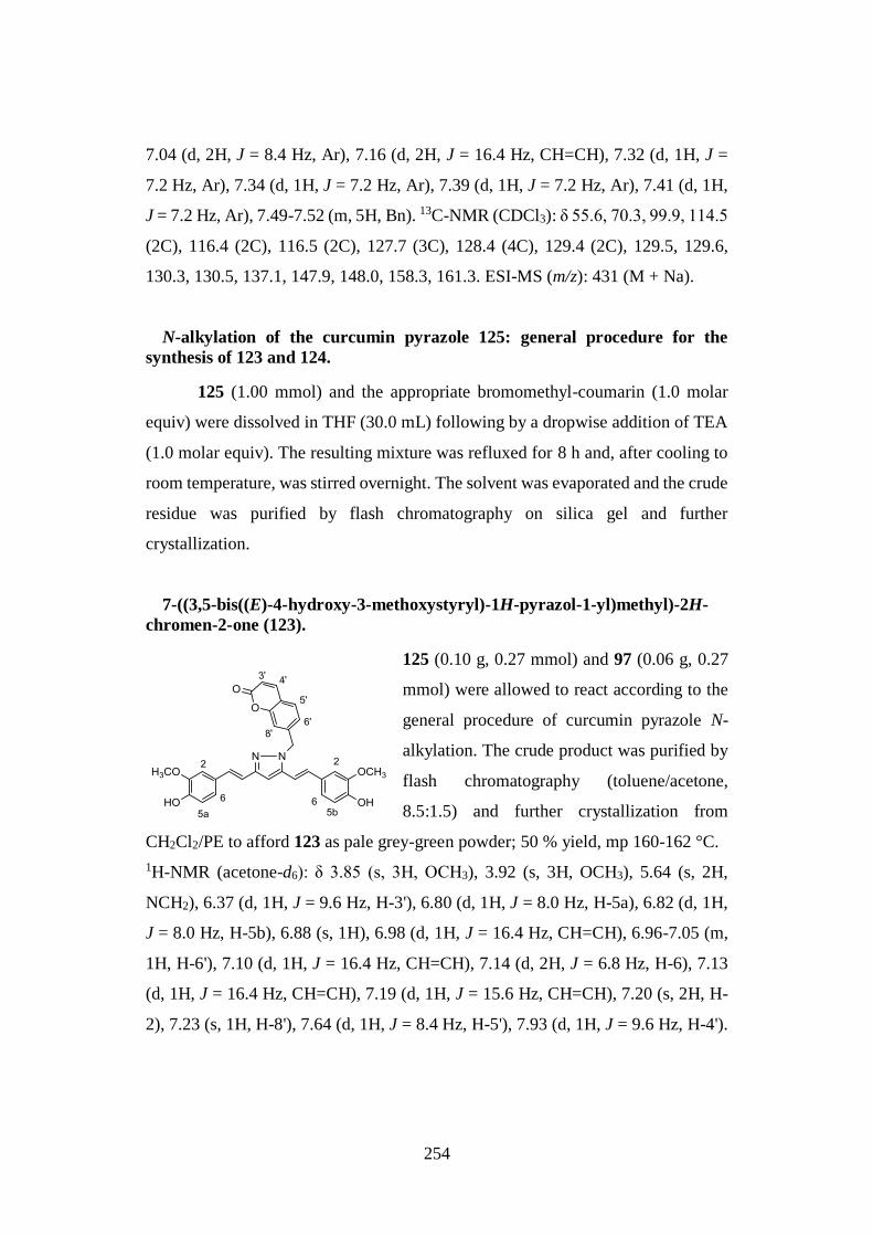
254
7.04 (d, 2H, J = 8.4 Hz, Ar), 7.16 (d, 2H, J = 16.4 Hz, CH=CH), 7.32 (d, 1H, J =
7.2 Hz, Ar), 7.34 (d, 1H, J = 7.2 Hz, Ar), 7.39 (d, 1H, J = 7.2 Hz, Ar), 7.41 (d, 1H,
J = 7.2 Hz, Ar), 7.49-7.52 (m, 5H, Bn). 13C-NMR (CDCl3): δ 55.6, 70.3, 99.9, 114.5
(2C), 116.4 (2C), 116.5 (2C), 127.7 (3C), 128.4 (4C), 129.4 (2C), 129.5, 129.6,
130.3, 130.5, 137.1, 147.9, 148.0, 158.3, 161.3. ESI-MS (m/z): 431 (M + Na).
N-alkylation of the curcumin pyrazole 125: general procedure for the
synthesis of 123 and 124.
125 (1.00 mmol) and the appropriate bromomethyl-coumarin (1.0 molar
equiv) were dissolved in THF (30.0 mL) following by a dropwise addition of TEA
(1.0 molar equiv). The resulting mixture was refluxed for 8 h and, after cooling to
room temperature, was stirred overnight. The solvent was evaporated and the crude
residue was purified by flash chromatography on silica gel and further
crystallization.
7-((3,5-bis((E)-4-hydroxy-3-methoxystyryl)-1H-pyrazol-1-yl)methyl)-2H-
chromen-2-one (123).
125 (0.10 g, 0.27 mmol) and 97 (0.06 g, 0.27
mmol) were allowed to react according to the
general procedure of curcumin pyrazole N-
alkylation. The crude product was purified by
flash chromatography (toluene/acetone,
8.5:1.5) and further crystallization from
CH2Cl2/PE to afford 123 as pale grey-green powder; 50 % yield, mp 160-162 °C.
1H-NMR (acetone-d6): δ 3.85 (s, 3H, OCH3), 3.92 (s, 3H, OCH3), 5.64 (s, 2H,
NCH2), 6.37 (d, 1H, J = 9.6 Hz, H-3'), 6.80 (d, 1H, J = 8.0 Hz, H-5a), 6.82 (d, 1H,
J = 8.0 Hz, H-5b), 6.88 (s, 1H), 6.98 (d, 1H, J = 16.4 Hz, CH=CH), 6.96-7.05 (m,
1H, H-6'), 7.10 (d, 1H, J = 16.4 Hz, CH=CH), 7.14 (d, 2H, J = 6.8 Hz, H-6), 7.13
(d, 1H, J = 16.4 Hz, CH=CH), 7.19 (d, 1H, J = 15.6 Hz, CH=CH), 7.20 (s, 2H, H-
2), 7.23 (s, 1H, H-8'), 7.64 (d, 1H, J = 8.4 Hz, H-5'), 7.93 (d, 1H, J = 9.6 Hz, H-4').
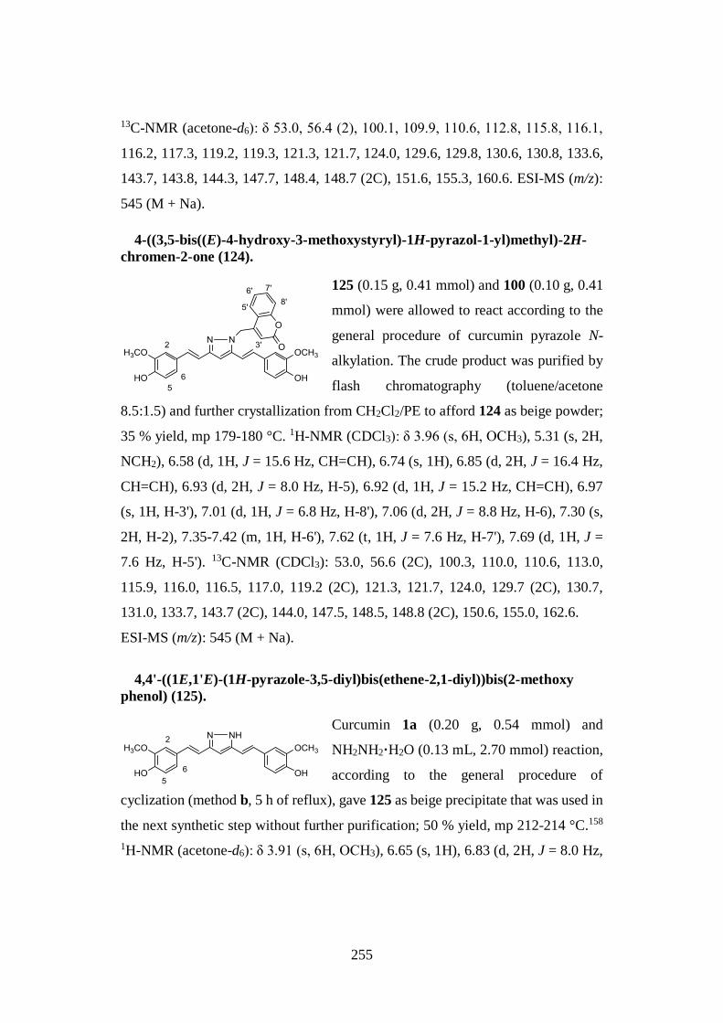
255
13C-NMR (acetone-d6): δ 53.0, 56.4 (2), 100.1, 109.9, 110.6, 112.8, 115.8, 116.1,
116.2, 117.3, 119.2, 119.3, 121.3, 121.7, 124.0, 129.6, 129.8, 130.6, 130.8, 133.6,
143.7, 143.8, 144.3, 147.7, 148.4, 148.7 (2C), 151.6, 155.3, 160.6. ESI-MS (m/z):
545 (M + Na).
4-((3,5-bis((E)-4-hydroxy-3-methoxystyryl)-1H-pyrazol-1-yl)methyl)-2H-
chromen-2-one (124).
125 (0.15 g, 0.41 mmol) and 100 (0.10 g, 0.41
mmol) were allowed to react according to the
general procedure of curcumin pyrazole N-
alkylation. The crude product was purified by
flash chromatography (toluene/acetone
8.5:1.5) and further crystallization from CH2Cl2/PE to afford 124 as beige powder;
35 % yield, mp 179-180 °C. 1H-NMR (CDCl3): δ 3.96 (s, 6H, OCH3), 5.31 (s, 2H,
NCH2), 6.58 (d, 1H, J = 15.6 Hz, CH=CH), 6.74 (s, 1H), 6.85 (d, 2H, J = 16.4 Hz,
CH=CH), 6.93 (d, 2H, J = 8.0 Hz, H-5), 6.92 (d, 1H, J = 15.2 Hz, CH=CH), 6.97
(s, 1H, H-3'), 7.01 (d, 1H, J = 6.8 Hz, H-8'), 7.06 (d, 2H, J = 8.8 Hz, H-6), 7.30 (s,
2H, H-2), 7.35-7.42 (m, 1H, H-6'), 7.62 (t, 1H, J = 7.6 Hz, H-7'), 7.69 (d, 1H, J =
7.6 Hz, H-5'). 13C-NMR (CDCl3): 53.0, 56.6 (2C), 100.3, 110.0, 110.6, 113.0,
115.9, 116.0, 116.5, 117.0, 119.2 (2C), 121.3, 121.7, 124.0, 129.7 (2C), 130.7,
131.0, 133.7, 143.7 (2C), 144.0, 147.5, 148.5, 148.8 (2C), 150.6, 155.0, 162.6.
ESI-MS (m/z): 545 (M + Na).
4,4'-((1E,1'E)-(1H-pyrazole-3,5-diyl)bis(ethene-2,1-diyl))bis(2-methoxy
phenol) (125).
Curcumin 1a (0.20 g, 0.54 mmol) and
NH2NH2·H2O (0.13 mL, 2.70 mmol) reaction,
according to the general procedure of
cyclization (method b, 5 h of reflux), gave 125 as beige precipitate that was used in
the next synthetic step without further purification; 50 % yield, mp 212-214 °C.158
1H-NMR (acetone-d6): δ 3.91 (s, 6H, OCH3), 6.65 (s, 1H), 6.83 (d, 2H, J = 8.0 Hz,
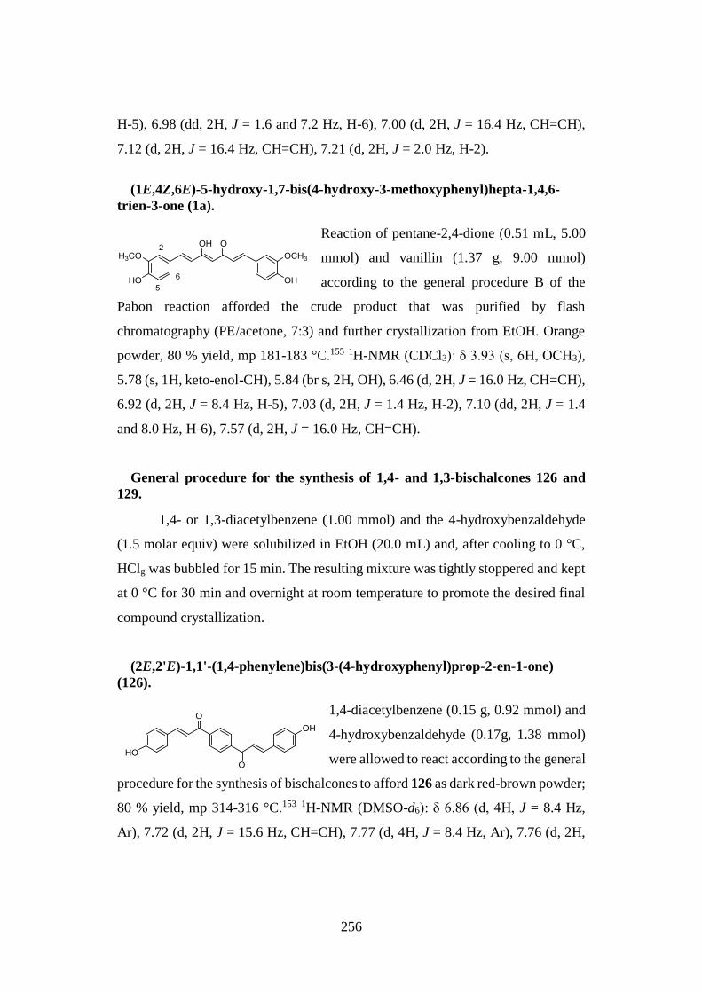
256
H-5), 6.98 (dd, 2H, J = 1.6 and 7.2 Hz, H-6), 7.00 (d, 2H, J = 16.4 Hz, CH=CH),
7.12 (d, 2H, J = 16.4 Hz, CH=CH), 7.21 (d, 2H, J = 2.0 Hz, H-2).
(1E,4Z,6E)-5-hydroxy-1,7-bis(4-hydroxy-3-methoxyphenyl)hepta-1,4,6-
trien-3-one (1a).
Reaction of pentane-2,4-dione (0.51 mL, 5.00
mmol) and vanillin (1.37 g, 9.00 mmol)
according to the general procedure B of the
Pabon reaction afforded the crude product that was purified by flash
chromatography (PE/acetone, 7:3) and further crystallization from EtOH. Orange
powder, 80 % yield, mp 181-183 °C.155 1H-NMR (CDCl3): δ 3.93 (s, 6H, OCH3),
5.78 (s, 1H, keto-enol-CH), 5.84 (br s, 2H, OH), 6.46 (d, 2H, J = 16.0 Hz, CH=CH),
6.92 (d, 2H, J = 8.4 Hz, H-5), 7.03 (d, 2H, J = 1.4 Hz, H-2), 7.10 (dd, 2H, J = 1.4
and 8.0 Hz, H-6), 7.57 (d, 2H, J = 16.0 Hz, CH=CH).
General procedure for the synthesis of 1,4- and 1,3-bischalcones 126 and
129.
1,4- or 1,3-diacetylbenzene (1.00 mmol) and the 4-hydroxybenzaldehyde
(1.5 molar equiv) were solubilized in EtOH (20.0 mL) and, after cooling to 0 °C,
HClg was bubbled for 15 min. The resulting mixture was tightly stoppered and kept
at 0 °C for 30 min and overnight at room temperature to promote the desired final
compound crystallization.
(2E,2'E)-1,1'-(1,4-phenylene)bis(3-(4-hydroxyphenyl)prop-2-en-1-one)
(126).
1,4-diacetylbenzene (0.15 g, 0.92 mmol) and
4-hydroxybenzaldehyde (0.17g, 1.38 mmol)
were allowed to react according to the general
procedure for the synthesis of bischalcones to afford 126 as dark red-brown powder;
80 % yield, mp 314-316 °C.153 1H-NMR (DMSO-d6): δ 6.86 (d, 4H, J = 8.4 Hz,
Ar), 7.72 (d, 2H, J = 15.6 Hz, CH=CH), 7.77 (d, 4H, J = 8.4 Hz, Ar), 7.76 (d, 2H,
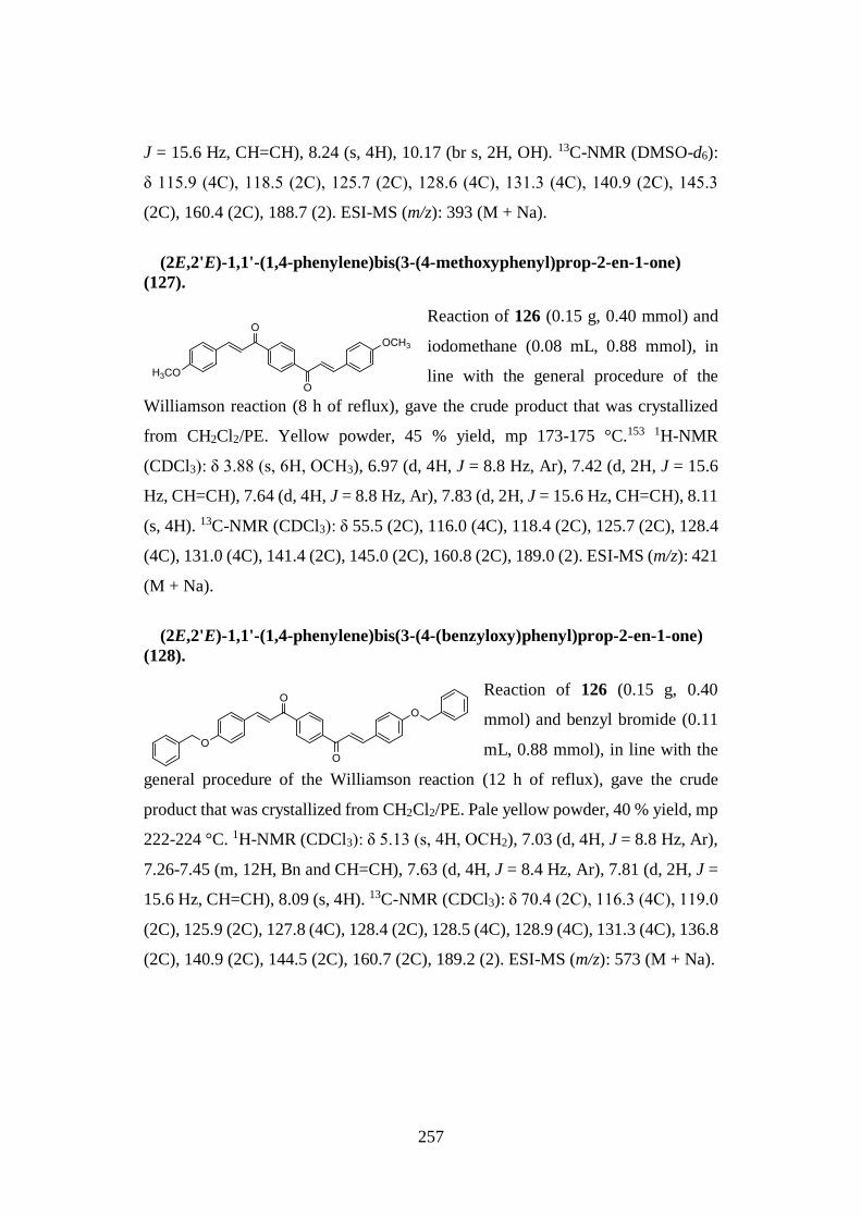
257
J = 15.6 Hz, CH=CH), 8.24 (s, 4H), 10.17 (br s, 2H, OH). 13C-NMR (DMSO-d6):
δ 115.9 (4C), 118.5 (2C), 125.7 (2C), 128.6 (4C), 131.3 (4C), 140.9 (2C), 145.3
(2C), 160.4 (2C), 188.7 (2). ESI-MS (m/z): 393 (M + Na).
(2E,2'E)-1,1'-(1,4-phenylene)bis(3-(4-methoxyphenyl)prop-2-en-1-one)
(127).
Reaction of 126 (0.15 g, 0.40 mmol) and
iodomethane (0.08 mL, 0.88 mmol), in
line with the general procedure of the
Williamson reaction (8 h of reflux), gave the crude product that was crystallized
from CH2Cl2/PE. Yellow powder, 45 % yield, mp 173-175 °C.153 1H-NMR
(CDCl3): δ 3.88 (s, 6H, OCH3), 6.97 (d, 4H, J = 8.8 Hz, Ar), 7.42 (d, 2H, J = 15.6
Hz, CH=CH), 7.64 (d, 4H, J = 8.8 Hz, Ar), 7.83 (d, 2H, J = 15.6 Hz, CH=CH), 8.11
(s, 4H). 13C-NMR (CDCl3): δ 55.5 (2C), 116.0 (4C), 118.4 (2C), 125.7 (2C), 128.4
(4C), 131.0 (4C), 141.4 (2C), 145.0 (2C), 160.8 (2C), 189.0 (2). ESI-MS (m/z): 421
(M + Na).
(2E,2'E)-1,1'-(1,4-phenylene)bis(3-(4-(benzyloxy)phenyl)prop-2-en-1-one)
(128).
Reaction of 126 (0.15 g, 0.40
mmol) and benzyl bromide (0.11
mL, 0.88 mmol), in line with the
general procedure of the Williamson reaction (12 h of reflux), gave the crude
product that was crystallized from CH2Cl2/PE. Pale yellow powder, 40 % yield, mp
222-224 °C. 1H-NMR (CDCl3): δ 5.13 (s, 4H, OCH2), 7.03 (d, 4H, J = 8.8 Hz, Ar),
7.26-7.45 (m, 12H, Bn and CH=CH), 7.63 (d, 4H, J = 8.4 Hz, Ar), 7.81 (d, 2H, J =
15.6 Hz, CH=CH), 8.09 (s, 4H). 13C-NMR (CDCl3): δ 70.4 (2C), 116.3 (4C), 119.0
(2C), 125.9 (2C), 127.8 (4C), 128.4 (2C), 128.5 (4C), 128.9 (4C), 131.3 (4C), 136.8
(2C), 140.9 (2C), 144.5 (2C), 160.7 (2C), 189.2 (2). ESI-MS (m/z): 573 (M + Na).
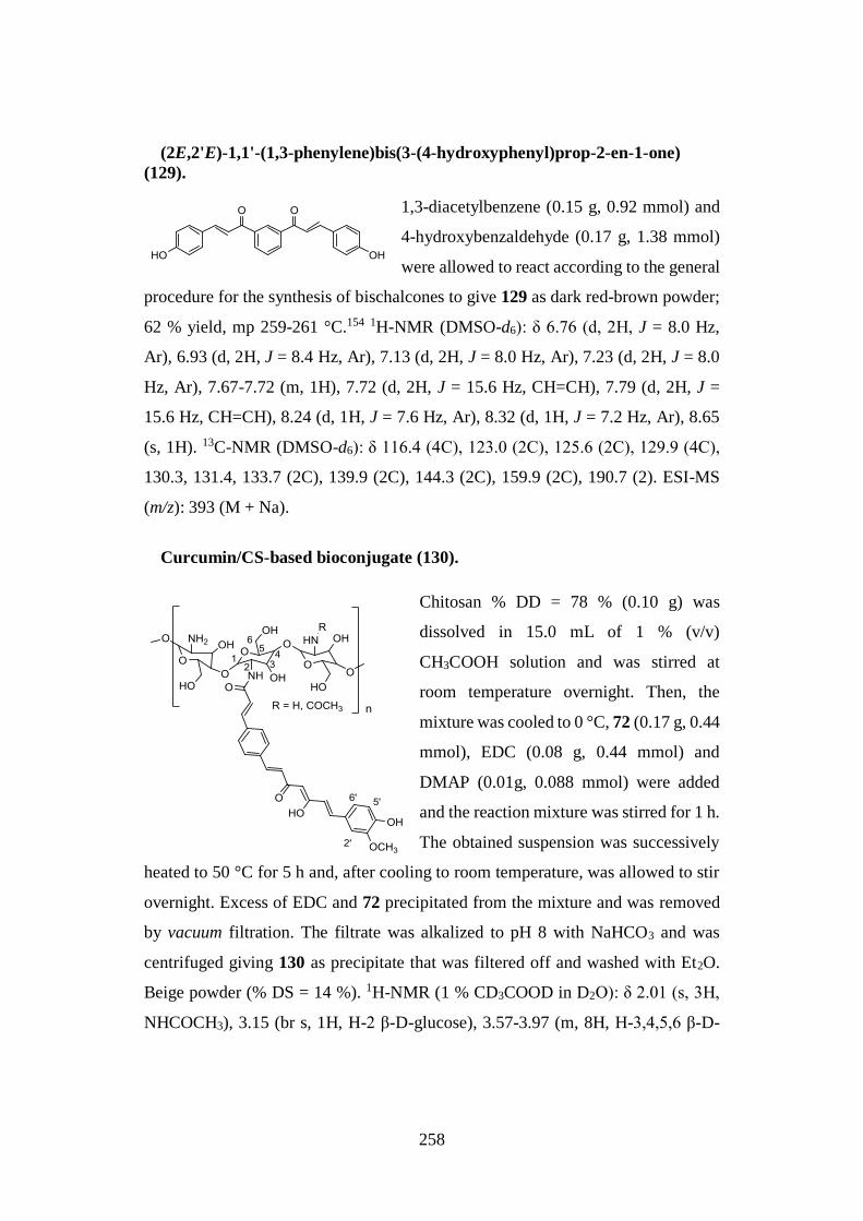
258
(2E,2'E)-1,1'-(1,3-phenylene)bis(3-(4-hydroxyphenyl)prop-2-en-1-one)
(129).
1,3-diacetylbenzene (0.15 g, 0.92 mmol) and
4-hydroxybenzaldehyde (0.17 g, 1.38 mmol)
were allowed to react according to the general
procedure for the synthesis of bischalcones to give 129 as dark red-brown powder;
62 % yield, mp 259-261 °C.154 1H-NMR (DMSO-d6): δ 6.76 (d, 2H, J = 8.0 Hz,
Ar), 6.93 (d, 2H, J = 8.4 Hz, Ar), 7.13 (d, 2H, J = 8.0 Hz, Ar), 7.23 (d, 2H, J = 8.0
Hz, Ar), 7.67-7.72 (m, 1H), 7.72 (d, 2H, J = 15.6 Hz, CH=CH), 7.79 (d, 2H, J =
15.6 Hz, CH=CH), 8.24 (d, 1H, J = 7.6 Hz, Ar), 8.32 (d, 1H, J = 7.2 Hz, Ar), 8.65
(s, 1H). 13C-NMR (DMSO-d6): δ 116.4 (4C), 123.0 (2C), 125.6 (2C), 129.9 (4C),
130.3, 131.4, 133.7 (2C), 139.9 (2C), 144.3 (2C), 159.9 (2C), 190.7 (2). ESI-MS
(m/z): 393 (M + Na).
Curcumin/CS-based bioconjugate (130).
Chitosan % DD = 78 % (0.10 g) was
dissolved in 15.0 mL of 1 % (v/v)
CH3COOH solution and was stirred at
room temperature overnight. Then, the
mixture was cooled to 0 °C, 72 (0.17 g, 0.44
mmol), EDC (0.08 g, 0.44 mmol) and
DMAP (0.01g, 0.088 mmol) were added
and the reaction mixture was stirred for 1 h.
The obtained suspension was successively
heated to 50 °C for 5 h and, after cooling to room temperature, was allowed to stir
overnight. Excess of EDC and 72 precipitated from the mixture and was removed
by vacuum filtration. The filtrate was alkalized to pH 8 with NaHCO3 and was
centrifuged giving 130 as precipitate that was filtered off and washed with Et2O.
Beige powder (% DS = 14 %). 1H-NMR (1 % CD3COOD in D2O): δ 2.01 (s, 3H,
NHCOCH3), 3.15 (br s, 1H, H-2 β-D-glucose), 3.57-3.97 (m, 8H, H-3,4,5,6 β-D-
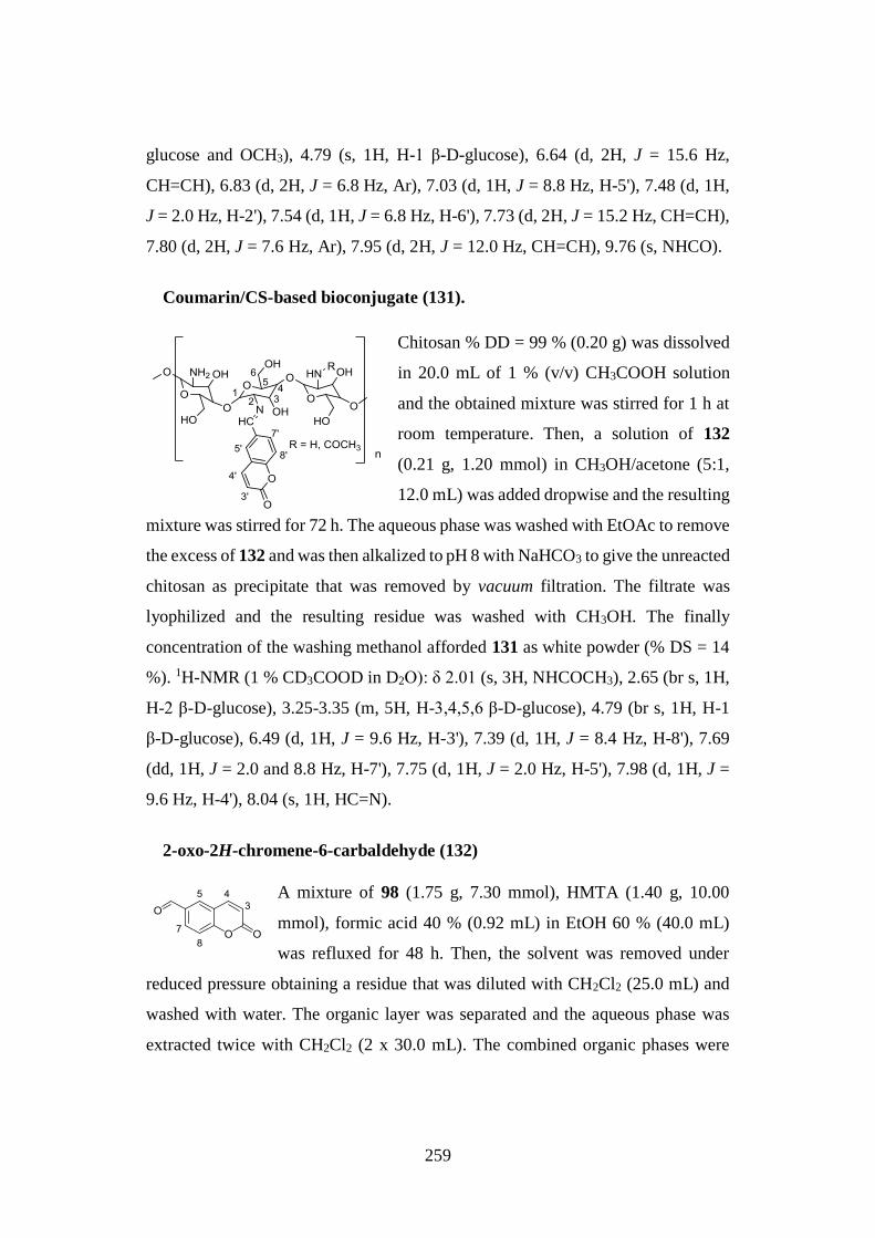
259
glucose and OCH3), 4.79 (s, 1H, H-1 β-D-glucose), 6.64 (d, 2H, J = 15.6 Hz,
CH=CH), 6.83 (d, 2H, J = 6.8 Hz, Ar), 7.03 (d, 1H, J = 8.8 Hz, H-5'), 7.48 (d, 1H,
J = 2.0 Hz, H-2'), 7.54 (d, 1H, J = 6.8 Hz, H-6'), 7.73 (d, 2H, J = 15.2 Hz, CH=CH),
7.80 (d, 2H, J = 7.6 Hz, Ar), 7.95 (d, 2H, J = 12.0 Hz, CH=CH), 9.76 (s, NHCO).
Coumarin/CS-based bioconjugate (131).
Chitosan % DD = 99 % (0.20 g) was dissolved
in 20.0 mL of 1 % (v/v) CH3COOH solution
and the obtained mixture was stirred for 1 h at
room temperature. Then, a solution of 132
(0.21 g, 1.20 mmol) in CH3OH/acetone (5:1,
12.0 mL) was added dropwise and the resulting
mixture was stirred for 72 h. The aqueous phase was washed with EtOAc to remove
the excess of 132 and was then alkalized to pH 8 with NaHCO3 to give the unreacted
chitosan as precipitate that was removed by vacuum filtration. The filtrate was
lyophilized and the resulting residue was washed with CH3OH. The finally
concentration of the washing methanol afforded 131 as white powder (% DS = 14
%). 1H-NMR (1 % CD3COOD in D2O): δ 2.01 (s, 3H, NHCOCH3), 2.65 (br s, 1H,
H-2 β-D-glucose), 3.25-3.35 (m, 5H, H-3,4,5,6 β-D-glucose), 4.79 (br s, 1H, H-1
β-D-glucose), 6.49 (d, 1H, J = 9.6 Hz, H-3'), 7.39 (d, 1H, J = 8.4 Hz, H-8'), 7.69
(dd, 1H, J = 2.0 and 8.8 Hz, H-7'), 7.75 (d, 1H, J = 2.0 Hz, H-5'), 7.98 (d, 1H, J =
9.6 Hz, H-4'), 8.04 (s, 1H, HC=N).
2-oxo-2H-chromene-6-carbaldehyde (132)
A mixture of 98 (1.75 g, 7.30 mmol), HMTA (1.40 g, 10.00
mmol), formic acid 40 % (0.92 mL) in EtOH 60 % (40.0 mL)
was refluxed for 48 h. Then, the solvent was removed under
reduced pressure obtaining a residue that was diluted with CH2Cl2 (25.0 mL) and
washed with water. The organic layer was separated and the aqueous phase was
extracted twice with CH2Cl2 (2 x 30.0 mL). The combined organic phases were
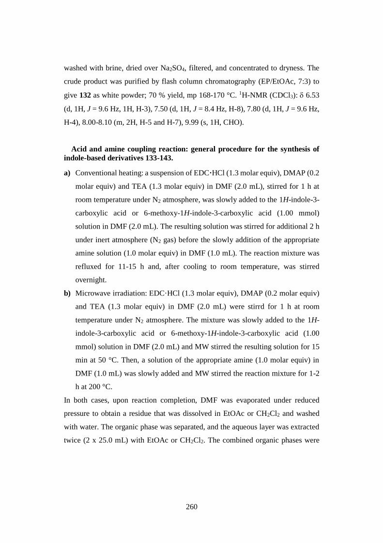
260
washed with brine, dried over Na2SO4, filtered, and concentrated to dryness. The
crude product was purified by flash column chromatography (EP/EtOAc, 7:3) to
give 132 as white powder; 70 % yield, mp 168-170 °C. 1H-NMR (CDCl3): 6.53
(d, 1H, J = 9.6 Hz, 1H, H-3), 7.50 (d, 1H, J = 8.4 Hz, H-8), 7.80 (d, 1H, J = 9.6 Hz,
H-4), 8.00-8.10 (m, 2H, H-5 and H-7), 9.99 (s, 1H, CHO).
Acid and amine coupling reaction: general procedure for the synthesis of
indole-based derivatives 133-143.
a) Conventional heating: a suspension of EDC·HCl (1.3 molar equiv), DMAP (0.2
molar equiv) and TEA (1.3 molar equiv) in DMF (2.0 mL), stirred for 1 h at
room temperature under N2 atmosphere, was slowly added to the 1H-indole-3-
carboxylic acid or 6-methoxy-1H-indole-3-carboxylic acid (1.00 mmol)
solution in DMF (2.0 mL). The resulting solution was stirred for additional 2 h
under inert atmosphere (N2 gas) before the slowly addition of the appropriate
amine solution (1.0 molar equiv) in DMF (1.0 mL). The reaction mixture was
refluxed for 11-15 h and, after cooling to room temperature, was stirred
overnight.
b) Microwave irradiation: EDC·HCl (1.3 molar equiv), DMAP (0.2 molar equiv)
and TEA (1.3 molar equiv) in DMF (2.0 mL) were stirrd for 1 h at room
temperature under N2 atmosphere. The mixture was slowly added to the 1H-
indole-3-carboxylic acid or 6-methoxy-1H-indole-3-carboxylic acid (1.00
mmol) solution in DMF (2.0 mL) and MW stirred the resulting solution for 15
min at 50 °C. Then, a solution of the appropriate amine (1.0 molar equiv) in
DMF (1.0 mL) was slowly added and MW stirred the reaction mixture for 1-2
h at 200 °C.
In both cases, upon reaction completion, DMF was evaporated under reduced
pressure to obtain a residue that was dissolved in EtOAc or CH2Cl2 and washed
with water. The organic phase was separated, and the aqueous layer was extracted
twice (2 x 25.0 mL) with EtOAc or CH2Cl2. The combined organic phases were
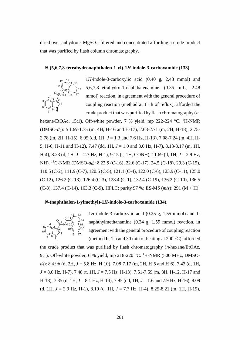
261
dried over anhydrous MgSO4, filtered and concentrated affording a crude product
that was purified by flash column chromatography.
N-(5,6,7,8-tetrahydronaphthalen-1-yl)-1H-indole-3-carboxamide (133).
1H-indole-3-carboxylic acid (0.40 g, 2.48 mmol) and
5,6,7,8-tetrahydro-1-naphthalenamine (0.35 mL, 2.48
mmol) reaction, in agreement with the general procedure of
coupling reaction (method a, 11 h of reflux), afforded the
crude product that was purified by flash chromatography (n-
hexane/EtOAc, 15:1). Off-white powder, 7 % yield, mp 222-224 °C. 1H-NMR
(DMSO-d6): δ 1.69-1.75 (m, 4H, H-16 and H-17), 2.68-2.71 (m, 2H, H-18), 2.75-
2.78 (m, 2H, H-15), 6.95 (dd, 1H, J = 1.3 and 7.6 Hz, H-13), 7.08-7.24 (m, 4H, H-
5, H-6, H-11 and H-12), 7.47 (dd, 1H, J = 1.0 and 8.0 Hz, H-7), 8.13-8.17 (m, 1H,
H-4), 8.23 (d, 1H, J = 2.7 Hz, H-1), 9.15 (s, 1H, CONH), 11.69 (d, 1H, J = 2.9 Hz,
NH). 13C-NMR (DMSO-d6): δ 22.5 (C-16), 22.6 (C-17), 24.5 (C-18), 29.3 (C-15),
110.5 (C-2), 111.9 (C-7), 120.6 (C-5), 121.1 (C-4), 122.0 (C-6), 123.9 (C-11), 125.0
(C-12), 126.2 (C-13), 126.4 (C-3), 128.4 (C-1), 132.4 (C-19), 136.2 (C-10), 136.5
(C-8), 137.4 (C-14), 163.3 (C-9). HPLC: purity 97 %; ES-MS (m/z): 291 (M + H).
N-(naphthalen-1-ylmethyl)-1H-indole-3-carboxamide (134).
1H-indole-3-carboxylic acid (0.25 g, 1.55 mmol) and 1-
naphthylmethanamine (0.24 g, 1.55 mmol) reaction, in
agreement with the general procedure of coupling reaction
(method b, 1 h and 30 min of heating at 200 °C), afforded
the crude product that was purified by flash chromatography (n-hexane/EtOAc,
9:1). Off-white powder, 6 % yield, mp 218-220 °C. 1H-NMR (500 MHz, DMSO-
d6): δ 4.96 (d, 2H, J = 5.8 Hz, H-10), 7.08-7.17 (m, 2H, H-5 and H-6), 7.43 (d, 1H,
J = 8.0 Hz, H-7), 7.48 (t, 1H, J = 7.5 Hz, H-13), 7.51-7.59 (m, 3H, H-12, H-17 and
H-18), 7.85 (d, 1H, J = 8.1 Hz, H-14), 7.95 (dd, 1H, J = 1.6 and 7.9 Hz, H-16), 8.09
(d, 1H, J = 2.9 Hz, H-1), 8.19 (d, 1H, J = 7.7 Hz, H-4), 8.25-8.21 (m, 1H, H-19),
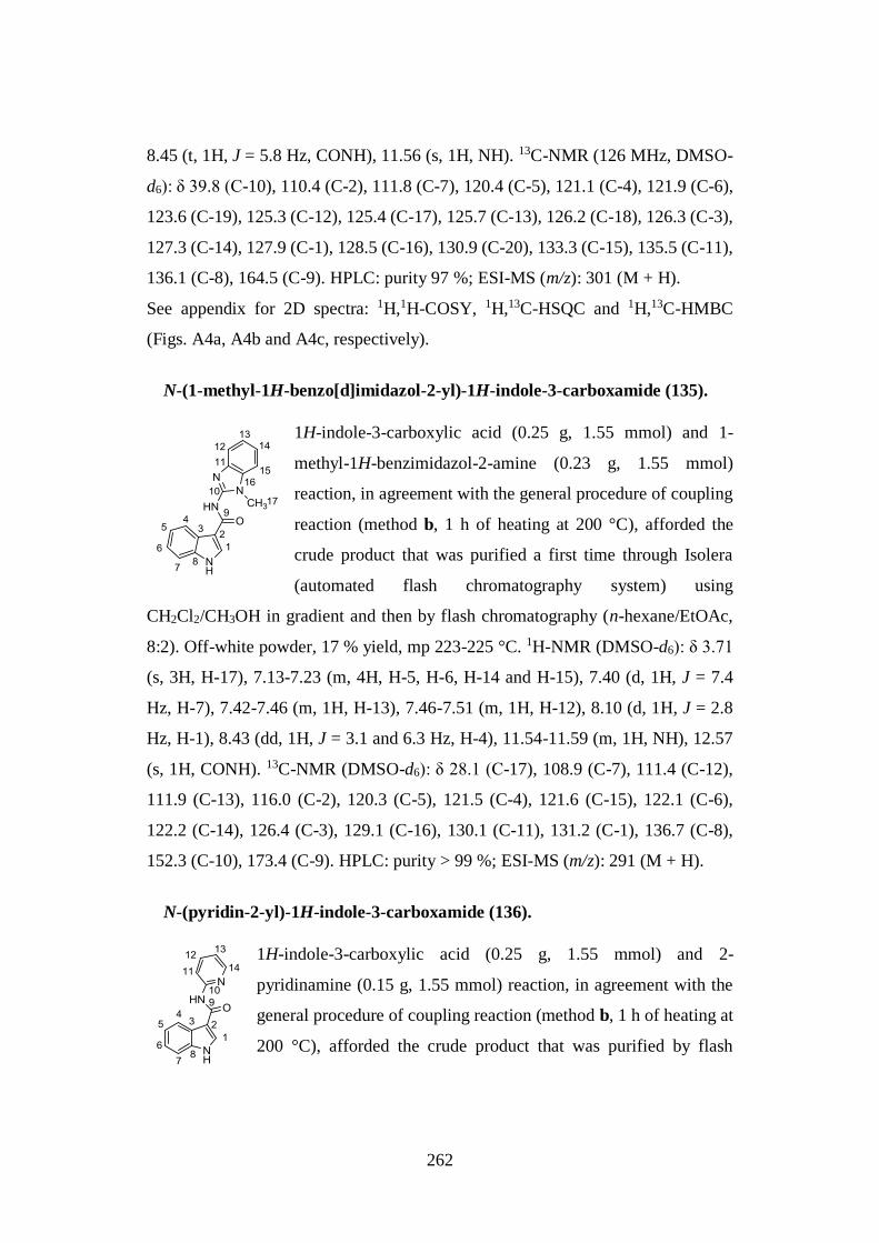
262
8.45 (t, 1H, J = 5.8 Hz, CONH), 11.56 (s, 1H, NH). 13C-NMR (126 MHz, DMSO-
d6): δ 39.8 (C-10), 110.4 (C-2), 111.8 (C-7), 120.4 (C-5), 121.1 (C-4), 121.9 (C-6),
123.6 (C-19), 125.3 (C-12), 125.4 (C-17), 125.7 (C-13), 126.2 (C-18), 126.3 (C-3),
127.3 (C-14), 127.9 (C-1), 128.5 (C-16), 130.9 (C-20), 133.3 (C-15), 135.5 (C-11),
136.1 (C-8), 164.5 (C-9). HPLC: purity 97 %; ESI-MS (m/z): 301 (M + H).
See appendix for 2D spectra: 1H,1H-COSY, 1H,13C-HSQC and 1H,13C-HMBC
(Figs. A4a, A4b and A4c, respectively).
N-(1-methyl-1H-benzo[d]imidazol-2-yl)-1H-indole-3-carboxamide (135).
1H-indole-3-carboxylic acid (0.25 g, 1.55 mmol) and 1-
methyl-1H-benzimidazol-2-amine (0.23 g, 1.55 mmol)
reaction, in agreement with the general procedure of coupling
reaction (method b, 1 h of heating at 200 °C), afforded the
crude product that was purified a first time through Isolera
(automated flash chromatography system) using
CH2Cl2/CH3OH in gradient and then by flash chromatography (n-hexane/EtOAc,
8:2). Off-white powder, 17 % yield, mp 223-225 °C. 1H-NMR (DMSO-d6): δ 3.71
(s, 3H, H-17), 7.13-7.23 (m, 4H, H-5, H-6, H-14 and H-15), 7.40 (d, 1H, J = 7.4
Hz, H-7), 7.42-7.46 (m, 1H, H-13), 7.46-7.51 (m, 1H, H-12), 8.10 (d, 1H, J = 2.8
Hz, H-1), 8.43 (dd, 1H, J = 3.1 and 6.3 Hz, H-4), 11.54-11.59 (m, 1H, NH), 12.57
(s, 1H, CONH). 13C-NMR (DMSO-d6): δ 28.1 (C-17), 108.9 (C-7), 111.4 (C-12),
111.9 (C-13), 116.0 (C-2), 120.3 (C-5), 121.5 (C-4), 121.6 (C-15), 122.1 (C-6),
122.2 (C-14), 126.4 (C-3), 129.1 (C-16), 130.1 (C-11), 131.2 (C-1), 136.7 (C-8),
152.3 (C-10), 173.4 (C-9). HPLC: purity > 99 %; ESI-MS (m/z): 291 (M + H).
N-(pyridin-2-yl)-1H-indole-3-carboxamide (136).
1H-indole-3-carboxylic acid (0.25 g, 1.55 mmol) and 2-
pyridinamine (0.15 g, 1.55 mmol) reaction, in agreement with the
general procedure of coupling reaction (method b, 1 h of heating at
200 °C), afforded the crude product that was purified by flash
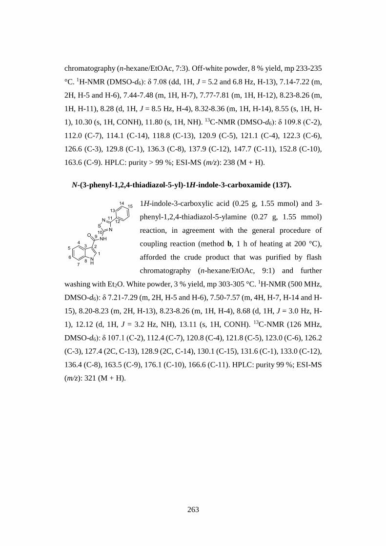
263
chromatography (n-hexane/EtOAc, 7:3). Off-white powder, 8 % yield, mp 233-235
°C. 1H-NMR (DMSO-d6): δ 7.08 (dd, 1H, J = 5.2 and 6.8 Hz, H-13), 7.14-7.22 (m,
2H, H-5 and H-6), 7.44-7.48 (m, 1H, H-7), 7.77-7.81 (m, 1H, H-12), 8.23-8.26 (m,
1H, H-11), 8.28 (d, 1H, J = 8.5 Hz, H-4), 8.32-8.36 (m, 1H, H-14), 8.55 (s, 1H, H-
1), 10.30 (s, 1H, CONH), 11.80 (s, 1H, NH). 13C-NMR (DMSO-d6): δ 109.8 (C-2),
112.0 (C-7), 114.1 (C-14), 118.8 (C-13), 120.9 (C-5), 121.1 (C-4), 122.3 (C-6),
126.6 (C-3), 129.8 (C-1), 136.3 (C-8), 137.9 (C-12), 147.7 (C-11), 152.8 (C-10),
163.6 (C-9). HPLC: purity > 99 %; ESI-MS (m/z): 238 (M + H).
N-(3-phenyl-1,2,4-thiadiazol-5-yl)-1H-indole-3-carboxamide (137).
1H-indole-3-carboxylic acid (0.25 g, 1.55 mmol) and 3-
phenyl-1,2,4-thiadiazol-5-ylamine (0.27 g, 1.55 mmol)
reaction, in agreement with the general procedure of
coupling reaction (method b, 1 h of heating at 200 °C),
afforded the crude product that was purified by flash
chromatography (n-hexane/EtOAc, 9:1) and further
washing with Et2O. White powder, 3 % yield, mp 303-305 °C. 1H-NMR (500 MHz,
DMSO-d6): δ 7.21-7.29 (m, 2H, H-5 and H-6), 7.50-7.57 (m, 4H, H-7, H-14 and H-
15), 8.20-8.23 (m, 2H, H-13), 8.23-8.26 (m, 1H, H-4), 8.68 (d, 1H, J = 3.0 Hz, H-
1), 12.12 (d, 1H, J = 3.2 Hz, NH), 13.11 (s, 1H, CONH). 13C-NMR (126 MHz,
DMSO-d6): δ 107.1 (C-2), 112.4 (C-7), 120.8 (C-4), 121.8 (C-5), 123.0 (C-6), 126.2
(C-3), 127.4 (2C, C-13), 128.9 (2C, C-14), 130.1 (C-15), 131.6 (C-1), 133.0 (C-12),
136.4 (C-8), 163.5 (C-9), 176.1 (C-10), 166.6 (C-11). HPLC: purity 99 %; ESI-MS
(m/z): 321 (M + H).
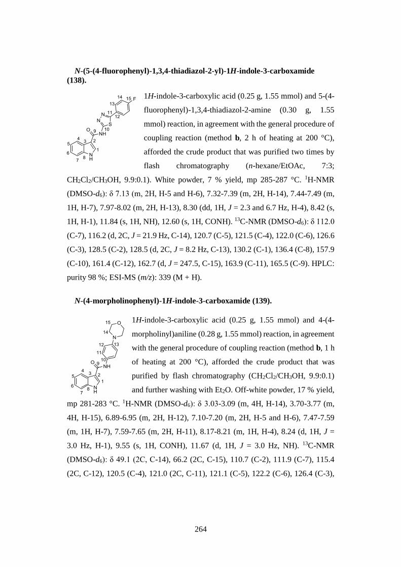
264
N-(5-(4-fluorophenyl)-1,3,4-thiadiazol-2-yl)-1H-indole-3-carboxamide
(138).
1H-indole-3-carboxylic acid (0.25 g, 1.55 mmol) and 5-(4-
fluorophenyl)-1,3,4-thiadiazol-2-amine (0.30 g, 1.55
mmol) reaction, in agreement with the general procedure of
coupling reaction (method b, 2 h of heating at 200 °C),
afforded the crude product that was purified two times by
flash chromatography (n-hexane/EtOAc, 7:3;
CH2Cl2/CH3OH, 9.9:0.1). White powder, 7 % yield, mp 285-287 °C. 1H-NMR
(DMSO-d6): δ 7.13 (m, 2H, H-5 and H-6), 7.32-7.39 (m, 2H, H-14), 7.44-7.49 (m,
1H, H-7), 7.97-8.02 (m, 2H, H-13), 8.30 (dd, 1H, J = 2.3 and 6.7 Hz, H-4), 8.42 (s,
1H, H-1), 11.84 (s, 1H, NH), 12.60 (s, 1H, CONH). 13C-NMR (DMSO-d6): δ 112.0
(C-7), 116.2 (d, 2C, J = 21.9 Hz, C-14), 120.7 (C-5), 121.5 (C-4), 122.0 (C-6), 126.6
(C-3), 128.5 (C-2), 128.5 (d, 2C, J = 8.2 Hz, C-13), 130.2 (C-1), 136.4 (C-8), 157.9
(C-10), 161.4 (C-12), 162.7 (d, J = 247.5, C-15), 163.9 (C-11), 165.5 (C-9). HPLC:
purity 98 %; ESI-MS (m/z): 339 (M + H).
N-(4-morpholinophenyl)-1H-indole-3-carboxamide (139).
1H-indole-3-carboxylic acid (0.25 g, 1.55 mmol) and 4-(4-
morpholinyl)aniline (0.28 g, 1.55 mmol) reaction, in agreement
with the general procedure of coupling reaction (method b, 1 h
of heating at 200 °C), afforded the crude product that was
purified by flash chromatography (CH2Cl2/CH3OH, 9.9:0.1)
and further washing with Et2O. Off-white powder, 17 % yield,
mp 281-283 °C. 1H-NMR (DMSO-d6): δ 3.03-3.09 (m, 4H, H-14), 3.70-3.77 (m,
4H, H-15), 6.89-6.95 (m, 2H, H-12), 7.10-7.20 (m, 2H, H-5 and H-6), 7.47-7.59
(m, 1H, H-7), 7.59-7.65 (m, 2H, H-11), 8.17-8.21 (m, 1H, H-4), 8.24 (d, 1H, J =
3.0 Hz, H-1), 9.55 (s, 1H, CONH), 11.67 (d, 1H, J = 3.0 Hz, NH). 13C-NMR
(DMSO-d6): δ 49.1 (2C, C-14), 66.2 (2C, C-15), 110.7 (C-2), 111.9 (C-7), 115.4
(2C, C-12), 120.5 (C-4), 121.0 (2C, C-11), 121.1 (C-5), 122.2 (C-6), 126.4 (C-3),
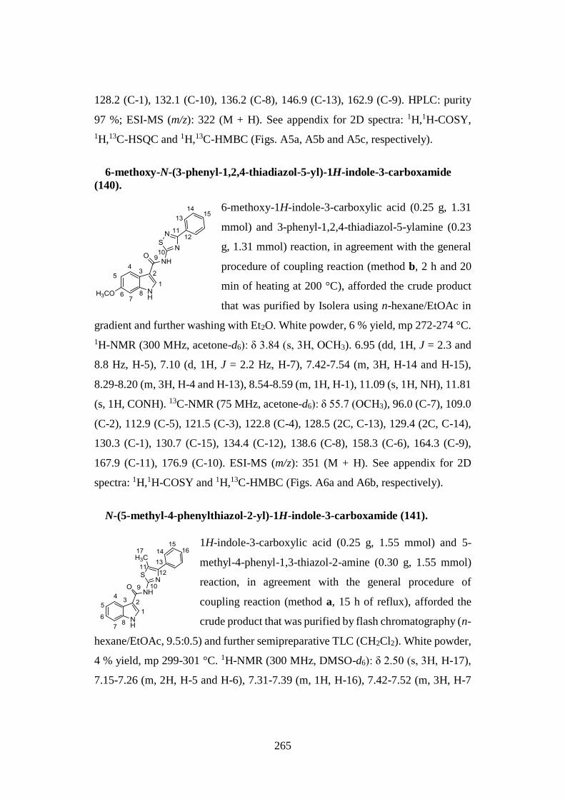
265
128.2 (C-1), 132.1 (C-10), 136.2 (C-8), 146.9 (C-13), 162.9 (C-9). HPLC: purity
97 %; ESI-MS (m/z): 322 (M + H). See appendix for 2D spectra: 1H,1H-COSY,
1H,13C-HSQC and 1H,13C-HMBC (Figs. A5a, A5b and A5c, respectively).
6-methoxy-N-(3-phenyl-1,2,4-thiadiazol-5-yl)-1H-indole-3-carboxamide
(140).
6-methoxy-1H-indole-3-carboxylic acid (0.25 g, 1.31
mmol) and 3-phenyl-1,2,4-thiadiazol-5-ylamine (0.23
g, 1.31 mmol) reaction, in agreement with the general
procedure of coupling reaction (method b, 2 h and 20
min of heating at 200 °C), afforded the crude product
that was purified by Isolera using n-hexane/EtOAc in
gradient and further washing with Et2O. White powder, 6 % yield, mp 272-274 °C.
1H-NMR (300 MHz, acetone-d6): δ 3.84 (s, 3H, OCH3). 6.95 (dd, 1H, J = 2.3 and
8.8 Hz, H-5), 7.10 (d, 1H, J = 2.2 Hz, H-7), 7.42-7.54 (m, 3H, H-14 and H-15),
8.29-8.20 (m, 3H, H-4 and H-13), 8.54-8.59 (m, 1H, H-1), 11.09 (s, 1H, NH), 11.81
(s, 1H, CONH). 13C-NMR (75 MHz, acetone-d6): δ 55.7 (OCH3), 96.0 (C-7), 109.0
(C-2), 112.9 (C-5), 121.5 (C-3), 122.8 (C-4), 128.5 (2C, C-13), 129.4 (2C, C-14),
130.3 (C-1), 130.7 (C-15), 134.4 (C-12), 138.6 (C-8), 158.3 (C-6), 164.3 (C-9),
167.9 (C-11), 176.9 (C-10). ESI-MS (m/z): 351 (M + H). See appendix for 2D
spectra: 1H,1H-COSY and 1H,13C-HMBC (Figs. A6a and A6b, respectively).
N-(5-methyl-4-phenylthiazol-2-yl)-1H-indole-3-carboxamide (141).
1H-indole-3-carboxylic acid (0.25 g, 1.55 mmol) and 5-
methyl-4-phenyl-1,3-thiazol-2-amine (0.30 g, 1.55 mmol)
reaction, in agreement with the general procedure of
coupling reaction (method a, 15 h of reflux), afforded the
crude product that was purified by flash chromatography (n-
hexane/EtOAc, 9.5:0.5) and further semipreparative TLC (CH2Cl2). White powder,
4 % yield, mp 299-301 °C. 1H-NMR (300 MHz, DMSO-d6): δ 2.50 (s, 3H, H-17),
7.15-7.26 (m, 2H, H-5 and H-6), 7.31-7.39 (m, 1H, H-16), 7.42-7.52 (m, 3H, H-7
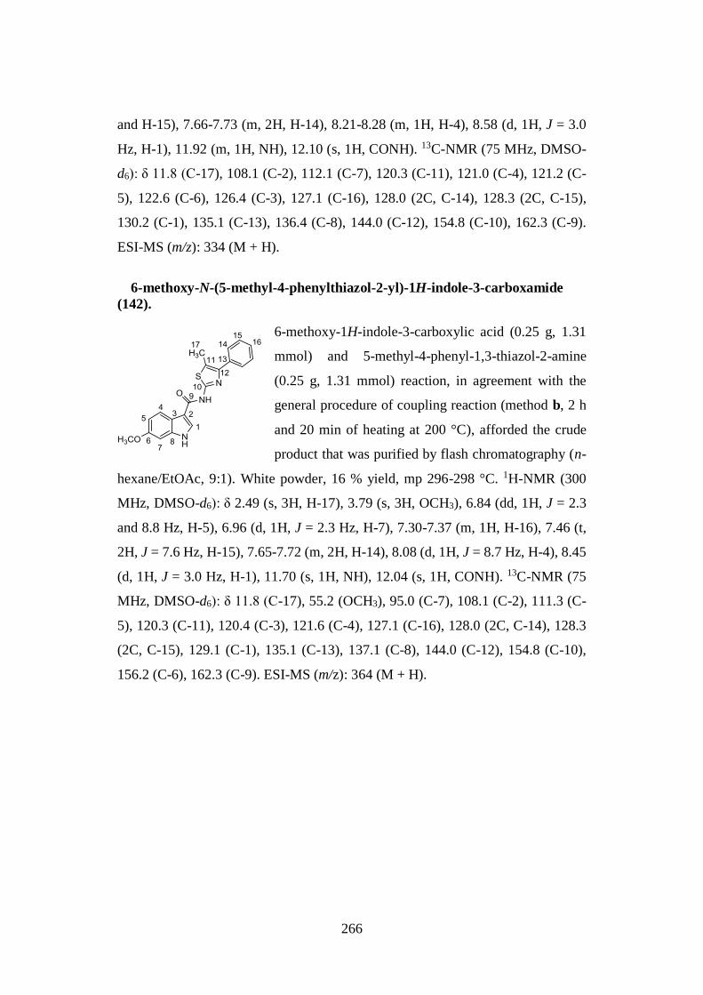
266
and H-15), 7.66-7.73 (m, 2H, H-14), 8.21-8.28 (m, 1H, H-4), 8.58 (d, 1H, J = 3.0
Hz, H-1), 11.92 (m, 1H, NH), 12.10 (s, 1H, CONH). 13C-NMR (75 MHz, DMSO-
d6): δ 11.8 (C-17), 108.1 (C-2), 112.1 (C-7), 120.3 (C-11), 121.0 (C-4), 121.2 (C-
5), 122.6 (C-6), 126.4 (C-3), 127.1 (C-16), 128.0 (2C, C-14), 128.3 (2C, C-15),
130.2 (C-1), 135.1 (C-13), 136.4 (C-8), 144.0 (C-12), 154.8 (C-10), 162.3 (C-9).
ESI-MS (m/z): 334 (M + H).
6-methoxy-N-(5-methyl-4-phenylthiazol-2-yl)-1H-indole-3-carboxamide
(142).
6-methoxy-1H-indole-3-carboxylic acid (0.25 g, 1.31
mmol) and 5-methyl-4-phenyl-1,3-thiazol-2-amine
(0.25 g, 1.31 mmol) reaction, in agreement with the
general procedure of coupling reaction (method b, 2 h
and 20 min of heating at 200 °C), afforded the crude
product that was purified by flash chromatography (n-
hexane/EtOAc, 9:1). White powder, 16 % yield, mp 296-298 °C. 1H-NMR (300
MHz, DMSO-d6): δ 2.49 (s, 3H, H-17), 3.79 (s, 3H, OCH3), 6.84 (dd, 1H, J = 2.3
and 8.8 Hz, H-5), 6.96 (d, 1H, J = 2.3 Hz, H-7), 7.30-7.37 (m, 1H, H-16), 7.46 (t,
2H, J = 7.6 Hz, H-15), 7.65-7.72 (m, 2H, H-14), 8.08 (d, 1H, J = 8.7 Hz, H-4), 8.45
(d, 1H, J = 3.0 Hz, H-1), 11.70 (s, 1H, NH), 12.04 (s, 1H, CONH). 13C-NMR (75
MHz, DMSO-d6): δ 11.8 (C-17), 55.2 (OCH3), 95.0 (C-7), 108.1 (C-2), 111.3 (C-
5), 120.3 (C-11), 120.4 (C-3), 121.6 (C-4), 127.1 (C-16), 128.0 (2C, C-14), 128.3
(2C, C-15), 129.1 (C-1), 135.1 (C-13), 137.1 (C-8), 144.0 (C-12), 154.8 (C-10),
156.2 (C-6), 162.3 (C-9). ESI-MS (m/z): 364 (M + H).
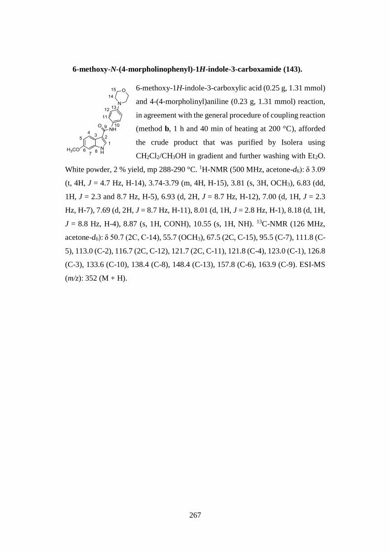
267
6-methoxy-N-(4-morpholinophenyl)-1H-indole-3-carboxamide (143).
6-methoxy-1H-indole-3-carboxylic acid (0.25 g, 1.31 mmol)
and 4-(4-morpholinyl)aniline (0.23 g, 1.31 mmol) reaction,
in agreement with the general procedure of coupling reaction
(method b, 1 h and 40 min of heating at 200 °C), afforded
the crude product that was purified by Isolera using
CH2Cl2/CH3OH in gradient and further washing with Et2O.
White powder, 2 % yield, mp 288-290 °C. 1H-NMR (500 MHz, acetone-d6): δ 3.09
(t, 4H, J = 4.7 Hz, H-14), 3.74-3.79 (m, 4H, H-15), 3.81 (s, 3H, OCH3), 6.83 (dd,
1H, J = 2.3 and 8.7 Hz, H-5), 6.93 (d, 2H, J = 8.7 Hz, H-12), 7.00 (d, 1H, J = 2.3
Hz, H-7), 7.69 (d, 2H, J = 8.7 Hz, H-11), 8.01 (d, 1H, J = 2.8 Hz, H-1), 8.18 (d, 1H,
J = 8.8 Hz, H-4), 8.87 (s, 1H, CONH), 10.55 (s, 1H, NH). 13C-NMR (126 MHz,
acetone-d6): δ 50.7 (2C, C-14), 55.7 (OCH3), 67.5 (2C, C-15), 95.5 (C-7), 111.8 (C-
5), 113.0 (C-2), 116.7 (2C, C-12), 121.7 (2C, C-11), 121.8 (C-4), 123.0 (C-1), 126.8
(C-3), 133.6 (C-10), 138.4 (C-8), 148.4 (C-13), 157.8 (C-6), 163.9 (C-9). ESI-MS
(m/z): 352 (M + H).

268
7. Appendix

269
7.1. 1D and 2D NMR SAMPLE COMPOUNDS
Figure A1a. 400 MHz, 1H-NMR spectra of compound 5 in DMSO-d6.
Figure A1b. 400 MHz, 1H-NMR spectrum of the 5-positive assay in DMSO-d6
with a 1:2 stoichiometric ratio of 5/cysteamine.
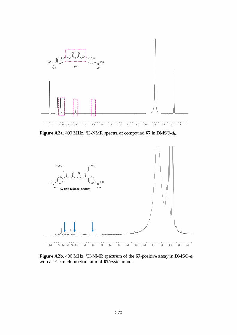
270
Figure A2a. 400 MHz, 1H-NMR spectra of compound 67 in DMSO-d6.
Figure A2b. 400 MHz, 1H-NMR spectrum of the 67-positive assay in DMSO-d6
with a 1:2 stoichiometric ratio of 67/cysteamine.
67-thia-Michael adduct
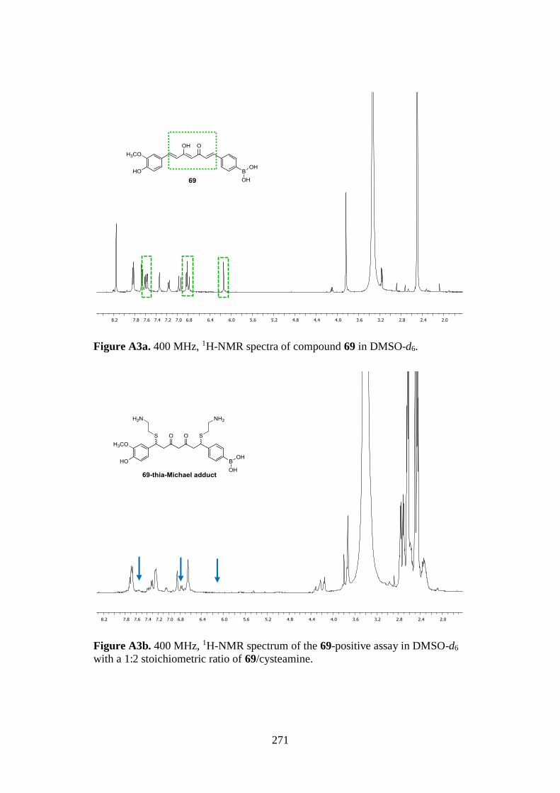
271
Figure A3a. 400 MHz, 1H-NMR spectra of compound 69 in DMSO-d6.
Figure A3b. 400 MHz, 1H-NMR spectrum of the 69-positive assay in DMSO-d6
with a 1:2 stoichiometric ratio of 69/cysteamine.
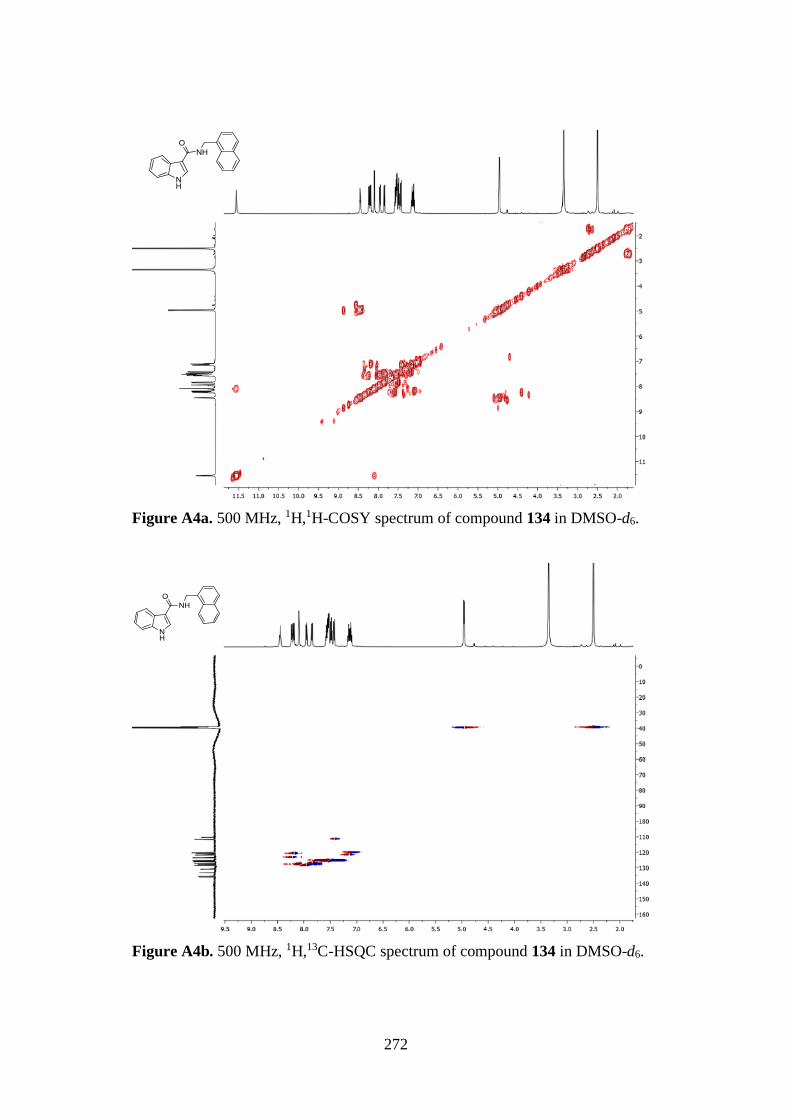
272
Figure A4a. 500 MHz, 1H,1H-COSY spectrum of compound 134 in DMSO-d6.
Figure A4b. 500 MHz, 1H,13C-HSQC spectrum of compound 134 in DMSO-d6.
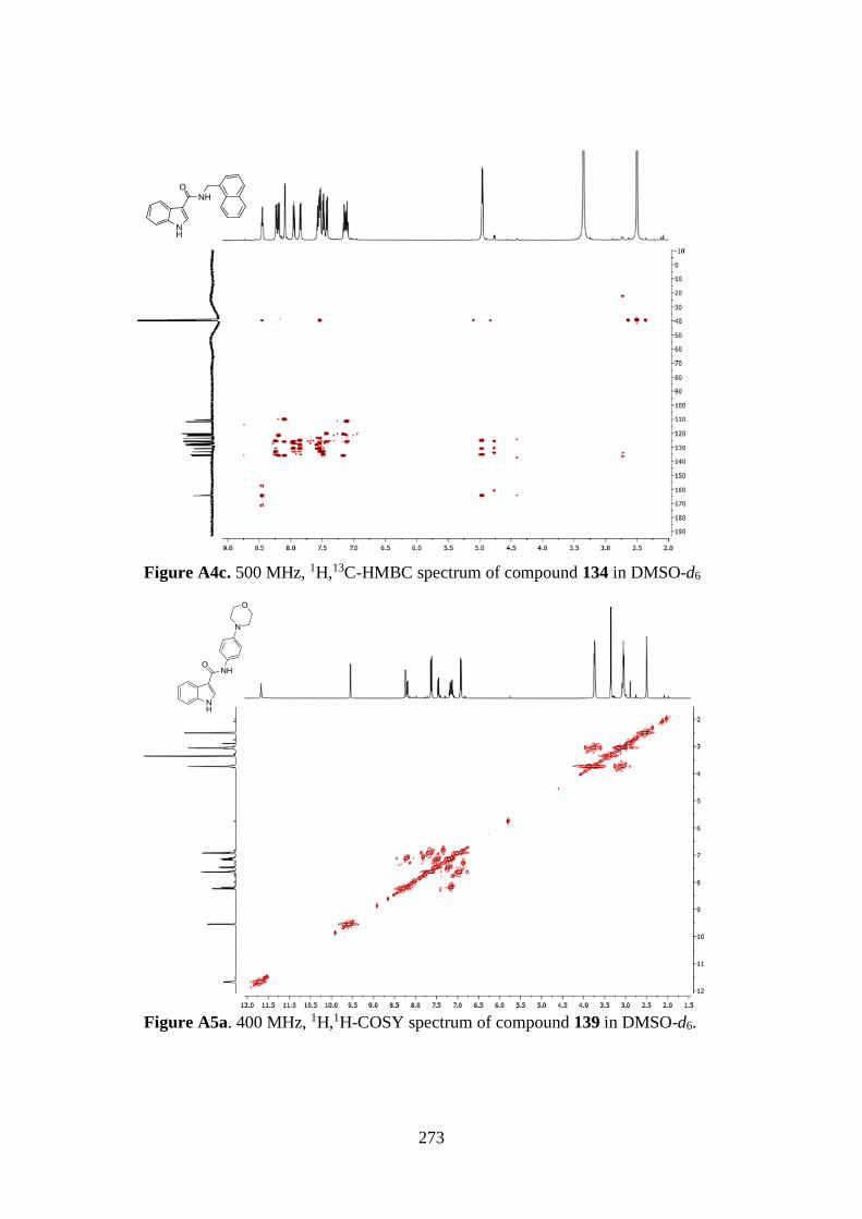
273
Figure A4c. 500 MHz, 1H,13C-HMBC spectrum of compound 134 in DMSO-d6
Figure A5a. 400 MHz, 1H,1H-COSY spectrum of compound 139 in DMSO-d6.
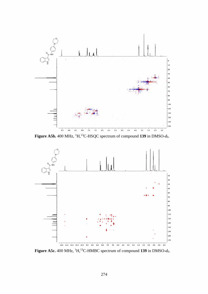
274
Figure A5b. 400 MHz, 1H,13C-HSQC spectrum of compound 139 in DMSO-d6.
Figure A5c. 400 MHz, 1H,13C-HMBC spectrum of compound 139 in DMSO-d6.
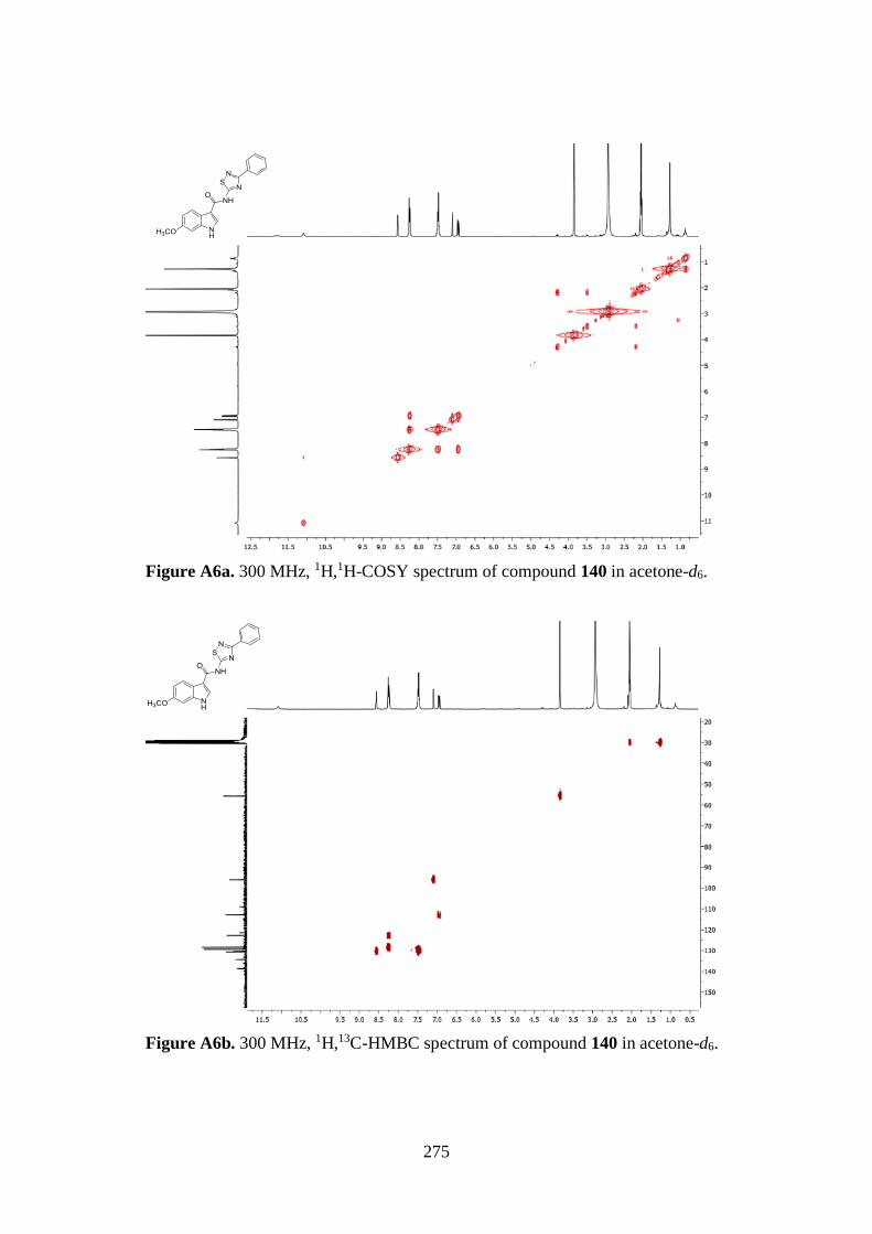
275
Figure A6a. 300 MHz, 1H,1H-COSY spectrum of compound 140 in acetone-d6.
Figure A6b. 300 MHz, 1H,13C-HMBC spectrum of compound 140 in acetone-d6.

276
8. Bibliographic references
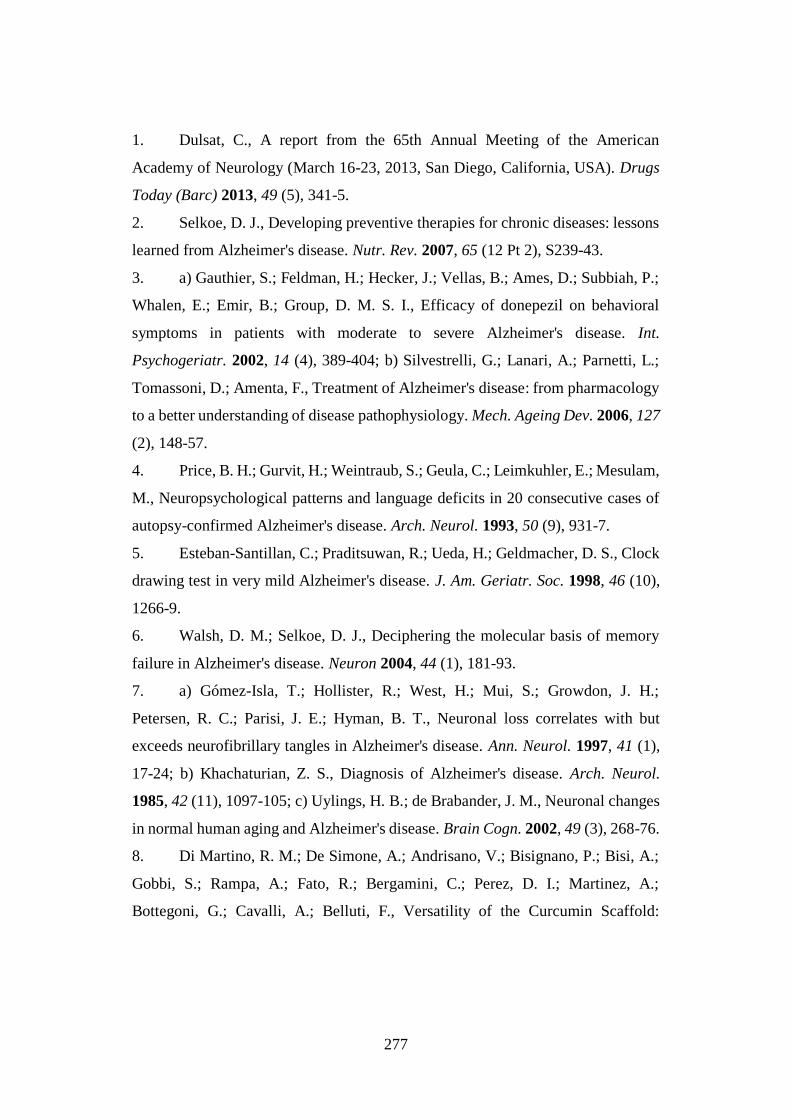
277
1. Dulsat, C., A report from the 65th Annual Meeting of the American
Academy of Neurology (March 16-23, 2013, San Diego, California, USA). Drugs
Today (Barc) 2013, 49 (5), 341-5.
2. Selkoe, D. J., Developing preventive therapies for chronic diseases: lessons
learned from Alzheimer's disease. Nutr. Rev. 2007, 65 (12 Pt 2), S239-43.
3. a) Gauthier, S.; Feldman, H.; Hecker, J.; Vellas, B.; Ames, D.; Subbiah, P.;
Whalen, E.; Emir, B.; Group, D. M. S. I., Efficacy of donepezil on behavioral
symptoms in patients with moderate to severe Alzheimer's disease. Int.
Psychogeriatr. 2002, 14 (4), 389-404; b) Silvestrelli, G.; Lanari, A.; Parnetti, L.;
Tomassoni, D.; Amenta, F., Treatment of Alzheimer's disease: from pharmacology
to a better understanding of disease pathophysiology. Mech. Ageing Dev. 2006, 127
(2), 148-57.
4. Price, B. H.; Gurvit, H.; Weintraub, S.; Geula, C.; Leimkuhler, E.; Mesulam,
M., Neuropsychological patterns and language deficits in 20 consecutive cases of
autopsy-confirmed Alzheimer's disease. Arch. Neurol. 1993, 50 (9), 931-7.
5. Esteban-Santillan, C.; Praditsuwan, R.; Ueda, H.; Geldmacher, D. S., Clock
drawing test in very mild Alzheimer's disease. J. Am. Geriatr. Soc. 1998, 46 (10),
1266-9.
6. Walsh, D. M.; Selkoe, D. J., Deciphering the molecular basis of memory
failure in Alzheimer's disease. Neuron 2004, 44 (1), 181-93.
7. a) Gómez-Isla, T.; Hollister, R.; West, H.; Mui, S.; Growdon, J. H.;
Petersen, R. C.; Parisi, J. E.; Hyman, B. T., Neuronal loss correlates with but
exceeds neurofibrillary tangles in Alzheimer's disease. Ann. Neurol. 1997, 41 (1),
17-24; b) Khachaturian, Z. S., Diagnosis of Alzheimer's disease. Arch. Neurol.
1985, 42 (11), 1097-105; c) Uylings, H. B.; de Brabander, J. M., Neuronal changes
in normal human aging and Alzheimer's disease. Brain Cogn. 2002, 49 (3), 268-76.
8. Di Martino, R. M.; De Simone, A.; Andrisano, V.; Bisignano, P.; Bisi, A.;
Gobbi, S.; Rampa, A.; Fato, R.; Bergamini, C.; Perez, D. I.; Martinez, A.;
Bottegoni, G.; Cavalli, A.; Belluti, F., Versatility of the Curcumin Scaffold:
278
Discovery of Potent and Balanced Dual BACE-1 and GSK-3β Inhibitors. J. Med.
Chem. 2016, 59 (2), 531-44.
9. Mattson, M. P., Pathways towards and away from Alzheimer's disease.
Nature 2004, 430 (7000), 631-9.
10. Desai, A. K.; Grossberg, G. T., Diagnosis and treatment of Alzheimer's
disease. Neurology 2005, 64 (12 Suppl 3), S34-9.
11. Mohamed, T.; Rao, P. P., Alzheimer's disease: emerging trends in small
molecule therapies. Curr. Med. Chem. 2011, 18 (28), 4299-320.
12. Hershenson, F. M.; Moos, W. H., Drug development for senile cognitive
decline. J. Med. Chem. 1986, 29 (7), 1125-30.
13. Rampa, A.; Belluti, F.; Gobbi, S.; Bisi, A., Hybrid-based multi-target
ligands for the treatment of Alzheimer's disease. Curr. Top. Med. Chem. 2011, 11
(22), 2716-30.
14. Scarpini, E.; Scheltens, P.; Feldman, H., Treatment of Alzheimer's disease:
current status and new perspectives. Lancet Neurol. 2003, 2 (9), 539-47.
15. Scott, L. J.; Goa, K. L., Galantamine: a review of its use in Alzheimer's
disease. Drugs 2000, 60 (5), 1095-122.
16. Cummings, J. L., Treatment of Alzheimer's disease: current and future
therapeutic approaches. Rev. Neurol. Dis. 2004, 1 (2), 60-9.
17. a) Golde, T. E., Disease modifying therapy for AD? J. Neurochem. 2006,
99 (3), 689-707; b) Grill, J. D.; Cummings, J. L., Current therapeutic targets for the
treatment of Alzheimer's disease. Expert Rev. Neurother. 2010, 10 (5), 711-28.
18. Pimplikar, S. W., Neuroinflammation in Alzheimer's disease: from
pathogenesis to a therapeutic target. J. Clin. Immunol. 2014, 34 Suppl 1, S64-9.
19. a) Hardy, J.; Allsop, D., Amyloid deposition as the central event in the
aetiology of Alzheimer's disease. Trends Pharmacol. Sci. 1991, 12 (10), 383-8; b)
Hardy, J. A.; Higgins, G. A., Alzheimer's disease: the amyloid cascade hypothesis.
Science 1992, 256 (5054), 184-5.
279
20. Hardy, J.; Selkoe, D. J., The amyloid hypothesis of Alzheimer's disease:
progress and problems on the road to therapeutics. Science 2002, 297 (5580), 353-
6.
21. Mattson, M. P., Cellular actions of beta-amyloid precursor protein and its
soluble and fibrillogenic derivatives. Physiol. Rev. 1997, 77 (4), 1081-132.
22. Silva, T.; Reis, J.; Teixeira, J.; Borges, F., Alzheimer's disease, enzyme
targets and drug discovery struggles: from natural products to drug prototypes.
Ageing Res. Rev. 2014, 15, 116-45.
23. a) Boddapati, S.; Levites, Y.; Sierks, M. R., Inhibiting β-secretase activity
in Alzheimer's disease cell models with single-chain antibodies specifically
targeting APP. J. Mol. Biol. 2011, 405 (2), 436-47; b) Willem, M.; Lammich, S.;
Haass, C., Function, regulation and therapeutic properties of beta-secretase
(BACE1). Semin. Cell Dev. Biol. 2009, 20 (2), 175-82.
24. a) Selkoe, D. J., Soluble oligomers of the amyloid beta-protein impair
synaptic plasticity and behavior. Behav. Brain Res. 2008, 192 (1), 106-13; b)
Dineley, K. T.; Kayed, R.; Neugebauer, V.; Fu, Y.; Zhang, W.; Reese, L. C.;
Taglialatela, G., Amyloid-beta oligomers impair fear conditioned memory in a
calcineurin-dependent fashion in mice. J. Neurosci. Res. 2010, 88 (13), 2923-32.
25. Lustbader, J. W.; Cirilli, M.; Lin, C.; Xu, H. W.; Takuma, K.; Wang, N.;
Caspersen, C.; Chen, X.; Pollak, S.; Chaney, M.; Trinchese, F.; Liu, S.; Gunn-
Moore, F.; Lue, L. F.; Walker, D. G.; Kuppusamy, P.; Zewier, Z. L.; Arancio, O.;
Stern, D.; Yan, S. S.; Wu, H., ABAD directly links Abeta to mitochondrial toxicity
in Alzheimer's disease. Science 2004, 304 (5669), 448-52.
26. Tamagno, E.; Parola, M.; Guglielmotto, M.; Santoro, G.; Bardini, P.; Marra,
L.; Tabaton, M.; Danni, O., Multiple signaling events in amyloid beta-induced,
oxidative stress-dependent neuronal apoptosis. Free Radic. Biol. Med. 2003, 35 (1),
45-58.
27. Evin, G.; Lessene, G.; Wilkins, S., BACE inhibitors as potential drugs for
the treatment of Alzheimer's disease: focus on bioactivity. Recent Pat. CNS Drug
Discov. 2011, 6 (2), 91-106.
280
28. Bennett, B. D.; Babu-Khan, S.; Loeloff, R.; Louis, J. C.; Curran, E.; Citron,
M.; Vassar, R., Expression analysis of BACE2 in brain and peripheral tissues. J.
Biol. Chem. 2000, 275 (27), 20647-51.
29. a) Fischer, F.; Molinari, M.; Bodendorf, U.; Paganetti, P., The disulphide
bonds in the catalytic domain of BACE are critical but not essential for amyloid
precursor protein processing activity. J. Neurochem. 2002, 80 (6), 1079-88; b)
Haniu, M.; Denis, P.; Young, Y.; Mendiaz, E. A.; Fuller, J.; Hui, J. O.; Bennett, B.
D.; Kahn, S.; Ross, S.; Burgess, T.; Katta, V.; Rogers, G.; Vassar, R.; Citron, M.,
Characterization of Alzheimer's beta-secretase protein BACE. A pepsin family
member with unusual properties. J. Biol. Chem. 2000, 275 (28), 21099-106.
30. Kacker, P.; Bottegoni, G.; Cavalli, A., Computational methods in the
discovery and design of BACE-1 inhibitors. Curr. Med. Chem. 2012, 19 (36), 6095-
111.
31. Mancini, F.; De Simone, A.; Andrisano, V., Beta-secretase as a target for
Alzheimer's disease drug discovery: an overview of in vitro methods for
characterization of inhibitors. Anal. Bioanal. Chem. 2011, 400 (7), 1979-96.
32. Tamagno, E.; Bardini, P.; Obbili, A.; Vitali, A.; Borghi, R.; Zaccheo, D.;
Pronzato, M. A.; Danni, O.; Smith, M. A.; Perry, G.; Tabaton, M., Oxidative stress
increases expression and activity of BACE in NT2 neurons. Neurobiol. Dis. 2002,
10 (3), 279-88.
33. Ghosh, A. K.; Devasamudram, T.; Hong, L.; DeZutter, C.; Xu, X.;
Weerasena, V.; Koelsch, G.; Bilcer, G.; Tang, J., Structure-based design of
cycloamide-urethane-derived novel inhibitors of human brain memapsin 2 (beta-
secretase). Bioorg. Med. Chem. Lett. 2005, 15 (1), 15-20.
34. Silvestri, R., Boom in the development of non-peptidic beta-secretase
(BACE1) inhibitors for the treatment of Alzheimer's disease. Med. Res. Rev. 2009,
29 (2), 295-338.
35. Belluti, F.; De Simone, A.; Tarozzi, A.; Bartolini, M.; Djemil, A.; Bisi, A.;
Gobbi, S.; Montanari, S.; Cavalli, A.; Andrisano, V.; Bottegoni, G.; Rampa, A.,
281
Fluorinated benzophenone derivatives: balanced multipotent agents for Alzheimer's
disease. Eur. J. Med. Chem. 2014, 78, 157-66.
36. Rampa, A.; Mancini, F.; De Simone, A.; Falchi, F.; Belluti, F.; Di Martino,
R. M.; Gobbi, S.; Andrisano, V.; Tarozzi, A.; Bartolini, M.; Cavalli, A.; Bisi, A.,
From AChE to BACE1 inhibitors: The role of the amine on the indanone scaffold.
Bioorg. Med. Chem. Lett. 2015, 25 (14), 2804-8.
37. a) Nagy, Z.; Esiri, M. M.; Jobst, K. A.; Morris, J. H.; King, E. M.;
McDonald, B.; Litchfield, S.; Smith, A.; Barnetson, L.; Smith, A. D., Relative roles
of plaques and tangles in the dementia of Alzheimer's disease: correlations using
three sets of neuropathological criteria. Dementia 1995, 6 (1), 21-31; b) Arriagada,
P. V.; Growdon, J. H.; Hedley-Whyte, E. T.; Hyman, B. T., Neurofibrillary tangles
but not senile plaques parallel duration and severity of Alzheimer's disease.
Neurology 1992, 42 (3 Pt 1), 631-9.
38. Iqbal, K.; Liu, F.; Gong, C. X.; Alonso, A. e. C.; Grundke-Iqbal, I.,
Mechanisms of tau-induced neurodegeneration. Acta Neuropathol. 2009, 118 (1),
53-69.
39. Mazanetz, M. P.; Fischer, P. M., Untangling tau hyperphosphorylation in
drug design for neurodegenerative diseases. Nat. Rev. Drug Discov. 2007, 6 (6),
464-79.
40. Reddy, P. H., Abnormal tau, mitochondrial dysfunction, impaired axonal
transport of mitochondria, and synaptic deprivation in Alzheimer's disease. Brain
Res. 2011, 1415, 136-48.
41. Cowan, C. M.; Quraishe, S.; Hands, S.; Sealey, M.; Mahajan, S.; Allan, D.
W.; Mudher, A., Rescue from tau-induced neuronal dysfunction produces insoluble
tau oligomers. Sci. Rep. 2015, 5, 17191.
42. Lee, H. G.; Perry, G.; Moreira, P. I.; Garrett, M. R.; Liu, Q.; Zhu, X.;
Takeda, A.; Nunomura, A.; Smith, M. A., Tau phosphorylation in Alzheimer's
disease: pathogen or protector? Trends Mol. Med. 2005, 11 (4), 164-9.
43. a) Hanks, S. K.; Hunter, T., Protein kinases 6. The eukaryotic protein kinase
superfamily: kinase (catalytic) domain structure and classification. FASEB J. 1995,
282
9 (8), 576-96; b) Cohen, P.; Alessi, D. R., Kinase drug discovery--what's next in
the field? ACS Chem. Biol. 2013, 8 (1), 96-104.
44. Palomo, V.; Soteras, I.; Perez, D. I.; Perez, C.; Gil, C.; Campillo, N. E.;
Martinez, A., Exploring the binding sites of glycogen synthase kinase 3.
Identification and characterization of allosteric modulation cavities. J. Med. Chem.
2011, 54 (24), 8461-70.
45. Rylatt, D. B.; Aitken, A.; Bilham, T.; Condon, G. D.; Embi, N.; Cohen, P.,
Glycogen synthase from rabbit skeletal muscle. Amino acid sequence at the sites
phosphorylated by glycogen synthase kinase-3, and extension of the N-terminal
sequence containing the site phosphorylated by phosphorylase kinase. Eur. J.
Biochem. 1980, 107 (2), 529-37.
46. Billingsley, M. L.; Kincaid, R. L., Regulated phosphorylation and
dephosphorylation of tau protein: effects on microtubule interaction, intracellular
trafficking and neurodegeneration. Biochem. J. 1997, 323 ( Pt 3), 577-91.
47. Lei, P.; Ayton, S.; Bush, A. I.; Adlard, P. A., GSK-3 in Neurodegenerative
Diseases. Int. J. Alzheimers Dis. 2011, 2011, 189246.
48. ter Haar, E.; Coll, J. T.; Austen, D. A.; Hsiao, H. M.; Swenson, L.; Jain, J.,
Structure of GSK3beta reveals a primed phosphorylation mechanism. Nat. Struct.
Biol. 2001, 8 (7), 593-6.
49. Llorens-Martín, M.; Jurado, J.; Hernández, F.; Avila, J., GSK-3β, a pivotal
kinase in Alzheimer disease. Front. Mol. Neurosci. 2014, 7, 46.
50. Martinez, A.; Castro, A.; Dorronsoro, I.; Alonso, M., Glycogen synthase
kinase 3 (GSK-3) inhibitors as new promising drugs for diabetes,
neurodegeneration, cancer, and inflammation. Med. Res. Rev. 2002, 22 (4), 373-84.
51. Martinez, A.; Gil, C.; Perez, D. I., Glycogen synthase kinase 3 inhibitors in
the next horizon for Alzheimer's disease treatment. Int. J. Alzheimers Dis. 2011,
2011, 280502.
52. Bidon-Chanal, A.; Fuertes, A.; Alonso, D.; Pérez, D. I.; Martínez, A.;
Luque, F. J.; Medina, M., Evidence for a new binding mode to GSK-3: allosteric
283
regulation by the marine compound palinurin. Eur. J. Med. Chem. 2013, 60, 479-
89.
53. Ittner, L. M.; Götz, J., Amyloid-β and tau--a toxic pas de deux in
Alzheimer's disease. Nat. Rev. Neurosci. 2011, 12 (2), 65-72.
54. Liu, Z.; Li, P.; Wu, J.; Wang, Y.; Li, P.; Hou, X.; Zhang, Q.; Wei, N.; Zhao,
Z.; Liang, H.; Wei, J., The cascade of oxidative stress and tau protein autophagic
dysfunction in Alzheimer’s disease. In Alzheimer's Disease-Challenges for
the Future, Zerr, I., Ed. 2015.
55. a) Marcus, D. L.; Thomas, C.; Rodriguez, C.; Simberkoff, K.; Tsai, J. S.;
Strafaci, J. A.; Freedman, M. L., Increased peroxidation and reduced antioxidant
enzyme activity in Alzheimer's disease. Exp. Neurol. 1998, 150 (1), 40-4; b) Omar,
R. A.; Chyan, Y. J.; Andorn, A. C.; Poeggeler, B.; Robakis, N. K.; Pappolla, M. A.,
Increased Expression but Reduced Activity of Antioxidant Enzymes in Alzheimer's
Disease. J. Alzheimers Dis. 1999, 1 (3), 139-145; c) Furuta, A.; Price, D. L.; Pardo,
C. A.; Troncoso, J. C.; Xu, Z. S.; Taniguchi, N.; Martin, L. J., Localization of
superoxide dismutases in Alzheimer's disease and Down's syndrome neocortex and
hippocampus. Am. J. Pathol. 1995, 146 (2), 357-67; d) Padurariu, M.; Ciobica, A.;
Hritcu, L.; Stoica, B.; Bild, W.; Stefanescu, C., Changes of some oxidative stress
markers in the serum of patients with mild cognitive impairment and Alzheimer's
disease. Neurosci. Lett. 2010, 469 (1), 6-10.
56. de Vries, H. E.; Witte, M.; Hondius, D.; Rozemuller, A. J.; Drukarch, B.;
Hoozemans, J.; van Horssen, J., Nrf2-induced antioxidant protection: a promising
target to counteract ROS-mediated damage in neurodegenerative disease? Free
Radic. Biol. Med. 2008, 45 (10), 1375-83.
57. Markesbery, W. R., Oxidative stress hypothesis in Alzheimer's disease. Free
Radic. Biol. Med. 1997, 23 (1), 134-47.
58. Sutherland, G. T.; Chami, B.; Youssef, P.; Witting, P. K., Oxidative stress
in Alzheimer's disease: Primary villain or physiological by-product? Redox Rep.
2013, 18 (4), 134-41.
284
59. Morales, I.; Guzmán-Martínez, L.; Cerda-Troncoso, C.; Farías, G. A.;
Maccioni, R. B., Neuroinflammation in the pathogenesis of Alzheimer's disease. A
rational framework for the search of novel therapeutic approaches. Front. Cell
Neurosci. 2014, 8, 112.
60. Wang, W. Y.; Tan, M. S.; Yu, J. T.; Tan, L., Role of pro-inflammatory
cytokines released from microglia in Alzheimer's disease. Ann. Transl. Med. 2015,
3 (10), 136.
61. Pan, X. D.; Zhu, Y. G.; Lin, N.; Zhang, J.; Ye, Q. Y.; Huang, H. P.; Chen,
X. C., Microglial phagocytosis induced by fibrillar β-amyloid is attenuated by
oligomeric β-amyloid: implications for Alzheimer's disease. Mol. Neurodegener.
2011, 6, 45.
62. a) Tenhunen, R.; Marver, H. S.; Schmid, R., The enzymatic conversion of
heme to bilirubin by microsomal heme oxygenase. Proc. Natl. Acad. Sci. USA 1968,
61 (2), 748-55; b) Maines, M. D., Heme oxygenase: function, multiplicity,
regulatory mechanisms, and clinical applications. FASEB J. 1988, 2 (10), 2557-68.
63. Yamazaki, H.; Tanji, K.; Wakabayashi, K.; Matsuura, S.; Itoh, K., Role of
the Keap1/Nrf2 pathway in neurodegenerative diseases. Pathol. Int. 2015, 65 (5),
210-9.
64. Torres-Lista, V.; Parrado-Fernández, C.; Alvarez-Montón, I.; Frontiñán-
Rubio, J.; Durán-Prado, M.; Peinado, J. R.; Johansson, B.; Alcaín, F. J.; Giménez-
Llort, L., Neophobia, NQO1 and SIRT1 as premorbid and prodromal indicators of
AD in 3xTg-AD mice. Behav. Brain Res. 2014, 271, 140-6.
65. Saharan, S.; Mandal, P. K., The emerging role of glutathione in Alzheimer's
disease. J. Alzheimers Dis. 2014, 40 (3), 519-29.
66. Kaspar, J. W.; Niture, S. K.; Jaiswal, A. K., Nrf2:INrf2 (Keap1) signaling
in oxidative stress. Free Radic. Biol. Med. 2009, 47 (9), 1304-9.
67. Jung, K. A.; Kwak, M. K., The Nrf2 system as a potential target for the
development of indirect antioxidants. Molecules 2010, 15 (10), 7266-91.
68. Ma, Q., Role of nrf2 in oxidative stress and toxicity. Annu. Rev. Pharmacol.
Toxicol. 2013, 53, 401-26.
285
69. Choi, B. H.; Kang, K. S.; Kwak, M. K., Effect of redox modulating NRF2
activators on chronic kidney disease. Molecules 2014, 19 (8), 12727-59.
70. Baird, L.; Dinkova-Kostova, A. T., The cytoprotective role of the Keap1-
Nrf2 pathway. Arch. Toxicol. 2011, 85 (4), 241-72.
71. Holland, R.; Fishbein, J. C., Chemistry of the cysteine sensors in Kelch-like
ECH-associated protein 1. Antioxid. Redox Signal. 2010, 13 (11), 1749-61.
72. Talalay, P.; De Long, M. J.; Prochaska, H. J., Identification of a common
chemical signal regulating the induction of enzymes that protect against chemical
carcinogenesis. Proc. Natl. Acad. Sci. USA 1988, 85 (21), 8261-5.
73. Wilson, A. J.; Kerns, J. K.; Callahan, J. F.; Moody, C. J., Keap calm, and
carry on covalently. J. Med. Chem. 2013, 56 (19), 7463-76.
74. Jain, A. K.; Jaiswal, A. K., Phosphorylation of tyrosine 568 controls nuclear
export of Nrf2. J. Biol. Chem. 2006, 281 (17), 12132-42.
75. Kanninen, K.; White, A. R.; Koistinaho, J.; Malm, T., Targeting Glycogen
Synthase Kinase-3β for Therapeutic Benefit against Oxidative Stress in Alzheimer's
Disease: Involvement of the Nrf2-ARE Pathway. Int. J. Alzheimers Dis. 2011,
2011, 985085.
76. Jain, A. K.; Jaiswal, A. K., GSK-3beta acts upstream of Fyn kinase in
regulation of nuclear export and degradation of NF-E2 related factor 2. J. Biol.
Chem. 2007, 282 (22), 16502-10.
77. Longenecker, K. L.; Roach, P. J.; Hurley, T. D., Three-dimensional
structure of mammalian casein kinase I: molecular basis for phosphate recognition.
J. Mol. Biol. 1996, 257 (3), 618-31.
78. a) Perez, D. I.; Gil, C.; Martinez, A., Protein kinases CK1 and CK2 as new
targets for neurodegenerative diseases. Med. Res. Rev. 2011, 31 (6), 924-54; b)
Flotow, H.; Graves, P. R.; Wang, A. Q.; Fiol, C. J.; Roeske, R. W.; Roach, P. J.,
Phosphate groups as substrate determinants for casein kinase I action. J. Biol.
Chem. 1990, 265 (24), 14264-9.
286
79. Flajolet, M.; He, G.; Heiman, M.; Lin, A.; Nairn, A. C.; Greengard, P.,
Regulation of Alzheimer's disease amyloid-beta formation by casein kinase I. Proc.
Natl. Acad. Sci. USA 2007, 104 (10), 4159-64.
80. Lewis, P. A., The function of ROCO proteins in health and disease. Biol.
Cell 2009, 101 (3), 183-91.
81. Deng, J.; Lewis, P. A.; Greggio, E.; Sluch, E.; Beilina, A.; Cookson, M. R.,
Structure of the ROC domain from the Parkinson's disease-associated leucine-rich
repeat kinase 2 reveals a dimeric GTPase. Proc. Natl. Acad. Sci. USA 2008, 105
(5), 1499-504.
82. Kawakami, F.; Shimada, N.; Ohta, E.; Kagiya, G.; Kawashima, R.;
Maekawa, T.; Maruyama, H.; Ichikawa, T., Leucine-rich repeat kinase 2 regulates
tau phosphorylation through direct activation of glycogen synthase kinase-3β.
FEBS J. 2014, 281 (1), 3-13.
83. Kim, B.; Yang, M. S.; Choi, D.; Kim, J. H.; Kim, H. S.; Seol, W.; Choi, S.;
Jou, I.; Kim, E. Y.; Joe, E. H., Impaired inflammatory responses in murine Lrrk2-
knockdown brain microglia. PLoS One 2012, 7 (4), e34693.
84. Bolognesi, M. L., Polypharmacology in a single drug: multitarget drugs.
Curr. Med. Chem. 2013, 20 (13), 1639-45.
85. Cavalli, A.; Bolognesi, M. L.; Minarini, A.; Rosini, M.; Tumiatti, V.;
Recanatini, M.; Melchiorre, C., Multi-target-directed ligands to combat
neurodegenerative diseases. J. Med. Chem. 2008, 51 (3), 347-72.
86. Zheng, H.; Fridkin, M.; Youdim, M., From single target to
multitarget/network therapeutics in Alzheimer's therapy. Pharmaceuticals (Basel)
2014, 7 (2), 113-35.
87. Morphy, R.; Rankovic, Z., Designed multiple ligands. An emerging drug
discovery paradigm. J. Med. Chem. 2005, 48 (21), 6523-43.
88. Bottegoni, G.; Favia, A. D.; Recanatini, M.; Cavalli, A., The role of
fragment-based and computational methods in polypharmacology. Drug Discov.
Today 2012, 17 (1-2), 23-34.
287
89. Costantino, L.; Barlocco, D., Privileged structures as leads in medicinal
chemistry. Curr. Med. Chem. 2006, 13 (1), 65-85.
90. DeSimone, R. W.; Currie, K. S.; Mitchell, S. A.; Darrow, J. W.; Pippin, D.
A., Privileged structures: applications in drug discovery. Comb. Chem. High
Throughput Screen. 2004, 7 (5), 473-94.
91. de Sá Alves, F. R.; Barreiro, E. J.; Fraga, C. A., From nature to drug
discovery: the indole scaffold as a 'privileged structure'. Mini Rev. Med. Chem.
2009, 9 (7), 782-93.
92. a) Borges, F.; Roleira, F.; Milhazes, N.; Santana, L.; Uriarte, E., Simple
coumarins and analogues in medicinal chemistry: occurrence, synthesis and
biological activity. Curr. Med. Chem. 2005, 12 (8), 887-916; b) Matos, M. J.;
Santana, L.; Uriarte, E.; Abreu, O. A.; E., Molina, E.; Yordi, E. G., Coumarins-
an important class of phytochemicals. In Phytochemicals-Isolation,
Characterization and Role in Human Health [Online] Rao, V., Ed. 2015.
93. Jameel, E.; Umar, T.; Kumar, J.; Hoda, N., Coumarin: A Privileged Scaffold
for the Design and Development of Antineurodegenerative Agents. Chem. Biol.
Drug Des. 2016, 87 (1), 21-38.
94. Batovska, D. I.; Todorova, I. T., Trends in utilization of the pharmacological
potential of chalcones. Curr. Clin. Pharmacol. 2010, 5 (1), 1-29.
95. Welsch, M. E.; Snyder, S. A.; Stockwell, B. R., Privileged scaffolds for
library design and drug discovery. Curr. Opin. Chem. Biol. 2010, 14 (3), 347-61.
96. Vasilevich, N. I.; Kombarov, R. V.; Genis, D. V.; Kirpichenok, M. A.,
Lessons from natural products chemistry can offer novel approaches for synthetic
chemistry in drug discovery. J. Med. Chem. 2012, 55 (16), 7003-9.
97. Chen, J.; Li, W.; Yao, H.; Xu, J., Insights into drug discovery from natural
products through structural modification. Fitoterapia 2015, 103, 231-41.
98. Hong, J., Role of natural product diversity in chemical biology. Curr. Opin.
Chem. Biol. 2011, 15 (3), 350-4.
99. Drahl, C.; Cravatt, B. F.; Sorensen, E. J., Protein-reactive natural products.
Angew. Chem. Int. Ed. Engl. 2005, 44 (36), 5788-809.
288
100. Gersch, M.; Kreuzer, J.; Sieber, S. A., Electrophilic natural products and
their biological targets. Nat. Prod. Rep. 2012, 29 (6), 659-82.
101. Amslinger, S., The tunable functionality of alpha,beta-unsaturated carbonyl
compounds enables their differential application in biological systems.
ChemMedChem. 2010, 5 (3), 351-6.
102. Silvermam, R. B., Model studies for a molecular
mechanism of action of oral anticoagulants. J. Am. Chem. Soc. 1981, 103, 3910-
3915.
103. Avonto, C.; Taglialatela-Scafati, O.; Pollastro, F.; Minassi, A.; Di Marzo,
V.; De Petrocellis, L.; Appendino, G., An NMR spectroscopic method to identify
and classify thiol-trapping agents: revival of Michael acceptors for drug discovery?
Angew. Chem. Int. Ed. Engl. 2011, 50 (2), 467-71.
104. Minassi, A.; Sánchez-Duffhues, G.; Collado, J. A.; Muñoz, E.; Appendino,
G., Dissecting the pharmacophore of curcumin. Which structural element is critical
for which action? J. Nat. Prod. 2013, 76 (6), 1105-12.
105. Ahmed T, G. A., Therapeutic potential of turmeric in Alzheimer's
disease: curcumin or curcuminoids? Phytother. Res. 2014, 28 (4), 517-525.
106. Shanmugam, M. K.; Rane, G.; Kanchi, M. M.; Arfuso, F.; Chinnathambi,
A.; Zayed, M. E.; Alharbi, S. A.; Tan, B. K.; Kumar, A. P.; Sethi, G., The
multifaceted role of curcumin in cancer prevention and treatment. Molecules 2015,
20 (2), 2728-69.
107. Esatbeyoglu, T.; Huebbe, P.; Ernst, I. M.; Chin, D.; Wagner, A. E.;
Rimbach, G., Curcumin--from molecule to biological function. Angew. Chem. Int.
Ed. Engl. 2012, 51 (22), 5308-32.
108. a) Oetari, S.; Sudibyo, M.; Commandeur, J. N.; Samhoedi, R.; Vermeulen,
N. P., Effects of curcumin on cytochrome P450 and glutathione S-transferase
activities in rat liver. Biochem. Pharmacol. 1996, 51 (1), 39-45; b) Tønnesen, H.
H.; Karlsen, J., Studies on curcumin and curcuminoids. VI. Kinetics of curcumin
degradation in aqueous solution. Z. Lebensm. Unters. Forsch. 1985, 180 (5), 402-
289
4; c) Price, L. C.; Buescher, R. W., Kinetics of alkaline degradation of the food
pigments curcumin and curcuminoids. J. Food Sci. 1997, 62 (2), 267-269.
109. Prasad, S.; Gupta, S. C.; Tyagi, A. K.; Aggarwal, B. B., Curcumin, a
component of golden spice: from bedside to bench and back. Biotechnol. Adv. 2014,
32 (6), 1053-64.
110. Anand, P.; Thomas, S. G.; Kunnumakkara, A. B.; Sundaram, C.; Harikumar,
K. B.; Sung, B.; Tharakan, S. T.; Misra, K.; Priyadarsini, I. K.; Rajasekharan, K.
N.; Aggarwal, B. B., Biological activities of curcumin and its analogues
(Congeners) made by man and Mother Nature. Biochem. Pharmacol. 2008, 76 (11),
1590-611.
111. Yallapu, M. M.; Jaggi, M.; Chauhan, S. C., Curcumin nanomedicine: a road
to cancer therapeutics. Curr. Pharm. Des. 2013, 19 (11), 1994-2010.
112. Kurien, B. T.; Singh, A.; Matsumoto, H.; Scofield, R. H., Improving the
solubility and pharmacological efficacy of curcumin by heat treatment. Assay Drug
Dev. Technol. 2007, 5, 567-576.
113. Aggarwal, B. B.; Sundaram, C.; Malani, N.; Ichikawa, H., Curcumin: the
Indian solid gold. Adv. Exp. Med. Biol. 2007, 595, 1-75.
114. Salem; M.; Rohanib, S.; Gillies, E. R., Curcumin, a promising anti-cancer
therapeutic: a review of its chemical properties, bioactivity and approaches to
cancer cell delivery. RSC Adv. 2014, 4, 10815-10829.
115. Chin, D.; Huebbe, P.; Pallauf, K.; Rimbach, G., Neuroprotective properties
of curcumin in Alzheimer's disease--merits and limitations. Curr. Med. Chem.
2013, 20 (32), 3955-85.
116. Huang; H.C.; Xu; K.; Jiang; ZF., Curcumin-mediated neuroprotection
against amyloid-β-induced mitochondrial dysfunction involves the inhibition of
GSK-3β. J. Alzheimers Dis. 2012, 32 (4), 981-996.
117. Jayasena, T.; Poljak, A.; Smythe, G.; Braidy, N.; Münch, G.; Sachdev, P.,
The role of polyphenols in the modulation of sirtuins and other pathways involved
in Alzheimer's disease. Ageing Res. Rev. 2013, 12 (4), 867-83.
290
118. Goel, A.; Kunnumakkara, A. B.; Aggarwal, B. B., Curcumin as
"Curecumin": from kitchen to clinic. Biochem. Pharmacol. 2008, 75 (4), 787-809.
119. Rojo, A. I.; Sagarra, M. R.; Cuadrado, A., GSK-3beta downregulates the
transcription factor Nrf2 after oxidant damage: relevance to exposure of neuronal
cells to oxidative stress. J. Neurochem. 2008, 105 (1), 192-202.
120. Ferrari, E.; Benassi, R.; Sacchi, S.; Pignedoli, F.; Asti, M.; Saladini, M.,
Curcumin derivatives as metal-chelating agents with potential multifunctional
activity for pharmaceutical applications. J. Inorg. Biochem. 2014, 139, 38-48.
121. Patra, D.; Barakat, C., Synchronous fluorescence spectroscopic study of
solvatochromic curcumin dye. Spectrochim. Acta A Mol. Biomol. Spectrosc. 2011,
79 (5), 1034-41.
122. Willmann, J. K.; van Bruggen, N.; Dinkelborg, L. M.; Gambhir, S. S.,
Molecular imaging in drug development. Nat. Rev. Drug Discov. 2008, 7 (7), 591-
607.
123. Nolting, D. D.; Nickels, M. L.; Guo, N.; Pham, W., Molecular imaging
probe development: a chemistry perspective. Am. J. Nucl. Med. Mol. Imaging 2012,
2 (3), 273-306.
124. a) Lavis, L. D.; Raines, R. T., Bright ideas for chemical biology. ACS Chem.
Biol. 2008, 3 (3), 142-55; b) Lavis, L. D.; Raines, R. T., Bright building blocks for
chemical biology. ACS Chem. Biol. 2014, 9 (4), 855-66.
125. Mutsuga, M.; Chambers, J. K.; Uchida, K.; Tei, M.; Makibuchi, T.;
Mizorogi, T.; Takashima, A.; Nakayama, H., Binding of curcumin to senile plaques
and cerebral amyloid angiopathy in the aged brain of various animals and to
neurofibrillary tangles in Alzheimer's brain. J. Vet. Med. Sci. 2012, 74 (1), 51-7.
126. Koronyo, Y.; Salumbides, B. C.; Black, K. L.; Koronyo-Hamaoui, M.,
Alzheimer's disease in the retina: imaging retinal aβ plaques for early diagnosis and
therapy assessment. Neurodegener. Dis. 2012, 10 (1-4), 285-93.
127. Chen, K.; Chen, X., Design and development of molecular imaging probes.
Curr. Top. Med. Chem. 2010, 10 (12), 1227-36.
291
128. Aulić, S.; Bolognesi, M. L.; Legname, G., Small-molecule theranostic
probes: a promising future in neurodegenerative diseases. Int. J. Cell. Biol. 2013,
2013, 150952.
129. a) Peng, H.; Liu, X.; Wang, G.; Li, M.; Bratlie, K. M.; Cochran, E.; Wang,
Q., Polymeric multifunctional nanomaterials for theranostics. J. Mater. Chem.
B 2015, 3, 6856-6870; b) Amiri, H.; Saeidi, K.; Borhani, P.; Manafirad, A.;
Ghavami, M.; Zerbi, V., Alzheimer's disease: pathophysiology and applications of
magnetic nanoparticles as MRI theranostic agents. ACS Chem. Neurosci. 2013, 4
(11), 1417-29.
130. Poduslo, J. F.; Wengenack, T. M.; Curran, G. L.; Wisniewski, T.;
Sigurdsson, E. M.; Macura, S. I.; Borowski, B. J.; Jack, C. R., Molecular targeting
of Alzheimer's amyloid plaques for contrast-enhanced magnetic resonance
imaging. Neurobiol. Dis. 2002, 11 (2), 315-29.
131. Rodrigues, S.; Dionísio, M.; López, C. R.; Grenha, A., Biocompatibility of
chitosan carriers with application in drug delivery. J. Funct. Biomater. 2012, 3 (3),
615-41.
132. Rinaudo, M., Chitin and chitosan: properties and applications. Prog. Polym.
Sci. 2006, 31 (7), 603-632.
133. Xia, W.; Liu, P.; Zhang, J.; Chen, J., Biological activities of chitosan and
chitooligosaccharides. Food Hydrocoll. 2011, 25 (2), 170-179.
134. Agnihotri, S. A.; Mallikarjuna, N. N.; Aminabhavi, T. M., Recent advances
on chitosan-based micro- and nanoparticles in drug delivery. J. Control Release
2004, 100 (1), 5-28.
135. Dodane, V.; Vilivalam, V. D., Pharmaceutical applications of chitosan.
Pharmaceutical Science and Technology Today 1998, 1 (6), 246-253.
136. Kumar, M. N. V. R., A review of chitin and chitosan applications. React.
Funct. Polym. 2000, 46 (1), 1-27.
137. Riva, R.; Ragelle, H.; des Rieux, A.; Duhem, N.; Jérome, C.; Préat, V.,
Chitosan and chitosan derivatives in drug delivery and tissue engineering. Adv.
Polym. Sci. 2011, 244, 19-44.
292
138. Wilson, B.; Samanta, M. K.; Santhi, K.; Kumar, K. P.; Ramasamy, M.;
Suresh, B., Chitosan nanoparticles as a new delivery system for the anti-Alzheimer
drug tacrine. Nanomedicine 2010, 6 (1), 144-52.
139. Fazil, M.; Md, S.; Haque, S.; Kumar, M.; Baboota, S.; Sahni, J. K.; Ali, J.,
Development and evaluation of rivastigmine loaded chitosan nanoparticles for brain
targeting. Eur. J. Pharm. Sci. 2012, 47 (1), 6-15.
140. Das, R. K.; Kasoju, N.; Bora, U., Encapsulation of curcumin in alginate-
chitosan-pluronic composite nanoparticles for delivery to cancer cells.
Nanomedicine 2010, 6 (1), 153-60.
141. Kerch, G., The potential of chitosan and its derivatives in prevention and
treatment of age-related diseases. Mar. Drugs 2015, 13 (4), 2158-82.
142. Basu, A.; Kunduru, K. R.; Abtew, E.; Domb, A. J., Polysaccharide-Based
Conjugates for Biomedical Applications. Bioconjug. Chem. 2015, 26 (8), 1396-412.
143. Lee, C. M.; Jang, D.; Kim, J.; Cheong, S. J.; Kim, E. M.; Jeong, M. H.; Kim,
S. H.; Kim, D. W.; Lim, S. T.; Sohn, M. H.; Jeong, Y. Y.; Jeong, H. J., Oleyl-
chitosan nanoparticles based on a dual probe for optical/MR imaging in vivo.
Bioconjug. Chem. 2011, 22 (2), 186-92.
144. Galbiati, A.; Tabolacci, C.; Morozzo Della Rocca, B.; Mattioli, P.; Beninati,
S.; Paradossi, G.; Desideri, A., Targeting tumor cells through chitosan-folate
modified microcapsules loaded with camptothecin. Bioconjug. Chem. 2011, 22 (6),
1066-72.
145. Qiu, X.; Liu, Z.; Shao, W. Y.; Liu, X.; Jing, D. P.; Yu, Y. J.; An, L. K.;
Huang, S. L.; Bu, X. Z.; Huang, Z. S.; Gu, L. Q., Synthesis and evaluation of
curcumin analogues as potential thioredoxin reductase inhibitors. Bioorg.
Med.Chem. 2008, 16 (17), 8035-41.
146. Kim, H. K.; Yang, C. H., Synthetic curcumin derivatives inhibit Jun-Fos-
DNA complex formation. Bull. Korean Chem. Soc. 2004, 25 (12), 1769-1774.
147. Khan, M. A.; El-Khatib, R.; Rainsford, K. D.; Whitehouse, M. W.,
Synthesis and anti-inflammatory properties of some aromatic and heterocyclic
aromatic curcuminoids. Bioorg. Chem. 2012, 40 (1), 30-8.
293
148. Gomes, Dde. C.; Alegrio, L. V.; Leon, L. L.; de Lima, M. E., Total synthesis
and anti-leishmanial activity of some curcumin analogues. Arzneimittelforschung
2002, 52 (9), 695-8.
149. Konno, H.; Endo, H.; Ise, S.; Miyazaki, K.; Aoki, H.; Sanjoh, A.;
Kobayashi, K.; Hattori, Y.; Akaji, K., Synthesis and evaluation of curcumin
derivatives toward an inhibitor of beta-site amyloid precursor protein cleaving
enzyme 1. Bioorg. Med. Chem. Lett. 2014, 24 (2), 685-90.
150. Tang, B.; Wang, Y.; Wang, C.; Sun, J.; Qi, W. Boron difluoride dye
fluorescent probe, synthesizing method of boron difluoride dye fluorescent probe
and application of boron difluoride dye fluorescent probe in detecting hydrogen ion
in cell CN 102603782 A, 2012.
151. Luo, J.; Ding, W.; Zhang, Y.; Yang, Z.; Li, Y.; Ding, L., Semisynthesis and
acaricidal activities of isoxazole and pyrazole derivatives of a natural product
bisdemethoxycurcumin. J. Pest. Sci. 2013, 38 (4), 214-219.
152. Changtam, C.; Hongmanee, P.; Suksamrarn, A., Isoxazole analogs of
curcuminoids with highly potent multidrug-resistant antimycobacterial activity.
Eur. J. Med. Chem. 2010, 45 (10), 4446-57.
153. Luo, H.; Yang, W.; Li, Y.; Zeng, H.; Yin, S. Process for preparation of
curcumin analogs bis[3-(substituted-phenyl)acryloyl]benzenes. CN 101475460 A,
2009.
154. Artico, M.; Di Santo, R.; Costi, R.; Novellino, E.; Greco, G.; Massa, S.;
Tramontano, E.; Marongiu, M. E.; De Montis, A.; La Colla, P., Geometrically and
conformationally restrained cinnamoyl compounds as inhibitors of HIV-1
integrase: synthesis, biological evaluation, and molecular modeling. J. Med. Chem.
1998, 41 (21), 3948-60.
155. Pabon, H. J. J., A synthesis of curcumin and related compounds. Recl. Trav.
Chim. Pays-Bas. 1964, 38 (4), 379-386.
156. Kolb, K. E.; Field, K. W.; Schatz, P. F., A One-Step Synthesis of Cinnamic
Acids Using Malonic Acid: The Verley-Doebner Modification of the Knoevenagel
Condensation. J. Chem. Educ. 1990, 67 (12), A304.
294
157. Cho, C. S., Palladium-catalyzed Sonogashira coupling reaction followed by
isomerization and cyclization. J. Organomet. Chem. 2005, 690 (17), 4094-4097.
158. Narlawar, R.; Pickhardt, M.; Leuchtenberger, S.; Baumann, K.; Krause, S.;
Dyrks, T.; Weggen, S.; Mandelkow, E.; Schmidt, B., Curcumin-derived pyrazoles
and isoxazoles: Swiss army knives or blunt tools for Alzheimer's disease?
ChemMedChem. 2008, 3 (1), 165-72.
159. Wang, X.; Kim, J. R.; Lee, S. B.; Kim, Y. J.; Jung, M. Y.; Kwon, H. W.;
Ahn, Y. J., Effects of curcuminoids identified in rhizomes of Curcuma longa on
BACE-1 inhibitory and behavioral activity and lifespan of Alzheimer's disease
Drosophila models. BMC Complement Altern. Med. 2014, 14, 88.
160. Baki, A.; Bielik, A.; Molnár, L.; Szendrei, G.; Keserü, G. M., A high
throughput luminescent assay for glycogen synthase kinase-3beta inhibitors. Assay
Drug Dev. Technol. 2007, 5 (1), 75-83.
161. Mercanti, G.; Ragazzi, E.; Toffano, G.; Giusti, P.; Zusso, M.,
Phosphatidylserine and curcumin act synergistically to down-regulate release of
interleukin-1β from lipopolysaccharide-stimulated cortical primary microglial
cells. CNS Neurol. Disord. Drug Targets 2014, 13 (5), 792-800.
162. Ma, H.; Deacon, S.; Horiuchi, K., The challenge of selecting protein kinase
assays for lead discovery optimization. Expert Opin. Drug Discov. 2008, 3 (6), 607-
621.
163. Di, L.; Kerns, E. H.; Fan, K.; McConnell, O. J.; Carter, G. T., High
throughput artificial membrane permeability assay for blood-brain barrier. Eur. J.
Med. Chem. 2003, 38 (3), 223-32.
164. Crivori, P.; Cruciani, G.; Carrupt, P. A.; Testa, B., Predicting blood-brain
barrier permeation from three-dimensional molecular structure. J. Med. Chem.
2000, 43 (11), 2204-16.
165. Lin, L.; Shi, Q.; Nyarko, A. K.; Bastow, K. F.; Wu, C. C.; Su, C. Y.; Shih,
C. C.; Lee, K. H., Antitumor agents. 250. Design and synthesis of new curcumin
analogues as potential anti-prostate cancer agents. J. Med. Chem. 2006, 49 (13),
3963-72.
295
166. Takahashi, M.; Tanaka, M.; Sakamoto, E.; Imai, M.; Funakoshi, K.; Sakai,
K.; Suemune, H., Application of Rh-catalyzed cyclization to the formation of a
chiral quaternary carbon. Chem. Pharm. Bull. (Tokyo) 2000, 48 (11), 1822-5.
Related Documents