Chimeras of Bet v 1 and Api g 1 reveal heterogeneous IgE responses in patients with birch pollen allergy Barbara Gepp, MSc, a Nina Lengger, a Merima Bublin, PhD, a Wolfgang Hemmer, PhD, b Heimo Breiteneder, PhD, a and Christian Radauer, PhD a Vienna, Austria Background: Characterization of IgE-binding epitopes of allergens and determination of their patient-specific relevance is crucial for the diagnosis and treatment of allergy. Objective: We sought to assess the contribution of specific surface areas of the major birch pollen allergen Bet v 1.0101 to binding IgE of individual patients. Methods: Four distinct areas of Bet v 1 representing in total 81% of its surface were grafted onto the scaffold of its homolog, Api g 1.0101, to yield the chimeras Api-Bet-1 to Api-Bet-4. The chimeras were expressed in Escherichia coli and purified. IgE binding of 64 sera from Bet v 1–sensitized subjects with birch pollen allergy was determined by using direct ELISA. Specificity was assessed by means of inhibition ELISA. Results: rApi g 1.0101, Api-Bet-1, Api-Bet-2, Api-Bet-3, and Api-Bet-4 bound IgE from 44%, 89%, 80%, 78%, and 48% of the patients, respectively. By comparing the amount of IgE binding to the chimeras and to rApi g 1.0101, 81%, 70%, 75%, and 45% of the patients showed significantly enhanced IgE binding to Api-Bet-1, Api-Bet-2, Api-Bet-3, and Api-Bet-4, respectively. The minority (8%) of the sera revealed enhanced IgE binding exclusively to a single chimera, whereas 31% showed increased IgE binding to all 4 chimeras compared with rApi g 1.0101. The chimeras inhibited up to 70% of IgE binding to rBet v 1.0101, confirming the specific IgE recognition of the grafted regions. Conclusion: The Bet v 1–specific IgE response is polyclonal, and epitopes are spread across the entire Bet v 1 surface. Furthermore, the IgE recognition profile of Bet v 1 is highly patient specific. (J Allergy Clin Immunol 2014;134:188-94.) Key words: Bet v 1.0101, Api g 1.0101, patient-specific IgE repertoire, chimera-based technology, IgE epitope mapping, birch pollen allergy Birch is one of the main elicitors of pollinosis in Europe. 1 More than 98% of patients with birch pollen allergy from Austria, Finland, and Sweden are sensitized to the major birch pollen allergen Bet v 1, 2 which belongs to the pathogenesis-related 10 family of plant pathogenesis-related proteins. 3 Additionally, more than 70% of patients with birch pollen allergy have adverse reactions to certain plant foods. 4 This cross-reactivity is caused by sensitization to Bet v 1 and binding of Bet v 1–specific IgE to homologous plant food allergens. Bet v 1–related proteins have been identified as major allergens in apple (Mal d 1), cele- ry (Api g 1), cherry (Pru av 1), and carrot (Dau c 1), among others. 5-8 Until now, little has been known about the nature of IgE- binding epitopes of Bet v 1 and related plant food allergens. Epitope mapping is crucial to understand immune responses to allergens and allergen cross-reactivity among homologous pro- teins. Furthermore, knowledge about pivotal IgE-binding re- gions provides the basic information required for the design of safe and effective reagents used for allergen-specific immuno- therapy, the only curative and specific approach in the treatment of allergy. 9 It was shown that IgE binding to Bet v 1 was highly dependent on the protein’s native conformation. 10,11 Thus the analysis of IgE-binding epitopes of Bet v 1 represents a chal- lenging task. Thus far, only 1 epitope was indirectly determined by means of the cocrystallization of Bet v 1.0112 and the Fab fragment of a murine mAb capable of blocking IgE binding to Bet v 1 by 40%. 11 This epitope covered the P-loop, a highly conserved region among pathogenesis-related 10 family mem- bers. 12 Furthermore, the contribution of the P-loop to IgE bind- ing of Bet v 1 was proved by means of site-directed mutagenesis of Bet v 1 and Pru av 1. Exchange of Glu45 in both proteins reduced IgE binding for most patients’ sera. 13,14 The existence of high and low IgE binding isoallergens of Bet v 1 and the gen- eration of hypoallergenic mutants led to the definition of further key residues important for IgE binding to Bet v 1 and Mal d 1. 15-17 Another strategy to identify epitopes is based on mimicking the epitope in its interaction with IgE by short pep- tides selected from random-peptide libraries. This so-called mimotope technology was applied to identify preferred IgE- binding regions of Bet v 1. 18,19 Engineering of chimeric pro- teins of Bet v 1 and homologous proteins represents a further approach for investigating B-cell epitopes. By using epitope grafting, 3 IgE-binding regions important for cross-reactivity between Bet v 1 and Mal d 1 were examined. 20-22 Because the diversity of the IgE response to Bet v 1 among individual patients with birch pollen allergy has never been investigated in detail, we aimed to determine the patient-specific IgE recognition profile of a large group of patients. We generated 4 chimeras of Bet v 1.0101 and its low-allergenic, nonsensitizing homolog Api g 1.0101 from celeriac. 23-25 On the basis of the known crystal structures of Bet v 1 26 and Api g 1, 27 4 selected Bet v 1–specific portions covering the major part of the molecular surface were grafted onto the Api g 1 scaffold. From a the Department of Pathophysiology and Allergy Research, Center for Pathophys- iology, Infectiology and Immunology, Medical University of Vienna, and b Floridsdor- fer Allergiezentrum, Vienna. Supported by grants SFB-F4608 (to H.B.) and P22559-B11 (to C.R.) from the Austrian Science Fund. Disclosure of potential conflict of interest: The authors declare that they have no relevant conflicts of interest. Received for publication December 21, 2012; revised December 3, 2013; accepted for publication December 19, 2013. Available online February 12, 2014. Corresponding author: Heimo Breiteneder, PhD, Department of Pathophysiology and Al- lergy Research, Medical University of Vienna, W€ ahringer G€ urtel 18-20, 1090 Vienna, Austria. E-mail: [email protected]. 0091-6749 Ó 2014 The Authors. Published by Elsevier Inc. on behalf of the American Academy of Allergy, Asthma & Immunology. This is an open access article under the CC BY license (http://creativecommons.org/licenses/by/3.0/). http://dx.doi.org/10.1016/j.jaci.2013.12.1073 188

Welcome message from author
This document is posted to help you gain knowledge. Please leave a comment to let me know what you think about it! Share it to your friends and learn new things together.
Transcript
Chimeras of Bet v 1 and Api g 1 reveal heterogeneous IgEresponses in patients with birch pollen allergy
Barbara Gepp, MSc,a Nina Lengger,a Merima Bublin, PhD,a Wolfgang Hemmer, PhD,b Heimo Breiteneder, PhD,a and
Christian Radauer, PhDa Vienna, Austria
Background: Characterization of IgE-binding epitopes ofallergens and determination of their patient-specific relevance iscrucial for the diagnosis and treatment of allergy.Objective: We sought to assess the contribution of specificsurface areas of the major birch pollen allergen Bet v 1.0101 tobinding IgE of individual patients.Methods: Four distinct areas of Bet v 1 representing in total81% of its surface were grafted onto the scaffold of its homolog,Api g 1.0101, to yield the chimeras Api-Bet-1 to Api-Bet-4. Thechimeras were expressed in Escherichia coli and purified. IgEbinding of 64 sera from Bet v 1–sensitized subjects with birchpollen allergy was determined by using direct ELISA.Specificity was assessed by means of inhibition ELISA.Results: rApi g 1.0101, Api-Bet-1, Api-Bet-2, Api-Bet-3, andApi-Bet-4 bound IgE from 44%, 89%, 80%, 78%, and 48% ofthe patients, respectively. By comparing the amount of IgEbinding to the chimeras and to rApi g 1.0101, 81%, 70%, 75%,and 45% of the patients showed significantly enhanced IgEbinding to Api-Bet-1, Api-Bet-2, Api-Bet-3, and Api-Bet-4,respectively. The minority (8%) of the sera revealed enhancedIgE binding exclusively to a single chimera, whereas 31%showed increased IgE binding to all 4 chimeras compared withrApi g 1.0101. The chimeras inhibited up to 70% of IgE bindingto rBet v 1.0101, confirming the specific IgE recognition of thegrafted regions.Conclusion: The Bet v 1–specific IgE response is polyclonal,and epitopes are spread across the entire Bet v 1 surface.Furthermore, the IgE recognition profile of Bet v 1 ishighly patient specific. (JAllergyClin Immunol 2014;134:188-94.)
Key words: Bet v 1.0101, Api g 1.0101, patient-specific IgErepertoire, chimera-based technology, IgE epitope mapping, birchpollen allergy
Birch is one of the main elicitors of pollinosis in Europe.1 Morethan 98% of patients with birch pollen allergy from Austria,
From athe Department of Pathophysiology and Allergy Research, Center for Pathophys-
iology, Infectiology and Immunology, Medical University of Vienna, and bFloridsdor-
fer Allergiezentrum, Vienna.
Supported by grants SFB-F4608 (to H.B.) and P22559-B11 (to C.R.) from the Austrian
Science Fund.
Disclosure of potential conflict of interest: The authors declare that they have no relevant
conflicts of interest.
Received for publication December 21, 2012; revised December 3, 2013; accepted for
publication December 19, 2013.
Available online February 12, 2014.
Corresponding author: Heimo Breiteneder, PhD, Department of Pathophysiology andAl-
lergy Research, Medical University of Vienna, W€ahringer G€urtel 18-20, 1090 Vienna,
Austria. E-mail: [email protected].
0091-6749
� 2014 The Authors. Published by Elsevier Inc. on behalf of the American Academy
of Allergy, Asthma & Immunology. This is an open access article under the CC BY
license (http://creativecommons.org/licenses/by/3.0/).
http://dx.doi.org/10.1016/j.jaci.2013.12.1073
188
Finland, and Sweden are sensitized to the major birch pollenallergen Bet v 1,2 which belongs to the pathogenesis-related 10family of plant pathogenesis-related proteins.3 Additionally,more than 70% of patients with birch pollen allergy haveadverse reactions to certain plant foods.4 This cross-reactivity iscaused by sensitization to Bet v 1 and binding of Bet v 1–specificIgE to homologous plant food allergens. Bet v 1–related proteinshave been identified as major allergens in apple (Mal d 1), cele-ry (Api g 1), cherry (Pru av 1), and carrot (Dau c 1), amongothers.5-8
Until now, little has been known about the nature of IgE-binding epitopes of Bet v 1 and related plant food allergens.Epitope mapping is crucial to understand immune responses toallergens and allergen cross-reactivity among homologous pro-teins. Furthermore, knowledge about pivotal IgE-binding re-gions provides the basic information required for the design ofsafe and effective reagents used for allergen-specific immuno-therapy, the only curative and specific approach in the treatmentof allergy.9
It was shown that IgE binding to Bet v 1 was highlydependent on the protein’s native conformation.10,11 Thus theanalysis of IgE-binding epitopes of Bet v 1 represents a chal-lenging task. Thus far, only 1 epitope was indirectly determinedby means of the cocrystallization of Bet v 1.0112 and the Fabfragment of a murine mAb capable of blocking IgE bindingto Bet v 1 by 40%.11 This epitope covered the P-loop, a highlyconserved region among pathogenesis-related 10 family mem-bers.12 Furthermore, the contribution of the P-loop to IgE bind-ing of Bet v 1 was proved by means of site-directed mutagenesisof Bet v 1 and Pru av 1. Exchange of Glu45 in both proteinsreduced IgE binding for most patients’ sera.13,14 The existenceof high and low IgE binding isoallergens of Bet v 1 and the gen-eration of hypoallergenic mutants led to the definition of furtherkey residues important for IgE binding to Bet v 1 and Mald 1.15-17 Another strategy to identify epitopes is based onmimicking the epitope in its interaction with IgE by short pep-tides selected from random-peptide libraries. This so-calledmimotope technology was applied to identify preferred IgE-binding regions of Bet v 1.18,19 Engineering of chimeric pro-teins of Bet v 1 and homologous proteins represents a furtherapproach for investigating B-cell epitopes. By using epitopegrafting, 3 IgE-binding regions important for cross-reactivitybetween Bet v 1 and Mal d 1 were examined.20-22
Because the diversity of the IgE response to Bet v 1 amongindividual patients with birch pollen allergy has never beeninvestigated in detail, we aimed to determine the patient-specificIgE recognition profile of a large group of patients. We generated4 chimeras of Bet v 1.0101 and its low-allergenic, nonsensitizinghomolog Api g 1.0101 from celeriac.23-25 On the basis of theknown crystal structures of Bet v 126 and Api g 1,27 4 selectedBet v 1–specific portions covering the major part of the molecularsurface were grafted onto the Api g 1 scaffold.
J ALLERGY CLIN IMMUNOL
VOLUME 134, NUMBER 1
GEPP ET AL 189
METHODS
Patients and control subjectsIn a retrospective study 64 residual serum samples of Austrian Bet v
1–sensitized patients with birch pollen allergy drawn during routine diagnosis
at the Floridsdorfer Allergiezentrum, Vienna, Austria, were included
(see Table E1 in this article’s Online Repository at www.jacionline.org).
The patients underwent no interventions related to the study. The use of
anonymized serum samples and clinical records without obtaining written
consent of the patients was approved by the ethics committee of the Medical
University of Vienna (approval no. 718/2010).
Patients were selected on the basis of a typical case history of birch pollen
allergy, positive skin prick test responses to birch pollen, and/or in vitro IgE
detection to rBet v 1 or birch pollen extract (>_0.35 kUA/L; ImmunoCAP,
Thermo-Fisher, Uppsala, Sweden). The average age of the patients was 34
years (range, 7-79 years). The sex distribution was balanced, with 56% female
and 44%male patients. History of food allergy to common birch pollen–asso-
ciated plant foods was assessed based on questioning by an experienced aller-
gist. Fifty-two percent (n5 33) had allergic symptoms after ingestion of plant
foods, with a single patient reporting celery allergy. Twenty-two percent (n514) did not report food allergies, and for the rest (n5 17), these data were not
available. As a negative control, sera from 7 nonallergic patients without his-
tories of type I allergy to common allergen sources were included.
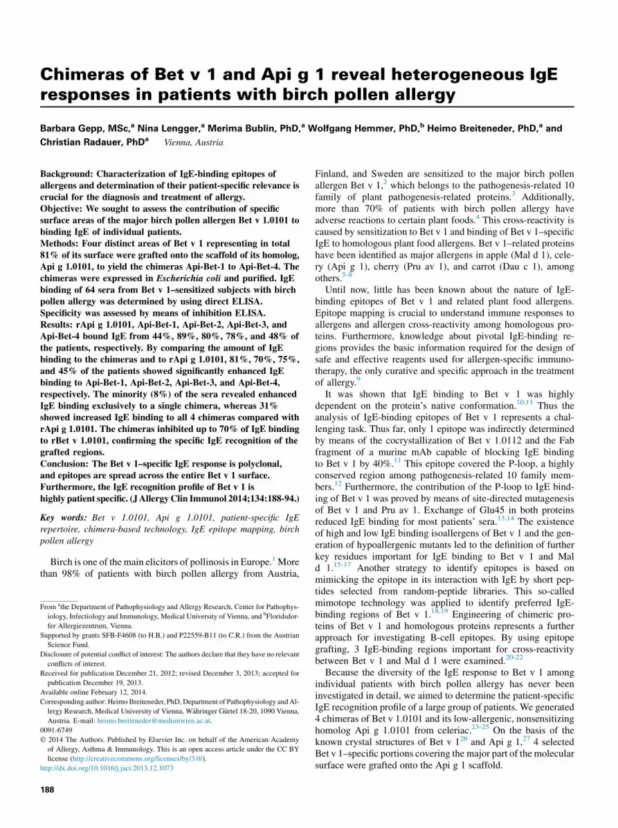
Design of the Api g 1–Bet v 1 chimerasChimeric proteins of Bet v 1.0101 and its homolog Api g 1.0101 were
generated to investigate IgE binding to defined Bet v 1.0101–specific surface
areas. Grafting of Bet v 1–specific surface areas onto the Api g 1.0101 scaffold
was achieved by replacing Api g 1.0101–specific solvent-accessible (>20%)
residues by corresponding Bet v 1.0101–specific residues (Fig 1).We generated
the chimeric protein Api-Bet-1 by grafting Glu45, the central residue of the pre-
viously identified P-loop epitope, and surrounding residues, identified by using
UCSF Chimera,28 onto Api g 1.0101. The region opposite the P-loop (Api-Bet-
2), the C-terminus and surrounding residues (Api-Bet-3), and the C-terminal a-
helix (Api-Bet-4) of Bet v 1.0101 were grafted in the same manner to generate
spatially well-distributed Bet v 1.0101–specific surface areas on Api g 1.0101.
Cloning, expression and purification, and
physicochemical analysis of the recombinant
proteinsProduction and analysis of the recombinant proteins was performed as
described in the Methods section in this article’s Online Repository at www.
jacionline.org.
IgE ELISAFor direct ELISA, microtiter plates (Maxisorp; Nalge Nunc International,
Roskilde, Denmark) were coated overnight at 48C with 1 mg/mL individual
chimeric proteins, a mixture of all chimeras (1 mg/mL each), and rApi g
1.0101 or rBet v 1.0101, respectively, in 50 mmol/L sodium carbonate buffer,
pH 9.6. After blocking of nonspecific binding sites, sera (1:10 dilution) were
incubated in duplicates overnight at 48C. Specific IgEwas detected by using an
alkaline phosphatase–conjugated mouse anti-human IgE mAb (BD Pharmin-
gen, San Jose, Calif), followed by color development with Sigma FAST
p-nitrophenyl phosphate tablets (Sigma-Aldrich, St Louis, Mo) and measure-
ment of the absorbance at 405 nm.
OD values were measured at several time points. For each serum, the
measurement with an OD of approximately 1.0 for Bet v 1.0101 was
normalized to a 1-hour substrate incubation period after subtracting the OD
values of the buffer controls (see Table E2 in this article’s Online Repository at
www.jacionline.org). Comparison of measurements at different times proved
that the OD values increased with time in a linear fashion (data not shown).
Hence normalized OD values were roughly proportional to allergen-specific
IgE concentrations.
Individual sera from 7 nonallergic donors were included as negative
controls. Normalized OD values exceeding themean negative control value by
more than 3 SDs were considered positive.
For each serum, specific IgE binding to the grafted regions of each chimera
was assessed by calculating the difference of the OD values of the chimera and
rApi g 1.0101. The difference was considered positive if it exceeded 3 times
the SD of the negative control value.
ELISA inhibitionFor inhibition ELISA, coating, blocking, and detection were performed, as
described above. Either Bet v 1.0101 or the chimeras were coated to the solid
phase. In inhibition assays, in which rBet v 1.0101 was coated, IgG was
removed in advance by means of incubation of prediluted sera on an anti-
human IgG (BD Pharmingen)–coated plate. Sera were diluted 30- to 100-fold.
Inhibition was performed by preincubating diluted sera with 10-fold serial
dilutions from 0.01 to 100 mg/mL of the individual chimeric proteins, a
mixture of all chimeras, and rApi g 1.0101 or rBet v 1.0101, respectively,
before they were applied to the plates.
For cross-inhibition between the chimeras, all chimeras, rApi g 1.0101, and
rBet v 1.0101 (1 mg/mL) were coated to the solid phase and incubated with
patients’ sera (diluted 10- to 60-fold). The supernatants were transferred to a
second plate, which was coated with all 4 chimeras or buffer only. IgE binding
to the second plate was detected, as described above.
Inhibition values were calculated as follows:
Inhibition ½%�5 ð12ODinhibited=ODnoninhibitedÞ3100:
ELISA with Bet v 1–specific mAbsBinding of Bet v 1–specific mAbs to rBet v 1, rApi g 1, and the chimeras
was tested by usingELISA, as described in theMethods section in this article’s
Online Repository.
Statistical analysesThe Friedman test (a5 .05) was performed to test whether the amount of
IgE binding to each of the 4 chimeras differed significantly from that to rApi g
1.0101. The relationship between the number of chimeras recognized better
than rApi g 1.0101 and the amount of rBet v 1–specific IgE present in patients’
sera was analyzed by performing Spearman correlation (a 5 .05).
RESULTS
Biochemical characterization of the recombinant
proteinsThe structural integrity of the recombinant proteins was
confirmed by means of circular dichroism spectroscopy, massspectrometry, and ELISA with Bet v 1–specific mAbs, asdescribed in the Results section in this article’s Online Repositoryat www.jacionline.org.
IgE-binding profiles of Bet v 1 are highly patient
specificIgE-binding capacities of rBet v 1.0101, rApi g 1.0101, and the
chimeras were determined by means of ELISA. All 64 tested seradisplayed rBet v 1–specific IgE, whereas only 44% of the serabound to rApi g 1 (Table I). Total IgE-binding capacities of all 4chimeras were significantly higher than that of rApi g 1 (P <.001;median OD for rApi g 1, 0.028; median ODs for the chimeras,0.076-0.222; see Fig E3 in this article’s Online Repository atwww.jacionline.org).
For each serum, specific IgE binding to the grafted regions ofeach chimera was assessed by calculating the difference of theELISAOD values of the chimera and the template rApi g 1.0101.The grafted regions of Api-Bet-1, Api-Bet-2, and Api-Bet-3 wererecognized by 70% to 81% of the sera, whereas only 45%recognized Api-Bet-4 (Table I).
Patients were categorized according to their binding patterns toevaluate individual IgE recognition profiles (Fig 2). Interestingly,
FIG 1. Sequence and structural comparison of the chimeras. A, Front and back views (rotated by 1808around a vertical axis) of the parent molecules and the chimeras are depicted. Colors indicating
mutated residues were mapped onto the Api g 1 surface. The models were prepared with UCSF Chimera.28
B, Multiple sequence alignment of proteins.
J ALLERGY CLIN IMMUNOL
JULY 2014
190 GEPP ET AL
only 5 (8%) of 64 sera exclusively recognized the grafted regionsof a single chimeric protein. Furthermore, 19 and 17 (30% and27%, respectively) sera showed increased IgE binding to 2 or 3chimeras, respectively, compared with rApi g 1.0101. The highestnumber of patients possessed IgE directed to all 4 grafted regions(20/64 [31%]). Only 3 (5%) sera did not bind to any grafted region.
Inhibition ELISA confirms IgE specificity for the
grafted areasData of 4 representative sera are depicted in Fig 3. Direct ELISA
data (Fig 3, A) show the highly patient-specific IgE-binding
patterns to the chimeric proteins. The percentage of Bet v1–specific IgE that bound to the chimeras was tested by using aninhibition ELISA (Fig 3, B) in which rBet v 1.0101 was coatedto the solid phase. Self-inhibition of rBet v 1.0101 was completeat inhibitor concentrations of 10 mg/mL. In most cases the chi-meras did not reach saturating inhibition, even at 100 mg/mL.Maximum inhibition values ranged from 7% to 71%, dependingon the serum and the inhibitor protein. A mixture of all 4 chimerasinhibited IgE binding to Bet v 1 by 55% to 77%. In contrast,rApi g 1 inhibited IgE binding to rBet v 1 by only 2% to 27%.
The 4 chimeras were coated to the solid phase to examine thepercentage of chimera-specific IgE that bound to the grafted areas
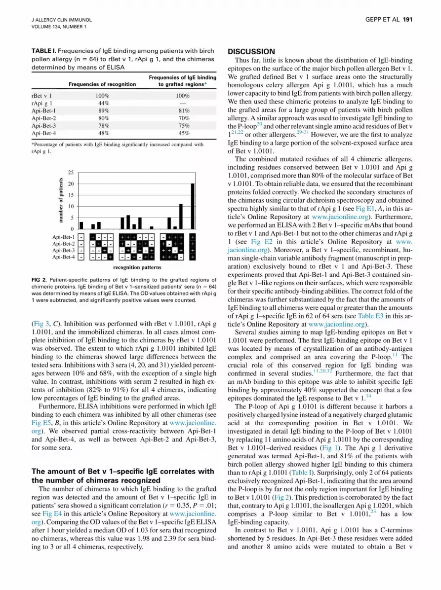
TABLE I. Frequencies of IgE binding among patients with birch
pollen allergy (n 5 64) to rBet v 1, rApi g 1, and the chimeras
determined by means of ELISA
Frequencies of recognition
Frequencies of IgE binding
to grafted regions*
rBet v 1 100% 100%
rApi g 1 44% —
Api-Bet-1 89% 81%
Api-Bet-2 80% 70%
Api-Bet-3 78% 75%
Api-Bet-4 48% 45%
*Percentage of patients with IgE binding significantly increased compared with
rApi g 1.
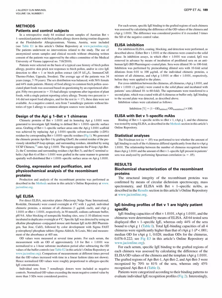
FIG 2. Patient-specific patterns of IgE binding to the grafted regions of
chimeric proteins. IgE binding of Bet v 1–sensitized patients’ sera (n 5 64)
was determined bymeans of IgE ELISA. The OD values obtainedwith rApi g
1 were subtracted, and significantly positive values were counted.
J ALLERGY CLIN IMMUNOL
VOLUME 134, NUMBER 1
GEPP ET AL 191
(Fig 3, C). Inhibition was performed with rBet v 1.0101, rApi g1.0101, and the immobilized chimeras. In all cases almost com-plete inhibition of IgE binding to the chimeras by rBet v 1.0101was observed. The extent to which rApi g 1.0101 inhibited IgEbinding to the chimeras showed large differences between thetested sera. Inhibitions with 3 sera (4, 20, and 31) yielded percent-ages between 10% and 68%, with the exception of a single highvalue. In contrast, inhibitions with serum 2 resulted in high ex-tents of inhibition (82% to 91%) for all 4 chimeras, indicatinglow percentages of IgE binding to the grafted areas.
Furthermore, ELISA inhibitions were performed in which IgEbinding to each chimera was inhibited by all other chimeras (seeFig E5, B, in this article’s Online Repository at www.jacionline.org). We observed partial cross-reactivity between Api-Bet-1and Api-Bet-4, as well as between Api-Bet-2 and Api-Bet-3,for some sera.
The amount of Bet v 1–specific IgE correlates with
the number of chimeras recognizedThe number of chimeras to which IgE binding to the grafted
region was detected and the amount of Bet v 1–specific IgE inpatients’ sera showed a significant correlation (r5 0.35, P5 .01;see Fig E4 in this article’s Online Repository at www.jacionline.org). Comparing the OD values of the Bet v 1–specific IgE ELISAafter 1 hour yielded a median OD of 1.03 for sera that recognizedno chimeras, whereas this value was 1.98 and 2.39 for sera bind-ing to 3 or all 4 chimeras, respectively.
DISCUSSIONThus far, little is known about the distribution of IgE-binding
epitopes on the surface of the major birch pollen allergen Bet v 1.We grafted defined Bet v 1 surface areas onto the structurallyhomologous celery allergen Api g 1.0101, which has a muchlower capacity to bind IgE from patients with birch pollen allergy.We then used these chimeric proteins to analyze IgE binding tothe grafted areas for a large group of patients with birch pollenallergy. A similar approach was used to investigate IgE binding tothe P-loop20 and other relevant single amino acid residues of Bet v121,22 or other allergens.29-31 However, we are the first to analyzeIgE binding to a large portion of the solvent-exposed surface areaof Bet v 1.0101.
The combined mutated residues of all 4 chimeric allergens,including residues conserved between Bet v 1.0101 and Api g1.0101, comprised more than 80% of the molecular surface of Betv 1.0101. To obtain reliable data, we ensured that the recombinantproteins folded correctly. We checked the secondary structures ofthe chimeras using circular dichroism spectroscopy and obtainedspectra highly similar to that of rApi g 1 (see Fig E1, A, in this ar-ticle’s Online Repository at www.jacionline.org). Furthermore,we performed an ELISAwith 2 Bet v 1–specific mAbs that boundto rBet v 1 and Api-Bet-1 but not to the other chimeras and rApi g1 (see Fig E2 in this article’s Online Repository at www.jacionline.org). Moreover, a Bet v 1–specific, recombinant, hu-man single-chain variable antibody fragment (manuscript in prep-aration) exclusively bound to rBet v 1 and Api-Bet-3. Theseexperiments proved that Api-Bet-1 and Api-Bet-3 contained sin-gle Bet v 1–like regions on their surfaces, which were responsiblefor their specific antibody-binding abilities. The correct fold of thechimeras was further substantiated by the fact that the amounts ofIgE binding to all chimeras were equal or greater than the amountsof rApi g 1–specific IgE in 62 of 64 sera (see Table E3 in this ar-ticle’s Online Repository at www.jacionline.org).
Several studies aiming to map IgE-binding epitopes on Bet v1.0101 were performed. The first IgE-binding epitope on Bet v 1was located by means of crystallization of an antibody-antigencomplex and comprised an area covering the P-loop.11 Thecrucial role of this conserved region for IgE binding wasconfirmed in several studies.11,20,32 Furthermore, the fact thatan mAb binding to this epitope was able to inhibit specific IgEbinding by approximately 40% supported the concept that a fewepitopes dominated the IgE response to Bet v 1.14
The P-loop of Api g 1.0101 is different because it harbors apositively charged lysine instead of a negatively charged glutamicacid at the corresponding position in Bet v 1.0101. Weinvestigated in detail IgE binding to the P-loop of Bet v 1.0101by replacing 11 amino acids of Api g 1.0101 by the correspondingBet v 1.0101–derived residues (Fig 1). The Api g 1 derivativegenerated was termed Api-Bet-1, and 81% of the patients withbirch pollen allergy showed higher IgE binding to this chimerathan to rApi g 1.0101 (Table I). Surprisingly, only 2 of 64 patientsexclusively recognized Api-Bet-1, indicating that the area aroundthe P-loop is by far not the only region important for IgE bindingto Bet v 1.0101 (Fig 2). This prediction is corroborated by the factthat, contrary to Api g 1.0101, the isoallergen Api g 1.0201, whichcomprises a P-loop similar to Bet v 1.0101,23 has a lowIgE-binding capacity.
In contrast to Bet v 1.0101, Api g 1.0101 has a C-terminusshortened by 5 residues. In Api-Bet-3 these residues were addedand another 8 amino acids were mutated to obtain a Bet v
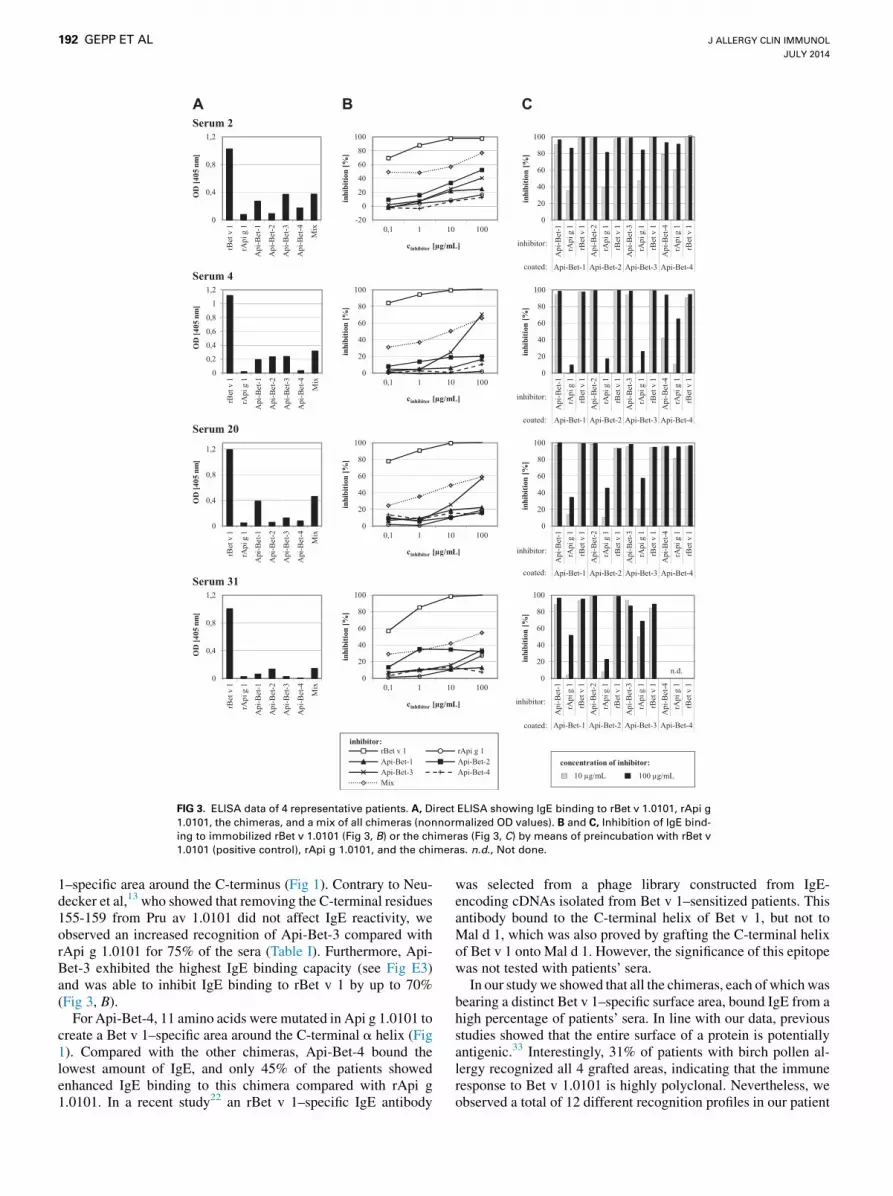
FIG 3. ELISA data of 4 representative patients. A, Direct ELISA showing IgE binding to rBet v 1.0101, rApi g
1.0101, the chimeras, and a mix of all chimeras (nonnormalized OD values). B and C, Inhibition of IgE bind-
ing to immobilized rBet v 1.0101 (Fig 3, B) or the chimeras (Fig 3, C) by means of preincubation with rBet v
1.0101 (positive control), rApi g 1.0101, and the chimeras. n.d., Not done.
J ALLERGY CLIN IMMUNOL
JULY 2014
192 GEPP ET AL
1–specific area around the C-terminus (Fig 1). Contrary to Neu-decker et al,13 who showed that removing the C-terminal residues155-159 from Pru av 1.0101 did not affect IgE reactivity, weobserved an increased recognition of Api-Bet-3 compared withrApi g 1.0101 for 75% of the sera (Table I). Furthermore, Api-Bet-3 exhibited the highest IgE binding capacity (see Fig E3)and was able to inhibit IgE binding to rBet v 1 by up to 70%(Fig 3, B).
For Api-Bet-4, 11 amino acids were mutated in Api g 1.0101 tocreate a Bet v 1–specific area around the C-terminal a helix (Fig1). Compared with the other chimeras, Api-Bet-4 bound thelowest amount of IgE, and only 45% of the patients showedenhanced IgE binding to this chimera compared with rApi g1.0101. In a recent study22 an rBet v 1–specific IgE antibody
was selected from a phage library constructed from IgE-encoding cDNAs isolated from Bet v 1–sensitized patients. Thisantibody bound to the C-terminal helix of Bet v 1, but not toMal d 1, which was also proved by grafting the C-terminal helixof Bet v 1 onto Mal d 1. However, the significance of this epitopewas not tested with patients’ sera.
In our studywe showed that all the chimeras, each of whichwasbearing a distinct Bet v 1–specific surface area, bound IgE from ahigh percentage of patients’ sera. In line with our data, previousstudies showed that the entire surface of a protein is potentiallyantigenic.33 Interestingly, 31% of patients with birch pollen al-lergy recognized all 4 grafted areas, indicating that the immuneresponse to Bet v 1.0101 is highly polyclonal. Nevertheless, weobserved a total of 12 different recognition profiles in our patient

J ALLERGY CLIN IMMUNOL
VOLUME 134, NUMBER 1
GEPP ET AL 193
sample, with the number of bound chimeras between 0 and 4(Fig 2).
Consistent with our results, a high patient-to-patient variationwas also observed when investigating IgE binding to the P-loopmutants of Pru av 1 and Api g 1.0101.13,23,34 Furthermore, Holmet al20 observed patient-specific IgE repertoires by grafting of aBet v 1–specific epitope containing the P-loop onto Mal d 1.Moreover, IgE binding to various Bet v 1mutants displayed diver-gent recognition patterns,16,35 and high heterogeneity of IgE spec-ificity could also be observed by comparing IgE binding to Bet v 1and homologous food proteins.19 However, most of these studieswere performed with small patient groups.
In a recent study of allergen-specific IgE from patientssensitized to the major house dust mite allergen Der p 2, it wasshown that the complexity of the allergic patients’ IgE repertoirecorrelated with the serum concentration of allergen-specificIgE.36 In accordance with this finding, we revealed that theamount of Bet v 1–specific IgE present in patients’ sera correlatedwith the number of chimeras recognized, a value representing thecomplexity of the IgE epitope repertoire (see Fig E4).
Reduction of IgE cross-linking on the surfaces of mast cells orbasophils by vaccine components during specific immunotherapyis crucial for preventing severe side effects. Therefore character-ization of IgE-binding epitopes is of paramount importance fordeveloping artificial hypoallergens or peptide vaccines for saferand more effective immunotherapy. Mapping of IgE epitopes byusing a chimera-based approach offers the possibility ofanalyzing a defined area (eg, a single IgE epitope) of an allergenwith polyclonal patients’ sera. Thus this technologymight be usedas diagnostic tool to determine the patient-specific response todefined epitopes of a major allergen or to cross-reactivehomologs. In addition, this will pave the way for a patient-tailored epitope-based therapy.
Taken together, this study demonstrates that it is possible tograft defined areas of a major allergen onto a low IgE-bindinghomolog to evaluate IgE binding to the grafted region. Further-more, investigation of a large group of patients with birch pollenallergy showed that the repertoire of Bet v 1–specific IgE is highlypatient specific and polyclonal. A single major epitope on Bet v1.0101 important for all patients with birch pollen allergy doesnot exist, and relevant IgE-binding epitopes are located across theentire surface of Bet v 1.0101.
Key messages
d The Bet v 1–specific IgE response is polyclonal, and therecognition profile is highly patient specific.
d The existence of a single major IgE epitope on Bet v 1 canbe excluded.
d The IgE epitopes are distributed across the entire surfaceof Bet v 1.
REFERENCES
1. D’Amato G, Cecchi L, Bonini S, Nunes C, Annesi-Maesano I, Behrendt H, et al.
Allergenic pollen and pollen allergy in Europe. Allergy 2007;62:976-90.
2. Moverare R, Westritschnig K, Svensson M, Hayek B, Bende M, Pauli G, et al.
Different IgE reactivity profiles in birch pollen-sensitive patients from six Euro-
pean populations revealed by recombinant allergens: an imprint of local sensitiza-
tion. Int Arch Allergy Immunol 2002;128:325-35.
3. Radauer C, Lackner P, Breiteneder H. The Bet v. 1 fold: an ancient, versatile scaf-
fold for binding of large, hydrophobic ligands. BMC Evol Biol 2008;8:286.
4. Geroldinger-Simic M, Zelniker T, Aberer W, Ebner C, Egger C, Greiderer A, et al.
Birch pollen-related food allergy: clinical aspects and the role of allergen-specific
IgE and IgG4 antibodies. J Allergy Clin Immunol 2011;127:616-22.
5. Vanek-Krebitz M, Hoffmann-Sommergruber K, Laimer da Camara Machado M,
Susani M, Ebner C, Kraft D, et al. Cloning and sequencing of Mal d 1, the major
allergen from apple (Malus domestica), and its immunological relationship to Bet v
1, the major birch pollen allergen. Biochem Biophys Res Commun 1995;214:
538-51.
6. Breiteneder H, Hoffmann-Sommergruber K, O’Riordain G, Susani M, Ahorn H,
Ebner C, et al. Molecular characterization of Api g 1, the major allergen of celery
(Apium graveolens), and its immunological and structural relationships to a group
of 17-kDa tree pollen allergens. Eur J Biochem 1995;233:484-9.
7. Scheurer S, Metzner K, Haustein D, Vieths S. Molecular cloning, expression and
characterization of Pru a 1, the major cherry allergen. Mol Immunol 1997;34:
619-29.
8. Hoffmann-Sommergruber K, O’Riordain G, Ahorn H, Ebner C, Laimer Da Camara
Machado M, Puhringer H, et al. Molecular characterization of Dau c 1, the Bet v 1
homologous protein from carrot and its cross-reactivity with Bet v 1 and Api g 1.
Clin Exp Allergy 1999;29:840-7.
9. Jutel M, Akdis CA. Immunological mechanisms of allergen-specific immuno-
therapy. Allergy 2011;66:725-32.
10. Focke M, Linhart B, Hartl A, Wiedermann U, Sperr WR, Valent P, et al. Non-
anaphylactic surface-exposed peptides of the major birch pollen allergen, Bet v
1, for preventive vaccination. Clin Exp Allergy 2004;34:1525-33.
11. Mirza O, Henriksen A, Ipsen H, Larsen JN, Wissenbach M, Spangfort MD, et al.
Dominant epitopes and allergic cross-reactivity: complex formation between a Fab
fragment of a monoclonal murine IgG antibody and the major allergen from birch
pollen Bet v 1. J Immunol 2000;165:331-8.
12. Liu J, Ekramoddoullah A. The family 10 of plant pathogenesis-related proteins:
their structure, regulation, and function in response to biotic and abiotic stresses.
Physiol Mol Plant Pathol 2006;68:3-13.
13. Neudecker P, Lehmann K, Nerkamp J, Haase T, Wangorsch A, Fotisch K, et al.
Mutational epitope analysis of Pru av 1 and Api g 1, the major allergens of cherry
(Prunus avium) and celery (Apium graveolens): correlating IgE reactivity with
three-dimensional structure. Biochem J 2003;376:97-107.
14. Spangfort MD, Mirza O, Ipsen H, Van Neerven RJ, Gajhede M, Larsen JN. Domi-
nating IgE-binding epitope of Bet v 1, the major allergen of birch pollen, charac-
terized by X-ray crystallography and site-directed mutagenesis. J Immunol 2003;
171:3084-90.
15. Ferreira F, Hirtenlehner K, Jilek A, Godnik-Cvar J, Breiteneder H, Grimm R, et al.
Dissection of immunoglobulin E and T lymphocyte reactivity of isoforms of the
major birch pollen allergen Bet v 1: potential use of hypoallergenic isoforms for
immunotherapy. J Exp Med 1996;183:599-609.
16. Ferreira F, Ebner C, Kramer B, Casari G, Briza P, Kungl AJ, et al. Modulation of
IgE reactivity of allergens by site-directed mutagenesis: potential use of hypoaller-
genic variants for immunotherapy. FASEB J 1998;12:231-42.
17. Ma Y, Gadermaier G, Bohle B, Bolhaar S, Knulst A, Markovic-Housley Z, et al.
Mutational analysis of amino acid positions crucial for IgE-binding epitopes of
the major apple (Malus domestica) allergen, Mal d 1. Int Arch Allergy Immunol
2006;139:53-62.
18. Ganglberger E, Grunberger K, Sponer B, Radauer C, Breiteneder H, Boltz-Nitu-
lescu G, et al. Allergen mimotopes for 3-dimensional epitope search and induction
of antibodies inhibiting human IgE. FASEB J 2000;14:2177-84.
19. Mittag D, Batori V, Neudecker P, Wiche R, Friis EP, Ballmer-Weber BK, et al. A
novel approach for investigation of specific and cross-reactive IgE epitopes on Bet
v 1 and homologous food allergens in individual patients. Mol Immunol 2006;43:
268-78.
20. Holm J, Ferreras M, Ipsen H, Wurtzen PA, Gajhede M, Larsen JN, et al. Epitope
grafting, re-creating a conformational Bet v 1 antibody epitope on the surface of
the homologous apple allergen Mal d 1. J Biol Chem 2011;286:17569-78.
21. Klinglmayr E, Hauser M, Zimmermann F, Dissertori O, Lackner P, Wopfner N,
et al. Identification of B-cell epitopes of Bet v 1 involved in cross-reactivity
with food allergens. Allergy 2009;64:647-51.
22. Hecker J, Diethers A, Schulz D, Sabri A, Plum M, Michel Y, et al. An IgE epitope
of Bet v 1 and Fagales PR10 proteins as defined by a human monoclonal IgE. Al-
lergy 2012;67:1530-7.
23. Wangorsch A, Ballmer-Weber BK, Rosch P, Holzhauser T, Vieths S. Mutational
epitope analysis and cross-reactivity of two isoforms of Api g 1, the major celery
allergen. Mol Immunol 2007;44:2518-27.
24. Bauermeister K, Ballmer-Weber BK, Bublin M, Fritsche P, Hanschmann KM,
Hoffmann-Sommergruber K, et al. Assessment of component-resolved in vitro
diagnosis of celeriac allergy. J Allergy Clin Immunol 2009;124:1273-81.
25. Kazemi-Shirazi L, Pauli G, Purohit A, Spitzauer S, Froschl R, Hoffmann-Sommer-
gruber K, et al. Quantitative IgE inhibition experiments with purified recombinant

J ALLERGY CLIN IMMUNOL
JULY 2014
194 GEPP ET AL
allergens indicate pollen-derived allergens as the sensitizing agents responsible for
many forms of plant food allergy. J Allergy Clin Immunol 2000;105:116-25.
26. Gajhede M, Osmark P, Poulsen FM, Ipsen H, Larsen JN, Joost van Neerven RJ,
et al. X-ray and NMR structure of Bet v 1, the origin of birch pollen allergy.
Nat Struct Biol 1996;3:1040-5.
27. Schirmer T, Hoffimann-Sommergrube K, Susani M, Breiteneder H, Markovic-
Housley Z. Crystal structure of the major celery allergen Api g 1: molecular anal-
ysis of cross-reactivity. J Mol Biol 2005;351:1101-9.
28. Pettersen EF, Goddard TD, Huang CC, Couch GS, Greenblatt DM, Meng EC, et al.
UCSF Chimera—visualization system for exploratory research and analysis.
J Comput Chem 2004;25:1605-12.
29. King TP, Jim SY, Monsalve RI, Kagey-Sobotka A, Lichtenstein LM, Spangfort
MD. Recombinant allergens with reduced allergenicity but retaining immunoge-
nicity of the natural allergens: hybrids of yellow jacket and paper wasp venom
allergen antigen 5s. J Immunol 2001;166:6057-65.
30. Karisola P, Alenius H, Mikkola J, Kalkkinen N, Helin J, Pentikainen OT, et al. The
major conformational IgE-binding epitopes of hevein (Hev b6.02) are identified by
a novel chimera-based allergen epitope mapping strategy. J Biol Chem 2002;277:
22656-61.
31. Albrecht M, Kuhne Y, Ballmer-Weber BK, Becker WM, Holzhauser T, Lauer I,
et al. Relevance of IgE binding to short peptides for the allergenic activity of
food allergens. J Allergy Clin Immunol 2009;124:328-36.
32. Gieras A, Cejka P, Blatt K, Focke-Tejkl M, Linhart B, Flicker S, et al. Mapping of
conformational IgE epitopes with peptide-specific monoclonal antibodies reveals
simultaneous binding of different IgE antibodies to a surface patch on the major
birch pollen allergen, Bet v 1. J Immunol 2011;186:5333-44.
33. Aalberse RC, Crameri R. IgE-binding epitopes: a reappraisal. Allergy 2011;66:
1261-74.
34. Scheurer S, Son DY, Boehm M, Karamloo F, Franke S, Hoffmann A, et al. Cross-
reactivity and epitope analysis of Pru a 1, the major cherry allergen. Mol Immunol
1999;36:155-67.
35. Holm J, Gajhede M, Ferreras M, Henriksen A, Ipsen H, Larsen JN, et al. Allergy
vaccine engineering: epitope modulation of recombinant Bet v 1 reduces IgE bind-
ing but retains protein folding pattern for induction of protective blocking-antibody
responses. J Immunol 2004;173:5258-67.
36. Willumsen N, Holm J, Christensen LH, Wurtzen PA, Lund K. The complexity of
allergic patients’ IgE repertoire correlates with serum concentration of allergen-
specific IgE. Clin Exp Allergy 2012;42:1227-36.
Related Documents