Excellence in specialist and community healthcare NICE CG95 ‘Chest pain of recent onset’ Kay Townsend RACPC CNS May 2018

Welcome message from author
This document is posted to help you gain knowledge. Please leave a comment to let me know what you think about it! Share it to your friends and learn new things together.
Transcript

Excellence in specialist and community healthcare
NICE CG95
‘Chest pain of recent onset’
Kay Townsend RACPC CNS May 2018
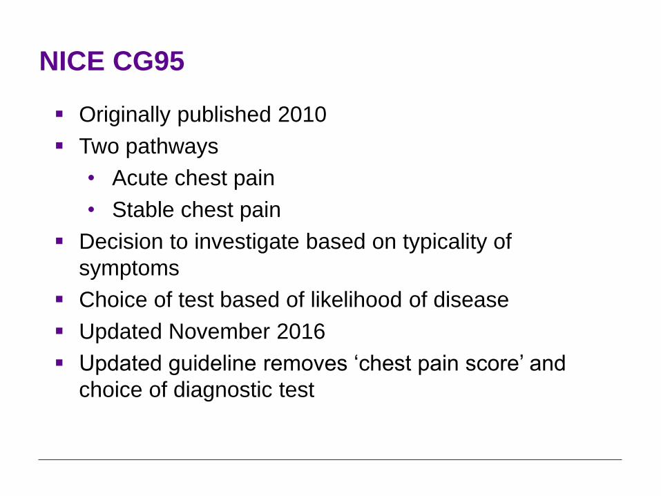
NICE CG95
Originally published 2010
Two pathways
• Acute chest pain
• Stable chest pain
Decision to investigate based on typicality of
symptoms
Choice of test based of likelihood of disease
Updated November 2016
Updated guideline removes ‘chest pain score’ and
choice of diagnostic test
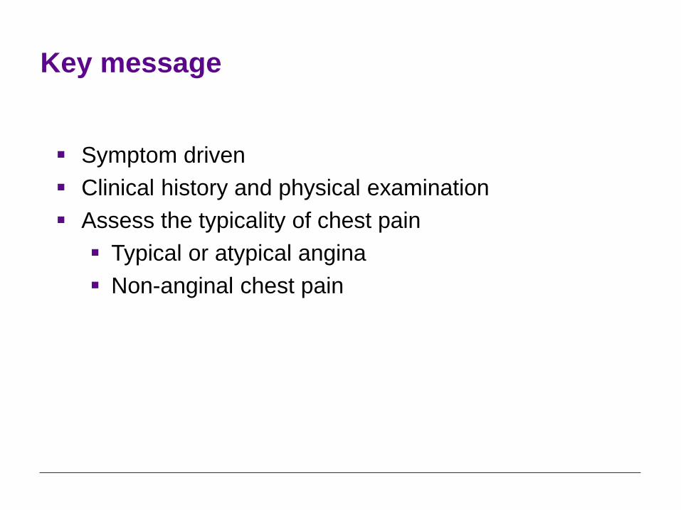
Key message
Symptom driven
Clinical history and physical examination
Assess the typicality of chest pain
Typical or atypical angina
Non-anginal chest pain

Clinical history and physical examination
Take a detailed clinical history documenting:
the age and sex of the person
the characteristics of the pain, including its location,
radiation, severity, duration and frequency, and factors
that provoke and relieve the pain
any associated symptoms, such as breathlessness
any history of angina, myocardial infarction, coronary
revascularisation or other cardiovascular disease and
any cardiovascular risk factors.

Clinical history and physical examination
Carry out a physical examination to:
identify risk factors for cardiovascular disease
identify signs of other cardiovascular disease
identify non-coronary causes of angina (for example,
severe aortic stenosis, cardiomyopathy) and
exclude other causes of chest pain.

Assess the typicality of chest pain
Anginal pain is:
constricting discomfort in the front of the chest, or in
the neck, shoulders, jaw or arms
precipitated by physical exertion
relieved by rest or GTN within about 5 minutes.
Assess the typicality of chest pain as follows:
Presence of three of the features is defined as typical
angina.
Presence of two of the features is defined as atypical
angina.
Presence of one or none of the features is defined as
non-anginal chest pain.
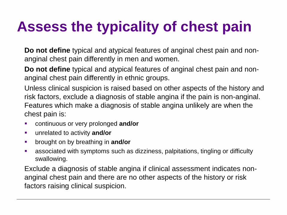
Assess the typicality of chest pain
Do not define typical and atypical features of anginal chest pain and non-
anginal chest pain differently in men and women.
Do not define typical and atypical features of anginal chest pain and non-
anginal chest pain differently in ethnic groups.
Unless clinical suspicion is raised based on other aspects of the history and
risk factors, exclude a diagnosis of stable angina if the pain is non-anginal.
Features which make a diagnosis of stable angina unlikely are when the
chest pain is:
continuous or very prolonged and/or
unrelated to activity and/or
brought on by breathing in and/or
associated with symptoms such as dizziness, palpitations, tingling or difficulty
swallowing.
Exclude a diagnosis of stable angina if clinical assessment indicates non-
anginal chest pain and there are no other aspects of the history or risk
factors raising clinical suspicion.
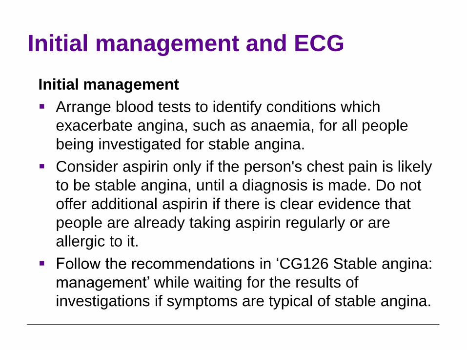
Initial management and ECG
Initial management
Arrange blood tests to identify conditions which
exacerbate angina, such as anaemia, for all people
being investigated for stable angina.
Consider aspirin only if the person's chest pain is likely
to be stable angina, until a diagnosis is made. Do not
offer additional aspirin if there is clear evidence that
people are already taking aspirin regularly or are
allergic to it.
Follow the recommendations in ‘CG126 Stable angina:
management’ while waiting for the results of
investigations if symptoms are typical of stable angina.

Initial management and ECG
ECG
For people in whom stable angina cannot be excluded on the basis of the
clinical assessment alone, take a resting 12-lead ECG as soon as
possible after presentation.
Do not rule out a diagnosis of stable angina on the basis of a normal
resting 12-lead ECG.
A number of changes on a resting 12-lead ECG are consistent with
coronary artery disease and may indicate ischaemia or previous
infarction. These include:
• pathological Q waves
• left bundle branch block
• ST-segment and T wave abnormalities (for example, flattening or inversion).
Note that the results may not be conclusive.
Consider any resting 12-lead ECG changes together with the clinical
history and risk factors.

Diagnostic investigations
If clinical assessment indicates typical or atypical angina,
offer diagnostic testing.
First-line
Second-line
Third-line
First-line: 64-slice CT coronary angiography
Offer 64-slice (or above) CT coronary angiography if:
clinical assessment indicates typical or atypical
angina, or
clinical assessment indicates non-anginal chest pain
but 12-lead resting ECG has been done and indicates
ST-T changes or Q waves.

Second-line: non-invasive functional
testing Offer non-invasive functional imaging for myocardial
ischaemia if 64-slice (or above) CT coronary angiography has shown coronary artery disease of uncertain functional significance or is non-diagnostic.
When offering non-invasive functional imaging for myocardial ischaemia use:
MPS with SPECT or
stress echocardiography or
first-pass contrast-enhanced magnetic resonance perfusion or
MRI for stress-induced wall motion abnormalities.
Third-line: invasive coronary angiography
Offer invasive coronary angiography as a third-line
investigation when the results of non-invasive functional
imaging are inconclusive.

Confirmed coronary artery disease
For people with confirmed coronary artery disease (for
example, previous myocardial infarction,
revascularisation, previous angiography) offer non-
invasive functional testing when there is uncertainty
about whether chest pain is caused by myocardial
ischaemia.
An exercise ECG may be used instead of functional
imaging.

Investigations that should not be used
Do not use MR coronary angiography for diagnosing
stable angina.
Do not use exercise ECG to diagnose or exclude stable
angina for people without known coronary artery
disease.
Non-anginal chest pain
Consider causes of chest pain other than angina (such
as gastrointestinal or musculoskeletal pain).
Only consider chest X-ray if other diagnoses, such as
a lung tumour, are suspected.
Do not offer diagnostic testing to people with non-
anginal chest pain on clinical assessment unless there
are resting ECG ST-T or Q waves.
If a diagnosis of stable angina has been excluded at
any point in the care pathway, but people have risk
factors for cardiovascular disease, follow the
appropriate guidance.
Stable angina
Confirm a diagnosis of stable angina and follow the
recommendations in CG126 ‘Stable angina:
management’ when:
significant coronary artery disease is found during
invasive or 64-slice (or above) CT coronary
angiography or
reversible myocardial ischaemia is found during non-
invasive functional imaging.

Other causes of chest pain
Investigate other causes of chest pain when:
significant coronary artery disease is not found during
invasive coronary angiography or 64-slice (or above)
CT coronary angiography or
reversible myocardial ischaemia is not found during
non-invasive functional imaging.

Other causes of angina
Consider investigating other causes of angina, such as
hypertrophic cardiomyopathy, in people with typical
angina-like chest pain and a low likelihood of coronary
artery disease.
Consider investigating other causes of angina, such as
hypertrophic cardiomyopathy or syndrome X in people
with typical angina-like chest pain if investigation
excludes flow-limiting disease in the epicardial
coronary arteries.

Future options?

HeartFlow FFRCT for estimating fractional flow reserve
The following recommendations are from NICE medical technologies
guidance on HeartFlow FFRCT for estimating fractional flow reserve from coronary CT angiography.
The case for adopting HeartFlow FFRCT for estimating fractional flow reserve from coronary CT angiography is supported by the evidence. The technology is non-invasive and safe, and has a high level of diagnostic accuracy.
HeartFlow FFRCT should be considered as an option for patients with stable, recent onset chest pain who are offered coronary CT angiography as part of the NICE chest pain pathway. Using HeartFlow FFRCT may avoid the need for invasive coronary angiography and revascularisation. For correct use, HeartFlow FFRCT requires access to 64-slice (or above) coronary CT angiography facilities.
Based on the current evidence and assuming there is access to appropriate coronary CT angiography facilities, using HeartFlow FFRCT may lead to cost savings of £214 per patient. By adopting this technology, the NHS in England may save a minimum of £9.1 million by 2022 through avoiding invasive investigation and treatment.

Excellence in specialist and community healthcare
Any questions?
Related Documents