CHEST INJURIES By Prof. Soliman Ali Hassan

Welcome message from author
This document is posted to help you gain knowledge. Please leave a comment to let me know what you think about it! Share it to your friends and learn new things together.
Transcript

CHEST INJURIES
By
Prof. Soliman Ali Hassan
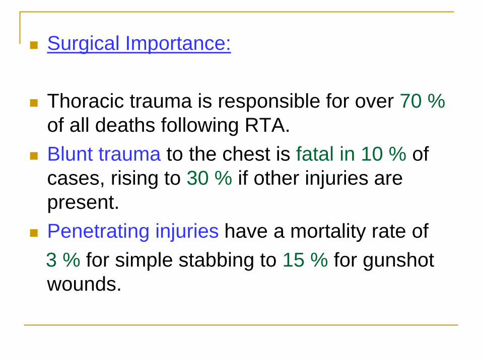
Surgical Importance:
Thoracic trauma is responsible for over 70 %
of all deaths following RTA.
Blunt trauma to the chest is fatal in 10 % of
cases, rising to 30 % if other injuries are
present.
Penetrating injuries have a mortality rate of
3 % for simple stabbing to 15 % for gunshot
wounds.

AETIOLOGY & MECHANISM OF INJURY
1- Blunt trauma:
Direct blow to the chest
Fall from a height
Car accident
2- penetrating trauma
Stab wound (low velocity)
Gun shots (high velocity)
3-Blast injury (explosive injuries)


Clinical types
1- simple rib fracture & flail chest
2- pneumothorax
3- haemothorax
4- contusion or laceration of the lung
5- cardiac injuries
6- injuries to major blood vessels
7- diaphragmatic injuries
8- oesophageal injuries

Pre-hospital care
Done by well trained paramedical personnel
( Basic Trauma Life Support Training Course).
Need ambulance cars, aircrafts, &
helicopters.

The prehospital medical services include
support of:
the patient’s airway, breathing, and
circulation (ABCs) & stoppage of external
hemorrhage.
iv lines, & iv fluids in the field.
Spinal protection (cervical & lumber),
immobilization, splinting.
Rapid transport to a trauma center.

Hospital care
The 3 R management plan is useful:
Resuscitation – Review – Repair

1- Resuscitation
ABCDE done by a team work:
Maintain a patent airway and restore the blood volume.
Maintain a patent airway
Remove an oral foreign body; dentures, food particles, etc.
Blood and secretions are removed from the oropharynx by suction.
Occlude sucking chest wound with dressing
Stop an external bleeding & maintain the circulation.
If laryngeal & pharyngeal reflexes are present forward tongue retraction with insertion of an oral (air way).
Absent reflexes endotracheal intubation


Endotracheal intubation

You can do:
Tracheostomy in cases of failed intubations
Laryngotomy when tracheostomy set is not
available ( insert needle just below the thyroid
cartilage)
Then do thorough tracheal & bronchial
aspiration.


Indications of ventilation in chest injuries:
Five main factors:
1- Multiple rib fractures with Severe Pain interfering
with coughing.
2- Flail chest multiple bilateral or two rows
fractures (unilateral).
3- Extensive pulmonary contusions.
4- Massive haemo or pneumothorax.
5- Associated head injuries with respiratory
depression
NB: Always do a chest tube before ventilation.

Urgent actions:
1- Severe pain strong analgesic is given ( morphia is contraindicated in associated head injuries)
2- Tension pneumothorax is deflated by a needle in 2nd IC
3- Thoractomy is done in case of uncontrollable bleeding.
4- If there is cardiac tamponade, needle aspiration of the pericardium is life saving until more controlled surgery to be performed.
5- In serious chest injuries, admit the patient to ICU.

5- Insert 5 tubes:
1) bilateral iv canulae (16G )
2) urethral catheter
3) NGT
4) CVP canula
Push IV fluids ( Ringer’s lactate)
Once the patient has been stabilized ;
Do the next step :
Review

REVIEW -2
It includes:
1- History taking: AMPLE
(allergy, medications, past history, last meal & event of the accident)
2- Measure "vital signs"
3- Assess roughly the level of consciousness:
AVPU (alertness, verbal response, pain response, or un response)

4- Do full clinical examination:
Inspection of the chest wall;
the frequency & pattern of breathing, external evidence of trauma of the thorax.
Palpation will detect;
surgical emphysema, paradoxical movement and a stove-in chest.
Auscultation and percussion should reveal;
the existence of a pneumo & or haemothorax, rib fractures & flail chest wall
&
Look for associated injuries

5- Investigations:
Blood samples for blood groups & cross
matching, CBC, electrolytes, etc.
X ray chest, CT & further treatment decided on
the basis of the patient’s condition and the
radiographic result.

After doing those measures ?
Classify your pat into:
1- Highest priority :
Cervical Fr.
Thoracic injuries.
Cardiac injuries.
External bleeding from big vessels.

2- High priority
Abd. Injuries
Head injuries (including brain injuries)
Fracture of long bones with extensive soft T. injuries.
3- Low priority
GIT injuries
Periph. Vascular& nerve injuries.
Fr. & dislocation
Facial & soft tissue injuries.

3- REPAIR (definitive treatment)
I- Chest wall injuries.
1- Localised rib fracture
Pain, tenderness & crepitus at the fracture site.
Uncomplicated fractures require analgesia or
an intercostal nerve blockade.
Encourage a normal respiratory movement
and effective coughing (physiotherapy).
Chest strapping or bed rest is no longer advised.

With plain X ray ; 2 ribs fractured

The same patient with 3 D.CT, 6 ribs fractured

2- Flail chest
More than 4 ribs are fractured in two places:
on one side of the chest (lateral)
(on either side of the sternum, anterior ) ;
called stove in chest
The flail segment moves paradoxically,
inwards during inspiration and outwards
during expiration, with side to side movement
of the heart & mediastinum ( mediastinal
flutter ); thereby reducing effective gas
exchange.

Flail chest: A- lat. Flail. B- ant. Flail (stove in)
Stove in

Treatment of flail chest:
If not affecting oxygenation, treat with analgesia & regular blood gas analysis until the flail segment stabilizes.
In case of impaired oxygenation, endotracheal intubation with:
Intermittent positive pressure breathing (IPPB)
for up to 3 weeks
Until the fractures become fixed.
Thoracotomy with fracture fixation is indicated if there is an underlying lung injury to be treated at the same time.

2- Pleural injuries
An erect chest X ray to confirm or exclude
pneumothorax or haemothorax.

Traumatic pneumothorax
Blunt or penetrating trauma to the chest wall may result in a lung laceration from a rib fracture.
Air comes from the lung or from the atmosphere
3 types:
1- simple
2- open
3- tension
Clinically; decreased air entry on the affected side & the trachea may be pushed to the opposite side.
There is an increased percussion note (resonance).

Simple pneumothorax
Limited amount of air
No mediastinal shift
If there is no marked dyspnea, treated
conservatively
For patient with dyspnea, do intercostal chest
tube

2- Open pneumothorax
Wound in chest wall (sucking chest wound) leading to communications between pleural space & atmosphere
During inspiration air passed outwards through the cutaneous wound.
Lung collapse & mediastinal shift to & fro
( mediastinal flutter)
1-The wound is closed at first with dressing then repaired surgically at OR.
2- Insert chest tube

Tension pneumothorax -3
Causes:
1- A small valve like wound of the visceral
pleura air accumulates in pleural
cavity during inspiration
2- A pleuro-cutaneous wound that allows air
to enter to but not pass out the pleural cavity.
3- Also, coughing, straining, or +ve pressure
ventilation may result in tension
pneumothorax.
Treatment is urgent and before X ray chest
can be taken.
A wide-bore needle introduced into the
affected side, in 2nd I.C. space will release
any air under tension and is life saving.
Then a wide-bore intercostal tube is
introduced in 5th IC space midaxillary and
directed to the apex of the pleural cavity.
A second drain may be introduced basally to
drain blood.

Tension pneumothorax with mediastinal shift


Traumatic haemothorax.
Due to blunt or penetrating injuries
Bleeding comes from intercostal or internal mammary vessels.
Chest tube using a wide bore tube (>28 Fr).
Continuing blood loss in excess of 200 mI / hour ,clotted or encysted blood require urgent thoracotomy .

Multiple rib fracture with haemthorax


Some special injuries:
1- First rib fracture.
Fracture of the first rib should alert the
clinician to a potentially serious chest injury,
such as injuries to the great vessels
The mortality rate associated with a fracture of
the first rib exceeds 30 %.

2- Injury of thoracic vertebrae
Usually stable
A quick neurological examination confirming the integrity of the nerve supply to the lower limbs
( paraplegia) should be performed and documented.

3- Lung contusions:
Persistent air leak require thoracotomy.
Avoid infection by early mobilization,
prophylactic antibiotics, suction drainage and
physiotherapy.

Haemthorax with left lung contusion

4- Major airways injuries
1- Injuries to major bronchi
2- Injury to the trachea
Infrequently seen.
There is usually a combination of surgical emphysema, haemoptysis and pneumothorax.
less than 25% of patients survive to reach hospital.
The treatment is exploration and repair if possible.


5- Diaphragmatic rupture
Caused by a high-speed blunt abdominal
trauma.
More commonly on the left hemi-diaphragm (the
right is protected by the liver).
Colon and stomach may herniate into the thorax
displacing the lung.
Bowel sounds may be heard in the chest and the
chest radiograph may reveal bowel gas in the
lung fields.

Diagnosis
A barium study will confirm the diagnosis.
Occasionally, the injury is overlooked and the
patient presents later on with a diaphragmatic
hernia.
Treatment is by thoracotomy to reduce the
bowel and repair the diaphragm .

6-Oesophageal injury
Perforation of oesophagus leads to
mediastinitis ( a very dangerous).
The management of penetrating trauma to
the oesophagus is by urgent repair.

7-Cardiac injury
Major injuries to the heart and great vessels from blunt trauma are frequently fatal.
Clinically;
Profuse external bleeding
Massive hemothorax
Cardiac tamponade ( Beck’s triad)
1- engorged neck veins
2- faint heart sounds
3- hypotension
There may be arrhythmias and signs of heart failure.


Treatment
1- Life saving measure
Aspiration of cardiac tamponade
Inert a long needle below & to the left of
xiphoid process, directed to the the tip of left
scapula
Left thoracotomy is indicated to deal with big
problems such as ruptured aorta , ruptured
cardiac chamber or massive haemothorax

The indications for thoracotomy
1- 500—1000 ml of fresh blood at the time of
initial drainage.
2- Continued bleeding (>100 ml/15 min) from
the intercostal drains.
3- Continued bleeding of >200 ml/h for 3 or
more hours from the intercostal drains.
4- Rupture of the bronchus, aorta, oesophagus
or diaphragm.
5- Cardiac tamponade (if needle aspiration is
unsuccessful)

Thoracotomy incision

Management of Penetrating injury
At first: wound dressing and intercostal tube.
Resuscitation as usual
In OR; explore the track of bullet and stab wounds in the chest to exclude damage to the heart, great vessels, and the diaphragm and abdominal viscera in addition to the lung injury.

Related Documents