Chemotherapy resistance in acute myeloid leukemia: the role of adhesion Joe C. Huang, M.D. Research Elective 8/09 -10/09 Mentor: Pamela S. Becker, M.D., Ph.D. Associate Professor of Medicine/Hematology Institute for Stem Cell and Regenerative Medicine University of Washington

Welcome message from author
This document is posted to help you gain knowledge. Please leave a comment to let me know what you think about it! Share it to your friends and learn new things together.
Transcript

Chemotherapy resistance in acute myeloid leukemia: the role of
adhesionJoe C. Huang, M.D.
Research Elective 8/09 -10/09
Mentor: Pamela S. Becker, M.D., Ph.D.
Associate Professor of Medicine/Hematology
Institute for Stem Cell and Regenerative Medicine
University of Washington

Acute myeloid leukemia (AML)
• Most common adult leukemia
• Incidence in 2007: 13, 410 cases
• Deaths in 2007: 8990
• For patients <60 yrs old, 30-35% survive >5 years
• For patients >60 yrs, <10% survive long-term

AML
• Principle cause of treatment failure is chemotherapy resistance despite initial complete remission rate of 65%
• Survival of AML cells in bone marrow following chemotherapy = minimal residual disease
• Minimal residual disease results in emergence of chemotherapy-resistant leukemia
• What role does the bone marrow provide in chemotherapy resistance?

Environment mediated drug resistance
• Bone marrow microenvironment promotes survival and proliferation of malignant cells, with adhesion sequestering malignant cells in a protective niche – de novo drug resistance independent of genetic changes caused by selective pressure of drug exposure (acquired drug resistance)

Role of CXCR-4
• Chemokine receptor expressed by normal and malignant hematopoetic cells – including NHL, myeloma, chronic and acute leukemias
• Ligand: stromal-derived factor-1 (SDF-1)
• SDF-1 is constitutively secreted by bone marrow cells

CXCR-4
• SDF-1 acts on normal and malignant hematopoetic progenitor cells that express its receptor
• Critical to homing and retention of malignant cells to the bone marrow microenvironment
• CXCR-4 expression correlated to prognosis in AML (low expression with longer relapse-free and overall survival)

Role of VLA-4
• Very late antigen-4, an α4β1 integrin
• Ligands: vascular cell adhesion molecule-1 (VCAM-1) and alternatively spliced fibronectin
• Migration of CD34+ cells and AML cells beneath marrow stroma
• Altered integrin expression involved in solid tumor metastasis (prostate, lung, breast) to bone and lodgment in bone marrow

VLA-4
• Integrin-mediated signaling protects against chemotherapy toxicity through activation of survival/anti-apoptotic pathways (PI3K/Ak/bcl-2, Wnt pathways) and induces quiescence
• Function of VLA-4 correlated to AML survival
• CXCR-4 enhances VLA-4 mediated adhesion and therefore both are potential therapeutic targets

Leukemia stem cells
• AML a disease of myeloid stem/progenitor cells
• Often CD34+ CD38- (variable) CD123+ CD96+ quiescent population
• Functional capacity of transplanted human LSC to re-populate lymphoid and myeloid lines of NOD/SCID mice
• Cells capable of self-renewal and propagation of the leukemia (theoretical)

Cytarabine (Ara C)
• Ara C – the most active nucleoside inhibitor in inducing remission in patients with AML
• S-phase specific, therefore activity dependent on cell cycling
• Ara C resistance likely due mechanisms that lead to decreased intra-cellular Ara CTP levels
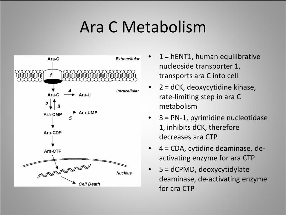
Ara C Metabolism
• 1 = hENT1, human equilibrative nucleoside transporter 1, transports ara C into cell
• 2 = dCK, deoxycytidine kinase, rate-limiting step in ara C metabolism
• 3 = PN-1, pyrimidine nucleotidase 1, inhibits dCK, therefore decreases ara CTP
• 4 = CDA, cytidine deaminase, de-activating enzyme for ara CTP
• 5 = dCPMD, deoxycytidylate deaminase, de-activating enzyme for ara CTP
Hypotheses
• Adhesion between primary AML cells and stromal cells protect leukemia cells from chemotherapy in vitro
• Disruption of adhesion interactions between AML cells and stromal cells enhance chemosensitivity of AML cells to Ara C
• Adhesion interactions between leukemia stem cells and stromal cells alters chemotherapy metabolism

Methods
• Primary AML samples from bone marrow or peripheral blood obtained from patients with newly-diagnosed AML and cryopreserved in liquid nitrogen
• Samples thawed and incubated in media supplemented by serum and cytokines
• Ficoll-hypaque separation of live cells

Methods
• Stromal cells obtained from HS-5 and HS27a cell lines and maintained in tissue culture
• Stromal cells plated and allowed to form monolayer prior to addition of AML cells in tissue culture plates
• Retronectin used as a recombinant ligand for VLA-4
Methods
• AML cells separated by magnetic bead column enrichment/depletion – CD34+, CD38-, CD14+
• CD34+CD38- AML stem cells sorted by flow cytometry and serially transplanted into NOD/SCID mice
• AML cells fluorescently labeled with CFSE prior to adhesion with stroma
• AML cells co-cultured with stromal cells prior to addition of chemotherapy
Methods
• Ara C tested at 5, 10, 20 μM
• AMD3100 (Plerixafor) for CXCR-4 inhibitor at 1 and 15 μM
• Anti-CD49d for anti-VLA-4 inhibitor at 10 μg/mL
• Cell viability determined via MTT reduction assay, trypan blue exclusion and CFSE-labeling of leukemia cells with cell counting on hemocytometer following trypsinization 24 – 72 hrs following exposure to chemotherapy

Methods
• RNA extraction, RT-PCR and DNA gel electrophoresis to evaluate expression of enzymes in Ara C metabolism on CD34+ vs. CD34-14+ cells and the effect of adhesion with retronectin
• Primers and probes for enzymes in Ara C metabolism obtained from Applied Biosystems Inc.
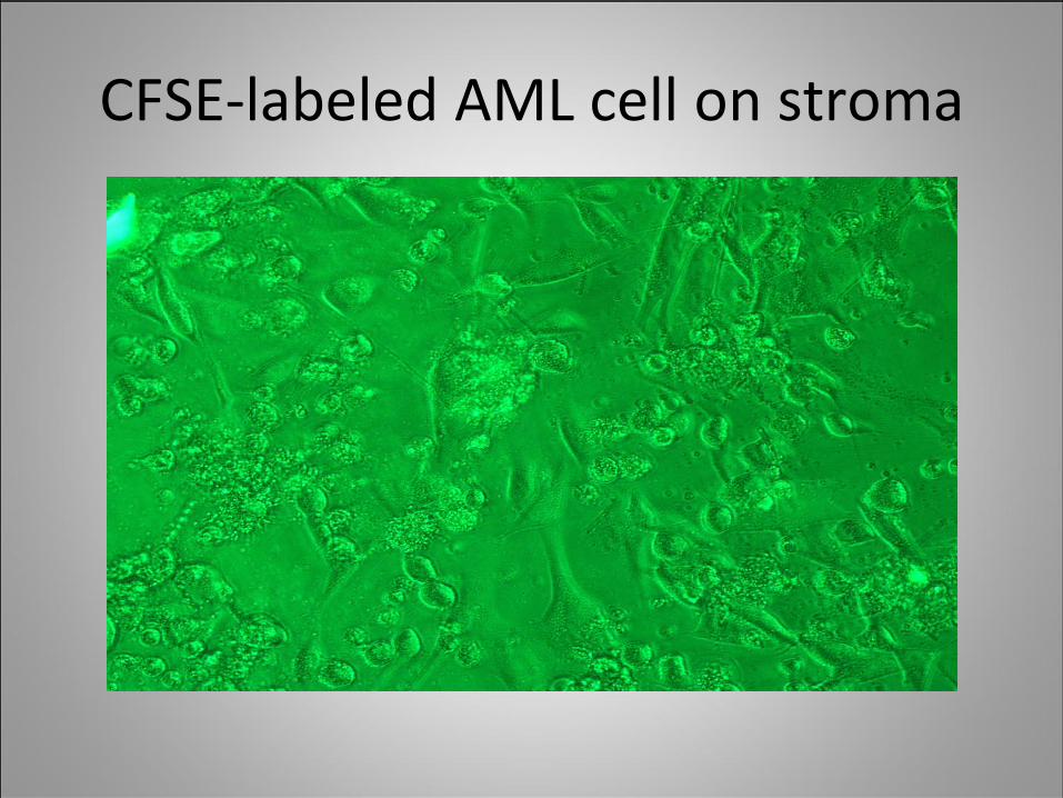
CFSE-labeled AML cell on stroma

CFSE-labeled AML cell on stroma
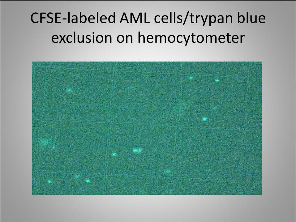
CFSE-labeled AML cells/trypan blue exclusion on hemocytometer
0
20
40
60
80
100
120
Control Ara C 0 + AMD3100
Ara C 5 Ara C 5 + AMD3100
Ara C 10 Ara C 10 + AMD3100
AraC 20 AraC 20 + AMD3100
Surv
ival
(% o
f con
trol
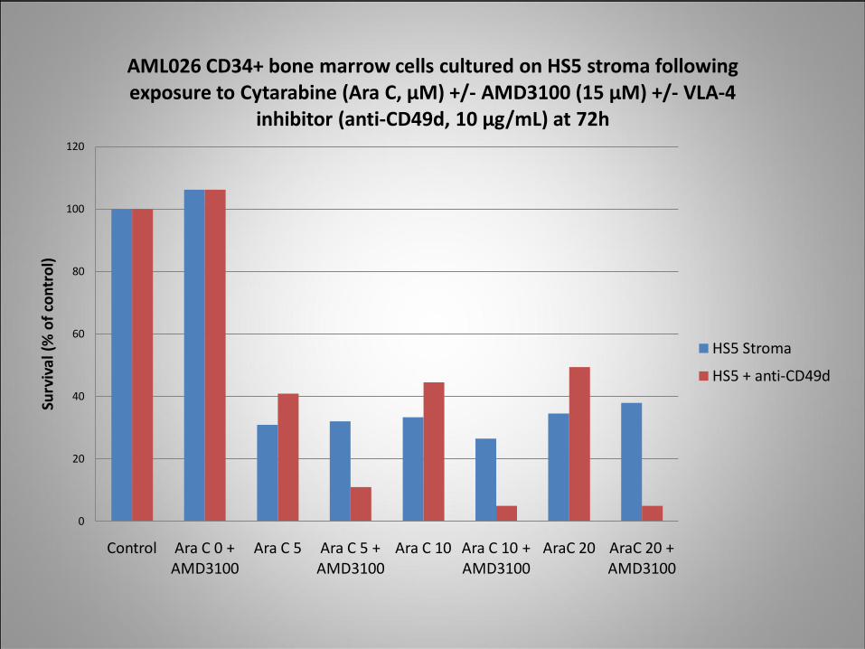
)AML026 CD34+ bone marrow cells cultured on HS5 stroma following exposure to Cytarabine (Ara C, µM) +/- AMD3100 (15 µM) +/- VLA-4
inhibitor (anti-CD49d, 10 µg/mL) at 72h
HS5 Stroma
HS5 + anti-CD49d

0
20
40
60
80
100
120
Control Ara C 20 Ara C 20 + AMD3100
Cell
surv
ival
(% o
f con
trol
)AML037 CD34+ bone marrow cells cultured on HS27a stroma after exposure to Cytarabine (Ara C, µM) +/- AMD3100 (1 µM) +/- VLA-4
inhibitor (anti-CD49d, 1 µM) x 24h
HS27a
HS27a + anti-CD49d
p = 0.09
p = 0.16
0
20
40
60
80
100
120
Control Ara C 20 Ara C 20 + AMD3100
Cell
surv
ival
(% o
f con
trol
)
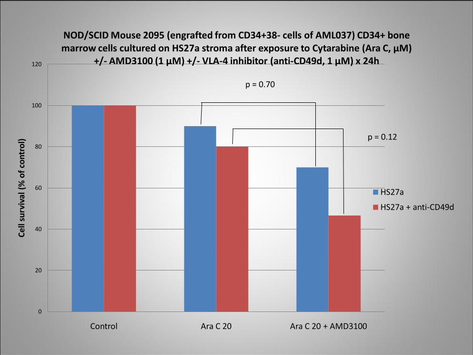
NOD/SCID Mouse 2095 (engrafted from CD34+38- cells of AML037) CD34+ bone marrow cells cultured on HS27a stroma after exposure to Cytarabine (Ara C, µM)
+/- AMD3100 (1 µM) +/- VLA-4 inhibitor (anti-CD49d, 1 µM) x 24h
HS27a
HS27a + anti-CD49d
p = 0.12
p = 0.70

0
20
40
60
80
100
120
Control Ara C 4 Ara C 4 + Clofar 2
Ara C 4 + Clofar 4
Clofar 4 Ara C 2 + Clofar 4
Ara C 7 + Clofar 3
Cell
surv
ival
(% c
ontr
ol)
AML029 CD34+ peripheral blood cells (with and without AMD3100 1 µM exposure) cultured on HS27a stroma and exposed to cytarabine (Ara C) and/or clofarabine at varying concentrations (µM) with survival at 72h
HS27a
HS27a + AMD3100
p = 0.13

0
20
40
60
80
100
120
Control Ara C 4 Ara C 4 + Clofar 2
Ara C 4 + Clofar 4
Clofar 4 Ara C 2 + Clofar 4
Ara C 7 + Clofar 3
Cell
surv
ival
(% o
f con
trol
)AML022 CD14+ peripheral blood cells (with and without AMD3100 1 µM
exposure) cultured on HS27a stroma and exposed to cytarabine (Ara C) and/or clofarabine at varying concentrations (µM) with survival at 72h
HS27a
HS27a + AMD3100

0
0.1
0.2
0.3
0.4
0.5
0.6
Control Ara C 5 Ara C 10 Ara C 20
Opt
ical
Den
sity
AML012 CD34+ vs CD34+38- peripheral blood cells cultured on retronectin following exposure to Cytarabine (Ara C) [µM] with cell
viability by MTT assay
CD34+ cells
CD34+38- cells
p = 0.01 p = 0.16 p = 0.28
p = 0.43

Adhesion and Ara C metabolism
• Do leukemia stem cells (CD34+) differ from more mature cells (CD34-14+) in expression of enzymes for Ara C drug metabolism?
• Does adhesion lead to altered chemotherapy metabolism?
• CD34+ cells vs CD34-14+ cells cultured on heat-inactivated 2% BSA or retronectin with and without AraC at 1 μM
• RNA extraction with RT-PCR and DNA gel electrophoresis performed to evaluate expression of hENT1, dCK, CDA, dCMPD, PN-1 with GAPDH as control
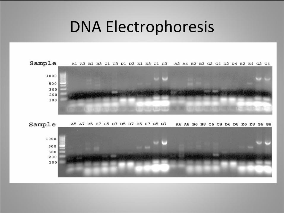
DNA Electrophoresis

RT-PCR Results
• CD34+ and CD34-14+ cells with greater expression of de-activating enzymes (CDA, dCMPD, PN-1) when cells are cultured on retronectin compared to BSA
• CD34-14+ cells with greater expression of de-activating enzyme dCMPD compared to CD34+ cells – CD34+ cells with likely decreased uptake of Ara C and therefore less expression of enzymes involved in Ara C inactivation

Conclusions
• Adhesion of CD34+ AML cells to HS5 or HS27a stromapromotes survival from chemotherapy
• Leukemia stem cells appear more resistant to chemotherapy
• For some patients with newly-diagnosed AML, disruption of adhesion between stromal cells and CD34+ AML cells with a combination of CXCR-4 and VLA-4 inhibitors appear to enhance sensitivity to Ara C
• Adhesion-mediated interactions may protect AML cells from chemotoxicity due to altered Ara C metabolism by increased expression of enzymes that degrade Ara C

Future Directions
• Characterizing the critical adhesion interactions between leukemia cells and stroma
• Use of multiple adhesion inhibitors with chemotherapy to improve killing of leukemia stem cells
• Measurement of Ara-CTP levels and the effect of adhesion on Ara-CTP accumulation rates
• Real-time RT-PCR of leukemia stem cells to evaluate the effect of adhesion on a variety of signaling pathways involved in chemotherapy uptake, metabolism, apoptosis, growth and differentiation

Support
• American Society of Hematology Research Trainee Award
• Becker Lab:
• Soumit Basu, M.D., Ph.D., Hematology & Oncology Fellow
• Sylvia Chien, Research Scientist
• Aric Zhao, Research Scientist

References
1. Horner MJ, Ries LAG, Krapcho M, et. al. SEER Cancer Statistics Review, 1975-2006, National Cancer Institute. Bethesda, MD, http://seer.cancer.gov/csr/1975_2006/, based on November 2008 SEER data submission, posted to the SEER web site, 2009.
2. Garrido SM, Appelbaum FR, Willman CL, et al. Acute myeloid leukemia cells are protected from spontaneous and drug-induced apoptosis by direct contact with a human stromal cell line (HS-5). Exp Hematol. 2001 Apr;29(4):448-57.
3. Matsunaga T, Takemoto N, Sato T, et. al. Nat Med. 2003 Sep;9(9):1158-65.
4. Meads, M et. al. The bone marrow microenvironment as a tumor sanctuary and contributor to drug resistance. Clinical Cancer Research 2008.
5. Spoo AC, Lubbert M, Wierda WG, et. al. Blood. 2007 109: 786-791.6. Becker PS, Kopecky KJ, Wilks AN, et. al. Blood. 2009 113: 866-874.
Related Documents