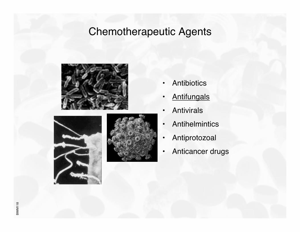
BIMM118 Chemotherapeutic Agents • Antibiotics • Antifungals • Antivirals • Antihelmintics • Antiprotozoal • Anticancer drugs

Welcome message from author
This document is posted to help you gain knowledge. Please leave a comment to let me know what you think about it! Share it to your friends and learn new things together.
Transcript
BIM
M11
8
Chemotherapeutic Agents
• Antibiotics• Antifungals• Antivirals• Antihelmintics• Antiprotozoal• Anticancer drugs
BIM
M11
8
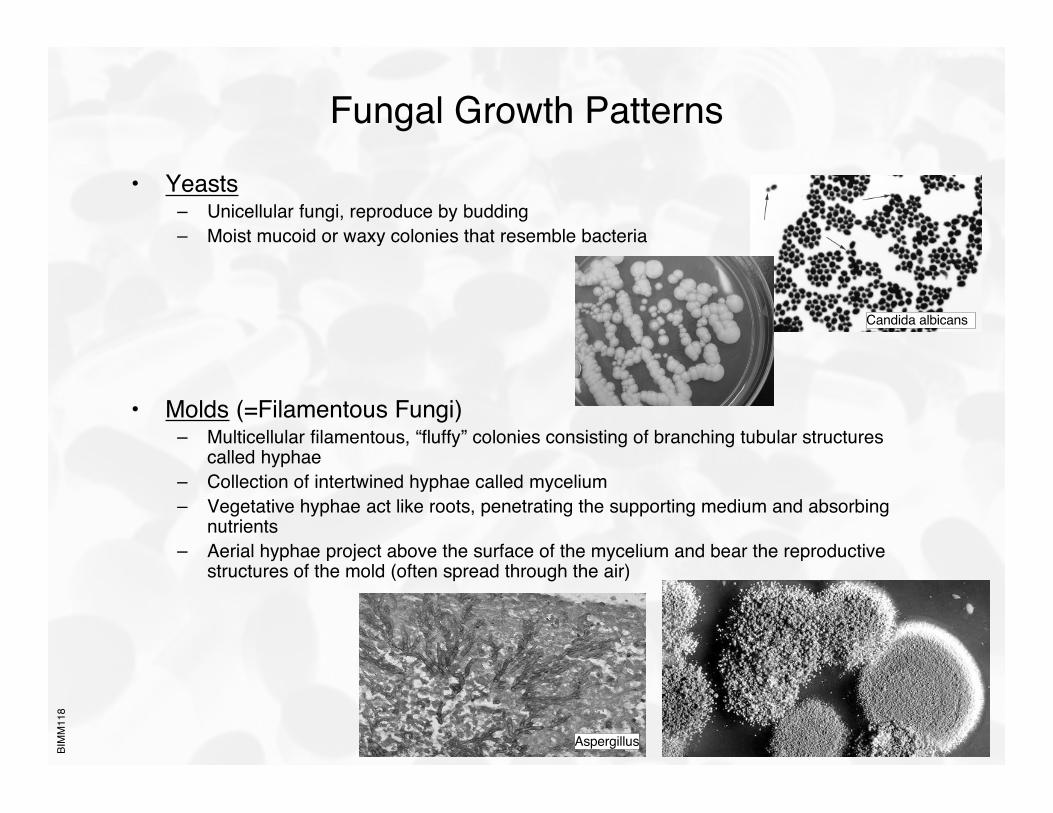
Fungal Growth Patterns
• Yeasts– Unicellular fungi, reproduce by budding– Moist mucoid or waxy colonies that resemble bacteria
• Molds (=Filamentous Fungi)– Multicellular filamentous, “fluffy” colonies consisting of branching tubular structures
called hyphae– Collection of intertwined hyphae called mycelium– Vegetative hyphae act like roots, penetrating the supporting medium and absorbing
nutrients– Aerial hyphae project above the surface of the mycelium and bear the reproductive
structures of the mold (often spread through the air)
Candida albicans
Aspergillus
BIM
M11
8
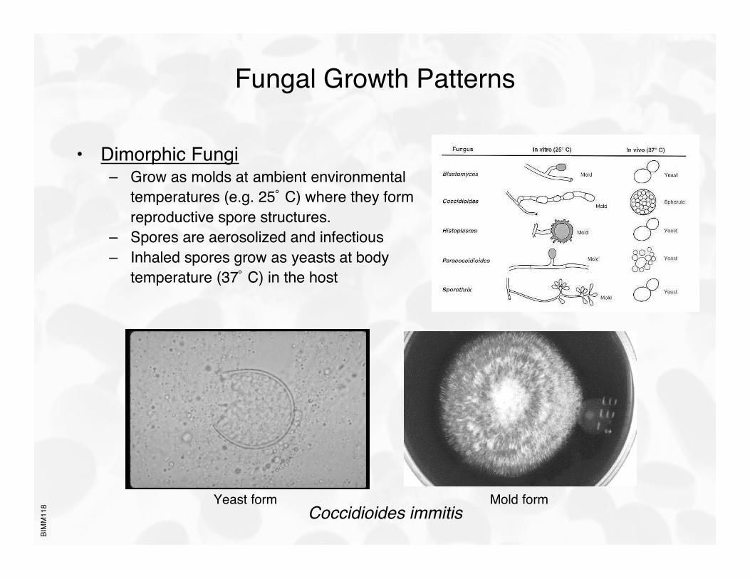
Fungal Growth Patterns
• Dimorphic Fungi– Grow as molds at ambient environmental
temperatures (e.g. 25˚ C) where they formreproductive spore structures.
– Spores are aerosolized and infectious– Inhaled spores grow as yeasts at body
temperature (37˚ C) in the host
Coccidioides immitisYeast form Mold form
BIM
M11
8
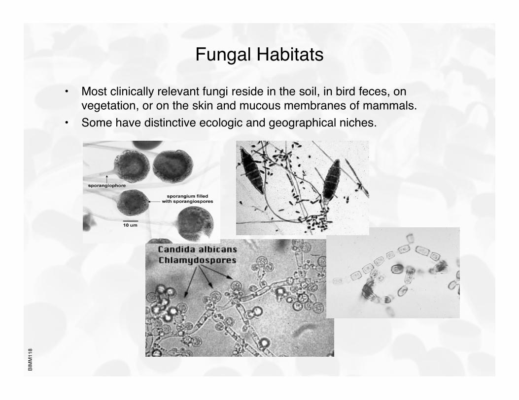
Fungal Habitats
• Most clinically relevant fungi reside in the soil, in bird feces, onvegetation, or on the skin and mucous membranes of mammals.
• Some have distinctive ecologic and geographical niches.
BIM
M11
8
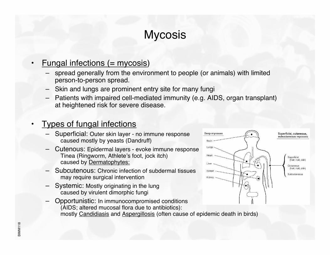
Mycosis
• Fungal infections (= mycosis)– spread generally from the environment to people (or animals) with limited
person-to-person spread.– Skin and lungs are prominent entry site for many fungi– Patients with impaired cell-mediated immunity (e.g. AIDS, organ transplant)
at heightened risk for severe disease.
• Types of fungal infections– Superficial: Outer skin layer - no immune response
caused mostly by yeasts (Dandruff)– Cutenous: Epidermal layers - evoke immune response
Tinea (Ringworm, Athlete’s foot, jock itch) caused by Dermatophytes:
– Subcutenous: Chronic infection of subdermal tissuesmay require surgical intervention
– Systemic: Mostly originating in the lungcaused by virulent dimorphic fungi
– Opportunistic: In immunocompromised conditions(AIDS; altered mucosal flora due to antibiotics):mostly Candidiasis and Aspergillosis (often cause of epidemic death in birds)
BIM
M11
8
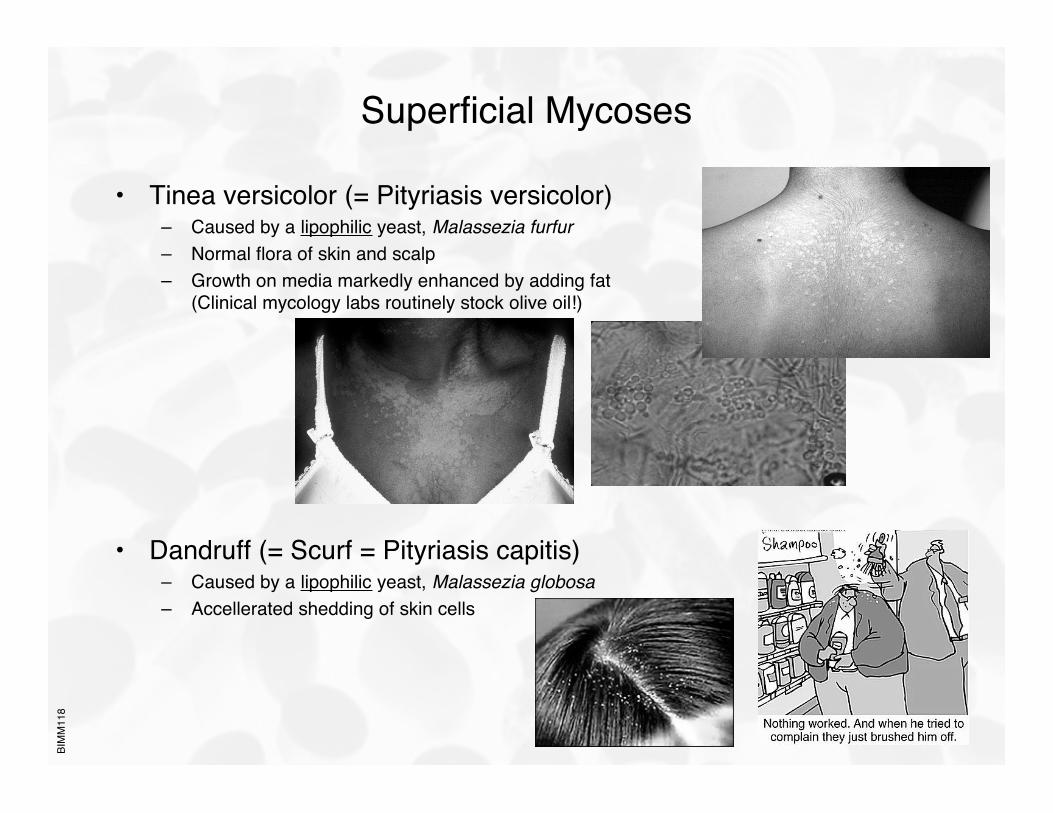
Superficial Mycoses
• Tinea versicolor (= Pityriasis versicolor)– Caused by a lipophilic yeast, Malassezia furfur– Normal flora of skin and scalp– Growth on media markedly enhanced by adding fat
(Clinical mycology labs routinely stock olive oil!)
• Dandruff (= Scurf = Pityriasis capitis)– Caused by a lipophilic yeast, Malassezia globosa– Accellerated shedding of skin cells
BIM
M11
8
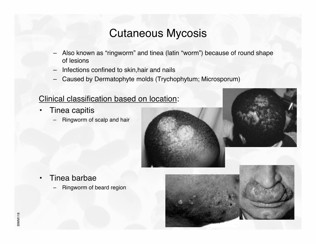
Cutaneous Mycosis– Also known as “ringworm” and tinea (latin “worm”) because of round shape
of lesions– Infections confined to skin,hair and nails– Caused by Dermatophyte molds (Trychophytum; Microsporum)
Clinical classification based on location:• Tinea capitis
– Ringworm of scalp and hair
• Tinea barbae– Ringworm of beard region
BIM
M11
8
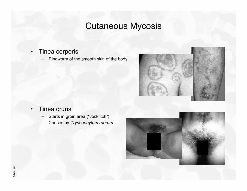
Cutaneous Mycosis
• Tinea corporis– Ringworm of the smooth skin of the body
• Tinea cruris– Starts in groin area (“Jock itch”)– Causes by Trychophytum rubrum
BIM
M11
8
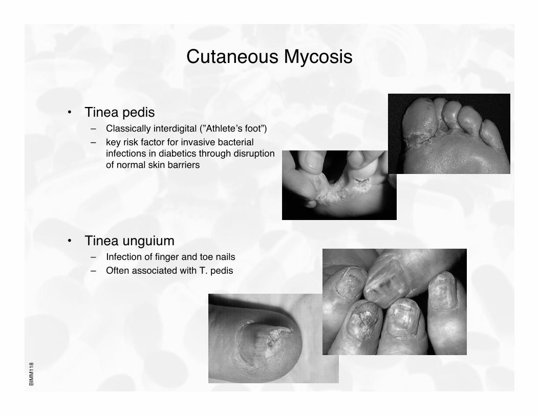
Cutaneous Mycosis
• Tinea pedis– Classically interdigital (”Athlete’s foot”)– key risk factor for invasive bacterial
infections in diabetics through disruptionof normal skin barriers
• Tinea unguium– Infection of finger and toe nails– Often associated with T. pedis
BIM
M11
8
Subcutaneous Mycoses
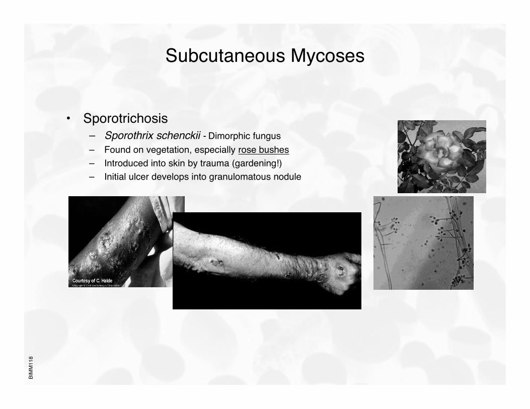
• Sporotrichosis– Sporothrix schenckii - Dimorphic fungus– Found on vegetation, especially rose bushes– Introduced into skin by trauma (gardening!)– Initial ulcer develops into granulomatous nodule
BIM
M11
8
Systemic Mycosis
– Infections are rare (high natural immunity)– Usually requires large inoculum– Often endemic to specific aereas
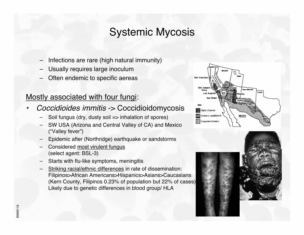
Mostly associated with four fungi:• Coccidioides immitis -> Coccidioidomycosis
– Soil fungus (dry, dusty soil => inhalation of spores)– SW USA (Arizona and Central Valley of CA) and Mexico
(“Valley fever”)– Epidemic after (Northridge) earthquake or sandstorms– Considered most virulent fungus
(select agent: BSL-3)– Starts with flu-like symptoms, meningitis– Striking racial/ethnic differences in rate of dissemination:
Filipinos>African Americans>Hispanics>Asians>Caucasians(Kern County, Filipinos 0.23% of population but 22% of cases)Likely due to genetic differences in blood group/ HLA
BIM
M11
8
Systemic Mycosis
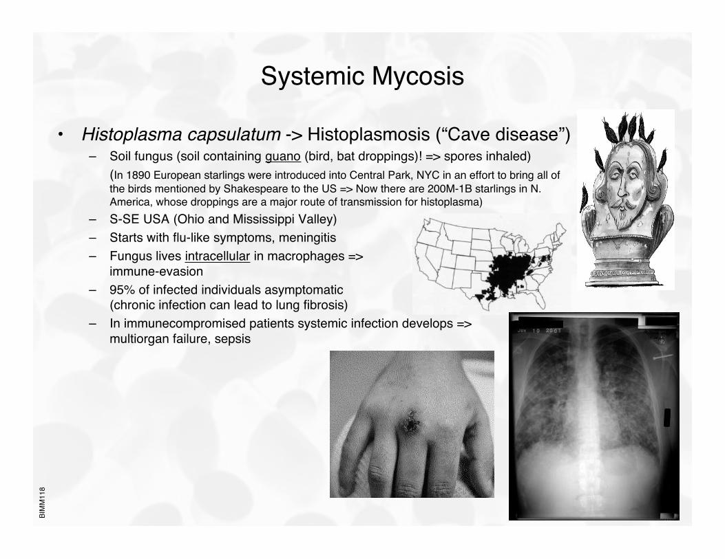
• Histoplasma capsulatum -> Histoplasmosis (“Cave disease”)– Soil fungus (soil containing guano (bird, bat droppings)! => spores inhaled)
(In 1890 European starlings were introduced into Central Park, NYC in an effort to bring all ofthe birds mentioned by Shakespeare to the US => Now there are 200M-1B starlings in N.America, whose droppings are a major route of transmission for histoplasma)
– S-SE USA (Ohio and Mississippi Valley)– Starts with flu-like symptoms, meningitis– Fungus lives intracellular in macrophages =>
immune-evasion– 95% of infected individuals asymptomatic
(chronic infection can lead to lung fibrosis)– In immunecompromised patients systemic infection develops =>
multiorgan failure, sepsis
BIM
M11
8
Systemic Mycosis
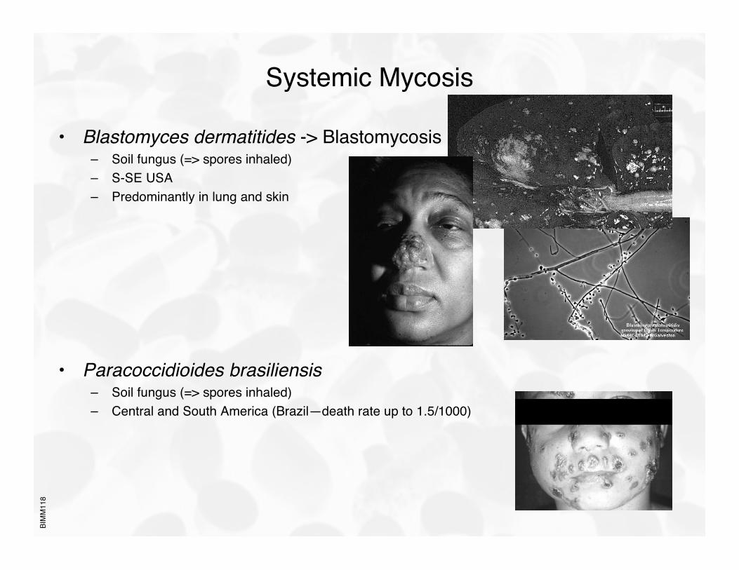
• Blastomyces dermatitides -> Blastomycosis– Soil fungus (=> spores inhaled)– S-SE USA– Predominantly in lung and skin
• Paracoccidioides brasiliensis– Soil fungus (=> spores inhaled)– Central and South America (Brazil—death rate up to 1.5/1000)
BIM
M11
8
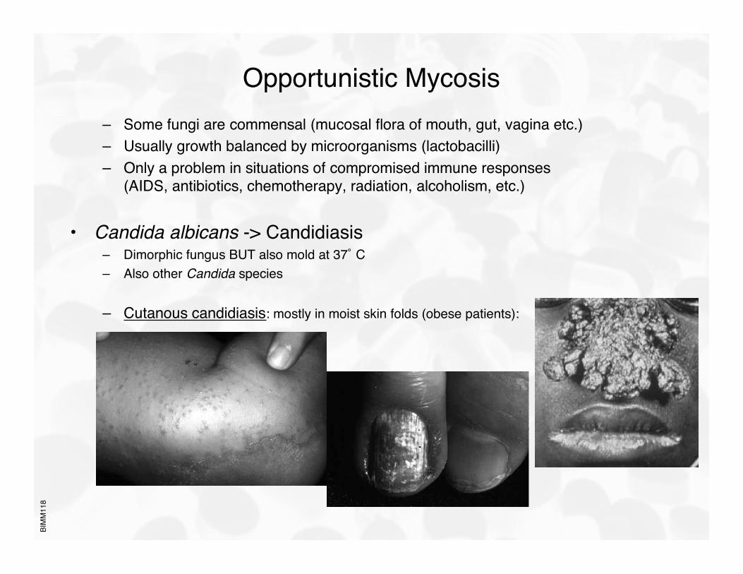
Opportunistic Mycosis– Some fungi are commensal (mucosal flora of mouth, gut, vagina etc.)– Usually growth balanced by microorganisms (lactobacilli)– Only a problem in situations of compromised immune responses
(AIDS, antibiotics, chemotherapy, radiation, alcoholism, etc.)
• Candida albicans -> Candidiasis– Dimorphic fungus BUT also mold at 37˚ C– Also other Candida species
– Cutanous candidiasis: mostly in moist skin folds (obese patients):
BIM
M11
8
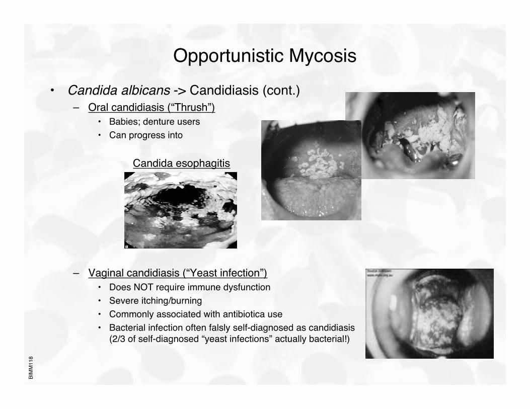
Opportunistic Mycosis• Candida albicans -> Candidiasis (cont.)
– Oral candidiasis (“Thrush”)• Babies; denture users• Can progress into
Candida esophagitis
– Vaginal candidiasis (“Yeast infection”)• Does NOT require immune dysfunction• Severe itching/burning• Commonly associated with antibiotica use• Bacterial infection often falsly self-diagnosed as candidiasis
(2/3 of self-diagnosed “yeast infections” actually bacterial!)
BIM
M11
8
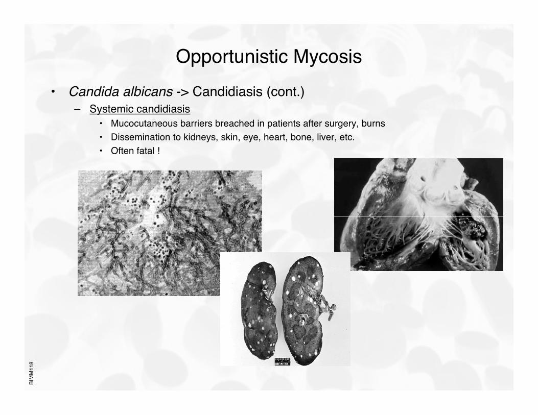
Opportunistic Mycosis• Candida albicans -> Candidiasis (cont.)
– Systemic candidiasis• Mucocutaneous barriers breached in patients after surgery, burns• Dissemination to kidneys, skin, eye, heart, bone, liver, etc.• Often fatal !
BIM
M11
8
Opportunistic Mycosis
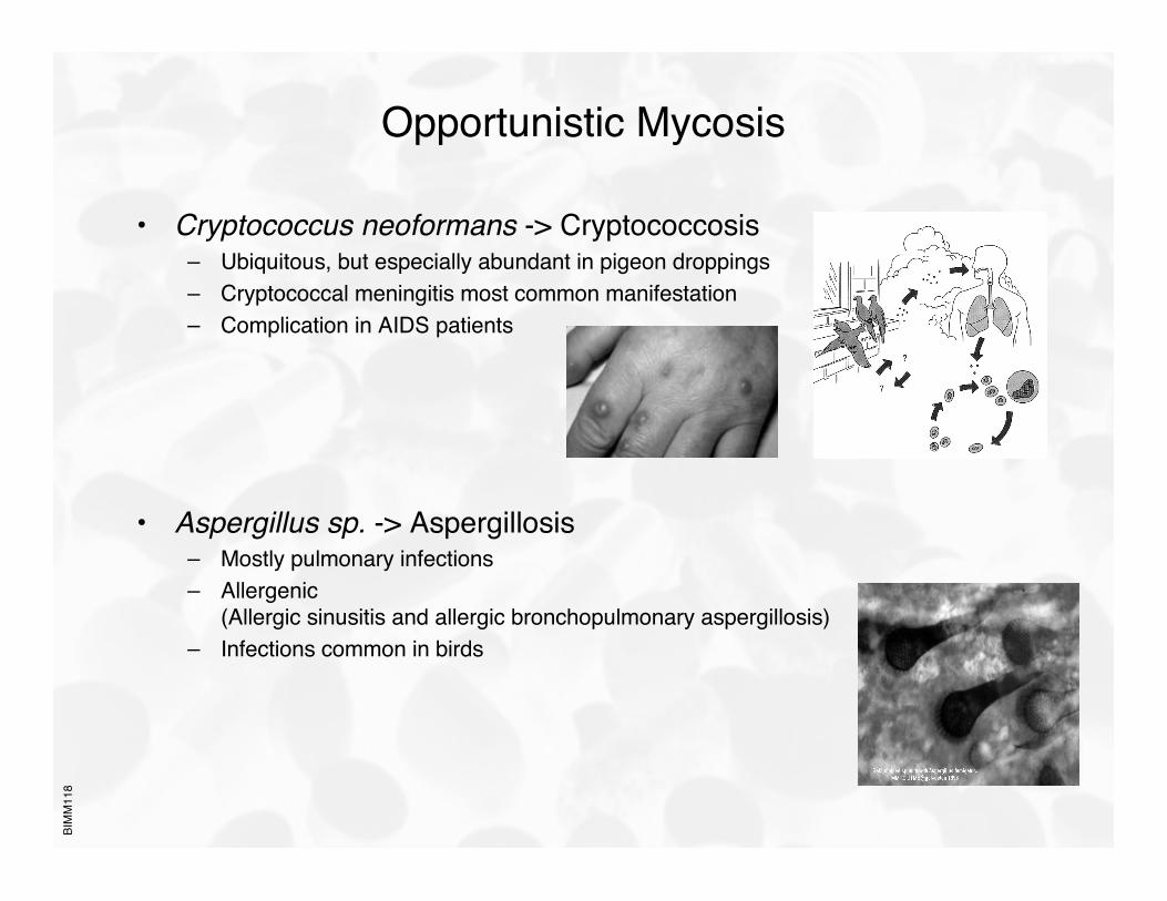
• Cryptococcus neoformans -> Cryptococcosis– Ubiquitous, but especially abundant in pigeon droppings– Cryptococcal meningitis most common manifestation– Complication in AIDS patients
• Aspergillus sp. -> Aspergillosis– Mostly pulmonary infections– Allergenic
(Allergic sinusitis and allergic bronchopulmonary aspergillosis)– Infections common in birds
BIM
M11
8
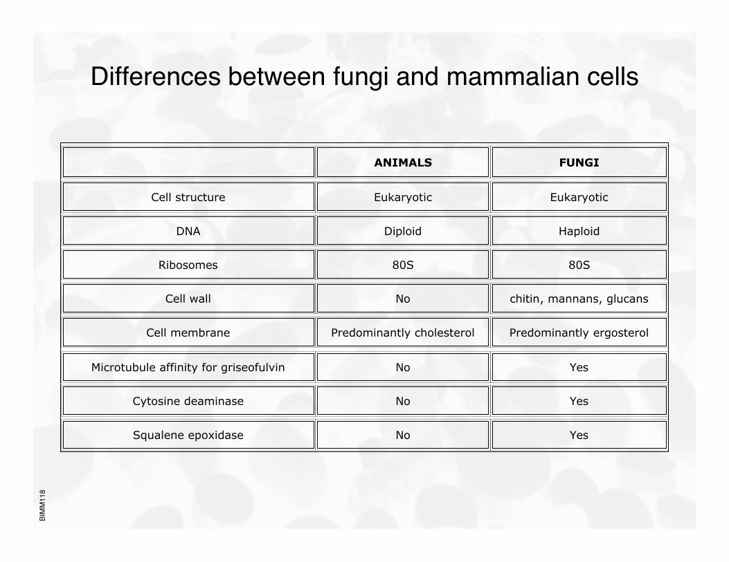
Differences between fungi and mammalian cells
ANIMALS FUNGI
Cell structure Eukaryotic Eukaryotic
DNA Diploid Haploid
Ribosomes 80S 80S
Cell wall No chitin, mannans, glucans
Cell membrane Predominantly cholesterol Predominantly ergosterol
Microtubule affinity for griseofulvin No Yes
Cytosine deaminase No Yes
Squalene epoxidase No Yes
BIM
M11
8
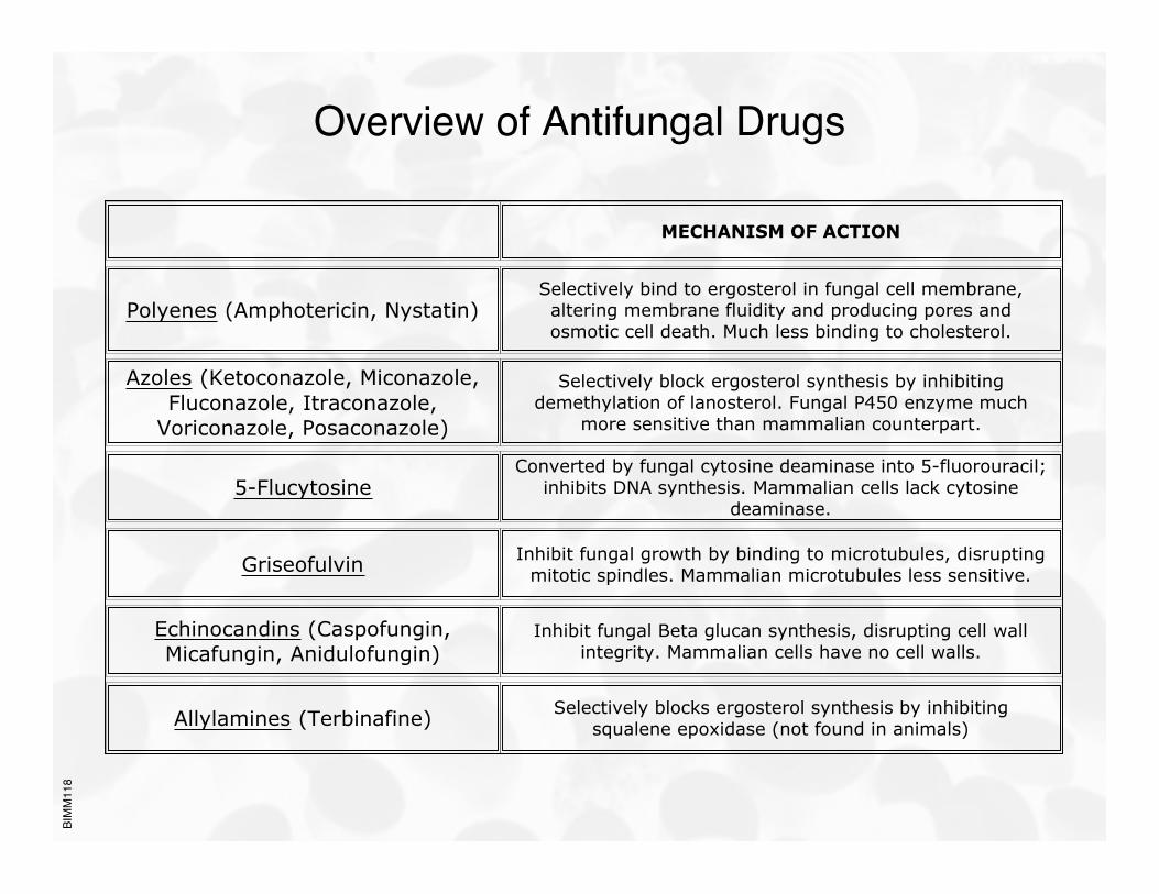
Overview of Antifungal Drugs
MECHANISM OF ACTION
Polyenes (Amphotericin, Nystatin)Selectively bind to ergosterol in fungal cell membrane,altering membrane fluidity and producing pores andosmotic cell death. Much less binding to cholesterol.
Azoles (Ketoconazole, Miconazole,Fluconazole, Itraconazole,
Voriconazole, Posaconazole)
Selectively block ergosterol synthesis by inhibitingdemethylation of lanosterol. Fungal P450 enzyme much
more sensitive than mammalian counterpart.
5-FlucytosineConverted by fungal cytosine deaminase into 5-fluorouracil;
inhibits DNA synthesis. Mammalian cells lack cytosinedeaminase.
Griseofulvin Inhibit fungal growth by binding to microtubules, disruptingmitotic spindles. Mammalian microtubules less sensitive.
Echinocandins (Caspofungin,Micafungin, Anidulofungin)
Inhibit fungal Beta glucan synthesis, disrupting cell wallintegrity. Mammalian cells have no cell walls.
Allylamines (Terbinafine) Selectively blocks ergosterol synthesis by inhibitingsqualene epoxidase (not found in animals)
BIM
M11
8
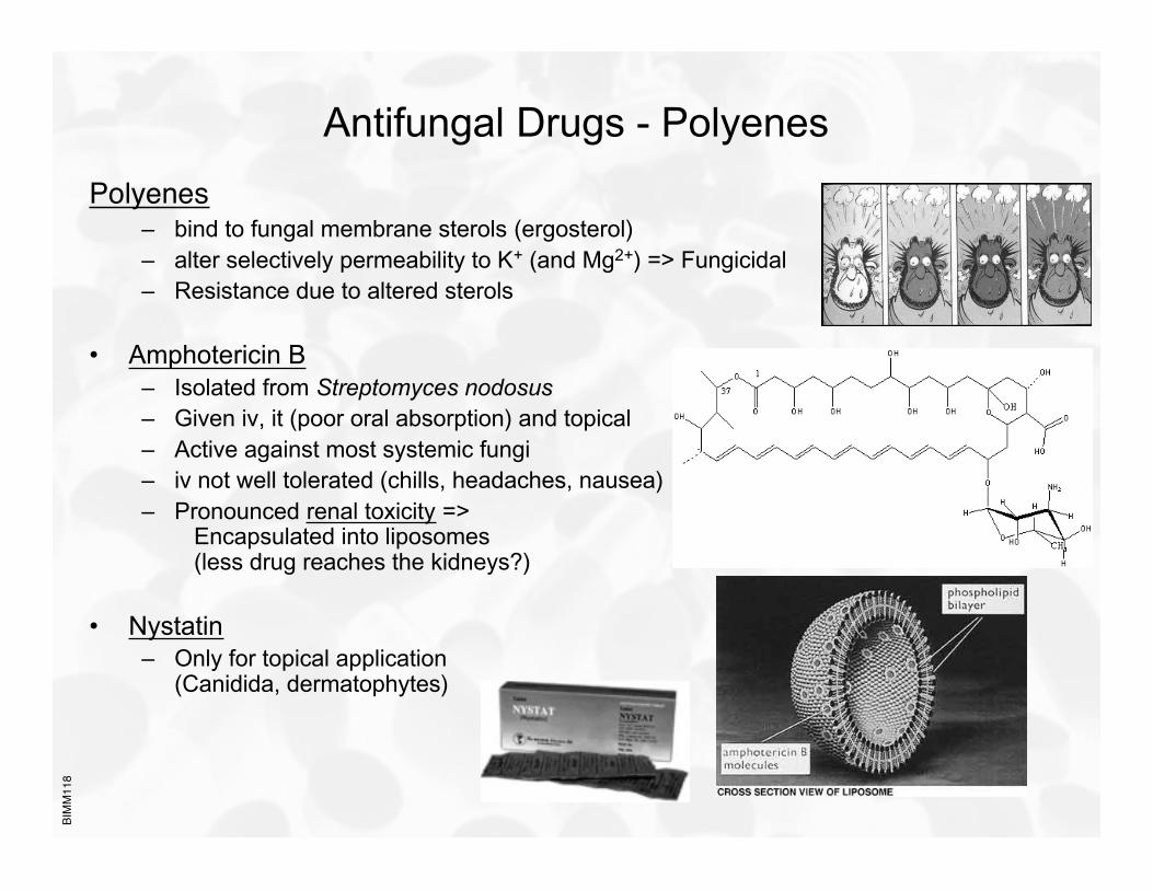
Antifungal Drugs - Polyenes
Polyenes– bind to fungal membrane sterols (ergosterol)– alter selectively permeability to K+ (and Mg2+) => Fungicidal– Resistance due to altered sterols
• Amphotericin B– Isolated from Streptomyces nodosus– Given iv, it (poor oral absorption) and topical– Active against most systemic fungi– iv not well tolerated (chills, headaches, nausea)– Pronounced renal toxicity =>
Encapsulated into liposomes(less drug reaches the kidneys?)
• Nystatin– Only for topical application
(Canidida, dermatophytes)
BIM
M11
8
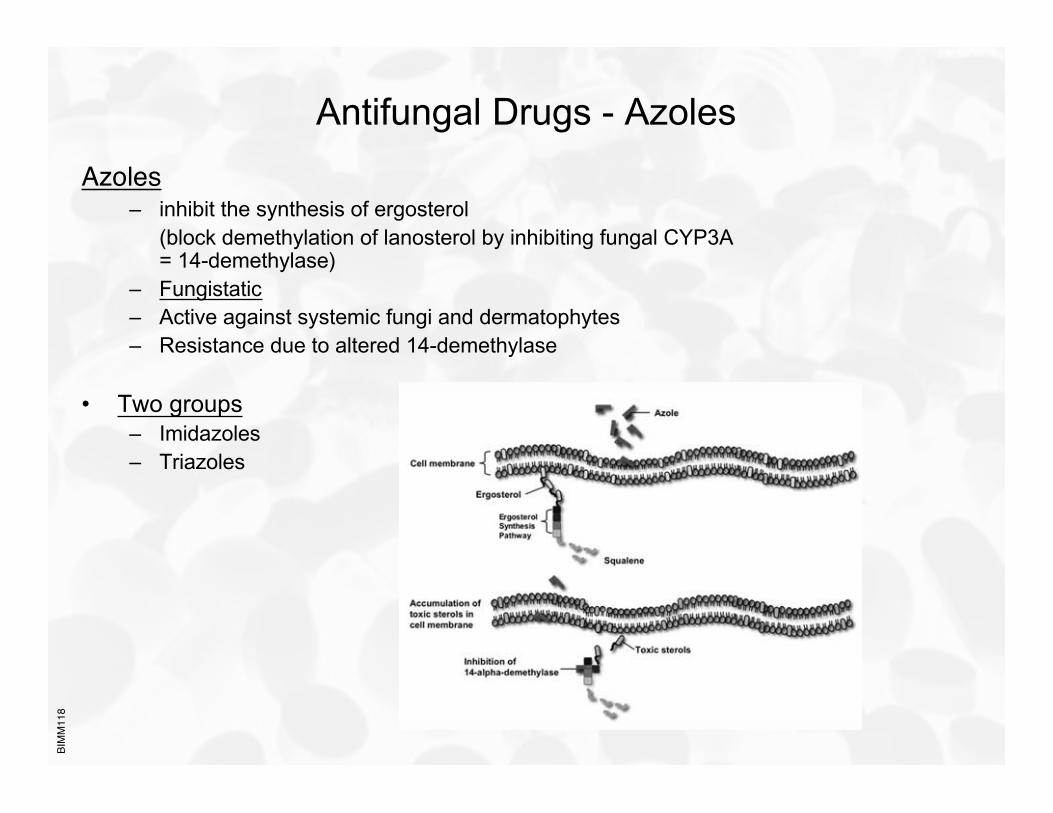
Antifungal Drugs - Azoles
Azoles– inhibit the synthesis of ergosterol
(block demethylation of lanosterol by inhibiting fungal CYP3A= 14-demethylase)
– Fungistatic– Active against systemic fungi and dermatophytes– Resistance due to altered 14-demethylase
• Two groups– Imidazoles– Triazoles
BIM
M11
8
Antifungal Drugs - Azoles
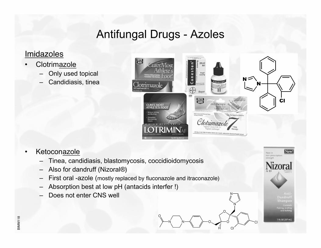
Imidazoles• Clotrimazole
– Only used topical– Candidiasis, tinea
• Ketoconazole– Tinea, candidiasis, blastomycosis, coccidioidomycosis– Also for dandruff (Nizoral®)– First oral -azole (mostly replaced by fluconazole and itraconazole)– Absorption best at low pH (antacids interfer !)– Does not enter CNS well
BIM
M11
8
Antifungal Drugs - Azoles
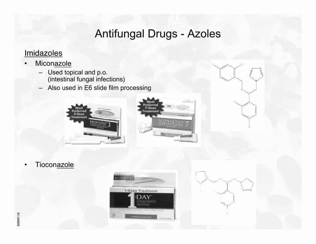
Imidazoles• Miconazole
– Used topical and p.o.(intestinal fungal infections)
– Also used in E6 slide film processing
• Tioconazole
BIM
M11
8
Antifungal Drugs - Azoles
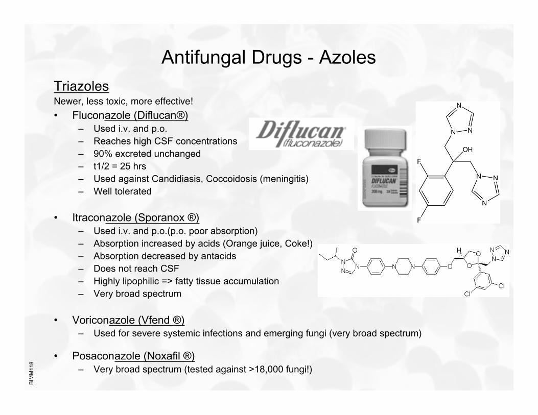
TriazolesNewer, less toxic, more effective!• Fluconazole (Diflucan®)
– Used i.v. and p.o.– Reaches high CSF concentrations– 90% excreted unchanged– t1/2 = 25 hrs– Used against Candidiasis, Coccoidosis (meningitis)– Well tolerated
• Itraconazole (Sporanox ®)– Used i.v. and p.o.(p.o. poor absorption)– Absorption increased by acids (Orange juice, Coke!)– Absorption decreased by antacids– Does not reach CSF– Highly lipophilic => fatty tissue accumulation– Very broad spectrum
• Voriconazole (Vfend ®)– Used for severe systemic infections and emerging fungi (very broad spectrum)
• Posaconazole (Noxafil ®)– Very broad spectrum (tested against >18,000 fungi!)
BIM
M11
8
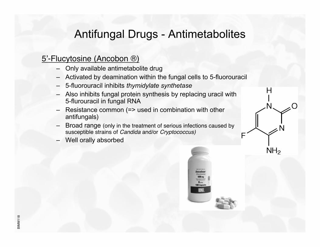
Antifungal Drugs - Antimetabolites
5’-Flucytosine (Ancobon ®)– Only available antimetabolite drug– Activated by deamination within the fungal cells to 5-fluorouracil– 5-fluorouracil inhibits thymidylate synthetase– Also inhibits fungal protein synthesis by replacing uracil with
5-flurouracil in fungal RNA– Resistance common (=> used in combination with other
antifungals)– Broad range (only in the treatment of serious infections caused by
susceptible strains of Candida and/or Cryptococcus)– Well orally absorbed
BIM
M11
8
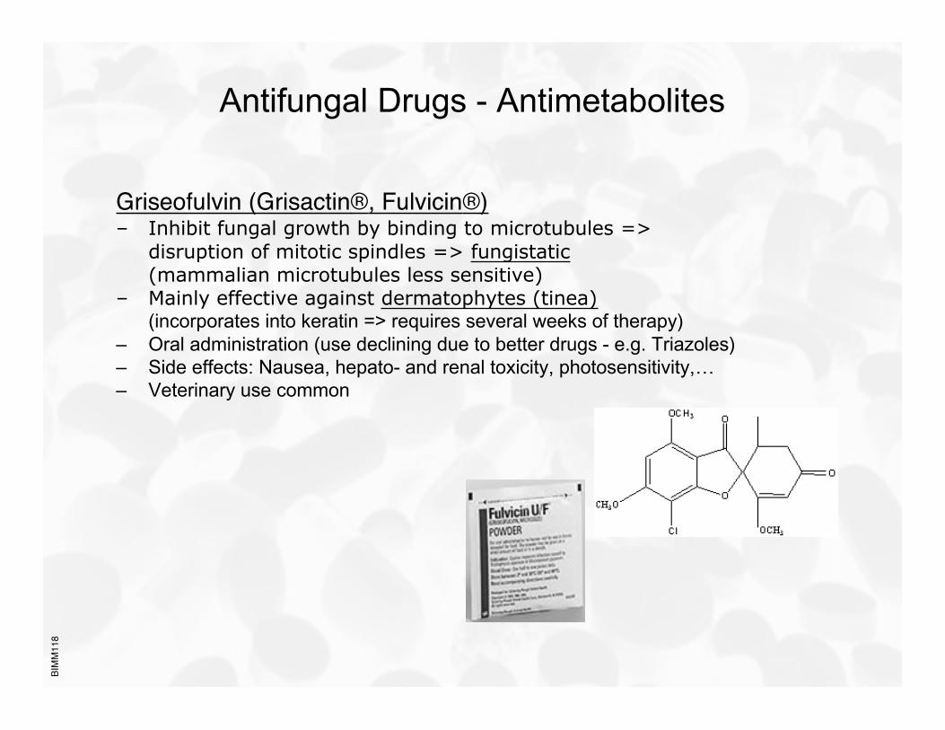
Antifungal Drugs - Antimetabolites
Griseofulvin (Grisactin®, Fulvicin®)– Inhibit fungal growth by binding to microtubules =>
disruption of mitotic spindles => fungistatic(mammalian microtubules less sensitive)
– Mainly effective against dermatophytes (tinea)(incorporates into keratin => requires several weeks of therapy)
– Oral administration (use declining due to better drugs - e.g. Triazoles)– Side effects: Nausea, hepato- and renal toxicity, photosensitivity,…– Veterinary use common
BIM
M11
8
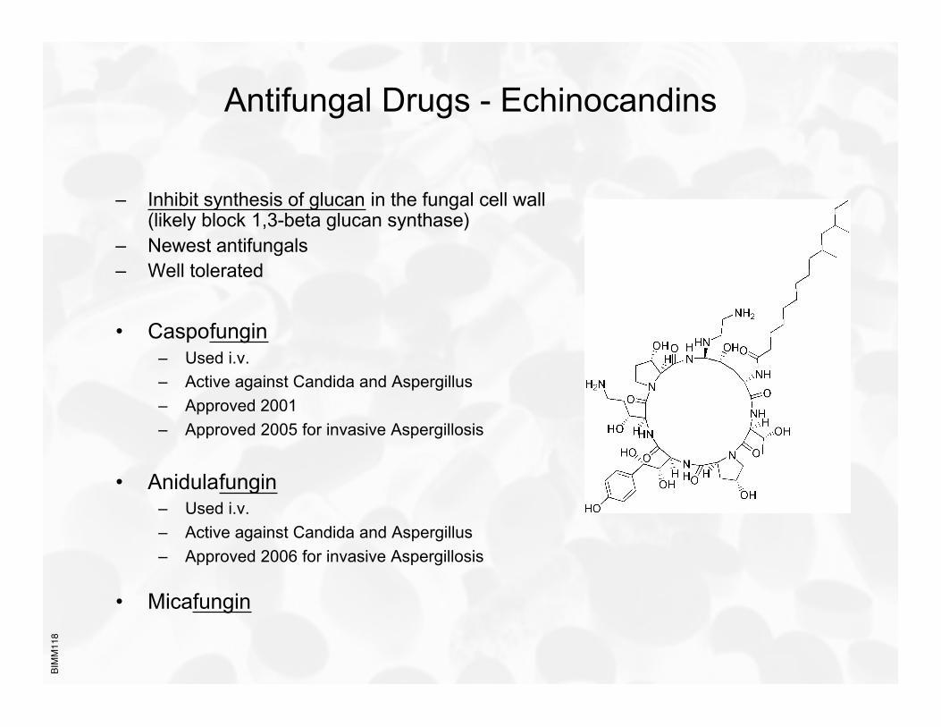
Antifungal Drugs - Echinocandins
– Inhibit synthesis of glucan in the fungal cell wall(likely block 1,3-beta glucan synthase)
– Newest antifungals– Well tolerated
• Caspofungin– Used i.v.
– Active against Candida and Aspergillus
– Approved 2001
– Approved 2005 for invasive Aspergillosis
• Anidulafungin– Used i.v.
– Active against Candida and Aspergillus
– Approved 2006 for invasive Aspergillosis
• Micafungin
BIM
M11
8
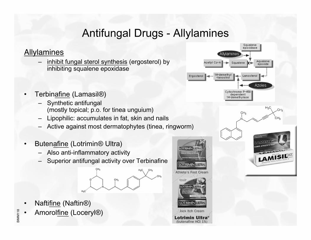
Antifungal Drugs - Allylamines
Allylamines– inhibit fungal sterol synthesis (ergosterol) by
inhibiting squalene epoxidase
• Terbinafine (Lamasil®)– Synthetic antifungal
(mostly topical; p.o. for tinea unguium)– Lipophilic: accumulates in fat, skin and nails– Active against most dermatophytes (tinea, ringworm)
• Butenafine (Lotrimin® Ultra)– Also anti-inflammatory activity– Superior antifungal activity over Terbinafine
• Naftifine (Naftin®)• Amorolfine (Loceryl®)

BIM
M11
8
Antifungals - Summary
Related Documents


![MBBS Antihelmintics [New] 2012](https://static.cupdf.com/doc/110x72/545a9cc7b1af9fb66e8b5edc/mbbs-antihelmintics-new-2012.jpg)
