REVIEWS OF GEOPHYSICS, VOL. 26, NO. 3, PAGES 407-444, AUGUST 1988 Chemistry of the Rock-Forming Silicates' Multiple-Chain, Sheet, and Framework Structures J. J. PAPIKE Institute for the Study of Mineral Deposits, South DakotaSchool of Mines and Technology, RapidCity The crystal chemistry of 16 groups of multiple-chain, sheet, and framework silicates is reviewed. Crystal structure drawings are presented to illustratecrystalchemical features necessary to interpret chemical data for each mineral group. The 16 silicate groups considered in thisreview are the amphibole; nonclassical, ordered pyriboles; mica; pyrophyllite-talc; chlorite;greenalite; minnesotaite; stilpnomel- ane; prehnite; silica polymorphs; feldspar; nepheline-kalsilite; leucite-analcite; sodalite group; cancrinite group; and scapolite. Electronmicroprobe analyses should be augmented by independent determinations of Fe2+/Fe 3+ andH20 for many of thesilicate groups discussed andby determinations of CO32-, SO42-, S 2-, andLi in some of the others. However, microprobe dataaugmented assuggested will still be ambiguous for some of the silicate groups considered herebecause the structures are not completely determined or are variable, with disparate domains and/or structural modulations, e.g., pyriboles, greena- lite, minnesotaite, and stilpnomelane. Nevertheless, the most rigorous way to interpret silicate mineral chemical data is basedon the crystal structures involved. CONTENTS Introduction ............................................. 407 Amphibole .............................................. 407 Pyriboles (nonclassical, ordered) ........................... 412 Mica ................................................... 413 Pyrophyllite-talc ......................................... 416 Chlorite ................................................. 418 Greenalite ............................................... 420 Minnesotaite ............................................ 422 Stilpnomelane ........................................... 422 Prehnite ................................................ 425 Quartz-tridymite-cristobalite .............................. 427 Feldspar ................................................ 429 Nepheline-kalsilite ....................................... 432 Leucite-analcite .......................................... 436 Sodalite group ........................................... 437 Cancrinite group ......................................... 439 Scapolite ................................................ 440 Concluding statement .................................... 442 Deer et al. [-1963a, 1962, 1963b] for mutiple-chain, sheet,and frameworksilicates, respectively. The Reviews in Mineralo•Iy series gives additional valuable information [Ribbe, 1983; Veblen, 1981; Veblen and Ribbe, 1982; Bailey, 1984]. Other amphibole reviews are by Cameronand Papike [1979] and Hawthorne[-1983].A general review of silicatecrystalchemis- try for 12 silicategroupsis provided by Papike and Cameron [1976]. The elements considered for each mineral group are tabu- lated in Table 1. Table 2 lists recommended normalization schemes for microprobedata and criteria for good analyses. The following text documents the rationale for these rec- ommendations. It is my hope, as with part 1 of this synthesis, that this review will help in the rigorous interpretation of chemical analyses of silicate minerals. INTRODUCTION The basic premise of this review of multiple-chain, sheet, and frameworksilicate structures is the same as that for part 1 of this synthesis [Papike, 1987]: that true understanding of the chemistry of minerals can be achieved only by reference to the crystal structures involved.To help investigators in the inter- pretation of silicate chemical data for those minerals con- sidered in this review, I haveassembled structure drawings for 16 groups of multiple-chain, sheet, and framework structures. Someof the drawings are new, and somehave beenadapted from previously published drawings. However, as in part 1 of this synthesis, all diagrams presentedhere have been selected to accurately portray sufficient detail to the non- crystallographer so that the mineral chemistry can be interpre- ted as rigorously as possible. Because of length limitations, referencing has been stream- lined. Referencesnecessaryto convey the essentials of the structural chemistry of each mineral group are emphasized. Readers who require more detailed information are referred to Copyright 1988 by the American Geophysical Union. Paper number 8R0240. 8755-1209/88/008 R-0240505.00 407 AMPHIBOLE The amphibole group has the general formula Ao_•B2CsT8022(OH , F, C1, 0)2 , where A containsNa, K at the A site, B contains Na, Li, Ca, Mn 2 +, Fe2 +, Mg at the M4 site, C contains Mn 2 +, Fe 2 +, Mg, Fe 3 +, Cr3 +, A1,Ti 4+ at the octahedrally coordinated M1, M2, and M3 sites, and T con- tains Si, A1 at the tetrahedral sites. The publishedwork on this important group of rock-forming silicates is voluminous, and only a relatively brief review is presentedhere. For more ex- tensive coverage the reader is referredto Deer et al. [1963a], Papike and Cameron [1976], Cameron and Papike [1979], Veblen [-1981], Veblen and Ribbe [-1982], and Hawthorne [1983]. Important aspects of the C2/m structure are illustrated in Figure la. The M1, M2, and M3 octahedra, which accommo- date the C cations, share edges to form octahedral bands parallel to ½. M1 and M3 are usually coordinated by four oxygens and two (OH, F) groups, whereas M2 is crYordinated by six oxygens. When present, A1 and Fe3 + usually preferthe M2 site, especially when Na occupiesthe adjacent M4 site. The M4 site, which accommodates the B cations, can be con- sidered six-coordinated (a distorted octahedron) when oc- cupied by Mn 2+, Fe2+, and Mg and eight-coordinated when occupied by Na or Ca. The double tetrahedral chains cross-

Welcome message from author
This document is posted to help you gain knowledge. Please leave a comment to let me know what you think about it! Share it to your friends and learn new things together.
Transcript
REVIEWS OF GEOPHYSICS, VOL. 26, NO. 3, PAGES 407-444, AUGUST 1988
Chemistry of the Rock-Forming Silicates' Multiple-Chain, Sheet, and Framework Structures
J. J. PAPIKE
Institute for the Study of Mineral Deposits, South Dakota School of Mines and Technology, Rapid City
The crystal chemistry of 16 groups of multiple-chain, sheet, and framework silicates is reviewed. Crystal structure drawings are presented to illustrate crystal chemical features necessary to interpret chemical data for each mineral group. The 16 silicate groups considered in this review are the amphibole; nonclassical, ordered pyriboles; mica; pyrophyllite-talc; chlorite; greenalite; minnesotaite; stilpnomel- ane; prehnite; silica polymorphs; feldspar; nepheline-kalsilite; leucite-analcite; sodalite group; cancrinite group; and scapolite. Electron microprobe analyses should be augmented by independent determinations of Fe2+/Fe 3+ and H20 for many of the silicate groups discussed and by determinations of CO32-, SO42-, S 2-, and Li in some of the others. However, microprobe data augmented as suggested will still be ambiguous for some of the silicate groups considered here because the structures are not completely determined or are variable, with disparate domains and/or structural modulations, e.g., pyriboles, greena- lite, minnesotaite, and stilpnomelane. Nevertheless, the most rigorous way to interpret silicate mineral chemical data is based on the crystal structures involved.
CONTENTS
Introduction ............................................. 407
Amphibole .............................................. 407 Pyriboles (nonclassical, ordered) ........................... 412 Mica ................................................... 413
Pyrophyllite-talc ......................................... 416 Chlorite ................................................. 418
Greenalite ............................................... 420 Minnesotaite ............................................ 422
Stilpnomelane ........................................... 422 Prehnite ................................................ 425
Quartz-tridymite-cristobalite .............................. 427 Feldspar ................................................ 429 Nepheline-kalsilite ....................................... 432 Leucite-analcite .......................................... 436
Sodalite group ........................................... 437 Cancrinite group ......................................... 439 Scapolite ................................................ 440 Concluding statement .................................... 442
Deer et al. [-1963a, 1962, 1963b] for mutiple-chain, sheet, and framework silicates, respectively. The Reviews in Mineralo•Iy series gives additional valuable information [Ribbe, 1983; Veblen, 1981; Veblen and Ribbe, 1982; Bailey, 1984]. Other amphibole reviews are by Cameron and Papike [1979] and Hawthorne [-1983]. A general review of silicate crystal chemis- try for 12 silicate groups is provided by Papike and Cameron [1976].
The elements considered for each mineral group are tabu- lated in Table 1. Table 2 lists recommended normalization
schemes for microprobe data and criteria for good analyses. The following text documents the rationale for these rec- ommendations.
It is my hope, as with part 1 of this synthesis, that this review will help in the rigorous interpretation of chemical analyses of silicate minerals.
INTRODUCTION
The basic premise of this review of multiple-chain, sheet, and framework silicate structures is the same as that for part 1 of this synthesis [Papike, 1987]: that true understanding of the chemistry of minerals can be achieved only by reference to the crystal structures involved. To help investigators in the inter- pretation of silicate chemical data for those minerals con- sidered in this review, I have assembled structure drawings for 16 groups of multiple-chain, sheet, and framework structures. Some of the drawings are new, and some have been adapted from previously published drawings. However, as in part 1 of this synthesis, all diagrams presented here have been selected to accurately portray sufficient detail to the non- crystallographer so that the mineral chemistry can be interpre- ted as rigorously as possible.
Because of length limitations, referencing has been stream- lined. References necessary to convey the essentials of the structural chemistry of each mineral group are emphasized. Readers who require more detailed information are referred to
Copyright 1988 by the American Geophysical Union.
Paper number 8R0240. 8755-1209/88/008 R-0240505.00
407
AMPHIBOLE
The amphibole group has the general formula Ao_•B2CsT8022(OH , F, C1, 0)2 , where A contains Na, K at the A site, B contains Na, Li, Ca, Mn 2 +, Fe 2 +, Mg at the M4 site, C contains Mn 2 +, Fe 2 +, Mg, Fe 3 +, Cr 3 +, A1, Ti 4 + at the octahedrally coordinated M1, M2, and M3 sites, and T con- tains Si, A1 at the tetrahedral sites. The published work on this important group of rock-forming silicates is voluminous, and only a relatively brief review is presented here. For more ex- tensive coverage the reader is referred to Deer et al. [1963a], Papike and Cameron [1976], Cameron and Papike [1979], Veblen [-1981], Veblen and Ribbe [-1982], and Hawthorne [1983].
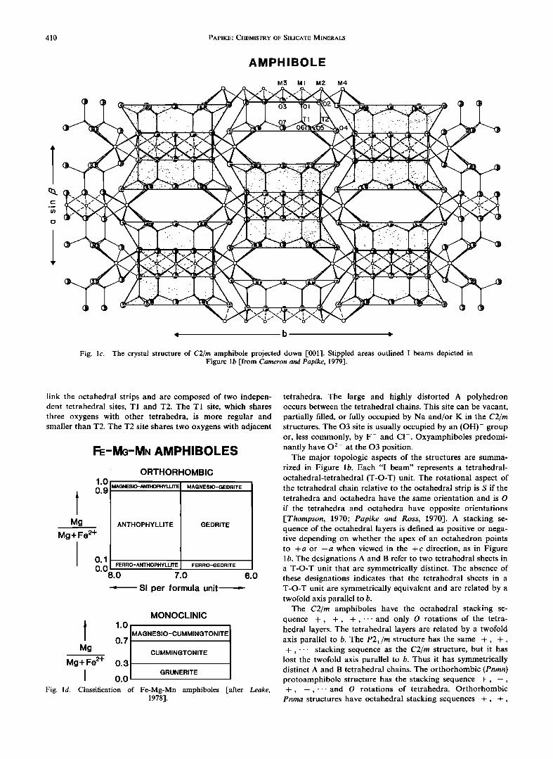
Important aspects of the C2/m structure are illustrated in Figure la. The M1, M2, and M3 octahedra, which accommo- date the C cations, share edges to form octahedral bands parallel to ½. M1 and M3 are usually coordinated by four oxygens and two (OH, F) groups, whereas M2 is crYordinated by six oxygens. When present, A1 and Fe 3 + usually prefer the M2 site, especially when Na occupies the adjacent M4 site. The M4 site, which accommodates the B cations, can be con- sidered six-coordinated (a distorted octahedron) when oc- cupied by Mn 2+, Fe 2+, and Mg and eight-coordinated when occupied by Na or Ca. The double tetrahedral chains cross-
408 PAPIKE' CHEMISTRY OF SILICATE MINERALS
TABLE 1. Inventory of Important Elements to Determine for Each Mineral
Si 4+ A13+ Fe 3+ Ti 4+ Mg 2+ Fe 2+ Mn 2+ Ca 2+ Ba 2+ Li + Na + K + Rb + Cs + H20 F- C1- CO3 2- SO4 2- S 2-
Amphibole group X X X* X X X X X Pyriboles X X X X X X Mica X X X* X X X X X
Pyrophyllite-talc X X X X X X X X Chlorite X X X* X X X X Greenalite X X X X X Minnesotaire X X X* X X X X
Stilpnomelane X X X* X X X X Prehnite X X X* X X X X X
Silica polymorphs X X X X X X X X Feldspar X X X* X X X X X Nepheline-kalsilite X X X X X X Leucite-analcite X X X X X X X
Sodalite group X X X X Cancrinite group X X X X Scapolite X X X
X* X X
X
X* X X X X
X X
X X
X
X X
X X
X X
X X
X X
X X
X X
X*
X*
X X X* X X*
X*
X*
X*
X* X*
X*
X X
X X* X X* X X* X*
X X* X* X
*Determinations recommended to supplement microprobe data.
TABLE 2. Recommended Normalization Schemes for Microprobe Data and Criteria for Good Analyses
Normalization
Fixed Number 100%
of Anhydrous Oxygens Fixed Number of Cations Sum
Criteria for Good Analyses
Total
Silicons Selected Cation Sum
Compare With Table
Analyses
Amphibole 23 no
Pyriboles Clinojimthompsonite 34 no Jimthompsonite 34 no Chesterite 57 no
Mica 11 no
Pyrophyllite 11 no Talc 11 no
Chlorite 14 no
Greenalite 7 no
12
12
2O
Si, •VAl = 8 Mn, Fe, Mg, Ti, Cr, A1 -> 5
octahedral cations in excess
of 5 (exoct) should sum to -<2
exoct, Ca < 2 exoct, Ca, Na, Li = 2 site occupancy (Na, K) -< 1
v• A1, Mg, Fe 2+, Mn, Ca = 10 v= A1, Mg, Fe 2+, Mn, Ca = 10 • A1, Mg, Fe 2+, Mn, Ca - 17 Y• Si, AI-> 4 Si-<4
octahedral cations -< 3 X cations -< 1 VIAl = 2
•Mg, Fe, AI- 3 Octahedral cations = 5.5-6.0
Si- 2.34-3.45
Octahedral
cations = 2.73-2.85 Si = 2.06-2.13
Minnesotaite 11 no X
Stilpnomelane Y• Si, A1, Fe 3+, Mg, X Fe 2+, Mn = 15
Prehnite 11 no
Quartz 2 yes Tridymite 2 yes
Feldspar 8 yes
Nepheline-kalsilite 16 yes Leucite 6 yes
Analcite 6 no
Sodalite group • Si, A1, Fe 3+ = 12 no Cancrinite group Y• Si, A1 = 12 no Scapolite • Si, A1 - 12 no
Ca + Na = 2
• Si, A1, Fe 3+, Ti, Mg, Fe 2+, Mn = 5
iVAl + •VFe3+ = 2(Mg, Fe 2+, Mn 2+, Ca) + Na + K
• Na, K, Ca, Ba, Fe 2+, Mg= 1
•AI, Si, Fe 3+,Ti= 4 • Si, A1, Fe 3+ = 8 •K, Na, Ca= 1 vSi A1, Fe 3+ Ti= 3 VNa, K, Ca = 1 • A1, Si = 3
It is assumed that Fe2+/Fe 3+ is determined independently, and therefore cations are not adjusted to estimate Fe2+/Fe 3+
PAPIKE.' CHEMISTRY OF SILICATE MINERALS 409
AMPHIBOLE
C sin •
A
O3
J b
Fig. la. The crystal structure of a C2/m amphibole projected down a [from Papike and Cameron, 1976].
I b
C 2/m
X, o8 // o8 ',,,
I b Pnmn P2•/m
PROTOAMPHIBOLE
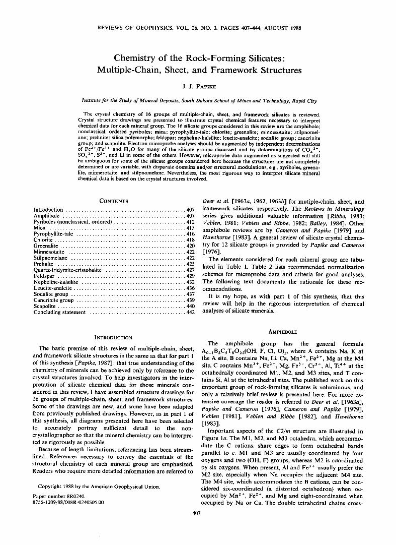
AMPHIBOLE I-BEAMS Fig. lb. Amphibole structural topology [from Papike and Cameron, 1976].
410 PAPIKE.' CHEMISTRY OF SILICATE MINERALS
AMPHIBOLE
M3 MI M2 M4
Fig. lc. The crystal structure of C2/m amphibole projected down [001]. Stippled areas outlined I beams depicted in Figure lb [from Cameron and Papike, 1979].
link the octahedral strips and are composed of two indepen- dent tetrahedral sites, T1 and T2. The T1 site, which shares three oxygens with other tetrahedra, is more regular and smaller than T2. The T2 site shares two oxygens with adjacent
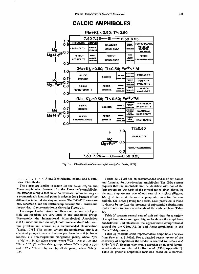
FE-MG-MN AMPHIBOLES
1.0
i 0.9 Mg
Mg + Fe 2-•
0.1 0.0
8.0
ORTHORHOMBIC
MAGNESIO-ANTHOPHYLLITE
ANTHOPHYLLITE
MAGNESIO-GEDRITE
GEDRITE
FERRO-ANTHOPHYLLITE FERRO-GEDRITE
7.0 6.0
-- Si per formula unit
MONOCLINIC
I 1.0 MAGNESIO-CUMMINGTONITE 0.7
Mg CUMMINGTONITE Mg+Fe 2+ 0.3
] 0.0 GRUNERITE Fig. ld. Classification of Fe-Mg-Mn amphiboles [after Leake,
1978].
tetrahedra. The large and highly distorted A polyhedron occurs between the tetrahedral chains. This site can be vacant, partially filled, or fully occupied by Na and/or K in the C2/m structures. The 03 site is usually occupied by an (OH)- group or, less commonly, by F- and C1-. Oxyamphiboles predomi- nantly have 0 2- at the 03 position.
The major topologic aspects of the structures are summa- rized in Figure lb. Each "I beam" represents a tetrahedral- octahedral-tetrahedral (T-O-T) unit. The rotational aspect of the tetrahedral chain relative to the octahedral strip is S if the tetrahedra and octahedra have the same orientation and is O
if the tetrahedra and octahedra have opposite orientations [Thompson, 1970' Papike and Ross, 1970]. A stacking se- quence of the octahedral layers is defined as positive or nega- tive depending on whether the apex of an octahedron points to +a or --a when viewed in the +c direction, as in Figure lb. The designations A and B refer to two tetrahedral sheets in a T-O-T unit that are symmetrically distinct. The absence of these designations indicates that the tetrahedral sheets in a T-O-T unit are symmetrically equivalent and are related by a twofold axis parallel to b.
The C2/m amphiboles have the octahedral stacking se- quence +, +, +,---and only O rotations of the tetra- hedral layers. The tetrahedral layers are related by a twofold axis parallel to b. The P2x/m structure has the same +, +, +,... stacking sequence as the C2/m structure, but it has
lost the twofold axis parallel to b. Thus it has symmetrically distinct A and B tetrahedral chains. The orthorhombic (Pnmn) protoamphibole structure has the stacking sequence q-, --, +, --,.--and O rotations of tetrahedra. Orthorhombic
Pnma structures have octahedral stacking sequences +, +,
PAPIKE.' CHEMISTRY OF SILICATE MINERALS 411
CALCIC AMPHIBOLES
1.0
I 0.9 Mg
Mg+ Fe 2+ 0.5 I
0.0
1.0
Mg+Fe2+013
0.0
I 1.0 0.7
M•:e2+0-5 Mg+ 0.3 I
0.0
(Na + K) A < 0.50; Ti < 0.50 7.50 7.25-.-• Si .------ 6.50 6.25
TREMOLITE ,-,=,•ou'nc .o..B•.œ.o,. 'rSC.E.- TSCHERMAKITE MAGNESIO- •Krnc
- ACTINOLITE senmemo (ALUMINO- .O..Br. ENOœ HORNBLENDE HOma.a• TSCHER- MAKITE)
- FERRO- FœRRO- FERRO- FœRRO- FERRO- •'19•UTIC MAKITIC
ACTINOLITE HORNBLENDE TSCHERMAKITE
(Na+K) A_>0.50; Ti<0.50; Fe3+_< V•AI PARGA81TIC
SILIClC EOEmT•C PARGASITE EDENITE .o.mm•
' EDENITE I'10RNBLENF FERROAN FERROAN
PARGA81TIC PARGASITE
- SILIClC FERRO- FœR.O- .o. mm• FERRO-EDENITE EDENITE œoœ.•TIC FœRRO- FERRO- •IORNBLENDE PARGA81TIC
.o.m, ,..o• PARGASITE
(Na+ K)^_> 0.50; Ti < 0.50; Fe3+>V•AI , !
MAGNE810-
HASTING- MAGNESIO- SILIClC EDENITE EDENITIC 81TIC .O.Na.a• HASTINGSITE
ß EDENITE HORNBLENDE MAGNE81AN
HASTING- MAGNESIAN 81TIC
HOma. e• HASTINGSITE ' SILICIC FERRO-
FERRO-EDENITE EDENITE EDENITIC HASTING- smc HASTINGSITE
I 1.0 Mg
Mg + Fe 2+ 0.5 I
0.0
7.E½0 7.;•5 • Si------G.50 6.'25
Ti >'0.50
KAERSUTITE
FERRO-KAERSUTITE
Fig. le. Classification of calcic amphiboles [after Leake, 1978].
--, --, +, +,..-'AandBtetrahedralchains, andOrota- tions of tetrahedra.
The a axes are similar in length for the C2/rn, P2•/rn, and Pnrnn amphibolcs' however, for the Pnrna orthoamphibolcs the distance along a that must be traversed before arriving at a symmetrically identical point is twice as long because of the different octahcdral stacking sequence. The T-O-T I beams are only schematic, and the relationship between the I beams and the polyhcdral representation is shown in Figure l c.
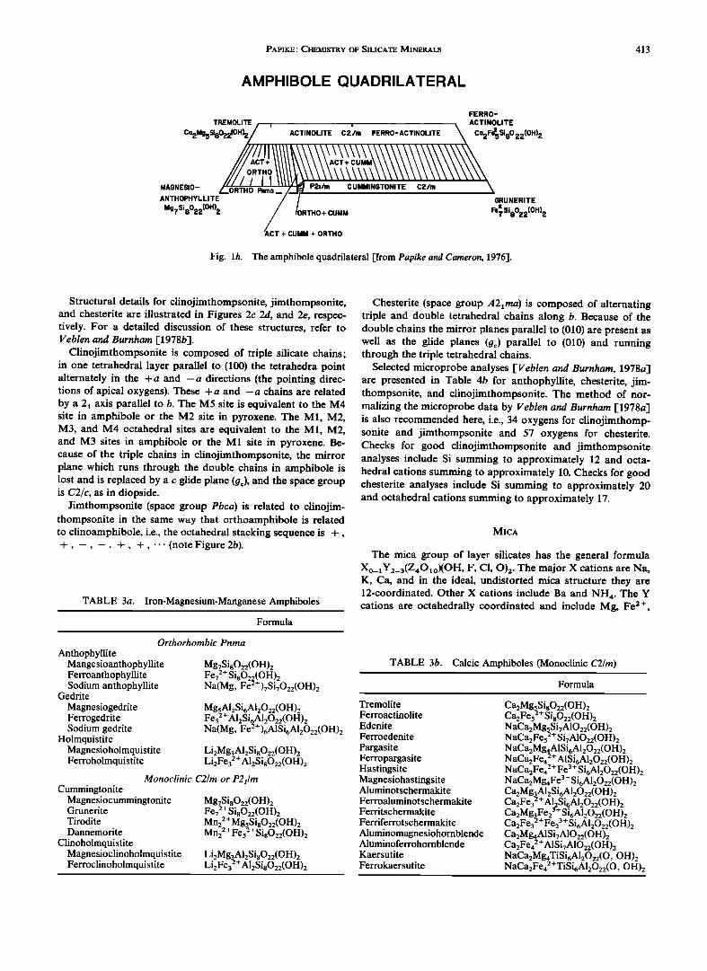
The range of substitutions and therefore the number of pos- sible end-members are very large in the amphibole group. Fortunately, the International Mincralogical Association (IMA) subcommittee on amphibolc nomenclature* addressed this problem and arrived at a recommended classification FLeake, 1978]. This system divides the amphiboles into four chemical groups in terms of atoms per formula unit (apfu) as follows' (1) iron-magnesium-manganese group, where B(Ca + Na) < 1.34, (2) calcic group, where B(Ca + Na) _> 1.34 and
nNa < 0.67, (3) sodic-calcic group, where n(Ca + Na)_> 1.34 and 0.67 < nNa < 1.34, and (4) alkali group, where nNa >_ 1.34.
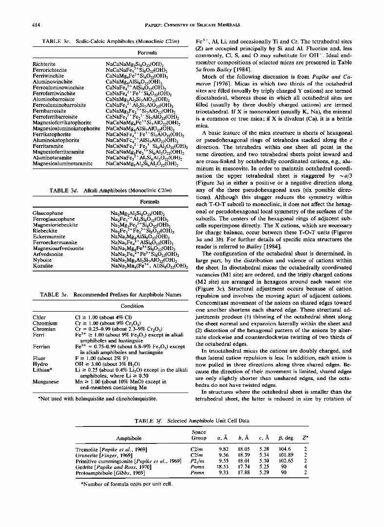
Tables 3a-3d list the 58 recommended end-member names
and formulae for rock-forming amphiboles. The IMA system requires that the amphibole first be identified with one of the four groups on the basis of the critical ratios given above. In the next step we use one of our sets of x-y plots (Figures ld-lg) to arrive at the most appropriate name for the am- phibole. See LeaIce [1978] for details. Last, provision is made to denote by prefixes the presence of substantial substitutions that are not essential constituents of the end-members (Table 3e).
Table 3f presents several sets of unit cell data for a variety of amphibole structure types. Figure lh shows the amphibole quadrilateral and illustrates the approximate compositional control for the C2/rn, P2•/rn, and Pnrna amphiboles in the Ca-Fe 2 +-Mg system.
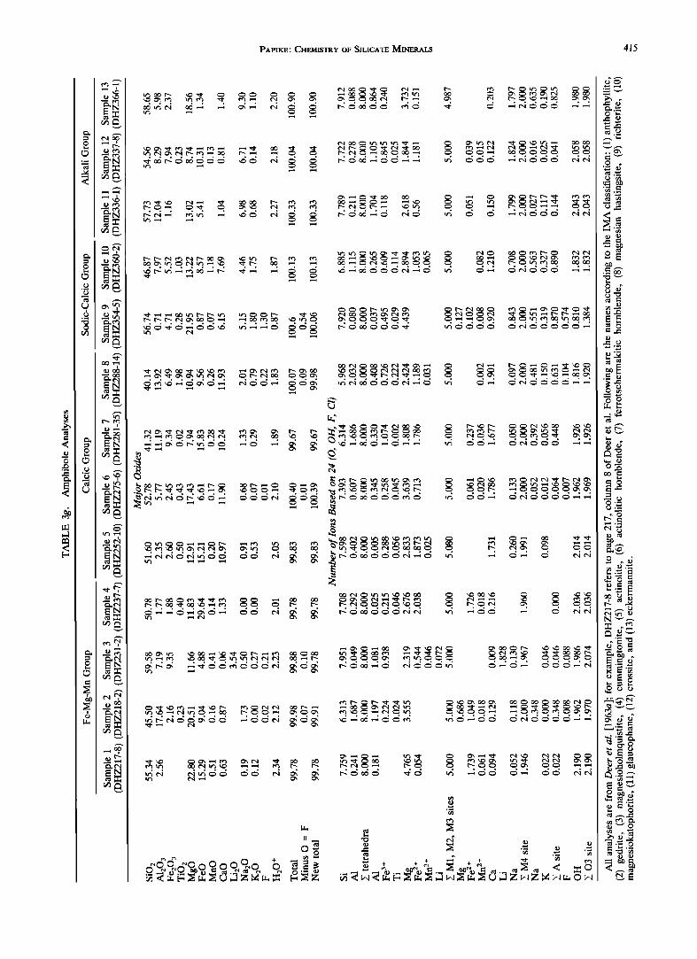
Table 3g presents some representative amphibole analyses from Deer et al. [1963a]. For a detailed recent review of the chemistry of amphiboles the reader is referred to Veblen and Ribbe [1982]. Readers who need a refresher on mineral formu- la calculations are referred to Deer et al. [1966, Appendix I]. Table 3g presents amphibole formulae based on a normal-
412 PAPIKE' CHEMISTRY OF SILICATE MINERALS
Mg+Fe 2+
NA-C^ AMPHIBOLES
1.0}
0.5
0.0 8.0
(Na+ K)A< 0.50 I
WlNCHITE BARROISITE
FERRO-
FERRO-BARROISITE WlNCHITE
i
7.5 6.5 6.0
Si p.f.u. •
Mg
Mg+Fe 2+
ALKALI AMPHIBOLES
0.0
0.5
1.0 0.0
(Na+ K)A2 0.50
FERRO-
ECKERMANNITE
ECKERMANNITE
ARFVEDSONITE
(KOZULITE IF Mn c •, 2.5)
MAGNESIO-
ARFVEDSONITE
0.5
Fe3+/(Fe3++ V•AI) • 1.0
1 Mg+Fe 2+
1.0!
0.5
0.0 8.0
(Na+ K) A > 0.50 I
MAGNESIO- MAGNESIO- •CHTERITE
KATOPHORITE TARAMITE
FERRO-
KATOPHORITE TARAMITE RICHTERITE
i
7.5 6.5 6.0
si p.f.u. •
Fig. If Classification of Na-Ca amphiboles [after Leake, 1978].
Mg
Mg + Fe 2+
0.0
0.5
1.0
(Na+ K)A< 0.50
FERRO- RIEBECKITE GLAUCOPHANE
CROSSITE
MAGNESIO- GLAUCOPHANE
RIEBECKITE
0.0 0.3 0.7 1.0
• Fe3+/(Fe 3+ + V•AI) •
Fig. 1•/. Classification of alkali amphiboles [after Leake, 1978].
ization to 24 (O, OH, F), and this is the recommended way to normalize amphibole analyses if reliable H20 and F analyses are available. If H20 and F are not analyzed, then the recom- mended normalization is to 23 oxygens or 46 negative charges. This is also the recommended way to normalize microprobe analyses. Of course, this assumes no oxyamphibole compo- nent, which can be a dangerous assumption in certain cases. Criteria for good analyses include: (1) Si + •VA1 should total to approximately eight; (2) the octahedral cations (Mn, Fe, Mg, Ti, Cr, A1) should sum to greater than or equal to five; (3) the octahedral cations in excess of the five needed to fill the
M1, M2, and M3 sites ("exoct") should be less than or equal to two; (4) exoct plus Ca should be less than or equal to two; (5) the M4 site occupancy (exoct, Ca, Na, Li) should sum to approximately two; and (6) the A site (Na, K) should sum to less than or equal to one. See Cameron and Papike [1979, p. 32] for additional charge balance criteria.
Note that there are various ways of estimating Fe 3+ from microprobe data (see Hawthorne [1983] for a review); how- ever, none are accurate, and none are recommended. I strongly recommend a direct determination of Fe2+/Fe 3+ [e.g., Goldich, 1984].
PYRIBOLES (NONCLASSICAL, ORDERED)
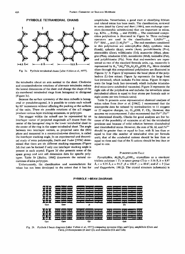
Much of this discussion concerning pyriboles and bio- pyriboles is extracted from a review by Veblen [1981]. The term biopyribole was introduced by Johannsen [1911] to col- lectively consider the micas (biotites), pyroxenes, and am- phiboles. Johannsen also used the term pyribole to refer to the biopyriboles excluding the micas. Thompson [1970, 1978] used
this terminology and extended it to include other minerals (hypothetical at that time) that could be thought of as as- sembled from slabs of pyroxene and mica. Here we consider only the nonclassical (excluding pyroxenes and amphiboles), ordered (ordered sequences of mixed double and triple chains along b) pyriboles.
The first silicates to be recognized with wider than a double chain were synthetic barium silicates [Katscher and Liebau, 1965; Liebau, 1980]. However, the tetrahedral chains in these structures are topologically distinct from those in pyroxenes and amphiboles. The first report of a structurally ordered, nonclassical biopyribole that can be constructed from mica and pyroxene units was of a synthetic reported by Drits et al. [1974]. This silicate contained Na and Mg at crystallographic sites analogous to the amphibole M4 site and the M1, M2, and M3 octahedral sites, respectively.
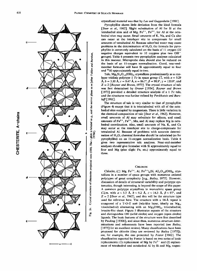
Veblen and Burnham [1975] discovered four new minerals in this series; these were intergrown with anthophyllite and cummingtonite in an alteration zone of a metamorphosed ul- tramafic body near Chester, Vermont. Structurally, the new minerals are similar to pyroxenes and amphiboles but contain either triple chains or both triple and double chains. Figure 2a provides a comparison of idealized pyroxene, amphibole, and the new triple chains. There is a remarkable analogy of these four new minerals to the pyroxene and amphibole groups. Figure 2b presents I beam diagrams comparing pyroxene, am- phibole, jimthompsonite (triple chains only), and chesterite (triple and double chains that alternate along b). Table 4a tabulates unit cell parameters, space groups, and the number of formula units per unit cell (Z) for jimthompsonite and chesterite.
PAPIKE' CHEMISTRY OF SILICATE MINERALS 413
AMPHIBOLE QUADRILATERAL
FERRO-
ag?SiaO22(OH)2 /ORTHO+ cuaa Fe'• Si8022(0H)2 AC•T + CUMM + ORTHO Fig. lh. The amphibole quadrilateral [from Papike and Cameron, 1976].
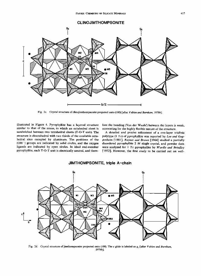
Structural details for clinojimthompsonite, jimthompsonite, and chesteritc are illustrated in Figures 2c 2d, and 2e, respec- tively. For a detailed discussion of these structures, refer to Veblen and Burnham [1978b].
Clinojimthompsonite is composed of triple silicate chains' in one tetrahedral layer parallel to (100) the tetrahedra point alternately in the +a and --a directions (the pointing direc- tions of apical oxygens). These + a and --a chains are related by a 2a axis parallel to b. The M5 site is equivalent to the M4 site in amphibole or the M2 site in pyroxene. The M1, M2, M3, and M4 octahedral sites are equivalent to the M1, M2, and M3 sites in amphibolc or the M1 site in pyroxene. Be- cause of the triple chains in clinojimthompsonitc, the mirror plane which runs through the double chains in amphibole is lost and is replaced by a c glide plane (go), and the space group is C2/c, as in diopside.
Jimthompsonite (space group Pbca) is related to clinojim- thompsonitc in the same way that orthoamphibole is related to clinoamphibole, i.e., the octahedral stacking sequence is +, +, -, - +, +,...(noteFigure2b).
TABLE 3a. Iron-Magnesium-Manganese Amphiboles
Formula
Orthorhombic Pnma
Anthophyllite Mangesioanthophyllite Ferroanthophyllite Sodium anthophyllite
Gedrite
Magnesiogedrite Ferrogedrite Sodium gedrite
Holmquistite Magnesioholmquistite Ferroholmquistite
Mg7Si8022(OH)2 Fe72+Si8022(OH)2 Na(Mg, Fe 2+)7Si7022(OH)2
MgsAI2Si6AI2022(OH)2 Fe52+A12Si6AI2022(OH)2 Na(Mg, Fe 2+)6AISi6AI2022(OH)2
Li2Mg3AI2Si8022(O H)2 Li2Fe32+A12Si8022( OH)2
Monoclinic C2/m or P2//m Cummingtonite
Magnesiocummingtonite Gruneritc Tirodite Dannemorite
Clinoholmquistite Magnesioclinoholmquistite Ferroclinoholmquistite
Mg7Si8022(OH)2 Fe72+Si8022(OH)2 Mn22+Mg5Si8022( OH)2 Mn22+Fe52+Si8022(OH)2
Li2Mg3AI2SiaO22(OH)2 Li2Fe32 + AI2Si8022(OH)2
Chesterite (space group A2•ma) is composed of alternating triple and double tetrahedral chains along b. Because of the double chains the mirror planes parallel to (010) are present as well as the glide planes (g½) parallel to (010) and running through the triple tetrahedral chains.
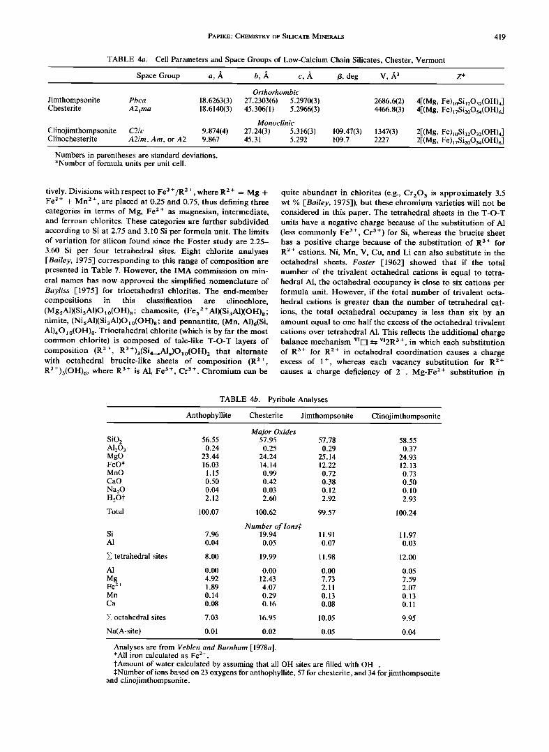
Selected microprobe analyses [Veblen and Burnham, 1978a] are presented in Table 4b for anthophyllite, chesterite, jim- thompsonitc, and clinojimthompsonite. The method of nor- malizing the microprobe data by Veblen and Burnham [1978a] is also recommended here, i.e., 34 oxygens for clinojimthomp- sonite and jimthompsonite and 57 oxygens for chesteritc. Checks for good clinojimthompsonite and jimthompsonite analyses include Si summing to approximately 12 and octa- hedral cations summing to approximately 10. Checks for good chesterite analyses include Si summing to approximately 20 and octahedral cations summing to approximately 17.
MICA
The mica group of layer silicates has the general formula Xo_•Ye_3(Z,•O•o)(OH, F, C1, O)e. The major X cations are Na, K, Ca, and in the ideal, undistorted mica structure they are 12-coordinated. Other X cations include Ba and NH,•. The Y cations are octahedrally coordinated and include Mg, Fe e+,
TABLE 3b. Calcic Amphiboles (Monoclinic C2/m)
Formula
Tremolite Ferroactinolite Edenite Ferroedenite
Pargasite Ferropargasite Hastingsite Magnesiohastingsite Aluminotschermakite Ferroaluminotschermakite Ferritschermakite Ferriferrotschermakite
Aluminomagnesiohornblende Aluminoferrohornblende Kaersutite
Ferrokaersutite
Ca2MgsSiaO22(OH)2 Ca2Fe52+Si8022(OH) 2 NaCa2MgsSi7AIO22(OH)2 NaCa2Fe52+Si7AIO22(OH)2 NaCa2Mg4AISi6AI2022(OH) 2 NaCa2Fe42 + AISi6AI2022 ( O H)2 NaCa2Fe42+Fe3+Si6AI2022(OH)2 NaCa2Mg4Fe 3+Si6A12022( OH)2 Ca2Mg3AI2Si6AI2022(OH)2 Ca2Fe32 + AI2 Si6A12022(O H) 2 Ca2Mg 3Fe23+Si6A12022( OH)2 Ca2Fe32 + Fe23 + Si6AI2022(OH)2 Ca2Mg4AISi7AIO22(OH)2 Ca2Fe42+AISi7AIO22(OH)2 NaCa2Mg4TiSi6AI2022(O, OH) 2 NaCa2Fe42+TiSi6AI2022(O, OH) 2
414 PAPIKE' CHEMISTRY OF SILICATE MINERALS
TABLE 3c. Sodic-Calcic Amphiboles (Monoclinic C2/m)
Formula
Richterite Ferrorichterite Ferriwinchite
Aluminowinchite Ferroaluminowinchite Ferroferriwinchite Aluminobarroisite
Ferroaluminobarroisite Ferribarroisite Ferroferribarroisite
Magnesioferrikatophorite
NaCaNaMgsSi8022(OH)2 NaCaNaFe52 + Si8022(OH) 2 CaNaMg4Fe 3 + Si8022(O H)2 CaNaMg4A1Si8022(OH)2 CaNaFe42 + A1Si80 22(O H)2 CaNaFe42 + Fe 3 + Si8022(OH) 2 CaNaMg3A12 Si7A1022(OH)2 CaNaFe32 + A12 Si7A1022( O H)2 CaNaMg3Fe23 + Si7A1022(OH)2 CaNaFe32 + Fe23+ Si7A1022(OH)2 N a CaN aMg4 F e 3 + S i7A10 22(O H)2
Magnesioaluminokatophorite NaCaNaMg4A1Si7A1022(OH)2 Ferrikatophorite NaCaNaFe42 +Fe 3 +Si7A1022(OH) 2 Aluminokatophorite NaCaNaFe42 + A1Si7A1022(OH)2 Ferritaramite NaCaNaFe32 +Fe23+ Si6A12022(OH) 2 Magnesioferritaramite NaCaNaMg3Fe23+ Si6A12022(OH)2 Aluminotaramite NaCaNaFe32 + A12Si6A12022(OH)2 Magnesioaluminotaramite NaCaNaMg3A12Si6A12022(OH)2
TABLE 3d. Alkali Amphiboles (Monoclinic C2/m)
Formula
Glaucophane Ferroglaucophane Magnesioriebeckite Riebeckite Eckermannite Ferroeckermannite
Magnesioarfvedsonite Arfvedsonite
Nyboite Kozulite
Na2Mg3A12S i8022 (O H)2 Na2Fe32 + Al2 Si8022(OH)2 Na2Mg3Fe23 + Si8022(OH) 2 Na2Fe32+Fe23+ Si8022(OH)2 NaNa2Mg4A1Si8022(OH)2 NaNa2 Fe42 + AISi8022(OH)2 NaNa2Mg4Fe 3+ Si8022(OH) 2 NaNa2Fe42+Fe3+ Si8022(OH)2 NaNa2Mg3A12Si7A1022(OH)2 NaNa2Mn4(Fe 3+ , A1)Si8022(OH) 2
TABLE 3e. Recommended Prefixes for Amphibole Names
Condition
Chlor Chromium Chromian Ferri
Ferrian
Fluor
Hydro Lithian*
Manganese
C1 -> 1.00 (about 4% CI) Cr -> 1.00 (about 9% Cr203) Cr = 0.25-0.99 (about 2.3-9% Cr203) Fe 3+ -> 1.00 (about 9% Fe203) except in alkali
amphiboles and hastingsite Fe 3+ - 0.75-0.99 (about 6.8-9% Fe203) except
in alkali amphiboles and hastingsite F -> 1.00 (about 2% F) OH -> 3.00 (about 3% H20 ) Li -> 0.25 (about 0.4% Li20 ) except in the alkali
amphiboles, where Li -> 0.50 Mn -> 1.00 (about 10% MnO) except in
end-members containing Mn
*Not used with holmquistite and clinoholmquistite.
Fe 3 +, A1, Li, and occasionally Ti and Cr. The tetrahedral sites (Z) are occupied principally by Si and A1. Fluorine and, less commonly, C1, S, and O may substitute for OH-. Ideal end- member compositions of selected micas are presented in Table 5a from Bailey [ 1984].
Much of the following discussion is from Papike and Ca- meron [1976]. Micas in which two thirds of the octahedral sites are filled (usually by triply charged Y cations) are termed dioctahedral, whereas those in which all octahedral sites are
filled (usually by three doubly charged cations) are termed trioctahedral. If X is monovalent (usually K, Na), the mineral is a common or true mica; if X is divalent (Ca), it is a brittle mica.
A basic feature of the mica structure is sheets of hexagonal or pseudohexagonal rings of tetrahedra stacked along the c direction. The tetrahedra within one sheet all point in the same direction, and two tetrahedral sheets point inward and are cross-linked by octahedrally coordinated cations, e.g., alu- minum in muscovite. In order to maintain octahedral coordi-
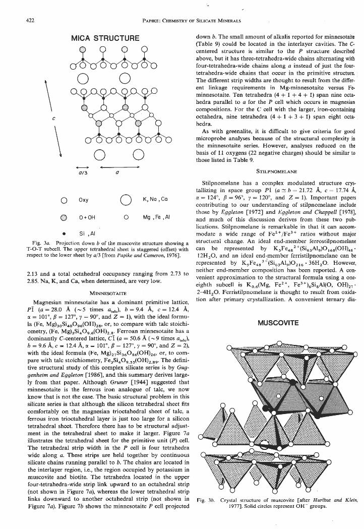
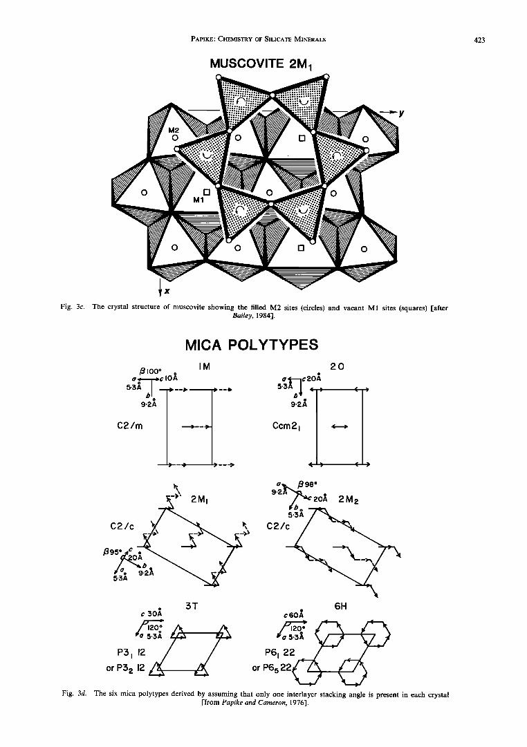
nation the upper tetrahedral sheet is staggered by •,-a/3 (Figure 3a) in either a positive or a negative direction along any of the three pseudohexagonal axes (six possible direc- tions). Although this stagger reduces the symmetry within each T-O-T subcell to monoclinic, it does not affect the hexag- onal or pseudohexagonal local symmetry of the surfaces of the subcells. The centers of the hexagonal rings of adjacent sub- cells superimpose directly. The X cations, which are necessary for charge balance, occur between these T-O-T units (Figures 3a and 3b). For further details of specific mica structures the reader is referred to Bailey [1984].
The configuration of the octahedral sheet is determined, in large part, by the distribution and valence of cations within the sheet. In dioctahedral micas the octahedrally coordinated vacancies (M1 site) are ordered, and the triply charged cations (M2 site) are arranged in hexagons around each vacant site (Figure 3c). Structural adjustment occurs because of cation repulsion and involves the moving apart of adjacent cations. Concomitant movement of the anions on shared edges toward one another shortens each shared edge. These structural ad- justments produce (1) thinning of the octahedral sheet along the sheet normal and expansion laterally within the sheet and (2) distortion of the hexagonal pattern of the anions by alter- nate clockwise and counterclockwise twisting of two thirds of the octahedral edges.
In trioctahedral micas the cations are doubly charged, and thus lateral cation repulsion is less. In addition, each anion is now pulled in three directions along three shared edges. Be- cause the direction of their movement is limited, shared edges are only slightly shorter than unshared edges, and the octa- hedra do not have twisted edges.
In structures where the octahedral sheet is smaller than the
tetrahedral sheet, the latter is reduced in size by rotation of
TABLE 3f. Selected Amphibole Unit Cell Data
Amphibole Space Group a, • b, • c, 3•
Tremolite [Papike et al., 1969] Grunerite [Finger, 1969] Primitive cummingtonite [Papike et al., 1969] Gedrite [Papike and Ross, 1970] Protoamphibole [Gibbs, 1969]
/3, deg
*Number of formula units per unit cell.
C2/m 9.82 18.05 5.28 104.6 C2/m 9.56 18.39 5.34 101.89
P2•/m 9.55 18.01 5.30 102.65 Pnma 18.53 17.74 5.25 90 Pnmn 9.33 17.88 5.29 90
g g<
PAPIKE' CHEMISTRY OF SILICATE MINERALS 415
416 PAPIKE' CHEMISTRY OF SILICATE MINERALS
PYRIBOLE TETRAHEDRAL CHAINS
F--4.5 J•-•
Fig. 2a. Pyribole tetrahedral chains [after Veblen et al., 1977].
the tetrahedra about an axis normal to the sheet. Clockwise
and counterclockwise rotations of alternate tetrahedra reduce
the lateral dimensions of the sheet and change the shape of the six-membered tetrahedral rings from hexagonal to ditrigonal (Figure 3c).
Because the surface symmetry of the mica subcells is hexag- onal or pseudohexagonal, it is possible to rotate each subcell by 60 ø increments without affecting the packing at the surfaces of the units. These six possible rotations of the a/3 stagger produce various layer stacking sequences, or polytypes.
The stagger within the subcell can be represented by an interlayer vector of projected magnitude a/3 drawn from the center of the hexagonal ring in the lower tetrahedral sheet to the center of the ring in the upper tetrahedral sheet. The angle between two interlayer vectors, as projected onto the (001) plane and measured in a counterclockwise direction, is called the interlayer stacking angle. In an experimental and theoreti- cal study of mica polymorpl•s, Smith and Yoder [1956] deter- mined that there are six different stacking sequences (Figure 3d) that can be formed if only one interlayer stacking angle is present in each crystal. Figure 3d also presents some of the space group and unit cell dimension data for specific poly- types. Table 5b [Bailey, 1984] documents the natural oc- currence of mica polytypes.
Unfortunately, the classification and nomenclature for micas has not been developed to the extent that it has for
amphiboles. Nevertheless, a good start at classifying lithium and related micas has been made. The classification, reviewed in some detail by Cern35 and Burr [1984], uses exchange oper- ators (isomorphic substitutions) that are important in micas, e.g., KNa_ x, FeMg_ x, and F(OH)_ x. The condensed compo- sition polyhedron is illustrated in Figure 3e. Three exchange operators are used in the classification' LiA1Fe 2 +_2, Fe 2+SiAl 2, and [] A12Fe 2+ The end-members considered -- -3'
in this polyhedron are siderophyllite (Sdp), synthetic mica (Synth), ephesite (Kep), annite (Ann), protolithionite (Pro), zinnwaldite (Znw), trilithionite (Tri), muscovite (Mus), phen- gite (Phn), celadonite (Cld), montdorite (Mtd), taeniolite (Fet), and polylithionite (Ply). Note that end-members are repre- sented as two of the standard formula units, e.g., muscovite is represented by K2VI(A14)IV(Si6A12)O2o(OH, F)4. More detailed slices through this compositional polyhedron are illustrated in Figures 3f-3i. Figure 3frepresents the basal plane of the poly- hedron (Li-free micas). Figure 3g represents the large front face (reversed), which includes Fe-free micas. Figure 3h repre- sents the large back face (reversed), which includes trioctahe- dral micas (zero octahedral vacancies). Figure 3i represents the right side of the polyhedron and includes the tetrasilicic micas (tetrahedral silicon is equal to four atoms per formula unit or eight atoms per two formula units).
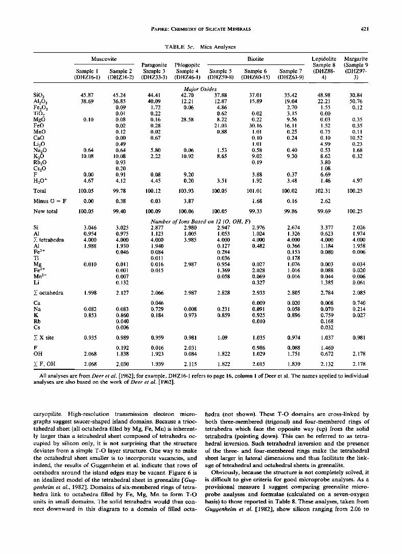
Table 5c presents some representative chemical analyses of micas taken from Deer et al. [1962]. I recommend that the microprobe data be reduced by normalization to 11 oxygens or 22 negative charges, i.e., Olo(OH, F, C1)2. However, this assumes no oxycomponent. I also recommend that Fe 2 +/Fe 3+ be determined directly. Checks for good analyses are few be- cause of the possibility of vacancies at all but the tetrahedral positions and because of solid solution between dioctahedral and trioctahedral micas. However, the sum of Si, A1, and Fe 3 + should be greater than or equal to four, with Si less than or equal to four (the number of tetrahedral sites per formula unit), that of the octahedral cations should be less than or equal to three and that of the X cations should be less than or equal to one.
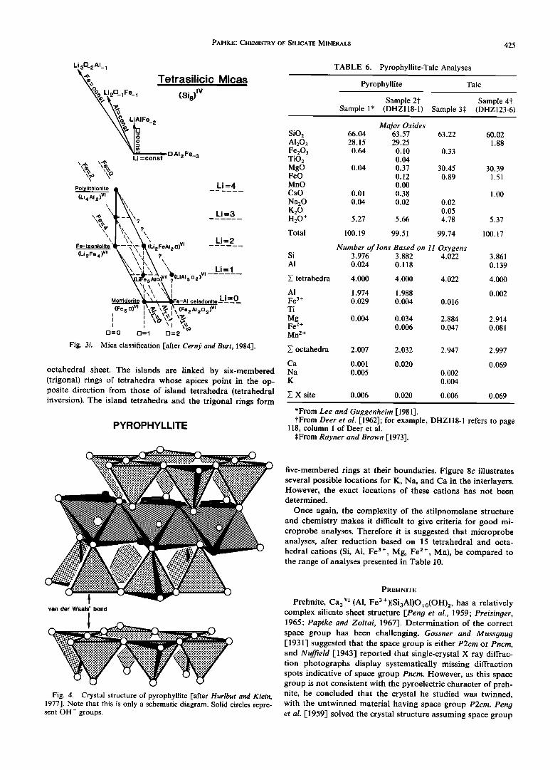
PYROPHYLLITE-TALC
Pyrophyllite, A12Si401o(OH)2, crystallizes as a one-layer triclinic polytype 1 Tc in space group Ci (a - 5.16 3, b -- 8.97 A, c = 9.35 A, • = 91.2 ø, fl = 100.5 ø, 7 = 89.6ø, and Z = 2 [Lee and Guggenheim, 1981]). The crystal structure (schematic) is
PYRIBOLE I-BEAM DIAGRAMS
Opx Oam Jt Ch
Cpx Cam Cjt Cch
Fig. 2b. Pyribole I beam diagrams [after Veblen et al., 1977] comparing pyroxene (Opx and Cpx), amphibole (Oam and Cam), jimthompsonite (Jt and Cjt), and chesterire (Ch and Cch).
PAPIKE' CHEMISTRY OF SILICATE MINERALS 417
CLINOJIMTHOMPSONITE
gc gc
I b/2 i
Fig. 2c. Crystal structure of clinojimthompsonite projected onto (100) [after Veblen and Burnham, 1978b].
illustrated in Figure 4. Pyrophyllite has a layered structure similar to that of the micas, in which an octahedral sheet is sandwiched between two tetrahedral sheets (T-O-T unit). The structure is dioctahedral with two thirds of the available octa-
hedral sites occupied by aluminum. The positions of the (OH-) groups are indicated by solid circles, and the oxygen ligands are indicated by open circles. In ideal end-member pyrophyllite, each T-O-T unit is electrically neutral, and there-
fore the bonding (Van der Waals') between the layers is weak, accounting for the highly flexible nature of the structure.
A detailed and precise refinement of a one-layer triclinic polytype (1 Tc) of pyrophyllite was reported by Lee and Gug- genheim [1981]. Raynet and Brown [1964] studied a partially disordered pyrophyllite 2 M single crystal, and powder data were analyzed for 1 Tc pyrophyllite by Wardle and Brindley [1972]. However, the first study to be carried out on well-
JIMTHOMPSONITE, triple A-chain
gc gc
MI M:• • M5
Fig. 2d. Crystal structure ofjimthompsonite projected onto (100). The c glide is labeled as gc [after Veblen and Burnham, 1978b].
418 PAPIKE' CHEMISTRY OF SILICATE MINERALS
crystallized material was that by Lee and Guggenheim [1981]. Pyrophyllite shows little deviation from the ideal formula
[Deer et al., 1962]. Slight substitution of A1 for Si at the tetrahedral sites and of Mg, Fe 2+, Fe 3+, for A1 at the octa- hedral sites may occur. Small amounts of K, Na, and Ca also can occur at the interlayer site to compensate for small amounts of tetrahedral A1. Because adsorbed water may cause problems in the determination of H20, the formula for pyro- phyllite is commonly calculated on the basis of 11 oxygen (22 negative charges equivalent to 10 oxygens plus two OH- groups). Table 6 presents two pyrophyllite analyses calculated in this manner. Microprobe data should also be reduced on the basis of an l 1-oxygen normalization. Good, near-end- member formulae will have Si approximately equal to four and VIA1 approximately equal to two.
Talc, Mg3Si,•Oxo(OH)2, crystallizes predominantly as a one- layer triclinic polytype 1 Tc in space group Ci, with a = 5.29 A, b = 5.30 A, c = 9.47 A, • = 86.1 ø, fl = 98.9 ø, 7 = 120-0ø, and Z = 2 [Rayner and Brown, 1973]. The crystal structure of talc was first determined by Gruner [1934]. Rayner and Brown [1973] provided a detailed structure analysis of a 1 Tc talc, and the structures was further refined by Perdikatsis and Burz- laff [1981].
The structure of talc is very similar to that of pyrophyllite (Figure 4) except that it is trioctahedral with all of the octa- hedral sites occupied by magnesium. There is little variation in the chemical composition of talc [Deer et al., 1962]. However, small amounts of A1 may substitute for silicon, and small amounts of Fe • +, Fe 3 +, Mn, and A1 may replace Mg in octa- hedral coordination. Also, small amounts of Na, K, and Ca may occur at the interlayer site to charge-compensate for tetrahedral A1. Because of problems with accurate determi- nation of H:O, chemical formulae should be calculated (as for pyrophyllite) on an l 1-oxygen normalization basis. Table 6 gives two representative talc analyses. Near-end-member analyses should give formulae with Si approximately equal to four and Mg (plus slight Fe, etc.) approximately equal to three.
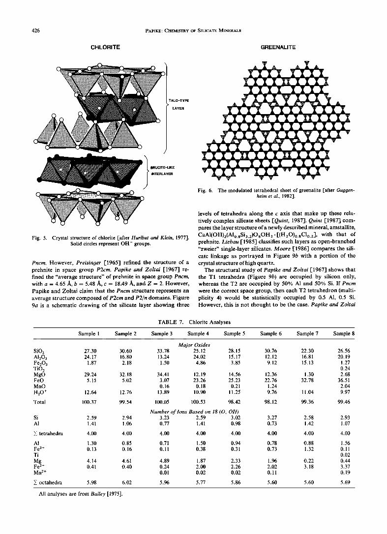
CHLORITE
Chlorite, (Z3 Mg, Fe 2+, A1, Fe3+)6(Si, A1),•O•o(OH)8, crys- tallizes in a number of space groups with numerous ordered polytypes of great complexity [e.g., Bailey, 1975]. However, discussion of details of structural variability and polytype sys- tematics, though interesting, is beyond the scope of this paper. A common polytype crystallizes in monoclinic space group C2/m, with a ,-, 5.3 /k, b-,• 9.2 /k, c ,-, 14.3 •, fl ,-, 97% and Z = 2 [Deer et al., 1962], and this will be the structure type used for reference here. The structure with a 14-/k repeat is composed of a T-O-T unit (talc-like layer, ideally an Mg 3, trioctahedral) alternating with an Mg3(OH)6, trioctahedral, brucite-like sheet. Figure 5 illustrates aspects of the structure and distinguishes OH (solid circles) and oxygen (open circles) ligands. The basic features of the structure were first described by Pauling [-1930b], and since then, numerous structure deter- minations and refinements have been reported (see Bailey, [-1975] for an excellent review). Many classifications have been proposed for chlorite (they are reviewed by Bailey [-1975]); see, for example, the one presented by Foster [1962]. The classification reported by Foster is based on two series of ionic replacements: (1) replacement of Mg by Fe 2 + and (2) replace- ment of tetrahedral and octahedral A1 by Si and Mg, respec-
PAPIKE.' CHEMISTRY OF SILICATE MINERALS 419
TABLE 4a. Cell Parameters and Space Groups of Low-Calcium Chain Silicates, Chester, Vermont
Space Group a, fk b, fk c, fk /3, deg V, •k 3 Z*
Orthorhombic
Jimthompsonite Pbca 18.6263(3) 27.2303(6) 5.2970(3) Chesterite A2•ma 18.6140(3) 45.306(1) 5.2966(3)
Monoclinic
Clinojimthompsonite C2/c 9.874(4) 27.24(3) 5.316(3) Clinochesterite A2/m, Am, or A2 9.867 45.31 5.292
2686.6(2) 4466.8(3)
109.47(3) 1347(3) 109.7 2227
4[(Mg, Fe)loSi12032(OH)4 ] 4[(Mg, Fe) 17Si20054(OH)6 ]
2[(Mg, Fe)loSi12032(OH)4 ] 2[(Mg, Fe)17Si20054(OH)6 ]
Numbers in parentheses are standard deviations. *Number of formula units per unit cell.
tively. Divisions with respect to Fe 2 +/R 2 +, where R 2 + = Mg + Fe 2 + + Mn 2 +, are placed at 0.25 and 0.75, thus defining three categories in terms of Mg, Fe 2 + as magnesian, intermediate, and ferroan chlorites. These categories are further subdivided according to Si at 2.75 and 3.10 Si per formula unit. The limits of variation for silicon found since the Foster study are 2.25- 3.60 Si per four tetrahedral sites. Eight chlorite analyses [Bailey, 1975] corresponding to this range of composition are presented in Table 7. However, the IMA commission on min- eral names has now approved the simplified nomenclature of Bayliss [1975] for trioctahedral chlorites. The end-member
compositions in this classification are clinochlore, (MgsA1)(Si3A1)O•o(OH)8; chamosite, (Fes 2+A1)(Si3A1)(OH)8; nimite, (NisA1)(Si3A1)O•o(OH)8; and pennantite, (Mn, A1)6(Si, A1),•O•0(OH)8. Trioctahedral chlorite (which is by far the most common chlorite) is composed of talc-like T-O-T layers of composition (R 2+, R3+)3(Si,•_xAlx)O•0(OH)2 that alternate with octahedral brucite-like sheets of composition (R 2+, R3+)3(OH)6, where R 3+ is A1, Fe 3+, Cr 3+. Chromium can be
quite abundant in chlorites (e.g., Cr20 3 is approximately 3.5 wt % [Bailey, 1975]), but these chromium varieties will not be considered in this paper. The tetrahedral sheets in the T-O-T units have a negative charge because of the substitution of A1 (less commonly Fe 3+, Cr 3+) for Si, whereas the brucite sheet has a positive charge because of the substitution of R 3+ for R 2 + cations. Ni, Mn, V, Cu, and Li can also substitute in the octahedral sheets. Foster [1962] showed that if the total number of the trivalent octahedral cations is equal to tetra- hedral A1, the octahedral occupancy is close to six cations per formula unit. However, if the total number of trivalent octa- hedral cations is greater than the number of tetrahedral cat- ions, the total octahedral occupancy is less than six by an amount equal to one half the excess of the octahedral trivalent
cations over tetrahedral A1. This reflects the additional charge balance mechanism vi[• • VI2R3 +, in which each substitution of R 3+ for R 2 + in octahedral coordination causes a charge excess of 1+, whereas each vacancy substitution for R 2+ causes a charge deficiency of 2-. Mg-Fe 2+ substitution in
TABLE 4b. Pyribole Analyses
Anthophyllite Chesterite Jimthompsonite Clinojimthompsonite
Major Oxides SiO2 56.55 57.95 57.78 58.55 A1203 0.24 0.25 0.29 0.37 MgO 23.44 24.24 25.14 24.93 FeO* 16.03 14.14 12.22 12.13 MnO 1.15 0.99 0.72 0.73 CaO 0.50 0.42 0.38 0.50 Na20 0.04 0.03 0.12 0.10 H20? 2.12 2.60 2.92 2.93
Total 100.07 100.62 99.57 100.24
Number of Ions$ Si 7.96 19.94 11.91 11.97 AI 0.04 0.05 0.07 0.03
tetrahedral sites 8.00 19.99 11.98 12.00
A1 0.00 0.00 0.00 0.05 Mg 4.92 12.43 7.73 7.59 Fe 2+ 1.89 4.07 2.11 2.07 Mn 0.14 0.29 0.13 0.13 Ca 0.08 0.16 0.08 0.11
v octahedral sites 7 03 16.95 10.05 9.95 /_• .
Na(A-site) 0.01 0.02 0.05 0.04
Analyses are from Veblen and Burnham [1978a]. *All iron calculated as Fe 2+.
?Amount of water calculated by assuming that all OH sites are filled with OH-. •Number of ions based on 23 oxygens for anthophyllite, 57 for chesterite, and 34 forjimthompsonite
and clinojimthompsonite.
420 PAPIKE: CHEMISTRY OF SILICATE MINERALS
TABLE 5a. Classification of the Micas
Subgroup, Species Ideal Formula Units
True Micas*
Trioctahedral
phlogopite biotite annite
ferriannite
polylithionite trilithionite taeniolite
zinnwaldite
masutomilite
hendricksite
KMg3(Si3A1)O•o(OH, F)2 K(Mgo.6_•.sFe2.4_l.2)(Si3A1)O•o(OH, F)2 KFe32+(Si3A1)O•o(OH, F)2 KFe32+ (Si3Fe3+)O 10(OH, F) 2 K(Li2A1)Si4Olo(F, OH)2 K(Li•.sAI•.5)(Si3A1)O •o(F, OH)2 K(Mg2Li)Si40•o(F, OH)2 K[Fel.5_0.52 +Lio.5_•.5(A1,
Fe 3+)](Si3.5_2.SAlo.5_•.5)O•o(F, OH)2 K(Mn •.0_0.52 + Li • .o_•.sA1)(Si3.5_3.0Alo.5_ • .0)O •0
(F, OH) 2 K(Zn, Mn)3(Si3A1)O•o(OH, F) 2
sodium phlogopite NaMg3(Si3A1)O•o(OH) 2 wonesite [(Na, K)o.5[•o.5][(Mg,
siderophyllite ephesite preiswerkite montdorite
Dioctahedral muscovite
paragonite phengite illite
chernykhite roscoelite celadonite
glauconite tobelite
Fe)2.sAlo.5](Si3A1)O •o(OH, F)2 K(Fe22+A1)(Si2A12)O•o(OH, F)2 Na(LiA12)(Si2A12)O •o(OH, F)2 Na(Mg2A1)(Si2A12)O •o(OH)2 K(Fe 2+, Mn, Mg)2.sSi4Olo(F, OH) 2
KA12(Si3A1)O•o(OH, F)2 NaA12(Si3AI)O•o(OH, F)2 K[AI•.5(Mg, Fe 2 +)0.5](Si3.SAlo.5)O•o(OH, F)2 -Ko.75(Al•.75Ro.252 +)(Si3.50A10.50)O•0(OH,
F)2 (Ba, Na, NH4)(V 3+, A1)2(Si, AI)40•0(OH) 2 K(V 3+, A1, Mg)2(Si3A1)O•0(OH) 2 K(Mg, Fe2+)(Fe 3+, A1)Si4Om(OH) 2 -• K(R • .333 + R0.672 + )(Si3.67A10.33) O •0(O H)2 (NH4, K)A12(Si3A1)O•o(OH)2
Trioctahedral clintonite kinoshitalite
anandite
bityite Dioctahedral
margarite
Brittle Micas?
Ca(Mg2A1)(SiAl3)O •o(OH, F)2 Ba(Mg, Mn, A1)3(Si2A12)O•o(OH, F) 2 BaFe32 + (Si3 Fe 3+)O j o(OH) S Ca(A12Li)(Si2A1Be)O•o(OH, F) 2
CaA12(Si2A12)O•o(OH, F)2
*X is a monovalent cation. ?X is a divalent cation.
chlorites can be complete. Foster's study showed that whereas most chlorites have H20 contents reflecting the ideal eight (OH) groups per formula unit, some analyses indicated less than eight suggesting an oxycomponent. Despite this observa- tion, microprobe data will normally be reduced assuming eight (OH) groups. In addition to trioctahedral chlorites a few examples of dioctahedral chlorites [Bailey, 1975] have been reported. However, these will not be discussed further here.
Microprobe analyses for chlorite should be reduced on a 14-oxygen (assumes 10 oxygen plus eight (OH), or 28 negative charges) basis, realizing that this ignores a possible oxycom- ponent. I strongly suggest that a direct determination of Fe 3 + [e.g., Goldich, 1984] be made because Fe 3+ substitution in chlorite can be significant. There are few firm criteria for as- sessing the accuracy of chlorite microprobe analyses. The sums will be low (•88%) because of the lack of an H20 determination. However, for most good (approximately trioc- tahedral) analyses the octahedral occupancy will vary between 5.5 and 6.0 atoms per formula unit (apfu), and silicon will fall in the range 2.25-3.60 apfu.
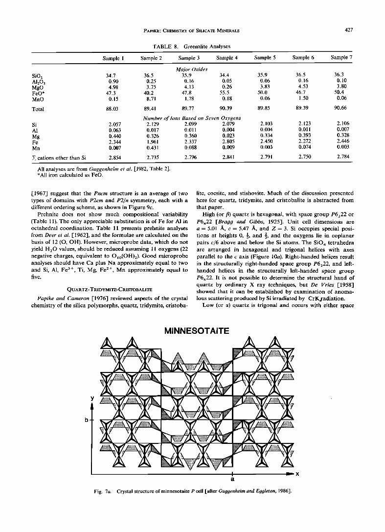
GREENALITE
Greenalite, (Fe, Mg, Mn)2.73_2.85Si2.06_2.13Os(OH)4 , is a complex iron silicate with a modulated layer structure com- posed of tetrahedral-octahedral (T-O) units somewhat analo- gous to that of antigorite, Mg3_xSi2Os(OH)½_2x , but with some significant differences. Gruner [1936] suggested that greenMite was the ferroan iron analogue of serpentine (an- rigorire). However, Floran and Papike [1975] found that elec- tron microprobe data for greenMite deviated from antigorite stoichiometry by having excess silicon (greater than 2.0) and deficient octahedral occupancy (less than 3.0) when the formu- la was calculated on the basis of seven oxygens (14 negative charges equivalent to O5(OH)½). Floran and Papike suggested that modulations involving tetrahedral inversions could ex- plain the observed chemical compositions.
The most detailed structural study of greenalite [Guggen- heim et al., 1982] reported new information about this impor- tant silicate but did not come up with a complete structural solution. Guggenheim et al. found that greenalite is not just an analogue of serpentine but that it is composed of a mixture of both a trigonal and a monoclinic phase. This is also true for the Mn analogue of greenalite, caryopilite. Satellites present on electron diffraction photographs indicated a modulation of the structure that ranged from 23 ]• in greenalite to 17 ]• in
TABLE 5b. Observed Natural Mica Structures
Abundance
Species High Medium Low
Trioctahedral Micas
Phlogopite 1M, 1Md 3T 2M• Manganoan phlogopite 1M 2M• Sodium phlogopite 1M Biotite 1M, 1Md 3T 2M• Annite 1M Ferriannite 1M
"Lepidolite" 1M, 2M 2 3T 2M• Taeniolite 1M, 3T 2M• Zinnwaldite 1M, 1Md 2M•,
3T
Masutomilite 1M 2M• Hendricksite 1M 2M• 3T Wonesite 1Md
Preiswerkite 2M• Siderophyllite 1M Ephesite 2M• 1M Clintonite 1M 3T, 2M• Anandite 20r 2M• ,
1M
Kinoshitalite 1M, 2M• Bityite 2M 1
Dioctahedral Micas
Muscovite 2M• 1M, 3T 1Md
Paragonite 2M l 3T Lithian muscovite 2M 1 Manganian muscovite 2M•, 3T 1M Chromian muscovite 2M•, 1M Phengite 1M, 1Md, 3T 2M1 2M 2 Roscoelite 1M
Chernykhite 2M• Tobelite 1M, 2M• Illire 1M, 1Md 3T Glauconite 1M, 1Md Celadonite 1M, 1Md Margarite 2M•
PAPIKE.' CHEMISTRY OF SILICATE MINERALS 421
TABLE 5c. Mica Analyses
Muscovite Biotite Lepidolite Margarite Paragonite Phlogopite Sample 8 (Sample 9
Sample 1 Sample 2 Sample 3 Sample 4 Sample 5 Sample 6 Sample 7 (DHZ88- (DHZ97- (DHZ16-1) (DHZ16-2) (DHZ33-3) (DHZ46-1) (DHZ59-8) (DHZ60-15) (DHZ63-9) 4) 3)
Major Oxides SiO 2 45.87 45.24 44.41 42.70 37.88 37.01 35.42 48.98 30.84 A120 3 38.69 36.85 40.09 12.21 12.87 15.89 19.04 22.21 50.76 Fe20 3 0.09 1.72 0.06 4.86 2.70 1.55 0.12 TiO 2 0.01 0.22 0.62 0.02 3.15 0.00 MgO 0.10 0.08 0.16 28.58 8.22 0.22 9.56 0.03 0.35 FeO 0.02 0.28 21.03 30.16 16.11 1.52 0.35 MnO 0.12 0.02 0.88 1.01 0.25 0.75 0.11 CaO 0.00 0.67 0.10 0.24 0.10 10.52
Li20 0.49 1.01 4.99 0.23 Na20 0.64 0.64 5.80 0.06 1.53 0.58 0.40 0.53 1.68 K20 10.08 10.08 2.22 10.92 8.65 9.02 9.30 8.62 0.32 Rb20 0.93 0.19 3.80 Cs20 0.20 1.08 F 0.00 0.91 0.08 9.20 3.88 0.37 6.69
H20+ 4.67 4.12 4.45 0.20 3.51 1.92 3.48 1.46 4.97
Total 100.05 99.78 100.12 103.93 100.05 101.01 100.02 102.31 100.25
Minus O = F 0.00 0.38 0.03 3.87 1.68 0.16 2.62
New total 100.05 99.40 100.09 100.06 100.05 99.33 99.86 99.69 100.25
Number of Ions Based on 12 (0, OH, F) Si 3.046 3.025 2.877 2.980 2.947 2.976 2.674 3.377 2.026 A1 0.954 0.975 1.123 1.005 1.053 1.024 1.326 0.623 1.974
E tetrahedra 4.000 4.000 4.000 3.985 4.000 4.000 4.000 4.000 4.000 A1 1.988 1.930 1.940 0.127 0.482 0.366 1.184 1.958 Fe 3+ 0.046 0.084 0.284 0.153 0.080 0.006 Ti 0.011 0.036 0.178
Mg 0.010 0.011 0.016 2.987 0.954 0.027 1.076 0.003 0.034 Fe 2+ 0.001 0.015 1.369 2.028 1.016 0.088 0.020 Mn 2 + 0.007 0.058 0.069 0.016 0.044 0.006 Li 0.132 0.327 1.385 0.061
• octahedra 1.998 2.127 2.066 2.987 2.828 2.933 2.805 2.784 2.085
Ca 0.046 0.009 0.020 0.008 0.740 Na 0.082 0.083 0.729 0.008 0.231 0.091 0.058 0.070 0.214 K 0.853 0.860 0.184 0.973 0.859 0.925 0.896 0.759 0.027 Rb 0.040 0.010 0.168 Cs 0.006 0.032
• X site 0.935 0.989 0.959 0.981 1.09 1.035 0.974 1.037 0.981
F 0.192 0.016 2.031 0.986 0.088 1.460 OH 2.068 1.838 1.923 0.084 1.822 1.029 1.751 0.672 2.178
• F, OH 2.068 2.030 1.939 2.115 1.822 2.015 1.839 2.132 2.178
All analyses are from Deer et al. [1962]; for example, DHZ16-1 refers to page 16, column 1 of Deer et al. The names applied to individual analyses are also based on the work of Deer et al. [1962].
caryopilite. High-resolution transmission electron micro- graphs suggest saucer-shaped island domains. Because a trioc- tahedral sheet (all octahedra filled by Mg, Fe, Mn) is inherent- ly larger than a tetrahedral sheet composed of tetrahedra oc- cupied by silicon only, it is not surprising that the structure deviates from a simple T-O layer structure. One way to make the octahedral sheet smaller is to incorporate vacancies, and indeed, the results of Guggenheim et al. indicate that rows of octahedra around the island edges may be vacant. Figure 6 is an idealized model of the tetrahedral sheet in greenalite [Gug- genheim et al., 1982]. Domains of six-membered rings of tetra- hedra link to octahedra filled by Fe, Mg, Mn to form T-O units in small domains. The solid tetrahedra would thus con-
nect downward in this diagram to a domain of filled octa-
hedra (not shown). These T-O domains are cross-linked by both three-membered (trigonal) and four-membered rings of tetrahedra which face the opposite way (up) from the solid tetrahedra (pointing down). This can be referred to as tetra- hedral inversion. Such tetrahedral inversion and the presence of the three- and four-membered rings make the tetrahedral sheet larger in lateral dimensions and thus facilitate the link- age of tetrahedral and octahedral sheets in greenalite.
Obviously, because the structure is not completely solved, it is difficult to give criteria for good microprobe analyses. As a provisional measure I suggest comparing greenalite micro- probe analyses and formulae (calculated on a seven-oxygen basis) to those reported in Table 8. These analyses, taken from Guqqenheim et al. [1982], show silicon ranging from 2.06 to
422 PAPIKE: CHEMISTRY OF SILICATE MINERALS
MICA STRUCTURE
\ o o
c
o o -013 o
0 Oxy 0 K,Na ,Ca
0 , .. ,.:"., O+OH 0 Mg ,Fe ,AI
• Si ,AI
Fig. 3a. Projection down b of the muscovite structure showing a T-O-T subcell. The upper tetrahedral sheet is staggered (offset) with respect to the lower sheet by a/3 [from Papike and Cameron, 1976].
2.13 and a total octahedral occupancy ranging from 2.73 to 2.85. Na, K, and Ca, when determined, are very low.
MINNESOTAITE
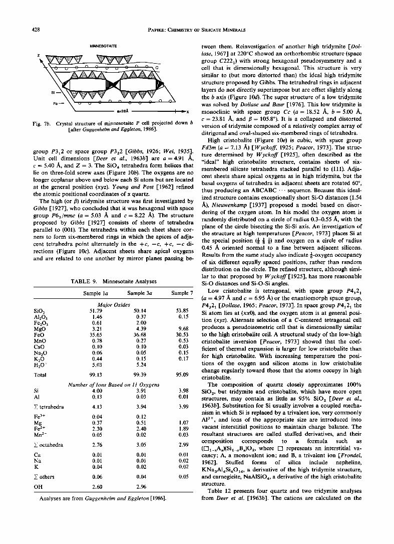
Magnesian minnesotaite has a dominant PrimItIve lattice, pi (a = 28.0 A (- 5 times ataIJ, b = 9.4 A, c = 12.4 A, IX = 101°, fJ = 127°, ')' = 90°, and Z = 1), with the ideal formula (Fe, Mg)30Si40096(OH)28' or, to compare with talc stoichiometry, (Fe, MghSi409.6(OHh.8' Ferroan minnesotaite has a dominantly C-centered lattice, cI (a = 50.6 A (-9 times a'olO>, b = 9.6 A, c = 12.4 A, IX = 101°, fJ = 127°, ')' = 90°, and Z = 2), with the ideal formula (Fe, Mg)z7Si36086(OH)z6' or, to compare with talc stoichiometry, Fe3Si409.ss(OHh.89' The definitive structural study of this complex silicate series is by Guggenheim and Eggleton [1986], and this summary derives largely from that paper. Although Gruner [1944] suggested that minnesotaite is the ferrous iron analogue of talc, we now know that is not the case. The basic structural problem in this silicate series is that although the silicon tetrahedral sheet fits comfortably on the magnesian trioctahedral sheet of talc, a ferrous iron trioctahedral layer is just too large for a silicon tetrahedral sheet. Therefore there has to be structural adjustment in the tetrahedral sheet to make it larger. Figure 7a illustrates the tetrahedral sheet for the primitive unit (P) cell. The tetrahedral strip width in the P cell is four tetrahedra wide along a. These strips are held together by continuous silicate chains running parallel to b. The chains are located in the interlayer region, i.e., the region occupied by potassium in muscovite and biotite. The tetrahedra located in the upper four-tetrahedra-wide strip link upward to an octahedral strip (not shown in Figure 7a), whereas the lower tetrahedral strip links downward to another octahedral strip (not shown in Figure 7a). Figure 7b shows the minnesotaite P cell projected
down b. The small amount of alkalis reported for minnesotaite (Table 9) could be located in the interlayer cavities. The Ccentered structure is similar to the P structure described , above, but it has three-tetrahedra-wide chains alternating with four-tetrahedra-wide chains along a instead of just the four· tetrahedra-wide chains that occur in the primitive structure. i
The different strip widths are thought to result from the differ- ' ent linkage requirements in Mg-minnesotaite versus Feminnesotaite. Ten tetrahedra (4 + 1 + 4 + 1) span nine octa· hedra parallel to a for the P cell which occurs in magnesian compositions. For the C cell with the larger, iron-containing octahedra, nine tetrahedra (4 + 1 + 3 + 1) span eight octa· , hedra.
As with greenalite, it is difficult to give criteria for good microprobe analyses because of the structural complexity in the minnesotaite series. However, analyses reduced on the basis of 11 oxygens (22 negative charges) should be similar to those listed in Table 9.
STILPNOMELANE
Stilpnomelane has a complex modulated structure crystallizing in space group pi (a ~ b = 21.72 A, c = 17.74 A, a = 124°, f3 = 96°, ')' = 120°, and Z = 1). Important papers contributing to our understanding of stilpnomelane include those by Eggleton [1972] and Eggleton and Chappell [1978], and much of this discussion derives from these two publications. Stilpnomelane is remarkable in that it can accommodate a wide range of FeZ + /Fe3+ ratios without major structural change. An ideal end-member ferrostilpnomelane can be represented by KsFe48z+(Si63AI9)OI6s(OH)48' 12HzO, and an ideal end-member ferristilpnomelane can be
represented by KsFe48 3+(Si63AI9)0216 . 36H zO. However, neither end-member composition has been reported. A convenient approximation to the structural formula using a one· eighth subcell is K O•6(Mg, Fe2+, Fe3+)6SisAl(O, OH)27' 2--4H20 . Ferristilpnomelane is thought to result from oxidation after primary crystallization. A convenient ternary dia-
MUSCOVITE
Fig. 3b. Crystal structure of muscovite [after Hurlbut and Klein, 1977]. Solid circles represent OH - groups.
PAPIKE' CHEMISTRY OF SILICATE MINERALS 423
MUSCOVITE 2M 1
M1
r'l
•x Fig. 3c. The crystal structure of muscovite showing the filled M2 sites (circles) and vacant M1 sites (squares) [after
Bailey, 1984].
MICA POLYTYPES
,• I00 ø a - -clO•
5.3•, ]
C2/m
IM 20 o
o•.--,c20A
ß __•, 5.3/• J .... 9.2•,
Ccm21
ST c 30•,
or PSa 12 . Fig. 3d. The six mica polytypes derived by assuming that only one interlayer stacking angle is present in each crystal
[from Papike and Cameron, 1976].
424 PAPIKE' CHEMISTRY OF SILICATE MINERALS
LiAIFe_ 2
eSiAl_ 2
EIAI2Fe_ 3
Li-Fe-AI Mica Volume
Subject to 0_•n•_2 4•_Si•_8 0•_Fe•_6 0•_AK_8.67
Ply 0_•Li_•4
Tri
(Li 2 FeAI2EI)
'Ke
Cid
Si----8
-.. ½/.,,. --•("e
Fig. 3e. Mica classification [after Cern)• and Burt, 1984]. The end- members are synthetic mica (Synth), siderophyllite (Sdp), ephesite (Kep), annite (Ann), protolithionite (Pro), zinnwaldite (Znw), trili- thionite (Tri), muscovite (Mus), phengite (Phn), celadonite (Cld), montdorite (Mtd), taeniolite (Fet), and polylithionite (Ply).
gram that compares greenalite, minnesotaite, and stilpnomel- ane compositions is illustrated in Figure 8a.
As mentioned above, an iron-bearing octahedral sheet has lateral dimensions too large to permit linkage of a continuous hexagonal sheet of silica tetrahedra [Eggleton, 1972-1. In stilp-
Si=const • Li313_2Ai_ 1
'• Li4[•_ 3 Si_ 1 -18i-1
AI3Li-18i-2 • ;• AI413-18i-3
polylithionite Si =8
• • •-x- • • Si=7 , ,
Muscovite •_'L _ • -; Si=6 • ...... • Trilithionite
• ILl3 AI3)VI(AI 2 Si
"•- '• -'•;" -4•*• si =5 ', •_
Na analog •-- - synth. by Francke (Li 2 AI 4 )VI(AI4 Si4 )Iv et al. (1982) )IV (AI4.67D1.33)VI(AI 4 Si4 I
I I I I I I
O=2 O=1 O=0
Fe-Free Micas
Fig. 3g. Mica classification l-after Cern)• and Burt, 1984].
nomelane (Figures 8b and 8c), linkage between the octahedral and the tetrahedral sheets is maintained over a distance of
seven linked tetrahedra, resulting in an island of 24 tetrahedra linked in a hexagonal array and articulating to a continuous
Trioctahedral Micas
/ Fe AI celadon,te Montdorite •,' '•-AI-celadonite' Si (Fe51::l)Vl (Si)8 Mg-analog
synth by S•ifert •/,/• and Schreyer (1971) //•/1' ;/'/•1 ./l /
..... IV• // O ) '(Fe S• / / Vl IV (Fes.5 0.5' 0.5 7.•/?7%•'-1--7 7•(FeA,.• ) (A,Si• .....
!... • II •','• (Fe.)VI(FeS'7)Iv •/L{' /J" '•-"1/
Annite •'?' ,' l?',' ,'1, Muscovite Si=6 Z Vl IV V• •V / /• / (AID ) (AI • ) (F%) (AI28 •) ••' / I/ ,'x /•/
•/•• / / • Si=5 (FesAi)V•(Ai38is)•v •-/--/•j • • -- • I .....
/
ß ß / / / / • / I Na analog S•deroDhylhte [5•/ / •/• / I -
(Fe4AI•VI(AI48i4)Iv -- et al. (1982) I J J (A•.67 D1.33)VI(AI4 Si4) Iv I I I I
I I
oVI:0 oVI:i oVI :2
'Fe-taeniolite' Polylithionite •i --8 (Li2Fe4)vl (Sis)lvj ' "•- •V•/ -IV '•-----" "•j • •/i(Li4AI2 ), (8i8•
•'//'i.r /, ./ / •0.. • //..• / /•:/
-' •,• ' / b/_ • • •' ./ •.-•'" •" • --•--•i"--•.•4--- • .....
,, ,., ,. ,, . ,- / _/.-1' /_. /..-r. /... • i
- ß //•'•/" ,L/.Zin•ite ," ' / .-" -. Annlte -•/ I 81--6 v, • .. •-'-•' - •'- •---:-•-'---,• •;i,•ioni• -----'- (Fe 6) (AI2Si6)iv T'P tro ol, ,' '•th nite' (Li,•Fe,,Al,,•l (LioAio)VllA•O.. dv
Vl / t 3 3 ß ß '2•'6 • [. ;,-•u- F- e•^•)v' -/•;; '7 / / •1 / ,• - [/.-,' •u, .-,"'I., X • - -
• ' P'- '•>,T'--T • :•? -7 ?>' - .... - I /_b-7,' .,'.','i I',,. I.?' / ,'. ,?- /i..t _. •.-
I" Y • •' / •: - i' -I t,,x.'< -
Siderophyllite •/"' ;? 'U''?' '_-•-K_;Phesite ß _ 8,=4 Vl iv Vl IV (Fe4AI 2 ) (AI48i4) (Li2AI4) (AI48i4)
I , Li=0 Li=l LiL2 Li=3 Li•4
Fig. 3f Mica classification [after Cern•;, and Burt, 1984]. Fig. 3h. Mica classification [after Cern)• and Butt, 1984].
PAPIKE: CHEMISTRY OF SILICATE MINERALS 425
Li 313_2 AI_ 1
e••½•2EI- 1Fe- 1 (SJ8)Iv •\% LiAIFe •,
Li =const 13
Tetrasilicic Micas
Li=4
Li=3
Li=2
Li=l
Li=O
Fig. 3i. Mica classification [after Cern)• and Butt, 1984].
octahedral sheet. The islands are linked by six-membered (trigonal) rings of tetrahedra whose apices point in the op- posite direction from those of island tetrahedra (tetrahedral inversion). The island tetrahedra and the trigonal rings form
PYROPHYLLITE
van der Waals' bond
Fig. 4. Crystal structure of pyrophyllite [after Hurlbut and Klein, 1977]. Note that this is only a schematic diagram. Solid circles repre- sent OH- groups.
TABLE 6. Pyrophyllite-Talc Analyses
Pyrophyllite Talc
Sample 27 Sample 47 Sample 1' (DHZl18-1) Sample 35 (DHZ123-6)
SiO 2 A1203 Fe203 TiO 2 MgO FeO
MnO
CaO
Na20 K20 H2 O+
Total
si A1
• tetrahedra
A1
Fe 3+ Ti
Mg Fe 2+ Mn 2+
Z octahedra
Ca Na
K
Major Oxides 66.04 63.57 63.22 60.02 28.15 29.25 1.88
0.64 0.10 0.33 0.04
0.04 0.37 30.45 30.39 0.12 0.89 1.51 0.00
0.01 0.38 1.00 0.04 0.02 0.02
0.05
5.27 5.66 4.78 5.37
100.19 99.51 99.74 100.17
Number of 1 OhS Based on 11 Oxygens 3.976 3.882 4.022 3.861 0.024 0.118 0.139
4.000 4.000 4.022 4.000
1.974 1.988 0.002 0.029 0.004 0.016
0.004 0.034 2.884 2.914 0.006 0.047 0.081
2.007 2.032 2.947 2.997
0.001 0.020 0.069 0.005 0.002
0.004
site 0.006 0.020 0.006 0.069
*From Lee and Guggenheim [1981]. ?From Deer et al. [1962]; for example, DHZl18-1 refers to page
118, column 1 of Deer et al. 5From Raynet and Brown [1973].
five-membered rings at their boundaries. Figure 8c illustrates several possible locations for K, Na, and Ca in the interlayers. However, the exact locations of these cations has not been determined.
Once again, the complexity of the stilpnomelane structure and chemistry makes it difficult to give criteria for good mi- croprobe analyses. Therefore it is suggested that microprobe analyses, after reduction based on 15 tetrahedral and octa-
hedral cations (Si, A1, Fe 3+, Mg, Fe 2+, Mn), be compared to the range of analyses presented in Table 10.
PREHNITE
Prehnite, Ca2 TM (A1, Fe3+)(Si3A1)O•o(OH)2, has a relatively complex silicate sheet structure [Peng et al., 1959; Preisinger, 1965; Papike and Zoltai, 1967]. Determination of the correct space group has been challenging. Gossnet and Mussgnug [1931] suggested that the space group is either P2cm or Pncm, and Nuffield [1943] reported that single-crystal X ray diffrac- tion photographs display systematically missing diffraction spots indicative of space group Pncm. However, as this space group is not consistent with the pyroelectric character of preh- nite, he concluded that the crystal he studied was twinned, with the untwinned material having space group P2cm. Peng et al. [1959] solved the crystal structure assuming space group
426 PAPIKE'. CHEMISTRY OF SILICATE MINERALS
CHLORITE GREENALITE
TALC-TYPE >
LAYER
?
BRUCITE-LIKE INTERLAYER
Fig. 5. Crystal structure of chlorite [after Hurlbut and Klein, 1977]. Solid circles represent OH- groups.
Pncm. However, Preisinger [1965] refined the structure of a prehnite in space group P2cm. Papike and Zoltai [1967] re- fined the "average structure" of prehnite in space group Pncm, with a = 4.65 A, b = 5.48 A, c = 18.49 A, and Z = 2. However, Papike and Zoltai claim that the Pncm structure represents an average structure composed of P2cm and P2/n domains. Figure 9a is a schematic drawing of the silicate layer showing three
Fig. 6. The modulated tetrahedral sheet of greenalite [after Guggen- heim et al., 1982].
levels of tetrahedra along the c axis that make up these rela- tively complex silicate sheets [Quint, 1987]. Quint [1987] com- pares the layer structure of a newly described mineral, amstallite, CaAl(OH)2(Alo.sSi3.2)O8OH2.[(H20)o.sClo.2], with that of prehnite. Liebau [1985] classifies such layers as open-branched "zweier" single-layer silicates. Moore [1986] compares the sili- cate linkage as portrayed in Figure 9b with a portion of the crystal structure of high quartz.
The structural study of Papike and Zoltai [1967] shows that the T1 tetrahedra (Figure 9b) are occupied by silicon only, whereas the T2 are occupied by 50% A1 and 50% Si. If Pncm were the correct space group, then each T2 tetrahedron (multi- plicity 4) would be statistically occupied by 0.5 A1, 0.5 Si. However, this is not thought to be the case. Papike and Zoltai
TABLE 7. Chlorite Analyses
Sample I Sample 2 Sample 3 Sample 4 Sample 5 Sample 6 Sample 7 Sample 8
Major Oxides SiO 2 27.30 30.60 33.78 25.12 28.15 30.76 22.30 26.56 A120 3 24.17 16.80 13.24 24.02 15.17 12.12 16.81 20.19 Fe20 3 1.87 2.18 1.50 4.86 3.85 9.12 15.13 1.27 TiO 2 0.24 MgO 29.24 32.18 34.41 12.19 14.56 12.36 1.30 2.68 FeO 5.15 5.02 3.07 23.26 25.23 22.76 32.78 36.51 MnO 0.16 0.18 0.21 1.24 2.04
H2 O+ 12.64 12.76 13.89 10.90 11.25 9.76 11.04 9.97
Total 100.37 99.54 100.05 100.53 98.42 98.12 99.36 99.46
Number of Ions Based on 18 (0, OH) Si 2.59 2.94 3.23 2.59 3.02 3.27 2.58 2.93 A1 1.41 1.06 0.77 1.41 0.98 0.73 1.42 1.07
E tetrahedra 4.00 4.00 4.00 4.00 4.00 4.00 4.00 4.00
AI 1.30 0.85 0.71 1.50 0.94 0.78 0.88 1.56 Fe 3+ 0.13 0.16 0.11 0.38 0.31 0.73 1.32 0.11 Ti 0.02
Mg 4.14 4.61 4.89 1.87 2.33 1.96 0.22 0.44 Fe 2+ 0.41 0.40 0.24 2.00 2.26 2.02 3.18 3.37 Mn 2+ 0.01 0.02 0.02 0.11 0.19
• octahedra 5.98 6.02 5.96 5.77 5.86 5.60 5.60 5.69
All analyses are from Bailey [1975].
PAPIKE' CHEMISTRY OF SILICATE MINERALS 427
TABLE 8. Greenalite Analyses
Sample 1 Sample 2 Sample 3 Sample 4 Sample 5 Sample 6 Sample 7
Major Oxides SiO2 34.7 36.5 35.9 34.4 35.9 36.5 36.3 A120 3 0.90 0.25 0.16 0.05 0.06 0.16 0.10 MgO 4.98 3.75 4.13 0.26 3.83 4.53 3.80 FeO* 47.3 40.2 47.8 55.5 50.0 46.7 50.4 MnO 0.15 8.71 1.78 0.18 0.06 1.50 0.06
Total 88.03 89.41 89.77 90.39 89.85 89.39 90.66
Si 2.O57 A1 0.063
Mg O.44O Fe 2.344 Mn 0.007
Number of Ions Based on Seven Oxygens 2.129 2.099 2.079 0.017 0.011 0.004 0.326 0.360 0.023 1.961 2.337 2.805 0.431 0.088 0.009
2.103 2.123 2.106 0.004 0.011 0.007 0.334 0.393 0.328 2.450 2.272 2.446 0.003 0.074 0.003
cations other than Si 2.854 2.735 2.796 2.841 2.791 2.750 2.784
All analyses are from Guggenheim et al. [1982, Table 2]. *All iron calculated as FeO.
[1967] suggest that the Pncm structure is an average of two types of domains with P2cm and P2/n symmetry, each with a different ordering scheme, as shown in Figure 9c.
Prehnite does not show much compositional variability (Table 11). The only appreciable substitution is of Fe for A1 in octahedral coordination. Table 11 presents prehnite analyses from Deer et al. [1962], and the formulae are calculated on the basis of 12 (O, OH). However, microprobe data, which do not yield H20 values, should be reduced assuming 11 oxygens (22 negative charges, equivalent to O•o(OH)2 ). Good microprobe analyses should have Ca plus Na approximately equal to two and Si, A1, Fe 3+, Ti, Mg, Fe 2+, Mn approximately equal to five.
QUARTZ-TRIDYMITE-CRISTOBALITE
Papike and Cameron [1976] reviewed aspects of the crystal chemistry of the silica polymorphs, quartz, tridymite, cristoba-
lite, coesite, and stishovite. Much of the discussion presented here for quartz, tridymite, and cristobalite is abstracted from that paper.
High (or fl) quartz is hexagonal, with space group P6222 or P6422 [Bragg and Gibbs, 1925]. Unit cell dimensions are a- 5.01 /•, c-- 5.47 /•, and Z = 3. Si occupies special posi- tions at heights 0, «, and •, and the oxygens lie in coplanar pairs c/6 above and below the Si atoms. The SiO,• tetrahedra are arranged in hexagonal and trigonal helices with axes parallel to the c axis (Figure 10a). Right-handed helices result in the structurally right-handed space group P6222, and left- handed helices in the structurally left-handed space group P6422. It is not possible to determine the structural hand of quartz by ordinary X ray techniques, but De Vries [1958] showed that it can be established by examination of anoma- lous scattering produced by Si irradiated by CrK•radiation.
Low (or 00 quartz is trigonal and occurs with either space
MINNESOTAITE
Fig. 7a. Crystal structure of minnesotaite P cell [after Guggenheim and Eggleton, 1986].
428 PAPIKE: CHEMISTRY OF SILICATE MINERALS
MINNESOTAITE
Z
a=28• I
Fig. 7b. Crystal structure of minnesotaite P cell projected down b [after Guggenheim and Eggleton, 1986].
group P3x2 or space group P322 [Gibbs, 1926; Wei, 1935]. Unit cell dimensions [Deer et al., 1963b] are a= 4.91 c - 5.40 II, and Z = 3. The SiO4 tetrahedra form helices that lie on three-fold screw axes (Figure 10b). The oxygens are no longer coplanar above and below each Si atom but are located at the general position (xyz). Young and Post [1962] refined the atomic positional coordinates of cz quartz.
The high (or fl) tridymite structure was first investigated by Gibbs [1927], who concluded that it was hexagonal with space group P63/mmc (a = 5.03 3, and c = 8.22 A). The structure proposed by Gibbs [1927] consists of sheets of tetrahedra parallel to (001). The tetrahedra within each sheet share cor- ners to form six-membered rings in which the apices of adja- cent tetrahedra point alternately in the +c, -c, +c, -c di- rections (Figure 10½). Adjacent sheets share apical oxygens and are related to one another by mirror planes passing be-
TABLE 9. Minnesotaite Analyses
Sample l a Sample 3a Sample 7
Major Oxides SiO2 51.79 50.14 53.85 A1203 1.46 0.37 0.15 Fe20 3 0.61 2.00 MgO 3.21 4.39 9.68 FeO 35.65 36.68 30.53 MnO 0.78 0.27 0.53 CaO 0.10 0.10 0.03
Na20 0.06 0.05 0.15 K20 0.44 0.15 0.17 H2 O+ 5.03 5.24
Total 99.13 99.39 95.09
Number of Ions Based on 11 Oxygens Si 4.00 3.91 3.98 AI 0.13 0.03 0.01
Y• tetrahedra 4.13 3.94 3.99
Fe 3+ 0.04 0.12 Mg 0.37 0.51 1.07 Fe 2+ 2.30 2.40 1.89 Mn 2+ 0.05 0.02 0.03
Y, octahedra 2.76 3.05 2.99
Ca 0.01 0.01 0.01 Na 0.01 0.01 0.02 K 0.04 0.02 0.02
others 0.06 0.04 0.05
OH 2.60 2.96
Analyses are from Guggenheim and Eggleton [1986].
tween them. Reinvestigation of another high tridymite [Dol- lase, 1967] at 220øC showed an orthorhombic structure (space group C222•) with strong hexagonal pseudosymmetry and a cell that is dimensionally hexagonal. This structure is very similar to (but more distorted than) the ideal high tridymite structure proposed by Gibbs. The tetrahedral rings in adjacent layers do not directly superimpose but are offset slightly along the b axis (Figure 10d). The super structure of a low tridymite was solved by Dollase and Baur [1976]. This low tridymite is monoclinic with space group Cc (a = 18.52 •, b = 5.00 •, c = 23.81 ?L and fl = 105.8ø). It is a collapsed and distorted version of tridymite composed of a relatively complex array of ditrigonal and oval-shaped six-membered rings of tetrahedra.
High cristobalite (Figure 10e) is cubic, with space group Fd3m (a = 7.13 /•) [Wyckoff, 1925; Peacor, 1973]. The struc- ture determined by Wyckoff [1925], often described as the "ideal" high cristobalite structure, contains sheets of six- membered silicate tetrahedra stacked parallel to (111). Adja- cent sheets share apical oxygens as in high tridymite, but the basal oxygens of tetrahedra in adjacent sheets are rotated 60 ø, thus producing an ABCABC ß ß ß sequence. Because this ideal- ized structure contains exceptionally short Si-O distances (1.54 •), Nieuwenkamp [1937] proposed a model based on disor- dering of the oxygen atom. In his model the oxygen atom is randomly distributed on a circle of radius 0.3-0.55/•, with the plane of the circle bisecting the Si-Si axis. An investigation of the structure at high temperatures [Peacot, 1973] places Si at the special position (• • •) and oxygen on a circle of radius 0.45 /• oriented normal to a line between adjacent silicons. Results from the same study also indicate •-oxygen occupancy of six different equally spaced positions, rather than random distribution on the circle. The refined structure, although simi- lar to that proposed by Wyckoff[1925], has more reasonable Si-O distances and Si-O-Si angles.
Low cristobalite is tetragonal, with space group P4•2• (a = 4.97/• and c = 6.95 ,•t) or the enantiomorph space group, P432 • [Do!!ase, 1965; Peacor, 1973]. In space group P4•2• the Si atom lies at (xxO), and the oxygen atom is at general posi- tion (xyz). Alternate selection of a C-centered tetragonal cell produces a pseudoisometric cell that is dimensionally similar to the high cristobalite cell. A structural study of the low-high cristobalite inversion [Peacor, 1973] showed that the coef- ficient of thermal expansion is larger for low cristobalite than for high cristobalite. With increasing temperature the posi- tions of the oxygen and silicon atoms in low cristobalite change regularly toward those that the atoms occupy in high cristobalite.
The composition of quartz closely approximates 100% SiO 2, but tridymite and cristobalite, which have more open structures, may contain as little as 95% SiO2 [Deer et al., 1963b]. Substitution for Si usually involves a coupled mecha- nism in which Si is replaced by a trivalent ion, very commonly A13+, and ions of the appropriate size are introduced into vacant interstitial positions to maintain charge balance. The resultant structures are called stuffed derivatives, and their composition corresponds to a formula such as ([Z]l_xAx)(Si•_xBx)O 2, where [Z] represents an interstitial va- cancy; A, a monovalent ion; and B, a trivalent ion [Frondel, 1962]. Stuffed forms of silica include nepheline, KNa3AI,,Si,[O•6, a derivative of the high tridymite structure, and carnegieite, NaAISiO4, a derivative of the high cristobalite structure.
Table 12 presents four quartz and two tridymite analyses from Deer et al. [1963b]. The cations are calculated on the
PAPIKE: CHEMISTRY OF SILICATE MINERALS 429
TALC-MINNESOTAITE SERIES
ß Ardua Lake area (Lesher, 1978) f=l Howells River area (Klein, 1974) ß Gunflint Iron Formation (Floran & Papike, 1975) O Dales Gorge Member, Hamersley Range (Miyano, 1978a)
STILPNOMELANE
& Ardua Lake area (Leaher, 1978) A Howells River area (Klein, 1974 and Klein & Fink, 1976) ß Gunflint Iron Formation (Floran & Papike, 1975)
GREENALITE
compositional field (Klein, 1974 and Floran
& Papike, 1975)
Homestake iron formation, So. Dak. (Kath & Papike, in preparation)
60 60
ß //-\x \•7 0 stdpnomelane // A \ 0 •.
• * '• •80
t zx --A• ,/xßwx • •90 ............ • ß - V) '•greenalite
alc-minneso•ite series "•--•---•• field Mg lO --2o- ao -4o 5o so - 7o so / oo Fe+Mn minnesotaite
Fig. 8a. Compositional ranges of stilpnomelane, talc-minnesotaite series, and greenalite [after Klein, 1983].
basis of two oxygens, which is also the way microprobe analy- ses should be reduced. Good quartz analyses will have Si approximately equal to one. However, tridymite analyses may show A1, Fe 3+, and Ti '•+ in tetrahedral coordination. These cation substitutions result in charge deficiencies at the tetra- hedral sites, e.g., one charge deficiency for each •VA1 and IVFe3 +. Thus the following charge balance equation should be obeyed:
IVA1 + IVFe3+ = 2(Mg, Fe 2+, Mn 2+, Ca) + Na + K site charge deficiency site charge excess
STILPNOMELANE
FELDSPAR
This discussion of feldspar crystal chemistry draws heavily on a paper by Papike and Cameron [-1976•. For a more com- plete and in-depth review the reader is referred to Ribbe •19833.
The compositions of most natural feldspars can be repre- sented in the ternary system KA1Si308 (Or)-NaA1Si3Os(Ab)- CaA12Si2Os(An ). Members of the series between KA1Si308 and NaA1Si308 are called alkali feldspars, and those between NaA1Si30 8 and CaAlISi20 8 are plagioclase feldspars. Each series is further subdivided into smaller compositional ranges; however, the names of the subdivisions are now used less commonly than the specific feldspar components An-Ab-Or.
Alkali feldspars generally contain less than 10 mol % An; likewise, plagioclases generally contain less than 10 mol % Or [Deer et al., 1963b-I. Ions that substitute in limited amounts for the major cations include Fe 3+ and Ti '•+ for IVA1; Rb and Cs for K, Na; Mg, Fe 2+, Sr, and Mn for Ca; and Ba for K. The less common barium feldspars form an isomorphous series between KA1Si30 s and BaAI•.Si•.O s (celsian); intermedi- ate members of the series are called hyalophane.
Fig. 8b. The modulated tetrahedral sheet of stilpnomelane [after Eggleton, 1972].
STILPNOMELANE
I
Fig. 8c. Crystal structure ofstilpnomelane [after Eggleton, 1972].
430 PAPIKE' CHEMISTRY OF SILICATE MINERALS
TABLE 10. Stilpnomelane Analyses
Sample N Sample P Sample J Sample F Sample V Sample A
Major Oxides SiO2 44.74 46.37 48.21 45.99 44.88 42.69 AI20 3 6.10 6.30 4.35 4.98 6.69 5.58 F%O3 2.92 5.38 13.03 20.98 25.46 33.70 MgO 5.00 8.07 9.11 3.42 7.16 3.89 FeO 24.52 20.77 14.00 12.85 2.73 0.30 MnO 4.40 0.66 0.27 0.59 0.53 2.43 CaO 0.00 0.28 0.28 0.12 0.79 0.13 Na20 0.30 0.13 0.06 0.42 0.12 0.07 K20 3.42 2.58 1.99 1.40 1.54 1.81 H2 O+ 8.87 8.46 7.32 8.80 7.83 Total 91.40 99.41 99.76 98.07 98.7 98.43
Cations Based on (Si, A!, Fe 3+, Mg, Fe 2+, Mn 2+) = 15 Si 7.82 7.92 8.17 8.22 7.90 AI 1.18 1.08 0.83 0.78 1.10
• tetrahedra 9.00 9.00 9.00 9.00 9.00
7.74 i .20
8.94
AI 0.08 0.19 0.03 0.27 0.29 Fe 3+ 0.39 0.69 1.65 2.82 3.37 4.60 Mg 1.30 2.06 2.29 0.91 1.88 1.04 Fe 2+ 3.58 2.96 1.98 1.92 0.40 0.05 Mn 0.65 0.11 0.04 0.09 0.08 0.37
x' octahedra 6.00 6.01 5.99 6.01 6.02 6.06
Ca 0.00 0.05 0.05 0.03 0.15 0.03 Na 0.05 0.04 0.03 0.15 0.04 0.03 K 0.76 0.57 0.43 0.32 0.34 0.42
others 0.81 0.66 0.51 0.50 0.53
All analyses are from Eggleton and Chappell [1978].
0.48
Adequate characterization of a feldspar requires knowledge of both the chemical composition and the structural state (dis- tribution of A1 and Si over distinct tetrahedral sites). Structur- al state depends on temperature of crystallization and on sub- sequent thermal history. In general, feldspars that crystallized at low temperatures or cooled slowly from high temperatures have low structural states (highly ordered Si-A1 distributions).
PREHNITE TETRAHEDRAL SHEET
b
Fig. 9a. Idealized tetrahedral sheet of prehnite projected down c [after Quint, 1987].
Those that cooled rapidly following crystallization at high temperatures have high structural states (disordered A1-Si dis- tributions). A continuum of structural states exists, each state differing principally in the distribution of Si and A1 over the independent tetrahedral sites in the crystal structures.
The two limiting structural states of potassium feldspar, KA1Si3Os, are (1) high-temperature sanidine, which is mono- clinic and has a completely disordered Si-A1 distribution, and (2) low-temperature maximum microcline, which is triclinic and has an ordered Si-A1 distribution. The monoclinic triclinic
transition takes place between 300 ø and 500øC, but the transi- tion temperature as determined in different studies varies con- siderably.
Adularia is a potassium feldspar that is distinctive because of its morphology and restricted paragenesis (e.g., pegmatites). Both triclinic and monoclinic varieties occur naturally, and
optical and structural parameters may vary in different parts of a single grain.
The structures of the KA1Si308 polymorphs can be de- scribed as variants of the high-temperature sanidine structure. Table 13a summarizes unit cell data for alkali feldspars. Sani- dine has space group symmetry C2/m, and each unit cell con- tains four KA1Si30 8 formula units. The 16 (Si, A1) atoms in the unit cell are distributed randomly over two crystallogra- phically distinct tetrahedral sites, T1 and T2, and the four potassium atoms occupy special positions on mirror planes perpendicular to b (Figure 11). The 32 oxygen atoms occur at special positions (four on two-fold axes and four on mirror planes) and at general positions. Potassium atoms occupy large irregularly shaped cavities within this tetrahedral frame- work. In sanidine each K is coordinated by six oxygens within 2.953 ]t and by an additional four within 3.14 ]t.

PAPIKE: CHEMISTRY OF SILICATE MINERALS 431
PREHNITE AI ORDERING in PREHNITE
c/2
CCa .50
¸ ¸
o o .50 .50
OCa .50
Fig. 9b. Average crystal structure of prehnite in space group Pncm. A1 is confined to T2 tetrahedron (hatched and stippled) [after Papike and Zoltai, 1967].
The structures of the lower-temperature polymorphs differ from the structure of sanidine principally in the degree of Si-A1 ordering. Concentration of A1 at a particular tetrahedral site results in the loss of both a twofold axis of rotation and a mirror plane, with a concomitant transition from monoclinic
to triclinic symmetry. The two tetrahedral sites of the mono- Si clinic phase become four symmetrically distinct tetrahedral AI sites (Tlo, Tlm, T2o, and T2m) in the triclinic phase. In mon- oclinic sanidine the 16 tetrahedral cations (4A1 + 12Si) are distributed randomly over eight T1 and eight T2 sites. In a A1 monoclinic orthoclase, A1 is partially ordered at the T1 site. Fe3+ Triclinic maximum microclinic has A1 strongly ordered at the Ti
Mg T 1 o site. Fe 2 +
NaA1Si30 8 has two extreme polymorphic modifications Mn that occur in nature, high and low albite. Low albite, which is structurally analogous to microcline, is triclinic and has a highly ordered Si-A1 distribution. High albite is triclinic to Ca very high temperatures and thus is not structurally analogous Na to sanidine. Its Si-A1 distribution is also highly disordered. Monoclinic albite, termed monalbite, is stable above • 1000øC. OH
The calcium end-member, anorthite, is primitive (space group Pi) at room temperature, with a -- 8.18 ]i, b -- 12.88 ]i,
AI
AI AI
AI
I a t P2cm P2/n
Fig. 9c. Possible ordering schemes for tetrahedral AI in space groups P2cm and P2/n. A1 tetrahedra are indicated by both hatching and stippling [after Papike and Zoltai, 1967].
TABLE 11. Prehnite Analyses
Sample 1 Sample 2 Sample 3 (DHZ264-1) (DHZ264-3) (DHZ264-6)
Major Oxides SiO2 42.76 42.86 41.67 A1203 24.83 24.41 24.44 Fe203 0.13 0.52 1.03 TiO2 0.01 0.12 MgO 0.07 0.03 0.25 FeO 1.12 0.28 0.32 MnO 0.05 0.06 CaO 26.84 26.89 27.25 Na20 0.03 0.32 0.18 H2 O+ 4.24 4.45 4.44
Total 100.07 99.83 99.70
Number of Ions Based on 12 (0, OH) 2.954 2.965 2.893 1.046 1.035 1.107
• tetrahedra 4.000 4.000 4.000
0.976 0.950 0.893 0.006 0.025 0.053
0.000 0.006 0.007 0.002 0.026 0.065 0.016 0.019 0.003 0.004
• octahedra 1.057
1.986
0.004
0.997 0.997
1.986 2.028 0.040 0.025
2.026 2.053
2.048 2.057
others 1.990
1.954
Analyses are from Deer et al. [1962]; for example, DHZ264-1 refers to page 264, column 1 of Deer et al.
432 PAPIKE' CHEMISTRY OF SILICATE MINERALS
HIGH QUARTZ TRIDYMITE SHEET
Fig. 10c. Portion of idealized tetrahedral sheet in high tridymite projected down c [from Papike and Cameron, 1976].
Fig. 10a. Crystal structure of high quartz projected down c [from Papike and Cameron, 1976].
c = 14.17/•, y = 93.2 ø , fi = 115.8 ø , V = 91.2ø, and Z = 8 [Deer et al., 1963b]. Si and A1 tetrahedra alternate throughout the structure, and this causes a doubling of the c axis. This order- ing is nearly perfect and is believed to persist up to melting temperatures. Intermediate plagioclase compositions have complex modulated structures with variable A1-Si ordering patterns and Ca-Na occupancies.
Table 13b gives some representative feldspar analyses from Deer et al. [1963b-]. Microprobe analyses should be normal- ized to eight oxygens. Analyses so reduced will have Na, K,
Ca, Ba, Fe 2+, Mg approximately equal to one and A1, Si, Fe 3 +, Ti approximately equal to four.
NEPHELINE-KALSILITE
The discussion of nepheline presented here is condensed from a review by Papike and Cameron [1976•; however, a new expanded discussion of kalsilite has been added. Natural nephelines, Na3KAI4Si40•6, are intermediate members of a solid solution series that extends between the pure sodium end-member NaAISiO4 and KA1SiO½ (kalsilite). Experimental work on this system by Tuttle and Smith [-1958• showed that pure sodium nepheline transforms at -•900øC to a high- temperature modification that inverts at 1245øC to the poly- morph carnegieite, whose structure is similar to that of cris- tobalite. Nepheline and kalsilite are not isomorphous, but the study by Tuttle and Smith •1958• showed that the substitution
LOW QUARTZ TRIDYMITE
o b
Fig. 10b. Crystal structure of low quartz projected down c [from Fig. 10d. Projection of the high tridymite structure down c [from Papike and Cameron, 1976]. Papike and Cameron, 1976].
PAPIKE: CHEMISTRY OF SILICATE MINERALS 433
CRISTOBALITE TABLE 13a. Cell Parameters of Alkali Feldspars
Fig. 10e. Projection of a portion of the high cristobalite structure onto (111) [from Papike and Cameron, 1976].
of K for Na in the nepheline structure increases from ,-, 25 mol % at 24øC to ,-,70 mol % at 1070øC, whereas the substitution of Na for K in the kalsilite structure increases from zero to 25
mol % over the same temperature interval.
a, /3, % a, A b, • c, A deg deg deg
Sanidine 8.56 13.03 7.17 90 116 90 Orthoclase 8.56 13.00 7.19 90 116 90 Intermediate 8.58 12.96 7.21 90.3 116 89.1
microcline Maximum 8.57 12.98 7.22 90.7 116 87.5
microcline
High albite 8.15 12.88 7.11 93.4 116.3 90.3 Low albite 8.14 12.79 7.16 94.3 116.6 87.6
From Deer et al. [1963b].
The nepheline structure can be described as a stuffed deriva- tive of high tridymite, in which A1 replaces approximately one half of the Si, and atoms such as K, Na, and Ca occupy interstitial voids to maintain charge balance. Buer•Ier et al. [1954] originally determined the structure, and Hahn and Buerger [1955] further refined it. Nepheline is hexagonal and has space group symmetry P63 (a--10.0 /•, c--8.4 /•, and Z = 2 [Deer et al., 1963b]), although some specimens possess high pseudosymmetry close to P63/m [Dollase, 1970; Foreman and Peacor, 1970]. There are four nonequivalent tetrahedral sites per unit cell: T1 and T2 occupy special positions on three-fold axes, and T3 and T4 occupy general positions. The T2-T3 and T1-T4 tetrahedra point in opposite directions along the c axis (Figure 12a). Within this tetrahedral frame- work are two distinct types of interstitial sites. One fourth of the sites are nearly hexagonal in geometry and are occupied
TABLE 12. Quartz-Tridymite Analyses
Quartz Tridymite
Sample 1 Sample 2 Sample 3 Sample 4 Sample 5 Sample 6 (DHZ192-1) (DHZ192-2) (DHZ192-3) (DHZ192-4) (DHZ192-5) (DHZ192-6)
Major Oxides SiO2 99.79 99.78 99.53 99.00 95.1 95.1 A120 3 0.042 0.02 2.4 2.70 Fe203 0.007 0.07 0.05 0.36 0.25 TiO2 0.048 0.015 0.26 0.28 MgO 0.008 0.09 0.3 0.03 FeO 0.04 0.05 0.36 MnO 0.009 0.02 0.003 0.00 CaO 0.010 0.40 0.2
Na20 0.80 0.67 m20 0.37 0.75
Total 99.91 100.0 99.65 99.38 99.99 99.98
Number of Ions Based on Two Oxygens Si 0.999 0.999 0.999 0.998 0.967 0.965 AI 0.001 0.000 0.029 0.032 Fe 3+ 0.000 0.001 0.000 0.003 0.002 Ti 4 + 0.000 0.000 0.002 0.002
Z tetrahedra 1.000 1.000 0.999 0.998 1.001 1.001
Mg 0.000 0.001 0.005 0.001 Fe2+ 0.000 0.000 0.003 Mn 2 + 0.000 0.000 0.000 0.000 Ca 0.000 0.004 0.002 Na 0.016 0.013
K 0.005 0.010
Z others 0.000 0.001 0.000 0.003 0.030 0.026
All analyses are from Deer et al. [1963b]; for example, DHZ192-1 refers to page 192, column 1 of Deer et al.
434 PAPIKE.' CHEMISTRY OF SILICATE MINERALS
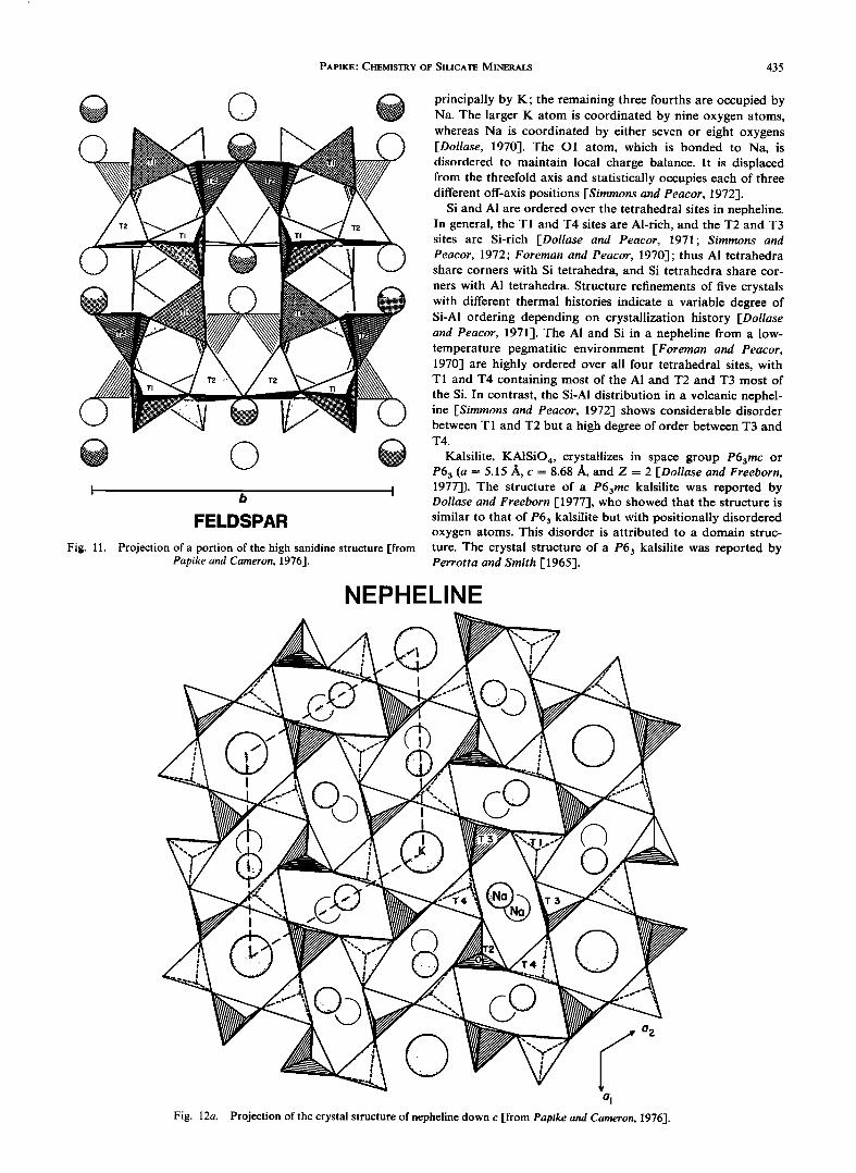
PAPIKE.' CHEMISTRY OF SILICATE MINERALS 435
e Q
e
I b I FELDSPAR
Fig. 11. Projection of a portion of the high sanidine structure [from Papike and Cameron, 1976].
principally by K; the remaining three fourths are occupied by Na. The larger K atom is coordinated by nine oxygen atoms, whereas Na is coordinated by either seven or eight oxygens [Dollase, 1970]. The O1 atom, which is bonded to Na, is disordered to maintain local charge balance. It is displaced from the threefold axis and statistically occupies each of three different off-axis positions [Simmons and Peacor, 1972].
Si and A1 are ordered over the tetrahedral sites in nepheline. In general, the T1 and T4 sites are Al-rich, and the T2 and T3 sites are Si-rich [Dollase and Peacor, 1971; Simmons and Peacor, 1972; Foreman and Peacor, 1970]; thus A1 tetrahedra share corners with Si tetrahedra, and Si tetrahedra share cor- ners with A1 tetrahedra. Structure refinements of five crystals with different thermal histories indicate a variable degree of Si-A1 ordering depending on crystallization history [Dollase and Peacor, 1971]. The A1 and Si in a nepheline from a low- temperature pegmatitic environment [Foreman and Peacor, 1970] are highly ordered over all four tetrahedral sites, with T1 and T4 containing most of the A1 and T2 and T3 most of the Si. In contrast, the Si-A1 distribution in a volcanic nephel- ine [Simmons and Peacor, 1972] shows considerable disorder between T1 and T2 but a high degree of order between T3 and T4.
Kalsilite, KA1SiO4, crystallizes in space group P63mc or P63 (a = 5.15 A, c -- 8.68 •, and Z = 2 [Dollase and Freeborn, 1977]). The structure of a P63mc kalsilite was reported by Dollase and Freeborn [1977], who showed that the structure is similar to that of P63 kalsilite but with positionally disordered oxygen atoms. This disorder is attributed to a domain struc- ture. The crystal structure of a P63 kalsilite was reported by Perrotta and Smith [1965].
NEPHELINE
o 2
Fig. 12a. Projection of the crystal structure of nepheline down c [from Papike and Cameron, 1976].
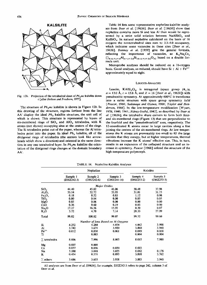
436 PAPIKE' CHEMISTRY OF SILICATE MINERALS
KALSILITE
Fig. 12b. Projection of the tetrahedral sheet of P63mc kalsilite down c [after Dollase and Freeborn, 1977].
The structure of P63mc kalsilite is shown in Figure 12b. In this drawing of the structure, regions farthest from the line AA' display the ideal P63 kalsilite structure, the unit cell of which is shown. This structure is represented by layers of six-membered rings of SiO,• and A10,• tetrahedra, with K atoms (not shown) occupying sites at the centers of the rings. The Si tetrahedra point out of the paper, whereas the A1 tetra- hedra point into the paper. In ideal P63 kalsilite, all of the ditrigonal rings of tetrahedra (the centers look like arrow- heads which show a direction) and oriented in the same direc- tion in any one tetrahedral layer. In P63mc kalsilite the orien- tation of the ditrigonal rings changes at the domain boundary AA'.
Table 14 lists some representative nepheline-kalsilite analy- ses from Deer et al. [1963b]. Deer et al. [1963b] show that nepheline contains more Si and less A1 than would be repre- sented by a strict solid solution between NaA1SiO,• and KalSiO 4. In natural nepheline calculated on the basis of 16 oxygens the nontetrahedral sites sum to 3.3-4.0 occupancy, which indicates some vacancies in these sites [Deer et al., 1963b]. Donnay et al. [1959] give the general formula,
reflecting the importance of vacancies, as KxNayCa z DS_(x+y+z)Al(x+y+2z)Si•6_(x+y+2z)032 based on a double for- mula unit.
Microprobe analyses should be reduced on a 16-oxygen basis. Good analyses, so reduced, should have Si + A1 + Fe 3 + approximately equal to eight.
LEUCITE-ANALCITE
Leucite, KA1Si20 6, is tetragonal (space group I4x/a, a = 13.0 ]i, c = 13.8 ]i, and Z = 16 [Deer et al., 1963b]) with pseudocubic symmetry. At approximately 600øC it transforms into a cubic structure with space group symmetry la3d [Peacor, 1968; Sadanaga and Ozawa, 1968; Taylor and Hen- derson, 1968]. In the low-temperature modification [Wyart, 1938, 1940, 1941; Ndray-Szab6, 1942], as described by Deer et al. 1-1963b], the tetrahedra share corners to form both four- and six-membered rings (Figure 13) that are perpendicular to the fourfold and the "pseudothreefold" axes, respectively. The 12-coordinated K atoms occur in large cavities along a line joining the centers of the six-membered rings. At low temper- atures the K atoms are presumably too small to fill the large cavities that they occupy, but at higher temperatures, thermal vibrations increase the K atoms' effective size. This, in turn,
results in an expansion of the collapsed structure and an in- crease in symmetry. Peacor [1968] refined the structure of the high-temperature polymorph.
TABLE 14. Nepheline-Kalsilite Analyses
Nepheline Kalsilite
Sample 1 Sample 2 Sample 3 Sample 4 Sample 5 (DHZ242-3) (DHZ242-6) (DHZ244-14) (DHZ253-2) (DHZ253-3)
Major Oxides SiO2 44.40 45.02 41.06 38.48 37.98 A1203 33.14 32.72 33.93 31.01 31.73 Fe20 3 0.18 0.52 0.83 1.12 0.98 TiO2 0.00 0.04 0.00 0.05 0.05 MgO 0.05 0.06 0.00 0.00 0.00 CaO 0.36 0.60 0.19 0.03 0.00
Na20 17.17 16.36 15.92 0.30 0.87 K20 3.72 4.70 7.14 28.33 27.99
Total 99.02 100.02 99.07 99.32 99.60
Number of Ions Based on 16 Oxygens Si 4.252 4.284 4.024 4.062 3.998 A1 3.742 3.671 3.920 3.860 3.940 Fe 3+ 0.012 0.038 0.061 0.089 0.038 Ti 0.003 0.004 0.004
• tetrahedra 8.006 7.996 8.005 8.015 7.980
Mg 0.007 0.008 Ca 0.037 0.056 0.020 0.003 Na 3.188 3.018 3.025 0.062 K 0.454 0.571 0.893 3.818
0.178
3.762
others 3.686 3.653 3.938 3.883 3.940
All analyses are from Deer et al. [1963b]' for example, DHZ242-3 refers to page 242, column 3 of Deer et al.
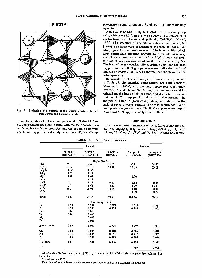
PAPIKE' CHEMISTRY OF SILICATE MINERALS 437
LEUCITE
Fig. 13. Projection of a portion of the leucite structure down c [from Papike and Cameron, 1976].
Selected analyses for leucite are presented in Table 15. Leu- cite compositions are close to ideal, with the main substitution involving Na for K. Microprobe analyses should be normal- ized to six oxygens. Good analyses will have K, Na, Ca ap-
proximately equal to one and Si, A1, Fe 3+, Ti approximately equal to three.
Analcite, NaA1Si20 6 ß H20 , crystallizes in space group Ia3d, with a = 13.7 A and Z = 16 [Deer et al., 1963b]; it is isostructural with leucite and poilucite, CsA1Si20 6 [Cerni•, 1974]. The structure of analcite was determined by Taylor [1930]. The framework of analcite is the same as that of leu- cite (Figure 13) and contains a set of 16 large cavities which form continuous channels parallel to three-fold symmetry axes. These channels are occupied by H•.O groups. Adjacent to these 16 large cavities are 24 smaller ones occupied by Na. The Na cations are octahedrally coordinated by four coplanar oxygens and two H•.O groups. A neutron diffraction study of analcite [Ferraris et al., 1972] confirms that the structure has cubic symmetry.
Representative chemical analyses of analcite are presented in Table 15. The chemical compositions are quite constant [Deer et al., 1963b-I, with the only appreciable substitution involving K and Ca for Na. Microprobe analyses should be reduced on the basis of six oxygens, and it is safe to assume that one H•.O group per formula unit is also present. The analyses of Table 15 [Deer et al., 1963b-I are reduced on the basis of seven oxygens because H20 was determined. Good microprobe analyses will have Na, K, Ca approximately equal to one and A1, Si approximately equal to three.
SODALITE GROUP
The most important members of the sodalite group are sod- lite, Nas['A16Si6024]C12- nosean, Nas['A16Si•O24]SO ½. and hatiyne, (Na, Ca)½_8(Al•Si•O2½)(SO½, S)•_2. Names and formu-
TABLE 15. Leucite-Analcite Analyses
Leucite Analcite
Sample 1 Sample 2 Sample 3 Sample 4 Sample 5 (DHZ280-4) (DHZ280-5) (DHZ280-7) (DHZ343-3) (DHZ343-6)
Major Oxides SiO2 55.4 54.66 56.39 55.11 54.10 A1203 23.3 23.15 23.10 22.86 23.68 Fe20 3 0.5* 0.36 TiO2 0.2 0.17 MgO 0.0 0.04 0.00 FeO 0.11 CaO 1.1 0.11 0.27 0.12 0.45 Na20 1.8 0.63 2.17 13.79 13.40 K20 18.3 20.04 18.05 0.18 0.34 H20 8.20 8.22
Total 100.6 99.27 99.98 100.26 100.19
Si
A1
Fe 3+ Ti
Mg Fe 2+
1.99
0.98
0.01
0.01
Number of Ions P 1.992 2.019 0.995 0.975 0.010
0.005
0.002
0.003
v tetrahedra 2.99 3.007
Ca 0.04 0.004 Na 0.13 0.045 K 0.84 0.932
others 1.01 0.981
H +
2.013 1.981 0.984 1.022
2.994 2.997 3.003
0.010 0.005 0.018 0.151 0.977 0.951 0.825 0.008 0.016
0.986 0.990 0.985
1.999 2.008
All analyses are from Deer et al. [1963b]; for example, DHZ280-4 refers to page 280, column 4 of Deer et al.
*Total iron as Fe 3+
?Number of ions is based on six oxygens for leucite and seven oxygens for analcite.
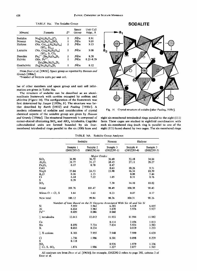
438 PAPIKE' CHEMISTRY OF SILICATE MINERALS
TABLE 16a. The Sodalite Group
Space Unit Cell Mineral Formula Z* Group Edge, ,i•
Sodalite Na8[h16Si6024]Cl 2 1 P,•3n 8.91 Nosean Na8[h16Si6024]SO 4 1 P,•3n 9.05 Ha•iyne (Na, Ca)4_8[h16Si6024 ] 1 P43n 9.13
(SO 4, S)1_2 Lazurite (Na, Ca)8[AlbSi6024 ] 1 P43n 9.08
(SO4, S, C1)2 Danalite Fe82+ [Be6Si6024]S 2 1 P43n 8.20 Helvite (Mn, Fe 2+ , 1 P43n 8.21-8.29
Zn)8[BebSi6024]S2 Genthelvite Zns[BebSi6024]S 2 1 P43n 8.12
From Deer et al. [ 1963b]. Space group as reported by Hassan and Grundy [1984a].
*Number of formula units per unit cell.
lae of other members and space group and unit cell infor- mation are given in Table 16a.
The structure of sodalite can be described as an alumi-
nosilicate framework with cavities occupied by sodium and chlorine (Figure 14). The configuration of the framework was first determined by Jaeger [1929a, b]. The structure was fur- ther described by Barth [1932] and Pauling [1930c]. A modern refinement of sodalite and consideration of crystal chemical aspects of the sodalite group are given by Hassan and Grundy [1984a]. The structural framework is composed of corner-shared alternating SiO,• and A10,• tetrahedra. Cagelike cuboctahedral units are formed bounded by six four- membered tetrahedral rings parallel to the six (100) faces and
a 2
SODALITE
Fig. 14. Crystal structure of sodalite [after Pauling, 1930c].
eight six-membered tetrahedral rings parallel to the eight (111) faces. These cages are stacked in eightfold coordination with each six-membered ring (each ring is parallel to one of the eight (111) faces) shared by two cages. The six-membered rings
TABLE 16b. Sodalite Group Analyses
Sodalite Nosean Ha•iyne
Sample 1 Sample 2 Sample 3 Sample 4 Sample 5 (DHZ292-2) (DHZ292-6) (DHZ295-1) (DHZ295-4) (DHZ295-5)
SiO 2 A1203 Fe203 CaO
Na20 K20 C1 S
SO3
Total
Major Oxides 36.99 36.72 36.69 31.77 31.17 28.45
0.17 0.70 0.47
0.63
25.84 24.53 23.90 0.16 1.13
6.44 7.22 1.05
0.39
101.76 101.47
7.30
98.49
32.18
27.11
10.26
16.34
0.08 0.31
14.10
100.38
Minus O = CI, S 1.64 1.63 0.23 0.07
New total 100.12 99.84 98.26 100.31
Si A1
Fe 3+
Number of lons Based on the 21 Oxygens Associated With Six Al and Six Si 5.959 5.961 6.203 6.018 6.034 5.965 5.670 5.976 0.020 0.086 0.060
X tetrahedra 12.013 12.012 11.933
34.04
28.27
Ca 0.114
Na 8.070 7.721 7.834 K 0.033 0.234
cations 8.103 7.955 7.948
C1 1.756 1.986 0.301 S 0.118
SO 4 0.926 • C1, S, SO 4 1.874 1.986 1.227
9.51 10.39 5.44
0.76
10.02
98.43
0.17
98.26
6.055
5.927
11.994 11.982
2.056 1.813
5.924 3.583 0.019 1.233
7.999 6.629
0.098 0.229
1.979 1.336
2.077 1.565
All analyses are from Deer et al. [1963b]' for example, DHZ292-2 refers to page 292, column 2 of Deer et al.
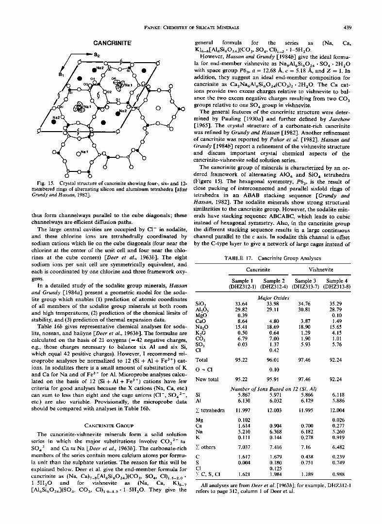
PAPIKE' CHEMISTRY OF SILICATE MINERALS 439
CANGRINITE
Fig. 15. Crystal structure of cancrinite showing four-, six- and 12- membered rings of alternating silicon and aluminum tetrahedra [after Grundy and Hassan, 1982].
thus form channelways parallel to the cube diagonals; these channelways are efficient diffusion paths.
The large central cavities are occupied by C1- in sodalite, and these chlorine ions are tetrahedrally coordinated by sodium cations which lie on the cube diagonals (four near the chlorine at the center of the unit cell and four near the chlo-
rines at the cube corners) [Deer et al., 1963b]. The eight sodium ions per unit cell are symmetrically equivalent, and each is coordinated by one chlorine and three framework oxy- gens.
In a detailed study of the sodalite group minerals, Hassan and Grundy [1984a] present a geometric model for the soda- lite group which enables (1) prediction of atomic coordinates of all members of the sodalite group minerals at both room and high temperatures, (2) prediction of the chemical limits of stability, and (3) prediction of thermal expansion data.
Table 16b gives representative chemical analyses for soda- lite, nosean, and hatiyne [Deer et al., 1963b]. The formulae are calculated on the basis of 21 oxygens (=42 negative charges, e.g., those charges necessary to balance six A1 and six Si, which equal 42 positive charges). However, I recommend mi- croprobe analyses be normalized to 12 (Si + A1 + Fe 3+) cat- ions. In sodalites there is a small amount of substitution of K
and Ca for Na and of Fe 3 + for A1. Microprobe analyses calcu- lated on the basis of 12 (Si + A1 + Fe 3+) cations have few criteria for good analyses because the X cations (Na, Ca, etc.) can sum to less than eight and the cage anions (CI-, SO4 2-, etc.) are also variable. Provisionally, the microprobe data should be compared with analyses in Table 16b.
CANCRINITE GROUP
The cancrinite-vishnevite minerals form a solid solution
series in which the major substitutions involve CO32-• SOa 2- and Ca • Na [Deer et al., 1963b]. The carbonate-rich members of the series contain more calcium atoms per formu- la unit than the sulphate varieties. The reason for this will be explained below. Deer et al. give the end-member formula for canefinite as (Na, Ca)7_s[A16Si602a](CO 3, SOa, C1)•.5_2.0- 1-5H 20 and for vishnevite as (Na, Ca, K)6_ ? [AI6Si602`r](SO a, CO 3, Cl)•.o_•. 5 ß 1-5H20. They give the
general formula for the series as (Na, Ca, K)6_8[A16Si602`r](CO3, SO,r, C1)•_2 ß 1-5H20.
However, Hassan and Grundy [1984b] give the ideal formu- la for end-member vishnevite as NasA16Si6024. SO 4 ß 2H20 with space group P63, a = 12.68 A, c = 5.18 A, and Z = 1. In addition, they suggest an ideal end-member composition for cancrinite as Ca2Na6A16Si6024(CO3) 2 ß 2H20. The Ca cat- ions provide two excess charges relative to vishnevite to bal- ance the two excess negative charges resulting from two CO 3 groups relative to one SO 4 group in vishnevite.
The general features of the cancrinite structure were deter-
mined by Pauling [1930a] and further defined by Jarchow [1965]. The crystal structure of a carbonate-rich cancrinite was refined by Grundy and Hassan [1982]. Another refinement of cancrinite was reported by Pahor et al. [1982]. Hassan and Grundy [1984b] report a refinement of the vishnevite structure and discuss important crystal chemical aspects of the cancrinite-vishnevite solid solution series.
The cancrinite group of minerals is characterized by an or- dered framework of alternating A10 4 and SiO 4 tetrahedra (Figure 15). The hexagonal symmetry, P63, is the result of close packing of interconnected and parallel sixfold rings of tetrahedra in an ABAB stacking sequence [Grundy and Hassan, 1982]. The sodalite minerals show strong structural similarities to the cancrinite group. However, the sodalite min- erals have stacking sequence ABCABC, which leads to cubic instead of hexagonal symmetry. Also, in the cancrinite group the different stacking sequence results in a large continuous channel parallel to the c axis. In sodalite this channel is offset
by the C-type layer to give a network of large cages instead of
TABLE 17. Cancrinite Group Analyses
Cancrinite Vishnevite
Sample 1 Sample 2 Sample 3 Sample 4 (DHZ312-1) (DHZ312-4) (DHZ313-7) (DHZ313-8)
Major Oxides SiO2 33.64 33.98 34.76 35.29 AI203 29.82 29.11 30.81 28.79 MgO 0.39 0.10 CaO 8.64 4.80 3.87 1.49
Na20 15.41 18.69 18.90 15.65 K20 0.50 0.64 1.29 4.15 CO2 6.79 7.00 1.90 1.01 SO3 0.03 1.37 5.93 5.76 C1 0.42
Total 95.22 96.01 97.46 92.24
O = CI 0.10
New total 95.22 95.91 97.46 92.24
Number of Ions Based on 12 (Si, Al) Si 5.867 5.971 5.866 6.118 AI 6.130 6.032 6.129 5.886
• tetrahedra 11.997 12.003 11.995 12.004
Mg 0.102 0.026 Ca 1.614 0.904 0.700 0.277
Na 5.210 6.368 6.182 5.260
K 0.111 0.144 0.278 0.919
• others 7.037 7.416 7.16 6.482
C 1.617 1.679 0.438 0.239
S 0.004 0.180 0.751 0.749 CI 0.125
•v C, S, CI 1.621 1.984 1.189 0.988
All analyses are from Deer et al. [1963b]' for example, DHZ312-1 refers to page 312, column 1 of Deer et al.
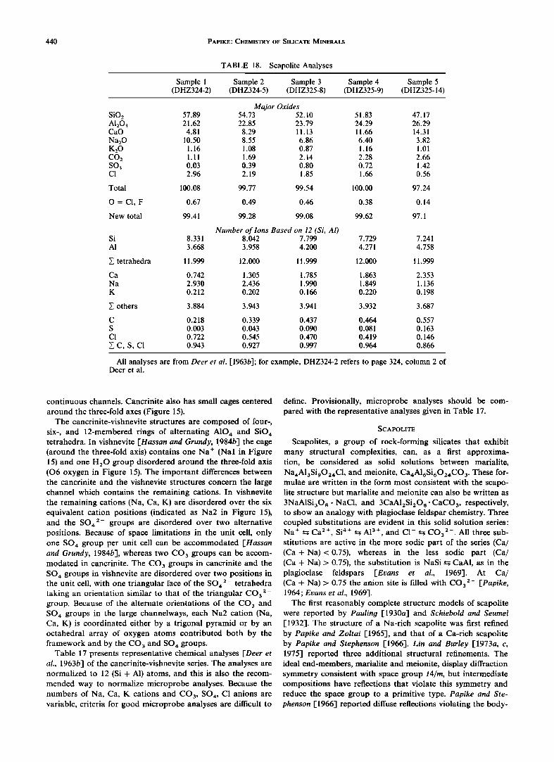
440 PAPIKE' CHEMISTRY OF SILICATE MINERALS
TABLE 18. Scapolite Analyses
Sample 1 Sample 2 Sample 3 Sample 4 Sample 5 (DHZ324-2) (DHZ324-5) (DHZ325-8) (DHZ325-9) (DHZ325-14)
Major Oxides SiO 2 57.89 54.73 A120 3 21.62 22.85 CaO 4.81 8.29
Na20 10.50 8.55 K20 1.16 1.08 CO2 1.11 1.69 SO3 0.03 0.39 C1 2.96 2.19
52 10
23 79
11 13
6 86 087
214
0.80 1.85
Total 100.08 99.77 99.54
O = C1, F 0.67 0.49 0.46
New total 99.41 99.28 99.08
51
24
11
6
1
2
0
1
100
83 47.17
29 26.29 66 14.31
40 3.82
16 1.01
28 2.66 .72 1.42
.66 0.56
.00 97.24
0.38 0.14
99.62 97.1
Number of Ions Based on 12 (Si, Al) Si 8.331 8.042 7.799 7.729 7.241 A1 3.668 3.958 4.200 4.271 4.758
X tetrahedra 11.999 12.000 11.999 12.000 11.999
Ca 0.742 1.305 1.785 1.863 2.353 Na 2.930 2.436 1.990 1.849 1.136 K 0.212 0.202 0.166 0.220 0.198
others 3.884 3.943 3.941 3.932 3.687
C 0.218 0.339 0.437 0.464 0.557 S 0.003 0.043 0.090 0.081 0.163 C1 0.722 0.545 0.470 0.419 0.146
• C, S, C1 0.943 0.927 0.997 0.964 0.866
All analyses are from Deer et al. [1963b]' for example, DHZ324-2 refers to page 324, column 2 of Deer et al.
continuous channels. Cancrinite also has small cages centered around the three-fold axes (Figure 15).
The cancrinite-vishnevite structures are composed of four-, six-, and 12-membered rings of alternating A104 and SiO4 tetrahedra. In vishnevite [Hassan and Grundy, 1984b] the cage (around the three-fold axis) contains one Na + (Nal in Figure 15) and one H20 group disordered around the three-fold axis (06 oxygen in Figure 15). The important differences between the cancrinite and the vishnevite structures concern the large channel which contains the remaining cations. In vishnevite the remaining cations (Na, Ca, K) are disordered over the six equivalent cation positions (indicated as Na2 in Figure 15), and the SO• 2- groups are disordered over two alternative positions. Because of space limitations in the unit cell, only one SO4 group per unit cell can be accommodated [Hassan and Grundy, 1984b], whereas two CO 3 groups can be accom- modated in cancrinite. The CO 3 groups in cancrinite and the SO4 groups in vishnevite are disordered over two positions in the unit cell, with one triangular face of the SO4 2- tetrahedra taking an orientation similar to that of the triangular CO3 2- group. Because of the alternate orientations of the CO 3 and SO4 groups in the large channelways, each Na2 cation (Na, Ca, K) is coordinated either by a trigonal pyramid or by an octahedral array of oxygen atoms contributed both by the framework and by the CO3 and SO4 groups.
Table 17 presents representative chemical analyses [Deer et al., 1963b] of the cancrinite-vishnevite series. The analyses are normalized to 12 (Si + A1) atoms, and this is also the recom- mended way to normalize microprobe analyses. Because the numbers of Na, Ca, K cations and CO3, SO4, C1 anions are variable, criteria for good microprobe analyses are difficult to
define. Provisionally, microprobe analyses should be com- pared with the representative analyses given in Table 17.
SCAPOLITE
Scapolites, a group of rock-forming silicates that exhibit many structural complexities, can, as a first approxima- tion, be considered as solid solutions between marialite, Na•A13Si902•C1, and meionite, Ca4A16Si602•CO 3. These for- mulae are written in the form most consistent with the scapo- lite structure but marialite and meionite can also be written as
3NaA1Si30 8 ß NaC1, and 3CaA12Si208- CaCO 3, respectively, to show an analogy with plagioclase feldspar chemistry. Three coupled substitutions are evident in this solid solution series: Na + -•Ca 2+, Si •+ --•A13+, and C!- •CO3 2-. All three sub- stitutions are active in the more sodic part of the series (Ca/ (Ca + Na)<0.75), whereas in the less sodic part (Ca/ (Ca + Na)> 0.75), the substitution is NaSi • CaA1, as in the plagioclase feldspars [Evans et al., 1969]. At Ca/ (Ca + Na) > 0.75 the anion site is filled with CO32- [Papike, 1964; Evans et al., 1969].
The first reasonably complete structure models of scapolite were reported by Pauling [1930a-I and Schiebold and Seumel [1932-1. The structure of a Na-rich scapolite was first refined by Papike and Zoltai [1965-1, and that of a Ca-rich scapolite by Papike and Stephenson [1966]. Lin and Burley [1973a, c, 1975-1 reported three additional structural refinements. The ideal end-members, marialite and meionite, display diffraction symmetry consistent with space group I4/m, but intermediate compositions have reflections that violate this symmetry and reduce the space group to a primitive type. Papike and Ste- phenson [1966] reported diffuse reflections violating the body-
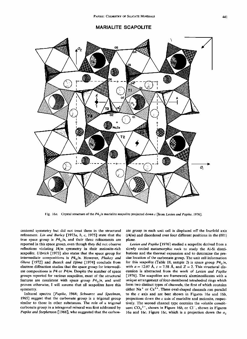
PAPIKE.' CHEMISTRY OF SILICATE MINERALS 441
MARIALITE SCAPOLITE
05
O2
o
Na,Ca
I
Fig. 16a. Crystal structure of the P42/n marialite scapolite projected down c [from Levien and Papike, 1976].
centered symmetry but did not treat them in the structural refinement. Lin and Burley 1-1973a, b, c, 1975] state that the true space group is P42/n, and their three refinements are reported in this space group, even though they did not observe reflections violating I4/rn symmetry in their meionite-rich scapolite. Ulbrich [1973] also states that the space group for intermediate compositions is P42/n. However, Phakey and Ghose [1972] and Buseck and Iijirna [1974] conclude from electron diffraction studies that the space group for intermedi- ate compositions is P4 or P4/rn. Despite the number of space groups reported for various scapolites, most of the structural features are consistent with space group P42/n, and until proven otherwise, I will assume that all scapolites have this symmetry.
Infrared spectra [Papike, 1964; Schwarcz and Speelrnan, 1965] suggest that the carbonate group is a trigonal group similar to those in other substances. The role of a trigonal carbonate group in a tetragonal mineral was first addressed by Papike and Stephenson [1966], who suggested that the carbon-
ate group in each unit cell is displaced off the fourfold axis (I4/rn) and disordered over four different positions in the (001) plane.
Levien and Papike [1976] studied a scapolite derived from a slowly cooled metamorphic rock to study the A1-Si distri- butions and the thermal expansion and to determine the pre- cise location of the carbonate group. The unit cell information for this scapolite (Table 18, sample 2) is space group P42/n, with a = 12.07 ,&, c = 7.58 ,&, and Z = 2. This structural dis- cussion is abstracted from the work of Levien and Papike [1976]. The scapolites are framework aluminosilicates with a unique arrangement of four-membered tetrahedral rings which form two distinct types of channels, the first of which contains either Na + or Ca 2 +. These oval-shaped channels run parallel to the c axis and are best shown in Figures 16a and 16b, projections down the c axis of marialite and meionite, respec- tively. The second channel type contains the volatile constit- uent CO32-, shown in Figure 16b, or CI-, shown in Figures 16a and 16c. Figure 16c, which is a projection down the a2
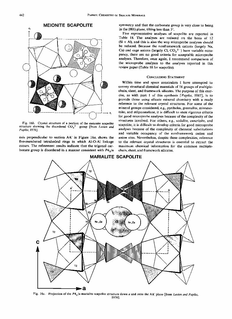
442 PAPIKE.' CHEMISTRY OF SILICATE MINERALS
MEIONITE SCAPOLITE
_, !• •'"• ..........
Fig. 16b. Crystal structure of a portion of the meionite scapolite structure showing the disordered CO32- group [from Levien and Papike, 1976].
axis perpendicular to section AA' in Figure 16a, shows the five-membered tetrahedral rings in which A1-O-A1 linkage occurs. The refinement results indicate that the trigonal car- bonate group is disordered in a manner consistent with P42/n
symmetry and that the carbonate group is very close to being in the (001)plane, tilting less than 3 ø.
Five representative analyses of scapolite are reported in Table 18. The analyses are reduced on the basis of 12 (Si + A1), and this is also the way microprobe analyses should be reduced. Because the nonframework cations (largely Na, Ca) and cage anions (largely C1, CO32-) have variable occu- pancy, there are no good criteria for acceptable microprobe analyses. Therefore, once again, I recommend comparison of the microprobe analyses to the analyses reported in this review paper (Table 18 for scapolite).
CONCLUDING STATEMENT
Within time and space constraints I have attempted to convey structural chemical essentials of 16 groups of multiple- chain, sheet, and framework silicates. The purpose of this exer- cise, as with part 1 of this synthesis [Papike, 1987], is to provide those using silicate mineral chemistry with a ready reference to the relevant crystal structures. For some of the mineral groups considered, e.g., pyriboles, greenalite, minneso- taite, and stilpnomelane, it is difficult to state rigorous criteria for good microprobe analyses because of the complexity of the structures involved. For others, e.g., sodalite, cancrinite, and scapolite, it is difficult to develop criteria for good microprobe analyses because of the complexity of chemical substitutions and variable occupancy of the nonframework cation and anion sites. Nevertheless, despite these complexities, reference to the relevant crystal structures is essential to extract the maximum chemical information for the common multiple- chain, sheet, and framework silicates.
MARIALITE SCAPOLITE
C
I _J
Fig. 16c. Projection of the P42/n marialite scapolite structure down a and onto the AA' plane [from Levien and Papike, 1976].
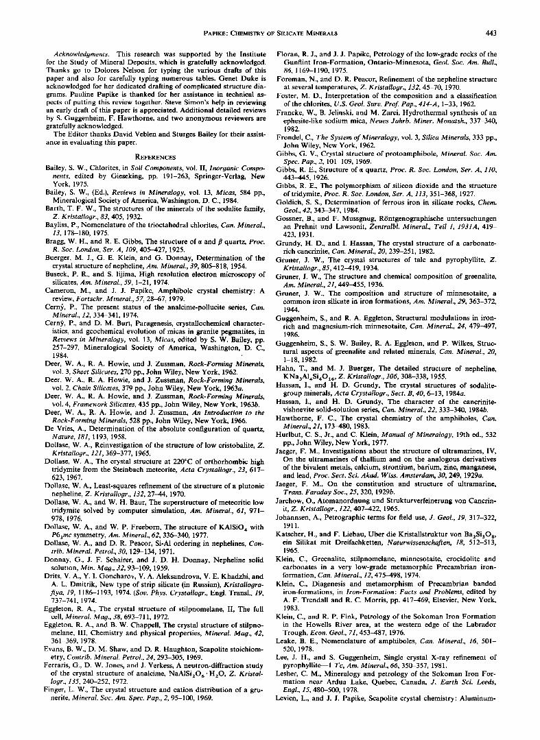
PAPIKE' CHEMISTRY OF SILICATE MINERALS 443
Acknowledgments. This research was supported by the Institute for the Study of Mineral Deposits, which is gratefully acknowledged. Thanks go to Dolores Nelson for typing the various drafts of this paper and also for carefully typing numerous tables. Genet Duke is acknowledged for her dedicated drafting of complicated structure dia- grams. Pauline Papike is thanked for her assistance in technical as- pects of putting this review together. Steve Simon's help in reviewing an early draft of this paper is appreciated. Additional detailed reviews by S. Guggenheim, F. Hawthorne, and two anonymous reviewers are gratefully acknowledged.
The Editor thanks David Veblen and Sturges Bailey for their assist- ance in evaluating this paper.
REFERENCES
Bailey, S. W., Chlorites, in Soil Components, vol. II, Inorganic Compo- nents, edited by Gieseking, pp. 191-263, Springer-Verlag, New York, 1975.
Bailey, S. W., (Ed.), Reviews in Mineralogy, vol. 13, Micas, 584 pp., Mineralogical Society of America, Washington, D.C., 1984.
Barth, T. F. W., The structures of the minerals of the sodalite family, Z. Kristallogr., 83, 405, 1932.
Bayliss, P., Nomenclature of the trioctahedral chlorites, Can. Mineral., 13, 178-180, 1975.
Bragg, W. H., and R. E. Gibbs, The structure of o• and fl quartz, Proc. R. Soc. London, Ser. A, 109, 405-427, 1925.
Buerger, M. J., G. E. Klein, and G. Donnay, Determination of the crystal structure of nepheline, Am. Mineral., 39, 805-818, 1954.
Buseck, P. R., and S. Iijima, High resolution electron microscopy of silicates, Am. Mineral., 59, 1-21, 1974.
Cameron, M., and J. J. Papike, Amphibole crystal chemistry: A review, Fortschr. Mineral., 57, 28-67, 1979.
Cerng, P., The present status of the analcime-pollucite series, Can. Mineral., 12, 334-341, 1974.
CernS', P., and D. M. Burt, Paragenesis, crystallochemical character- istics, and geochemical evolution of micas in granite pegmatites, in Reviews in Mineralogy, vol. 13, Micas, edited by S. W. Bailey, pp. 257-297, Mineralogical Society of America, Washington, D.C., 1984. ;
Deer, W. A., R. A. Howie, and J. Zussman, Rock-Forming Minerals, vol. 3, Sheet Silicates, 270 pp., John Wiley, New York, 1962.
Deer, W. A., R. A. Howie, and J. Zussman, Rock-Forming Minerals, vol. 2, Chain Silicates, 379 pp., John Wiley, New York, 1963a.
Deer, W. A., R. A. Howie, and J. Zussman, Rock-Forming Minerals, vol. 4, Framework Silicates, 435 pp., John Wiley, New York, 1963b.
Deer, W. A., R. A. Howie, and J. Zussman, An Introduction to the Rock-Forming Minerals, 528 pp., John Wiley, New York, 1966.
De Vries, A., Determination of the absolute configuration of quartz, Nature, 181, 1193, 1958.
Dollase, W. A., Reinvestigation of the structure of low cristobalite, Z. Kristallogr., 121, 369-377, 1965.
Dollase, W. A., The crystal structure at 220øC of orthorhombic high tridymite from the Steinbach meteorite, Acta Crystallogr., 23, 617- 623, 1967.
Dollase, W. A., Least-squares refinement of the structure of a plutonic nepheline, Z. Kristallogr., 132, 27-44, 1970.
Dollase, W. A., and W. H. Baur, The superstructure of meteoritic low tridymite solved by computer simulation, Am. Mineral., 61, 971- 978, 1976.
Dollase, W. A., and W. P. Freeborn, The structure of KAISiO 4 with P63mc symmetry, Am. Mineral., 62, 336-340, 1977.
Dollase, W. A., and D. R. Peacor, Si-AI ordering in nephelines, Con- trib. Mineral. Petrol., 30, 129-134, 1971.
Donnay, G., J. F. Schairer, and J. D. H. Donnay, Nepheline solid solution, Min. Mag., 32, 93-109, 1959.
Drits, V. A., Y. I. Goncharov, V. A. Aleksandrova, V. E. Khadzhi, and A. L. Dmitrik, New type of strip silicate (in Russian), Kristallogra- fiya, 19, 1186-1193, 1974. (Soy. Phys. Crystallogr., Engl. Transl., 19, 737-741, 1974.
Eggleton, R. A., The crystal structure of stilpnomelane, II, The full cell, Mineral. Mag., 38, 693-711, 1972.
Eggleton, R. A., and B. W. Chappell, The crystal structure of stilpno- melane, III, Chemistry and physical properties, Mineral. Mag., 42, 361-369, 1978.
Evans, B. W., D. M. Shaw, and D. R. Haughton, Scapolite stoichiom- etry, Contrib. Mineral. Petrol., 24, 293-305, 1969.
Ferraris, G., D. W. Jones, and J. Yerkess, A neutron-diffraction study of the crystal structure of analcime, NaA1Si206- H20 , Z. Kristal- logr., 135, 240-252, 1972.
Finger, L. W., The crystal structure and cation distribution of a gru- nerite, Mineral. Soc. Am. Spec. Pap., 2, 95-100, 1969.
Floran, R. J., and J. J. Papike, Petrology of the low-grade rocks of the Gunflint Iron-Formation, Ontario-Minnesota, Geol. Soc. Am. Bull., 86, 1169-1190, 1975.
Foreman, N., and D. R. Peacor, Refinement of the nepheline structure at several temperatures, Z. Kristallogr., 132, 45-70, 1970.
Foster, M.D., Interpretation of the composition and a classification of the chlorites, U.S. Geol. Surv. Prof Pap., 414-A, 1-33, 1962.
Francke, W., B. Jelinski, and M. Zarei, Hydrothermal synthesis of an ephesite-like sodium mica, Neues Jahrb. Miner. Monatsh., 337-340, 1982.
Frondel, C., The System of Mineralogy, vol. 3, Silica Minerals, 333 pp., John Wiley, New York, 1962.
Gibbs, G. V., Crystal structure of protoamphibole, Mineral. Soc. Am. Spec. Pap., 2, 101-109, 1969.
Gibbs, R. E., Structure of o• quartz, Proc. R. Soc. London, Ser. A, 110, 443-445, 1926.
Gibbs, R. E., The polymorphism of silicon dioxide and the structure of tridymite, Proc. R. Soc. London, Ser. A, 113, 351-368, 1927.
Goldich, S.S., Determination of ferrous iron in silicate rocks, Chem. Geol., 42, 343-347, 1984.
Gossner, B., and F. Mussgnug, R6ntgenographische untersuchungen an Prehnit und Lawsonit, Zentralbl. Mineral., Tell 1, 1931A, 419- 423, 1931.
Grundy, H. D., and I. Hassan, The crystal structure of a carbonate- rich cancrinite, Can. Mineral., 20, 239-251, 1982.
Gruner, J. W., The crystal structures of talc and pyrophyllite, Z. Kristallogr., 85, 412-419, 1934.
Gruner, J. W., The structure and chemical composition of greenalite, Am. Mineral., 21, 449-455, 1936.
Gruner, J. W., The composition and structure of minnesotaite, a common iron silicate in iron formations, Am. Mineral., 29, 363-372, 1944.
Guggenheim, S., and R. A. Eggleton, Structural modulations in iron- rich and magnesium-rich minnesotaite, Can. Mineral., 24, 479-497, 1986.
Guggenheim, S., S. W. Bailey, R. A. Eggleton, and P. Wilkes, Struc- tural aspects of greenalite and related minerals, Can. Mineral., 20, 1-18, 1982.
Hahn, T., and M. J. Buerger, The detailed structure of nepheline, KNa3AI,•Si,•O•6, Z. Kristallogr., 106, 308-338, 1955.
Hassan, I., and H. D. Grundy, The crystal structures of sodalite- group minerals, Acta Crystallogr., Sect. B, 40, 6-13, 1984a.
Hassan, I., and H. D. Grundy, The character of the cancrinite- vishnevite solid-solution series, Can. Mineral., 22, 333-340, 1984b.
Hawthorne, F. C., The crystal chemistry of the amphiboles, Can. Mineral., 21, 173-480, 1983.
Hurlbut, C. S., Jr., and C. Klein, Manual of Mineralogy, 19th ed., 532 pp., John Wiley, New York, 1977.
Jaeger, F. M., Investigations about the structure of ultramarines, IV, On the ultramarines of thallium and on the analogous derivatives of the bivalent metals, calcium, strontium, barium, zinc, manganese, and lead, Proc. Sect. Sci. Akad. Wiss. Amsterdam, 30, 249, 1929a.
Jaeger, F. M., On the constitution and structure of ultramarine, Trans. Faraday Soc., 25, 320, 1929b.
Jarchow, O., Atomanordnung und Strukturverfeinerung von Cancrin- it, Z. Kristallogr., 122, 407-422, 1965.
Johannsen, A., Petrographic terms for field use, J. Geol., 19, 317-322, 1911.
Katscher, H., and F. Liebau, Uber die Kristallstruktur yon Ba2Si30 8, ein Silikat mit Dreifachketten, Naturwissenschaften, 18, 512-513, 1965.
Klein, C., Greenalite, stilpnomelane, minnesotaite, crocidolite and carbonates in a very low-grade metamorphic Precambrian iron- formation, Can. Mineral., 12, 475-498, 1974.
Klein, C., Diagenesis and metamorphism of Precambrian banded iron-formations, in Iron-Formation: Facts and Problems, edited by A. F. Trendall and R. C. Morris, pp. 417-469, Elsevier, New York, 1983.
Klein, C., and R. P. Fink, Petrology of the Sokoman Iron Formation in the Howells River area, at the western edge of the Labrador Trough, Econ. Geol., 71, 453-487, 1976.
Leake, B. E., Nomenclature of amphiboles, Can. Mineral., 16, 501- 520, 1978.
Lee, J. H., and S. Guggenheim, Single crystal X-ray refinement of pyrophyllite--1 Tc, Am. Mineral., 66, 350-357, 1981.
Lesher, C. M., Mineralogy and petrology of the Sokoman Iron For- mation near Ardua Lake, Quebec, Canada, J. Earth $ci. Leeds, Engl., 15, 480-500, 1978.
Levien, L., and J. J. Papike, Scapolite crystal chemistry: Aluminum-
444 PAPIKE.' CHEMISTRY OF SILICATE MINERALS
silicon distributions, carbonate group disorder, and thermal ex- pansion, Am. Mineral., 61, 864-877, 1976.
Liebau, F., Classification of silicates, in Reviews in Mineralo•Ty, vol. 5, Orthosilicates, edited by P. H. Ribbe, pp. 1-24, Mineralogical So- ciety of America, Washington, D.C., 1980.
Liebau, F., Structural Chemistry of Silicates, Structure, Bonding7, and Classification, 347 pp., Springer-Verlag, New York, 1985.
Lin, S. B., and B. J. Burley, Crystal structure of a sodium and chlorine-rich scapolite, Acta Crystallogr., Sect. B, 29, 1272-1278, 1973a.
Lin, S. B., and B. J. Burley, On the weak reflections violating body- centered symmetry in scapolites, Tschermaks Mineral. Petro•Tr. Mitt., 20, 28-44, 1973b.
Lin, S. B., and B. J. Burley, The crystal structure of meionite, Acta Crystallo•Tr., Sect. B, 29, 2024-2026, 1973c.
Lin, S. B., and B. J. Burley, The crystal structure of an intermediate scapolite-wernerite, Acta Crystallo•Tr., Sect. B, 31, 1806-1814, 1975.
Miyano, T., Effects of CO 2 on mineralogical differences in some low- grade metamorphic iron-formations, Geochem. d., 12, 201-211, 1978.
Moore, P. B., Quartz: Variations on a theme, Am. Mineral., 71, 540- 546, 1986.
Nfiray-Szab6, S., Die Struktur des Leucits KA1Si20 6, Z. Kristallo•r., 104, 39-44, 1942.
Nieuwenkamp, W., Uber die Struktur von Hoch-Kristobalit, Z. Kri- stallo•r., 96, 454-458, 1937.
Nuffield, E. W., Prehnite from Ashcroft, British Columbia, Toronto Univ. Stud. Geol. Ser., 48, 49-64, 1943.
Pahor, N. B., M. Calligaris, G. Nardin, and L. Randaccio, Structure of a basic cancrinite, Acta Crystallo•Tr., Sect. B, 38, 893-895, 1982.
Papike, J. J., The crystal structure and crystal chemistry of scapolite, Ph.D. thesis, Univ. of Minn., Minneapolis, 1964.
Papike, J. J., Chemistry of the rock-forming silicates: Ortho, ring, and single-chain structures, Rev. Geophys., 25, 1483-1526, 1987.
Papike, J. J., and M. Cameron, Crystal chemistry of silicate minerals of geophysical interest, Rev. Geophys., 14, 37-80, 1976.
Papike, J. J., and M. Ross, Gedrites: Crystal structures and intracry- stalline cation distributions, Am. Mineral., 55, 1945-1972, 1970.
Papike, J. J., and N. C. Stephenson, The crystal structure of mizzon- ite, a calcium- and carbonate-rich scapolite, Am. Mineral., 51, 1014- 1027, 1966.
Papike, J. J., and T. Zoltai, The crystal structure of a marialite scapo- lite, Am. Mineral., 50, 641-655, 1965.
Papike, J. J., and T. Zoltai, Ordering of tetrahedral aluminum in prehnite, Ca•(Al, Fe3+)[Si3A10•0](OH)•, Am. Mineral., 52, 974- 984, 1967.
Papike, J. J., M. Ross, and J. R. Clark, Crystal-chemical characterization of clinoamphiboles based on five new structure refinements, Mineral. Soc. Am. Spec. Pap., 2, 117-136, 1969.
Pauling, L., The structure of some sodium and calcium alumi- nosilicates; Proc. Natl. Acad. Sic. U.S. A., 16, 453-459, 1930a.
Pauling, L., The structure of chlorites, Proc. Natl. Acad. Sci. U. S. A., 16, 578-582, 1930b.
Pauling, L., The structure of sodalite and helvite, Z. Kristallo•Tr., 74, 213-225, 1930c.
Peacor, D. R., A high-temperature single-crystal diffractometer study of leucite, (K, Na)A1Si:O 6, Z. Kristallo•Tr., 127, 213-224, 1968.
Peacor, D. R., High-temperature single-crystal study of the cristoba- lite inversion, Z. Kristallo•Tr., 138, 274-298, 1973.
Peng, Sze-Tung, Chou Kung-Du, and Tang You-Chi, The structure of prehnite, Hua Hsueh Hsueh Pao, 25, 56-63, 1969.
Perdikatsis, B., and H. Burzlaff, Strukturverfeinerung am Talk Mg3[(OH):Si,•O•0 ], Z. Kristallo•r., 156, 177-186, 1981.
Perrotta, A. J., and J. V. Smith, The crystal structure of kalsilite, KA1SiO½, Mineral. Ma•7., 35, 588-595, 1965.
Phakey, P. P., and S. Ghose, Scapolite: Observation of anti-phase domain structure, Nature London Phys. Sci., 238, 78-80, 1972.
Preisinger, A., Prehnit--Ein neuer Schichtsilikattyp, Tschermaks Min- eral. Petro•Ir. Mitt., 10, 491-504, 1965.
Quint, R., Description and crystal structure of amstallite, CaAl(OH)2[A10.8Si3.:O8(OH): ] .[(H:O)0.8C10.:], a new mineral from Amstall, Austria, Neues Jahrb. Miner. Monatsh., 6, 253-262, 1987.
Rayner, J. H., and G. Brown, Structure of pyrophyllite, Clays Clay Miner., 13, 73-84, 1964.
Rayner, J. H., and G. Brown, The crystal structure of talc, Clays Clay Miner., 21, 103-114, 1973.
Ribbe, P. H. (Ed.), Reviews in Mineralo•Ty, vol. 2, Feldspar Mineralo•Ty, 2nd ed., 362 pp., Mineralogical Society of America, Washington, D.C., 1983.
Sadanaga, R., and T. Ozawa, Thermal transition of leucite, Mineral. J., 5, 321-333, 1968.
Schiebold, E., and G. Seumel, Uber die Kristallstruktur von Skapol- ith, Z. Kristallo•Tr., 81, 110-134, 1932.
Schwarcz, H. P., and E. L. Speelman, Determination of sulfur and carbon coordination in scapolite by infra-red absorption spec- trophotometry, Am. Mineral., 50, 656-666, 1965.
Seifert, F., and W. Schreyer, Synthesis and stability of micas in the system K20-MgO-SiO:-H:O and their relation to phlogopite, Contrib. Mineral Petrol., 30, 196-215, 1971.
Simmons, W. B., Jr., and D. R. Peacor, Refinement of the crystal structure of a volcanic nepheline, Am. Mineral., 57, 1711-1719, 1972.
Smith, J. V., and H. S. Yoder, Jr., Experimental and theoretical stud- ies of the mica polymorphs, Mineral. Mag., 31, 209-235, 1956.
Taylor, D., and C. M. B. Henderson, The thermal expansion of the leucite group of minerals, Am. Mineral., 53, 1476-1489, 1968.
Taylor, W. H., The structure of analcite (NaA1Si:O 6 ß H•_O), Z. Kri- stallo•r., 74, 1, 1930.
Thompson, J. B., Jr., Geometrical possibilities for amphibole struc- tures: Model biopyriboles (abstract), Am. Mineral., 55, 292-293, 1970.
Thompson, J. B., Jr., Biopyriboles and polysomatic series, Am. Min- eral., 63, 239-249, 1978.
Tuttle, O. F., and J. V. Smith, The nepheline-kalsilite system, 2, Phase relations, Am. J. Sci., 256, 571-589, 1958.
Ulbrich, H. H., Crystallographic data and refractive indices of scapo- lites, Am. Mineral., 58, 81-92, 1973.
Veblen, D. R. (Ed.), Reviews in Mineralo•Ty, vol. 9A, Amphiboles and Other Hydrous Pyriboles--Mineralo•Ty, 372 pp., Mineralogical So- ciety of America, Washington, D.C., 1981.
Veblen, D. R., and C. W. Burnham, Triple-chain biopyriboles: Newly discovered intermediate products of the retrograde anthophyllite- talc transformation, Chester, VT (abstract), Eos Trans. AGU, 56, 1076, 1975.
Veblen, D. R., and C. W. Burnham, New biopyriboles from Chester, Vermont, I, Descriptive mineralogy, Am. Mineral., 63, 1000-1009, 1978a.
Veblen, D. R., and C. W. Burnham, New biopyriboles from Chester, Vermont, II, The crystal chemistry of jimthompsonite, clinojim- thompsonite, and chesterite, and the amphibole-mica reaction, Am. Mineral., 63, 1053-1973, 1978b.
Veblen, D. R., and P. H. Ribbe (Eds.), Reviews in Mineralo•Ty, vol. 9B, Amphiboles: Petrology and Experimental Phase Relations, 390 pp., Mineralogical Society of America, Washington, D.C., 1982.
Veblen, D. R., P. R. Buseck, and C. W. Burnham, Asbestiform chain silicates: New minerals and structural groups, Science, 198, 359- 365, 1977.
Wardle, R., and G. W. Brindley, The crystal structures of pyrophyllite 1Tc and of its dehydroxylate, Am. Mineral., 57, 732-750, 1972.
Wei, P., The structure of •-quartz, Z. Kristallo•Tr., 92, 355-362, 1935. Wyart, J., Etude sur la leucite, Bull. Soc. Fr. Mineral., 61, 228-238,
1938.
Wyart, J., Etude cristallographique d'une leucite artificielle; structure atomique et symm6trie du min6ral, Bull. Soc. Ft. Mineral., 63, 5-17, 1940.
Wyart, J., Structure atomique de la leucite, C. R. Acad. Sci., 282, 356-359, 1941.
Wyckoff, R. W. G., The crystal structure of the high-temperature form of cristobalite (SiO2), Am. J. Sci., 9, 448-459, 1925.
Young, R. A., and B. Post, Electron density and thermal effects in alpha quartz, Acta Crystallo•Tr., 15, 337-346, 1962.
J. J. Papike, South Dakota School of Mines and Technology, 501 East Saint Joseph Street, Rapid City, SD 57701.
(Received December 9, 1987; accepted March 23, 1988.)
Related Documents


![Improved Photoactivity of Pyroxene Silicates by Cation ... · rock-forming silicates as they werefound in almostevery igne-ous rock.[19–21] Moreover, it is believed that pyroxenes](https://static.cupdf.com/doc/110x72/5ed92b706714ca7f476946a6/improved-photoactivity-of-pyroxene-silicates-by-cation-rock-forming-silicates.jpg)