Hindawi Publishing Corporation Journal of Nanomaterials Volume 2013, Article ID 587021, 10 pages http://dx.doi.org/10.1155/2013/587021 Research Article Iron Oxide Magnetic Nanoparticles: Characterization and Toxicity Evaluation by In Vitro and In Vivo Assays Alina Mihaela Prodan, 1,2 Simona Liliana Iconaru, 3 Carmen Steluta Ciobanu, 3 Mariana Carmen Chifiriuc, 4 Mihai Stoicea, 2 and Daniela Predoi 3 1 Carol Davila University of Medicine and Pharmacy, 8 Eroii Sanitari, Sector 5, 050474 Bucharest, Romania 2 Emergency Hospital Floreasca, Bucharest 5, Calea Floreasca Nr 8, Sector 1, 014461 Bucarest, Romania 3 National Institute of Materials Physics, 105 bis Atomistilor, P.O. Box MG 07, 077125 Bucuresti-Magurele, Romania 4 Microbiology Department, Faculty of Biology, University of Bucharest, Aleea Portocalelor 1-3, 60101 Bucharest, Romania Correspondence should be addressed to Daniela Predoi; [email protected] Received 21 August 2013; Revised 4 October 2013; Accepted 5 October 2013 Academic Editor: In-Kyu Park Copyright © 2013 Alina Mihaela Prodan et al. is is an open access article distributed under the Creative Commons Attribution License, which permits unrestricted use, distribution, and reproduction in any medium, provided the original work is properly cited. e aim of this study was to evaluate the biological properties of iron oxide nanoparticles (IO-NPs) obtained in the aqueous suspension. e iron oxide nanoparticles were characterized by scanning electron microscopy (SEM) and transmission electron microscopy (TEM). e biocompatibility of the iron oxide was demonstrated by the in vitro quantification of HeLa cells viability using propidium iodide (PI) and fluorescein diacetate (FdA) and the MTT colorimetric assay. e toxicity of small size iron oxide nanoparticles was also evaluated by means of histological examination on male Brown Norway rats aſter intraperitoneal injection. At the tested concentrations, the nanoparticles proved to be not cytotoxic on HeLa cells. e rat’s behavior, as well as the histopathological aspect of liver, kidney, lung, and spleen tissues at 48h aſter intraperitoneal injection did not present any modifications. e in vivo and in vitro assays suggested that the IO-NPs could be further used for developing new in vivo medical applications. 1. Introduction Nowadays, finding new approaches for solving pressing problems in the field of medical science is the focus of research institutes everywhere. e most studied materials with promising potential in the field of biomedical applica- tions are those with magnetic properties. Magnetic materials, especially iron oxides nanoparticles, are known since ancient times to have many spectacular properties, but in the last decade the properties that they possess at nanometric scale have been the starting point of great potential applications such as drug delivery, magnetic cell separation, tumor label- ing and cell labeling. e most common forms of iron oxides, magnetite and maghemite (Fe 3 O 4, -Fe 2 O 3 ), are studied due to the outstanding properties they exhibit at nanometric scale (high specific surface area, superparamagnetism, etc.) [1–5]. e nanometric dimensions of these materials makes them ideal candidates for surface engineering and functionaliza- tion. Surface enhancement and functionalization facilitate the use of these nanomaterials in biomedical applications, for example, as contrast agents for magnetic resonance imaging (MRI) [6, 7], tissue-specific release of therapeutic agents, targeted drug delivery in tumor therapy [8], hyperthermia, cell labeling [9], magnetic cell sorting [10], and magnetic field assisted radionuclide therapy [11]. In the past few years, superparamagnetic iron oxide nanoparticles with controlled and enhanced surface chem- istry properties have been used successfully as contrast agents for magnetic resonance imaging in vivo [12, 13]. e new direction of research aims to develop new compounds based on iron oxide nanoparticles for in vivo biomedical applications. Recent studies in the field of malignant tumors are focused on developing a new drug delivery systems based on iron oxide nanoparticles in order to avoid damaging the healthy cells around the tumor mass in the process of cancerous cell destruction. ese types of nanosystems based on iron oxide nanoparticles have the ability to heat up,

Welcome message from author
This document is posted to help you gain knowledge. Please leave a comment to let me know what you think about it! Share it to your friends and learn new things together.
Transcript
Hindawi Publishing CorporationJournal of NanomaterialsVolume 2013, Article ID 587021, 10 pageshttp://dx.doi.org/10.1155/2013/587021
Research ArticleIron Oxide Magnetic Nanoparticles: Characterization andToxicity Evaluation by In Vitro and In Vivo Assays
Alina Mihaela Prodan,1,2 Simona Liliana Iconaru,3 Carmen Steluta Ciobanu,3
Mariana Carmen Chifiriuc,4 Mihai Stoicea,2 and Daniela Predoi3
1 Carol Davila University of Medicine and Pharmacy, 8 Eroii Sanitari, Sector 5, 050474 Bucharest, Romania2 Emergency Hospital Floreasca, Bucharest 5, Calea Floreasca Nr 8, Sector 1, 014461 Bucarest, Romania3 National Institute of Materials Physics, 105 bis Atomistilor, P.O. Box MG 07, 077125 Bucuresti-Magurele, Romania4Microbiology Department, Faculty of Biology, University of Bucharest, Aleea Portocalelor 1-3, 60101 Bucharest, Romania
Correspondence should be addressed to Daniela Predoi; [email protected]
Received 21 August 2013; Revised 4 October 2013; Accepted 5 October 2013
Academic Editor: In-Kyu Park
Copyright © 2013 Alina Mihaela Prodan et al. This is an open access article distributed under the Creative Commons AttributionLicense, which permits unrestricted use, distribution, and reproduction in any medium, provided the original work is properlycited.
The aim of this study was to evaluate the biological properties of iron oxide nanoparticles (IO-NPs) obtained in the aqueoussuspension. The iron oxide nanoparticles were characterized by scanning electron microscopy (SEM) and transmission electronmicroscopy (TEM). The biocompatibility of the iron oxide was demonstrated by the in vitro quantification of HeLa cells viabilityusing propidium iodide (PI) and fluorescein diacetate (FdA) and the MTT colorimetric assay. The toxicity of small size ironoxide nanoparticles was also evaluated by means of histological examination on male Brown Norway rats after intraperitonealinjection. At the tested concentrations, the nanoparticles proved to be not cytotoxic on HeLa cells. The rat’s behavior, as well asthe histopathological aspect of liver, kidney, lung, and spleen tissues at 48 h after intraperitoneal injection did not present anymodifications. The in vivo and in vitro assays suggested that the IO-NPs could be further used for developing new in vivomedicalapplications.
1. Introduction
Nowadays, finding new approaches for solving pressingproblems in the field of medical science is the focus ofresearch institutes everywhere. The most studied materialswith promising potential in the field of biomedical applica-tions are those withmagnetic properties. Magnetic materials,especially iron oxides nanoparticles, are known since ancienttimes to have many spectacular properties, but in the lastdecade the properties that they possess at nanometric scalehave been the starting point of great potential applicationssuch as drug delivery, magnetic cell separation, tumor label-ing and cell labeling.Themost common forms of iron oxides,magnetite and maghemite (Fe
3O4,𝛾-Fe2O3), are studied due
to the outstanding properties they exhibit at nanometric scale(high specific surface area, superparamagnetism, etc.) [1–5].The nanometric dimensions of these materials makes themideal candidates for surface engineering and functionaliza-tion. Surface enhancement and functionalization facilitate
the use of these nanomaterials in biomedical applications, forexample, as contrast agents for magnetic resonance imaging(MRI) [6, 7], tissue-specific release of therapeutic agents,targeted drug delivery in tumor therapy [8], hyperthermia,cell labeling [9], magnetic cell sorting [10], andmagnetic fieldassisted radionuclide therapy [11].
In the past few years, superparamagnetic iron oxidenanoparticles with controlled and enhanced surface chem-istry properties have been used successfully as contrastagents for magnetic resonance imaging in vivo [12, 13]. Thenew direction of research aims to develop new compoundsbased on iron oxide nanoparticles for in vivo biomedicalapplications. Recent studies in the field of malignant tumorsare focused on developing a new drug delivery systems basedon iron oxide nanoparticles in order to avoid damagingthe healthy cells around the tumor mass in the process ofcancerous cell destruction.These types of nanosystems basedon iron oxide nanoparticles have the ability to heat up,
2 Journal of Nanomaterials
delivering toxic amounts of thermal energy to tumors, or aschemotherapy and radiotherapy enhancement agents, wherea controlled degree of tissue warming leads to an effective celldestruction [14, 15].
In agreementwith Pisanic II et al. [16],magnetic nanopar-ticles could be used as tools in a wide variety of biomedicalapplications. On the other hand, Pisanic II et al. showedthat failure to fully and properly evaluate nanostructureson an individual case-by-case basis may lead to lack ofparameter control in in vitro experiments, as well as incorrectassumptions concerning their biocompatibility and biosafetyof their in vivo use [16]. In order to improve the knowledgeon cytotoxicity of iron oxide nanoparticles, we performed anin vivo toxicity study (48 h) by administration by intraperi-toneal injection of 𝛾-Fe
2O3dispersion at concentrations of
0.7mL/kg, 1.7mL/kg, and 3.7mL/kg.The aim of this study was to develop iron oxide nanopar-
ticles by an adapted coprecipitation method [17–22] withcontrollable parameters and enhanced biocompatible prop-erties for in vivo applications. Scanning electron microscopy(SEM) and transmission electron microscopy (TEM) studieshave been conducted to obtain information about the size,structure, and morphology of IO-NPs. The biocompatibilityof the iron oxide was evaluated using in vitro and in vivoassays, consisting in the quantification of HeLa cells viabilityand the histological evaluation of the nanoparticles effects onthe male Brown Norway rat’s tissues.
2. Experimental Section
2.1. Materials. Ferrous chloride tetrahydrate (FeCl2⋅4H2O),
ferric chloride hexahydrate (FeCl3⋅6H2O), natrium hydrox-
ide (NaOH), and chlorhidric acid (HCl) were purchasedfrom Merck. Deionized water was used in the synthesis ofnanoparticles and for rinsing the clusters.
2.2. Synthesis of Iron Oxide Ferrofluid. Iron oxide nanopar-ticles were prepared by coprecipitation [17–22]. Ferrouschloride tetrahydrate (FeCl
2⋅4H2O) in 2M HCl and ferric
chloride hexahydrate (FeCl3⋅6H2O) were mixed at 100∘C
(Fe2+/Fe3+ = 1/2). The mixture was dropped into 200mLof NaOH (2mol⋅L−1) solution under vigorous stirring forabout 30min. The precipitate of magnetite (black precipitateimmediately formed) was converted into 𝛾-Fe
2O3particles
by repeated treatment with HNO3(2mol⋅L−1) and FeNO
3
(0.3mol⋅L−1) solutions [23]. The acidic precipitate was iso-lated by decantation on a magnet, separated by centrifuga-tion (6000 rpm), then washed in acetone, and dispersed indeionized water at pH = 2.5. The final ion concentrationwas 0.38mol⋅L−1. For biological investigations, the pH wasadjusted to 7 using aqueous ammonia. The iron content ofthe suspensions was determined by redox-titration [23].
2.3. Characterization of Nanoparticles. The morphology ofthe obtained material was studied using a Quanta InspectF scanning electron microscope (SEM), operating at 25 kVin vacuum. The elemental local analysis was performedusing an energy dispersive spectroscopy (EDS) detector
from EDAX. The operating conditions were an acceleratingvoltage between 2 and 25 keV (depending of the signal/noiseratio) for samples tilted at 25∘ in order to get the optimaltake off angle (30∘) allowing a dead time around 20–30%and a collecting time of 90–120 s. Transmission electronmicroscopy (TEM) images for these samples were recordedusing a FEI Tecnai 12 equipped with a low dose digital camerafrom Gatan. The specimen for TEM imaging was preparedby ultramicrotomy in order to obtain a thin section of about60 nm.Thepowder was embedded in an epoxy resin (polaron612) before microtomy.
2.4. Cytotoxicity Assay. Quantification of cell viability wasperformed using propidium iodide (PI) and fluorescein diac-etate (FdA). Briefly, 5×10⁴HeLa cells were seeded in eachwellof a 24-well plate and after 24 h, the monolayers were treatedwith a suspension of 𝛾-Fe
2O3(200𝜇L) nanoparticles diluted
100 times. The effects on cellular viability were evaluatedafter 48 h by adding 100 𝜇L PI (0.1mg/mL) and 100 𝜇L FdA(0.1mg/mL) and fluorescence studies have been performedusing Observer D1 Carl Zeiss microscope. The cell viabilitywas established by the ratio between viable (green) and deadcells (red) counted on several microscopic fields [24].
The cell viability was determined by MTT colorimetricassay developed by Mosmann for in vitro cytotoxicity andcell proliferation measurements [25]. It was reported thatthe mitochondrial enzyme succinate-dehydrogenase withinviable cells is able to cleave theMTT salt into formazan, a bluecolored product. The amount of formazan produced, readon scanning multiwell spectrophotometer, is proportional tothe number of viable cells present [25–27]. The cells werecultured in the medium (2.5 × 10⁵ cells/mL) containing ironoxide nanoparticles for 12, 24, and 72 hours periods. Culturemedium without iron oxide nanoparticles served as controlin each experiment. The different final concentrations ofthe suspension of iron oxide nanoparticles were preparedin cell growth medium. Concentration ranges were 10, 20,and 30 𝜇g/mL. The medium from each well was removedby aspiration, the cells were washed with 200𝜇L phosphatebuffer saline solution (PBS)/well, and then 50𝜇L of 1mg/mLMTT solution was added on each well. After 2 h of incu-bation, the MTT solution from each well was removed byaspiration. A volume of 50 𝜇L isopropanol was added andthe plate was shaken to dissolve formazan crystals. Theoptical density at 595 nm, for each well, was then determinedusing a Tecan multiplate reader (Tecan GENios, Grodic,Germany). The percent of viable cells cultured on the ironoxide nanoparticles was calculated in comparison with acontrol sample; the cells cultured on uncoated culture plasticvessels, being considered to have a viability of 100%.
2.5. Animals. Male Brown Norway rats (weighing ∼300 ±10 g) were purchased from the National Institute of Researchand Development for Microbiology and Immunology “Can-tacuzino,” Bucharest.The rats were housed in an environmentcontrolled for temperature (22 ± 2∘C), light (12 h light/darkcycles), and humidity (60 ± 10%). The animals were main-tained under specific pathogen free-conditions in accordancewith NIH Guide for the Care and Use of laboratory Animals.
Journal of Nanomaterials 3
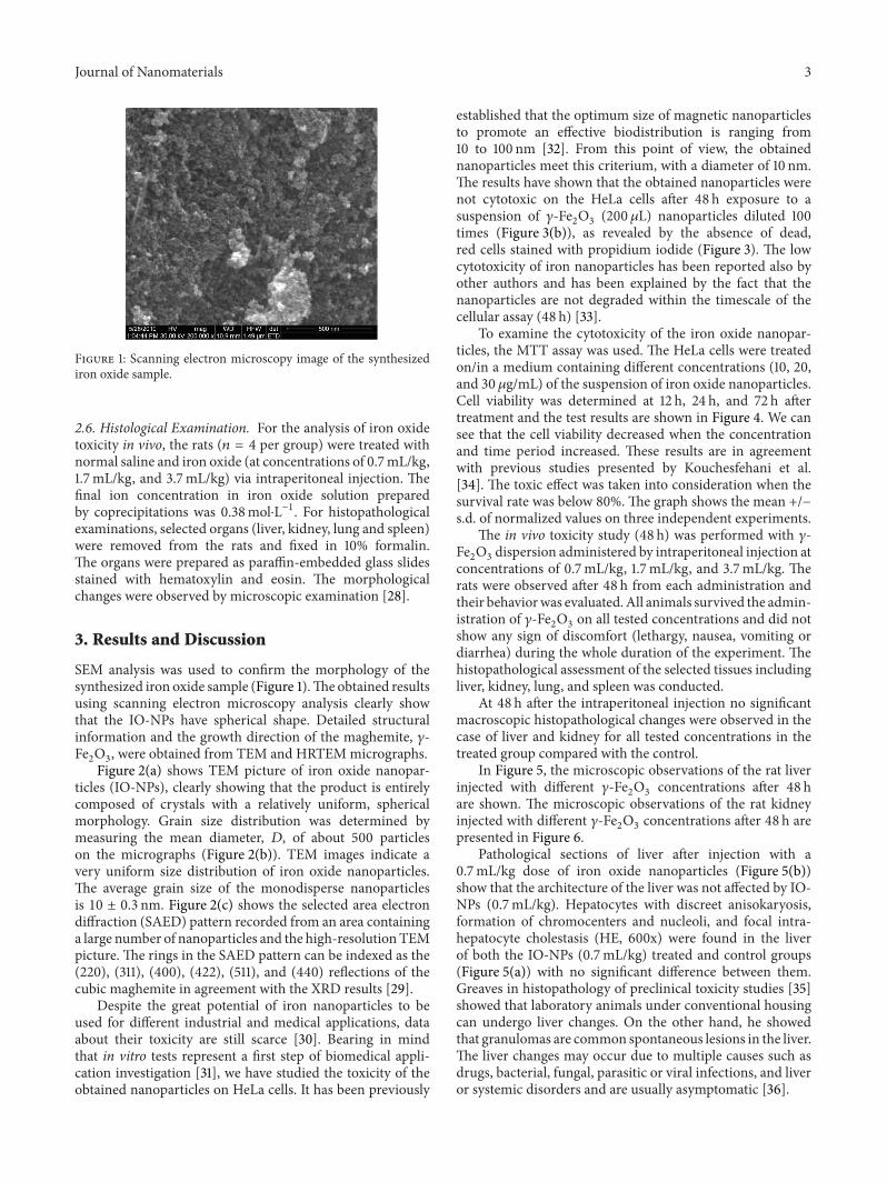
Figure 1: Scanning electron microscopy image of the synthesizediron oxide sample.
2.6. Histological Examination. For the analysis of iron oxidetoxicity in vivo, the rats (𝑛 = 4 per group) were treated withnormal saline and iron oxide (at concentrations of 0.7mL/kg,1.7mL/kg, and 3.7mL/kg) via intraperitoneal injection. Thefinal ion concentration in iron oxide solution preparedby coprecipitations was 0.38mol⋅L−1. For histopathologicalexaminations, selected organs (liver, kidney, lung and spleen)were removed from the rats and fixed in 10% formalin.The organs were prepared as paraffin-embedded glass slidesstained with hematoxylin and eosin. The morphologicalchanges were observed by microscopic examination [28].
3. Results and Discussion
SEM analysis was used to confirm the morphology of thesynthesized iron oxide sample (Figure 1).The obtained resultsusing scanning electron microscopy analysis clearly showthat the IO-NPs have spherical shape. Detailed structuralinformation and the growth direction of the maghemite, 𝛾-Fe2O3, were obtained from TEM and HRTEMmicrographs.
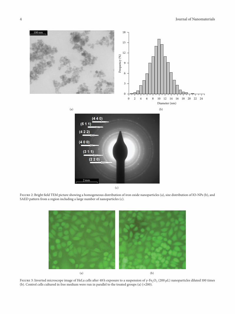
Figure 2(a) shows TEM picture of iron oxide nanopar-ticles (IO-NPs), clearly showing that the product is entirelycomposed of crystals with a relatively uniform, sphericalmorphology. Grain size distribution was determined bymeasuring the mean diameter, 𝐷, of about 500 particleson the micrographs (Figure 2(b)). TEM images indicate avery uniform size distribution of iron oxide nanoparticles.The average grain size of the monodisperse nanoparticlesis 10 ± 0.3 nm. Figure 2(c) shows the selected area electrondiffraction (SAED) pattern recorded from an area containinga large number of nanoparticles and the high-resolutionTEMpicture. The rings in the SAED pattern can be indexed as the(220), (311), (400), (422), (511), and (440) reflections of thecubic maghemite in agreement with the XRD results [29].
Despite the great potential of iron nanoparticles to beused for different industrial and medical applications, dataabout their toxicity are still scarce [30]. Bearing in mindthat in vitro tests represent a first step of biomedical appli-cation investigation [31], we have studied the toxicity of theobtained nanoparticles on HeLa cells. It has been previously
established that the optimum size of magnetic nanoparticlesto promote an effective biodistribution is ranging from10 to 100 nm [32]. From this point of view, the obtainednanoparticles meet this criterium, with a diameter of 10 nm.The results have shown that the obtained nanoparticles werenot cytotoxic on the HeLa cells after 48 h exposure to asuspension of 𝛾-Fe
2O3(200𝜇L) nanoparticles diluted 100
times (Figure 3(b)), as revealed by the absence of dead,red cells stained with propidium iodide (Figure 3). The lowcytotoxicity of iron nanoparticles has been reported also byother authors and has been explained by the fact that thenanoparticles are not degraded within the timescale of thecellular assay (48 h) [33].
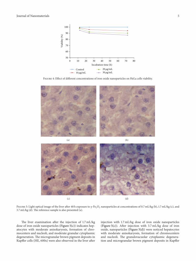
To examine the cytotoxicity of the iron oxide nanopar-ticles, the MTT assay was used. The HeLa cells were treatedon/in a medium containing different concentrations (10, 20,and 30 𝜇g/mL) of the suspension of iron oxide nanoparticles.Cell viability was determined at 12 h, 24 h, and 72 h aftertreatment and the test results are shown in Figure 4. We cansee that the cell viability decreased when the concentrationand time period increased. These results are in agreementwith previous studies presented by Kouchesfehani et al.[34]. The toxic effect was taken into consideration when thesurvival rate was below 80%. The graph shows the mean +/−s.d. of normalized values on three independent experiments.
The in vivo toxicity study (48 h) was performed with 𝛾-Fe2O3dispersion administered by intraperitoneal injection at
concentrations of 0.7mL/kg, 1.7mL/kg, and 3.7mL/kg. Therats were observed after 48 h from each administration andtheir behaviorwas evaluated.All animals survived the admin-istration of 𝛾-Fe
2O3on all tested concentrations and did not
show any sign of discomfort (lethargy, nausea, vomiting ordiarrhea) during the whole duration of the experiment. Thehistopathological assessment of the selected tissues includingliver, kidney, lung, and spleen was conducted.
At 48 h after the intraperitoneal injection no significantmacroscopic histopathological changes were observed in thecase of liver and kidney for all tested concentrations in thetreated group compared with the control.
In Figure 5, the microscopic observations of the rat liverinjected with different 𝛾-Fe
2O3concentrations after 48 h
are shown. The microscopic observations of the rat kidneyinjected with different 𝛾-Fe
2O3concentrations after 48 h are
presented in Figure 6.Pathological sections of liver after injection with a
0.7mL/kg dose of iron oxide nanoparticles (Figure 5(b))show that the architecture of the liver was not affected by IO-NPs (0.7mL/kg). Hepatocytes with discreet anisokaryosis,formation of chromocenters and nucleoli, and focal intra-hepatocyte cholestasis (HE, 600x) were found in the liverof both the IO-NPs (0.7mL/kg) treated and control groups(Figure 5(a)) with no significant difference between them.Greaves in histopathology of preclinical toxicity studies [35]showed that laboratory animals under conventional housingcan undergo liver changes. On the other hand, he showedthat granulomas are common spontaneous lesions in the liver.The liver changes may occur due to multiple causes such asdrugs, bacterial, fungal, parasitic or viral infections, and liveror systemic disorders and are usually asymptomatic [36].
4 Journal of Nanomaterials
100nm
(a)
0 2 4 6 8 10 12 14 16 18 20 22 24Diameter (nm)
0
3
6
9
12
15
18
Freq
uenc
y (%
)
(b)
2mm
(c)
Figure 2: Bright field TEM picture showing a homogeneous distribution of iron oxide nanoparticles (a), size distribution of IO-NPs (b), andSAED pattern from a region including a large number of nanoparticles (c).
(a) (b)
Figure 3: Inverted microscope image of HeLa cells after 48 h exposure to a suspension of 𝛾-Fe2O3(200𝜇L) nanoparticles diluted 100 times
(b). Control cells cultured in free medium were run in parallel to the treated groups (a) (×200).
Journal of Nanomaterials 5
50
60
70
80
90
100
Viab
ility
(%)
0 10 20 30 40 50 60 70 80Incubation time (h)
Control10𝜇g/mL
20𝜇g/mL30𝜇g/mL
Figure 4: Effect of different concentrations of iron oxide nanoparticles on HeLa cells viability.
(a) (b)
(c) (d)
Figure 5: Light optical image of the liver after 48 h exposure to 𝛾-Fe2O3nanoparticles at concentrations of 0.7mL/kg (b), 1.7mL/kg (c), and
3.7mL/kg (d). The reference sample is also presented (a).
The liver examination after the injection of 1.7mL/kgdose of iron oxide nanoparticles (Figure 5(c)) indicates hep-atocytes with moderate anisokaryosis, formation of chro-mocenters and nucleoli, and moderate granular cytoplasmicdegeneration. The microgranular brown pigment deposits inKupffer cells (HE, 600x) were also observed in the liver after
injection with 1.7mL/kg dose of iron oxide nanoparticles(Figure 5(c)). After injection with 3.7mL/kg dose of ironoxide, nanoparticles (Figure 5(d)) were noticed hepatocyteswith moderate anisokaryosis, formation of chromocentersand nucleoli. The granulovacuolar cytoplasmic degenera-tion and microgranular brown pigment deposits in Kupffer
6 Journal of Nanomaterials
(a) (b)
(c) (d)
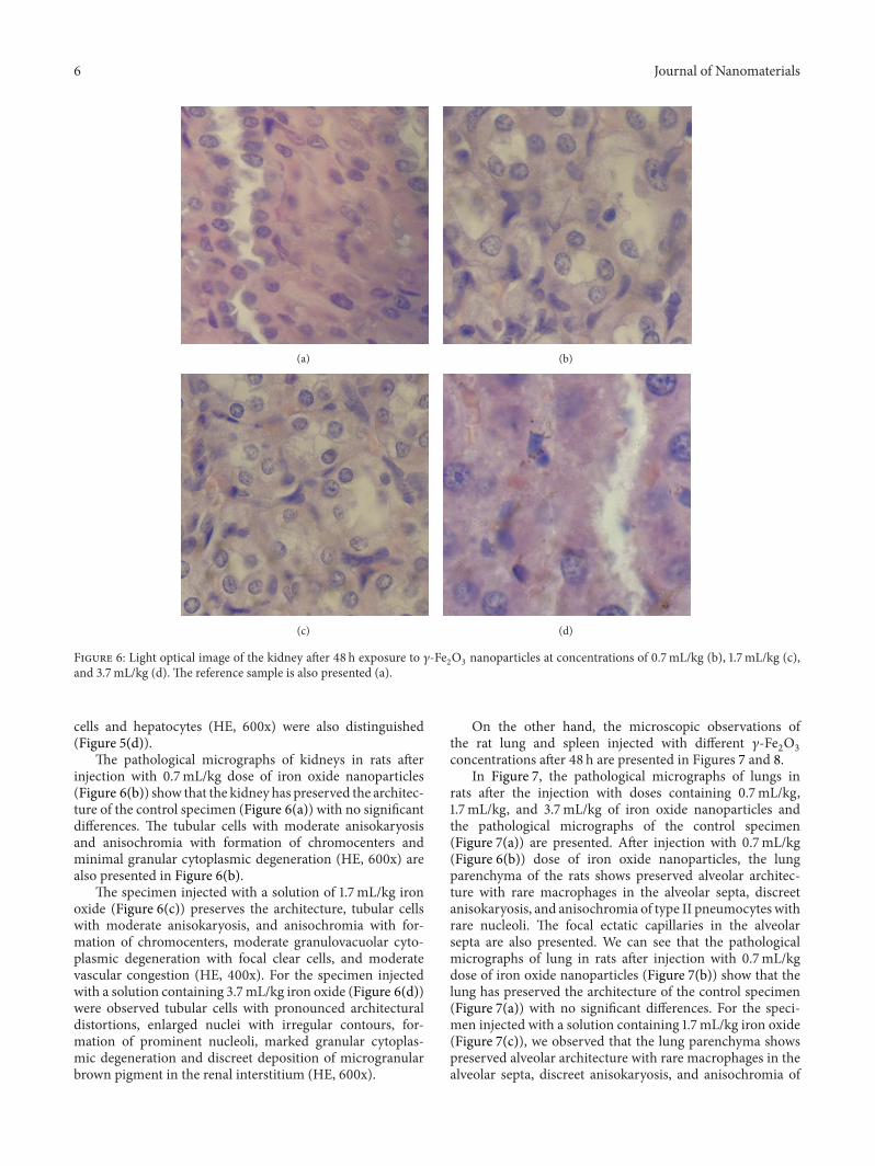
Figure 6: Light optical image of the kidney after 48 h exposure to 𝛾-Fe2O3nanoparticles at concentrations of 0.7mL/kg (b), 1.7mL/kg (c),
and 3.7mL/kg (d). The reference sample is also presented (a).
cells and hepatocytes (HE, 600x) were also distinguished(Figure 5(d)).
The pathological micrographs of kidneys in rats afterinjection with 0.7mL/kg dose of iron oxide nanoparticles(Figure 6(b)) show that the kidney has preserved the architec-ture of the control specimen (Figure 6(a)) with no significantdifferences. The tubular cells with moderate anisokaryosisand anisochromia with formation of chromocenters andminimal granular cytoplasmic degeneration (HE, 600x) arealso presented in Figure 6(b).
The specimen injected with a solution of 1.7mL/kg ironoxide (Figure 6(c)) preserves the architecture, tubular cellswith moderate anisokaryosis, and anisochromia with for-mation of chromocenters, moderate granulovacuolar cyto-plasmic degeneration with focal clear cells, and moderatevascular congestion (HE, 400x). For the specimen injectedwith a solution containing 3.7mL/kg iron oxide (Figure 6(d))were observed tubular cells with pronounced architecturaldistortions, enlarged nuclei with irregular contours, for-mation of prominent nucleoli, marked granular cytoplas-mic degeneration and discreet deposition of microgranularbrown pigment in the renal interstitium (HE, 600x).
On the other hand, the microscopic observations ofthe rat lung and spleen injected with different 𝛾-Fe
2O3
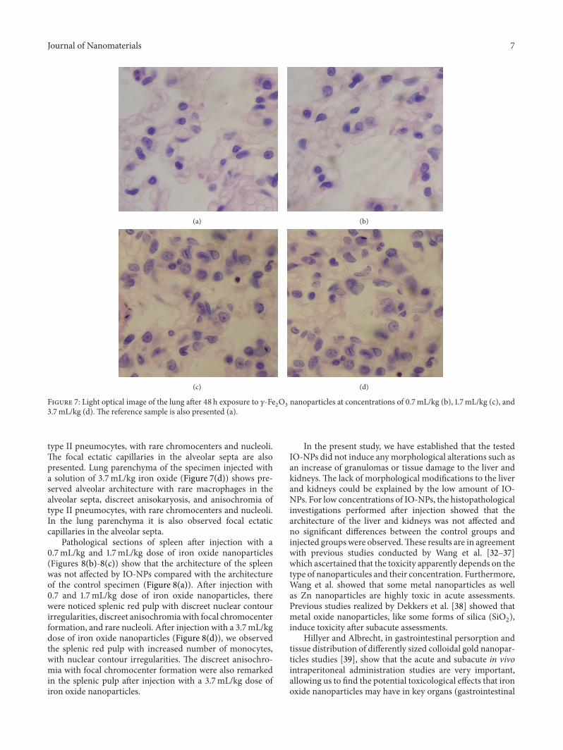
concentrations after 48 h are presented in Figures 7 and 8.In Figure 7, the pathological micrographs of lungs in
rats after the injection with doses containing 0.7mL/kg,1.7mL/kg, and 3.7mL/kg of iron oxide nanoparticles andthe pathological micrographs of the control specimen(Figure 7(a)) are presented. After injection with 0.7mL/kg(Figure 6(b)) dose of iron oxide nanoparticles, the lungparenchyma of the rats shows preserved alveolar architec-ture with rare macrophages in the alveolar septa, discreetanisokaryosis, and anisochromia of type II pneumocytes withrare nucleoli. The focal ectatic capillaries in the alveolarsepta are also presented. We can see that the pathologicalmicrographs of lung in rats after injection with 0.7mL/kgdose of iron oxide nanoparticles (Figure 7(b)) show that thelung has preserved the architecture of the control specimen(Figure 7(a)) with no significant differences. For the speci-men injected with a solution containing 1.7mL/kg iron oxide(Figure 7(c)), we observed that the lung parenchyma showspreserved alveolar architecture with rare macrophages in thealveolar septa, discreet anisokaryosis, and anisochromia of
Journal of Nanomaterials 7
(a) (b)
(c) (d)
Figure 7: Light optical image of the lung after 48 h exposure to 𝛾-Fe2O3nanoparticles at concentrations of 0.7mL/kg (b), 1.7mL/kg (c), and
3.7mL/kg (d). The reference sample is also presented (a).
type II pneumocytes, with rare chromocenters and nucleoli.The focal ectatic capillaries in the alveolar septa are alsopresented. Lung parenchyma of the specimen injected witha solution of 3.7mL/kg iron oxide (Figure 7(d)) shows pre-served alveolar architecture with rare macrophages in thealveolar septa, discreet anisokaryosis, and anisochromia oftype II pneumocytes, with rare chromocenters and nucleoli.In the lung parenchyma it is also observed focal ectaticcapillaries in the alveolar septa.
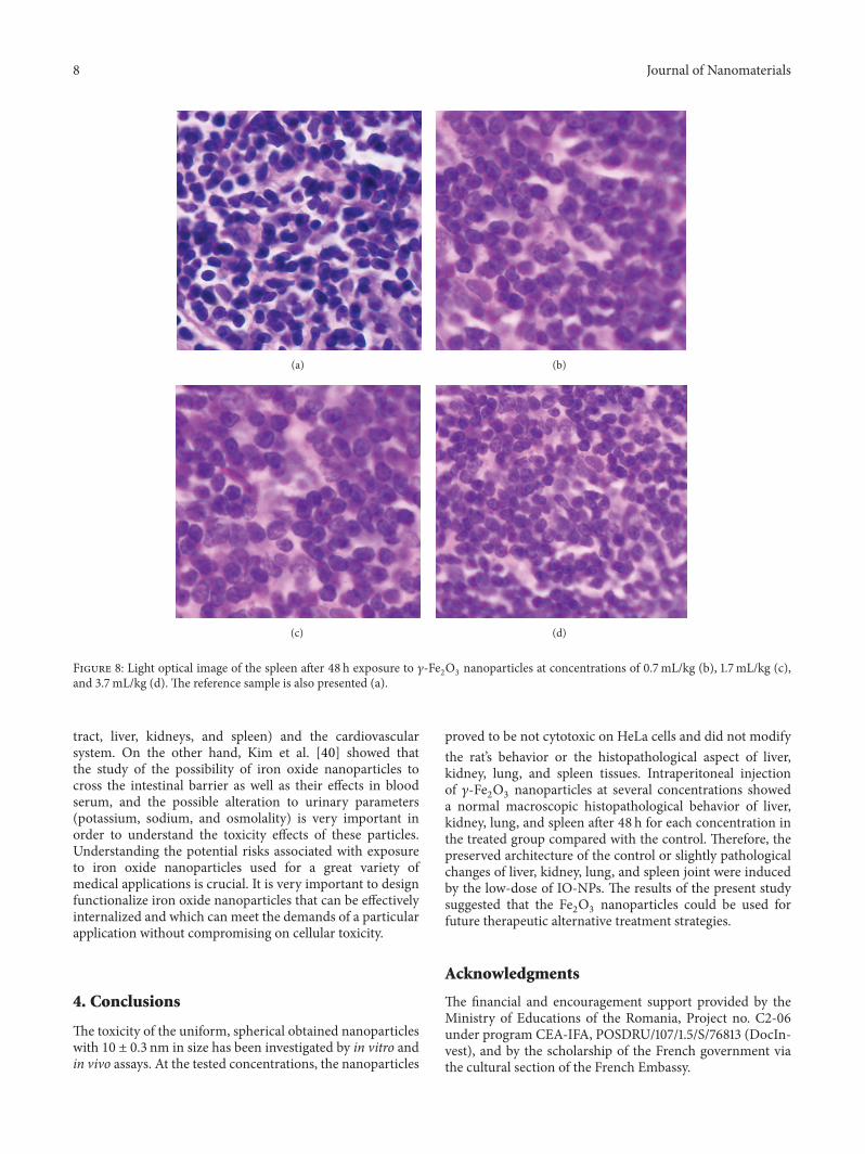
Pathological sections of spleen after injection with a0.7mL/kg and 1.7mL/kg dose of iron oxide nanoparticles(Figures 8(b)-8(c)) show that the architecture of the spleenwas not affected by IO-NPs compared with the architectureof the control specimen (Figure 8(a)). After injection with0.7 and 1.7mL/kg dose of iron oxide nanoparticles, therewere noticed splenic red pulp with discreet nuclear contourirregularities, discreet anisochromiawith focal chromocenterformation, and rare nucleoli. After injection with a 3.7mL/kgdose of iron oxide nanoparticles (Figure 8(d)), we observedthe splenic red pulp with increased number of monocytes,with nuclear contour irregularities. The discreet anisochro-mia with focal chromocenter formation were also remarkedin the splenic pulp after injection with a 3.7mL/kg dose ofiron oxide nanoparticles.
In the present study, we have established that the testedIO-NPs did not induce anymorphological alterations such asan increase of granulomas or tissue damage to the liver andkidneys. The lack of morphological modifications to the liverand kidneys could be explained by the low amount of IO-NPs. For low concentrations of IO-NPs, the histopathologicalinvestigations performed after injection showed that thearchitecture of the liver and kidneys was not affected andno significant differences between the control groups andinjected groupswere observed.These results are in agreementwith previous studies conducted by Wang et al. [32–37]which ascertained that the toxicity apparently depends on thetype of nanoparticules and their concentration. Furthermore,Wang et al. showed that some metal nanoparticles as wellas Zn nanoparticles are highly toxic in acute assessments.Previous studies realized by Dekkers et al. [38] showed thatmetal oxide nanoparticles, like some forms of silica (SiO
2),
induce toxicity after subacute assessments.Hillyer and Albrecht, in gastrointestinal persorption and
tissue distribution of differently sized colloidal gold nanopar-ticles studies [39], show that the acute and subacute in vivointraperitoneal administration studies are very important,allowing us to find the potential toxicological effects that ironoxide nanoparticles may have in key organs (gastrointestinal
8 Journal of Nanomaterials
(a) (b)
(c) (d)
Figure 8: Light optical image of the spleen after 48 h exposure to 𝛾-Fe2O3nanoparticles at concentrations of 0.7mL/kg (b), 1.7mL/kg (c),
and 3.7mL/kg (d). The reference sample is also presented (a).
tract, liver, kidneys, and spleen) and the cardiovascularsystem. On the other hand, Kim et al. [40] showed thatthe study of the possibility of iron oxide nanoparticles tocross the intestinal barrier as well as their effects in bloodserum, and the possible alteration to urinary parameters(potassium, sodium, and osmolality) is very important inorder to understand the toxicity effects of these particles.Understanding the potential risks associated with exposureto iron oxide nanoparticles used for a great variety ofmedical applications is crucial. It is very important to designfunctionalize iron oxide nanoparticles that can be effectivelyinternalized and which can meet the demands of a particularapplication without compromising on cellular toxicity.
4. Conclusions
The toxicity of the uniform, spherical obtained nanoparticleswith 10 ± 0.3 nm in size has been investigated by in vitro andin vivo assays. At the tested concentrations, the nanoparticles
proved to be not cytotoxic on HeLa cells and did not modifythe rat’s behavior or the histopathological aspect of liver,kidney, lung, and spleen tissues. Intraperitoneal injectionof 𝛾-Fe
2O3nanoparticles at several concentrations showed
a normal macroscopic histopathological behavior of liver,kidney, lung, and spleen after 48 h for each concentration inthe treated group compared with the control. Therefore, thepreserved architecture of the control or slightly pathologicalchanges of liver, kidney, lung, and spleen joint were inducedby the low-dose of IO-NPs. The results of the present studysuggested that the Fe
2O3nanoparticles could be used for
future therapeutic alternative treatment strategies.
Acknowledgments
The financial and encouragement support provided by theMinistry of Educations of the Romania, Project no. C2-06under program CEA-IFA, POSDRU/107/1.5/S/76813 (DocIn-vest), and by the scholarship of the French government viathe cultural section of the French Embassy.
Journal of Nanomaterials 9
References
[1] S. Laurent, D. Forge, M. Port et al., “Magnetic iron oxide nano-particles: synthesis, stabilization, vectorization, physic chem-ical characterizations and biological applications,” ChemicalReviews, vol. 108, no. 6, pp. 2064–2110, 2008.
[2] S. Mornet, S. Vasseur, F. Grasset, and E. Duguet, “Magneticnanoparticle design for medical diagnosis and therapy,” Journalof Materials Chemistry, vol. 14, no. 14, pp. 2161–2175, 2004.
[3] A. M. Prodan, S. L. Iconaru, C. M. Chifiriuc et al., “Magneticproperties and biological activity evaluation of iron oxidenanoparticles,” Journal of Nanomaterials, vol. 2013, Article ID893970, 7 pages, 2013.
[4] E. Katz and I. Willner, “Integrated nanoparticle-biomoleculehybrid systems: synthesis, properties, and applications,” Ange-wandte Chemie, vol. 43, no. 45, pp. 6042–6108, 2004.
[5] S. Laurent, S. Dutz, U. O. Hafeli, and M. Mahmoudi, “Magneticfluid hyperthermia: focus on superparamagnetic iron oxidenanoparticles,” Advances in Colloid and Interface Science, vol.166, no. 1-2, pp. 8–23, 2011.
[6] C. H. Cunningham, T. Arai, P. C. Yang, M. V. McConnell, J. M.Pauly, and S. M. Conolly, “Positive contrast magnetic resonanceimaging of cells labeled with magnetic nanoparticles,”MagneticResonance in Medicine, vol. 53, no. 5, pp. 999–1005, 2005.
[7] S. A. Anderson, R. K. Rader, W. F. Westlin et al., “Mag-netic resonance contrast enhancement of neovasculature withalpha(v)beta(3)-targeted nanoparticles,”Magnetic Resonance inMedicine, vol. 44, no. 3, pp. 433–439, 2000.
[8] B. Polyak andG. Friedman, “Magnetic targeting for site-specificdrug delivery: applications and clinical potential,” Expert Opin-ion on Drug Delivery, vol. 6, no. 1, pp. 53–70, 2009.
[9] R. Weissleder, H.-C. Cheng, A. Bogdanova, and A. BogdanovJr., “Magnetically labeled cells can be detected by MR imaging,”Journal ofMagnetic Resonance Imaging, vol. 7, no. 1, pp. 258–263,1997.
[10] E. A. Schellenberger, F. Reynolds, R. Weissleder, and L. Joseph-son, “Surface-functionalized nanoparticle library yields probesfor apoptotic cells,” ChemBioChem, vol. 5, no. 3, pp. 275–279,2004.
[11] A. R. Jalilian, A. Panahifar, M. Mahmoudi, M. Akhlaghi, and A.Simchi, “Preparation and biological evaluation of [67Ga]-labeled- superparamagnetic nanoparticles in normal rats,”Radiochimica Acta, vol. 97, no. 1, pp. 51–56, 2009.
[12] C. Corot, P. Robert, J.-M. Idee, and M. Port, “Recent advancesin iron oxide nanocrystal technology for medical imaging,”Advanced Drug Delivery Reviews, vol. 58, no. 14, pp. 1471–1504,2006.
[13] C. Fan, W. Gao, Z. Chen et al., “Tumor selectivity of stealthmulti-functionalized superparamagnetic iron oxide nanoparti-cles,” International Journal of Pharmaceutics, vol. 404, no. 1-2,pp. 180–190, 2011.
[14] Q. A. Pankhurst, J. Connolly, S. K. Jones, and J. Dobson, “Appli-cations of magnetic nanoparticles in biomedicine,” Journal ofPhysics D, vol. 36, no. 13, pp. R167–R181, 2003.
[15] R. K. Gilchrist,W. D. Shorey, R. C. Hanselman, J. C. Parrott, andC. B. Taylor, “Selective inductive heating of lymph,” Annals ofSurgery, vol. 146, pp. 596–606, 1957.
[16] T. R. Pisanic II, J. D. Blackwell, V. I. Shubayev, R. R. Finones, andS. Jin, “Nanotoxicity of iron oxide nanoparticle internalizationin growing neurons,”Biomaterials, vol. 28, no. 16, pp. 2572–2581,2007.
[17] R. Massart, “Magnetic fluids and process for obtaining them,”US Patent 4329241, 1982.
[18] R.Massart, “Preparation of aqueousmagnetic liquids in alkalineand acidic media,” IEEE Transactions on Magnetics, vol. 17, pp.1247–1248, 1981.
[19] R. Massart, J. Roger, and V. Cabuil, “New trends in chemistryof magnetic colloids: polar and non polar magnetic fluids,emulsions, capsules and vesicles,” Brazilian Journal of Physics,vol. 25, no. 2, pp. 135–141, 1995.
[20] D. Predoi and C. Valsangiacom, “Thermal studies of magneticspinel iron oxide in solution,” Journal of Optoelectronics andAdvanced Materials, vol. 9, no. 6, pp. 1797–1799, 2007.
[21] D. Zins, V. Cabuil, and R. Massart, “New aqueous magneticfluids,” Journal ofMolecular Liquids, vol. 83, no. 1–3, pp. 217–232,1999.
[22] D. Predoi, “A study on iron oxide nanoparticles coated withdextrin obtained by coprecipitation,”Digest Journal of Nanoma-terials and Biostructures, vol. 2, no. 1, pp. 169–173, 2007.
[23] S. Mornet, F. Grasset, J. Portier, and E. Duguet, “Maghemite@silica nanoparticles for biological applications,” European Cellsand Materials, vol. 3S2, article 110, 2002.
[24] A. M. Grumezescu, E. Andronescu, A. Ficai, C. Bleotu, and M.C. Chifiriuc, “Chitin based biomaterial for antimicrobial ther-apy: fabrication, characterzation and in vitro profile basedintercation with eukaryotic and prokaeryotic cells,” BiointerfaceResearch in Applied Chemistry, vol. 2, p. 446, 2012.
[25] T. Mosmann, “Rapid colorimetric assay for cellular growth andsurvival: application to proliferation and cytotoxicity assays,”Journal of Immunological Methods, vol. 65, no. 1-2, pp. 55–63,1983.
[26] F. Denizot and R. Lang, “Rapid colorimetric assay for cell gro-wth and survival: modifications to the tetrazolium dye pro-cedure giving improved sensitivity and reliability,” Journal ofImmunological Methods, vol. 89, no. 2, pp. 271–277, 1986.
[27] H. Wan, R. L. Williams, P. J. Doherty, and D. F. Williams, “Thecytotoxicity evaluation of Kevlar and silicon carbide by MTTassay,” Journal of Materials Science, vol. 5, no. 6-7, pp. 441–445,1994.
[28] B. Su, S. L. Xiang, J. Su et al., “Diallyl disulfide increases histoneacetylation and P21WAF1 expression in human gastric cancercells in vivo and in vitro,” Biochemical Pharmacology, vol. 1, no.7, pp. 1–10, 2012.
[29] C. S. Ciobanu, S. L. Iconaru, E. Gyorgy et al., “Biomedical prop-erties and preparation of iron oxide-dextran nanostructures byMAPLE technique,” Chemistry Central Journal, vol. 6, article 17,2012.
[30] O. Krystofova, J. Sochor, O. Zitka et al., “Effect of magnetic nan-oparticles on tobacco BY-2 cell suspension culture,” Interna-tional Journal of Environmental Research and Public Health, vol.10, no. 1, pp. 47–71, 2013.
[31] L. L. C. Estevanato, J. R. Da Silva, A. M. Falqueiro et al.,“Co-nanoencapsulation of magnetic nanoparticles and selolfor breast tumor treatment: in vitro evaluation of cytotoxicityand magnetohyperthermia efficacy,” International Journal ofNanomedicine, vol. 7, pp. 5287–5299, 2012.
[32] M. L. B. Carneiro, E. S. Nunes, R. C. A. Peixoto et al., “Free Rho-dium (II) citrate and rhodium (II) citrate magnetic carriersas potential strategies for breast cancer therapy,” Journal ofNanobiotechnology, vol. 9, article 11, 2011.
[33] L. Gu, R. H. Fang, M. J. Sailor, and J.-H. Park, “In vivo clearanceand toxicity of monodisperse iron oxide nanocrystals,” ACSNano, vol. 6, no. 6, pp. 4947–4954, 2012.
10 Journal of Nanomaterials
[34] H. M. Kouchesfehani, S. rKiani, A. A. Rostami, and R. Fakheri,“Cytotoxic effect of iron oxide nanoparticles on mouse embry-onic stem cells byMTT assay,” Iranian Journal of Toxicology, vol.7, no. 21, pp. 849–853, 2013.
[35] P. Greaves, “Liver and pancreas,” inHistopathology of PreclinicalToxicity Studies, pp. 457–503, Academic Press, Elsevier, NewYork, NY, USA, 2007.
[36] A. Shiga, Y. Ota, Y. Ueda et al., “Study on the pathogenesis offoreign body granulomatous inflammation in the livers ofsprague-dawley rats,” Journal of Toxicologic Pathology, vol. 23,no. 4, pp. 253–260, 2010.
[37] B.Wang,W. Feng,M.Wang et al., “Acute toxicological impact ofnano- and submicro-scaled zinc oxide powder on healthy adultmice,” Journal of Nanoparticle Research, vol. 10, no. 2, pp. 263–276, 2008.
[38] S. Dekkers, P. Krystek, R. J. B. Peters et al., “Presence and risksof nanosilica in food products,”Nanotoxicology, vol. 5, no. 3, pp.393–405, 2011.
[39] J. F. Hillyer and R. M. Albrecht, “Gastrointestinal persorptionand tissue distribution of differently sized colloidal goldnanoparticles,” Journal of Pharmaceutical Sciences, vol. 90, no.12, pp. 1927–1936, 2001.
[40] Y. S. Kim, J. S. Kim,H. S. Cho et al., “Twenty-eight-day oral toxi-city, genotoxicity, and gender-related tissue distribution of silvernanoparticles in Sprague-Dawley rats,” Inhalation Toxicology,vol. 20, no. 6, pp. 575–583, 2008.

Submit your manuscripts athttp://www.hindawi.com
ScientificaHindawi Publishing Corporationhttp://www.hindawi.com Volume 2014
CorrosionInternational Journal of
Hindawi Publishing Corporationhttp://www.hindawi.com Volume 2014
Polymer ScienceInternational Journal of
Hindawi Publishing Corporationhttp://www.hindawi.com Volume 2014
Hindawi Publishing Corporationhttp://www.hindawi.com Volume 2014
CeramicsJournal of
Hindawi Publishing Corporationhttp://www.hindawi.com Volume 2014
CompositesJournal of
NanoparticlesJournal of
Hindawi Publishing Corporationhttp://www.hindawi.com Volume 2014
Hindawi Publishing Corporationhttp://www.hindawi.com Volume 2014
International Journal of
Biomaterials
Hindawi Publishing Corporationhttp://www.hindawi.com Volume 2014
NanoscienceJournal of
TextilesHindawi Publishing Corporation http://www.hindawi.com Volume 2014
Journal of
NanotechnologyHindawi Publishing Corporationhttp://www.hindawi.com Volume 2014
Journal of
CrystallographyJournal of
Hindawi Publishing Corporationhttp://www.hindawi.com Volume 2014
The Scientific World JournalHindawi Publishing Corporation http://www.hindawi.com Volume 2014
Hindawi Publishing Corporationhttp://www.hindawi.com Volume 2014
CoatingsJournal of
Advances in
Materials Science and EngineeringHindawi Publishing Corporationhttp://www.hindawi.com Volume 2014
Smart Materials Research
Hindawi Publishing Corporationhttp://www.hindawi.com Volume 2014
Hindawi Publishing Corporationhttp://www.hindawi.com Volume 2014
MetallurgyJournal of
Hindawi Publishing Corporationhttp://www.hindawi.com Volume 2014
BioMed Research International
MaterialsJournal of
Hindawi Publishing Corporationhttp://www.hindawi.com Volume 2014
Nano
materials
Hindawi Publishing Corporationhttp://www.hindawi.com Volume 2014
Journal ofNanomaterials
Related Documents