Chemical Pathology Lecture Notes 2011 University of Cape Town Preface This manuscript constitutes a series of lecture notes prepared and updated by members of the Division of Chemical Pathology at UCT. These notes are intended to provide a clear and concise reference of the basic biochemical principles and clinical application of the field of Chemical Pathology/Clinical Biochemistry. These lectures have been developed specifically for undergraduate medical student teaching. Included in lectures 1 -11, 14, 18, 20, 22- 25 and 27 are a series of related questions and answers that can be used as an excellent tool to test knowledge and provide insight into a wide range of clinical problems. These lectures are published under Creative Commons Copyright (Attribution-Non Commercial- Share alike -2.5-South Africa) and may be reproduced for teaching purposes in this exact format provided they are not distributed for profit and that all original authors are acknowledged Experts in the field who may find this resource useful are invited to contribute corrections, submit revisions of lectures or submit new lectures on relevant topics. In this way it is hoped that this work will improve and remain current as teaching tool. George van der Watt Acting Head of Department Division of Chemical Pathology University of Cape Town. Contact: e-mail: [email protected]

Welcome message from author
This document is posted to help you gain knowledge. Please leave a comment to let me know what you think about it! Share it to your friends and learn new things together.
Transcript
Chemical Pathology Lecture Notes 2011
University of Cape Town
Preface
This manuscript constitutes a series of lecture notes prepared and updated by members of the
Division of Chemical Pathology at UCT. These notes are intended to provide a clear and concise
reference of the basic biochemical principles and clinical application of the field of Chemical
Pathology/Clinical Biochemistry. These lectures have been developed specifically for undergraduate
medical student teaching.
Included in lectures 1 -11, 14, 18, 20, 22- 25 and 27 are a series of related questions and answers
that can be used as an excellent tool to test knowledge and provide insight into a wide range of clinical
problems.
These lectures are published under Creative Commons Copyright (Attribution-Non Commercial-
Share alike -2.5-South Africa) and may be reproduced for teaching purposes in this exact format
provided they are not distributed for profit and that all original authors are acknowledged
Experts in the field who may find this resource useful are invited to contribute corrections, submit
revisions of lectures or submit new lectures on relevant topics. In this way it is hoped that this work will
improve and remain current as teaching tool.
George van der Watt
Acting Head of Department
Division of Chemical Pathology
University of Cape Town.
Contact:
e-mail: [email protected]
2
Table of Contents
Table of Contents .......................................................................................................................................... 1
Lecture 1: Introductory Lecture And Basic Statistics. .................................................................................... 3
Lecture 2: Acid-Base Balance .................................................................................................................... 17
Lecture 3: Disorders Of Renal Function ..................................................................................................... 47
Lecture 4: Disorders Of Water And Sodium Balance ................................................................................. 65
Lecture 5: Disorders Of Potassium Metabolism ......................................................................................... 79
Lecture 6: Carbohydrate Metabolsim And Diabetes ................................................................................... 91
Lecture 7: Calcium, Magnesium And Phosphate Metabolism...................................................................126
Lecture 8: Iron Metabolism ....................................................................................................................... 150
Lecture 9: Lipid And Lipoprotein Metabolism And Dyslipidaemias ............................................................ 170
Lecture 10: Obesity ................................................................................................................................... 200
Lecture 11: Biochemistry Of Alcohol Abuse ............................................................................................. 211
Lecture 12: Disorders Of Protein Metabolism ............................................................................................ 221
Lecture 13: Cerebrospinal Fluid ................................................................................................................. 247
Lecture 14: Chemical Pathology Of Liver Disease ................................................................................... 255
Lecture 15: Enzymology............................................................................................................................. 281
Lecture 16: Chemical Pathology Of The Gastro-Intestinal Tract ............................................................... 293
Lecture 17: Endocrinology 1: Pituitary And Hypothalamus ........................................................................ 310
Lecture 18: Endocrinology 2: Pituitary Diseases ....................................................................................... 315
Lecture 19: Endocrinology 3: Adrenal Diseases ........................................................................................ 327
Lecture 20: Endocrinology 4: Adrenal Dysfunction: Hypercortisolism And Hyperaldosteronism .............. 334
Lecture 21: Endocrinology 5: Hypoadrenalsim And Congenital Adrenal Hyperplasia ............................... 345
Lecture 22: Endocrinology 6: Thyroid ....................................................................................................... 352
Lecture 23: Endocrinology 7: Disorders Of Gonadal Function ................................................................. 361
Lecture 24: Vitamins ................................................................................................................................. 372
Lecture 25: Uric Acid And Gout ................................................................................................................ 390
Lecture 26: Inherited Metabolic Diseases .................................................................................................. 400
Lecture 27- Disorders Of Porphyrin Metabolism ....................................................................................... 420
Lecture 28: Tumour Markers ..................................................................................................................... 433
Lecture 29: Pregnancy And Parental Screening ........................................................................................ 448
3
Lecture 1: Introductory Lecture And Basic Statistics
PROF E HARLEY.B. UPDATED PROF TS PILLAY 2007
WHAT IS CHEMICAL PATHOLOGY?
Chemical Pathology (also known as Clinical Biochemistry/Clinical Chemistry) is the study of the biochemical
basis of disease, and the application of biochemical and molecular techniques in diagnosis. An allied
subspecialty of Chemical Pathology is Metabolic Medicine which deals with metabolic disease in all its
manifestations. The Division of Chemical Pathology at the University of Cape Town is involved in teaching at
undergraduate and postgraduate levels and provides diagnostic laboratory services for Groote Schuur
Hospital.
An understanding of the biochemical mechanisms of disease states has provided modern medicine with a
rational basis for diagnosis and therapy. The course will equip you with the conceptual tools you will require to
understand the meaning and interpretation of diagnostic tests, as well as introducing new advances in
molecular aspects of medicine. Chemical Pathology is a logical, scientific subject, and is at the interface
between the practice of medicine and cutting-edge scientific developments. NB: More than 75% of medical
diagnoses require the services of the laboratory.
What is the role of Chemical Pathology in health care?
Chemical Pathology is the branch of pathology dealing with the biochemical basis of disease and the use of
biochemical tests for diagnosis and management. Doctors in the specialty have dual responsibilities. First
there is the provision of a reliable analytical service, for example measuring serum electrolytes, indices of
liver function, hormones, drugs and tumour markers in hundreds of patient samples every day. Many of these
analyses are performed on automated analysers, usually operated by technologists, but the management of
the process (and the staff), assurance of quality and provision of guidance on the selection of tests and
assessment of the significance of the results (particularly with some of the less generally familiar tests) are
the province of the chemical pathologist.
Secondly, Chemical Pathologists have an important clinical role, not only advising on the management of
patients with metabolic disturbances but in several countries now, they are increasingly having direct
responsibility for such patients in out-patient clinics and on the wards.
Chemical Pathology not only brings together science and medicine, it relates to all the medical specialities.
Chemical Pathologists are frequently consulted about further investigation or management of patients found
to have biochemical abnormalities on 'routine' testing. They frequently have to deal with investigating patients
with dyslipidaemias, diabetes and hypertension, review ward patients receiving artificial nutrition, discuss the
introduction of a new diagnostic test with consultant colleagues, review the quality of the laboratory's
analytical service and manage research projects of trainees. (Adapted from Dr William Marshall.)
Under application of biochemical techniques can be listed the following:
• Diagnosis: tests can be used to help differentiate between various possibilities in the differential diagnosis based on the initial history and examination when the patient first presents.

4
• Screening: detection of disease before it is clinically evident, e.g. testing all infants at birth for a specific inherited disease (phenylketonuria, thyroid deficiency)
• Monitoring: following the progression of disease processes, checking against adverse drug effects (e.g. hypokalaemia with diuretic therapy), or response to therapy (glucose levels in diabetes mellitus).
• Prognosis: providing information on disease susceptibility, e.g. cholesterol to predict heart disease.
MEASUREMENTS IN CHEMICAL PATHOLOGY: concentration of a substance
There are 2 types of units of concentration, molar units, and mass units. The former is preferable, since it is a better comparative descriptor of the concentration of a substance, but unfortunately many countries still cling to the older mass units, usually grams/100 ml, and it is often necessary to convert.
I mole of a compound corresponds to a mass (in grams) equal to the molecular weight of that compound,
e.g. 1 mole of NaCl = (23+35) =58 grams of the salt
or, 1mole/liter (mol/l) of NaCl = 58 g/l or 5.8 g/100ml
Abbreviations:
1 mol/l = 1 M = 1 molar
1 mmol/l = 1 mM = 1 millimolar = 10-3 M
1 µmol/l = 1 µM = 1 micromolar = 10-6 M
1 nM = 1 nanomolar = 10-9 M
1 pM = 1 picomolar = 10-12 M
Concentrations of some analytes (to demonstrate the range of concentrations in clinical chemistry)
serum Na+ 140 mM serum glucose 8 mM
serum Mg2+ 1 mM serum albumin 0.6 mM
serum Fe3+ 20 µM serum cortisol 500 nM
serum [H+] 40 nM plasma ACTH 50 pM
METHODS USED IN THE CHEMICAL PATHOLOGY LABORATORY
• Colorimetric methods:
Analyte reacts with a dye, changing its absorption spectrum (colour). Measured with a
spectrophotometer. Rapid & easily automated. Cheap. Concentration range: mM to µM.
Examples: urea, creatinine, phosphate, albumin, total Ca2+
• Ion-selective electrodes:
Membrane selectively permeable to an ion generates a membrane potential proportional to
concentration of free ion. Rapid & easily automated. Cheap. Concentration range: mM to nM
Examples: Na+ , H+ (pH meter), free Ca2+
• Enzymatic:
Example: lactate dehydrogenase (LDH) catalyses the reaction
5
lactate + NAD ↔ pyruvate + NADH + H+
The concentration of NADH is easily measurable by its UV absorbance, allowing the rate of the
reaction to be measured. An end-point method can be used to measure the concentration of a
substrate (e.g. the amount of NADH formed will be equal to the initial lactate concentration) or a
rate method can measure the amount of enzyme.
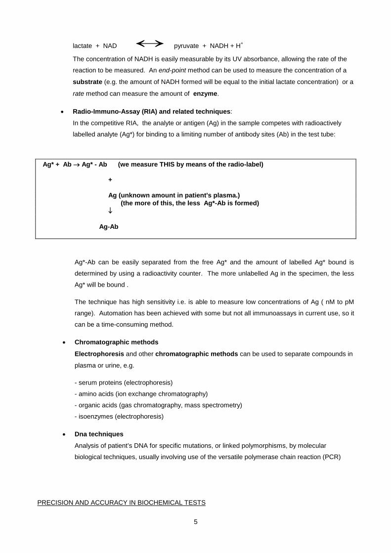
• Radio-Immuno-Assay (RIA) and related techniques:
In the competitive RIA, the analyte or antigen (Ag) in the sample competes with radioactively
labelled analyte (Ag*) for binding to a limiting number of antibody sites (Ab) in the test tube:
Ag* + Ab → Ag* - Ab (we measure THIS by means of the radio-label) + Ag (unknown amount in patient's plasma.) (the more of this, the less Ag*-Ab is formed) ↓ Ag-Ab
Ag*-Ab can be easily separated from the free Ag* and the amount of labelled Ag* bound is
determined by using a radioactivity counter. The more unlabelled Ag in the specimen, the less
Ag* will be bound .
The technique has high sensitivity i.e. is able to measure low concentrations of Ag ( nM to pM
range). Automation has been achieved with some but not all immunoassays in current use, so it
can be a time-consuming method.
• Chromatographic methods
Electrophoresis and other chromatographic methods can be used to separate compounds in
plasma or urine, e.g.
- serum proteins (electrophoresis)
- amino acids (ion exchange chromatography)
- organic acids (gas chromatography, mass spectrometry)
- isoenzymes (electrophoresis)
• Dna techniques
Analysis of patient’s DNA for specific mutations, or linked polymorphisms, by molecular
biological techniques, usually involving use of the versatile polymerase chain reaction (PCR)
PRECISION AND ACCURACY IN BIOCHEMICAL TESTS
6
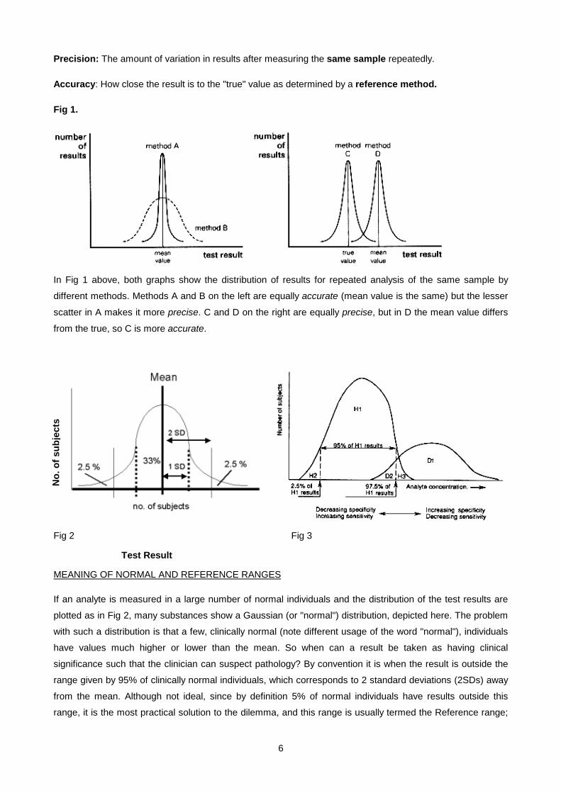
Precision: The amount of variation in results after measuring the same sample repeatedly.
Accuracy: How close the result is to the "true" value as determined by a reference method.
Fig 1.
In Fig 1 above, both graphs show the distribution of results for repeated analysis of the same sample by
different methods. Methods A and B on the left are equally accurate (mean value is the same) but the lesser
scatter in A makes it more precise. C and D on the right are equally precise, but in D the mean value differs
from the true, so C is more accurate.
Fig 2 Fig 3
Test Result
MEANING OF NORMAL AND REFERENCE RANGES
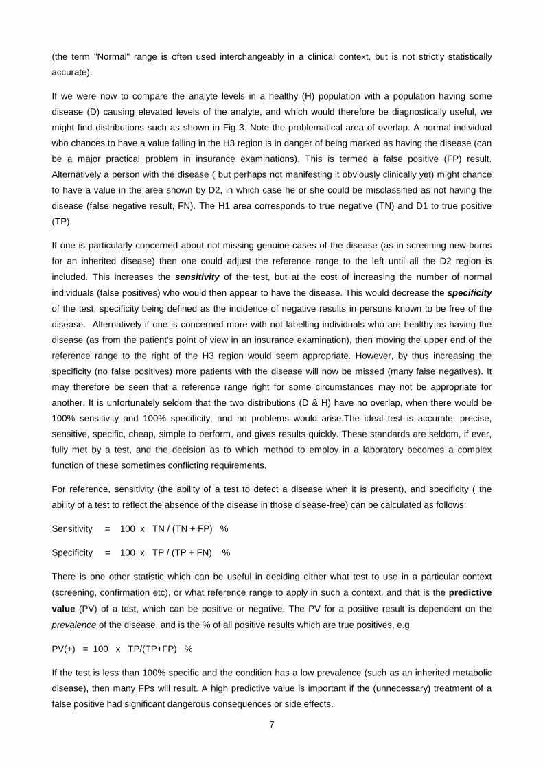
If an analyte is measured in a large number of normal individuals and the distribution of the test results are
plotted as in Fig 2, many substances show a Gaussian (or "normal") distribution, depicted here. The problem
with such a distribution is that a few, clinically normal (note different usage of the word "normal"), individuals
have values much higher or lower than the mean. So when can a result be taken as having clinical
significance such that the clinician can suspect pathology? By convention it is when the result is outside the
range given by 95% of clinically normal individuals, which corresponds to 2 standard deviations (2SDs) away
from the mean. Although not ideal, since by definition 5% of normal individuals have results outside this
range, it is the most practical solution to the dilemma, and this range is usually termed the Reference range;
No.
of s
ubje
cts
7
(the term "Normal" range is often used interchangeably in a clinical context, but is not strictly statistically
accurate).
If we were now to compare the analyte levels in a healthy (H) population with a population having some
disease (D) causing elevated levels of the analyte, and which would therefore be diagnostically useful, we
might find distributions such as shown in Fig 3. Note the problematical area of overlap. A normal individual
who chances to have a value falling in the H3 region is in danger of being marked as having the disease (can
be a major practical problem in insurance examinations). This is termed a false positive (FP) result.
Alternatively a person with the disease ( but perhaps not manifesting it obviously clinically yet) might chance
to have a value in the area shown by D2, in which case he or she could be misclassified as not having the
disease (false negative result, FN). The H1 area corresponds to true negative (TN) and D1 to true positive
(TP).
If one is particularly concerned about not missing genuine cases of the disease (as in screening new-borns
for an inherited disease) then one could adjust the reference range to the left until all the D2 region is
included. This increases the sensitivity of the test, but at the cost of increasing the number of normal
individuals (false positives) who would then appear to have the disease. This would decrease the specificity
of the test, specificity being defined as the incidence of negative results in persons known to be free of the
disease. Alternatively if one is concerned more with not labelling individuals who are healthy as having the
disease (as from the patient's point of view in an insurance examination), then moving the upper end of the
reference range to the right of the H3 region would seem appropriate. However, by thus increasing the
specificity (no false positives) more patients with the disease will now be missed (many false negatives). It
may therefore be seen that a reference range right for some circumstances may not be appropriate for
another. It is unfortunately seldom that the two distributions (D & H) have no overlap, when there would be
100% sensitivity and 100% specificity, and no problems would arise.The ideal test is accurate, precise,
sensitive, specific, cheap, simple to perform, and gives results quickly. These standards are seldom, if ever,
fully met by a test, and the decision as to which method to employ in a laboratory becomes a complex
function of these sometimes conflicting requirements.
For reference, sensitivity (the ability of a test to detect a disease when it is present), and specificity ( the
ability of a test to reflect the absence of the disease in those disease-free) can be calculated as follows:
Sensitivity = 100 x TN / (TN + FP) %
Specificity = 100 x TP / (TP + FN) %
There is one other statistic which can be useful in deciding either what test to use in a particular context
(screening, confirmation etc), or what reference range to apply in such a context, and that is the predictive
value (PV) of a test, which can be positive or negative. The PV for a positive result is dependent on the
prevalence of the disease, and is the % of all positive results which are true positives, e.g.
PV(+) = 100 x TP/(TP+FP) %
If the test is less than 100% specific and the condition has a low prevalence (such as an inherited metabolic
disease), then many FPs will result. A high predictive value is important if the (unnecessary) treatment of a
false positive had significant dangerous consequences or side effects.

8
The PV for a negative result should be maximised if one does not want to miss a patient who has the
disease, it being the proportion of all negative results which are true negatives. It is given by
PV(-) = 100 x TN/(TN+FN) % and implies that the test should maximise sensitivity.
SPECIMEN HANDLING IN CHEMICAL PATHOLOGY
If the physician is to have confidence that the laboratory result is correct and meaningful, he/she needs to
ensure that the specimen is taken in an appropriate way and that it gets to the laboratory in optimum
condition and in good time. As an old saying goes: "There is many a slip 'twixt the cup and the lip"
Blood and additives:
Anti-coagulated Blood (centrifuged) → cells + plasma
Clotted blood (centrifuged) → (cells + clot) + serum (lacks fibrinogen & some clotting factors,
otherwise same as plasma).
Anticoagulants:
Heparin - inhibits clotting factor action
Ca2+ chelators: - EDTA, citrate, oxalate. Chelators bind to and remove free Ca2+ which is required for
clotting factor activation.
All these anticoagulants are anions which are added as their Na+, K+ or Li+ salts - these samples are
therefore NOT suitable for measurement of Na+, K+ or Li+, so serum should be used for these electrolytes.
Fluoride tube is used for blood glucose measurement as it inhibits glycolysis. A blood sample taken without
this inhibitor will continue to metabolise the glucose giving a lower glucose level than it should (the sample
would also resemble one from a patient with lactate acidosis, red cells metabolising glucose to lactate)
Urine additives: Random or 24-hour urine collections usually need a special bottle containing the correct
additive. Azide or toluene are often used to prevent bacterial growth. For urine Ca2+, Mg2+ and phosphate
(Pi) the bottle must contain acid (HCl) since Ca2+ and Mg2+ form an insoluble precipitate with Pi at alkaline
pH. For urate, the urine needs to be alkalinised because URATE is much more soluble than URIC ACID.
Contamination
1. In the patient. Taking blood from a vein where the patient has a drip installed peripherally can give very
strange results ("drip arm").
2. In the tube (wrong additive)
Separation of red cells from serum/plasma
For most tests delay of a few hours does not matter. After a 12 h delay the specimen is classified as a "non
separated specimen" (NSS). NSS typically shows false high K+, Pi and LDH (leakage from RBCs).
9
Haemolysis - produces much the same changes as NSS and in addition haemoglobin is released. A clue to
this is where serum remains red after the blood is spun.
Labile analytes
Special precautions have to be taken when measuring labile constituents. EXAMPLES:
Blood gases : blood has to be taken anaerobically and into a stoppered tube (to prevent CO2 escaping) and
placed on ice to prevent lactic acid production.
Q: What acid/base disturbances would appear to result if these precautions are not followed?
Peptide hormones are susceptible to protease degradation, and require addition of protease inhibitors.
Plasma ammonia rapidly rises after sampling due to breakdown of glutamine.
Other factors which can influence the value of an analyte and may need to be taken into account in
the interpretation of results are the following:
Factor Example
• Age Alkaline phosphatase - elevated in growing children
• Gender Levels of sex hormones, also uric acid.
• Pregnancy Hormone levels, glucose.
• Posture Albumin - probably the reason why most (recumbent) hospital patients have low albumin
values
• Exercise Creatine kinase
• Fasting Glucose levels may be elevated if not fasting.
• Time of day Cortisol.
10
REFERENCE RANGES OF ANALYTES WHICH ARE FREQUENTLY ENCOUNTERED AND THEREFORE USEFUL TO MEMORIZE
ACID-BASE pH 7,36 - 7,44
pCO2 4,5 - 6.1 kPa
Std. Bicarbonate 22 - 26 mmol/l
Base excess -2,5 to +2,5 mmol/l
pO2 10,0 - 16,0 kPa
SODIUM 135 - 145 mmol/l
POTASSIUM 3,5 - 5,0 mmol/l
CHLORIDE 97 - 107 mmol/l
CALCIUM 2,1 - 2,6 mmol/l
UREA 1 - 6 mmol/l
CREATININE 5 - 115 µmol/l
SERUM OSMOLALITY 275 - 297 mol/kg
GLUCOSE (fasting) 3,9 - 6,1 mmol/l
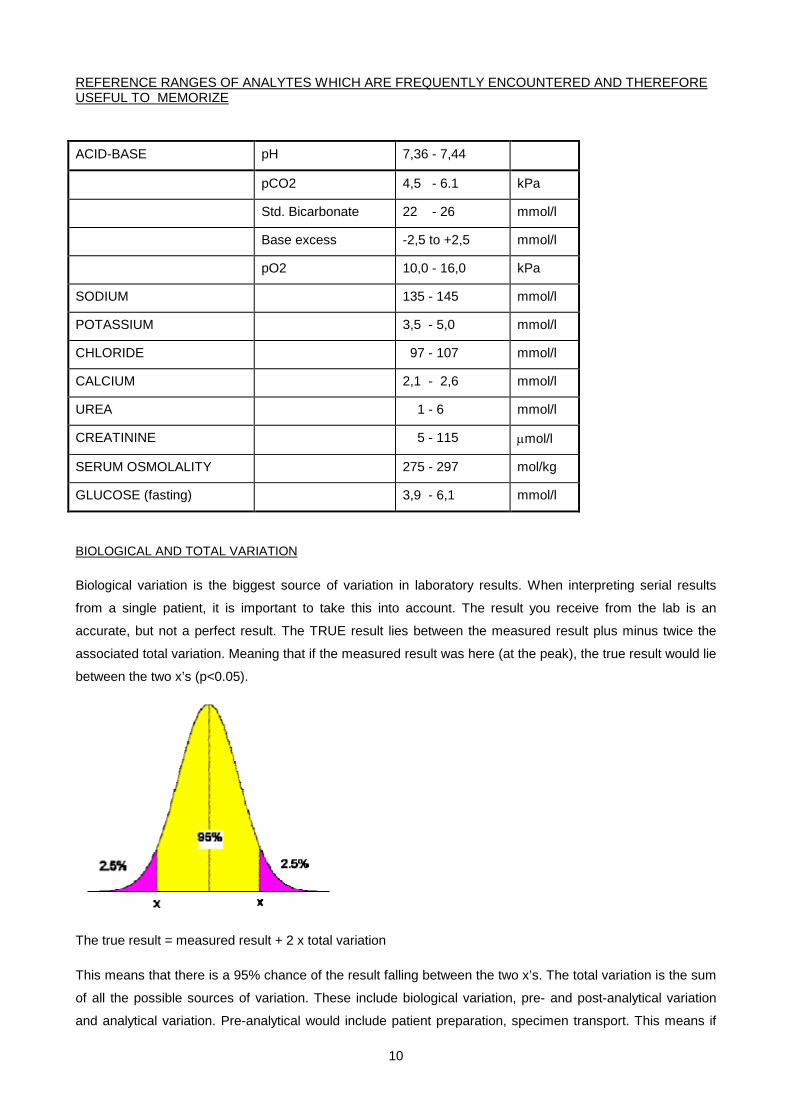
BIOLOGICAL AND TOTAL VARIATION
Biological variation is the biggest source of variation in laboratory results. When interpreting serial results
from a single patient, it is important to take this into account. The result you receive from the lab is an
accurate, but not a perfect result. The TRUE result lies between the measured result plus minus twice the
associated total variation. Meaning that if the measured result was here (at the peak), the true result would lie
between the two x’s (p<0.05).
The true result = measured result + 2 x total variation
This means that there is a 95% chance of the result falling between the two x’s. The total variation is the sum
of all the possible sources of variation. These include biological variation, pre- and post-analytical variation
and analytical variation. Pre-analytical would include patient preparation, specimen transport. This means if
11
you did the same healthy person’s test 100 times, the results would dispersed about the mean. 95 times, the
result would fall between the two “x’s”.
Understanding this dispersion influences one’s interpretation of a result.
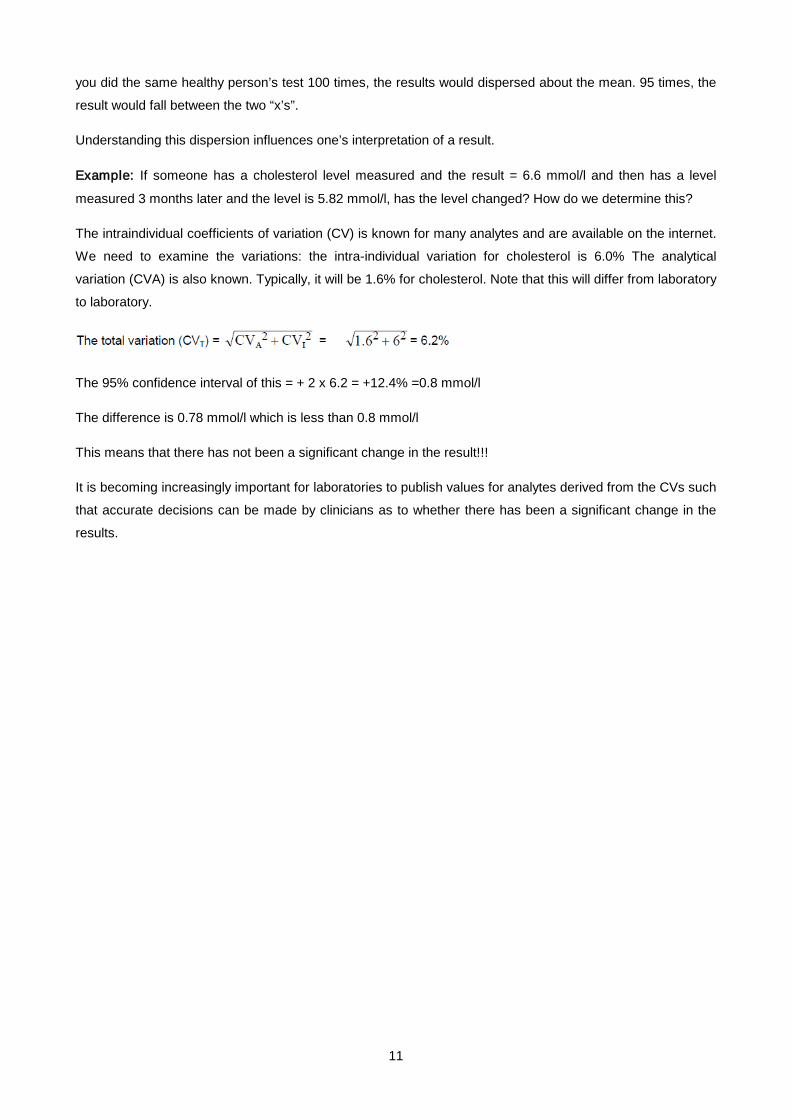
Example: If someone has a cholesterol level measured and the result = 6.6 mmol/l and then has a level
measured 3 months later and the level is 5.82 mmol/l, has the level changed? How do we determine this?
The intraindividual coefficients of variation (CV) is known for many analytes and are available on the internet.
We need to examine the variations: the intra-individual variation for cholesterol is 6.0% The analytical
variation (CVA) is also known. Typically, it will be 1.6% for cholesterol. Note that this will differ from laboratory
to laboratory.
The 95% confidence interval of this = + 2 x 6.2 = +12.4% =0.8 mmol/l
The difference is 0.78 mmol/l which is less than 0.8 mmol/l
This means that there has not been a significant change in the result!!!
It is becoming increasingly important for laboratories to publish values for analytes derived from the CVs such
that accurate decisions can be made by clinicians as to whether there has been a significant change in the
results.
12
SMALL GROUP TEACHING. LECTURE 1- INTRODUCTION TO CHEMICAL PATHOLOGY: QUESTIONS
CONVERSION OF UNITS
In order to interconvert between MOLAR and MASS units all that is needed is the molecular weight (MW) of the compound. Remember the essential relationship:
1 mol of a compound has a mass of MW g therefore
1 mol/l = MW g/l and 1 mmol/l = MW mg/l and 1 µmol/l = MW µg/l
etc., etc.
Abbreviations: 1 mol/l = 1 molar = 1 M, 1 mmol/l = 1 millimolar = 1 mM, 1 µmol/l = 1 micromolar = 1 µM etc.
The expression "g %" ("grams percent") means g per 100 ml.
EXAMPLES:
1. Convert a serum cortisol value of 500 nmol/l into µg/ml, given that MW of cortisol is 320
500 nmol/l = 500 x MW ng/l = 500 x 320 ng/l
= 160000 ng/l = 160 ng/ml = 0.16 µg/ml
2. Convert a plasma glucose level of 36 mg/dl to molar units. The MW of glucose is 180.
36 mg/dl
= 360 mg/l = 360/MW mmol/l = 360/180 mmol/l = 2 mmol/l
EXERCISES:
1. Convert a serum iron level of 112 µg/dl to µmol/l. The MW of iron is 56.
2. The reference range for serum testosterone in adult males is 10 - 35 nmol/l. Convert this to ng/ml given that the MW of testoterone is 300.
3. 3. A patient's urine has a Ca2+ concentration of 80 mg/dl. What is this in mmoles/l, given that the MW of Ca2+ is 40 ?.
4. 4. An average "western" diet has a salt intake of about 150 mmol/day. How much NaCl is this in grams? (MW of NaCl = 58).
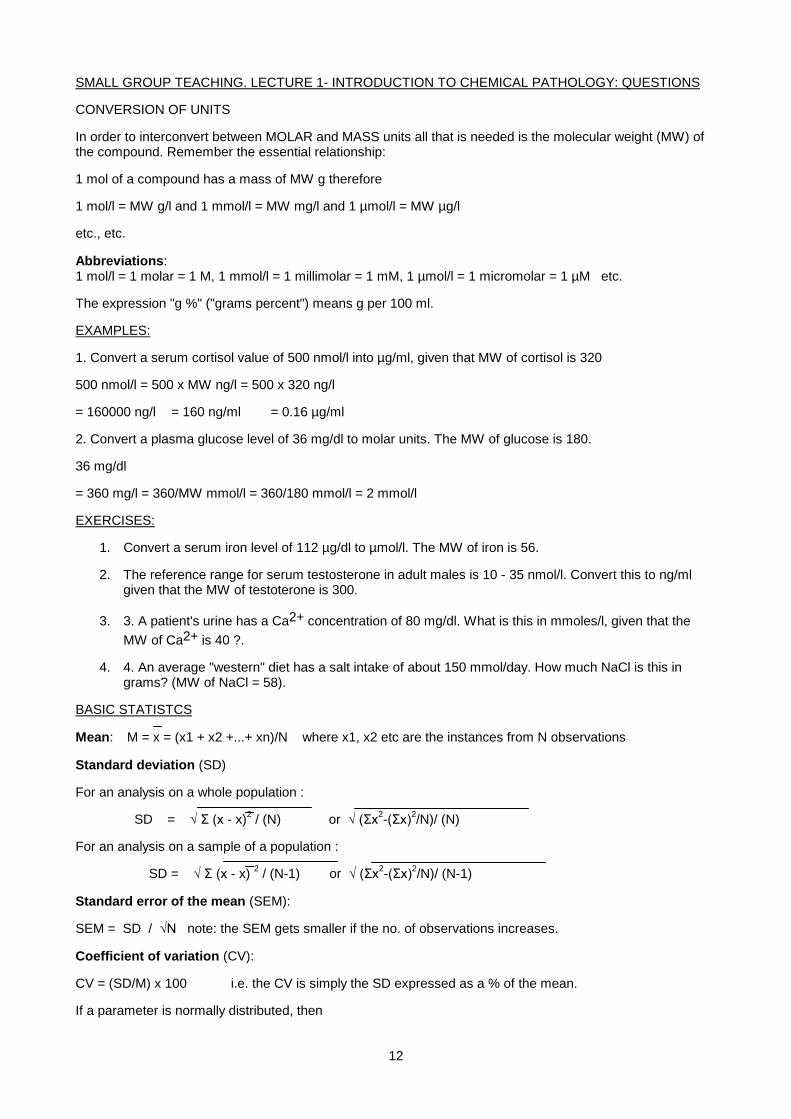
BASIC STATISTCS
Mean: M = x = (x1 + x2 +...+ xn)/N where x1, x2 etc are the instances from N observations
Standard deviation (SD)
For an analysis on a whole population :
SD = √ Σ (x - x)2 / (N) or √ (Σx2-(Σx)2/N)/ (N)
For an analysis on a sample of a population :
SD = √ Σ (x - x) 2 / (N-1) or √ (Σx2-(Σx)2/N)/ (N-1)
Standard error of the mean (SEM):
SEM = SD / √N note: the SEM gets smaller if the no. of observations increases.
Coefficient of variation (CV):
CV = (SD/M) x 100 i.e. the CV is simply the SD expressed as a % of the mean.
If a parameter is normally distributed, then
13
* approx. 95% of values fall within 2 SD of the mean
* 2.5% exceed the mean by more than 2SD
* 2.5% are less than the mean by more than 2SD
* approx. 66% of values fall within 1 SD of the mean.
EXERCISES:
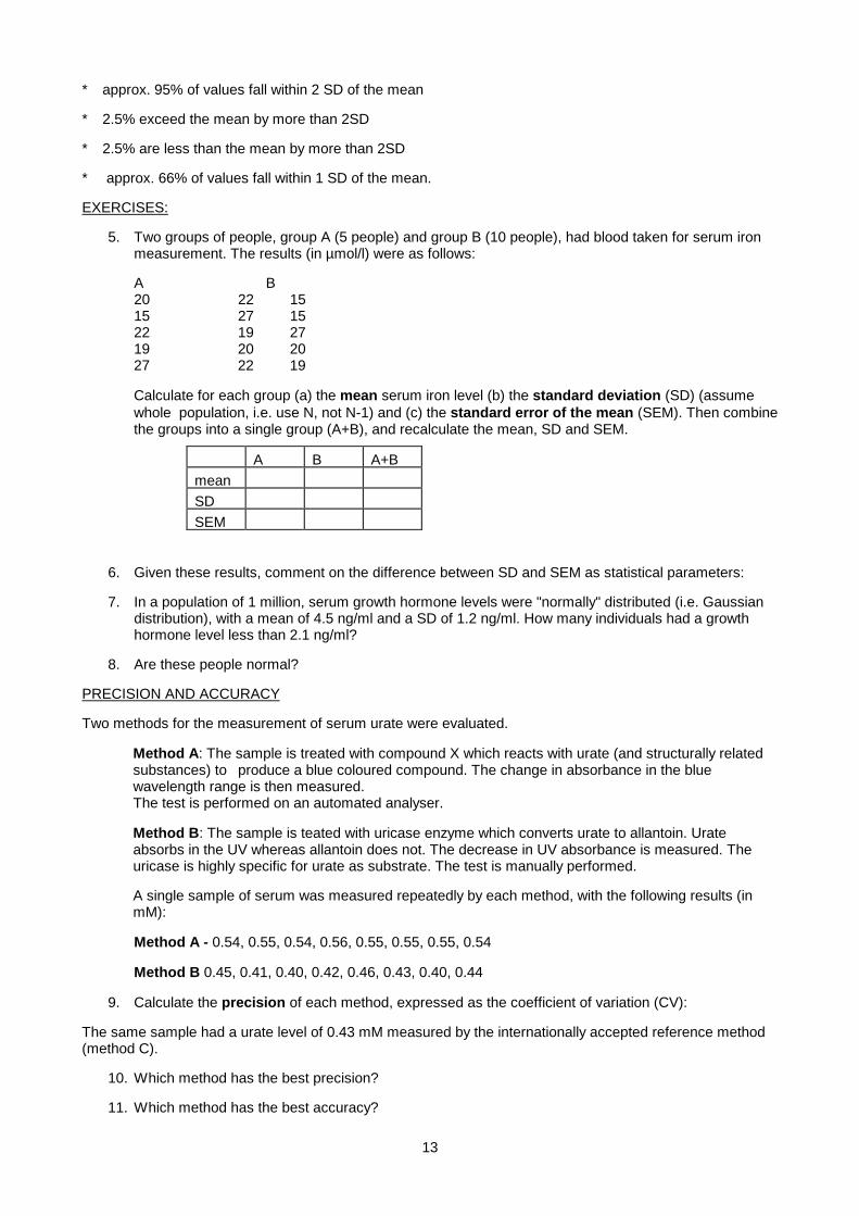
5. Two groups of people, group A (5 people) and group B (10 people), had blood taken for serum iron measurement. The results (in µmol/l) were as follows:
A B 20 22 15 15 27 15 22 19 27 19 20 20 27 22 19
Calculate for each group (a) the mean serum iron level (b) the standard deviation (SD) (assume whole population, i.e. use N, not N-1) and (c) the standard error of the mean (SEM). Then combine the groups into a single group (A+B), and recalculate the mean, SD and SEM.
A B A+B mean SD SEM
6. Given these results, comment on the difference between SD and SEM as statistical parameters:
7. In a population of 1 million, serum growth hormone levels were "normally" distributed (i.e. Gaussian distribution), with a mean of 4.5 ng/ml and a SD of 1.2 ng/ml. How many individuals had a growth hormone level less than 2.1 ng/ml?
8. Are these people normal?
PRECISION AND ACCURACY
Two methods for the measurement of serum urate were evaluated.
Method A: The sample is treated with compound X which reacts with urate (and structurally related substances) to produce a blue coloured compound. The change in absorbance in the blue wavelength range is then measured. The test is performed on an automated analyser.
Method B: The sample is teated with uricase enzyme which converts urate to allantoin. Urate absorbs in the UV whereas allantoin does not. The decrease in UV absorbance is measured. The uricase is highly specific for urate as substrate. The test is manually performed.
A single sample of serum was measured repeatedly by each method, with the following results (in mM):
Method A - 0.54, 0.55, 0.54, 0.56, 0.55, 0.55, 0.55, 0.54
Method B 0.45, 0.41, 0.40, 0.42, 0.46, 0.43, 0.40, 0.44
9. Calculate the precision of each method, expressed as the coefficient of variation (CV):
The same sample had a urate level of 0.43 mM measured by the internationally accepted reference method (method C).
10. Which method has the best precision?
11. Which method has the best accuracy?

14
12. Can you suggest any possible reasons for these differences?
13. What other factors would need to be considered before deciding on whether to use method A or B in a particular laboratory?
REFERENCE RANGES
14. Serum glucose levels were measured in a group of 200 volunteers after an overnight fast. The results were: mean serum glucose = 6.8 mmol/l; SD = 0.6 mmol/l If this group is representative of the general population, what percentage of the population would be expected to have a fasting glucose level greater than 8.0 mmol/l?
15. Suggest a possible reference range for fasting blood glucose based on the above data:
16. From the data in exercise 5 of the Basic Statistics section, suggest suitable reference ranges for serum iron for groups A, B and A+B.
SENSITIVITY AND SPECIFICITY IN LABORATORY TESTS
A test result is termed positive if it is abnormal i.e. outside the reference range, and negative if it falls within the reference range. A true positive (TP) is a positive result in a person who has the disease being tested for. A false positive (FP) is a positive (abnormal) result in a normal person. Similarly, a true negative (TN) is a negative test in a normal person, and a false negative (FN) is a negative test in a person with the disease.
Sensitivity, specificity and predictive value are precisely defined terms in clinical chemistry:
• SENSITIVITY = { TP / (TP + FN) } x 100 (i.e. the % of patients with the disease who have a positive test)
• SPECIFICITY = { TN / (TN + FP) } x 100 (i.e. the % of normal subjects who have a negative test)
• The PREDICTIVE VALUE of a positive test is defined by: PV(+) = { TP / (TP + FP) } x 100 (i.e. the percentage of all positives which were true positives)
• The PREDICTIVE VALUE of a negative test is defined by: PV(-) = { TN / (TN + FN) x 100
EXERCISES:
17. The data on fasting blood glucose levels in normal volunteers (see above) was used to construct a reference range. The mean ± 2SD was chosen as the reference range for this purpose (6.8 ± 1.2 mmol/l). This test was then used to screen a population of a village (1000 people) for diabetes mellitus, a disease characterised by an elevated fasting blood glucose. How many people (should have) had a positive test for diabetes (glucose > 8 mM)?
18. Of these, subsequent tests confirmed diabetes in only 5 subjects (the True Positives). Assuming that no cases of diabetes were undetected, calculate the sensitivity and specificity of the test.
19. What was the ratio of false positives to true positives?
20. Calculate the predictive values of a positive and a negative test.
21. Do you think this was an appropriate reference range to use for screening for diabetes in the general population?
22. Could the number of false positives be reduced by changing the reference range?
23. If the upper limit of the reference range was increased, what effect would this have on
a) the sensitivity of the test?
b) the specificity of the test?
24. Serum T4 (thyroid hormone) levels were reported to have a sensitivity of 95% and a specificity of 95% as an index of hypothyroidism. In a population of 100,000 the prevalence of hypothyroidism was 0.1%.
15
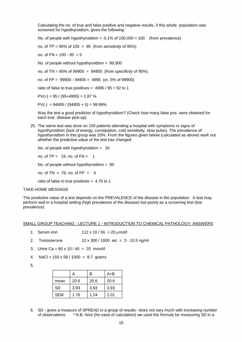
Calculating the no. of true and false positive and negative results, if this whole population was screened for hypothyroidism, gives the following:
No. of people with hypothyroidism = 0.1% of 100,000 = 100 (from prevalence)
no. of TP = 95% of 100 = 95 (from sensitivity of 95%)
no. of FN = 100 - 95 = 5
No. of people without hypothyroidism = 99,900
no. of TN = 95% of 99900 = 94905 (from specificity of 95%)
no. of FP = 99900 - 94905 = 4995 (or, 5% of 99900)
ratio of false to true positives = 4995 / 95 = 52 to 1
PV(+) = 95 / (95+4995) = 1.87 %
PV(-) = 94905 / (94905 + 5) = 99.99%
Was the test a good predictor of hypothyroidism? (Check how many false pos. were obtained for each true disease pick-up)
25. The same test was done on 100 patients attending a hospital with symptoms or signs of hypothyroidism (lack of energy, constipation, cold sensitivity, slow pulse). The prevalence of hypothyroidism in this group was 20%. From the figures given below (calculated as above) work out whether the predictive value of the test has changed
No. of people with hypothyroidism = 20
no. of TP = 19, no. of FN = 1
No. of people without hypothyroidism = 80
no. of TN = 76, no. of FP = 4
ratio of false to true positives = 4.75 to 1
TAKE-HOME MESSAGE
The predictive value of a test depends on the PREVALENCE of the disease in the population. A test may perform well in a hospital setting (high prevalence of the disease) but poorly as a screening test (low prevalence).
SMALL GROUP TEACHING : LECTURE 1 - INTRODUCTION TO CHEMICAL PATHOLOGY: ANSWERS
1. Serum iron 112 x 10 / 56 = 20 µmol/l
2. Testosterone 10 x 300 / 1000 etc = 3 - 10.5 ng/ml
3. Urine Ca = 80 x 10 / 40 = 20 mmol/l
4. NaCl = 150 x 58 / 1000 = 8.7 grams
5.
A B A+B
mean 20.6 20.6 20.6
SD 3.93 3.93 3.93
SEM 1.76 1.24 1.01
6. SD - gives a measure of SPREAD in a group of results –does not vary much with increasing number of observations. * N.B. here (for ease of calculation) we used the formula for measuring SD in a
16
whole population, rather than a sample (when we use N-1 instead of N to avoid bias). Good student statisticians may rightly object!
7. SEM - gives a measure of the certainty of the mean - more observations, lower error.
8. 2.5% have a GH level less than 2SD below the mean. Most of these people will be normal, but they may include a subset of people with GH deficiency.
9. mean SD CV
method A: 0.5475 0.0066 1.2% (1.3% if using N-1)
method B 0.426 0.0212 5.0 % (5.3% " )
10. A has greater precision.
11. B has greater accuracy.
12. Method A (simple colorimetric method) might be less specific e.g.compound X reacts with other substances in serum leading to higher values, whereas the enzymatic method is very specific and hence accurate. The precision depends on many factors e.g. number of pipetting steps etc.
13. Other factors to consider would include:
- cost
- time and expertise required to perform the test
- Is accuracy or precision more important in this test?
- can it be easily automated?
14. 2.5 % should have a level greater than 8 (2 SDs above mean).
15. Suitable ref. range would be mean ± 2SD i.e. 6.8 ± 1.2 mmol/l, or 5.6 - 8.0.
16. Groups A, B and A+B all have same mean and SD so the same range will be applicable (mean ± 2SD). Range is therefore 12.7-28.5
17. 25 would have had a blood glucose >8 mmol/l (2.5% of 1000).
18. FN given as zero (no cases undetected), therefore TN = 1000-25 = 975, FP = 25 - 5 = 20
Sensitivity = 5/5 = 100 % (all cases detected)
Specificity = 975 / (975 + 20) = 98 % (TN / (TN + FP))
19. Ratio of false positives to true positives: 4 to 1 (20 / 5)
20. PV(+) = 5 / 25 = 20 %, PV(-) = 975 / 975 = 100%
21. The PV(+) of 20% (and the high rate of false positives) means that this test is unsuitable as a screening test.
22. The no. of FP would be decreased by increasing the upper limit of the ref. range.
23. If the upper limit is increased, this would
(a) decrease the sensitivity (some cases might be missed)
(b) increase the specificity (fewer false positives).
24. The PV(+) of 1.87 % is almost useless as a predictor! The PV(-) of 99.99% seems very good, but is actually little better than a "blind" prediction based purely on prevalence.
25. PV(+) = 19/(19 + 4) = 82.6%, PV(-) = 76/(76 + 1) = 98.7%
The predictive value of a positive test is vastly improved by selecting the test population (in effect increasing the prevalence of the disease in the test population).
17
Lecture 2: Acid-Base Balance
DR HELENE VREEDE 2007
AIMS OF THE LECTURES
1. Understand the basic biochemistry and physiology of acid-base balance.
2. Understand human acid-base balance and interpret clinical acid-base data,
3. Understand the diseases that cause acid-base disturbance.
LECTURE CONTENT
1. CONCEPTS AND VOCABULARY OF ACID-BASE BALANCE
Hydrogen ion concentration and concept of pH
Sources of hydrogen ions
Background to buffers
Definition of terms
Strengths of acids
Definition of a buffer
Physiological buffers
Henderson-Hasselbalch equation
Role of haemoglobin - transport of CO2 and buffering
Role of the kidney in handling bicarbonate and hydrogen
2. ASSESSING ACID-BASE BALANCE
Normal values
Concepts and vocabulary of acid-base imbalance
Anion gap
Interpretation of pO2
Laboratory acid-base analysis
Interpreting acid-base data
3. ACID-BASE DISORDERS
Metabolic acidosis
Metabolic alkalosis
Respiratory acidosis
Respiratory alkalosis
18
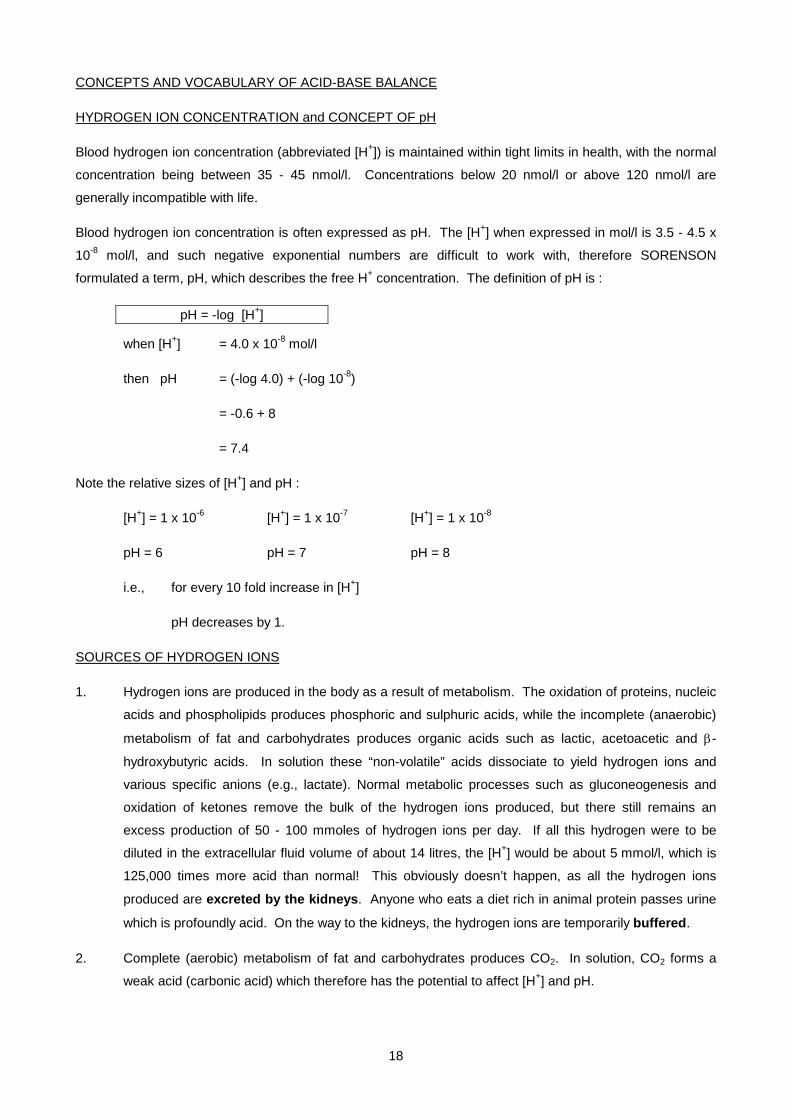
CONCEPTS AND VOCABULARY OF ACID-BASE BALANCE
HYDROGEN ION CONCENTRATION and CONCEPT OF pH
Blood hydrogen ion concentration (abbreviated [H+]) is maintained within tight limits in health, with the normal
concentration being between 35 - 45 nmol/l. Concentrations below 20 nmol/l or above 120 nmol/l are
generally incompatible with life.
Blood hydrogen ion concentration is often expressed as pH. The [H+] when expressed in mol/l is 3.5 - 4.5 x
10-8 mol/l, and such negative exponential numbers are difficult to work with, therefore SORENSON
formulated a term, pH, which describes the free H+ concentration. The definition of pH is :
pH = -log [H+]
when [H+] = 4.0 x 10⁻⁸ mol/l
then pH = (-log 4.0) + (-log 10-8)
= -0.6 + 8
= 7.4
Note the relative sizes of [H+] and pH :
[H+] = 1 x 10⁻⁶ [H+] = 1 x 10⁻⁷ [H+] = 1 x 10⁻⁸
pH = 6 pH = 7 pH = 8
i.e., for every 10 fold increase in [H+]
pH decreases by 1.
SOURCES OF HYDROGEN IONS
1. Hydrogen ions are produced in the body as a result of metabolism. The oxidation of proteins, nucleic
acids and phospholipids produces phosphoric and sulphuric acids, while the incomplete (anaerobic)
metabolism of fat and carbohydrates produces organic acids such as lactic, acetoacetic and β-
hydroxybutyric acids. In solution these “non-volatile” acids dissociate to yield hydrogen ions and
various specific anions (e.g., lactate). Normal metabolic processes such as gluconeogenesis and
oxidation of ketones remove the bulk of the hydrogen ions produced, but there still remains an
excess production of 50 - 100 mmoles of hydrogen ions per day. If all this hydrogen were to be
diluted in the extracellular fluid volume of about 14 litres, the [H+] would be about 5 mmol/l, which is
125,000 times more acid than normal! This obviously doesn’t happen, as all the hydrogen ions
produced are excreted by the kidneys. Anyone who eats a diet rich in animal protein passes urine
which is profoundly acid. On the way to the kidneys, the hydrogen ions are temporarily buffered.
2. Complete (aerobic) metabolism of fat and carbohydrates produces CO2. In solution, CO2 forms a
weak acid (carbonic acid) which therefore has the potential to affect [H+] and pH.
19
This process produces 15,000 - 20,000 mmoles of CO2 per day. CO2 is however volatile, and under
normal circumstances is transported to the lungs in the blood and is rapidly excreted by the
lungs. Only if respiratory function is impaired do problems occur.
BACKGROUND TO BUFFERS
Before we can define a buffer, or describe what a buffer does and how it does it, there are certain concepts
that must be understood.
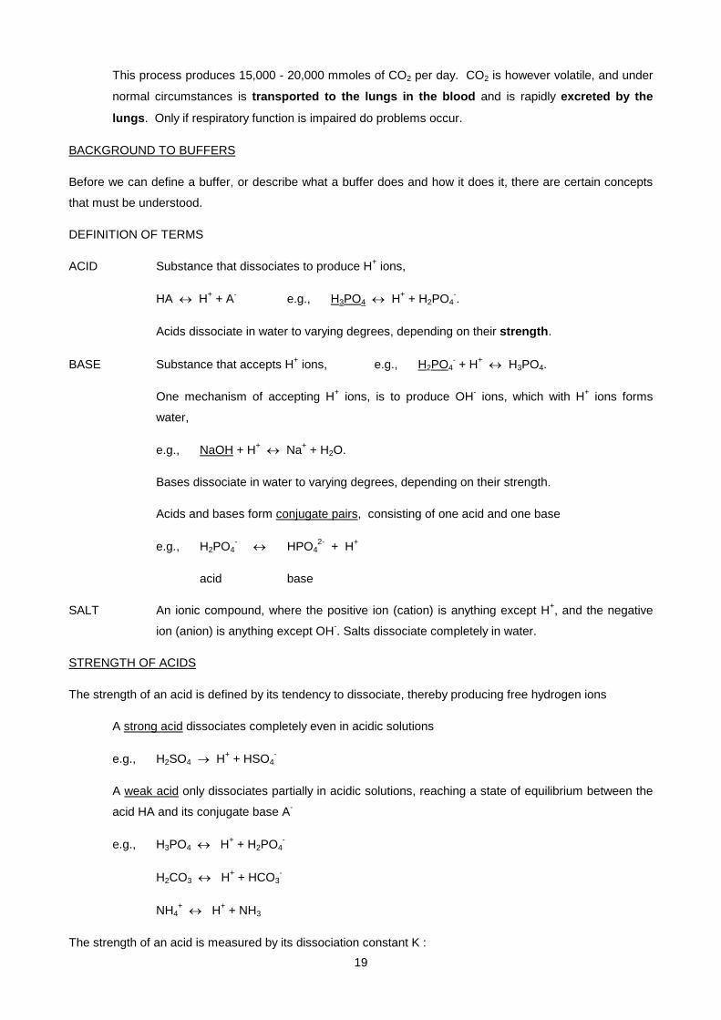
DEFINITION OF TERMS
ACID Substance that dissociates to produce H+ ions,
HA ↔ H+ + A- e.g., H3PO4 ↔ H+ + H2PO4-.
Acids dissociate in water to varying degrees, depending on their strength.
BASE Substance that accepts H+ ions, e.g., H2PO4- + H+ ↔ H3PO4.
One mechanism of accepting H+ ions, is to produce OH- ions, which with H+ ions forms
water,
e.g., NaOH + H+ ↔ Na+ + H2O.
Bases dissociate in water to varying degrees, depending on their strength.
Acids and bases form conjugate pairs, consisting of one acid and one base
e.g., H2PO4- ↔ HPO4
2- + H+
acid base
SALT An ionic compound, where the positive ion (cation) is anything except H+, and the negative
ion (anion) is anything except OH-. Salts dissociate completely in water.
STRENGTH OF ACIDS
The strength of an acid is defined by its tendency to dissociate, thereby producing free hydrogen ions
A strong acid dissociates completely even in acidic solutions
e.g., H2SO4 → H+ + HSO4-
A weak acid only dissociates partially in acidic solutions, reaching a state of equilibrium between the
acid HA and its conjugate base A-
e.g., H3PO4 ↔ H+ + H2PO4-
H2CO3 ↔ H+ + HCO3-
NH4+ ↔ H+ + NH3
The strength of an acid is measured by its dissociation constant K :

20
K
HA ↔ H+ + A-
[H+] [A-]
K = -------------- and pK = -log K
[HA]
For a strong acid, K is large (> 1) and pK is small (< 0)
For a weak acid, K is small (< 10-3) and pK is large (> 3)
DEFINITION OF A BUFFER
A buffer is a solution containing a conjugate acid-base pair, made up of a weak acid and its salt, which
minimises changes in pH.
the weak acid (HA) - this dissociates partially into H+ and A-
its salt (e.g., NaA) - this dissociates fully and yields the maximum amount of the conjugate
base (A-)
HA ↔ H+ + A- plus NaA → Na+ + A- gives HA ↔ H+ + Na+ + A-
dissociates partly dissociates fully lots of A- present
Buffers bind or release hydrogen ions depending on the surrounding hydrogen ion concentration, by shifting
the equilibrium of the reaction.
HA ↔ H+ + A-
in presence of excess H+ in presence of deficient H+
equilibrium shifts towards acid equilibrium shifts towards base
HA ← H+ + A- HA → H+ + A-
excess free H+ removed, [A-] decreases deficient free H+ replaced, [A-] increases
Buffers thus minimise changes in free hydrogen ion concentration, and thus minimise changes in pH.
Buffering is however only a short-term solution - any excess hydrogen ions must eventually be excreted from
the body.
PHYSIOLOGICAL BUFFERS
21
The body contains a number of buffers. Proteins can act as buffers by binding hydrogen ions, and
haemoglobin in red blood cells, in particular, has a high capacity for binding hydrogen ions.
proteins Pr- + H+ ↔ PrH
haemoglobin Hb- + H+ ↔ HbH
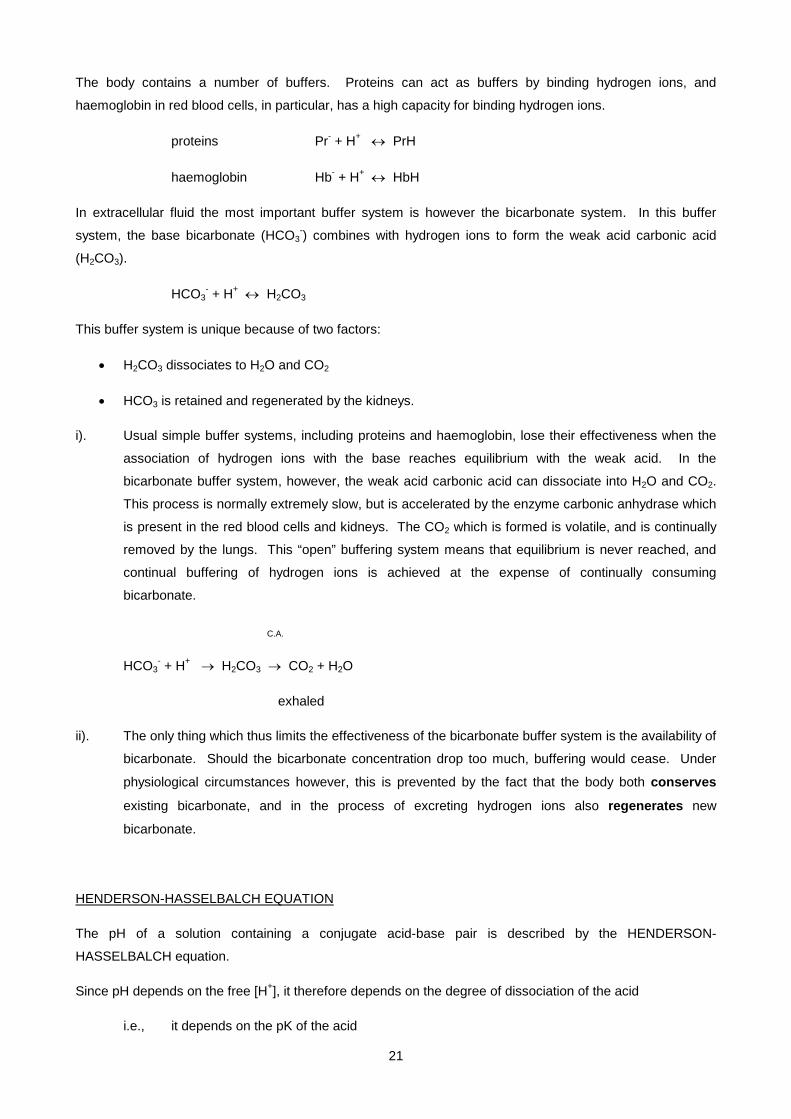
In extracellular fluid the most important buffer system is however the bicarbonate system. In this buffer
system, the base bicarbonate (HCO3-) combines with hydrogen ions to form the weak acid carbonic acid
(H2CO3).
HCO3- + H+ ↔ H2CO3
This buffer system is unique because of two factors:
• H2CO3 dissociates to H2O and CO2
• HCO3 is retained and regenerated by the kidneys.
i). Usual simple buffer systems, including proteins and haemoglobin, lose their effectiveness when the
association of hydrogen ions with the base reaches equilibrium with the weak acid. In the
bicarbonate buffer system, however, the weak acid carbonic acid can dissociate into H2O and CO2.
This process is normally extremely slow, but is accelerated by the enzyme carbonic anhydrase which
is present in the red blood cells and kidneys. The CO2 which is formed is volatile, and is continually
removed by the lungs. This “open” buffering system means that equilibrium is never reached, and
continual buffering of hydrogen ions is achieved at the expense of continually consuming
bicarbonate.
C.A.
HCO3- + H+ → H2CO3 → CO2 + H2O
exhaled
ii). The only thing which thus limits the effectiveness of the bicarbonate buffer system is the availability of
bicarbonate. Should the bicarbonate concentration drop too much, buffering would cease. Under
physiological circumstances however, this is prevented by the fact that the body both conserves
existing bicarbonate, and in the process of excreting hydrogen ions also regenerates new
bicarbonate.
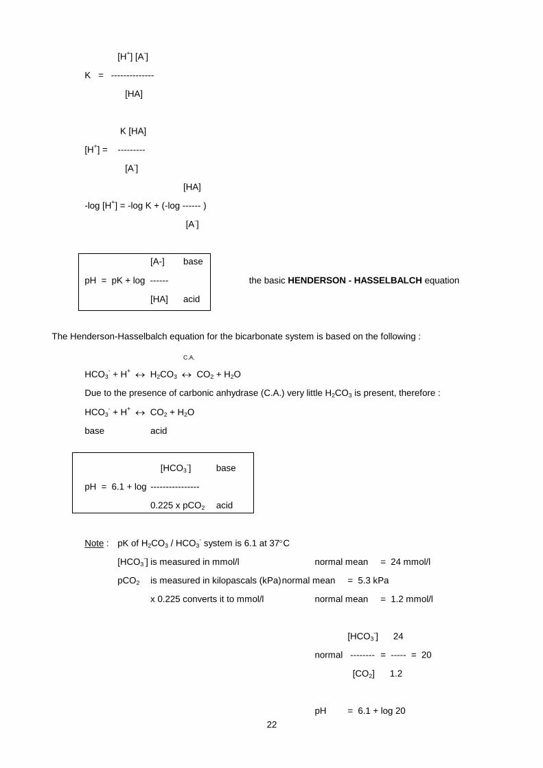
HENDERSON-HASSELBALCH EQUATION
The pH of a solution containing a conjugate acid-base pair is described by the HENDERSON-
HASSELBALCH equation.
Since pH depends on the free [H+], it therefore depends on the degree of dissociation of the acid
i.e., it depends on the pK of the acid
22
[H+] [A-]
K = --------------
[HA]
K [HA]
[H+] = ---------
[A-]
[HA]
-log [H+] = -log K + (-log ------ )
[A-]
[A-] base
pH = pK + log ------ the basic HENDERSON - HASSELBALCH equation
[HA] acid
The Henderson-Hasselbalch equation for the bicarbonate system is based on the following :
C.A.
HCO3- + H+ ↔ H2CO3 ↔ CO2 + H2O
Due to the presence of carbonic anhydrase (C.A.) very little H2CO3 is present, therefore :
HCO3- + H+ ↔ CO2 + H2O
base acid
[HCO3-] base
pH = 6.1 + log ----------------
0.225 x pCO2 acid
Note : pK of H2CO3 / HCO3- system is 6.1 at 37°C
[HCO3-] is measured in mmol/l normal mean = 24 mmol/l
pCO2 is measured in kilopascals (kPa) normal mean = 5.3 kPa
x 0.225 converts it to mmol/l normal mean = 1.2 mmol/l
[HCO3-] 24
normal -------- = ----- = 20
[CO2] 1.2
pH = 6.1 + log 20

23
= 6.1 + 1.3
= 7.4
NB. The more stable the HCO3 / CO2 ratio is, the more stable the pH is.
ROLE OF HAEMOGLOBIN - TRANSPORT OF CO2 AND BUFFERING
CO2, produced by complete (aerobic) metabolism of fat and carbohydrates, diffuses out of cells into the ECF.
In the ECF a small amount combines with water to form carbonic acid, thereby increasing the [H+] and
decreasing the pH of the ECF. In red blood cells metabolism is anaerobic and no CO2 is formed. CO2
therefore diffuses into red blood cells down a concentration gradient. In the red blood cells the majority of the
CO2 combines with water to form carbonic acid, due to the presence of carbonic anhydrase. The carbonic
acid dissociates to form hydrogen ions and bicarbonate ions, and the hydrogen ions are bound by the
haemoglobin. Deoxygenated haemoglobin binds hydrogen ions more strongly than oxygenated haemoglobin,
and in fact the binding of hydrogen ions to haemoglobin facilitates the release of oxygen (the Bohr effect).
The overall effect of this process is that CO2 is converted to bicarbonate in red blood cells. The bicarbonate
diffuses out of the red blood cells along a concentration gradient, to be replaced by chloride ions (the chloride
shift). In the lungs, the reverse occurs, because of the low partial pressure of CO2 in the alveoli: bicarbonate
diffuses into the red cells, combines with hydrogen ions released when haemoglobin binds oxygen, and is
converted into CO2, which diffuses into the alveoli to be excreted. The role of haemoglobin is therefore to
transport O2 and by converting CO2 to bicarbonate, to minimise changes in the HCO3 / CO2 ratio between
venous and arterial blood, which helps to minimise pH changes.

24
ROLE OF THE KIDNEY IN HANDLING OF BICARBONATE AND HYDROGEN
As mentioned previously, eventually all hydrogen ions produced must be excreted from the body. Buffering
of hydrogen ions is a vital short-term solution to the problem of maintaining a constant and normal pH, and
adequate amounts of bicarbonate must therefore be maintained to preserve the blood’s buffering capacity.
The kidneys are involved in all these processes - excretion of hydrogen ions, conservation (reabsorption) of
existing bicarbonate ions, and regeneration of new bicarbonate ions to replace those used up in the buffering
process. The mechanisms for bicarbonate reabsorption and regeneration are very similar and easily
confused. Note that the difference is in net hydrogen ion excretion.
Reabsorption of bicarbonate
The glomerular filtrate contains the same concentration of bicarbonate ions as the plasma. The luminal
surface of the renal tubular cells is impermeable to bicarbonate, and direct reabsorption can thus not occur.
Within the renal tubular cells, CO2 combines with water to form carbonic acid, due to the presence of
carbonic anhydrase. The carbonic acid dissociates to form hydrogen ions and bicarbonate ions. The
hydrogen ions are secreted into the tubular lumen in exchange for sodium ions, while the bicarbonate ions
pass across the basal border of the cells into the interstitial fluid together with the sodium ions. In the tubular
lumen the hydrogen ions combine with the filtered bicarbonate, form carbonic acid and then CO2 and water,
some of which filters back into the renal tubular cell. The net effect of this process is that filtered bicarbonate
is reabsorbed, but although there is hydrogen ion secretion, there is no net hydrogen ion excretion. This
process takes place as long as filtered bicarbonate is present in the tubular lumen, which is essentially in the
proximal renal tubule.
Excretion of hydrogen and regeneration of bicarbonate
After filtered bicarbonate is fully reabsorbed, hydrogen ion secretion into the tubular lumen causes a net
excretion of hydrogen ions. This process uses the same mechanism as described above, but requires the
presence of urinary buffers, otherwise the hydrogen ion gradient created would prevent further hydrogen ion
secretion. The main urinary buffer is phosphate, which is present predominantly as HPO42-. This combines
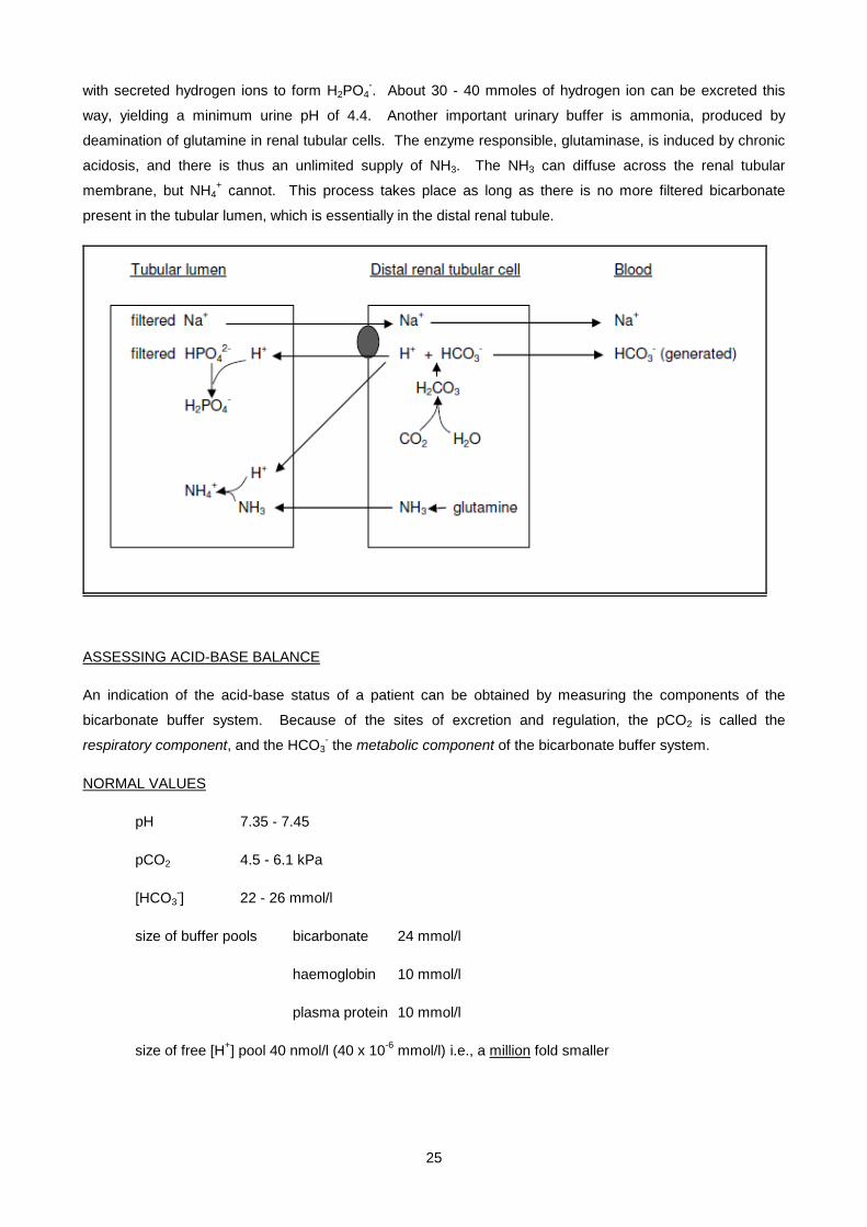
25
with secreted hydrogen ions to form H2PO4-. About 30 - 40 mmoles of hydrogen ion can be excreted this
way, yielding a minimum urine pH of 4.4. Another important urinary buffer is ammonia, produced by
deamination of glutamine in renal tubular cells. The enzyme responsible, glutaminase, is induced by chronic
acidosis, and there is thus an unlimited supply of NH3. The NH3 can diffuse across the renal tubular
membrane, but NH4+ cannot. This process takes place as long as there is no more filtered bicarbonate
present in the tubular lumen, which is essentially in the distal renal tubule.
ASSESSING ACID-BASE BALANCE
An indication of the acid-base status of a patient can be obtained by measuring the components of the
bicarbonate buffer system. Because of the sites of excretion and regulation, the pCO2 is called the
respiratory component, and the HCO3- the metabolic component of the bicarbonate buffer system.
NORMAL VALUES
pH 7.35 - 7.45
pCO2 4.5 - 6.1 kPa
[HCO3-] 22 - 26 mmol/l
size of buffer pools bicarbonate 24 mmol/l
haemoglobin 10 mmol/l
plasma protein 10 mmol/l
size of free [H+] pool 40 nmol/l (40 x 10-6 mmol/l) i.e., a million fold smaller
26
CONCEPTS AND VOCABULARY OF ACID-BASE IMBALANCE
ACIDOSIS A condition characterised by a decrease in blood pH. The condition can be of metabolic or
respiratory origin.
[HCO3-]
Since pH = 6.1 + log ----------------
0.225 x pCO2
Therefore a decrease in pH can be due to:
• a decrease in [HCO3-] (metabolic acidosis)
• an increase in pCO2 (respiratory acidosis)
ALKALOSIS A condition characterised by an increase in blood pH. The condition can be of metabolic or
respiratory origin.
[HCO3-]
Since pH = 6.1 + log ----------------
0.225 x pCO2
Therefore an increase in pH can be due to
• an increase in [HCO3-] (metabolic alkalosis)
• a decrease in pCO2 (respiratory alkalosis)
COMPENSATION
These simple relationships of bicarbonate and CO2 to pH are complicated by the physiological mechanisms
which have evolved to attempt to bring the pH back to normal. These physiological mechanisms are
operated by that part of the acid-base balance system which is not affected by the primary disease process.
These compensatory changes bring the blood pH back towards normal, by bringing the HCO3/CO2 ratio back
towards normal. When lung function is compromised, the kidneys attempt to increase the excretion of
hydrogen ions via the renal route. This is known as metabolic compensation for the primary respiratory
disorder. Metabolic compensation is slow to take effect, coming into effect over 2 - 4 days.
• In a respiratory acidosis (decreased pH due to increased pCO2), more H+ is excreted and
more HCO3- is generated by the kidneys, increasing blood HCO3
-
• In a respiratory alkalosis (increased pH due to decreased pCO2), less H+ is excreted and less
HCO3- is generated by the kidneys, decreasing blood HCO3
-
When there are metabolic disorders, some compensation is possible by the lungs by altering the rate and
depth of respiration, which is affected directly by the blood pH. This is known as respiratory compensation for

27
the primary metabolic disorder. Respiratory compensation is quick to take effect, coming into effect within 15
- 30 minutes.
• In a metabolic acidosis (decreased pH due to decreased HCO3-), the rate and depth of
respiration are increased (hyperventilation), decreasing blood pCO2
• In a metabolic alkalosis (increased pH due to increased HCO3-) the rate and depth of
respiration are decreased (hypoventilation), increasing blood pCO2
If compensation is complete, the pH returns to normal, although the bicarbonate and CO2 concentrations are
abnormal, sometimes grossly so. Compensation is however often partial, in which case there is a change in
both bicarbonate and CO2 concentrations, but the pH is still abnormal.
Note The compensatory change in the one component is always in the same direction as the pathological
change in the other component.
ACTUAL and STANDARD BICARBONATE
The actual bicarbonate is the bicarbonate concentration actually found in the patient’s blood. It is calculated
by the blood gas analyser from the blood sample's measured pH and pCO2, by using the Henderson-
Hasselbalch equation. However, the problem with using actual bicarbonate in interpreting acid-base results,
is that one cannot readily assess whether a metabolic change is present or not. This is because the actual
bicarbonate concentration is not just influenced by the renal mechanisms described before, it is also slightly
influenced by a physiological shift.
CO2 + H2O ↔ H2CO3 ↔ HCO3- + H+
Therefore as CO2 increases, HCO3- also increases slightly.
This is a physiological shift, not a metabolic (renal) change.
A mechanism was therefore invented whereby the physiological shift of CO2 to HCO3- in the patient’s blood
sample could be reversed, so that one could assess whether a metabolic change was present or not. If after
the reversal of the physiological shift an increased bicarbonate concentration is still present, then it is
evidence of a metabolic (renal) change in the body. After the pH and pCO2 are measured (from which the
actual bicarbonate concentration is calculated), the blood sample in the blood gas analyser is exposed to a
normal pCO2 concentration of 5.3 kPa. After equilibration at this normal pCO2, the pH is measured again and
a new bicarbonate concentration is calculated. This is called the standard bicarbonate.
The standard bicarbonate (abbreviated SBC) is thus not the bicarbonate concentration actually found in the
patient’s blood, but is the bicarbonate concentration calculated from the pH of a blood sample equilibrated to
a pCO2 of 5.3 kPa, and is the value used to assess whether a metabolic change is present.
e.g., pH = 7.24 - decreased, therefore this is an acidosis
pCO2 = 9.3 - increased, therefore this is a respiratory acidosis
actual HCO3- = 29 - increased, but is this metabolic compensation or just a physiological shift?

28
standard HCO3- = 24 - normal, therefore there is no metabolic compensation
Blood gas analyses reports usually give the SBC, not the actual bicarbonate concentration.
CORRECTION
Once the primary disease has been treated, the part of the acid-base balance system that was diseased
must rid the body of the accumulated acid or base which caused the original pH abnormality. In addition, the
non-diseased part of the acid-base system must rid the body of the compensatory changes. Correction is
slow to take effect, coming into effect many days after successful treatment.
e.g., after successfully treating a respiratory acidosis, the lungs must excrete the excess CO2, and the
kidneys must excrete the excess HCO3- that accumulated during metabolic compensation.
ANION GAP
The anion gap is a concept which is useful in establishing the cause of a metabolic acidosis. Blood is always
electroneutral, even if there is an acid-base disturbance, i.e., it always contains an equal number of positive
cations and negative anions.
Normally: the millimolar sum of all cations = the millimolar sum of all anions
140 mM Na+ 105 mM Cl-
4 mM K+ 25 mM HCO3-
2 mM Ca2+ 2 mM PO43-
1 mM Mg2+ 15 mM proteins-
< 1 mM organic anions-
Although almost all these electrolytes can be measured, we commonly only measure Na+, K+, Cl- and HCO3-.
When we subtract the commonly measured anions from the commonly measured cations, we find an “anion
gap” of about 10 - 20 mmol/l.
(Na+ + K+) - (Cl- + HCO3-) = "anion gap" = 10 - 20 mmol/l
The "anion gap" therefore reflects the concentration of those anions which are actually present in serum, but
are routinely unmeasured, including negatively charged proteins (mainly albumin), phosphates, sulphates and
organic acids. If the "anion gap" is bigger than normal, it is because one of these unmeasured anions is
increased.
In a metabolic acidosis, when the excess production of hydrogen ions causes the concentration of
bicarbonate anion to fall, another anion must take its place to maintain electroneutrality. Sometimes another
anion is produced at the same time as the hydrogen ion which consumed the bicarbonate, such as lactate
anion in a lactic acidosis. If no such additional anion is produced, the kidney reabsorbs more chloride and
the increased chloride maintains electroneutrality. When an increased anion gap is present, this is described
as an "increased anion gap acidosis". When a normal anion gap is present, this is described as a "normal
anion gap acidosis" or a "hyperchloraemic acidosis".

29
e.g., Na+ 140
K+ 5 145
Cl- 110
HCO3- 10 - 120
25 increased anion gap
indicates excess unmeasured anions- i.e., an increased anion gap acidosis
NB. In any metabolic acidosis, calculate the anion gap to distinguish between different causes of the
acidosis!
INTERPRETATION OF pO2
Although these lectures are primarily about acid-base balance and therefore the components of the
bicarbonate buffer system (pH, HCO3- and pCO2), no complete interpretation of acid-base balance is possible
without also looking at the pO2. Although both carbon dioxide and oxygen are transported between the
alveoli and the blood stream (albeit in opposite directions), their respective partial pressures do not
necessarily change in a reciprocal fashion. There are two reasons for this. First, carbon dioxide is generally
more diffusible than oxygen, with the result that in pulmonary diseases that cause increased diffusion
distances, such as pulmonary oedema and interstitial lung disease, oxygen diffusion is reduced more than
carbon dioxide diffusion. Thus these diseases result in a low pO2, but not necessarily in a raised pCO2.
Second, while carbon dioxide is carried in the blood mainly in solution (as bicarbonate), very little oxygen is
carried in solution in the blood, but is bound to haemoglobin which is normally fully saturated with oxygen.
Hyperventilation can therefore not increase the pO2 significantly, but can reduce the pCO2. A raised pO2 is
only seen in patients given supplementary oxygen which results in increased inspired pO2. A reduced pO2
(hypoxaemia) can be caused by :
Cause Mechanism
• decreased alveolar ventilation
e.g., depressed respiratory centre
decreased alveolar pO2
• venous-to-arterial shunting of blood e.g.,
cyanotic congenital heart disease dilution of high pO2 arterial blood with low pO2 venous blood
• impaired diffusion
e.g., pulmonary oedema
decreased diffusion of O2 across alveolus into blood
• ventilation/perfusion imbalance
e.g., pneumonia
blood perfusing non-aerated areas is not oxygenated
(and CO2 is not removed *)
30
*Blood leaving the poorly ventilated areas will have a low pO2 and a high pCO2. The effect on pCO2 can be
partially compensated by hyperventilation of normally ventilated and perfused alveoli, depending on the
severity of the ventilation-perfusion imbalance.
• With mild to moderate imbalance, hyperventilation of normally ventilated and perfused alveoli
can remove the excess CO2, (the low pO2 cannot be increased because the haemoglobin is
already fully saturated), resulting in low pO2 and normal or even low pCO2.
• With severe imbalance, such hyperventilation is not sufficient to remove the excess CO2,
resulting in low pO2 and high pCO2.
LABORATORY ACID-BASE (or blood gas) ANALYSIS
When you request an acid-base or blood gas analysis, always remember to send
- arterial or capillary blood: to measure arterial pO2 and pCO2 values
- a heparinised sample: most O2 is carried in red blood cells so we need an anticoagulated sample
- in a sealed syringe: to prevent O2 and CO2 diffusing out of the sample
- on ice : to prevent ongoing red cell metabolism from generating a lactic acidosis
1. pH Measured with a pH electrode.
Tells you whether an acidosis or an alkalosis is present.
2. pCO2 Measured with a CO2 electrode.
Tells you whether there is a change in the respiratory component.
3. HCO3-
- Actual HCO3- Calculated from the H-H equation using the measured pH and pCO2.
- Standard HCO3- Calculated from the H-H equation after remeasuring the pH at a pCO2 of
5.3 kPa.
Tells you whether there is a change in the metabolic component.
4. pO2 Measured with an O2 electrode.
Tells you more about the respiratory system.
INTERPRETING ACID-BASE DATA
1. Look at pH and decide whether it is normal, low (acidosis) or high (alkalosis).
2. Look at pCO2 and SBC and decide which is the primary abnormality i.e., which change can cause
this pH?
base decreased base increased base
31
pH ∝ -------- decreased pH increased pH
acid increased acid decreased acid
3. Look at the non-causative component and decide whether compensation is present or not.
Compensatory change is always in the same direction as the primary change.
4. In respiratory acid-base disturbances, look at the pO2 to assess respiratory function.
pH pCO2 Actual HCO3- SBC Interpretation
7.35 - 7.45 4.5 - 6.1 kPa 22 - 26 mmol/l 22 - 26 mmol/l
7.49 7.3 40 37
7.24 9.3 29 24
7.54 3.2 20 24
7.15 3.2 8 10
7.50 3.2 18 22
7.02 5.3 10 10
7.32 9.1 34 29
7.59 5.3 37 37
METABOLIC ACIDOSIS
In a metabolic acidosis there is a decrease in the blood pH caused by a decrease in the bicarbonate
concentration. The decrease in the bicarbonate concentration can be caused by one of two mechanisms -
the loss of bicarbonate, or the accumulation of hydrogen. In evaluating the possible causes of a metabolic
acidosis, it can be useful to determine the anion gap, since some causes of metabolic acidosis are
associated with a normal anion gap, while other causes are associated with an increased anion gap.
COMMON CAUSES OF METABOLIC ACIDOSIS
1. DIARRHOEA
Small intestinal fluid is rich in bicarbonate, due to pancreatic exocrine secretion. Once in the colon,
NaCl reabsorption takes place in exchange for K+ and further HCO3- secretion. Stool water thus
contains high concentrations of potassium and bicarbonate. With normal stool consistency the
volume of stool water lost is small, therefore normal GIT losses of potassium and bicarbonate are
negligible. However, with diarrhoea, large volumes of stool water are lost from the body. In acute
diarrhoea with a rapid transit time, where less time than usual would have been available for water
and NaCl reabsorption, the main losses are of water, sodium and bicarbonate. In chronic diarrhoea,
where more time would have been available for water and NaCl reabsorption and KHCO3 secretion,
32
the main losses are of potassium and bicarbonate. Thus in both acute and chronic diarrhoea
bicarbonate is lost. Since no additional anion has accumulated, the kidney reabsorbs more chloride
and the increased chloride maintains electroneutrality. Diarrhoea is therefore a cause of a normal
anion gap acidosis.
2. DIABETIC KETOACIDOSIS (DKA)
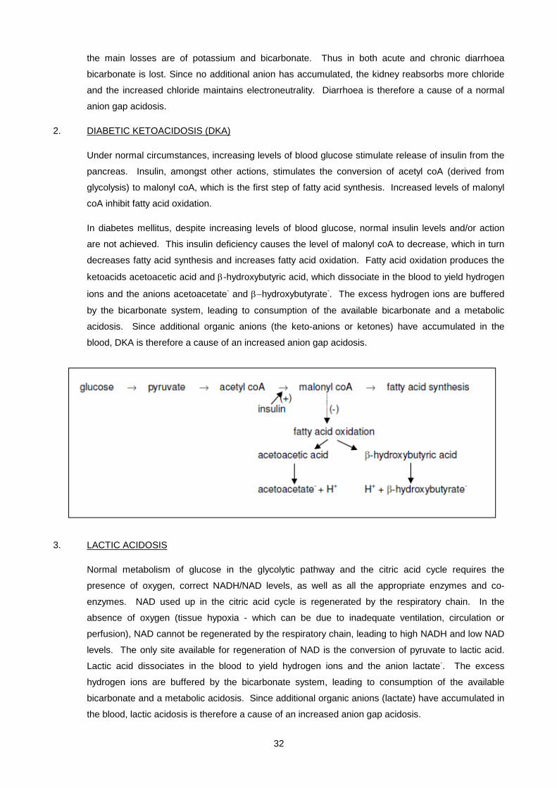
Under normal circumstances, increasing levels of blood glucose stimulate release of insulin from the
pancreas. Insulin, amongst other actions, stimulates the conversion of acetyl coA (derived from
glycolysis) to malonyl coA, which is the first step of fatty acid synthesis. Increased levels of malonyl
coA inhibit fatty acid oxidation.
In diabetes mellitus, despite increasing levels of blood glucose, normal insulin levels and/or action
are not achieved. This insulin deficiency causes the level of malonyl coA to decrease, which in turn
decreases fatty acid synthesis and increases fatty acid oxidation. Fatty acid oxidation produces the
ketoacids acetoacetic acid and β-hydroxybutyric acid, which dissociate in the blood to yield hydrogen
ions and the anions acetoacetate- and β−hydroxybutyrate-. The excess hydrogen ions are buffered
by the bicarbonate system, leading to consumption of the available bicarbonate and a metabolic
acidosis. Since additional organic anions (the keto-anions or ketones) have accumulated in the
blood, DKA is therefore a cause of an increased anion gap acidosis.
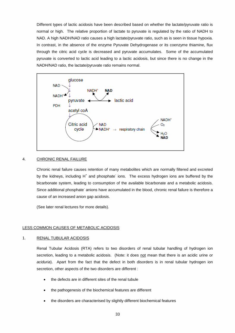
3. LACTIC ACIDOSIS
Normal metabolism of glucose in the glycolytic pathway and the citric acid cycle requires the
presence of oxygen, correct NADH/NAD levels, as well as all the appropriate enzymes and co-
enzymes. NAD used up in the citric acid cycle is regenerated by the respiratory chain. In the
absence of oxygen (tissue hypoxia - which can be due to inadequate ventilation, circulation or
perfusion), NAD cannot be regenerated by the respiratory chain, leading to high NADH and low NAD
levels. The only site available for regeneration of NAD is the conversion of pyruvate to lactic acid.
Lactic acid dissociates in the blood to yield hydrogen ions and the anion lactate-. The excess
hydrogen ions are buffered by the bicarbonate system, leading to consumption of the available
bicarbonate and a metabolic acidosis. Since additional organic anions (lactate) have accumulated in
the blood, lactic acidosis is therefore a cause of an increased anion gap acidosis.
33
Different types of lactic acidosis have been described based on whether the lactate/pyruvate ratio is
normal or high. The relative proportion of lactate to pyruvate is regulated by the ratio of NADH to
NAD. A high NADH/NAD ratio causes a high lactate/pyruvate ratio, such as is seen in tissue hypoxia.
In contrast, in the absence of the enzyme Pyruvate Dehydrogenase or its coenzyme thiamine, flux
through the citric acid cycle is decreased and pyruvate accumulates. Some of the accumulated
pyruvate is converted to lactic acid leading to a lactic acidosis, but since there is no change in the
NADH/NAD ratio, the lactate/pyruvate ratio remains normal.
4. CHRONIC RENAL FAILURE
Chronic renal failure causes retention of many metabolites which are normally filtered and excreted
by the kidneys, including H+ and phosphate- ions. The excess hydrogen ions are buffered by the
bicarbonate system, leading to consumption of the available bicarbonate and a metabolic acidosis.
Since additional phosphate- anions have accumulated in the blood, chronic renal failure is therefore a
cause of an increased anion gap acidosis.
(See later renal lectures for more details).
LESS COMMON CAUSES OF METABOLIC ACIDOSIS
1. RENAL TUBULAR ACIDOSIS
Renal Tubular Acidosis (RTA) refers to two disorders of renal tubular handling of hydrogen ion
secretion, leading to a metabolic acidosis. (Note: it does not mean that there is an acidic urine or
aciduria). Apart from the fact that the defect in both disorders is in renal tubular hydrogen ion
secretion, other aspects of the two disorders are different :
• the defects are in different sites of the renal tubule
• the pathogenesis of the biochemical features are different
• the disorders are characterised by slightly different biochemical features

34
• the disorders are associated with different diseases
i) PROXIMAL RENAL TUBULAR ACIDOSIS (RTA type II)
Normally, the proximal renal tubular cells secrete hydrogen ions (in exchange for sodium ions), which
combine with filtered bicarbonate, in order to reabsorb the filtered bicarbonate. Luminal fluid leaving
the proximal tubule no longer contains any bicarbonate. Recall that the net effect of this process in
the proximal renal tubule is that filtered bicarbonate is reabsorbed, but there is no net hydrogen ion
excretion. In PRTA the secretion of hydrogen ions from the proximal renal tubular cells is defective.
With less hydrogen ion secretion, not all the filtered bicarbonate can be reabsorbed and some is lost
in the urine. This lack in conservation of filtered bicarbonate causes a decreased blood bicarbonate
concentration (metabolic acidosis), with the amount of bicarbonate remaining being an indication of
the severity of the defect. The luminal fluid leaving the proximal tubule contains unabsorbed
bicarbonate and sodium. The excess bicarbonate is excreted in the urine, while the excess sodium
is reabsorbed in the distal renal tubule in exchange for potassium, which leads to hypokalaemia. The
secretion of hydrogen ions from the distal tubular cells is normal, and by distal hydrogen ion secretion
the normal daily acid load can be excreted. Urine pH is usually greater than 5.5, but if there is a
significant acid load to be excreted, the urine pH can drop below 5.5.
The biochemical features of PRTA are therefore:
• blood - normal anion gap metabolic acidosis
hypokalaemia
• urine - inappropriately alkaline urine for the degree of acidosis
urine pH can drop below 5.5 if there is a significant acid load to be excreted
PRTA is associated with a number of disorders, such as cystinosis, SLE, multiple myeloma, heavy
metal poisoning and Wilson’s disease. It may form part of the Fanconi syndrome (collection of
proximal renal tubular defects including bicarbonaturia, glycosuria, phosphaturia and aminoaciduria,
which is associated with a number of inherited or acquired conditions).
ii) DISTAL RENAL TUBULAR ACIDOSIS (RTA TYPE I)
Normally, the distal renal tubular cells secrete hydrogen ions (in exchange for sodium ions), which
combine with urinary buffers and acidify the urine. This mechanism excretes a net acid load. Normal
luminal fluid (urine) can reach a pH as low as 4.4 in the presence of a metabolic acidosis. In
addition, the distal renal tubular cells secrete potassium ions (also in exchange for sodium) by a
separate active mechanism. In DRTA the proximal tubular cells function normally and all filtered
bicarbonate is reabsorbed. The luminal fluid leaving the proximal tubule contains no bicarbonate and
normal amounts of sodium. However, the secretion of hydrogen ions from the distal renal tubular
cells is defective. With less hydrogen ion secretion, the daily acid load can not be excreted, and
urine pH is always greater than 5.5. The lack in hydrogen ion excretion and bicarbonate regeneration
35
causes a decreased blood bicarbonate concentration (metabolic acidosis). In the absence of distal
hydrogen ion secretion, distal renal tubular sodium absorption can now only take place in exchange
for potassium secretion, which leads to hypokalaemia.
The biochemical features of DRTA are therefore :
• blood - normal anion gap metabolic acidosis
hypokalaemia
• urine - inappropriately alkaline urine for the degree of acidosis
urine pH always greater than 5.5, irrespective of severity of acidosis
DRTA is associated with a number of disorders, such as SLE, Sjogren’s syndrome, heavy metal
poisoning and amphotericin B toxicity. It does not form part of the Fanconi syndrome.
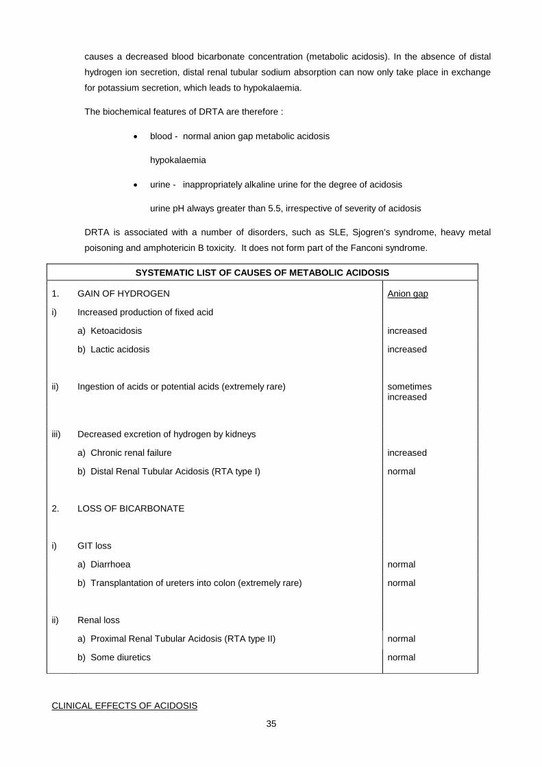
SYSTEMATIC LIST OF CAUSES OF METABOLIC ACIDOSIS
1. GAIN OF HYDROGEN Anion gap
i) Increased production of fixed acid
a) Ketoacidosis increased
b) Lactic acidosis increased
ii) Ingestion of acids or potential acids (extremely rare) sometimes increased
iii) Decreased excretion of hydrogen by kidneys
a) Chronic renal failure increased
b) Distal Renal Tubular Acidosis (RTA type I) normal
2. LOSS OF BICARBONATE
i) GIT loss
a) Diarrhoea normal
b) Transplantation of ureters into colon (extremely rare) normal
ii) Renal loss
a) Proximal Renal Tubular Acidosis (RTA type II) normal
b) Some diuretics normal
CLINICAL EFFECTS OF ACIDOSIS
36
• Increased [H+] stimulates the respiratory centre and causes hyperventilation. This causes deep, rapid
and gasping respiration known as Kussmaul breathing. This is a physiological compensatory response
which decreases the pCO2 and therefore returns the pH towards normal.
• Increased [H+] commonly causes hyperkalaemia. Intracellular polyanions such as proteins- and
glycogen- normally bind hydrogen and potassium ions. In an acidosis, excess hydrogen ions move into
cells, displacing potassium ions Increased [H+] causes increased neuromuscular irritability. There is thus
a risk of cardiac arrhythmias, especially in the presence of hyperkalaemia.
• Increased [H+] depresses consciousness, which can progress to coma and death.
TREATMENT OF METABOLIC ACIDOSIS
1. Treat the primary cause.
2. Treat any dehydration and hyperkalaemia which are commonly present.
3. Treat the acidosis if severe (pH < 7.0) by administering NaHCO3
Beware of the following dangers in correcting acidosis by administering HCO3-, especially if it is done too fast
1. The brain may become more acidotic than before. As soon as bicarbonate is administered it
increases the blood pH. This switches off the stimulus for hyperventilation and the pCO2 increases.
The higher pCO2 equilibrates across the blood-brain-barrier faster than the higher [HCO3-], thereby
dropping the pH of the brain.
2. Hyperkalaemia may change to dangerous hypokalaemia. Hyperkalaemia, if present, is caused by
displacement of potassium ions out of cells upon the influx of excess hydrogen ions. On correcting
the acidosis, this process is reversed and potassium ions move back into cells, which can cause
hypokalaemia.
METABOLIC ALKALOSIS
In a metabolic alkalosis there is an increase in the blood pH caused by an increase in the bicarbonate
concentration. The increase in the bicarbonate concentration can be caused by one of two mechanisms -
the gain of bicarbonate, or the loss of hydrogen ions.
COMMON CAUSES OF METABOLIC ALKALOSIS
1. VOMITING
Gastric fluid is rich in hydrochloric acid. Strictly speaking this is an extravascular pool of hydrogen,
the loss of which would not directly affect intravascular hydrogen ion concentration. However, loss of
gastric fluid is followed by the production of new hydrochloric acid. Inside the gastric parietal cells,
carbonic acid dissociates into hydrogen and bicarbonate ions. The hydrogen ions are secreted into
the stomach by the proton pump, while the bicarbonate ions diffuse into the circulation, causing the
post-prandial “alkaline tide”. With vomiting there is thus an accumulation of bicarbonate (as long as
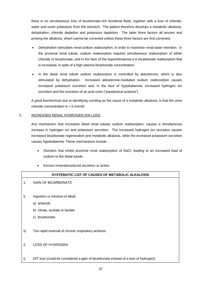
37
there is no simultaneous loss of bicarbonate-rich duodenal fluid), together with a loss of chloride,
water and some potassium from the stomach. The patient therefore develops a metabolic alkalosis,
dehydration, chloride depletion and potassium depletion. The latter three factors all worsen and
prolong the alkalosis, which cannot be corrected unless these three factors are first corrected.
• Dehydration stimulates renal sodium reabsorption, in order to maximise renal water retention. In
the proximal renal tubule, sodium reabsorption requires simultaneous reabsorption of either
chloride or bicarbonate, and in the face of the hypochloraemia it is bicarbonate reabsorption that
is increased, in spite of a high plasma bicarbonate concentration.
• In the distal renal tubule sodium reabsorption is controlled by aldosterone, which is also
stimulated by dehydration. Increased aldosterone-mediated sodium reabsorption causes
increased potassium excretion and, in the face of hypokalaemia, increased hydrogen ion
excretion and the excretion of an acid urine (“paradoxical aciduria”).
A good biochemical clue to identifying vomiting as the cause of a metabolic alkalosis, is that the urine
chloride concentration is < 5 mmol/l.
2. INCREASED RENAL HYDROGEN ION LOSS
Any mechanism that increases distal renal tubular sodium reabsorption, causes a simultaneous
increase in hydrogen ion and potassium secretion. The increased hydrogen ion secretion causes
increased bicarbonate regeneration and metabolic alkalosis, while the increased potassium secretion
causes hypokalaemia. These mechanisms include :
• Diuretics that inhibit proximal renal reabsorption of NaCl, leading to an increased load of
sodium to the distal tubule.
• Excess mineralocorticoid secretion or action.
SYSTEMATIC LIST OF CAUSES OF METABOLIC ALKALOSIS
1. GAIN OF BICARBONATE
i) Ingestion or infusion of alkali
a) antacids
b) citrate, acetate or lactate
c) bicarbonate
ii) Too-rapid reversal of chronic respiratory acidosis
2. LOSS OF HYDROGEN
i) GIT loss (could be considered a gain of bicarbonate instead of a loss of hydrogen)
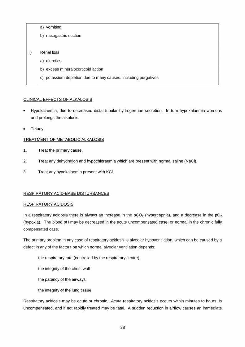
38
a) vomiting
b) nasogastric suction
ii) Renal loss
a) diuretics
b) excess mineralocorticoid action
c) potassium depletion due to many causes, including purgatives
CLINICAL EFFECTS OF ALKALOSIS
• Hypokalaemia, due to decreased distal tubular hydrogen ion secretion. In turn hypokalaemia worsens
and prolongs the alkalosis.
• Tetany.
TREATMENT OF METABOLIC ALKALOSIS
1. Treat the primary cause.
2. Treat any dehydration and hypochloraemia which are present with normal saline (NaCl).
3. Treat any hypokalaemia present with KCl.
RESPIRATORY ACID-BASE DISTURBANCES
RESPIRATORY ACIDOSIS
In a respiratory acidosis there is always an increase in the pCO2 (hypercapnia), and a decrease in the pO2
(hypoxia). The blood pH may be decreased in the acute uncompensated case, or normal in the chronic fully
compensated case.
The primary problem in any case of respiratory acidosis is alveolar hypoventilation, which can be caused by a
defect in any of the factors on which normal alveolar ventilation depends:
the respiratory rate (controlled by the respiratory centre)
the integrity of the chest wall
the patency of the airways
the integrity of the lung tissue
Respiratory acidosis may be acute or chronic. Acute respiratory acidosis occurs within minutes to hours, is
uncompensated, and if not rapidly treated may be fatal. A sudden reduction in airflow causes an immediate

39
increase in pCO2 and decrease in pO2, which can cause coma and death. Examples include choking, acute
exacerbations of asthma, and severe bronchopneumonia.
Chronic respiratory acidosis is usually long-standing, and is accompanied by maximal metabolic
compensation. The pCO2 is increased, but so is the bicarbonate due to increased renal hydrogen ion
excretion, therefore the pH is normal or only slightly reduced. Examples include chronic bronchitis and
emphysema.
SYSTEMATIC LIST OF CAUSES OF RESPIRATORY ACIDOSIS
1. DEPRESSION OF RESPIRATORY CENTRE
i) Drugs such as morphine, barbiturates, alcohol or general anaesthetics
ii) Head injury
iii) Intracerebral disease or tumours
2. PHYSICAL INABILITY TO VENTILATE
i) Crush injury to chest, flail chest
ii) Muscle paralysis, muscle relaxants
3. AIRWAY OBSTRUCTION
i) Asthma
ii) Chronic obstructive airways disease (COAD) - emphysema and chronic bronchitis
iii) Foreign body inhalation
4. PULMONARY DISEASE CAUSING DECREASED CO2 and O2 EXCHANGE
i) Severe pneumonia
ii) Severe lung collapse
iii) Severe pulmonary fibrosis
TREATMENT OF RESPIRATORY ACIDOSIS
1. Treat the underlying cause, e.g., remove any physical airway obstruction
2. Improve alveolar ventilation and lower the pCO2 by using physiotherapy, bronchodilators, antibiotics
and assisted ventilation if necessary. Beware of lowering the pCO2 too rapidly in chronic conditions,
since the renal compensatory changes takes several days to reverse - otherwise the patient would
develop a residual metabolic alkalosis.
3. Improve the hypoxia. In acute conditions, this must be done urgently by using oxygen at high
concentrations, or even assisted ventilation. In chronic conditions, beware of removing the hypoxia
which may be the only stimulus to which the respiratory centre still responds.

40
RESPIRATORY ALKALOSIS
In a respiratory alkalosis there is always a decrease in the pCO2 (hypocapnia), but hypoxia is not invariably
present. The blood pH may be increased in the acute uncompensated case, or normal in the chronic fully
compensated case.
The primary problem in any case of respiratory alkalosis is respiratory hyperstimulation, when the rate of
excretion of CO2 exceeds the rate of production of CO2.
Respiratory alkalosis may be acute or chronic. Acute respiratory alkalosis shows a decrease in pCO2, and a
small (physiological) decrease in actual bicarbonate, but no metabolic compensation (normal SBC). The
blood pH is increased. Examples include hysterical overventilation and pulmonary oedema.
Chronic respiratory alkalosis, which is rare, is accompanied by metabolic compensation. The pCO2 is
decreased, but so is the bicarbonate due to decreased renal hydrogen ion excretion, therefore the blood pH
is normal or only slightly increased.
SYSTEMATIC LIST OF CAUSES OF RESPIRATORY ALKALOSIS
1. DIRECT STIMULATION OF RESPIRATORY CENTRE
i) Drugs e.g., salicylates
ii) Anxiety, hysteria
iii) Brain stem disease
2. MECHANICAL OVERVENTILATION
3. HYPOXIA
i) High altitude
ii) Anaemia
iii) Pulmonary disease causing decreased O2 diffusion
- Pulmonary oedema
iv) Pulmonary disease causing decreased O2 and CO2 exchange (ventilation/perfusion imbalance) *
- Mild pneumonia
- Mild lung collapse
- Mild pulmonary fibrosis
Note: The conditions marked with * can have a respiratory acidosis if severe.
For explanation see discussion on page 12 of the notes.
TREATMENT OF RESPIRATORY ALKALOSIS
Treat the underlying cause
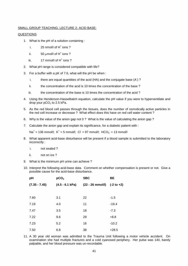
41
SMALL GROUP TEACHING. LECTURE 2- ACID BASE:
QUESTIONS
1. What is the pH of a solution containing :
i. 25 nmol/l of H+ ions ?
ii. 50 µmol/l of H+ ions ?
iii. 17 mmol/l of H+ ions ?
2. What pH range is considered compatible with life?
3. For a buffer with a pK of 7.6, what will the pH be when :
i. there are equal quantities of the acid (HA) and the conjugate base (A-) ?
ii. the concentration of the acid is 10 times the concentration of the base ?
iii. the concentration of the base is 10 times the concentration of the acid ?
4. Using the Henderson-Hasselbalch equation, calculate the pH value if you were to hyperventilate and drop your pCO2 to 2.5 kPa.
5. As the red blood cell passes through the tissues, does the number of osmotically active particles in the red cell increase or decrease ? What effect does this have on red cell water content ?
6. Why is the value of the anion gap not 0 ? What is the value of calculating the anion gap ?
7. Calculate the anion gap and explain its significance, for a diabetic patient with :
Na+ = 136 mmol/l; K+ = 5 mmol/l; Cl- = 97 mmol/l; HCO3- = 13 mmol/l
8. What apparent acid-base disturbance will be present if a blood sample is submitted to the laboratory incorrectly :
i. not sealed ?
ii. not on ice ?
9. What is the minimum pH urine can achieve ?
10. Interpret the following acid-base data. Comment on whether compensation is present or not. Give a possible cause for the acid-base disturbance.
pH
(7.35 - 7.45)
pCO2
(4.5 - 6.1 kPa)
SBC
(22 - 26 mmol/l)
BE
(-2 to +2)
7.60 3.1 22 -1.5
7.19 4.0 11 -19.4
7.47 3.5 18 -7.3
7.22 9.6 29 +8.8
7.23 5.2 16 -10.2
7.50 6.8 38 +28.5
11. A 30 year old woman was admitted to the Trauma Unit following a motor vehicle accident. On examination she had multiple fractures and a cold cyanosed periphery. Her pulse was 140, barely palpable, and her blood pressure was un-recordable.
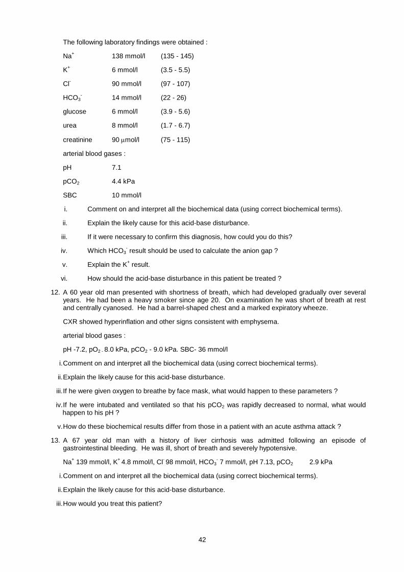
42
The following laboratory findings were obtained :
Na+ 138 mmol/l (135 - 145)
K+ 6 mmol/l (3.5 - 5.5)
Cl- 90 mmol/l (97 - 107)
HCO3- 14 mmol/l (22 - 26)
glucose 6 mmol/l (3.9 - 5.6)
urea 8 mmol/l (1.7 - 6.7)
creatinine 90 µmol/l (75 - 115)
arterial blood gases :
pH 7.1
pCO2 4.4 kPa
SBC 10 mmol/l
i. Comment on and interpret all the biochemical data (using correct biochemical terms).
ii. Explain the likely cause for this acid-base disturbance.
iii. If it were necessary to confirm this diagnosis, how could you do this?
iv. Which HCO3- result should be used to calculate the anion gap ?
v. Explain the K+ result.
vi. How should the acid-base disturbance in this patient be treated ?
12. A 60 year old man presented with shortness of breath, which had developed gradually over several years. He had been a heavy smoker since age 20. On examination he was short of breath at rest and centrally cyanosed. He had a barrel-shaped chest and a marked expiratory wheeze.
CXR showed hyperinflation and other signs consistent with emphysema.
arterial blood gases :
pH -7.2, pO2 - 8.0 kPa, pCO2 - 9.0 kPa. SBC- 36 mmol/l
i. Comment on and interpret all the biochemical data (using correct biochemical terms).
ii. Explain the likely cause for this acid-base disturbance.
iii. If he were given oxygen to breathe by face mask, what would happen to these parameters ?
iv. If he were intubated and ventilated so that his pCO2 was rapidly decreased to normal, what would happen to his pH ?
v. How do these biochemical results differ from those in a patient with an acute asthma attack ?
13. A 67 year old man with a history of liver cirrhosis was admitted following an episode of gastrointestinal bleeding. He was ill, short of breath and severely hypotensive.
Na+ 139 mmol/l, K+ 4.8 mmol/l, Cl- 98 mmol/l, HCO3- 7 mmol/l, pH 7.13, pCO2 2.9 kPa
i. Comment on and interpret all the biochemical data (using correct biochemical terms).
ii. Explain the likely cause for this acid-base disturbance.
iii. How would you treat this patient?
43
14. A 60 year old man complains of a 4 week history of increasing weakness, as well as mild polyuria and polydipsia. On examination, weakness of the limbs was confirmed. He was normotensive and adequately hydrated.
Na+137 mmol/l, K+ 1.7 mmol/l, Cl- 120 mmol/l, HCO3- 8 mmol/l, urea 7.4 mmol/l, pH 7.2, pCO2
2.8 kPa, SBC 9.5 mmol/l
i. Comment on and interpret all the biochemical data (using correct biochemical terms).
ii. Is the combination of hypokalaemia with acidosis unusual ? In which conditions can this occur ?
iii. Explain the likely cause for this biochemical picture.
iv. What other investigations might be helpful ?
15. An 18 year old woman was admitted to hospital repeatedly over a period of 6 weeks with a history of nausea, weight loss, weakness and “fainting”. Each episode had similar biochemical findings and responded to potassium supplementation.
Na+ 140 mmol/l, K+ 2.4 mmol/l, Cl- 87 mmol/l, urea 7 mmol/l, creat 50 umol/l, HCO3- 39 mmol/l, pH
7.53, pCO2 6.4 kPa
i. Comment on and interpret all the biochemical data (using correct biochemical terms).
ii. What do the biochemical features suggest ?
iii. What would analysis of the patient’s urine reveal ?
iv. Explain the likely cause for this biochemical picture.
16. A young man was involved in a road traffic accident and sustained severe chest injuries. He was struggling to breathe and in great distress, but fully conscious.
pH 7.24, pCO2 8 kPa, pO2 8 kPa, SBC 25 mmol/l
i. Comment on and interpret all the biochemical data (using correct biochemical terms).
ii. Work out the possible causes for this acid-base disturbance.
17. A young woman is admitted to hospital unconscious following a fall. Her respiratory rate is persistently rapid.
pH 7.48, pCO2 3.9 kPa, pO2 12 kPa, SBC19 mmol/l
i. Comment on and interpret all the biochemical data (using correct biochemical terms).
ii. Work out the possible causes for this acid-base disturbance.
18. A young man had been complaining to his GP of tiredness and weight loss. On questioning he admitted to excessive thirst and passing more urine than normal. He was scheduled for assessment in hospital the following day, but by then he was feeling drowzy and unwell, and vomiting. On admission he was found to have a BP of 95/60 with a pulse rate of 120/min and cold extremities. His breathing was deep and sighing.
Na+ 130 mmol/l K+ 5.8 mmol/l, Cl- 105 mmol/l, urea 18 mmol/l, creat 140 umol/l, glu 32 mmol/l, HCO3-
5 mmol/l, pH 7.05, pCO22 kPa
i. Comment on and interpret all the biochemical data (using correct biochemical terms).
ii. Work out the possible causes for this acid-base disturbance.
19. A young woman is admitted to hospital 8 hours after taking an aspirin overdose.
Na+ 140 mmol/l, K+ 4.7 mmol/l, Cl- 110 mmol/l, HCO3- 11 mmol/l, pH 7.48. pCO2 2 kPa
i) Comment on and interpret all the biochemical data (using correct biochemical terms).
ii) Work out the possible cause for this acid-base disturbance.

44
SMALL GROUP TEACHING. LECTURE 2- ACID BASE 1:
ANSWERS
1. i. 7.6, ii. 4.3, iii.1.8
2. [H+] of 20 to 120 nmol/l = pH of 6.9 - 7.7
3. i. 7.6, ii. 6.6, iii. 8.6
4. Acute change in pCO2, so no time to compensate. HCO3- is therefore normal.
[HCO3-]
pH = pK + log ------------------
[0.225 x pCO2]
[24]
pH = pK + log --------------- = 7.73
[0.225 x 2.5]
5. As the red blood cell passes through the tissues, CO2 and H2O are converted to HCO3- and H+, i.e.,
additional osmotically active particles are produced. The HCO3- is exchanged for Cl- (chloride shift)
which is also osmotically active. Water therefore enters the red blood cell and the cell swells.
6. The “anion gap” is a measure of all the charges on proteins and other unmeasured anions. The normal charge carried on proteins is about 10 - 20, hence that is the size of the “anion gap”.
Calculating the anion gap is valuable in distinguishing between various causes of metabolic acidosis.
7. (136 + 5) - (97 + 13) = 31 i.e., increased anion gap.
This means that unmeasured anions are present, probably ketone anions.
8. i. CO2 will diffuse out of the sample, therefore decreased pCO2, therefore “respiratory alkalosis”. ii. Ongoing red cell metabolism will produce a lactic acidosis, i.e., “metabolic acidosis”.
9. 4.4 (maximum H+ gradient of 1000 mmol/l = maximum pH gradient of 3)
10. .
pH pCO2 SBC BE Interpretation
1 7.60 3.1 22 -1.5 alkalosis, respiratory, no metabolic compensation
2 7.19 4.0 11 -19.4 acidosis, metabolic, partial respiratory compensation
3 7.47 3.5 18 -7.3 alkalosis, respiratory, partial metabolic compensation
4 7.22 9.6 29 +8.8 acidosis, respiratory, partial metabolic compensation
5 7.23 5.2 16 -10.2 acidosis, metabolic, no respiratory compensation
6 7.50 6.8 38 +28.5 alkalosis, metabolic, partial respiratory compensation
Some possible causes
45
1.acute resp alk - anxiety, hysteria, acute pulmonary oedema, lung collapse
2.chronic met acid - any cause (lactic acidosis, DKA, diarrhoea, RTA)
3.chronic resp alk - brain stem disease, living at high altitude, anaemia, pulmonary fibrosis
4.chronic resp acid - COAD, head injury, intracerebral tumour, pneumonia, pulmonary fibrosis
5.acute met acid - early lactic acidosis, early DKA
6.chronic met alk - vomiting with dehydration, diuretics, purgative abuse, Conn's
11.
i. Metabolic acidosis with partial respiratory compensation. Anion gap = 40 i.e., increased.
Mild hyperkalemia. Increased urea and normal creatinine.
ii. Clinically shocked. Probable cause - lactic acidosis due to poor tissue perfusion.
Pre-renal uraemia indicates poor renal perfusion
iii. Seldom necessary to do, but can measure lactate.
iv. Actual bicarbonate, not SBC.
v. Acidosis leads to H+ shifting into cells, thereby displacing K+ from intracellular anionic binding sites.
vi. Rehydration; treat patient’s physical injuries. Bicarbonate therapy probably not necessary. Monitor pH, HCO3
- and especially K+ levels.
12.
i. Respiratory acidosis with partial metabolic compensation. Hypoxia.
ii. COAD - based on history, examination, CXR findings and compatible acid-base findings.
The history is chronic, therefore there has been ample time for metabolic compensation to occur.
iii. The pO2 would increase, and this would reduce one of the stimuli to his respiratory centre, i.e., the hypoxic stimulus. This would reduce his ventilatory drive and ventilatory rate and therefore further increase his pCO2, worsening the respiratory acidosis.
iv. The mechanism described above would not operate, because his ventilatory rate would be maintained artificially. However, rapid correction of his pCO2 to normal while his HCO3
- is still high would lead to a metabolic alkalosis.
v. Acute condition, therefore no metabolic compensation. At the same level of pCO2, the pH would be much lower.
13.
i. Metabolic acidosis with partial respiratory compensation.
Anion gap = (139+4.8) - (98+7) = 38.8 i.e., increased.
ii. Lactic acidosis on the basis of poor tissue perfusion caused by massive GIT bleed, probably from oesophageal varices.
iii. Resuscitate with normal saline and give blood transfusion. Treat his underlying conditions (cirrhosis and GIT haemorrhage).
14.
i. Metabolic acidosis with partial respiratory compensation.
Anion gap = (137+1.7) - (120+8) = 10.7 i.e., normal.

46
Severe hypokalaemia. Mild renal impairment.
ii. Yes it is more common for hypokalaemia to be associated with alkalosis, and for hyperkalaemia to be associated with acidosis. The combination of hypokalemia and acidosis can occur in RTA.
iii. RTA.
iv. Urine pH on acid loading (if urine pH can drop below 5.5 then it is proximal RTA).
Urine HCO3-, phosphate, glucose and amino acids (if these substances are increased then
Fanconi syndrome is present, part of proximal RTA).
Look for the cause.
15.
i. Metabolic alkalosis with partial respiratory compensation.Severe hypokalemia.
Increased urea and low creatinine.
ii. Low chloride suggests vomiting as the cause of the metabolic alkalosis.
Pre-renal uraemia indicates poor renal perfusion and suggests dehydration.
Low creatinine indicates small muscle bulk.
iii. Low urine Cl- (< 5 mmol/l) and acid urine pH.
iv. Bulimia.
16.
i. Respiratory acidosis with no metabolic compensation. Hypoxia.
ii. Acute respiratory acidosis in this situation could have been caused by a crush injury to the chest, multiple rib fractures causing a "flail" chest segment, or acute lung collapse. Acute airway obstruction would also be a possibility. Head injury would be a less likely possibility, since the patient is conscious.
17.
i. Respiratory alkalosis with almost complete metabolic compensation. No hypoxia.
ii. Chronic respiratory alkalosis without hypoxia can only be due to hyperstimulation of the respiratory centre. Possible causes include the obvious one of brain stem injury, but the possibility of drug ingestion must not be overlooked.
18.
i. Hyponatraemia. Hyperkalaemia. Increased urea and creatinine, with greater increase in urea. Hyperglycaemia. Metabolic acidosis with partial respiratory compensation. Anion gap = (130+5.8) - (105+5) = 25.8 i.e., increased
ii. The features are typical of DKA, with signs of fluid depletion and Kussmaul breathing.
19.
i. Respiratory alkalosis. The presence of the very low bicarbonate at first suggests almost complete metabolic compensation, but in fact there has not been sufficient time for that to occur. The other possibility is a concomitant metabolic acidosis, i.e., a mixed acid-base disturbance. Anion gap = (140+4.7) - (110+11) = 23.7 i.e., increased
ii. A mixed acid-base disturbance is characteristic of salicylate poisoning - initial respiratory stimulation causes a respiratory alkalosis, but later salicylate causes a metabolic acidosis with a high anion gap (salicylate anion accumulates).
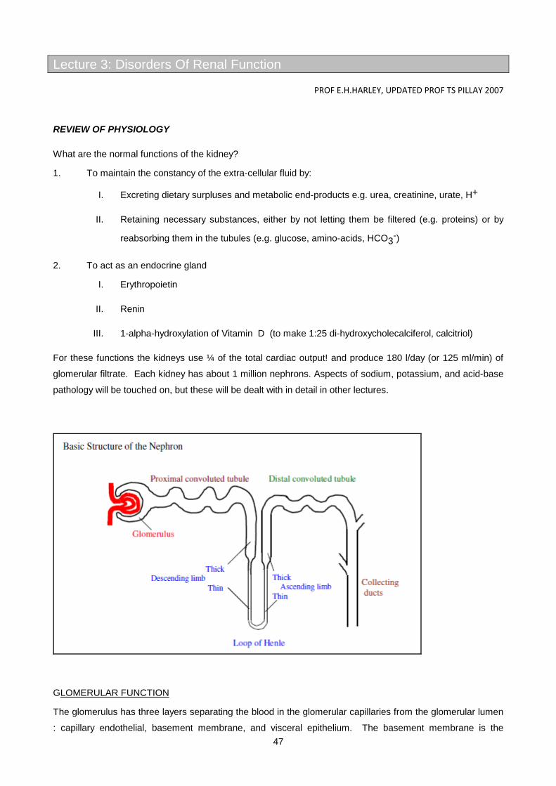
47
Lecture 3: Disorders Of Renal Function
PROF E.H.HARLEY, UPDATED PROF TS PILLAY 2007
REVIEW OF PHYSIOLOGY
What are the normal functions of the kidney?
1. To maintain the constancy of the extra-cellular fluid by:
I. Excreting dietary surpluses and metabolic end-products e.g. urea, creatinine, urate, H+
II. Retaining necessary substances, either by not letting them be filtered (e.g. proteins) or by
reabsorbing them in the tubules (e.g. glucose, amino-acids, HCO3-)
2. To act as an endocrine gland
I. Erythropoietin
II. Renin
III. 1-alpha-hydroxylation of Vitamin D (to make 1:25 di-hydroxycholecalciferol, calcitriol)
For these functions the kidneys use ¼ of the total cardiac output! and produce 180 l/day (or 125 ml/min) of
glomerular filtrate. Each kidney has about 1 million nephrons. Aspects of sodium, potassium, and acid-base
pathology will be touched on, but these will be dealt with in detail in other lectures.
GLOMERULAR FUNCTION
The glomerulus has three layers separating the blood in the glomerular capillaries from the glomerular lumen
: capillary endothelial, basement membrane, and visceral epithelium. The basement membrane is the
48
primary barrier. An overall negative charge due to abundant sialic acid groups in the glomerular layers helps
prevent large anions, such as most proteins, from crossing. The hydrostatic pressure across the membrane
which carries the ultrafiltrate is only about 1 kPa, and if blood pressure falls only moderately, the oncotic
pressure due to the plasma proteins is sufficient to cause filtration to slow or even stop. This explains the
oliguria in shocked patients.
(NB polyuria = more than normal urine; oliguria = less than normal; anuria = no urine).
TUBULAR FUNCTION
1. PROXIMAL TUBULE
Designed to reabsorb only what you need - 80% of sodium and water - high capacity active sodium uptake,
with chloride following passively. A low plasma chloride can limit recovery of sodium- important later for
understanding how this can cause or perpetuate alkalosis after prolonged vomiting.
- K+ : 95% absorbed usually, but diet dependant
- phosphate : active reabsorption which is inhibited by PTH
- HCO3- : mostly absorbed, but see acid-base lectures for details
- glucose and amino acid absorption is normally nearly complete
- also secretes: organic acids, urate, drugs
The Fanconi syndrome is loss (inherited or acquired) of proximal tubular functions and is characterised
(logically from the above) by glycosuria, amino aciduria, and phosphaturia. May also have acidosis and
polyuria. Classic cause is cystinosis, an inherited disease of lysosomal membrane transport, but it can also
be aquired.
2. LOOP OF HENLE
Only found in birds and mammals. Its counter-current multiplier effect is the basis for creating either
dilute or concentrated urine. Key features are an active NaCl pump in the thick ascending limb, and
water impermeability of the whole of the ascending limb.
Fluid emerges hypotonic at the end of the loop of Henle, at about ½ the osmolality of body fluids (~
120-150 mosmoles/l, as compared with 250-300 mosmoles/l in plasma). Further salt may be
removed in the collecting ducts under conditions of water diuresis, causing further dilution.
3. DISTAL CONVOLUTED TUBULE (DCT)
Little change in volume or concentration
Secretion of Aldosterone (see renin-angiotensin system) causes sodium to be exchanged for K+
and/or H+. Conn's syndrome, Addisons disease, and RTA type I all affect DCT function.
49
4. COLLECTING DUCTS
Anti Diuretic Hormone (ADH, Vasopressin, Pitressin), synthesised in the hypothalamus and released
from the posterior pituitary in response to an increase in extracellular osmolality, increases water
permeability of tubular cells (and urea permeability in the lower medullary part).
ADH linked pathology exerts its effect here and consists of
4.1 Diabetes insipidus
: pituitary form, where no ADH is synthesized due to damage to the pituitary.
: nephrogenic form, where renal tubular cells do not respond to normal levels of ADH. Both
forms give rise to polyuria with dilute urine.
4.2 Syndrome of inappropriate secretion of ADH (SIADH): low output of inappropriately concentrated urine in the presence of hypervolaemia and dilutional hyponatraemia.
TESTS OF RENAL FUNCTION
GLOMERULAR FUNCTION TESTS:
These depend on examination of substances which depend on glomerular function for their
elimination:
• SERUM CREATININE: Filtered at the glomerulus and eliminated without significant
reabsorption or secretion in the tubules (not altogether true but works in practise). Derived
from creatine phosphate in muscle. Serum levels are related to muscle mass, and
influenced by dietary meat intake. Increases as renal mass is lost in chronic renal disease,
but not in a linear fashion. Increases in acute renal failure.
• CREATININE CLEARANCE (Cr Cl): Owing to creatinine not being significantly reabsorbed or
secreted by the renal tubules, Cr Cl provides a measure of the glomerular filtration rate
(GFR). It is calculated as follows:
Cr Cl. = (Urine Creatinine conc. x volume) / (Plasma Creatinine conc.)
Note that the units are a flow rate, ml/min. It can be difficult to measure well since the timed
urine collection (should be over 24 hrs) is in practice notoriously unreliable and requires good
patient and staff cooperation. However, Cr Cl. changes more linearly in proportion to renal
mass loss than does plasma creatinine or urea (Cr Cl decreases, the latter increase) so is
the best measure of progress in chronic renal failure. It is of no value in acute renal failure
since it needs a steady state situation to obtain a meaningful result. Normal Cr Cl. is about
120 ml/min.
• BLOOD UREA: Derived in the liver from protein breakdown: is a useful measure of
decreased filtration. About 30-40 % is normally reabsorbed in tubules. Levels are affected

50
by high protein intake, catabolic states, post-surgery and trauma, and gastro-intestinal
haemorrhage, all of which cause increased urea production from protein.
• Estimated GFR (eGFR): Laboratories are making increasing use of estimated GFR derived
from creatinine estimations, more especially because of the problems with measuring
creatinine clearance. There are a number of equations that have been introduced. These
make use of the patient’s weight and creatinine level to estimate the GFR. The most popular
equation in use is the MDRD (Modification of Diet in Renal disease) equation. This is only
used in adults (>18 years) estimated GFR (eGFR) should be calculated using the 4-variables
(i.e. serum creatinine concentration, age, gender and ethnic origin). It is recommended that
the isotope dilution mass spectrometry (ID-MS) traceable version of the Modification
of Diet in Renal Disease (MDRD) equation should be used:
GFR (mL/min/1.73 m2) = 175 x [serum creatinine (umol/L) x 0.011312]-1.154 x [age]-
0.203 x [1.212 if black] x [0.742 if female]
The MDRD cannot be used in children. Here, the child’s height has to be taken into
account and the Schwartz equation should be used. The precision and accuracy of EGFR
decreases as GFR increases.
CAUSES OF ABNORMAL SERUM UREA TO CREATININE RATIO
Increased Decreased
High protein intake Low protein intake
G.I. haemorrhage Dialysis (urea crosses dialysis membrane easier)
Hypercatabolic state Severe liver disease (decreased urea synthesis)
Dehydration
Urinary Stasis
Muscle wasting or amputation
1. TUBULAR FUNCTION TESTS
1.1 Urinary Na+ concentration. Normally low relative to serum concentration unless on a dietary
high salt intake.
1.2 Concentration tests (usually after Pitressin), and Dilution tests, after a water load. Ratio of
osmolality (or urea) in urine relative to that in plasma is a simple practical measure.
2.3 Acidification tests, after administration of NH4Cl ( → NH3 + H+ + Cl- ). Seldom done except
in differentiation of type I and II renal tubular acidoses (RTA, see later).
MISCELLANEOUS TESTS:
3.1 Microscopy : look for casts, cells, or crystals
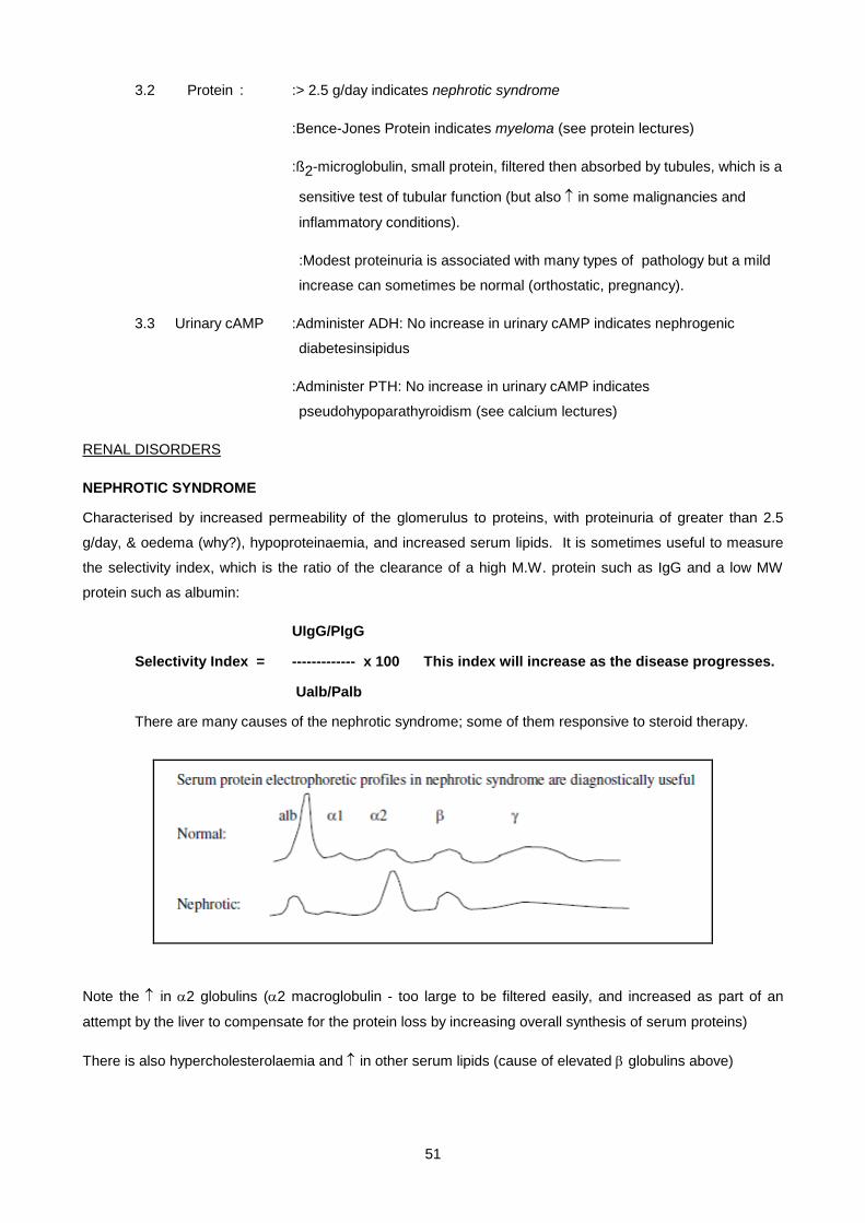
51
3.2 Protein : :> 2.5 g/day indicates nephrotic syndrome
:Bence-Jones Protein indicates myeloma (see protein lectures)
:ß2-microglobulin, small protein, filtered then absorbed by tubules, which is a
sensitive test of tubular function (but also ↑ in some malignancies and
inflammatory conditions).
:Modest proteinuria is associated with many types of pathology but a mild
increase can sometimes be normal (orthostatic, pregnancy).
3.3 Urinary cAMP :Administer ADH: No increase in urinary cAMP indicates nephrogenic
diabetesinsipidus
:Administer PTH: No increase in urinary cAMP indicates
pseudohypoparathyroidism (see calcium lectures)
RENAL DISORDERS
NEPHROTIC SYNDROME
Characterised by increased permeability of the glomerulus to proteins, with proteinuria of greater than 2.5
g/day, & oedema (why?), hypoproteinaemia, and increased serum lipids. It is sometimes useful to measure
the selectivity index, which is the ratio of the clearance of a high M.W. protein such as IgG and a low MW
protein such as albumin:
UIgG/PIgG
Selectivity Index = ------------- x 100 This index will increase as the disease progresses.
Ualb/Palb
There are many causes of the nephrotic syndrome; some of them responsive to steroid therapy.
Note the ↑ in α2 globulins (α2 macroglobulin - too large to be filtered easily, and increased as part of an
attempt by the liver to compensate for the protein loss by increasing overall synthesis of serum proteins)
There is also hypercholesterolaemia and ↑ in other serum lipids (cause of elevated β globulins above)

52
ACUTE RENAL FAILURE (ARF)
Definition: Urine output less than 450 ml/day (in adult) with a rising blood urea (N = 1.7 - 6.7 mmoles/l)
The blood urea typically rises by 5 mmoles/l/day, but in surgical, trauma, or gastro-intestinal bleeding it can
rise by up to 15 mmoles/l/day.
ARF is a common medical emergency with some critical urgent decisions facing the clinician (i.e. - you): the
diagnostic problem is : has the patient got :
1. Pre-renal failure (defect before the kidney)
2. Intra-renal failure (defect in the kidney e.g. acute tubular necrosis, glomerulonephritis)
3. Post-renal failure (defect after the kidney, e.g. prostatic enlargement, urolithiasis).
Causes under these headings include:
Pre-renal Intra-renal Post-renal
Hypovolaemia Acute tubular necrosis (ischaemic or
toxic)
Bilateral ureteric
obstruction
Decreased cardiac
output
Acute glomerulonephritis Urethral obstruction
Renovascular obstruction Interstitial nephritis
Intrarenal vasoconstriction (e.g. sepsis)
Tubular obstruction (e.g. uric acid crystals)
To decide between these three categories of acute renal failure is urgent because
1. Pre-renal failure if not rapidly treated can progress to the much more serious intra-renal failure (acute
tubular necrosis).
2. Some aspects of the treatment of intra-renal failure are the opposite of those for pre-renal failure.
The diagnostic strategy is to assess tubular function in a situation where glomerular malfunction is
predominating - reflex glomerular shutdown to varying degrees always accompanies acute tubular necrosis,
probably by renal circulatory redistribution to avoid the disastrous polyuria of a “pure” tubular malfunction.
Tubular function will be defective in Intra-renal failure but normal (for a while) in pre-renal failure. Some
appropriate tests to distinguish between these two are therefore:
PRE-RENAL INTRA-RENAL
Urinary sodium concentration * < 20 > 40 mmol/l
53
Urine/plasma osmolality ratio > 1.4 < 1.1
Urine/plasma urea ratio > 14 < 10
* The urinary Na+ is low in pre-renal failure because the low blood volume causes a marked stimulation of
aldosterone-mediated Na+ uptake. In Intra-renal failure the damaged tubules can't respond fully.
MANAGEMENT OF ACUTE RENAL FAILURE
1. Post-renal - relieve the obstruction, but then watch out for subsequent polyuria (why should this occur ?)
2. Pre-renal - restore blood volume, then blood pressure and GFR will return to normal levels.
3. Acute tubular necrosis -
3.1 Water : if blood volume is low, replace with care, since fluid overload can lead to
cardiac failure, maintain balance with 500 ml/day for insensible loss, plus previous
day's volume of urine output, monitor by weighing patient daily
3.2 Na+ : if oliguric – restrict, in diuretic phase - may need to administer Na+ ++.
3.3 K+ :if oliguric - restrict - may even have to dialyse, in diuretic phase - administer if
hypokalaemic.
3.4 H+ :high anion gap metabolic acidosis, may need bicarbonate to neutralise a severe
acidosis, but danger of Na+ overload if too much NaHCO3 is given in oliguric
phase.
3.5 Dialysis is indicated if
: blood urea is greater than 50 mmol/l and rising
: bicarbonate is less than 10 mmol/l
: K+ is greater than 7.0 mmol/l (or ECG changes
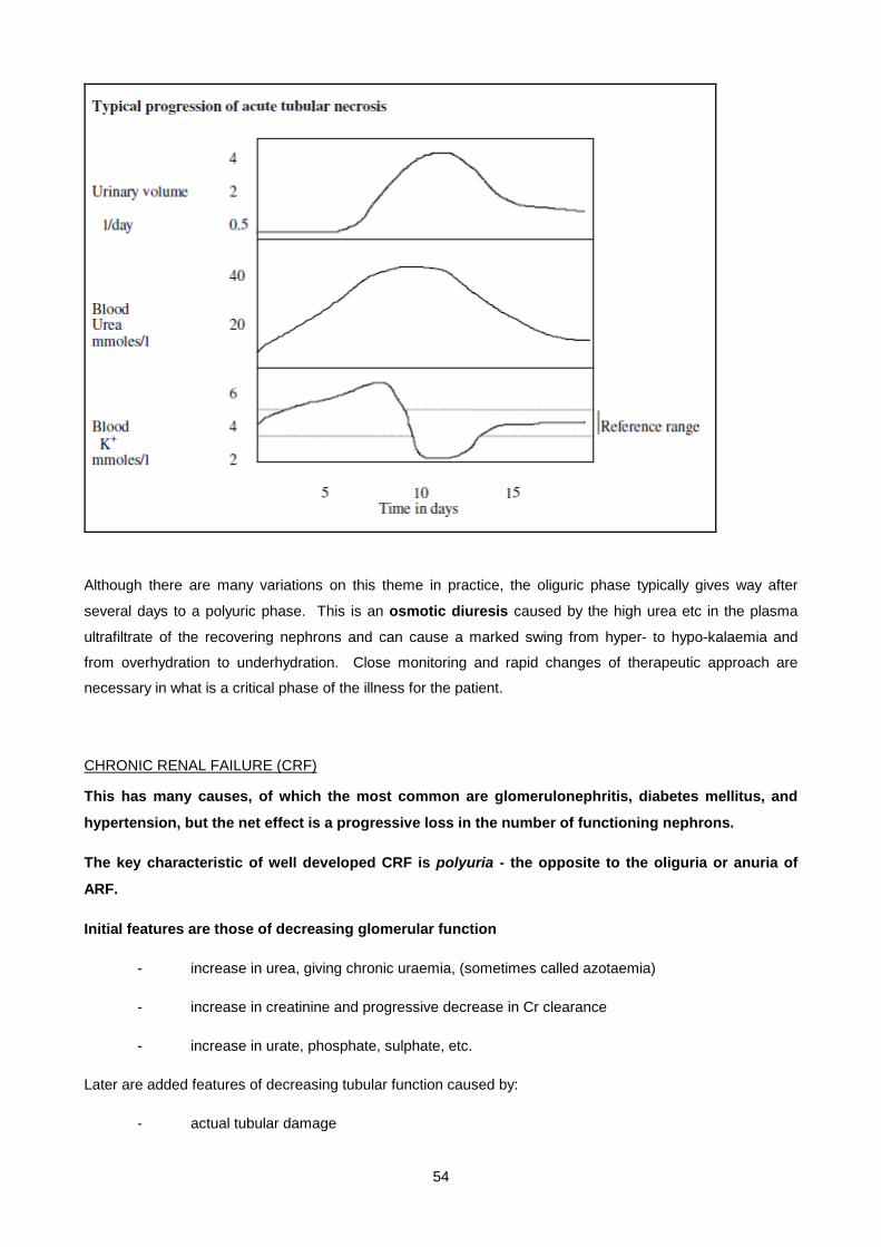
54
Although there are many variations on this theme in practice, the oliguric phase typically gives way after
several days to a polyuric phase. This is an osmotic diuresis caused by the high urea etc in the plasma
ultrafiltrate of the recovering nephrons and can cause a marked swing from hyper- to hypo-kalaemia and
from overhydration to underhydration. Close monitoring and rapid changes of therapeutic approach are
necessary in what is a critical phase of the illness for the patient.
CHRONIC RENAL FAILURE (CRF)
This has many causes, of which the most common are glomerulonephritis, diabetes mellitus, and
hypertension, but the net effect is a progressive loss in the number of functioning nephrons.
The key characteristic of well developed CRF is polyuria - the opposite to the oliguria or anuria of
ARF.
Initial features are those of decreasing glomerular function
- increase in urea, giving chronic uraemia, (sometimes called azotaemia)
- increase in creatinine and progressive decrease in Cr clearance
- increase in urate, phosphate, sulphate, etc.
Later are added features of decreasing tubular function caused by:
- actual tubular damage
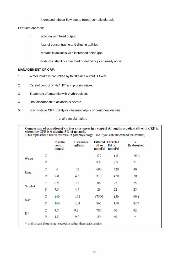
55
- increased tubular flow due to (urea) osmotic diuresis
Features are then:
- polyuria with fixed output
- loss of concentrating and diluting abilities
- metabolic acidosis with increased anion gap
- sodium instability - overload or deficiency can easily occur
MANAGEMENT OF CRF:
1. Water intake is controlled by thirst since output is fixed.
2. Careful control of Na+, K+ and protein intake.
3. Treatment of anaemia with erythropoietin.
4. Oral bicarbonate if acidosis is severe.
5. In end-stage CRF : dialysis - haemodialysis or peritoneal dialysis
: renal transplantation.
56
MISCELLANEOUS TOPICS:
POLYURIA
1. Water diuresis (urinary osmolality <200)
1.1 Compulsive water drinking
1.2 Diabetes insipidus - neurogenic or nephrogenic
2. Osmotic diuresis (urinary osmolality + 300)
Caused by the presence of incompletely reabsorbed solutes in the tubular lumen:
2.1 Na+ - dietary, iatrogenic, diuretics, salt-losing nephritis.
2.2 Urea – CRF, recovery phase of acute tubular necrosis or post-renal failure
2.3 Glucose (diabetes mellitus)
2.4 Mannitol,and some other therapeutic agents.
RENAL STONES (Nephrolithiasis)
Causes
1. A high concentration of a substance in the urine due to:
- low urine volume
- high excretion rate
2. pH changes
- alkaline urine predisposes to Ca deposition (e.g. infection)
- acid urine predosposes to uric acid deposition.
3. Stagnation, usually due to obstruction.
Types of stones (or calculi)
1. Calcium - oxalate (+ phosphate)
- phosphate
2. Uric acid - in about 10% of gouty cases. May be associated with low urinary pH due to inadequate buffer production.
3. Rare forms - cystine: cystinuria, a transport defect of dibasic amino acids and cysteine
- xanthine: xanthine oxidase deficiency
- 2,8 dihydroxyadenine: Adenine Phosphoribosyl Transferase (APRT - a
purine salvage enzyme) deficiency
Calculi are only partly mineral; up to 60% may consist of protein, the rest being varying proportions of
calcium, magnesium, ammonium, phosphate, etc.
57
Treatment
- fluids ++ to keep urine dilute (in all cases)
- Potassium citrate and thiazide diuretics may help prevent Ca stone formation - urinary citrate helps
solubilise calcium.
- alkalinisation may help prevent uric acid (but not 2,8 dihydroxyadenine) stone formation
RENAL ACIDOSIS
There are 2 components to H+ excretion by the kidney: (i) reabsorption of filtered bicarbonate in the proximal
tubule, and (ii) H+ secretion in the distal tubule.
1. URAEMIC ACIDOSIS.
Seen in acute or chronic renal failure. Decreased H+ excretion due to both glomerular and tubular
failure. Increased anion gap due to retention of phosphate, sulphate and other anions. Acidosis
develops only when the GFR falls below about 20 ml/min. (plasma creatinine >350 µM). Associated
with hyperkalemia.
2. RENAL TUBULAR ACIDOSIS (RTA).
A group of disorders characterized by tubular dysfunction, with normal or perhaps slightly decreased
glomerular function. The picture is that of a normal anion gap (i.e. hyperchloraemic) metabolic
acidosis, in the presence of a normal or near-normal plasma creatinine. The urine pH is often
inappropriately high in the face of the systemic acidosis (but NOT always - see below).
Type 1 (distal) RTA:
Due to inability of distal nephron to excrete H+. The urine pH is inappropriately high (pH >
5.5), but does not contain significant bicarbonate. Associated with hypokalemia,
nephrocalcinosis and rickets. Hypokalemia occurs because there is an increase in K+/Na+
exchange compensating for the defect in H+/Na+ exchange in the distal tubule. There are
genetic and acquired forms (e.g. heavy metal toxicity).
Type 2 (proximal) RTA:
Due to defective proximal bicarbonate reabsorption. The renal threshold for bicarbonate is
decreased (normal value 24 mmol/l). Whenever the plasma bicarbonate level exceeds the
(lowered) renal threshold (e.g. 16 mmol/l), the urine contains large amounts of bicarbonate
and the urine pH is inappropriately high (called renal bicarbonate wasting). However, if the
plasma bicarbonate drops to the level of renal threshold, then all filtered bicarbonate can be
reabsorbed, and the urine pH can be appropriately acidic (< 5.5) since distal tubular H+
excretion is normal. Associated with hypokalemia, due to the increased delivery of Na+ to the
distal tubule where Na+/K+ exchange occurs. Proximal RTA may be isolated or may be
associated with other proximal tubular defects: glycosuria, phosphaturia and amino aciduria -
the Fanconi syndrome. Can be genetic (e.g. with cystinosis) or acquired (e.g. toxins).
58
(Type 3 RTA is a mixed form of types 1 and 2 - not recognised nowadays as a specific entity.)
The acidosis due to mineralocorticoid deficiency or to renal resistance to mineralocorticoid action is
sometimes called Type 4 RTA. Associated with hyperkalemia. Mineralocorticoid resistance may be a
specific genetic entity (pseudohypoaldosteronism, see below) or may result from generalised tubular
damage.
RARE INHERITED DISORDERS OF THE RENAL TUBULES
This section provides additional voluntary reading, but despite dealing with rare disorders it provides
interesting insights into more detailed physiological mechanisms of renal function:
1. Liddle's syndrome, or pseudohyperaldosteronism: Hypertension, hypokalaemic metabolic
alkalosis, low renin and aldosterone, risk of heart disease. Autosomal dominant. Plasma volume
expansion due to abnormal Na reabsorption (amiloride sensitive) downstream of the
mineralocorticoid receptor. Cause is an increase in the number of Epithelial Na channels (ENaC) in
distal tubule at apical surface of the cells.
2. Pseudohypoaldosteronism type 1 (PHA1): Failure To Thrive (FFT), hyponatraemia with salt
wasting, dehydration and profound hyperkalaemic metabolic acidosis. Renin & aldosterone are
elevated. Sporadic, Autosomal Dominant (AD), or Autosomal Recessive (AR). Mutations in (any of
the 3 subunits of the) ENaC gene abolishing function. Effects similar to those of the diuretic
amiloride.
3. Bartter's syndrome: FTT, polyuria, nephrocalcinosis, short stature, and mental retardation.
Hypokalaemic metabolic alkalosis with renal salt wasting, nephrogenic Diabetes Insipidus, and
hypercalciuria. Sporadic or AR. Profound activation of the renin-angiotensin-aldosterone axis +
hyperplasia of the juxtoglomerular apparatus with excessive intra-renal production of prostaglandin
E2. Primary abnormality is in epithelial cells of the medullary thick ascending limb (mTAL) of the
nephron, with mutations causing loss of function of one or other of the following:
a) The sodium-potassium-chloride triple cotransporter (NKCCT) responsible for elecroneutral
Na and Cl reabsorption in the mTAL (loop diuretics such as furosemide work here - giving
diuresis, kaliuresis, salt wasting, concentrating defects and hypercalciuria).
b) The ATP-regulated potassium channel, ROMK1, through which K is re-secreted back to the
lumen. This channel is tightly coupled to NKCCT, so mutations in either can cause Bartter's
syndrome. The consequence of the impaired mTAL NaCl absorption and resulting
hypovolaemia activates the renin-aldosterone system to produce an increase in distal tubular
Na reabsorption (via ENaC) and kaliuresis, plus metabolic alkalosis. Hypercalciuria is
explained by the fact that 20% of Ca absorption is coupled to Na absorption in the mTAL.
c) A thick-ascending limb baso-lateral chloride channel (CLCKB). Despite significant
hypercalciuria, nephrocalcinosis is not a feature.

59
4. Gitelman's syndrome: Hypokalaemic metabolic alkalosis, but (in contrast to Bartter's) also
hypocalciuria and hypomagnesaemia as invarient features. Sporadic or AR, presenting in late
childhood or early adult life. Not especialy hypervolaemic despite some activation of renin-
aldosterone axis . Mg excretion high, causing hypomagnesaemia. Defect in a distal convoluted tubule
thiazide-sensitive sodium-chloride cotransporter (NCCT), hence syndrome resembles chronic
thiazide diuretic use. Heterozygotes may be predisposed to hypokalaemic alkalosis on loop or
thiazide diuretic therapy.
5. X-linked hypercalciuric nephrolothiasis (Dent's disease): hypercalciuria, low-MW proteinuria,
progressive nephrocalcinosis, nephrolithiasis, and renal failure. Fanconi-type generalised proximal
tubulopathy and rickets. Renal transplant is an effective therapy. Defect is in a member of the
voltage-gated chloride channels (CLC5), although mechanism is uncertain - maybe electrical or
osmotic gradients become unfavorable inhibiting reabsorption functions. Carrier females show LMW
proteinuria and sometimes hypercalciuria.
60
SMALL GROUP TEACHING. LECTURE 3- : DISORDERS OF RENAL FUNCTION:
QUESTIONS
1. What substances is the kidney normally working to (i) retain, (ii) get rid of?
2. What maintains a normal GFR and what effect does plasma oncotic pressure have on this?
3. What is the source of urea (and draw its chemical structure). In which organ(s) does this pathway operate? What are the other forms in which animals eliminate nitrogen waste?
4. What is the source of creatinine and how is it formed?
5. Is there a difference in the renal handling of urea and creatinine?
6. What hormones does the kidney produce?
7. How does the kidney make concentrated and dilute urine
8. How and where does ADH act, and what disorders of ADH function are known?
9. If a substance had a renal clearance of 160 ml/min what would this tell you about its handling by the kidney?
10. What are the two most commonly performed serum analyses to give information on glomerular function?
11. What factor(s) OTHER THAN RENAL FUNCTION affect serum urea levels?
12. Explain the clinical value of measuring creatinine clearance; what different information does it give from measuring just serum creatinine?
13. Suggest possible conditions under which the following results might be obtained:
(i) A high urea with a normal creatinine:
(ii) A high creatinine with a normal urea:
14. How does acidification with NH4Cl work Is it a test used in clinical practise?
15. What would you expect the urinary Na+ concentration to be in:
(i) A patient who has had a severe haemorrhage (low plasma volume)
(ii) A patient who has impaired tubular function.
16. What is the value of measuring proteins in the urine?
17. What is the Fanconi syndrome, and what anatomical part of the kidney is affected?
18. A mother donates one of her kidneys for transplant to one of her children who has a severe kidney disease. How will her own renal function be affected?
19. If the normal Creatinine clearance is 120 ml/min what would you suggest are likely values in patients with loss of:
(i) 50 %
(ii) 90% of renal functional mass?
What sort of changes might you expect to see in plasma urea and/or creatinine?
20. What is the usual cause of glomerulonephritis, and how might it present?
21. A 4 year old child presents with facial oedema a few weeks after a flu-like illness? What single test will be most informative?
22. What is the differential diagnosis of polyuria?
23. Describe the biochemical features of acute tubular necrosis during
61
i. The acute phase
ii. The recovery phase:
24. What are the main biochemical similarities and differences between pre-renal failure and acute tubular necrosis (intrinsic renal failure)?
25. Why is it important to differentiate between these two conditions at an early phase, and what are the key tests to help in this differentiation?
26. List the biochemical disturbances which are characteristically seen in chronic renal insufficiency.
27. What is the treatment for
i) A moderately severe Chronic Renal Failure
ii) End stage renal failure?
28. What substances can cause an osmotic diuresis, and under what conditions?
29. The urine pH can fall to lower values (ie become more acid) in one of the two main forms of renal tubular acidosis than in the other; how? Which is easier to treat and with what?
30. What are kidney stones composed of? Which types of stone are radio-opaque, and which are radiolucent?
31. What factors predispose to the formation of kidney stones?
32. What are the principles of treatment for renal stones?
33. Give two causes of heavy proteinuria.
34. 56 year old female. Symptoms : tiredness, weakness, developing over a long period. Several years previously she had developed backache due to lumbar disc prolapse, and had habitually consumed large quantities of analgesic tablets.
serum: Na+ 140 (N 135-145) K+ 5.5 (N 3.5-5.5) Cl- 100 (N 97-107) Bicarbonate 16 (N 22-26) urea 33 mM (N 1.7-6.7) creatinine 900 µM (N 75-115) calcium 1.9 mM (N 2.1-2.6) albumin 40 g/l (N 30-50) inorganic phos. 4.2 mM (N 0.8-1.4) urate 0.57 mM (N 0.12-0.5)
urine: Na+ 50 mmol/l K+ 30 mmol/l urea 120 mmol/l creat 4.0 mmol/l (4000 µmoles/l) osmol. 330 mosm/kg urine output: 3 litres/24h
The findings were similar 2 months previously at an outpatient clinic visit.
i. Calculate the creatinine clearance.
ii. The patient has a normal 24h urinary output of urea and creatinine, but markedly elevated plasma values. Explain this apparent paradox.
iii. Comment on the plasma bicarbonate concentration.
iv. Comment on the plasma urate and phosphate.
62
v. Suggest a cause for the hypocalcemia.
vi. Calculate the daily sodium output. What could happen if her sodium intake fell substantially below this value?
vii. Comment on the plasma K+. Is it important to monitor this regularly?
35. A 6-month old male infant was investigated for poor feeding and failure to gain weight. There was occasional regurgitation of food but no diarrhoea. There were no problems during labour and the infant appeared normal after birth. The following biochemical results were obtained:
Serum: Na+ 134 mmol/l, K+3.0mmol/l, Cl- 115mmol/l, HCO3- 12mmol/l, creatinine 75 µmol/l, pH 7.2, pCO2 3.8 kPa, Bicarbonate 11 mmol/l, URINE pH 6.7
Discuss the biochemical findings in this case, and suggest a diagnosis. Would any further investigations be useful? Discuss treatment.
36. 30 year old male. 48 h after motor accident. Multiple injuries. Serum: urea 20.5 mmol/l, creat 450 µmol/l, Na+ 140 mmol/l, K+ 6.0 mmol/l HCO3 15 mmol/l Urine:urea 74 mmol/l (N 250-600 mmoles/day), creat 1500 µmol/l (N 7-17 mmoles/day), Na+ 90 mmol/l, Cl- 100 mmol/l, urine osmol 350 mosm/l, urine output 400 ml/24 h
i. Comment on the urea and creatinine output.
ii. What diagnosis is suggested, and by what specific tests ?
iii. Why is the plasma potassium elevated?
iv. What is the danger associated with hyperkalemia?
v. What can be done to treat the hyperkalemia acutely?
vi. Comment on this patient's fluid requirements. Would an infusion of several litres of intravenous fluid be beneficial?
SMALL GROUP TEACHING. LECTURE 3- : DISORDERS OF RENAL FUNCTION:
ANSWERS
1.
i. Retain sugars, amino-acids, bicarbonate, water (!), and to variable extent (diet) Na+ and K+.
ii. Get rid of urea, urate, creatinine, sulphate, phosphate (usually), H+.
2. Hydrostatic pressure - plasma oncotic pressure counters this and results in no flow at only moderately low BP.
3. Protein; liver (kidney). Other forms: allantoin (most sensible mammals), ammonia (some fish); urate (reptiles and birds).
4. Muscle creatine phosphate. Mostly non-enzymatically.
5. Urea about 40% reabsorbed (clearance about 70 ml/min).
6. Erythropoetin (lack contributes to anaemia of CRF). Renin. 1- hydroxylation of D3 (lack contributes to hypocalcaemia of CRF).
7. Detailed functions of loop of Henle, medulla urea, collecting ducts and ADH.
8. Collecting ducts. Diabetes insipidus, SIADH.
9. It would mean that not only is it not being absorbed by the tubules but there is also active tubular secretion.
63
10. Urea and creatinine.
11. LOW : Liver failure, urea cycle disorders, low body mass, low protein intake, dialysis.
HIGH : GI haemorrhage, high protein intake.
12. Measure of GFR - useful in CRF (not in acute) - linear with respect to loss of renal mass. Serum Cr useful in acute renal failure and in late CRF, when it starts rising steeply.
13.
i. High protein intake,GI bleed, hypercatabolic state, dehydration/urinary stasis (urea more diffusible - leaks back from concentrated urine).
ii. Low protein intake, Dialysis (urea more diffusible), severe liver disease.
14. NH4CL → NH3 (metabolised to urea) + H+ + Cl-. Seldom used except for differentiation between types I & II RTA.
15.
i. low – pre-renal failure
ii. high.
16. Discuss the various causes - nephrotic, glom nephritis, BJP.
17. Glycosuria, phosphaturia, amino aciduria. Proximal.
18. No pathology expected. Her GFR (& Creatinine Clearance) will drop, but less (eventually) than 50% due to compensatory hypertrophy (see also next question).
19.
i. 60 ml/min
ii. 12 ml/min.
Plasma urea - little change at 50% loss - marked rise at 90% loss.
20. Auto-immune disease. Haematuria. Nephrotic syndrome.
21. Urinary protein.
22. See notes. (Polydipsia, DI, osmotic).
23. See notes.
24. Similarities: Urea & Creatinine up, acidosis, oliguria. Differences are all in the urine: Na+, osm, urea.
25. Pre-renal is reversible (if caught early). Treatment quite different. Tests: urinary Na+, urinary/plasma urea or osmolality ratios.
26. High serum urea, cr, etc. Mild acidosis. Polyuria. Na (and K) lability. Hypocalcaemia, anaemia.
27.
i. Dietary and monitoring
ii. Dialysis, renal transplant.
28. Na+, (dietary, diuretics), urea (CRF, release of post-renal obstruction, diuretic phase of ATN), glucose (DM), mannitol (iatrogenic, i/v).
29. When plasma bicarbonate levels fall below renal threshold in type II. Type I easier to treat with small quantities of bicarb, since then the distal tubule dosn’t have to excrete H+ (in II bicarb just runs straight through).
30. See notes.
64
31. See notes
32. Fluids +++; citrate (to chelate and solubilise urinary Ca);.treat primary cause where possible; alkalinise for uric acid stones.
33. Nephrotic Syndrome, Myeloma with Bence-Jones protein.
34.
i. 4000 x 3000/(900 x 60 x 24) = 9 ml/min
ii. Students often have a problem with understanding this. (I use the tap, sink, blocked outlet analogy).
iii. Anion gap metabolic acidosis associated with CRF
iv. Elevations both in keeping with CRF
v. Hydroxylation of vitamin D3 defective.
vi. Would become hyponatraemic.
vii. Not usually a major problem until very late in CRF, but avoid K+ rich foods (proteins).
35. Inappropriately alkaline urine in face of acidosis in a child suggests RTA (creatinine normal). Hypokalaemia consistent with type I or II (but not 4 – mineralocorticoid def.). Check for Fanconi (urinary glucose, phos, amino acids), urinary bicarb or CO2 (after bicarb loading if necesary). In type II acidification can allow urine pH to reach low levels when bicarb threshold is reached. If type I treat with bicarb. Renal transplant can help in cystinosis.
36.
i. Low output . That is why they are accumulating in plasma.
ii. Acute tubular necrosis. High urinary Na, urinary plasma urea ratio 74/20.5 = 3.6 (<10 = intra-renal failure, >14 = pre-renal). U/P osm ratio also probably close to or less than the 1.1 level.
iii. Release from damaged tissues, acidosis, poor renal elimination.
iv. Cardiac arrest.
v. Glucose/insulin
vi. Tight control on fluid balance needed. Several litres i/v would NOT be beneficial (might be lethal!)
65
Lecture 4: Disorders Of Water And Sodium Balance
DR PETER BERMAN 2007
IMPORTANCE OF A NORMAL PLASMA [NA+]
All cells (with the notable exception of the collecting ducts of the renal medulla) are freely permeable to water.
Thus, an osmotic gradient between a cell and its environment cannot develop, and extracellular osmolality is
always equal to intracellular (normally 280-290 mosmol/kg).
Though the osmolality of the two compartments is equal, the solutes responsible for it are very different.
Whereas Na+ion, along with its counter-anions Cl- & HCO3-, is the major contributor to extracellular fluid
(ECF) osmolality, intracellular fluid (ICF) osmolality derives mainly from K+ & Mg2+as cations, and organic
phosphates, including ATP, creatine phosphate and glycolytic intermediates as anions. The difference is
largely attributable to the Na+/K+ATPase, coupled with the impermeability of cells to Na+ions, so that
ECF[Na+] is generally >> ICF[Na+] (140mM versus 10mM).
Since ECF[Na+] is the major determinant of ECF osmolality, any increase in ECF[Na+] results in water
moving from cells into the ECF, which expands the ECF at the expense of the ICF, and leads to intracellular
dehydration. The converse occurs if ECF[Na+] falls; water passes into cells from the ECF, leading to cell
swelling. Bear in mind that the determinant of water movement is ECF Na+ concentration ([Na]), not total Na+
content.
Changes in cell volume are particularly important in the case of the brain, where cerebral dehydration causes
the brain to pull away from the meninges, rupturing meningeal vessels, while cerebral swelling (oedema)
causes equally catastrophic compression of the brain against the rigid vault of the skull. In either case,
affected individuals exhibit neurological symptoms; from altered behavior, irritability, and impaired level of
consciousness, to convulsions and coma.
Given time, the brain is able to adapt to these water shifts by appropriately altering its content of 'osmolytes'.
This heterogeneous collection of small molecules, including ions and a number of organic compunds,
accumulate in neuronal cells exposed to high ECF osmolality, minimizing water loss, whereas they fall in
brain cells chronically exposed to low ECF osmolality, thereby minimizing water influx.
IMPORTANCE OF A NORMAL ECF VOLUME
In the case of isotonic loss or gain of ECF, there is no change in ECF[Na+]), hence no change in ECF
osmolality and no change in ICF volume. Thus the ECF bears the full brunt of such loss or accumulation.
Since plasma is part of the ECF (about a ¼), any change in ECF volume is reflected by an equivalent change
in blood volume. Thus, a decrease in ECF volume leads to a fall in blood pressure, poor tissue perfusion,
shock and renal shutdown, while an expanded ECF gives rise to hypertension, oedema, or accumulation of
fluid in alveoli (pulmonary oedema). As an example, a patient who is 10% dehydrated from mainly water loss
(eg- running a marathon on a hot day), will lose fluid from both ICF and ECF, and be less haemodynamically
compromised than another patient who is 10% dehydrated from isotonic fluid loss (eg- acute diarrhoea).

66
The distribution of ECF fluid between interstitial and plasma compartments, normally about 3 to 1, is
determined by plasma albumin level, since albumin exerts an oncotic pressure that holds water in the plasma
compartment. When plasma albumin level falls, due to, say, cirrhosis or nephrotic syndrome, the decreased
oncotic pressure leads to shift of fluid from plasma into interstitium, with resulting in peripheral oedema and a
contracted blood volume. ICF volume is unchanged.
REGULATION OF HYDRATION STATUS
Let us review the homeostatic mechanisms whereby normal ECF and ICF volume is maintained. This allows
an understanding of what happens in disorders where these homeostatic mechanisms break down, as well
as the secondary adaptive responses to such breakdown.
MECHANISM SOURCE STIMULUS EFFECT
1. GFR kidney permits Na &
water excretion
2. Aldosterone adrenal ↓ renal perfusion renal Na & water
retention
3. ADH# hypothalamus ↑ ECF tonicity ↓↓↓ blood
volume
pure water
retention
4. ANF* cardiac atria ↑ blood volume renal Na & water
excretion
# = anti-diuretic hormone * = atrial natriuretic factor
CONTROL OF THE HYPOTHALAMIC OSMOSTAT- OSMOLALITY VS TONICITY
The hypothalamic osmostat, that controls both ADH release and the sensation of thirst, is acutely sensitive to
small changes in plasma osmolality. ADH secretion is abolished at osmolalities below 280mosm/kg, but
increases steeply as osmolality increases by a few mosm/kg above this value, leading to a profound drop in
urine volume and increase in urine osmolality. Are all osmotically-active particles equally potent in this
regard? Not actually, since the osmostat is, in reality, a cell volume receptor, and thus responds only to
osmotically-active substances confined to the ECF. For example, in pure water loss, ECF [Na+] increases,
the hypothalamic cell shrinks and activates the osmostat. Glucose has the same effect, since in diabetes
mellitus, glucose is confined to the ECF. Mannitol, used to treat cerebral oedema, behaves in the same way.
On the other hand, substances like urea and ethanol, which equilibrate across cell membranes, have no
effect on the osmostat, despite making a significant contribution to plasma osmolality; eg. in renal failure and
drunken stupor, urea and ethanol, respectively, can increase plasma osmolality by over 50mosmol/kg,
without activating the osmostat at all. The term, hypertonicity, rather than hyperosmolality, is sometimes
used to indicate an increased "effective" ECF osmolality, and is due to solutes, like Na+ and glucose, that are
confined to the ECF.

67
NORMAL WATER AND NA+ REQUIREMENTS
With all these systems working normally, what are the body's obligatory water and Na requirements?
There is an obligatory daily water loss from the skin, lungs, urine and GIT, so that under temperate climatic
conditions, a healthy adult requires a minimum water intake of just over a litre/day to remain in water balance.
OBLIGATORY LOSSES SOURCES
Skin 500ml
Lungs 400ml
Gut 100ml water from metabolism 400ml
Kidneys 500ml minimum in diet 1100ml
-------------------------------- ------------------------------------------------------
TOTAL 1500ml TOTAL 1500ml
Na+, on the other hand, is very well conserved by a healthy kidney, so that obligatory Na+ loss is
<10mmol/day. This suggests that during our evolutionary history, Na+ was in very short supply. Na+ intake
in a modern diet can be up to 100-200mmol/day. Although excess Na+ is readily excreted by the normal
kidney, there is evidence implicating excess Na+ intake in a variety of diseases, including hypertension, renal
stones and even osteoporosis.
We now consider the clinical syndromes of dehydration and overhydration.
DEHYDRATION
Dehydration is usefully subdivided according to how much Na has been lost along with the water; i.e. whether
fluid lost has been water only (hypotonic fluid loss), or has been accompanied by an equivalent loss of Na+
(isotonic fluid loss). These are two extremes – real clinical scenarios fall somewhere in between.
HYPOTONIC FLUID LOSS
Examples of hypotonic fluid loss include:
1. Diabetes Insipidus (DI) – both hypothalamic or nephrogenic
2. Osmotic diuresis (eg. diabetes mellitus)
3. Excess sweating, fever or exercise in a hot climate
4. Hyperventilation or assisted ventilation with unhumidified air
5. Cessation of water intake with ongoing obligatory water loss eg.in infants, elderly or unconscious patients.
Since water in excess of Na+ is lost from the ECF, ECF[Na+] rises, water moves in from the ICF. Thus,
signs of cerebral dehydration (confusion, etc) may be present. Increased plasma tonicity ('effective'
osmolality) rapidly evokes an immediate ADH and thirst response. Urine becomes maximally concentrated,
with urine osmolality approaching 1000 mosmol/kg (except, of course, when the cause of the water loss is
diabetes insipidus). Movement of water from ICF to ECF minimizes the depletion in blood volume, so the
68
signs of circulatory collapse (tachycardia, low BP, oliguria) are less pronounced, & occur later than would be
the case with isotonic fluid loss. Nevertheless, when blood volume has become sufficiently depleted, renal
underperfusion evokes renin release, which, via the angiotensin pathway, stimulates aldosterone secretion.
Aldosterone promotes Na+ uptake by the distal tubule and results in an extremely low urine [Na+] (<10mM).
This is a valuable clue that the hypernatraemia is due to water depletion rather than excess salt intake, since
in the latter condition, urine [Na] is very high (>100mM).
Fluids with a low Na+ content should be used for replacement - either water by mouth, or, if unable to drink,
5% glucose or 1/2N NaCl intravenously. Beware of over-rapid correction if the hypernatraemia has been long-
standing, since accumulated osmolytes pose the risk of iatrogenic cerebral oedema.
ISOTONIC FLUID LOSS
Examples of isotonic fluid loss include:
• Haemorrhage
• Burns
• GIT loss (diarrhoea, vomiting, fistula)
• Renal loss (diuretics, polyuric recovery phase of acute renal failure, Addisons disease)
• Effusion of ECF into body spaces (hematoma, ascites, pancreatitis)
Since water loss is accompanied by an equivalent amount of Na+, there is no immediate change in plasma [Na+],
and hence no movement of fluid from the ICF. Thus cerebral dehydration is not a problem. However, since all
the fluid lost comes from the ECF, circulatory collapse is more pronounced than in hypotonic fluid loss. Patients
can present shocked, pale and clammy, with tachycardia and hypotension. GFR may drop rapidly, with a falling
urine output, and a progressive rise in plasma [urea] and [creatinine]. Signs of haemo-concentration (↑
[haemoglobin], ↑ [total plasma protein]) may be present (unless, of course, haemorrhage is the cause of the fluid
loss). The fall in renal perfusion triggers early aldosterone release via the renin/angiotensin mechanism (except
in Addisons disease of course, where lack of aldosterone is the basic problem). As with hypotonic fluid loss,
urine is therefore highly concentrated, with a very low [Na] (<10mmol/l). Since plasma osmolality is unchanged,
there is no osmotic stimulus for early ADH release such as occurs in hypotonic fluid loss. ADH release only
occurs once blood volume depletion is severe. This ADH response, and administration of salt-free fluids, may
lead to subsequent HYPOnatraemia (in contrast to the hypernatraemia of hypotonic fluid loss).
Treatment, provided the patient has not development acute renal failure, consists of rapidly replacing the
ECF deficit with intravenous isotonic saline (0.9%NaCl). Continue until the patient is haemodynamically
stable, with a normal urine output. There is no danger of cerebral oedema from over-rapid replacement since
infused fluid is isotonic. The major danger is delay in treatment allowing acute renal failure (tubular necrosis
to develop. After restoring ECF volume, identify & treat the underlying problem (eg stop diuretics, give
minerallo-corticoid replacement for Addisons, etc).
COMPARISON BETWEEN HYPOTONIC WITH ISOTONIC FLUID LOSS:

69
HYPOTONIC ISOTONIC
plasma [Na+] ↑↑ normal to ↓
hematocrit slightly ↑ ↑↑↑
ECF volume ↓ ↓↓↓
plasma [urea] normal to ↑ ↑
urine output ↓↓↓ ↓
thirst early late
tachycardia & hypotension late early
fluid replacement cautious rapid
OVERHYDRATION
As with water depletion, water overload can be conveniently sub-divided into pure water overload and isotonic
water overload (water plus an equivalent amount of Na+).
PURE WATER OVERLOAD
Water intoxication from excess intake is rare, since the capacity of a healthy individual to excrete a water load
is considerable (at least 1 litre/hour). Once plasma tonicity falls by a few mosmol/kg., ADH secretion ceases,
and a large volume (1 litre/hour) of dilute urine (50 mosmol/kg) is excreted, until the excess water has been
eliminated. This process obviously requires intact renal function. Failure to excrete a water load results in
immediate dilutional hyponatraemia, and may lead to cerebral oedema, particularly when it develops acutely.
Causes of pure water overload include:
1. Water intake at a rate in excess of the kidney’s ability to excrete it; generally >1litre/hour. This
occasionally arises as a mental disorder (psychogenic polydipsia) or in dedicated beer drinkers.
2. Water intake in patients with salt-losing forms of renal disease (‘salt-losing nephritis’) or those on
diuretics that impair tubular salt reabsorption. Ability to excrete water depends on intact renal salt
reabsorption,
3. Syndrome of inappropriate ADH (SIADH), due to the failure of a low plasma osmolality to suppress
ADH secretion.
SIADH
70
SIADH may be due to:
1. Intracranial pathology (head injury, haemorrhage, meningitis, encephalitis, or brain tumor), where there is
direct stimulation of hypothalamic ADH release.
2. Pulmonary pathology (pneumonia, TB, assisted ventilation), where volume receptors in the pulmonary
vascular bed falsely report a message of vascular depletion to the hypothalamus.
3. Ectopic production of ADH by tumors, particularly bronchial carcinomas.
4. Cortisol deficiency – since these hormones antagonize ADH, a deficiency of either will result in
unopposed ADH action.
5. Pain, from trauma or surgery, stimulates ADH release.
6. Drugs, including psychoactive drugs (antidepressants, narcotics, carbamazepine), sulphonylureas,
oxytocin for labour induction, vincristine for chemotherapy.
Findings in SIADH
Although patients with SIADH are over-hydrated, they are not frankly oedematous. Retained water is shared
between the ECF and ICF, and ECF expansion is eventually limited by release of atrial natriuretic factor (ANF)
from cardiac atria, which promotes a saline diuresis. Their major problem is hyponatraemia, leading to cerebral
oedema and consequent depressed level of consciousness. The more rapid the onset of hyponatraemia, the
more severe is the cerebral oedema, since slow onset allows the brain time to decrease its osmolyte content.
Urine is typically inappropriately concentrated despite low plasma osmolality, and its [Na+] is high in the face of
hyponatraemia (the effect of ANF).
MANAGEMENT OF PURE WATER OVERLOAD
Restriction of water intake is the logical treatment, and, in mild cases, may be sufficient. If symptoms of water
intoxication are present, ADH may be specifically antagonized by the drug demeclocycline. Alternatively,
intravenous hypertonic saline (5% NaCl) or mannitol will rapidly remove intracellular water.
Two complications may arise during treatment:
Firstly, rapid infusion on hypertonic NaCl can overload the vascular bed (ECF) and lead to pulmonary oedema.
Secondly, over-rapid correction of plasma osmolality may cause cerebral dehydration, particularly if
hyponatraemia has been long-standing and the brain has responded by decreasing its level of osmolytes.
CENTRAL PONTINE MYELINOSIS is an acute, potentially fatal neurological condition that occurs if chronic
hyponatraemia is too rapidly corrected. During treatment, the risk of cerebral oedema from the primary
disorder should be balanced against the risk of causing central pontine myelinosis by increasing plasma [Na+]
too fast; as a general guide, it should not be increased by >1mmol/l/hr, or 10mmol/l/day.
ISOTONIC FLUID OVERLOAD
In this condition, accumulation of water is accompanied by an equivalent retention of Na+. Since there is no
osmotic force to drive excess fluid into the cells, the ECF bears the entire brunt of the fluid overload. It may

71
manifest as hypertension, or peripheral and/or pulmonary oedema, but never with neurological manifestations
from intra-cerebral water shifts, as seen in pure water overload.
Major causes of isotonic fluid overload include:
1. Administration of excess isotonic fluid, particularly to patients with impaired renal function.
2. Hyperaldosteronism, which is the inappropriate secretion of aldosterone despite an expanded ECF
volume. It may be either primary or secondary.
PRIMARY HYPERALDOSTERONISM (CONN'S SYNDROME)
This is autonomous secretion of aldosterone, typically by an adrenal adenoma, resulting in excessive Na+
and water reabsorption in the distal convoluted tubule, with concomitant loss of K+ and H+. There is
generally a hypokalaemic alkalosis, with plasma Na normal to slightly increased. Plasma renin levels are
appropriately suppressed, and the high plasma aldosterone levels are unaffected by posture or suppressed
by salt loading or ACE-inhibitors. Patients are typically hypertensive, but since ECF volume expansion is
limited by ANF, oedema is not a feature. Treatment is surgical resection of the tumor. It is an uncommon but
TREATABLE cause of hypertension, and is mandatory to exclude in a young hypertensive.. Individuals
exposed to excess non-aldosterone minerallocorticoid (liquorice, 11-deoxy-corticosterone (11-DOC)), present
with the same clinical and biochemical picture, except that plasma aldosterone levels are appropriately
suppressed.
SECONDARY HYPERALDOSTERONISM
This implies excessive aldosterone secretion in response to renin, so the basic problem is persistent renin
secretion in the absence of ECF volume depletion , i.e. renin secretion is, in a sense, inappropriate. How
can this arise?
1. A renin-secreting tumor (rare).
2. A disturbance in the blood supply to one or both kidneys, eg. renal artery stenosis
3. Abnormal shift of fluid from plasma into the interstitial space. Leakage of
fluid from the vascular bed into the tissues, depleting plasma volume, which stimulates renin
secretion. Despite a depleted plasma volume, total ECF volume is grossly expanded. This is the
commonest cause of secondary hyperaldosteronism by far, and merits some discussion.
Leakage from the capillary bed occurs either because of increased hydrostatic pressure, forcing fluid out
(congestive cardiac failure), or a decreased plasma oncotic pressure, due to a low [albumin. Common
causes of a low albumin include liver cirrhosis, nephrotic syndrome, protein-losing enteropathy and protein
malnutrition (kwashiorkor). The major clinical manifestation of this disorder is severe peripheral oedema,
though pulmonary oedema and large effusions in serous cavities (pleural, ascitic) can also occur. Despite
being whole body Na+ overloaded, they are typically hyponatraemic, since a contracted plasma volume acts
as a stimulus for ADH release, which leads to water retention. In fact, this form of secondary
hyperaldosteronism is probably the commonest cause of hyponatraemia seen in clinical practice. Treatment
72
is aimed at reversing the underlying condition, and at reducing the whole body Na and water load by diuretic
therapy and limiting salt intake.
PRIMARY Na+ OVERLOAD
This is fairly unusual. Examples include:
1. Administration of hypertonic NaCl or NaHCO3 to unconscious patients.
2. Infants given feeds in which salt has been inadvertently substituted for sugar.
3. Shipwrecked sailors drinking seawater in desperation.
Intake of salt unaccompanied by water leads to hypernatraemia, which immediately shifts fluid from ICF to
ECF, causing cerebral dehydration with its accompanying neurological symptoms. Provided renal function is
intact, a prompt saline diuresis ensues, leading to whole body dehydration in which the ICF bears the brunt.
Typical findings are hypernatraemia with a high urine Na+ (100-200mmol/l). This is in sharp contrast to the
hypernatraemia of pure water loss, where renal Na+ retention produces very low urine [Na+] (<10mmol/l).
PSEUDOHYPONATRAEMIA
Water contributes 93% to the volume of normal plasma. The remainder is mainly protein, with lipid
contributing <0.1%. In certain disease states, plasma lipid can increase enormously, to account for 5 or even
10% of plasma volume. Since [Na+] in the AQUEOUS PHASE of plasma is normal, there is no tendency for
fluid shifts.. However, since most methods for plasma [Na+] measure the Na+ content of a fixed volume of
plasma, such samples will yield misleadingly low [Na+]. This is termed pseudohyponatraemia and requires
no treatment aimed at normalising the [Na+]. Since osmolality depends on solute concentraion in the aqueous
phase, plasma osmolality is unaffected in pseudohyponatraemia, If any doubt remains, lipid can be removed
by ultracentrifugation, and the Na+ measurement repeated on the infranatant.
SICK-CELL SYNDROME
Seriously ill patients often develop a mild hyponatraemia. Some ascribe it to failure of the Na/K ATPase to
extrude Na+. A more plausible explanation is that loss of intracellular osmolytes causes re-setting of the
hypothalamic osmostat, such that ADH secretion only cuts out at lower plasma osmolality. This form of
hyponatraemia requires no treatment beyond that of the underlying condition.
WATER DEPRIVATION TEST
This test is used in patients presenting with polyuria and low urine osmolality, in which the diagnosis of
diabetes insipidus (either hypothalamic or nephrogenic) is being considered. Under medical supervision,
patients are deprived of water, serial urines collected, and their osmolalities measured. When urine
osmolality plateaus out, (i.e. there is no further increase in osmolality of sequential urine specimens), ADH is
administered, and the osmolality of the next urine noted.

73
Interpretation: In normal subjects and psychogenic polydipsia, urine osmolality rises progressively during
water deprivation until it reaches the high hundreds (700-800mosmol/kg). Plasma [Na+] and body weight
show negligible change.
In diabetes insipidus (DI), urine osmolality plateaus out at a much lower value (eg. 200-300 mosmol/kg),
depending on the severity. There is a progressive increase of plasma Na+ and loss of body weight, and
patients soon become thirsty and distressed from dehydration. If the DI is neurogenic, urine osmolality
increases after ADH is given, whereas in nephrogenic DI, it remains unchanged.
PRACTICAL ISSUES
WHEN TO MEASURE PLASMA Na+
Although plasma Na+ is very frequently measured, there are only really three scenarios where it is likely
to be of value:
1. Dehydrated patients or those with a history of fluid loss
2. Patients receiving fluids intravenously, especially infants, the elderly and the unconscious.
3. Patients with unexplained alteration in level of consciousness, confusion or irritability.
INTERPRETING A PLASMA Na+ RESULT
As should be clear by now, an isolated plasma [Na+] measurement is of little clinical value in the absence of
additional information. For instance, a [Na+] of 125 could be due to:
• primary water overload (e.g. SIADH)
• diarrhoea or diuretic therapy, with replacement of water only
• diabetes mellitus, where glucose draws water from ICF into ECF, diluting the Na+
• gross lipidaemia.
Thus, to properly interpret a plasma Na+ result, one must consider factors including:
1. The history - has there been:
• a head injury to suggest SIADH?
• diarrhoea and/or vomiting?
• polyuria or polydipsia to suggest diabetes (mellitus or insipidis)?
• diuretic therapy?
2. The clinical examination
• level of consciousness?
• hydration status - are there signs of water overload (oedema, distended neck veins,hypertension, pulmonary oedema) or dehydration (loss of skin turgor, dry mouth, tachycardia, low BP, concentrated urine, oliguria)?
• rapid weight changes, fluid intake/output recordings?
74
• pigmentation to suggest Addisons disease?
• features of cardiac failure, liver cirrhosis or nephrotic syndrome, to suggest secondary hyperaldosteronism?
3. Urine findings
• appearance - dilute or concentrated?
• osmolality?
• [Na+] concentration?
• any glucose or protein?
4. Blood findings
• haemoconcentration (increased haemoglobin and protein)?
• renal function (urea and creatinine levels)?
• milky appearance (lipidaemia)?
• K+ abnormalities (Addisons or primary aldosteronism)?
• low cortisol or high TSH to account for SIADH?
OSMOLAR GAP
Osmolality is determined by the sum of all solutes present in a fluid, and is usually measured by freezing
point depression. Plasma osmolality can generally be predicted quite accurately from the formula:
Osmolality = 2x [Na+] + [urea] + [glucose]
A significant disparity between measured and calculated osmolality is refered to as an "osmolar gap", and is
usually due to the presence of an osmotically active substance, the commonest by far in clinical practice
being ethanol.
75
SMALL GROUP TEACHING, LECTURE 4 DISORDERS OF WATER AND SODIUM BALANCE: QUESTIONS
1. Explain and differentiate between the concepts of osmolarity, osmolality and tonicity.
i. How is the approximate molarity calculated?
ii. What is the osmolar gap?
iii. When is an increased osmolar gap of clinical significance.
2. Describe the mechanisms of urine dilution and concentration.
3. In a normal adult subject, what is the maximum and what is the minimum achievable urine osmolality?
4. Explain the water and electrolyte changes which occur on administering.
i. normal saline (0.9% NaCl 150 mmol/l);
ii. tap water orally;
iii. 5% dextrose i.v.
5. Outline your approach to the differential diagnosis of :
i. hyponatraemia;
ii. hypernatraemia.
6. Write down the criteria necessary for diagnosing SIADH.
7. Explain the concept of pseudohyponatraemia. Should this condition be treated
8. Describe and explain the biochemical abnormalities which occur in :
i. osmotic diuresis
ii. acute blood loss
9. Explain these results obtained on a 30 year old male patient
Test Results Reference Range
Sodium 97 135 - 145 mmol/l
Potassium >10 3.0 - 4.7 mmol/l
Chloride 86 99 - 112 mmol/l
Total CO2 14 19 - 29 mmol/l
Calcium 0 2.2 - 2.6 mmol/l
Magnesium 0 0.6 - 1.0 mmol/l
Phosphate 0 0.6 - 1.2 mmol/l
Alkaline Phosphatase (ALP) 0 <120 U/l
Lactate Dehydrogenase (LD) 0 <320 U/l
Glucose 75 3.0 - 5.5 mmol/l
10. A 5 month old bottle-fed infant with severe diarrhoea has the following serum biochemistry:
Reference Range
Sodium 153 mmol/l (135-145)
Potassium 2.2 mmol/l (3.3-5.2)
76
Chloride 116 mmol/l (99-113)
Total CO2 13 mmol/l (19-29)
Urea 7.8 mmol/l (<5.5)
Creatinine 44 µmol/l (<45)
i. What is the likely fluid status of this child?
ii. Which constituents confirm this state?
iii. How would you manage the child?
iv. The normal range for creatinine in adults is up to 120mol/l. Why is this lower in a child?
11. A six week old male infant presented at Red Cross Hospital with projectile vomiting. Examination revealed a sausage-like mass in the epigastrium.
Serum: Reference Range Blood gases: Reference Range
Sodium 131mmol/l (135-145) pH 7.58 (7.37-7.43)
Potassium 2.1mmol/l (3.3-5.2) Pco2 45 mmHg (29-37)
Chloride 75mmol/l (99-113) Po2 60 mmHg (68-84)
AHCO3- 41 mmol/l (17-28)
Urine:
Sodium 8mmol/l
Potassium 25mmol/l
Chloride 6mmol/l
i. What is the volume status of this child ?
ii. Which results confirm this ?
iii. Does alkalosis fit this picture of hydration ?
iv. How could hyponatraemia develop in this situation ?
v. Why are U-Na and U-Cl divergent ?
12. A 60 year old man was admitted to hospital for evaluation of weakness, anorexia and haemoptysis (he gave a 20 year history of smoking 2 packs of cigarettes per day). The chest X-ray showed a left hilar mass. He had no oedema. Skin turgor appeared normal.
The following serum biochemistry results were reported:
Reference Range
Sodium 112mmol/l, Potassium 3.9 mmol/l, Chloride 84 mmol/l, Total CO2 21 mmol/l, Urea 1.9 mmol/l, Creatinine 60 µmol/l, Osmolality 240mmol/kg (280-295)
i. What is your diagnosis?
ii. What features are present that support your diagnosis and which would you like to know about that are not presented here?
iii. How would you treat his hyponatraemia?
13. A male aged 52 years has a 6 months history of loss of appetite, weight loss, fatigue and episodic abdominal pain. BP 105/60. Pulse 100/min.
Serum: Sodium 125 mmol/l, (135-145), Potassium 5.2mmol/l (3.3-5.2), Total CO2 16 mmol/l (19-29), Urea 8.2 mmol/l (2.6-8.0), Creatinine102 µmol/l (<120)

77
i. What is your differential diagnosis? Why?
ii. What would you expect his U-Na to be ?
iii. If his urinary sodium were low, how would you explain this?
iv. If high, how would you explain his urinary sodium?
v. How would you confirm your diagnosis?
vi. What is the treatment ?
SMALL GROUP TEACHING, LECTURE 4 DISORDERS OF WATER AND SODIUM BALANCE: ANSWERS
1.
i. Osmolarity is the number of osmoles dissolved in 1l of water and is expressed as mosmoles per litre: the total volume is thus 1l minus the solute volume. Osmolality refers to the number of osmoles per kilogram of water and is expressed as mosmol/kg: the total volume is thus 1l plus the solutes Tonicity refers to the effective osmolality i.e. that portion which is held on one side of a cell membrane and can thus cause fluid shifts. An isotonic fluid is always iso-osmolar whereas the converse does not hold. A suspension of red blood cells in an iso-osmotic urea solution will haemolyse: urea will equilibrate pulling water into the RBC.
ii. Twice the sodium plus potassium plus glucose plus urea. Note that urea should be excluded to determine the effective osmolarity
iii. The difference between the measured osmolality and the calculated osmolarity
iv. Poisoning e.g. methanol
2. Absorption of NaCl without water in the thick ascending limb of Henle for maximal dilution plus switch off ADH . For concentration NaCl absorption in the TAL to increase medullary osmolality plus the presence of ADH.
3. 50 mosmol/l and 1400 mosmol/l
4.
i. This will expand extracellular volume, but have no effect on the sodium concentration nor osmolality (iso-osmotic). There will be no movement across the cell membrane
ii. The addition of water will expand and dilute both the ICF and ECF
iii. This is a way of administering water to the body. 5%dextrose is iso-osmotic, but will be metabolised to cause net administration of water. Thus can cause hyponatraemia. Initially glucose will cause movement of water from the ICF, but in the presence of insulin will be taken up and metabolised leaving a net addition of pure water which will equilabrate across the cell membrane.
5. See lecture notes
6. Hypo-osmolar hyponatraemia. Inappropriately raised U-osmol. Normal potassium/acid base
Normal renal, adrenal, thyroid function
7. Solutes as expressed in molar concentration actually 7% solute + 93% water ( 70ml solute plus 930ml water to make up a litre). In hypertriglyceridaemia and Hyperproteinaemia more water replaced with solute thus instead of 930ml containing proportionate amount of measured solutes, now lower volume of water with proportionately lesser amount of solute. Measurement error. Treat primary condition, not lab result
8.
i. Causes extracellular fluid loss: thus fluid shifts from ICF to ECF. Also greater water loss via kidneys causes additional electrolyte loss

78
ii. Loss is iso-osmotic/isotonic. Thus contraction of intravascular volume which does not cause fluid shifts. However lower GFR and diminished perfusion of the kidney.
9. Drip contamination with - 5% glucose + potassium
10.
i. Dehydrated
ii. Hypernatraemia, elevated urea with normal creatinine
iii. Stop milk feeds: oral rehydration ( “invented” by a pathologist in Zambia – Maurice King). IV rehydration if necessary. Encourage Mum that breast feeding would have been better
iv. Creatinine is proportionate to muscle mass
11.
i. Dehydrated
ii. Urea >8.0 mmol/l and U-Cl < 6mmol/l
iii. Yes, due to secondary hyperaldosteronism caused by volume contraction
iv. Replacement of electrolyte loss with water due to thirst stimulus
v. Obligatory loss of Na with bicarbonate, but maximum drive still present to conserve as much water in the proximal tubule as possible. Thus U-Cl reflects this component.
12.
i. SIADH due to lung Ca
ii. Hypo-osmolar hyponatraemia, normal potassium and bicarbonate, normal renal function. Would like U-osmol >100 mosmol/l, U-Na > 20mmol/l, normal thyroid and adrenal function
iii. Fluid restriction
13.
i. Addisons disease, diabetes . Hyperkalaemic acidosis, dehydration ( pre-renal failure), hyponatraemia
ii. High
iii. Dehydration causing maximum reabsorption of Na in the proximal tubule
iv. Absence of aldosterone with delivery of sodium to the distal tubule
v. Synacthen test
vi. Replacement with mineralocorticoid (fludrocortisone) and glucocorticoid (hydrocortisone)
79
Lecture 5: Disorders Of Potassium Metabolism
DR PETER BERMAN & PROF ERIC HARLEY 2007
WHOLE BODY POTASSIUM DISTRIBUTION
In contrast to sodium (Na+), most potassium (K+) in the body is located within cells, as shown in the table below:
ICF ECF
_________________________________________________________________________
volume (litres) 28 14
K+ concentration (mmol/l) 150 5 ( i.e. levels are 30 x higher in the cell)
K+ content (as a % of total) 98% 2%
_________________________________________________________________________
Plasma K+ is a small fraction of, and hence not always an accurate reflection of whole body K+ status. For
example, in diabetic ketoacidosis, accumulated H+ ions force K+ ions out of cells into the plasma, from where
they are filtered by the kidney and lost in the urine. In this situation, although plasma K+ is normal or high,
intracellular K+ (and hence total body K+) may be severely depleted. Why is intracellular [K+] so much higher?
As a result of two forces, both acting to drive K+ into cells:
1. Intracellular anions, consisting of organic phosphate in various forms (creatine phosphate, ATP,
phosphorylated glycolytic intermediates) as well as protein, provide binding sites for cations (e.g. K+).
2. The Na/K ATPase in the cell membrane pumps Na+ out and K+ in against their respective concentration
gradients, thus ensuring that K+, rather than Na+, remains in the cell and balances the intracellular anions.
K+ AND THE CELL MEMBRANE POTENTIAL
K+ leaks out down its concentration gradient through K+ channels in the cell membrane, leaving the ICF
negative relative to the ECF. This continues until an equilibrium is reached, where the outwardly-directed
concentration gradient is matched by an inwardly-directed electrical force. The voltage at equilibrium is the
resting membrane potential, and can be predicted from the Nernst equation, i.e.
Membrane potential = - 60 x log[K+in]/[K+
out]
= - 60 x log 30 = - 60 x 1.5 = - 90 mV (inside negative)
Thus, membrane potential is directly related to the ratio of [K+in] to [K+
out], and hence is affected by the
following factors:
• Hypokalaemia enhances K+ efflux from the cell by increasing the K+ concentration gradient from IN to
OUT. K+ efflux hyperpolarizes excitable cells, making it increasingly difficult to initiate an action
potential. This slows or even blocks nerve conduction, resulting in muscle weakness and/or diastolic
cardiac arrest. e.g. with a serum K+ of 2.5 (half normal) the membrane potential is -60 x log 60 = -60
x 1.78 = -107 mV

80
• Hyperkalaemia opposes K+ efflux, reducing membrane potential, and facilitating depolarization. In
the heart this hyperexcitable state can lead to ectopic beats and ventricular fibrillation.
• Certain drugs, the sulphonylureas, used to treat diabetes, specifically block K+ channels in pancreatic
β-cells. By inhibiting K+ efflux down its concentration gradient, these drugs decrease cell membrane
polarization and facilitate depolarization, which triggers insulin release. The physiological blocker for
these K+ channels is ATP, which explains how glucose, by increasing intracellular ATP in the β-cell,
stimulates insulin release.
Apart from its role in generating a membrane potential, K+ ions are also important for the normal function of a
number of intracellular enzymes, for example, the glycolytic enzyme, pyruvate kinase. Thus intracellular K+
depletion leads to cell injury and dysfunction independent of its effect on cell excitability. Rhabdomyolysis
(skeletal muscle necrosis) and renal tubular dysfunction can result from K+ deficiency within the muscle and
renal tubular cells, respectively.
K+ HOMEOSTASIS
The kidney plays a central role in maintaining K+ balance. Filtered K+ is practically all reabsorbed in the
proximal convoluted tubule (PCT) and Loop of Henle. Hence, net K+ excretion depends on the SECRETION
of K+ by the distal convoluted tubule (DCT), a passive process depending on a transtubular electrical gradient
(lumen negative) created by Na+ absorption under the influence of aldosterone (see
diagram below).
A NUMBER OF FACTORS AFFECT RENAL K+ EXCRETION. THESE INCLUDE:
• Na+ delivery to the distal convoluted tubule (DCT). K+ cannot be excreted by the DCT unless
there is Na+ present with which it can exchange. When the GFR is reduced, a greater fraction of
filtered Na+ is reabsorbed proximally, thereby reducing Na+ delivery distally. Since less Na+ is
available to exchange for K+ or H+, both these cations are retained. This effect contributes to the
hyperkalaemic acidosis of acute renal failure. The opposite occurs when excess Na+ arrives at the
DCT. Diuretics, like lasix, that impair Na+ reabsorption in the Loop of Henle, leads to enhanced Na+
81
exchange for K+ and H+ in the distal tubule, and hypokalaemic alkalosis. Alternatively, in metabolic
alkalosis, usually due to vomiting, a decrease in filtered Cl- ions impairs proximal tubular Na+
reabsorption (since Na+ cannot be absorbed without an accompanying Cl-). Excess Na+ is delivered
distally, and the enhanced exchange of H+ and K+ ions for these Na+ ions perpetuates the alkalosis
and also leads to hypokalaemia.
• Aldosterone. Reabsorption of Na+ in exchange for K+ and H+ in the DCT is under control of the
steroid hormone, aldosterone. Hence in Conn’s syndrome, the result of an aldosterone-producing
tumor of the adrenal cortex, we see hypokalaemic alkalosis, whereas in patients with adrenal failure
(Addison’s disease) or those treated with diuretics that act as aldosterone antagonists
(spironolactone), hyperkalaemia and acidosis develop. It is worth remembering that excess
aldosterone can only cause K+ depletion if Na+ delivery to the DCT is normal or excessive -
hypokalaemia is NOT a feature of secondary hyperaldosteronism due to decreased effective
intravascular volume (shock, congestive cardiac failure, hypoalbuminaemic states) where glomerular
filtration is reduced, and hence distal Na+ delivery decreased. Aldosterone secretion is directly
stimulated by extracellular K+, independent of its control by the renin-angiotensin system, and this
provides an important defence against hyperkalaemia.
• Hydrogen ion availability. Since Na+ is reabsorbed in the DCT in exchange for either K+ or H+, a
reciprocal relationship exists between K+ and H+ excretion. The more H+ available for exchange, the
less K+ will be excreted. Thus acidosis predisposes to K+ retention, whereas in alkalosis, or when H+
ion secretion by the renal tubule is impaired (distal renal tubular acidosis or acetazolamide therapy),
hypokalaemia often ensues.The competition between K+ and H+ ions for renal secretion - indeed for
binding sites in all cells - explains why hypokalaemia and alkalosis usually occur together. Whether
the primary problem is alkalosis, enhancing urinary K+ losses and K+ transfer into cells, or whether K+
depletion is the primary event, leading to excess H+ ion excretion (paradoxical aciduria), the end
result is the same - a hypokalaemic alkalosis. Exceptions to this rule (i.e. hypokalaemia with acidosis)
are unusual, and indicate either acute diarrhoea (loss of fluid rich in both HCO3- and K+), or renal
tubular acidosis, where a defect in H+ secretion by the DCT leads to both H+ retention (acidosis) and
excessive loss of K+ in exchange for Na+ (hypokalaemia).
Consider the following disorders in which abnormalities of plasma K+ and HCO3- co-exist:
↓ plasma K+ ↑ plasma K+
__________________________________________________________________
↑ plasma HCO3- minerallocorticoid excess chronic resp acidosis
K+ losing diuretics
vomiting
↓ plasma HCO3- diarrhoea Addisons disease *
renal tubular acidosis K+ sparing diuretics *
acetazolamide Rx NH4Cl; arginineHCl *
diversion urine → GIT acute renal failure #
ketoacidosis #
82
* = normal anion gap (hyperchloraemic)
# = increased anion gap
ENDOCRINE REGULATION OF K+ HOMEOSTASIS
Three hormones are known to affect serum K+:
• Aldosterone promotes K+ excretion mainly by the renal mechanism described, but also stimulates
Na+/K+ exchange in the GIT and sweat glands. Hyperkalaemia directly stimulates aldosterone
release.
• Insulin promotes K+ entry into cells as a consequence of its stimulation of glucose uptake and
protein synthesis. Once glucose enters cells, it is promptly phosphorylated (to glucose-6-phosphate)
and metabolized via intermediates that are all phosphorylated. These phosphorylated intermediates,
together with proteins, provide negatively charged binding sites, which causes K+ to promptly enter
cells, and the plasma K+ to fall. The ability of insulin (given with glucose to avoid hypoglycaemia) to
rapidly correct hyperkalaemia is exploited therapeutically. Hyperkalaemia stimulates insulin release,
whereas hypokalaemia suppresses insulin release (by hyperpolarizing the β-cell), and can even
result in reversible glucose intolerance.
• Catecholamines enhance K+ uptake into cells by stimulating glycogenolysis (increases the
concentration of phosphorylated sugars that provide K+ binding sites). This effect is exploited by
using β-adrenergic drugs to treat hyperkalaemia.
NA+ AND K+ HOMEOSTASIS COMPARED
The ability to conserve Na+ is superior to that of K+. Whereas daily Na+ loss can be reduced to <5mmol
/day, urinary K+ excretion cannot decrease below 20 mmol/day. Since normal GIT K+ losses amount to 20
mmol/day, daily K+ intake must be >40 mmol for an individual to remain in K+ balance. This is generally not a
problem, as an average diet contains well in excess of this - from 100-200 mmol/day.
However, since GIT secretions are rich in K+, GIT fluid losses can rapidly lead to negative K+ balance in
patients with protracted vomiting, diarrhoea, or GIT fistulae. Chronic diarrhoea is particularly likely to cause
hypokalaemia, since, unlike acute diarrhoea, there is time for colonic Na+/K+ exchange under the influence of
aldosterone.
HYPOKALAEMIA
Hypokalaemia is a common and potentially serious metabolic disturbance in general clinical practice.
Many of its clinical effects can be explained by hyperpolarization of various cell types.
CLINICAL FEATURES OF HYPOKALAEMIA
83
The organs most affected by hypokalaemia are muscle of all types (myocardium, skeletal muscle, GIT
smooth muscle), kidney, and pancreatic β-cells.
• Cardiac arrythmias can be induced by hypokalaemia, and are often heralded by tell-tale ECG
features, including an increased PR-interval (slowed atrio-ventricular conduction) or flattened or
inverted T-waves (delayed repolarization). Since digitalis, a drug frequently prescribed for cardiac
failure, impairs function of the Na+/K+ ATPase, it aggravates hypokalaemia-induced arrythmias.
Hence patients with cardiac failure treated with digitalis and diuretics should have their serum K+
monitored, and be supplemented with KCl if necessary.
• Muscle weakness may be profound - to the point of mimicking paralysis. It usually involves
peripheral muscles, sparing facial muscles and muscles of respiration. Occasionally, patients with
gross intracellular K+ depletion of muscle develop rhabdomyolysis, an acute, painful necrosis of
skeletal muscle, with liberation of myoglobin that may secondarily damage renal tubules.
• Git smooth muscle weakness manifests as constipation, progressing to paralytic ileus.
• Polyuria and dehydration. K+ is required for salt and water reabsorption via the Na+/K+/2Cl- co-
transporter in the thick ascending limb of the loop of Henle.
• Glucose intolerance. Hypokalaemia impairs insulin release (fortunately, since insulin would
aggravate hypokalaemia). It can mimic diabetes mellitus and resolves promptly once K+ is replaced.
CAUSES OF HYPOKALAEMIA
Hypokalaemia is conveniently classified according to whether it is due to diminished intake, increased losses
(renal or GIT) or redistribution into cells.
1. DECREASED K+ INTAKE
On its own, this is a rare cause of hypokalaemia, since K+ is present in most foods, particularly meat and fruit.
It may be a factor in severe malnutrition associated with:
Starvation – eg anorexia nervosa
Alcoholism
Parenteral nutrition, without adequate K+ supplementation.
2. Renal K+ loss
• Diuretic therapy is probably the commonest cause of hypokalaemia. Any diuretic that impairs
tubular Na+ reabsorption will increase distal Na+ delivery and Na+/K+ exchange. Diuretics in this
category include thiazide and loop diuretics. On the other hand, diuretics that interfere with distal
Na+/K+ exchange, including spironolactone and amiloride ('potassium-sparing diuretics), tend to
produce hyperkalaemia.
• Minerallocorticoid excess, due to an aldosterone-secreting adrenal adenoma, or to secondary
hyperaldosteronism where there is adequate distal Na+ delivery to permit K+ exchange (eg. unilateral
84
renal artery stenosis), will develop hypokalaemia. Plasma aldosterone is high. Note that in
secondary hyperaldosteronism due to dehydration or hypoalbuminaemia, where ECF volume is
contracted, distal Na+ delivery is decreased, and hypokalaemia does not occur. Steroids other than
aldosterone can account for excess minerallocorticoid activity. This includes 11-deoxy
corticosterone, secreted by the adrenal in certain forms of Cushings syndrome and congenital
adrenal hyperplasia, possesses minerallocorticoid activity. Liquorice mimicks the effect of
aldosterone by an interesting mechanism. Its active principle, glycyrrhizic acid, is not an aldosterone
agonist per se, but rather a potent inhibitor of 11-βHSD (11-βhydroxy-steroid dehydrogenase), the
enzyme converting cortisol to cortisone. Since cortisol (but not cortisone) is able to activate the
aldosterone receptor, this enzyme normally prevents non-specific activation of the aldosterone
receptor by cortisol. By inhibiting 11-βHSD, liquorice allows cortisol to increase sufficiently to
stimulate the aldosterone receptor. In this case, aldosterone is appropriately suppressed.
• Renal tubular acidosis (RTA) type 1 is an inability of the distal tubule to secrete protons (acidify the
urine). Thus Na+ absorption can only occur in exchange for K+, which progressively depletes the
body’s K+ stores.
In type 2 RTA, where the defect lies in proximal tubular HCO3- reabsorption, excess Na+
(accompanying the HCO3- ) is delivered to the DCT allowing more Na+/K+ exchange and K+ loss. This
is exacerbated if an attempt is made to correct low plasma HCO3- levels with oral NaHCO3.
• The diuretic phase of acute renal failure is associated with short-lived but substantial K+ losses.
Clearance of urea retained during the anuric phase, results in osmotic diuresis, with passage of up to
10-15 litres urine/day, until urea is cleared (2-3 days). Adequate proximal Na+ reabsorption cannot
occur at this flow rate, and major urinary losses of both Na+ and K+ occur.
• Bartters syndrome is an inherited disorder of NaCl absorption due to a defect in the Na+/K+/2Cl-
transporter of the ascending limb of the loop of Henle. Failure to reabsorb Na+ leads to excess Na+
delivery to the DCT and a high aldosterone due to ECF volume depletion. These are precisely the
factors that promote urinary K+ loss (it’s like being permanently on lasix).
• Magnesium deficiency causes a secondary renal K+ leak. The mechanism is presumably via
impaired proximal Na+ absorption, and the condition disappears on correcting the magnesium deficit.
3. GASTRO-INTESTINAL K+ LOSS
Since GIT secretions are rich in K+, loss of such secretions will lead directly to K+ depletion. For example:
• Diarrhoea often causes hypokalaemia, particularly chronic diarrhoea, such as induced by purgative
abuse, where there is time for colonic Na+/K+ exchange.
• Enteric fistulae that discharge small intestinal contents, eg. ileostomy or colostomy.
• Vomiting is a frequent cause of hypokalaemia. As well as the direct loss of K+ in the vomitus,
hypokalaemia is aggravated by the accompanying alkalosis and chloride (Cl -) depletion. Since Na+
absorption in the PCT requires equimolar uptake of Cl- to preserve electroneutrality, substitution of
85
HCO3- for Cl- in the glomerular filtrate impairs proximal Na+ reabsorption. Thus Na+ must be
reabsorbed distally, in exchange for K+ or H+ , aggravating the hypokalaemia and alkalosis.
Hypokalaemia secondary to Cl- depletion exhibits an extremely low urine [Cl-]; typically <5mmol/L.
• Villous adenoma of the rectum, a benign tumour oozing K+ rich secretions, is a rare cause of GIT
K+ loss.
A useful test to distinguish renal from extra-renal (i.e. GIT) K+ loss is to measure urine [K+]. Low urine K+
(<20mM) indicates extra-renal loss, whereas if K+ loss has a renal basis, urine K+ will be higher.
4. MOVEMENT OF K+ FROM ECF TO ICF
Acute alkalosis
Diabetic acidosis treated with insulin
β-adrenergic drugs
In contrast to hypokalaemia as a result of K+ depletion, hypokalaemia from ECF→ICF shifts develops more
acutely and can normalize rapidly.
TREATMENT of HYPOKALAEMIA
Under most circumstances, plasma [K+] reflects whole body K+ content reasonably well, but can on occasion
be misleading - eg. in untreated diabetic ketoacidosis, hyperkalaemia may be present despite profound
intracellular K+ depletion. A problem in correcting large deficits in intracellular K+ is the relatively small ECF
compartment through which such K+ replacement has to pass - at no stage should ECF [K+] rise higher than
6mM because of the risk of cardiac arrythmia. For this reason, K+ should where possible be replaced orally.
If intravenous infusion is required, K+ must be infused slowly (<20mmol/hr), well-mixed, in dilute solution
(<40mmol/l). It is wise to monitor plasma K+ repeatedly during intravenous infusion - continuous ECG
monitoring may be useful, since ECG signs of hyperkalaemia will manifest before biochemical results
become available. K+ should NEVER be administered undiluted – in fact, injection of concentrated KCl is a
standard method of execution in the US – as in the movie ‘Dead Man Walking’.
HYPERKALAEMIA
CAUSES OF HYPERKALAEMIA: These can be divided into excess intake, impaired renal excretion,
redistribution of K+ from ICF to ECF, or spurious.
1. EXCESSIVE K+ INTAKE
Excess oral intake rarely causes hyperkalaemia, as the healthy kidney readily excretes a K+ load. Only
patients with renal impairment or those treated with K+ sparing diuretics are at risk from excess intake.
Hyperkalaemia may also follow overenthusiastic intravenous K+ replacement for hypokalaemia, or rapid
transfusion of stored blood, in which K+ has leaked out of the RBCs.
2. DECREASED RENAL K+ EXCRETION
86
• Acute renal failure is often complicated by severe hyperkalaemia during its oliguric phase. This is
hardly surprising in view of the central role of the kidney in K+ homeostasis. In chronic renal failure,
GIT K+ excretion is stepped up, which often maintains a normal, or only marginally elevated, plasma
[K+].
• Certain drugs interfere with the ability of the kidney to excrete K+. For example: K+-sparing diuretics,
including spironolactone or amiloride, interfere with the ability of the distal tubular to secrete K+;
spironolactone by inhibiting aldosterone action on the DCT, and amiloride by blocking the
luminal Na+ channels through which Na+ enters the DCT cells. Angiotensin converting enzyme
(ACE) inhibitors and angiotensin II antagonists, used to treat hypertension, interfere with
aldosterone release.Prostaglandin inhibitors (indomethacin) impair release of renin.
• Adrenal insufficiency, whether congenital, as in a 21-hydroxylase defect, or acquired, as in
Addison’s disease, will manifest with a contracted ECF volume (salt-losing state), hyperkalaemia and
metabolic acidosis.
• Hyporeninaemic hypoaldosteronism is a primary failure of renin secretion, usually seen in
association with diabetes mellitus. Hyperkalaemia is its major biochemical feature.
3. MOVEMENT OF K+ FROM ICF TO ECF
• Acute tissue injury may result in K+ efflux, eg. crush injury to muscle or acute haemolysis.
Liberation of myoglobin or haemoglobin, respectively, into the plasma can cause secondary renal
damage, which in turn aggravates hyperkalaemia. Tumor lysis syndrome (see protein lecture notes)
is a further example of hyperkalaemia due to K+ release from acutely damaged cells.
• Acidosis will displace K+ from intracellular sites into the plasma. The acidosis may be part of a
systemic disease (ketoacidosis, lactic acidosis), or result from ingestion of acid-forming substances,
such as ammonium chloride or arginine hydrochloride.
• Depolarizing muscle relaxants, like succinyl choline (scoline), can cause a transient hyperkalaemia
by preventing K+ re-uptake from acutely depolarized muscle cells.
4. Spurious hyperkalaemia
This refers to the reporting of hyperkalaemia in a patient in whom circulating K+ is, in fact, normal. It has a
variety of causes:
• Release from red cells during or after venepuncture. Haemolysis can occur during difficult
venepuncture, and is apparent from the red discoloration of the plasma. Even without any visible
discoloration, a few hours delay in separating plasma from red cells allows significant K+ to leak out.
Since intracellular K+ is >100mmol/l, even a minor leak will increase plasma [K+] substantially.
• Drip arm contamination describes the situation whereby blood sampled from one site is
contaminated with fluid being infused into a nearby site (eg - the cubital fossa and back of the hand,
respectively). If the fluid happens to have a high K+ content (as it may well do in a patient being
treated for hypokalaemia), a false hyperkalaemia may be reported.
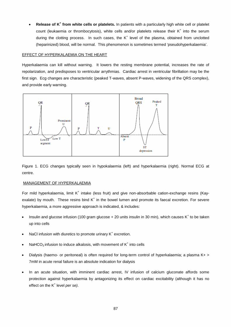
87
• Release of K+ from white cells or platelets. In patients with a particularly high white cell or platelet
count (leukaemia or thrombocytosis), white cells and/or platelets release their K+ into the serum
during the clotting process. In such cases, the K+ level of the plasma, obtained from unclotted
(heparinized) blood, will be normal. This phenomenon is sometimes termed ‘pseudohyperkalaemia’.
EFFECT OF HYPERKALAEMIA ON THE HEART
Hyperkalaemia can kill without warning. It lowers the resting membrane potential, increases the rate of
repolarization, and predisposes to ventricular arrythmias. Cardiac arrest in ventricular fibrillation may be the
first sign. Ecg changes are characteristic (peaked T-waves, absent P-waves, widening of the QRS complex),
and provide early warning.
Figure 1. ECG changes typically seen in hypokalaemia (left) and hyperkalaemia (right). Normal ECG at
centre.
MANAGEMENT OF HYPERKALAEMIA
For mild hyperkalaemia, limit K+ intake (less fruit) and give non-absorbable cation-exchange resins (Kay-
exalate) by mouth. These resins bind K+ in the bowel lumen and promote its faecal excretion. For severe
hyperkalaemia, a more aggressive approach is indicated, & includes:
• Insulin and glucose infusion (100 gram glucose + 20 units insulin in 30 min), which causes K+ to be taken
up into cells
• NaCl infusion with diuretics to promote urinary K+ excretion.
• NaHCO3 infusion to induce alkalosis, with movement of K+ into cells
• Dialysis (haemo- or peritoneal) is often required for long-term control of hyperkalaemia; a plasma K+ >
7mM in acute renal failure is an absolute indication for dialysis
• In an acute situation, with imminent cardiac arrest, IV infusion of calcium gluconate affords some
protection against hyperkalaemia by antagonizing its effect on cardiac excitability (although it has no
effect on the K+ level per se).

88
SMALL GROUP TEACHING, LECTURE 5 DISORDERS OF POTASSIUM BALANCE
QUESTIONS
1. What is the mechanism and site of action of each of the following groups of diuretics:
i. thiazides;
ii. loop diuretics
iii. potassium sparing diuretics
2. What are the complications associated with diuretic therapy?.
3. Comparing loop diuretics with thiazides, which affects the concentrating ability of the kidney?
4. Explain these factitious results:
Test Spec 1 Spec 2 Reference Range
Sodium 141 145 135 - 145 mmol/l
Potassium 10.3 8.3 3.0 - 4.7 mmol/l
Chloride 101 109 99 – 112 mmol/l
Total CO2 28 22 19 – 29 mmol/l
Calcium 0.5 2.4 2.2 - 2.6 mmol/l
Magnesium 0.1 1.5 0.6 - 1.0 mmol/l
Phosphate 1.1 5.2 0.6 - 1.2 mmol/l
Alkaline Phosphatase (ALP) 12 75 <120 U/l
Lactate Dehydrogenase (LD) - 450 <320 U/l
Glucose - 1.5 3.0 - 5.5 mmol/l
5. A 45 year old woman complains of drinking more than 10 litres of water per day.
Results are:
Reference Range
Plasma sodium 132 mmol/l (135-145)
Plasma osmolality 274 mmol/kg (280-295)
Urine osmolality 80 mmol/kg -
i. Name six important clinical disorders associated with polydipsia.
ii. What information can be derived from the above laboratory data about likely causes in this patient.
iii. Outline the investigations you would do to establish a diagnosis.
6. A 12 year old boy was brought to casualty with a 3 day history of severe diarrhoea which was thought to be due to food poisoning. He appeared drowsy and was clinically dehydrated BP 95/60 Pulse 100/min.
Blood tests were as follows: Reference Range
Sodium 156 mmol/l (135-145)
Potassium 2.7 mmol/l (3.3-5.2)
89
Total CO2 15 mmol/l (19-29)
Chloride 116 mmol/l (99-113)
Urea 15 mmol/l (2.6-8.0)
Creatinine 115 umol/l (<120)
Random glucose 4.2 mmol/l (3.0-6.0 fasting)
i. Explain the sodium result.
ii. Explain the urea/creatinine results.
iii. What would you expect his urinary sodium to be ?
iv. What is the cause of the low serum potassium?
v. Give one other clinical condition in which you would find a similar Total CO2 result together with a low potassium.
vi. Calculate the anion gap.
vii. Interpret the result of the anion gap.
viii. Calculate the osmolarity.
ix. What changes occur in the brain in response to this abnormality?
x. Outline the principles of treatment for this boy.
SMALL GROUP TEACHING, LECTURE 5 DISORDERS OF POTASSIUM BALANCE
ANSWERS
1.
i. Thiazides: act on the coupled Na+/Cl- channels of the distal tubule.
ii. Loop diuretcs: furosemide or ethacrynic acid prevent NaCl reabsorption in the TAL.
iii. Potassium sparing diuretics: amiloride acts on electrogenic Na+channels, spironolactone opposes aldosterone action by competition for receptor.
2. Complications associated with diuretic therapy are:
• Volume depletion
• Azotaemia
• Metabolic alkalosis
• Hypokalaemia ( especially important in digitalis treatment and severe liver disease)
• Hyperkalaemia and metabolic acidosis ( spironolactone and amiloride)
• Hyperuricaemia
• Hyponatraemia
• Hypomagnesaemia
3. Loop diuretics affect the concentrating ability since NaCl reabsorption in the TAL is essential for establishing the hyperosmotic medullary interstitium necessary for water reabsorption. I.e. ADH effect is impaired with loop diuretics, but intact with thiazide diuretics
4. Specimen 1 = EDTA tube, Specimen 2 = “old” blood
90
5.
i. Six important clinical disorders associated with polydipsia.
• Psychogenic polydipsia
• Cranial diabetes insipidus
• Nephrogenic diabetes insipidus
• Diabetes mellitus
• Hypercalcaemia
• Hypokalaemia
ii. Information that can be derived from the above laboratory data about likely causes in this patient is
• Hypo-osmolar hyponatraemia with fully dilute urine
• Thus kidney and ADH are intact
• Likely to be intake
iii. Investigations to establish a diagnosis would be
• 8 hour water deprivation test under supervision
• S-osmol, U-osmol, and weight
6.
i. Sodium result probably dehydration due to altered mental state and probably not responding to thirst impulse.
ii. Urea/creatinine results indicate that he patient is in pre-renal failure
iii. Urinary sodium should be <10mmol/l
iv. Causes of the low serum potassium include:
• Loss of potassium in diarrhoea
• Secondary hyperaldosteronism causing distal exchange for sodium (water) for potassium and hydrogen ions
v. One would find a similar Total CO2 result together with a low potassium in renal tubular acidosis
vi. The anion gap = 128
vii. Anion gap interpretation: high anion gap probably due to underperfusion because of volume depletion
viii. Osmolarity = 331.
ix. Brain changes expected in this abnormality are: intracellular dehydration followed by formation of idiogenic osmoles
x. Principles of treatment include Fluid replacement with normal saline and potassium replacement

91
Lecture 6: Carbohydrate Metabolsim And Diabetes
DR PETER BERMAN AND PROF TS PILLAY
LECTURE OUTLINE
OVERVIEW
INSULIN SYNTHESIS, SECRETION AND METABOLIC ACTION
Insulin secretion
Metabolic actions of insulin
Glucagon-like insulinotropic peptide
Ketone bodies
Glucose transport
PREVENTING HYPOGLYCAEMIA
Counter-regulatory hormones
DIABETES MELLITUS
History
Causes of hyperglycaemia
Metabolic syndrome
Other variants
Diagnosis of diabetes
COMPLICATIONS OF DIABETES
DKA
Biochemical derangements in DKA
Hyperosmolar Non-Ketotic Coma (HONK)
Hyperlipidaemia and diabetes
Long term complications of diabetes
Macrovascular disease
Advanced Glycosylation End Products
Glycosuria
Summary of quantitative glucose methods
ANTI-DIABETIC DRUGS
Insulin
Oral agents
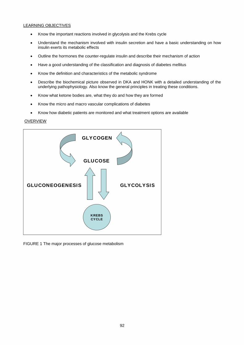
92
LEARNING OBJECTIVES
• Know the important reactions involved in glycolysis and the Krebs cycle
• Understand the mechanism involved with insulin secretion and have a basic understanding on how insulin exerts its metabolic effects
• Outline the hormones the counter-regulate insulin and describe their mechanism of action
• Have a good understanding of the classification and diagnosis of diabetes mellitus
• Know the definition and characteristics of the metabolic syndrome
• Describe the biochemical picture observed in DKA and HONK with a detailed understanding of the underlying pathophysiology. Also know the general principles in treating these conditions.
• Know what ketone bodies are, what they do and how they are formed
• Know the micro and macro vascular complications of diabetes
• Know how diabetic patients are monitored and what treatment options are available
OVERVIEW
GLUCOSE
GLYCOGEN
KREBSCYCLE
GLYCOLYSISGLUCONEOGENESIS
FIGURE 1 The major processes of glucose metabolism
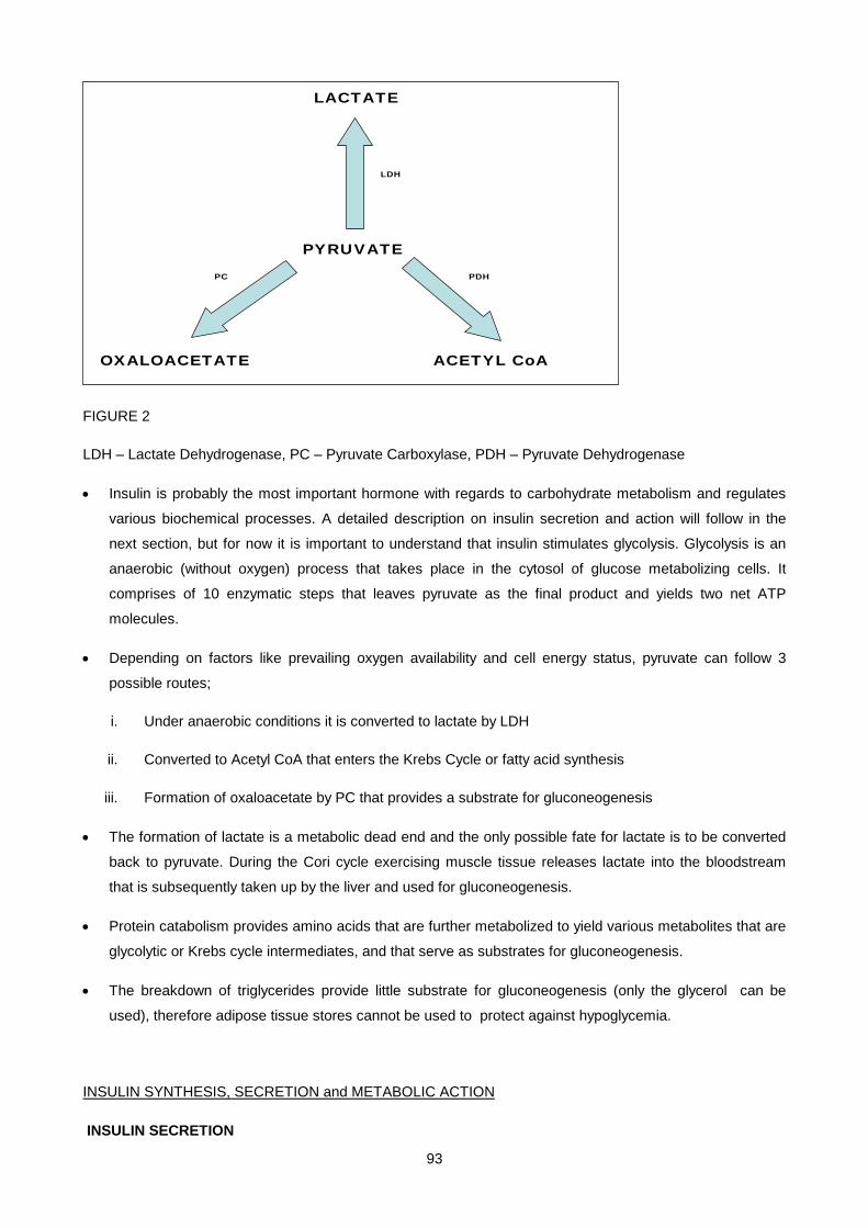
93
PYRUVATE
OXALOACETATE ACETYL CoA
LACTATE
PC
LDH
PDH
FIGURE 2
LDH – Lactate Dehydrogenase, PC – Pyruvate Carboxylase, PDH – Pyruvate Dehydrogenase
• Insulin is probably the most important hormone with regards to carbohydrate metabolism and regulates
various biochemical processes. A detailed description on insulin secretion and action will follow in the
next section, but for now it is important to understand that insulin stimulates glycolysis. Glycolysis is an
anaerobic (without oxygen) process that takes place in the cytosol of glucose metabolizing cells. It
comprises of 10 enzymatic steps that leaves pyruvate as the final product and yields two net ATP
molecules.
• Depending on factors like prevailing oxygen availability and cell energy status, pyruvate can follow 3
possible routes;
i. Under anaerobic conditions it is converted to lactate by LDH
ii. Converted to Acetyl CoA that enters the Krebs Cycle or fatty acid synthesis
iii. Formation of oxaloacetate by PC that provides a substrate for gluconeogenesis
• The formation of lactate is a metabolic dead end and the only possible fate for lactate is to be converted
back to pyruvate. During the Cori cycle exercising muscle tissue releases lactate into the bloodstream
that is subsequently taken up by the liver and used for gluconeogenesis.
• Protein catabolism provides amino acids that are further metabolized to yield various metabolites that are
glycolytic or Krebs cycle intermediates, and that serve as substrates for gluconeogenesis.
• The breakdown of triglycerides provide little substrate for gluconeogenesis (only the glycerol can be
used), therefore adipose tissue stores cannot be used to protect against hypoglycemia.
INSULIN SYNTHESIS, SECRETION and METABOLIC ACTION
INSULIN SECRETION
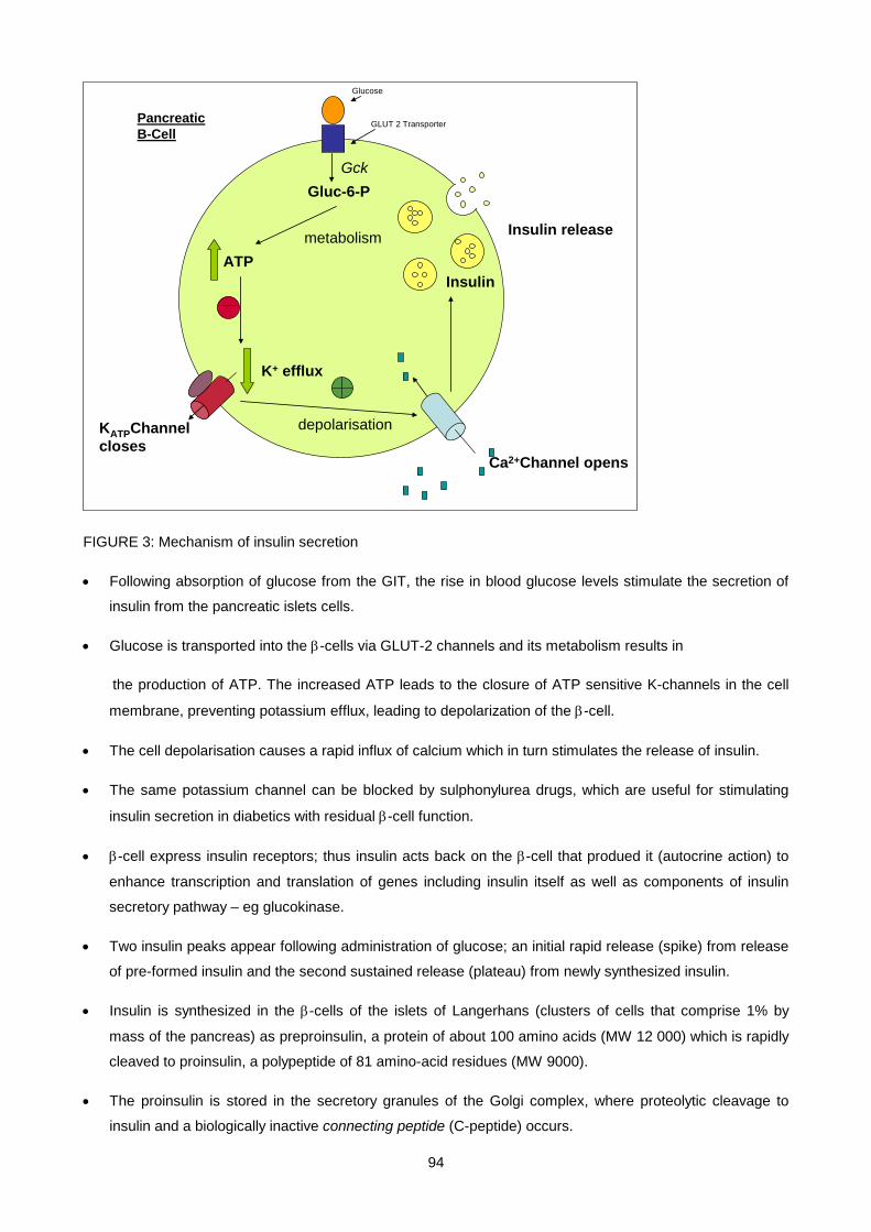
94
Gluc-6-PGck
metabolismATP
KATPChannelcloses
K+ efflux
Ca2+Channel opens
depolarisation
Insulin
Pancreatic B-Cell
Insulin release
Glucose
GLUT 2 Transporter
FIGURE 3: Mechanism of insulin secretion
• Following absorption of glucose from the GIT, the rise in blood glucose levels stimulate the secretion of
insulin from the pancreatic islets cells.
• Glucose is transported into the β-cells via GLUT-2 channels and its metabolism results in
the production of ATP. The increased ATP leads to the closure of ATP sensitive K-channels in the cell
membrane, preventing potassium efflux, leading to depolarization of the β-cell.
• The cell depolarisation causes a rapid influx of calcium which in turn stimulates the release of insulin.
• The same potassium channel can be blocked by sulphonylurea drugs, which are useful for stimulating
insulin secretion in diabetics with residual β-cell function.
• β-cell express insulin receptors; thus insulin acts back on the β-cell that produed it (autocrine action) to
enhance transcription and translation of genes including insulin itself as well as components of insulin
secretory pathway – eg glucokinase.
• Two insulin peaks appear following administration of glucose; an initial rapid release (spike) from release
of pre-formed insulin and the second sustained release (plateau) from newly synthesized insulin.
• Insulin is synthesized in the β-cells of the islets of Langerhans (clusters of cells that comprise 1% by
mass of the pancreas) as preproinsulin, a protein of about 100 amino acids (MW 12 000) which is rapidly
cleaved to proinsulin, a polypeptide of 81 amino-acid residues (MW 9000).
• The proinsulin is stored in the secretory granules of the Golgi complex, where proteolytic cleavage to
insulin and a biologically inactive connecting peptide (C-peptide) occurs.

95
• Insulin was the first hormone to be (a) sequenced (b) measured by RIA and (c) produced by recombinant
DNA technology.
METABOLIC ACTIONS OF INSULIN:
1. Promotes glucose uptake
In the muscle and adipose tissue, insulin causes glucose transporting channels (GLUT4) to
movefrom the cytosol to the cell membrane, to facilitate glucose uptake by these cells. Once inside
the cells, glucose is immediately phosphorylated and converted to glycogen or used for energy.
Phosphorylation traps glucose and commits it to further metabolism within the cell.
2. Promotes glycolysis
Insulin promotes glycolysis by stimulating formation of a glucose metabolite, fructose 2,6-
bisphosphate. This compound is a potent activator of phosphofructokinase-1 (PFK-1), the rate-
limiting step in glycolysis. In addition, fructose 2,6-biphosphate inhibits fructose 1,6-biphosphatase,
the enzyme that catalyzes the reverse reaction of PFK-1. Thus glucose metabolized to pyruvate
cannot be converted back to glucose via gluconeogenesis (fructose 1,6-biphosphatase catalyzes a
gluconeogenic reaction).
Fructose 6-phosphate
Fructose 1,6-biphosphate
PFK-1 Fructose 1,6-biphosphatase
Insulin
+
+ -
Gluconeogenesis
Glycolysis
Fructose 2,6-biphosphatase
FIGURE 4: Insulin regulation of glycolysis
3. Increases glycogen synthesis
In the liver, insulin does not affect glucose transport directly (as it does in muscle and fat), but
promotes glucose uptake by stimulating its storage as glycogen. This is achieved by activating
glycogen synthase, the enzyme that catalyses glycogen synthesis, while simultaneously deactivating
glycogen phosphorylase, an enzyme that facilitates glycogen breakdown (see fig 1).

96
4. Increases triglyceride synthesis
Acetyl CoA carboxylase catalyses formation of malonyl CoA, the first and rate limiting step of de novo
fatty acid synthesis, and is activated by insulin. Furthermore, malonyl CoA inhibits breakdown of
newly formed fatty acids by preventing their transport into mitochondria (where β oxidation takes
place). It does so by inhibiting the fatty acid transporter protein (carnitine palmitoyl transferase I),
located in the outer mitochondrial membrane. The newly formed fatty acids are esterified to
triglyceride and then packaged as fat-rich particles known as very low density lipoproteins
(VLDL) and exported into the bloodstream to be taken up and used by muscle or stored in
adipose tissue. The triglyceride uptake is stimulated by insulin, which activates the relevant
enzyme, lipoprotein lipase. In the adipose tissue, the stored triglycerides are hydrolysed to fatty acids
and glycerol by hormone sensitive lipase, which is activated by several hormones antagonistic to
insulin (counterregulatory hormones like adrenalin and growth hormone).
If not re-esterified, these fatty acids are released into the blood. Re-esterification requires α glycerol-
phosphate, produced from dihydroxyacetone phosphate (DHAP), a glucose metabolite. By
promoting entry of glucose into adipocytes, insulin inhibits fatty acid efflux.
Glycolysis
Acetyl CoA
Glucose
Pyruvate
PDH
Fatty Acid Synthesis
KrebsCycle
Malonyl CoA
Insulin
+
+
CPT-1
-
Fatty acid transport into mitochondria
FIGURE 5: Insulin regulation of fatty acid synthesis
KETONE BODIES
• The compounds categorized as ketone bodies are acetoacetic acid, β-hydroxybutyric acid and acetone
(acetone is formed spontaneously from acetoacetate and cannot be further metabolized).
• Ketones are formed in liver mitochondria during the fasting state when insulin levels are low. They are
derived from βoxidation of free fatty acids. They are water soluble compounds and do not need special
carrier proteins like lipoproteins or albumin; they can be regarded as a water soluble form of fat.
97
• Peripheral tissue like heart and muscle convert ketone bodies to acetyl CoA which can enter the Krebs
cycle and provide an alternative source of fuel to glucose in times of food deprivation.
• Insulin blocks ketogenesis by two mechanisms
i. Inhibits fatty acid release from the adipocytes as described in paragraph 2.2.4.
ii. Blocks entry of fatty acids into the liver mitochondria by boosting the levels of malonyl CoA. This
prevents the liver from oxidizing the fatty acids it has just synthesized (see also paragraph 2.2.4).
GLUCAGON-LIKE INSULINOTROPIC PEPTIDE
Oral administration of glucose results in a bigger insulin response than an equivalent dose administered
intravenously. This has been attributed to GLP-1 (Glucagon-like insulinotropic peptide), a hormone released
into the bloodstream from the duodenum in response to dietary carbohydrate ingestion.
GLUCOSE TRANSPORT
Transport of glucose into cells is modulated by two families of proteins. The intestinal sodium/glucose
cotransporter is responsible for uptake of glucose and galactose in the small intestines and their
reabsorption by the kidneys. This transporter uses an electrochemical sodium gradient to transport glucose
against its concentration gradient (logic behind using a salt and sugar solution for rehydration). The second
family of glucose transporter is the facilitated glucose transporters (GLUT) found in most cells. They are
designated GLUT1 to GLUT7 based on the order in which they were identified.
Facilitative Human Glucose Transporters
NAME TISSUE DISTRIBUTION
GLUT 1 Wide distribution, erythrocyte, fetal tissues and brain and kidneys.
GLUT 2 Liver and pancreatic β-cells.
GLUT 3 Wide distribution ,especially in neurons, placenta and brain.
GLUT 4 Skeletal muscle, cardiac muscle and adipose tissue.
GLUT 5 Small intestines (transports fructose not glucose)
GLUT 6 Pseudogene not expressed at protein level.
GLUT 7 Microsomal-diffusion of glucose out of ER of gluconeogenic tissues.
PREVENTING HYPOGLYCEMIA
Tissues like the brain and erythrocytes have an obligate glucose requirement and are constantly consuming
glucose. So in the absence of glucose intake (fasting), blood glucose levels must be maintained
endogenously. Insulin secretion is switched off and all its effects of enhancing glucose utilisation are
reversed;
98
• Glucose entry into muscle and adipose tissue is reduced due to lack of functioning GLUT-4 transporters.
This denies the muscle access to glucose, and spares it for tissues like the brain that utilize non-insulin
dependent glucose transporters (GLUTs 1and 3).
• Lack of functional GLUT4 in adipocytes leads to insufficient α-glycerol phosphate for re-esterification of
fatty acids. Free fatty acids leave the adipocyte and travel to the liver. In the liver, low insulin causes level
of malonyl CoA to fall, which lifts the inhibition on CPT1 and allows fatty acids to be transported into
hepatic mitochondria and be oxidized to ketone bodies (ketogenesis).
• Lack of insulin causes the liver to switch from a glucose-consuming to a glucose-producing organ; first
through glycogenolysis, then, once glycogen is depleted, from glucogenic amino acids like alanine
derived from the breakdown of skeletal muscle proteins (gluconeogenesis). Hepatic gluconeogenesis is
stimulated by de-repression of fructose 1,6-bisphosphatase arising from low levels of fructose 2,6-
bisphosphate in the absence of insulin.
COUNTER-REGULATORY HORMONES
These are hormones released when blood glucose levels fall, and are there, along with lack of insulin, to
ensure maintenance of a normal glucose level in the face of food deprivation. Their actions are in many
respects opposite that of insulin.
Glucagon
A 29 amino acid polypeptide hormone secreted by the α-cell of the pancreatic islets. Unlike insulin, the amino
acid sequence of glucagon is the same in all mammalian species examined to date.
This is the first hormone to be released when glucose levels fall and is important in the hour to hour
maintenance of adequate glucose levels between meals and at night. It primarily acts on the liver, where it
stimulates both glycogenolysis and gluconeogenesis.
Regulation of secretion:
The α-cell is responsive to a number of stimuli that signal actual or potential hypoglycaemia.
i. Low blood glucose is the primary stimuli for glucagon release. So during an overnight fast, elevated
glucagon levels prevent hypoglycaemia.
ii. Protein-derived amino acids stimulate both insulin and glucagon. The glucagon effectively
antagonises insulin and prevents hypoglycaemia that would otherwise result from an insulin
secretory response to the protein meal (arginine is very potent).
iii. Adrenalin from the adrenal medulla and noradrenalin produced by the sympathetic innervation of
the pancreas stimulates release of glucagon in anticipation of increased glucose demands.
Adrenaline
• A tyrosine-derived molecule from the adrenal medulla that counteracts hypoglycaemia by:
i. activating glycogenolysis in muscle and liver
99
ii. stimulating adipocyte lipolysis (via hormone sensitive lipase)
iii directly inhibiting insulin release.
This ensures blood glucose remains high in cases of emergency with increased energ demands.
• Adrenaline release is triggered by lower blood glucose levels than occur during normal fasting and so is
only required for emergency correction of hypoglycaemia. It is therefore not involved in routine blood
glucose regulation (except in diabetics with pancreatic damage who develop a glucagon deficiency).
• Clinical features of hypoglycaemia are mainly attributable to adrenaline.
• Tumours of the adrenal medulla known as phaeocromocytoma secrete excess adrenalin and/or
noradrenaline, and frequently lead to hyperglycaemia.
Cortisol
• A steroid hormone released from the adrenal cortex in response to ACTH. It promotes protein catabolism
in order to provide substrate for gluconeogenesis. By activating the
gluconeogenic enzymes in the liver, it ensures that alanine delivered to the liver from the muscle is
efficiently converted to glucose. Patients with Cushing`s syndrome have increased cortisol which may
causes hyperglycaemia.
Growth Hormone
• A peptide hormone from the anterior pituitary. It promotes lipolysis (hydrolysis of triglycerides to free fatty
acids and glycerol) in adipose tissue, thereby providing an alternative fuel to glucose. In chronic
starvation, GH limits muscle protein breakdown by promoting use of fatty acids derived from fat stores as
an energy alternative. GH has an insulin-like action on protein metabolism; like insulin, it stimulates
protein synthesis (via insulin-like growth factor-1 (IGF-1)).
• Fasting stimulates release of GH, and glucose administration lowers GH (except in acromegaly).
Other Hormones
• Somatostatin from the δ-cells of the pancreatic islets. Not directly involved in carbohydrate metabolism
but inhibits GH release. It also inhibits the release of both insulin and glucagon.
• Thyroxine from the thyroid gland also not directly involved but is known to stimulate glycogenolysis
DIABETES MELLITUS
HISTORY
• A state of chronic hyperglycaemia (excess glucose) caused by a defect in insulin secretion, action or both, that results in various metabolic disturbances.
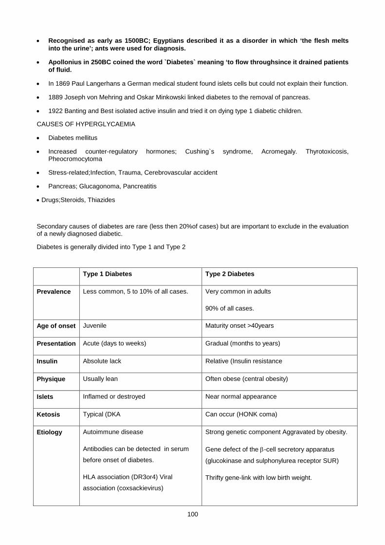
100
• Recognised as early as 1500BC; Egyptians described it as a disorder in which ‘the flesh melts into the urine’; ants were used for diagnosis.
• Apollonius in 250BC coined the word `Diabetes` meaning ‘to flow throughsince it drained patients of fluid.
• In 1869 Paul Langerhans a German medical student found islets cells but could not explain their function.
• 1889 Joseph von Mehring and Oskar Minkowski linked diabetes to the removal of pancreas.
• 1922 Banting and Best isolated active insulin and tried it on dying type 1 diabetic children.
CAUSES OF HYPERGLYCAEMIA
• Diabetes mellitus
• Increased counter-regulatory hormones; Cushing`s syndrome, Acromegaly. Thyrotoxicosis, Pheocromocytoma
• Stress-related;Infection, Trauma, Cerebrovascular accident
• Pancreas; Glucagonoma, Pancreatitis
• Drugs;Steroids, Thiazides
Secondary causes of diabetes are rare (less then 20%of cases) but are important to exclude in the evaluation of a newly diagnosed diabetic.
Diabetes is generally divided into Type 1 and Type 2
Type 1 Diabetes Type 2 Diabetes
Prevalence Less common, 5 to 10% of all cases. Very common in adults
90% of all cases.
Age of onset Juvenile Maturity onset >40years
Presentation Acute (days to weeks) Gradual (months to years)
Insulin Absolute lack Relative (Insulin resistance
Physique Usually lean Often obese (central obesity)
Islets Inflamed or destroyed Near normal appearance
Ketosis Typical (DKA Can occur (HONK coma)
Etiology Autoimmune disease
Antibodies can be detected in serum
before onset of diabetes.
HLA association (DR3or4) Viral
association (coxsackievirus)
Strong genetic component Aggravated by obesity.
Gene defect of the β-cell secretory apparatus
(glucokinase and sulphonylurea receptor SUR)
Thrifty gene-link with low birth weight.
101
Part of the metabolic syndrome.
SYNDROME X / METABOLIC SYNDROME
This is a cluster of risk factors for heart disease associated with insulin resistance described by Gerald Reaven in
1988. Factors include;
i. Obesity BMI >28 (weight in kg divided by height in meters squared). Waist : (Men -ideal <94: high risk-
>102cm)(Women-ideal< 80: high risk- >88cm)
ii. Hypertension
iii. Dyslipidaemia: (hypertriglyceridemia, small dense LDL,and low HDL)
iv. Insulin resistance: When normal amount of insulin fails to maintain glucose within normal limits. This is
often associated with type II diabetes mellitus.
v. High fibrinogen levels.
vi. Hyperuricaemia
Thrifty Gene- was once important for survival. It is thought to regulate hormonal fluctuations to accommodate
seasonal changes. This facilitates the storage of fat when food is available and its utilisation when food is scarce.
Low birth weight babies can grow to be obese when food is available (LIMA –Native Americans)
OTHER VARIANTS OF DIABETES
Maturity onset diabetes of the young (MODY)
Mody is Type 2 diabetes that develops in typically type 1 age group. Four genetic defects have been identified, one
of which is a mutation on the glucokinase gene. Glucokinase catalyzes the conversion of glucose to glucose-6-
phosphate and eventually the metabolites that stimulate the insulin secretion in the β-cell. Thus glucokinase serves
as the glucose sensor for the β-cell. The defect means that increased levels of glucose are required to elicit the
usual secretion of insulin (possibly a Vmax mutation).
Gestational diabetes mellitus
• During pregnancy, the pancreatic function might not be sufficient to overcome both the insulin resistance
created by the anti-insulin hormones secreted by the placenta (oestrogen, prolactin, human placental lactogen,
cortisol and progesterone).
• Diabetic patients who become pregnant do not have gestational diabetes but have “diabetes mellitus and
pregnancy” and should be treated accordingly, before, during and after pregnancy.
Risk Factors;
Family history of diabetes in a first degree relative
102
Obesity
Advance maternal age
Previous adverse pregnancies e.g.still birth,macrosomia.
Screening is recommended between 24-28 weeks: 50gm of glucose given orally regardless of time of last meal.
Glucose is measured after one hour.Patients with ≥7.7mmol/l should have OGTT done.
DIAGNOSIS OF DIABETES
The 2006 diagnostic criteria for diabetes as suggested by the American diabetes Association (ADA)1
1. Symptoms of diabetes plus a random (casual) plasma glucose concentration ⋝ 11.1 mmol/l. Random is
defined as any time of day without regard to time since last meal. The classic symptoms of diabetes include
polyuria, polydipsia, and unexplained weight loss.
;
OR
2. Fasting plasma glucose (FPG) ≥ 7.0 mmol/l. Fasting is defined as no caloric intake for at least 8 h.
OR
3. 2-h postload glucose ≥ 11.1 mmol/l during an OGTT. The test should be performed as described by WHO,
using a glucose load containing the equivalent of 75 g anhydrous glucose dissolved in water.
*In the absence of unequivocal hyperglycemia, these criteria should be confirmed by repeat testing on a different
day.
OGTT PROTOCOl
• The patient should have a normal mixed diet for the previous three days prior to the test. The patient is fasted
overnight and blood is drawn for glucose measurement before 75 grams is administered orally in 200ml of
water. Blood glucose is measured every thirty minutes for two hours. Subjects should rest during the test and
should not smoke or eat.
• Glucose is drawn into sodium fluoride tubes (grey top). Sodium fluoride is an enzyme inhibitor that prevents
glucose from being metabolized by the cells. Flouride ions prevent glycolysis by binding magnesium that is
required by the glycolytic enzymes.
*Important;
1 Diagnosis and classification of diabetes: DIABETES CARE, VOLUME 29, SUPPLEMENT 1, JANUARY 2006

103
• In normal subjects, plasma glucose rises and returns to the fasting level by two hours. Plasma Glucose > 11.1
mmol/l at two hours confirms diabetes mellitus. If the two hour value lies between 7.8 and 11.1 mmol/l the
patient is labeled as impaired glucose tolerant (IGT). Although not frankly diabetic, this patient should be
followed up as they usually become diabetic at a later stage.
• Patients with a fasting glucose levels between normal and diabetic (5.6 and 7 mmol/l according to the ADA) are
labeled as impaired fasting glucose (IFG). This is analogues to impaired glucose tolerance.
MODIFIED OGTT
• This is done for pregnant patients. 100grams is given instead of 75 grams and plasma glucose is measured,
before and at hourly intervals for 3 hours.
Gestational diabetes is diagnosed if at least two of the following four cut off values are exceeded:-
• fasting >5.3 mmol/l
• one hr >10 mmol/l
• two hrs > 8.6 mmol/l
• three hrs > 7.8 mmol/l
• The normal fasting levels are lower in pregnancy due to the constant fetal siphoning of glucose and
gluconeogenic amino acids, while the post glucose levels are higher due to the anti insulin effect of human
placental lactogen (HPL) and other anti-insulin hormones.
• Glycosuria is present in diabetics whenever the renal threshold for glucose (10mmol/l) is exceeded. As renal
threshold varies in health and disease, glycosuria is not as reliable an indicator of diabetes as plasma glucose.
• Renal threshold is lower in pregnancy, due to increased glomerular filtration rate (GFR) which is higher in
pregnancy and so more glucose is filtered and some of the glucose will be excreted in urine.
RENAL GLYCOSURIA
This is a benign condition in patients with normal blood glucose levels. This is due to a defect in the tubular
threshold for reabsorption of glucose in the proximal tubules.
COMPLICATIONS OF DIABETES
DIABETIC KETOACIDOSIS (DKA)
This is the hallmark of Insulin dependent diabetes mellitus (Type1), characterized by hyperglycaemia,
hyperosmolarity, low pH, ketonuria and ketonemia.
This acute metabolic emergency can:
• Develop in diabetics as a result of insufficient/ interruption of insulin therapy.
• Be precipitated by severe physical stress (trauma , infection) resulting in the production of the counter regulatory
hormones like cortisol.
104
• Also present in previously undiagnosed diabetics.
Presentation:
Patients will be distressed and dehydrated secondary to polyuria, nausea and vomiting and present with
hypotension, tachycardia, dry skin, and poor urinary output.
Breathing is rapid and deep, referred to as Kussmaul`s breathing. The decrease in pH in metabolic acidosis
stimulates the respiratory centre.
A smell of acetone is apparent in the breath due to the presence of ketones.
BIOCHEMICAL DERANGEMENTS IN DKA
HYPERGLYCEMIA
• Glucose levels can be between 20-50mmol/l or even higher. This is because the lack of insulin prevents
the entry of glucose into insulin dependent cells (GLUT4) like the muscle and the adipocytes, which are
major glucose consumers.
The liver synthesizes glucose (gluconeogenesis) from muscle derived alanine and this leads to a further
rise in glucose levels.
• The enhanced gluconeogenesis leads to higher urea levels, this coupled with hypovolumia, compromises
renal function.
• Despite loss of glucose in urine and cessation of food intake due to drowsiness and vomiting, glucose
levels continue to increase. Once the polyuric patient becomes oliguric, the rate of glucose increase
accelerates.
METABOLIC ACIDOSIS.
• The ketone bodies acetoacetate and β-hydroxybutyrate are overproduced and cause a fall in the blood
pH, usually to between 7.0 and 7.2, but can be as low as 6.9. Ketone formation is a physiological
response to starvation. However in starvation, the body fully metabolises them to energy, CO2 and H2O
with no net gain of H+.
• In diabetes, the ketone bodies are lost in the urine with the retention of H+. An elevation of ketone body
concentration results in acidaemia (the carboxyl group of a ketone body has a pK around 4). Therefore
each ketone body loses a proton as it circulates in the blood which lowers the pH of the body.
• The hydrogen ions released from these acids are buffered by bicarbonate – thus plasma bicarbonate
(HCO3) is markedly reduced often to around 10mmol/l or less. Blood pCO2 is generally low as a result of
respiratory compensation for the acidosis (hyperventilation). Spontaneous decarboxylation of
acetoacetate yield acetone, a volatile ketone that imparts a smell to the breath that is characteristic of
ketoacidosis. Since bicarbonate is to a large extent replaced by the corresponding organic anion,
acetoacetate and β- hydroxybutyrate, an increased plasma anion gap develops.

105
• Severity of ketoacidosis can conveniently be assessed by testing the plasma with ketostix, a
commercially available dip stick that provides a semi quantitative measure of ketone body levels
(Nitroprusside test). However it detects only acetoacetates. Under normal circumstances, the ratio of β-
hydroxybutyrates, to acet oacetate in plasma is fairly constant (3:1) in favour of β-hydroxybutyrates. In
severely shocked patients, tissue anoxia increases the ratio of NADH to NAD which pushes the
equilibrium in the direction of hydroxybutyrate causing an underestimation of ketoacidosis.
• A similar phenomenon occurs in acute ethanol intoxication (see alcohol lecture notes).
Acetoacetate + NADH β-hydroxybutyrate + NAD +H
It is essential to note that patients can be mistaken to be getting better because it measures
acetoacetates only or could be getting worse during treatment as β-hydroxybutyrate is converted to
acetoacetate during treatment with insulin. It is therefore essential that the ketostix is supported by other
tests.
LACTIC ACIDOSIS
Patients are often severely dehydrated with associated with poor perfusion resulting in lactic acidosis.
SODIUM AND WATER
Significant water loss due to glucose induced osmotic diuresis will cause plasma sodium to rise. This is offset
by hyperglycaemia drawing water out of the ECF leading to dilutional hyponatraemia and loss of urinary
sodium. Thus depending on the interplay of these factors, plasma sodium can be low, normal or high. As a
general principle, the higher the sum of sodium and glucose, the more severe the water depletion. It is
possible to estimate the extent of dehydration from the measured sodium and glucose.
1 First eliminate the dilutional effect caused by glucose drawing water into the ECF by adding 3 mmol/l
to the sodium for every 10mmol/l increase in glucose. This gives the value of sodium if the glucose
was normal.
2 Use this corrected sodium value to estimate water loss from the following formula:-
∆v/TBW = 1-140/Na
∆v = water deficit TBW = original total body water, about 40 litres.
Example, a diabetic patient with plasma sodium of 150mmol/l and glucose of 65mmol/l
Corrected sodium is equal to 150 + (65-5) X 3/10 (where 5 = normal glucose)
= 150 + 18 = 168
Using the formula to calculate the extent of pure water loss,
∆v/40 = 1 – 140/168 = 1/6
∆v = 40/6 (approx 7 litres)
106
In the normal situations, if there is no dehydration, 1 – 140/140 = 0
POTASSIUM AND OTHER ELECTROLYTES
• Potassium tends to leave the cells during ketoacidosis. Intracellular K binding sites including protein and
phosphorylated glycolytic intermediates are depleted. Accumulating H ions displace K from these binding
sites. In normal renal function these K ions are cleared by the kidney leaving a normal or slightly elevated
plasma K at presentation of DKA, obscuring the profound intracellular K depeletion. Once the factors
causing K efflux are reversed by treatment, K will re-enter the cells and hypokalaemia will rapidly
manifest, requiring vigorous K replacement. All DKA patients require K replacement.
• Phosphate and magnesium are other intracellular ions that are lost during the development of
ketoacidosis.
• During late glycolysis, inorganic phosphates are incorporated. This translates into a sudden drop of
phosphate in DKA patients after treatment with insulin. This has been implicated as a cause of sudden
death in DKA patients. Therefore consider phosphate repletion with life threatening hypophosphataemia.
ABNORMAL RENAL FUNCTION
As dehydration worsens, renal perfusion deteriorates leading to a plasma urea and creatinine increase.
*A falsely elevated creatinine level can be caused by high serum acetoacetate concentrations. Acetoacetate
interfere with the Jaffe reaction that is most commonly utilized in creatinine measurements.
TREATMENT OF DKA
The mainstay of treatment for DKA includes;
• Rehydration,
• Insulin administration
• Monitor and replace lost intracellular ions
• Look for and treat predisposing causes, eg infection.
• These patients are normally extremely dehydrated and may require large amounts of IV fluids (> 6 liters/
24 h). Potassium replacement is imperative and should be initiated immediately after the initial
resuscitation period has ended.
• It is important to realize that high and continuous doses of insulin may cause the blood glucose level to
normalize rapidly. However, insulin therapy should not be terminated until the patient has a normal (or
near normal) pH and urinary ketones are undetectable. Under these circumstances dextrose containing
IV fluids should be administered whilst continuing with insulin therapy.

107
• Bicarbonate replacement is a controversial subject and can be administered in severe cases of acidosis
(pH <7.0).This is rarely necessary because once insulin is given; metabolism of circulating ketone anions
spontaneously generates an equimolar quantity of bicarbonate.
Potential dangers of administering intravenous NaHCO3 during the acute phase include
i. overshot alkalosis once ketones are metabolised
ii. Increased affinity of haemoglobin for oxygen leading to tissue anoxia (haemoglobin has higher
affinity for oxygen in alkali conditions leading to non release of oxygen to the tissues).
iii. Temporary worsening of cerebral acidosis – a sudden increase in blood pH abolishes the
hyperventilatory drive since the respiratory centre responds to changes in blood pH. (Cerebral pH
paradoxically falls because the CO2, equilibrates across the blood-barrier faster than HCO3)
*Please refer to any standard medical textbook for comprehensive treatment protocols.
HYPEROSMOLAR NON-KETOTIC COMA (HONK)
• This variant of diabetic coma is characterised by severe hyperglycaemia causing dehydration often
worse than DKA but without ketoacidosis. It develops in elderly type two diabetics who have enough
insulin to prevent ketone formation. It can be explained by the differential sensitivity of ketogenesis and
gluconeogenesis to inhibition by insulin. Ketogenesis is sensitive to insulin whereas inhibition of
gluconeogenesis needs more insulin. Therefore insulin is enough to prevent ketogenesis but not
gluconeogenesis and hence the liver continues to put out glucose resulting in increased osmolarity.
• Complications that develop are secondary to severe dehydration. These include hyperviscosity of the
blood leading to decreased tissue perfusion with lactic acidosis, renal shutdown and cerebral thrombosis.
The principle of treatment in these patients is the same as those for DKA.
• Thromboembolism is increased because of hyperosmolarity and therefore anticoagulation is required.
HYPERLIPIDAEMIA IN DIABETICs
• Lack of insulin causes uncontrolled lipolysis in adipose tissue and release of free fatty acids. Once taken
up by the liver, the free fatty acids have two possible fates
1 β-oxidation to ketone bodies
2. Re-esterification to triglycerides which is packaged and transported to adipose tissue as very
low-density lipoprotein (VLDL).
• While ketogenesis predominates in type one patients because of lack of insulin causing low levels of
malonyl CoA, in type two diabetics the re-estification pathway is favoured leading to excessive hepatic
VLDL production.
• VLDL removal by adipose tissue depends on lipoprotein lipase an enzyme requiring insulin for its
activation. Therefore increased production in type two patients with decreased clearance will lead to
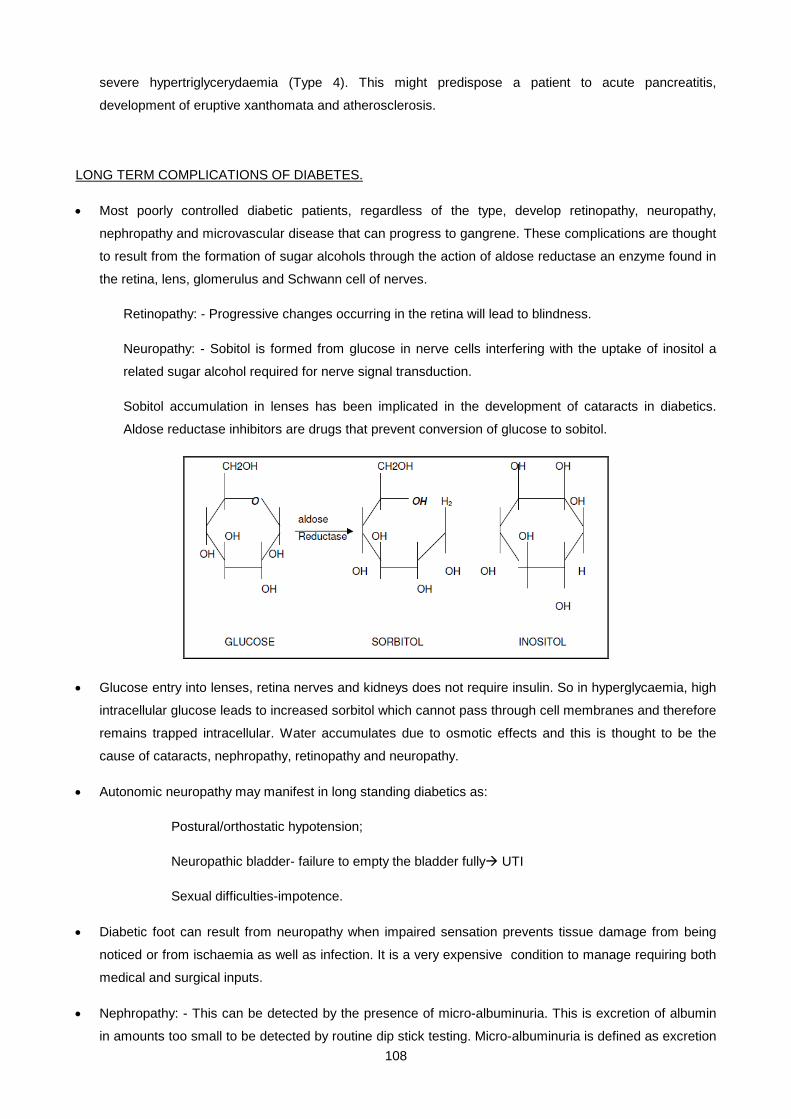
108
severe hypertriglycerydaemia (Type 4). This might predispose a patient to acute pancreatitis,
development of eruptive xanthomata and atherosclerosis.
LONG TERM COMPLICATIONS OF DIABETES.
• Most poorly controlled diabetic patients, regardless of the type, develop retinopathy, neuropathy,
nephropathy and microvascular disease that can progress to gangrene. These complications are thought
to result from the formation of sugar alcohols through the action of aldose reductase an enzyme found in
the retina, lens, glomerulus and Schwann cell of nerves.
Retinopathy: - Progressive changes occurring in the retina will lead to blindness.
Neuropathy: - Sobitol is formed from glucose in nerve cells interfering with the uptake of inositol a
related sugar alcohol required for nerve signal transduction.
Sobitol accumulation in lenses has been implicated in the development of cataracts in diabetics.
Aldose reductase inhibitors are drugs that prevent conversion of glucose to sobitol.
• Glucose entry into lenses, retina nerves and kidneys does not require insulin. So in hyperglycaemia, high
intracellular glucose leads to increased sorbitol which cannot pass through cell membranes and therefore
remains trapped intracellular. Water accumulates due to osmotic effects and this is thought to be the
cause of cataracts, nephropathy, retinopathy and neuropathy.
• Autonomic neuropathy may manifest in long standing diabetics as:
Postural/orthostatic hypotension;
Neuropathic bladder- failure to empty the bladder fully UTI
Sexual difficulties-impotence.
• Diabetic foot can result from neuropathy when impaired sensation prevents tissue damage from being
noticed or from ischaemia as well as infection. It is a very expensive condition to manage requiring both
medical and surgical inputs.
• Nephropathy: - This can be detected by the presence of micro-albuminuria. This is excretion of albumin
in amounts too small to be detected by routine dip stick testing. Micro-albuminuria is defined as excretion
109
of 30 to 300 mg albumin per day and is believed to be an early indicator of kidney involvement that may
respond to ACE inhibitors and low protein diet. Another early sign of renal involvement is hyporenin,
hypoaldosteronism manifesting as hyperkalemia. Renin is from the juxtaglumerula cells. When the kidney
is involved, renin production is low and the renin – angiotensin system will not be functional leading to
reduced aldosterone.
MACROVASCULAR DISEASE:
• Atherosclerosis is very common in both type I and type II. This may cause coronary heart disease and
more commonly peripheral vascular disease. Diabetes accounts for half of all non-traumatic amputations.
ADVANCED GLYCOSYLATION END PRODUCTS (AGES)
• Non enzymatic glycosylation of tissue proteins is the covalent attachment of glucose to free amino acids
on the protein with subsequent chemical rearrangements. These modifications can seriously impact on
protein function. However some of the glycosylation products are useful in monitoring glycaemia, e.g.
Haemoglobin A1 (HbA1c) and fructosamine.
• HbA1c is glycosylated haemoglobin. The newly synthesised glucose is sugar free and haemoglobin is
glycosylated during red cell circulation when glucose attaches to the N terminal end of the β- globin chain
at a rate proportional to the blood glucose concentration at that time. The extent of haemoglobin
glycosylation ranges from less < 5% in non-diabetics to >20% in poorly controlled diabetics. HbA1c
migrates faster electrophoretically than unmodified haemoglobin because of the attachment of the amino
group which abolishes the positive charge.
• Spuriously low values can be seen in chronic haemolysis, and in patients with conditions like sickle cell
disease and G6PD deficiency. Fructosamine is a better test in such conditions.
• Fructosamine is the glycosylation of albumin. It has a shorter half life, i.e. 2 to 3 weeks. These two
methods can be used for long term monitoring of glucose control (HbA1c- 2 to 4 months and fructosamin,
2 weeks).
GLYCOSURIA
• Glucose appears in urine either because;
i. the blood level is high exceeding the threshold level (10mmol/l)
ii. renal glycosuria which is harmless
iii. increased GFR in pregnancy and
iv. in generalised proximal renal tubular defect (Fanconi Syndrome)
SUMMARY OF QUANTITATIVE GLUCOSE METHODS
Glucose Oxidase Method: The glucose is oxidized by glucose oxidase (GOD) in the presence of atmospheric
oxygen to gluconolactone and H2O2 (hydrogen peroxide). A peroxidase enzyme in the presence of a chromogenic

110
oxygen acceptor, oxidizes the hydrogen peroxide resulting in the formation of a colored compound. The colour
intensity is directly proportional to the glucose concentration.
Hexokinase Method: Glucose is phosphorylated by ATP in the presence of hexokinase and Mg2+. The glucose-6-
phosphate is oxidised by glucose-6-phosphate dehydrogenase (G6PD) to 6-phosphogluconate in
the presence of nicotinamide-adenine dinucleotide phosphate (NADP). The amount of NADPH produced is directly
proportional to the amount of glucose in the sample and is measured at 340nm.
ANTI-DIABETIC DRUGS
INSULIN
• Insulin is the mainstay of treatment in type 1 diabetes but is also used in type 2 when oral
hypoglycaemics become ineffective.
• The initial source of insulin for clinical use in humans was from cows, pigs or fish pancreases. Insulin
from these sources is effective in humans as they are nearly identical to human insulin (two amino acid
difference for bovine insulin, one amino acid difference for porcine). Insulin is obviously a protein which
has been very strongly conserved across evolutionary time.
• Human insulin is now manufactured for widespread clinical use using genetic engineering techniques,
which significantly reduces impurity reaction problems.
ORAL AGENTS
Sulphonylureas
• These drugs were discovered by chance when it was noted that a sulphonamide derivative used for
treating typhoid, caused marked hypoglycaemia.
• Sulphonylureas increases insulin secretion by closing the ATP-sensitive K channel on the β-cell
membrane resulting in its depolarization and release of insulin. Examples include glibenclamide and
chlorpropamide (not safe in renal impairment - can cause prolonged hypoglycaemia in patients with renal
impairment).
• Meglitinides are related to sulfonylureas. The amplification of insulin release is shorter and more intense,
and they are taken with meals to boost the insulin response after each meal.
Biguanides
• Improves insulin sensitivity by increasing glucose utilisation by skeletal muscle and also decreases
hepatic gluconeogenesis. Exactly how biguanides go about in causing these effects are not well
understood but a study in 2001(Zhou G et al) has indicated that they probably work by increasing cellular
AMP-kinase levels. Metformin is the most widely known and used drug in this class and is frequently
prescribed to overweight patients as it also promotes weight loss.
α-Glucosidase Inhibitors
111
• Inhibit the enzymes that catalyse the metabolism of carbohydrates in the intestines resulting in poor
absorption of carbohydrates and a reduction postprandial glucose rise. Good for overweight patients.
• Undigested starch may reach the colon where it is fermented, resulting in abdominal discomfort and
flatulance. This drug is used less commonly nowadays.
Thiazolidinediones
• A chance observation has also lead to the discovery of these drugs. A clofibrate analogue, ciglitazone
was unexpectedly found to lower blood glucose.
• They increase insulin sensitivity by binding to a nuclear receptor called PPAR-γ (peroxisome proliferator –
activated receptor-γ).
• PPAR-( forms part of a nuclear receptor family that regulates the expression of several genes and
encodes proteins that regulate adipocyte differentiation as well as lipid and glucose metabolism in
adipose and skeletal muscle. Examples are drugs like rosiglitazone and pioglitazone.
New developments
• The search for an oral form of insulin continues, since it would have enormous financial implications for
the pharmaceutical company who can develop such a drug.
• Other experimental drugs with novel mechanism of action include drugs that increase glukokinase
sensitivity and fructose 1.6-biphosphatase inhibitors.
HYPOGLYCAEMIA
Classically, to diagnose hypoglycaemia, three conditions must be satisfied (Whipple's triad). These are:
1. Symptoms of hypoglycaemia.
2. Low blood glucose (<2.2 mmol/l)
3. Prompt reversal of symptoms on administering glucose.
SYMPTOMS OF HYPOGLYCAEMIA:
1. Central nervous system dysfunction due to hypoglycaemia per se (neuroglycopenia).
2. Effects of catecholamines released in response to hypoglycaemia
Symptoms of neuroglycopenia include disorientation, mental detachment, dizziness, parasthesia, ataxia,
diplopia, that can progress quickly to convulsions and/or coma. Chronic hypoglycaemia may present with
psychiatric disturbances, including memory loss, personality changes, or dementia.
Catecholamine effects include palpitations, tachycardia, sweating, tremor, pallor, and serve as a warning of
impending neuroglycopaenia. Blunted sympathetic response may occur in patients taking beta-adrenergic
112
blockers, or in diabetics with autonomic neuropathy. These patients can slip into a coma without any warning
symptoms.
Factors affecting severity of neuroglycopenic symptoms include:
1. Age - neonates are more tolerant of, and the elderly more sensitive to, hypoglycaemia.
2. Previous glucose levels – symptoms are more pronounced if glucose falls rapidly from a previously high
level, as seen during treatment of diabetic coma (? down-regulation of GLUT-1 in brain).
Hypoglycaemia may be classified according to whether it is provoked by some exogenous agent (induced),
or occurs spontaneously during fasting.
INDUCED HYPOGLYCAEMIA
By induced, we mean the hypoglycaemia is caused by some exogenous agent - a drug, toxin, or foodstuff.
DRUGS
• Insulin - usually occurs in diabetics on insulin treatment that miss meals, undertake strenuous
exercise, overdose inadvertently (eg visual impairment caused by diabetes), or develop renal
dysfunction (prolonged insulin half-life). Deliberate self-administration of insulin by medical or
nursing personel, or anyone who has access to insulin (eg. family member of a diabetic), as an
attention-seeking strategy, is not unknown, & should always be considered.
• Sulphonylureas, esp. chlorpropamide in individuals with impaired renal function.
• Alcohol - Ingestion of excess ethanol by a starved or malnourished subject may lead to profound
hypoglycaemia, often many hours after the binge. Ethanol specifically impairs gluconeogenesis, so
hypoglycaemia only occurs once hepatic glycogen stores are exhausted, and may thus appear to be
a fasting (endogenous) hypoglycaemia. Liver dysfunction may be a contributing factor in chronic
alcoholism. Alcohol can also potentiate glucose-induced insulin secretion, and lead to a reactive
hypoglycaemia.
• Acute carbohydrate intake (Reactive Hypoglycaemia) - Rapid absorption of ingested
carbohydrate can cause excessive insulin release, and overshoot hypoglycaemia. Particularly
common following gastric bypass surgery. Can also occur following abrupt termination of high
glucose content parenteral feeds or dialysis against a high glucose dialysate.
• Hypoglycin - Akee nuts are eaten in the West Indies. Unripe nuts contain a compound, hypoglycin,
that inhibits fatty acid oxidation., causing unoxidized fatty acids to accumulate in the liver as their
CoA esters. This depletes acetyl CoA needed to activate gluconeogenesis. Inability to oxidize fat
increases the demand for glucose as a fuel. The combination of increased utilization of glucose and
decreased production, leads to profound hypoglycaemia, in a disorder known as Jamaican vomiting
sickness.
• Leucine - Endogenous or Fasting Hypoglycaemia
INSULINOMA
113
Tumours arising from ß-cells of the pancreatic islets can secrete insulin autonomously - i.e. independent of
prevailing blood glucose. They typically present with repeated hypoglycaemic attacks in the fasted state, eg
before waqking in the morning. The tumors are generally benign, and may be part of the multiple endocrine
neoplasia type I (MEN I) syndrome, where they coexist with other islet cell tumours of the pancreas
(gastrinoma), or with tumours of the parathyroid and pituitary (prolactinoma, acromegaly)... the 3p’s.
Diagnosis is confirmed by demonstrating inappropriately high insulin levels (>10 mU/litre) IN THE
PRESENCE of hypoglycaemia (<2.2 mmol/litre). Random measurement of insulin is of little diagnostic value.
Thus, when confronted by a patient with unexplained hypoglycaemia, remember to take a sample for glucose
AND insulin before administering glucose. It is a window of opportunity. In an asymptomatic patient, one
can try to induce hypoglycaemia by a prolonged fast (up to 72 hours), while encouraging physical activity.
While healthy subjects do not become hypoglycaemic despite such drastic manoevres, patients with
insulinoma typically develop hypoglycaemia with inappropriately high insulin levels.
Plasma C-peptide assays are useful in distinguishing exogenous insulin administration from an insulinoma.
Whereas commercially available insulin is free of C-peptide, endogenous insulin, such as from an
insulinoma, is secreted with an equimolar amounts of C-peptide.
Ketosis usually accompanies hypoglycaemia, as an appropriate response to starvation. Since it is blocked
by insulin, an inappropriately low plasma ketone body and fatty acid level in the face of hypoglycaemia is a
diagnostic clue of hyperinsulinism (insulin inhibits lipolysis & ketogenesis). It also explains why symptoms of
hypoglycaemia are worse when due tio hyperinsulinism, since ketones, an alternative energy source, are
denied to the tissues,
Treatment of choice for benign insulinomas (the majority) is surgical resection - not always so easy because
they may be small and buried deep within the pancreas.
Medical treatment with the drug, diazoxide, reduces insulin secretion in both normal and neoplastic ß-cells,
but may cause generalized hairiness (hypertrichosis) as an unwanted side-effect. Somatostatin, a peptide
hormone, also inhibits insulin secretion, but has to be given by injection, is expensive, and patients often
develop tolerance (it needs to be given in increasing amounts to retain efficacy). Occasionally,
streptozotocin, a potent cytotoxin, is used to destroy ß-cells, but it causes permanent diabetes and
widespread organ damage, so its use is confined to inoperable, malignant insulinomas.
NON-PANCREATIC NEOPLASMS
Hypoglycaemic attacks may occur in patients with malignant disease - particularly malignant hepatomas and
retroperitoneal sarcomas. Such tumors secrete insulin-like growth factor 2 (IGF-2), which, at high
concentrations, cross-reacts with the insulin receptor. Since IGF-2 is immunologically distinct from insulin, it
is not measured by the usual insulin immonoassays. Thus insulin appears appropriately suppressed during
hypoglycaemia.
ENDOCRINE DISEASE
A deficiency in any of the hormones that antagonize insulin can cause fasting hypoglycaemia. Examples
include. Addison’s disease (lack of cortisol) or pituitary damage (lack of ACTH and growth hormone). Lack of

114
glucagon, due to total pancreatectomy or chronic pancreatitis, explains the predisposition of pancreatic
diabetes to hypoglycaemia, and makes control of their blood glucose with insulin difficult ('brittle diabetes').
SEVERE LIVER AND RENAL DISEASE
Since the liver is the organ primarily responsible for maintaining plasma glucose in the fasting state (first by
glycogenolysis, then by gluconeogenesis), hypoglycaemia can develop as a complication of liver necrosis in
eg. viral hepatitis, paracetamol poisoning, or congestive cardiac failure.The kidneys are a major route of
insulin elimination, as well as a site for gluconeogenesis, which explains the hypoglycaemia occasionally
encountered in end-stage renal failure.
HYPOGLYCAEMIA IN CHILDHOOD
Hypoglycaemia in childhood may be due to any of the above causes (eg. insulinoma, ACTH deficiency).
However, certain types of hypoglycaemia are specific to the younger age group.
• Neonatal hypoglycaemia: Transient hypoglycaemia may occur in otherwise healthy neonates.
Blood glucose is maintained In utero by placental glucose transfer from the mother, but, after
delivery, depends on hepatic glycogen reserves until feeding commences. Premature and small-for-
dates babies are particularly susceptible, since their hepatic glycogen stores are low, and they feed
poorly. Babies born to poorly controlled diabetic mothers are exposed to high glucose levels in utero.
This induces islet cell hyperplasia, and results in rebound hypoglycaemia immediately after birth,
when maternal glucose delivery is abruptly terminated. Tendency to hypoglycaemia resolves within a
few days.
• Nesidioblastosis (Persistent hyperinsulinaemic hypoglycaemia of infancy – PHHI): This
developmental abnormality, presenting in infants, is due to diffuse proliferation of ß-cells throughout
the pancreas. The underlying defect lies in the sulphonylurea receptor (SUR), a membrane protein
that regulates the K+ channel in β-cells. Inability to open these K+ channels keeps the β-cells in a
constant state of depolarization, causing continuous (constitutive) insulin release. Biochemical
presentation of nesidioblastosis is similar to insulinoma (inappropriately high insulin secretion during
hypoglycaemia), except that hypoglycaemia is continuous rather than sporadic, and requires constant
infusion of large amounts of glucose to prevent irreversible brain damage (>12mg/kg/min). Hence
these babies are often fat. Nesidioblastosis may respond to diazoxide therapy in the short-term,
although it often needs total pancreatectomy to effect permanent cure.
• Leucine sensitivity: Some children experience hypoglycaemic episodes after protein ingestion. It is
due to leucine in the protein directly stimulating insulin secretion. The condition is termed ‘leucine
intolerance’.
• Ketotic hypoglycaemia of infancy: This relatively common cause of childhood hypoglycaemia is
due to failure of skeletal muscle to release adequate alanine for hepatic gluconeogenesis. Plasma
alanine level is low, and hypoglycaemia responds promptly to alanine infusion, whereas in
gluconeogenic enzyme deficiencies (e.g. glucose-6-phosphatase), it does not. Hypoglycaemia can
be prevented by regular feeds, and the condition improves with age. Insulin is appropriately
115
suppressed, which explains the ketosis (as opposed to nesidioblastosis, where insulin is high and
ketones are absent).
• Glycogen storage diseases (GSD): These are a group of conditions resulting from a defect in an
enzyme involved in glycogenolysis (breakdown of glycogen). The common forms are type 1 and type
3.
o Type 1, also called von Gierke's disease, is due to a lack of hepatic glucose-6-
phosphatase, the ultimate step in both glycogenolysis and gluconeogenesis. It presents as
growth failure, enormous hepatomegaly (liver stuffed with glycogen) and fasting
hypoglycaemia. Associated biochemical features include lactic acidosis and elevated
plasma triglyceride (this being the only way the liver rid itself of glycogen) and uric acid.
Glucose administration results in a characteristic fall in plasma lactate, presumably via
insulin-mediated inhibition of glycogenolysis. Hypoglycaemic attacks and stunted growth
are much improved by nocturnal tube feeding.
o Type 3 is due to a deficiency of glycogen debranching enzyme, that degrades glycogen
beyond its branch points (1,6-glycosidic bonds). Affected children present with
hepatomegaly, fasting hypoglycaemia and ketosis (as opposed to lactate acidosis in type 1).
Typically, glucagon injection increases blood glucose in the fed, but not fasted, state, since
glycogen straight chains can still be degraded. Since only glycogenolysis, and not
gluconeogenesis, is affected, hypoglycaemic attacks are less severe than in type 1 GSD, in
that glucose can still be synthesized from protein.
• Galactosaemia: This inherited disorder of galactose metabolism is commonly due to deficiency of
galactose-1-phosphate uridyl transferase, an enzyme involved in conversion of galactose to glucose.
Galactose-1-phosphate accumulates in the liver after ingestion of milk or milk products, and causes
hypoglycaemia by inhibiting glycogenolysis (at the level of phosphoglucomutase). It presents after
birth as soon as babies are fed milk. Affected children fail to thrive, and continued ingestion of milk
products can lead to mental retardation, cirrhosis, renal tubular dysfunction, and cataracts.
• Hereditary Fructose Intolerance: An inborn deficiency of fructose-1-phosphate aldolase results in
accumulation of fructose-1-phosphate in the liver whenever fructose (fruit sugar) is ingested. It
inhibits gluconeogensis (at the level of fructose 1,6 bis-phosphatase) and glycogenolysis (inorganic
phosphate, needed for glycogen phosphorylase, is trapped as fructose-1-phosphate), and leads to
hypoglycaemia. It presents on weaning infants from milk onto a diet including sugar and fruit, and if
unrecognised, may lead to failure to thrive, liver cirrhosis and renal tubular damage. Affected
individuals display a lifelong aversion to sweet foods (hence have excellent teeth). During attacks,
fructose is present in the urine. Galactose and fructose is readily detected in the urine of infants by
the non-specific copper reduction dipstick method (Clinitest). This gives it an advantage, in a
paediatric context, over the specific glucose oxidase dipstick (Clinistix), which will miss these sugars.

116
SMALL GROUP TEACHING: LECTURE 6: CARBOHYDRATE METABOLSIM AND DIABETES
QUESTIONS
1. What are the common presenting symptoms of type 1 diabetes? In each case, give the biochemical basis.
2. Tabulate the biochemical similarities and differences between starvation and uncontrolled diabetes mellitus.
3. Ensure that you understand the difference between plasma free fatty acids and triglycerides, in terms of their origins and fates. Explain why both fatty acid AND triglyceride levels are typically increased in uncontrolled diabetes.
4. Which endocrine diseases can lead to secondary diabetes? Explain the mechanism in each case.
5. Highlight the clinical and biochemical differences between type 1 and type 2 diabetes.
6. Explain the indications for, and interpretation of, the GTT.
7. How is the execution and the interpretation of a GTT modified when used in pregnancy? What are the biochemical reasons for this modification?
8. What is the end-point(s) of insulin Rx in diabetic ketoacidosis?
9. Describe the underlying biochemical differences between DKA and hyperosmolar coma, and explain why they typically occur in type 1 and type 2, respectively.
10. You go out for supper and enjoy a T-bone steak. By morning, all the ingested protein has been converted to glucose and stored as glycogen. Describe the metabolic pathways involved, and how metabolite flux is co-ordinated by the relevant hormones.
11. Describe recent theories of the causation of long-term complications of diabetes. What are 'advanced glycosylation end-products (AGEs)’?
12. Describe the early renal changes in, and diagnosis of, diabetic nephropathy.
13. What makes hyperglycaemic control difficult in pancreatic diabetes (= brittle diabetes)?
14. Why can spuriously low HbA1c results be obtained in haemolytic anaemia?
15. Explain how the extent and duration of polyuria influences recovery from ketoacidosis with insulin Rx.
16. A number of glucose transporters (GLU-Ts) have been described, including GLUT-2 and GLUT-4. What is their importance in normal blood glucose homeostasis? How do their biochemical properties particularly suit their function?
17. When is it appropriate to administer glucose containing fluids (e.g. 5% dextrose water) to diabetics in ketoacidosis?
18. What would you do if confronted in the emergency unit by a patient in coma with suspected hypoglycaemia? Why?
19. What is the value of the non-specific copper reduction test for urine glucose when a specific glucose oxidase test exists?
20. Explain how 'binge' drinkers may develop severe hypoglycaemia many hours after ethanol ingestion. What simple test may distinguish this condition from an insulinoma?
21. Explain the paradox that, while fasting glucose levels tend to be lower in normal pregnancy, postprandial levels are higher.
22. How may a delay in separating plasma in a blood sample give a spurious glucose result? What precautions prevent this occurrence? What other biochemical parameters might be affected by delay in separation?
23. What is C-peptide? Indicate the diagnostic value of C-peptide estimations.
24. What is the importance of obtaining a good dietary history in neonates and infants with hypoglycaemia?
117
25. What should be considered in a previously well-controlled diabetic who develops frequent hypoglycaemic attacks?
26. How do sulphonylureas stimulate insulin release?
27. Explain the principle behind the glucagon test in diagnosing and differentiating glycogen storage diseases.
28. Why does glucose infusion decrease plasma lactate in glucose-6-phosphatase deficiency?
29. 15 year old boy, admitted in a comatose state. His mother stated that he had complained of excessive thirst from about a week previously. She thought he had lost weight over the past few weeks. On the day of admission, he had vomited repeatedly and become drowsy. Examination: Comatose, with deep and rapid breathing. Breath smelled of acetone. Signs of dehydration were present: loss of skin tugor, dry mouth, sunken eyes. Pulse 110, BP 90/50. Dip-stick test on urine showed glucose 3+, ketones 4+, pH 5.
Plasma: Na 130 mmol/l, K 5.8mmol/L, Cl 100mmol/L, Urea 18mmol/L, Creatinine 140umol/l, Glucose 32 mmol/l, pH 7.05, pCO2 2.0 kPa, SBC 5 mmol/l, BExcess -26 mmol/L
i. What type of acid-base disturbance is present?
ii. Calculate the anion gap. Comment on its value.
iii. What is/are the likely reason(s) for the elevated urea and creatinine?
iv. What treatment is appropriate?
v. Which biochemical parameters should be frequently monitored during treatment?
vi. Why is the K+ slightly increased? Do you think it may change during treatment? Why?
vii. Why is the Na+ low? Does is necessarily imply Na+ depletion?
viii. The HCO3- is low. Should it be corrected with NaHco3 Rx?
ix. What type of diabetes is likely here?
30. A middle aged widow, living alone, was found semi-conscious by her son. He had last seen her a week before, when she had seemed well. On examination, she was extremely dehydrated but not ketotic. Respiration was normal. She was not a known diabetic. Treated with fluids and insulin.
Plasma Pre-treatment 5 h post-treatment
Na 148 160 mmol/l K 4.6 4.3 mmol/l Cl 118 130 mmol/l HCO3 18 23 mmol/l Urea 30 12 mmol/l Total protein 90 76 g/l
Osmo. 380 350 mosmol/l kg Glu. 54 12 mmol Ketones -ve -ve
i. What is the diagnosis?
ii. What is the cause of the coma?
iii. Why did the Na+ rise after treatment? Can you predict how much the Na+ will rise from the measured fall in glucose (from 54 to 12 mmol/l)? How well does this agree with the actual increase in Na+?
118
iv. Do you think the dehydration was more severe in this case than the previous one (case 1)? Why?
v. Comment on the total protein.
vi. Why is it important in this case to lower the extracellular osmolality slowly?
vii. Why is the fall in serum K+ less impressive than that usually observed during treatment of ketoacidosis?
viii. What is the appropriate continuation therapy?
ix. Is there an acid-base disturbance present? If so, how may it have arisen?
31. A 23-year old medical student complained of frequent attacks of light-headedness, associated with sweating, trembling and a rapid heart rate. From a careful history, his GP made the diagnosis of hypoglycemia, and prescribed glucose tablets. Comment on this diagnosis and treatment.
32. A woman telephoned for an ambulance when she was unable to rouse her husband one morning; she noticed that his left leg and arm were jerking. In the hospital emergency room, he was seen to be pale and sweaty, with a rapid poor-volume pulse. His blood glucose concentration was 0.8 mmol/L. He regained consciousness when give a bolus of glucose intravenously, but then became confused and required a continuous glucose infusion for several hours to prevent hypoglycaemia.His wife revealed that she had been becoming increasingly worried about her husband. Formerly a man of equable temperament, over the past six months he had frequently arrived home in a bad mood, taken little notice of his wife and young child and sat in sullen silence until his evening meal. After eating he would behave quite normally, apparently with no recollection of his previous behaviour. On the two mornings immediately prior to admission, she had found him sitting up in bed, apparently conscious but staring vacantly at the wall and not speaking; she had managed to get him to drink his usual cup of sweet tea and he had rapidly recovered. A presumptive diagnosis of insulinoma was made and was confirmed by the finding of a serum insulin concentration of 480 pmol/L at a time when he was hypoglycaemic. He had hepatomegaly, and the serum alkaline phosphatase activity was raised (see results below). A coeliac axis angiogram demonstrated a large filling defect in the liver; at laparotomy, the liver was found to have extensive tumour deposits, shown on histological examination to be characteristic of an insulinoma. A single small tumour was present in the pancreas. No operative treatment was possible; he initially responded well to cytotoxic drugs but relapsed and died six months later.
The following results were obtained during an attack:
Glucose 1.5 mM
Insulin 480 pmol/ l (during hypoglycaemia should be <20 pmol/l)
C-peptide 2200 pmol/l (during hypoglycaemia should be <100 pmol/l)
Alk phos 600 u/l (N.R. 30-120 u/l)
LDH 250 u/l (N.R. 60-200 u/l)
AST 22 u/l (N.R. 0-40 U/L)
GGT 310 u/l (N.R. 0-50 u/l)
ß hydroxy butyrate 0.06 mM (N.R. 0-0.2 mM)
Prolactin Normal
PTH Normal
CT Scan of pituitary Normal
Abdominal ultrasound - numerous space-filling defects in liver
i. Is this the usual course of an insulinoma?
ii. What was the value of the C-peptide analysis in establishing the diagnosis?
119
iii. Ketogenesis is the normal response to hypoglycaemia. Why was the ß-hydroxy butyrate low in this case?
iv. Of the enzymes, why was the Alk Phos and GGT disproportionately increased?
v. Why was the prolactin and PTH measured, and the pituitary examined?
vi. What do you consider appropriate treatment in this case?
33. A 26 year old woman complained of dizziness, palpitations and sweating if she missed a meal or exercised strenuously. She had a past history of glycosuria during pregnancy (? gestational diabetes), for which she was prescribed glibenclamide. Fasting plasma glucose levels of 1.7, 1.5 and 1.3 mmol/l had been recorded. Results after a 12 h fast (hypoglycaemic symptoms present) were as follows:
Plasma Glucose 1.6 mmol/l (3.0-5.5 mmol/l)
Insulin 210 pmol/l (<20 pmol/l if hypoglycaemic)
C-peptide 900 pmol/l (<100 pmol/l if hypoglycaemic)
A diagnosis of insulinoma was made, but a laparotomy and partial pancreatectomy revealed no abnormality. After these procedures proved fruitless, tests revealed glibenclamide to be present in the urine.
i. What was the correct diagnosis in this case?
ii. Do you think the glibenclamide had been prescribed appropriately?
iii. What allows us to exclude insulin self-administration?
iv. How does glibenclamide induce hypoglycaemia?
34. A one month old boy was brought to the hospital by his mother. He had persistent vomiting after feeds and had failed to gain weight since birth. On examination the child was mildly jaundiced and hepatomegaly was present. While in hospital the baby had a generalized convulsion. A dextrostix reading indicated hypoglycemia immediately after the seizure. Blood and urine were collected, and glucose was administered intravenously.
The following results were obtained:
Plasma : Na 134, K 4.5, Urea 2.4, Glucose 1.1
Urine sugar: Clinistix (glucose oxidase): negative
Clinitest (Copper reduction): positive
i. What diagnosis is suggested?
ii. What further tests should be done to confirm this diagnosis?
iii. Indicate the metabolic pathway which is affected in this disorder.
iv. What is the pattern of inheritance?
v. What is the treatment, and the consequences of failing to treat?
35. 64 year-old man with a past history of pulmonary TB with severe lung destruction - Bronchiectasis, cor-pulmonale. Now suffered grand mal seizure at home. Brought to emergency unit, lab glucose 0.4 mM.
Rx with 10% Dextrose, 18 hours later - further seizure. No alcohol. No liver or renal disease Not diabetic
On examination:
disorientated, tremor, cyanosed, plethoric, oedematous, clubbed
Respiratory: bilateral crackles

120
GIT: 2 cm hepatomegaly
CVS : pulse 104, tricuspid regurgitation, cor pulmonale
Investigations Electrolytes and urea normal
Total bilirubin 41 µM (<17)
Conjugated " 22 µM (<2)
Alk Phos 187 u/l (60-120)
AST/ALT 19/10 u/l (<40)
Albumin 33 g/l (35-50)
INR 1,2 (0,8-1,2)
Chest X-ray Extensive parenchymal destruction.No tumour.
Special Investigations
Insulin 280 pmol/l (< 20 pmol/l during hypoglycaemia)
Growth hormone 20 ng/ml (<5 ng/ml)
Cortisol 655 nmol/l (280-700 nmol/l)
Free fatty acids 0,6 mM (<1mmol/l)
ßOH butyrate 0,3 mM (<0,2mmol/l)
α feto protein <10 ng/ml (<10 ng/ml)
On further questioning, it turned out that the patient's wife was a diabetic and the patient's children, who apparently gave out the medicines, swapped the patient's diuretic Rx (for treating his right heart failure) with his wife's glibenclamide (oral sulphonylurea). Both are little white tablets.
i. Apart from insulin, why were the other hormones measured? What did they show?
ii. Would C-peptide assay have been of any value?
iii. Why did the patient have another seizure, despite being given 10% Dextrose?
iv. How did a thorough history taken by the intern save the patient an unnecessary operation?
v. Is there any single biochemical test you may want to do on his wife?
36. An elderly man was found unrousable one morning by fellow inmates of a derelict house where they slept. He had been drunk the previous evening and although this was not uncommon, he had never before been so stuporose in the morning. An ambulance was called and he was admitted to hospital, and found to be profoundly hypoglycaemic. He did not appear inebriated and responded rapidly to intravenous glucose. He refused further treatment and discharged himself later the same day. Plasma measurements made at the time included:
Glucose: 1.8 mmol/l (fasting levels 3.5-6.0 mmol/l)
β-hydroxybutyrate: 4.2 mmol/l (0 – 0.2 mmol/l).
ethanol: 1.0 mmol/l (=0.0046%) (legal limit 0.05%)
Serum insulin: 11 pmol/l (<20 pmol/l during hypoglycaemia)
Serum cortisol: 850 nmol/l (140-700 nmol/l)
Growth hormone: 22 ng/ml (<5ng/ml)
i. Is there any obvious endocrine cause for the hypoglycaemic attack?
ii. Is the blood ethanol level compatible with ethanol-induced hypoglycaemia?
121
iii. What is the significance of the β-hydroxybutyrate?
iv. Would you expect the keto-stix test to be strongly positive?
SMALL GROUP TEACHING: LECTURE 6: CARBOHYDRATE METABOLSIM AND DIABETES
ASWERS
1. Polyuria - osmotic diuresis; polydipsia - ↑ effective plasma osmolality (tonicity); weight loss - gluconeogenesis; tendency to infections - ↑ ECF glucose; visual disturbance - swings in ECF glucose induce volume changes of lens.
2. Similarities
insulin levels low, ↓ glucose uptake in non-insulin dependent tissue (muscle, fat), ↑ lipolysis, ↑ ketogenesis,↑ gluconeogenesis
Differences
blood glucose levels,glycosuria
EXTENT of ketogenesis and acidosis
blood lipid levels
To summarize: Diabetes mellitus is an exaggerated starvation response in presence of hyperglycaemia.
3. Free fatty acids
↓ insulin; ↓ glucose entry into adipose tissue; ↓ ∝ glycero-P; ↓ re-esterification of FFA, ↑ efflux of FFA from adipose tissue
Triglyceride
↑ FFA delivery to liver; FFA esterified and exported as VLDL; ↓ lipoprotein lipase in adipose tissue. ↑ circulating triglyceride levels. Some contribution to hepatic VLDL from glucose →fat conversion in type 2.
4. Cushings syndrome → excess cortisol → gluconeogenesis (GNG).
Acromegaly → GH → insulin antagonist, lipolysis offers alternative fuel (Randall effect).
Phaeochromocytoma → adrenalin → ↑glycogenolysis/GNG, inhibits insulin release.
5. IDDM NIDDM
thin fat
young old
insulin deficiency insulin resistance
islets destroyed islets often normal
small genetic component strong genetic predisposition
(HLA linkage)
tendency → ketosis not usually ketosis-prone
coma usually ketoacidosis coma often hyperosmolar
Rx insulin Rx diet, drugs, maybe insulin
6. Indications:
122
Random blood glucose <11.1 or fasting glucose <7,0 mmol/l. If still suspect DM, do GTT. Interpretation:
DM: 2 hour glucose >11.1 mM.
Impaired GT : 2 hour glucose 7.8-11.1 mM
Normal : 2 hour glucose <7.8 mM
7. Fetus continuously siphoning glucose from maternal circulation. Hence give 100g vs 75g, and sample every h for 3h.
Interpretation: fasting levels lower, and postprandial levels higher, in pregnancy ( ↑ fetal uptake of glucose and amino acids, and anti-insulin effect of HCS, respectively).
8. Normalization plasma glucose (10-15 mM) AND disappearance of ketones.
9. Lipolysis + ketogenesis exquisitely sensitive to insulin, gluconeogenesis not so. Hence in hyperosmolar coma (typical of type 2), insulin sufficient to prevent ketogenesis, but not gluconeogenesis.
10. Protein digestion → amino acids. Amino acids stimulate glucagon which activates gluconeogenesis in liver, amino acids → glucose. Amino acids and glucagon stimulate insulin, which promotes glucose → glycogen. Because gluconeogenesis less sensitive to insulin, insulin doesn't prevent amino acid conversion to glucose.
11.
i. Glucose → sorbitol, blocking inositol import into cells, especially nerve.
ii. Glycosylation of free amino groups (lysine) → rearrangements to keto-amine Cross-linking of proteins → advanced glycosylation end-products (AGE's).
12. GFR. ↑ Renal size. Microalbuminuria (30-300 mg/day), hyporeninaemic hypoaldosteronism.
13. Absence of glucagon blunts response to hypoglycaemia. Attempts at strict control lead to frequent hypoglycaemia.
14. Rapid turnover of RBcs results in half life of < 120 days and hence less time for glycation and a lower HbA1c.
15. Polyuria ↑→ ketone anion loss - less potential for Hco3- regeneration.
16. GLUT-2 in liver and pancreas. High Km. Direction of flux dependent on blood glucose concentration in the physiological range.
GLUT-4, low Km transporter in muscle and fat, normally sequestrated within cytosol, translocated → cell membrane in response to insulin.
GLUT-2 in pancreas → insulin release proportional to blood glucose level.
" " liver → glucose uptake or release dependent on ambient blood glucose level.
GLUT-4 - muscle and fat → glucose uptake dependent on insulin.
17. When glucose is under control, to
i. Replace water without Na.
ii. To complete ketone metabolism and regeneration of HCO3-.
18. First take blood for glucose and insulin (and, if necessary, C-peptide)
then give intravenous glucose.
It’s a good opportunity to diagnose insulinoma or factitious hypoglycaemia
123
19. To detect non-glucose reducing substances, e.g. fructose, galactose, homogentisic acid, in certain inherited disorders.
20. Metabolism of ethanol in liver blocks gluconeogenesis. Presence of ketones in plasma or urine.
21. Fasting glucose lower - fetus continuously siphons off maternal glucose and aminoacid substrates for gluconeogenesis. Post prandial higher - human chorionic somatomammotropin (HCS) act as insulin antagonist (homologous to growth hormone), causing insulin resistance.
22. Consumption of glucose by RBC, WBC, platelets. Add fluoride to poison glycolysis (binds Mg ions required for enolase activity). K+, PO4--, Mg++, LD.
23.
i. Distinguish exogenous insulin administration from insulinoma
ii. Insulin-induced C-peptide suppression test for insulinoma.
24. Induced by:milk (galactossaemia), fruit/sugar (fructose intolerance), protein (leucine sensitivity).
25. Diabetic nephropathy (↓ insulin clearance). Visual deterioration (cataracts/retinopathy), causing errors in insulin administration). Circulating insulin antibodies (slow release of insulin post-prandially). Unaccustomed increase in physical activity (eg working out at gym before breakfast)
26. Blocks K+ channels, ( via the sulphonylurea receptor, SUR), decreases membrane potential, promotes ß cell depolarization.
27. Glucagon stimulates hepatic glycogen phosphorylase, increasing blood glucose in normal subjects. In G-6-Pase deficiency (type I), there is no glucose response under any circumstances.
In debrancher deficiency (type III), there is no response after prolonged fasting, whereas glucose increases if glucagon is given soon after a meal (while glycogen ends are still straight chains that can be cleaved by glycogen phosphorylase).
28. Glucose stimulates insulin, which inhibits glycogenolysis.
29.
i. Compensated metabolic acidosis
ii. Increased. Ketone anion accumulation.
iii. Urea - gluconeogenesis and pre-renal failure. Creatinine - ↓ GFR or artefact due to aceto-acetate.
iv. Isotonic NaCl, then hypotonic NaCl or 5% dextrose
Insulin infusion
Electrolyte, esp K+ replacement
v. Acid-base, glucose, Na and K, urea/creat.
vi. Efflux from cells. Yes, decrease. Entry into cells.
vii. ECF expansion by osmotic effect of glucose.
viii. No. Will regenerate with ketone anion metabolism.
ix. Type 1 diabetes (age, ketosis, recent weight loss).
x. Yes; alcoholic ketoacidosis
30.
i. Hyperglycaemic hyperosmolar coma.
ii. Cerebral dehydration.
124
iii. Glucose enters cells, water moves from ECF to ICF. Sodium will rise by 3mM for every 10 mM fall in glucose (based on the relative sizes of the ECF and ICF). Since glucose drops by 54-12 = 42 mM, Na+ will rise by 42 x 3/10 = 12.6 mM
Actual Na increases by 12.
iv. Yes - Na+ and glucose are appreciably higher
v. ECF depletion → increase haematocrit and total protein
vi. Prevent cerebral oedema ( ↑ idiogenic osmols in brain).
vii. Less acidosis to displace ICF K+. Hence no sudden ↑ in pH to cause K+ influx.
viii. ½N saline or 5% glucose to lower Na and replenish ICF water.
ix. Mild metabolic acidosis from renal impairment and/or lactic acidosis
31. Cannot diagnose hypoglycaemia from symptoms alone. Need to confirm with blood glucose value and prompt response to glucose. If truly hypoglycaemic, need proper investigation to exclude e.g. insulinoma, not just symptomatic Rx.
32.
i. Not usually malignant. Often permanently cured by surgery
ii. Confirmed endogenous hyperinsulinism
iii. Hyperinsulinaemic state
iv. Focal intrahepatic biliary obstruction
v. Checking for MEN I syndrome
vi. No surgery. Control of hypoglycaemic symptoms with diazoxide, somatostatin. Maybe, ablate ß cells with streptozotocin.
33.
i. Surreptitious sulphonylurea ingestion.
ii. No. Diabetes in pregnancy cannot be diagnosed by glycosuria alone, since renal glycosuria is common. Must be confirmed by blood glucose. If present, gestational diabetes is treated with insulin, since it carries no risk of fetal malformation.
iii. Increased C-peptide.
iv. Blocks K+ channels → ß cell depolarization.
34.
i. Galactosaemia
ii. Positive identification of galactose - e.g. by chromatography. Confirm with RBC gal-1-P uridyltransferase activity.
iii. Gal → Gal-1-P → UDP-gal → UDP-glu → glycogen
iv. Autosomal Recessive
v. Diet free of milk or milk products. Progressive mental retardation, cataracts, cirrhosis.
35.
i. Appropriate response to hypoglycaemia. ∴ No endocrine cause for hypoglycaemia.
ii. Low C-peptide would exclude exogenous insulin. High proinsulin would suggest insulinoma.
iii. Sulphonylureas are long-acting (up to 24h)
125
iv. Prevented laparotomy and possibly partial pancreatectomy.
v. Serum K+ (hypokalaemia may result from long-term diuretic Rx)
36.
i. No. Endocrine responses are all appropriate
ii. Yes. Hypoglycaemia can develops after most of the ethanol has been oxidized
iii. That hyperinsulinism is not a factor
iv. No. The chemical equilibrium is shifted in favour of β-hydroxybutyrate by the high NADH/NAD ratio.
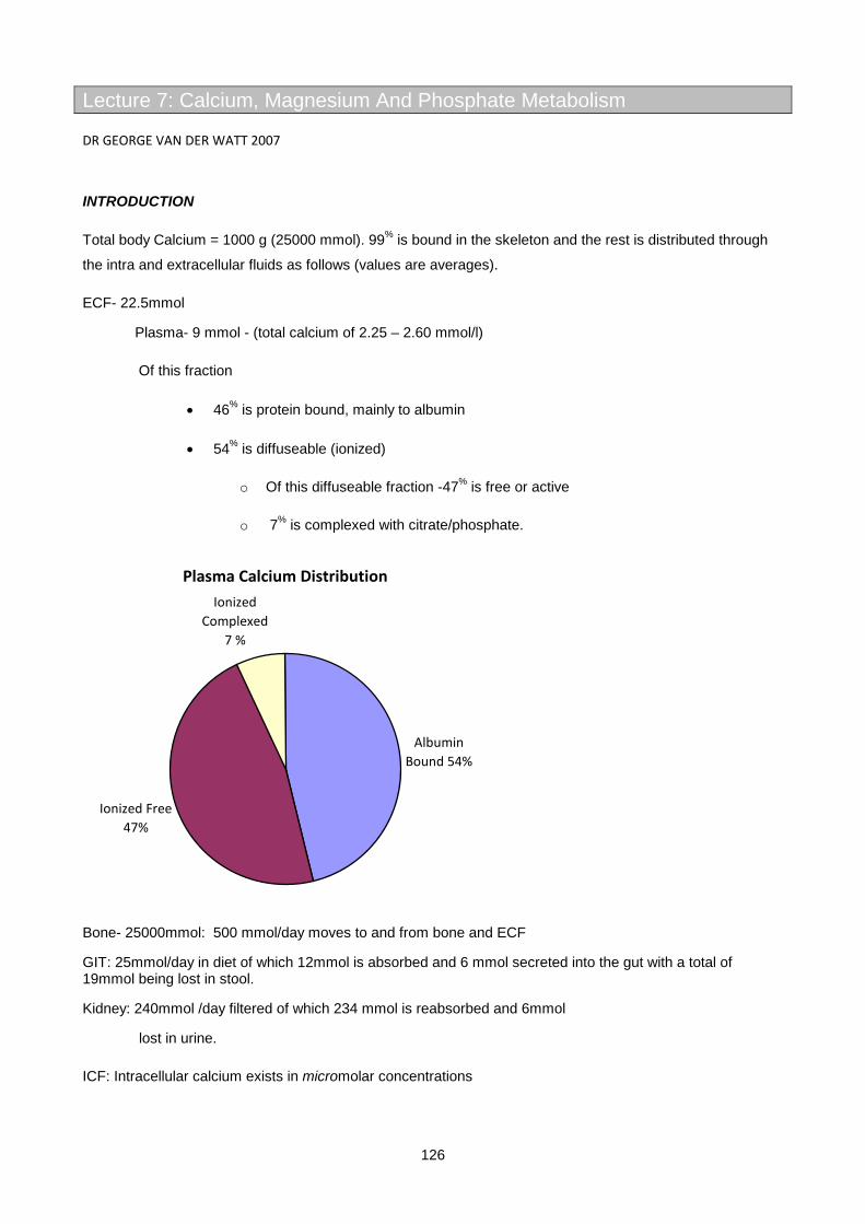
126
Lecture 7: Calcium, Magnesium And Phosphate Metabolism
DR GEORGE VAN DER WATT 2007
INTRODUCTION
Total body Calcium = 1000 g (25000 mmol). 99% is bound in the skeleton and the rest is distributed through
the intra and extracellular fluids as follows (values are averages).
ECF- 22.5mmol
Plasma- 9 mmol - (total calcium of 2.25 – 2.60 mmol/l)
Of this fraction
• 46% is protein bound, mainly to albumin
• 54% is diffuseable (ionized)
o Of this diffuseable fraction -47% is free or active
o 7% is complexed with citrate/phosphate.
Plasma Calcium Distribution
Albumin Bound 54%
Ionized Free 47%
Ionized Complexed
7 %
Bone- 25000mmol: 500 mmol/day moves to and from bone and ECF
GIT: 25mmol/day in diet of which 12mmol is absorbed and 6 mmol secreted into the gut with a total of 19mmol being lost in stool.
Kidney: 240mmol /day filtered of which 234 mmol is reabsorbed and 6mmol
lost in urine.
ICF: Intracellular calcium exists in micromolar concentrations

127
Functions Example
Structural Bone and teeth
Neuromuscular Control of excitability
Release of neurotransmitters
Initiation of muscle contraction
Signaling Intracellular 2nd messenger
Enzymatic Coenzyme for coagulation factors
PHYSIOLOGY OF BONE TURNOVER
Bone consists of osteoid, a matrix of collagen on which inorganic calcium is deposited as hydroxyapatites -
Ca10[PO4]6[OH]2 (general formula).
Type 1 collagen in bone is synthesized by osteoblasts with N and C terminal extensions that are are cleaved
before the collagen is assembled into three (2xά1 and 1xά2) linear intertwined polypeptide chains, the
released N and C terminal propeptides of type I collagen can be measured in serum as markers of bone
formation.
The collagen chains are strengthened by cross linkages called pyridinoline and deoxypyridinoline (DPD)
cross links. These cross links are formed by hydroxylysine and lysine residues that are activated by the
enzyme lysyl oxidase. Hydroxylysine is made by lysine hydroxylase, an enzyme that requires ascorbate, this
explains the osteoporosis that is seen in prolonged scurvy. When collagen is digested by osteoclast
proteases the DPD groups resist cleavage and are released into the circulation. Urinary DPD levels are
therefore a good marker of bone resorption. N-terminal cross linked telopeptides of type 1 collagen (NTx) are
also released by osteoclasts and can be measured in serum and urine as resorptive markers, they consist of
a DPD group with two small specific peptide fragments still attached that are recognized by a specific
antibody. Another good serum marker of bone resorption are crosslaps (CTx), these 8 amino acid peptide
fragments are released by osteoclasts from the C terminal of type 1 collagen and are unique in that they
contain a hydroxylysine and an aspartate residue that forms a peptide linkage through its ß-carboxyl group as
opposed to the conventional ά-carboxyl, this feature is recognized by an antibody in an immunoassay.
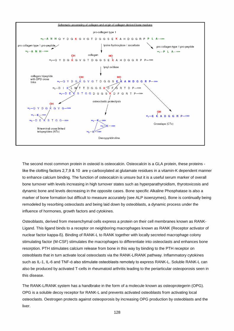
128
The second most common protein in osteoid is osteocalcin. Osteocalcin is a GLA protein, these proteins -
like the clotting factors 2,7,9 & 10 are γ-carboxylated at glutamate residues in a vitamin-K dependent manner
to enhance calcium binding. The function of osteocalcin is unsure but it is a useful serum marker of overall
bone turnover with levels increasing in high turnover states such as hyperparathyroidism, thyrotoxicosis and
dynamic bone and levels decreasing in the opposite cases. Bone specific Alkaline Phosphatase is also a
marker of bone formation but difficult to measure accurately (see ALP isoenzymes). Bone is continually being
remodeled by resorbing osteoclasts and being laid down by osteoblasts, a dynamic process under the
influence of hormones, growth factors and cytokines.
Osteoblasts, derived from mesenchymal cells express a protein on their cell membranes known as RANK-
Ligand. This ligand binds to a receptor on neighboring macrophages known as RANK (Receptor activator of
nuclear factor kappa-ß). Binding of RANK-L to RANK together with locally secreted macrophage colony
stimulating factor (M-CSF) stimulates the macrophages to differentiate into osteoclasts and enhances bone
resorption. PTH stimulates calcium release from bone in this way by binding to the PTH receptor on
osteoblasts that in turn activate local osteoclasts via the RANK-L/RANK pathway. Inflammatory cytokines
such as IL-1, IL-6 and TNF-ά also stimulate osteoblasts remotely to express RANK-L. Soluble RANK-L can
also be produced by activated T-cells in rheumatoid arthritis leading to the periarticular osteoporosis seen in
this disease.
The RANK-L/RANK system has a handbrake in the form of a molecule known as osteoprotegerin (OPG).
OPG is a soluble decoy receptor for RANK-L and prevents activated osteoblasts from activating local
osteoclasts. Oestrogen protects against osteoporosis by increasing OPG production by osteoblasts and the
liver.
129
Tumour cells use all manner of tricks to stimulate bone resorption to create space for themselves within the
bone matrix. Plasma cells in myeloma secrete cytokines that stimulate osteoblasts to express RANK-L, they
also express a proteoglycan called syndecan-1 that mops up any local OPG. Some tumours secrete soluble
RANK-L that can remotely activate osteoclasts, whereas others like breast and prostate cancers directly
express RANK-L. Squamous cell and other malignancies that often metastasize to bone secrete parathyroid
hormone related peptide (PTH-RP), this protein shares homology with the 1st 13 N terminal amino acids of
PTH and bind the PTH receptor on osteoblasts stimulating bone resorption via the RANK-L/RANK pathway.
All the above mechanisms explain why hypercalcaemia is most often associated with a tumour (if parathyroid
adenoma is considered a tumour).
Activated osteoclasts attach to an area of bone and at their ruffled border pump out H+ ions via an H+
ATPase, this generates a localized Ph of 4.5 that aids in dissolving bone mineral. Cathepsin-K a lysosomal
protease is also secreted and digests the collagen matrix of bone. All the products of resorbtion are taken up
by the cell and re-secreted on the non absorbing surface into the circulation.
H+ ions are generated by carbonic anhydrase (CA) from CO2 and water. The cellular pH is maintained by
energy independent exchange of HCO3- for Cl- and electro-neutrality is maintained by CL moving along its
concentration and electrical gradient, passively into the resorbing space.
Bisphosphonates (eg alendronate) are pyrophosphate analogues used to treat osteoporosis and other
disorders of high bone turnover (Pagets, tumour hypercalcaemia). They are incorporated into bone by
osteoblasts. When osteoclasts resorb bone they are exposed to high levels of the drug that interferes with
cholesterol synthesis. This leads to dysfunction of the ruffled border of the osteoclasts and inhibits bone
resorption.
New experimental therapies used to treat osteoporosis include inhibitors of cathepsin-k and carbonic
anhydrase, antibodies that block RANK and injections of recombinant OPG. Interestingly, the only therapy
that actually increases bone formation is pulsatile PTH injections, why this is so is poorly understood because
chronic high PTH exposure leads to excessive bone resorption.
PLASMA CALCIUM
Calcium occurs in plasma in three forms namely
• protein bound - 46%
• complexed to citrate and phosphate - 7%
• free ions - 47% : this is the physiologically active part
Albumin has 12 calcium binding sites and hydrogen ions compete for these, it therefore follows that in
alkalosis free ionized active calcium will fall and in acidosis it will rise as calcium is displaced off the binding
sites by H+, if the change is rapid, symptoms of hypo or hypercalcaemia can develop with pH changes,
despite no change in total serum calcium. Furthermore it is important to realize that since albumin acts as a
“carrier” of Ca2+, the total serum Calcium can vary widely in states of hyper or hypoalbuminaemia with no
significant change in ionized calcium (provided the pH remains constant).
130
When we measure calcium we are interested in the physiologically active fraction as this determines whether
a patient is hyper or hypocalcaemic – we can do this in one of two ways
1. Measure the free ionized calcium directly with an ion sensitive electrode – this is the best way to
determine calcium status but is technically demanding as the pH must be kept constant after
drawing blood to prevent errors. A sample for ionized calcium must be handled the same as a blood
gas specimen in that fresh anticoaglulated whole blood must be sealed in a syringe without gas
bubbles and transported on ice to the laboratory.
2. Correct the total serum calcium to a standardized albumin of 40g/l. This requires simultaneous
measurement of total calcium and albumin. After correction all total calciums can be compared
directly to the reference range. Corrected calciumis calculated as follows:
If [alb] < 40g/l - then corrected Calcium = [total Ca] + 0.02 x (40 - alb) in mmol/l
If [alb] > 45g/l then corrected Calcium = [total Ca] – 0.02 x (alb - 45) in mmol/l
ECF calcium concentrations are kept within narrow limits by the following:
• Calcium Sensing Receptor (CASR)
• Parathyroid Hormone (PTH)
• Vitamin D
(Calcitonin from the C cells of the thyroid plays a minor role in calcium physiology but is useful as a serum
tumour marker for medullary carcinoma of the thyroid.)
1. CALCIUM SENSING RECEPTOR (CASR)
• These receptors are found in the parathyroid gland, the kidney, brain and C cells of the thyroid.
• They are G-protein coupled receptors (GPCRs) that sense ECF ionised calcium concentration and
when activated by calcium binding they activate Gqα . The resultant activation of phospholipase-C
results in cleavage of membrane phosphatidy-inositol-di-phosphate (PIP2) to diacylglycerol (DAG)
and inosine tri-phosphate (IP3). IP3 binds to the endoplasmic reticulum and triggers intra-cytoplasmic
calcium release. DAG activates protein kinase C. The combination of these two 2nd messenger
systems gives rise to the following actions within target tissues.
1. Decreased PTH secretion by parathyroid glands
2. Inactivation of the ROM-K luminal potassium channel in the thick ascending limb of the renal
tubule. Function of this channel facilitates Na+, Cl-, water and Ca2+ absorption
It therefore follows that hypercalcaemia should suppress PTH levels and give rise to a diuresis with
hypercalciuria

131
2. PARATHYROID HORMONE (PTH)
• An 84 AA polypeptide secreted by the parathyroid glands in response to a drop in ionized serum
calcium.
• PTH activates the PTH-receptor with the first 6 N-terminal AAs but requires amino acids’s 7-34 on
the N-terminal to bind the receptor.
• PTH receptors are also GPCRs and are found principally on osteoblasts and renal tubular cells. is
mediated through so called calcium sensing receptors (CASRs). These are G-protein coupled
receptors that suppress PTH synthesis when bound to calcium, the reverse occurs when calcium
levels are low.
THE MAIN EFFECTS OF PTH ARE TO INCREASE SERUM CA2+ AND DECREASE SERUM PHOSPHATE.
Actions of PTH
Target Organ
Action Effect
Bone Rapid release of calcium by stimulation of osteoclasts via PTH-R on osteoblasts acting via RANK-L and RANK
Increase plasma ionized calcium
Kidney Decrease proximal tubular phosphate reabsorbtion by decreasing luminal sodium-phosphate (NaPPi) transporters
Decrease plasma phosphate with increased urinary phosphate excretion
Activate alpha-1-hydroxylase with increased production of 1,25-di-OH-vit-D
• Increased GIT calcium and phosphate reabsorption
• Increased renal calcium reabsorption
• Decreased PTH secretion (-ve feedback loop)
Decrease HCO3- reabsorbtion Metabolic acidosis - helps to increase ionized calcium
It is important to realize that PTH secretion and its actions are magnesium dependent and severe
hypomagnesemia can mimic hypoparathyriodism. PTH is rapidly cleared from plasma with a t1/2 of 5 min by
hepatic and renal metabolism.
3. CHOLECALCIFEROL OR VITAMIN D
Pre-Vit D is formed from the ultra violet photolysis of 7-dehydrocholesterol in the skin. Pre-Vit D3 then
thermally isomerises to Vit-D3 (cholecalciferiol) from where it enters the circulation. Cholecalciferol also
occurs naturally in the diet. It is 25-hydroxylated in the liver (not subject to feedback control) and in the kidney
it is alpha-1-hydroxylated to its active form - 1,25-dihydroxycholecalciferol by the regulated enzyme α-1-
hydroxylase
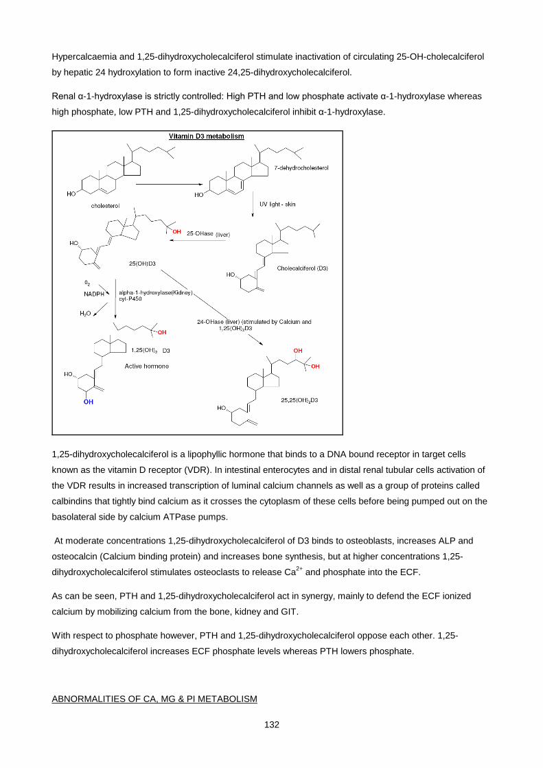
132
Hypercalcaemia and 1,25-dihydroxycholecalciferol stimulate inactivation of circulating 25-OH-cholecalciferol
by hepatic 24 hydroxylation to form inactive 24,25-dihydroxycholecalciferol.
Renal α-1-hydroxylase is strictly controlled: High PTH and low phosphate activate α-1-hydroxylase whereas
high phosphate, low PTH and 1,25-dihydroxycholecalciferol inhibit α-1-hydroxylase.
1,25-dihydroxycholecalciferol is a lipophyllic hormone that binds to a DNA bound receptor in target cells
known as the vitamin D receptor (VDR). In intestinal enterocytes and in distal renal tubular cells activation of
the VDR results in increased transcription of luminal calcium channels as well as a group of proteins called
calbindins that tightly bind calcium as it crosses the cytoplasm of these cells before being pumped out on the
basolateral side by calcium ATPase pumps.
At moderate concentrations 1,25-dihydroxycholecalciferol of D3 binds to osteoblasts, increases ALP and
osteocalcin (Calcium binding protein) and increases bone synthesis, but at higher concentrations 1,25-
dihydroxycholecalciferol stimulates osteoclasts to release Ca2+ and phosphate into the ECF.
As can be seen, PTH and 1,25-dihydroxycholecalciferol act in synergy, mainly to defend the ECF ionized
calcium by mobilizing calcium from the bone, kidney and GIT.
With respect to phosphate however, PTH and 1,25-dihydroxycholecalciferol oppose each other. 1,25-
dihydroxycholecalciferol increases ECF phosphate levels whereas PTH lowers phosphate.
ABNORMALITIES OF CA, MG & PI METABOLISM
133
1. HYPERCALCAEMIA.
Two causes dominate this diagnosis, namely Primary Hyperparathyroidism and Malignancy. Together
they are responsible for 90 % of cases. Hypercalcaemia is often asymptomatic and symptoms are usually
attached to the diagnosis retrospectively.
Signs and symptoms of hypercalcaemia.
Weakness, tiredness, lassitude and muscle weakness.
Mental changes ranging from depression and impaired concentration to psychosis and coma.
Nausea, vomiting, constipation, anorexia, abdominal pain.
Polyuria (NB diff dx of nephrogenic DI), dehydration , renal calculi and nephrocalcinosis.
Cardiac arrythmias, hypertension, increased QT interval.
CAUSES OF HYPERCALCAEMIA
Common Less common Rare
Malignancy
Primary hyper PTH
Thyrotoxicosis
Vit D3 intoxication
Thiazide diuretics
Sarcoidoses
Familial hypercalcuric hypercalcemia
Tertiary hyperPTH
(post-kidney transplant)
Milk-alkali syndrome
Lithium
Tuberculosis
Immobilization
Adrenal failure
MALIGNANT DISEASE AND HYPERCALCAEMIA
Tumour hypercalcaemia is usually due to one of the following mechanisms
• Local osteolysis – multiple myeloma often causes lytic bone lesions as the plasma cells involved
stimulate localized bone resorption by osteoblasts.
• PTH related protein (PTHrP) - PTHrP is a polypeptide of varying length (139, 141 or 173 AA’s)
produced from a single gene that is identical to PTH for the 1st 13 amino acids on the N-terminal end,
the rest of the protein is totally different. Physiologically it is synthesized locally by tissue where it acts
in a paracrine fashion, to regulate cartilage growth, tooth development, lactation and placental
calcium transport. In malignant tissue however, it may be overproduced and enter the circulation in
significant amounts where it has an endocrine effect, similar to PTH, causing hypercalcaemia.

134
Tumour PTH-RP production is often suspected in a patient with an malignancy and hypercalcaemia,
an appropriately suppressed PTH and unexplained hypophosphataemia.
• Other osteoclastic activating factors, like interleukins 1 and 6, TGF-Beta and Tissue Necrosis
Factor (TNF) stimulate osteoclasts to resorb bone and are most often produced by haematological
malignancies eg myeloma, leukaemias and lymphoma’s. Prostaglandin E2 activates osteolysis in
breast cancer patients.
• 1,25-dihydroxycholecalciferol (Vit D) is often overproduced by lymphoma tissue (usually Hodgkin
lymphoma). This is thought to be due to overproduction of alpha-1-OH-ase by these cells or due to
them activating the same enzyme in normal macrophages via interferon-gamma production. These
patients have a high calcium and a high or high normal phosphate, an appropriately suppressed PTH
and a high LDH.(Monocytes/macrophages are the only other tissue to have significant alpha-1-
hydroxylase levels and hypercalcaemia can be seen in other granulomatous diseases like
tuberculosis and sarcoidosis)
• A surprising number of patients with cancer have coexistant primary hyperparathyroidism as well
and their hypercalcaemia is not related to the malignancy per se.
PRIMARY HYPERPARATHYROIDISM
Occurring in 1/1000 individuals especially post menopausal women. This condition is most often due to a
parathyroid adenoma, sometimes due to diffuse hyperplasia of the parathyroids and rarely due to a PTH
secreting parathyroid carcinoma. Some adenomas are familial (part of multiple endocrine neoplasia or MEN
syndromes).
Patients often have subtle symptoms such as non specific bone pain, kidney stones and symptoms of
lethargy and depression. The adenomas are usually small and rarely palpable. Surgical excision is the
treatment of choice.
The diagnosis is usually made by the finding of a high or high/normal PTH in the face of hypercalcaemia.
Approximately 20% of patients will have radiological evidence of hyper-PTH, 20% can have a raised ALP and
most have a mild hypophosphataemia with phosphaturia, although phosphate can be normal or high if renal
damage has occurred. In rare instances plasma calcium can be normal due to renal disease, hypovitaminosis
D3 or hypothyroidism.
SECONDARY AND TERTIARY HYPERPARATHYROIDISM
Chronic renal disease and vitamin D3 deficiencies give rise to low 1,25-dihydroxycholecalciferol levels. This
in turn leads to low calcium levels. As a result, the parathyroid glands are stimulated to produce PTH. Here
the high PTH occurs with a low or low normal calcium (secondary hyperPTH).
If this process continues for a long time the gland can become hyperplastic and become insensitive to
calcium mediated negative feedback. This is termed tertiary hyperPTH. This can result in severe

135
hypercalcaemia in patients who receive renal transplants where the new kidneys are able to produce1,25-
dihydroxycholecalciferol via alpha-1-hydroxylase activity which is driven by the high circulating PTH levels.
OTHER CAUSES OF HYPERCALCAEMIA
• Thyrotoxicosis stimulates osteoclastic activity, raising s-calcium levels and causing osteoporosis.
• Vitamin D toxicity can occur with oral overdose of vit-D, overzealous treatment with Vitamin D in renal
failure patients and with sarcoidosis, where increased alpha-1-hydroxlase activity occurs in the
macrophages of the sarcoid granulomas. This occurs in other diseases where granulomas which are
full of active macrophages occur, as in tuberculosis. (Sarcoidosis is incidentally diagnosed by
increased levels of ACE in the serum.)
• Thiazide diuretics decrease renal calcium excretion and can cause hypercalcaemia with
hypocalcuria.
• Immobilization - When active bone is immobilized, resorbtion continues but formation decreases
because axial pressure is needed to stimulate osteoblasts. This results in hypercalcaemia among
fast growing adolescents and patients with Paget’s disease when immobilized. Both of these have
high levels of active bone turnover.
• Disease affecting the CASR.
o Familial Benign Hypocalciuric Hypercalcaemia (FBHH) - autosomally dominant inherited loss
of function of the calcium sensing receptors in the parathyroids and kidneys. These patients
need higher levels of calcium to suppress PTH secretion. They therefore have
hypercalcaemia with normal PTH levels and hypocalciuria. The diagnosis is usually
suspected when there is a strong family history of hypercalcaemia (penetrance is 100%).
The way to make the diagnosis is to measure the fractional excretion of calcium, FeCa =
(Urine Ca x Serum Creat) / (Serum Ca x Urine Creat). In FBHH it is < 0.01 (1%). Children of
two parents with FBHH may inherit two defective CASR genes – these children present with
neonatal severe hypercalcaemia and hyperparathyroidism as they cannot sense ECF
calcium.
o Lithium interferes with CASR function and patients on lithium can have the identical findings
to patients with FBHH therefore lithium must always be excluded when working patients up
for hyperparathyroidism.
INVESTIGATION OF HYPERCALCAEMIA
X-rays can reveal signs of chronic hyperparathyroidism, Pagets disease, expose some malignancies and
help diagnose tuberculosis and sarcoidosis.
136
Serum phosphate is useful as it is often low with hyperparathyroidism or PTH-RP production. A high calcium
with a high phosphate is an unusual finding and is usually due to renal failure secondary to another cause for
hypercalcaemia (myeloma)
It is useful to calculate the solubility product where
[K] = [Ca] X [Pi] (in mmol/l)
If K is greater than 4.6 then a risk of metastatic tissue calcification exists due to the formation of insoluble
CaPO4 crystals in ECF.
The hypercalcaemia of malignancy can be differentiated from hyperparathyroidism by:
more severe and sudden onset
sometimes responds to corticosteroid treatment
produce1,25-dihydroxycholecalciferol level is usually normal or low as opposed to high in hyper-PTH
TREATMENT of HYPERCALCAEMIA
Life threatening hypercalcaemia, defined as a total calcium of > 3.7mmol/l, is treated with normal saline
volume repletion and once well hydrated, with furosemide, which has the effect of blocking calcium
reabsorbtion whilst promoting diuresis. If needed, dialysis is also an option.
In the longer term bisphosphonates can be given, which help trap Ca2+ in bone and inhibit bone resorbtion.
As with all diseases, treat the cause; ie parathyroidectomy for primary hyperPTH.
2. HYPOCALCAEMIA
Causes Clinical features
Artefact (blood in EDTA tube)
Vit D deficiency
- Dietary deficiency
- Malabsorbtion
- Lack of UV light
Disordered Vit D metabolism
- Renal failure
- Anticonvulsant treatment
Alpha-1-hydroxylase deficiency
HypoPTH
PseudohyperPTH
Magnesium deficiency
Paraesthesiae and numbness
Cramps and spasms (tetany)
Laryngeal stridor
Convulsions
Increased QT time
+ve Chvostek and Trosseau signs
137
Acute pancreatitis
Massive blood transfusion (citrate)
The majority of patients seen with hypocalcaemia have
• Renal Failure
• Impaired Vitamin D metabolism
• Hypomagnesaemia
• Hypoparathyroidism
The symptoms of hypocalcaemia are mainly due to increased muscle and nerve excitability and rapid
hypoventilation with alkalosis can elicit all of these symptoms by decreasing ionized calcium.
In Chvostek’s sign the facial muscles twitch when the facial nerve is tapped.
In Trosseau’s sign carpal spasm develops when a BP cuff is applied between systolic and diastolic pressures
for three minutes (can you work out why?)
• EDTA in blood collection tubes chelates calcium
• Citrate used as anticoagulant in blood transfusions can cause hypocalcaemia with large
transfusions.
• Phenobarbitone and phenytoin induce HME’s resulting in increased inactivation of vitamin D; they
also interfere with vitamin D and calcium absorption from the GIT thereby causing hypocalcaemia.
Patients on these drugs generally spend less time out in direct sunlight.
• Vitamin D is a fat soluble vitamin requiring adequate lipase, bile salts and a normal small intestine
for absorbtion ( see GIT lectures), malbsorbtion of fat or a D3 deficient diet can therefore cause
hypocalcaemia.
• In acute pancreatitis lipases released into tissues cleave tri-acyl-glycerides into glycerol and free
fatty acids (FFA), the -ve carboxyl groups on these FFA’s form insoluble salts with calcium ions
causing hypocalcaemia.
• Hypomagnesemia results in impaired PTH secretion and impairs PTH’s action on tissues causing a
hypocalcaemia resistant to calcium and Vit D therapy.
• Hypoparathyroidism can be congenital as in Di George syndrome where it goes with immune
deficiency and thymic aplasia.
• Aquired hypoparathyroidism can be due to autoimmune gland destruction, surgical gland removal or
due to infiltrative gland destruction as in hemochromatosis.
138
• Patients with pseudohypoparathyroidism are heterozygous for a loss of function mutation in the Gsα
subunit of the PTH receptor. This gene happens to be paternally imprinted in the kidney. This means
that the paternal gene is inactivated in the kidney. Therefore if a patient inherits the disease from
their mother they develop a full PTH resistant phenotype with a syndrome called Allbrights hereditary
osteodystrophy ( short stature, round face, subcutaneous ossifications and short fingers often with a
shortened 4th metacarpal) as well as biochemical hypoparathyroidism with low Ca and high PO4
(due to renal PTH resistance). This is known as pseudohypoparathyroidism type 1A. If however a
patient inherits the disease from their father they only manifest with Allbrights hereditary
osteodystrophy but not biochemical PTH resistance because the maternal allele is functional in the
kidney. These patients have pseudopseudohypoparathyroidism. To confuse matters, a third group of
patients have been described who have PTH resistance but no osteodystrophy. They have
pseudohypoparathyroidism type 1B. The way to diagnose this disease biochemically is to give them
a large IV PTH dose and measure urinary cAMP. If biochemical PTH resistance is present, the
cAMP levels remain low.
Vit D deficiency results in rickets in children and osteomalacia in adults, and is discussed below.
Hypocalcaemia is common in patients with end stage renal disease often as part of renal osteodystrophy,
discussed below.
TREATMENT OF HYPOCALCAEMIA
As per usual, treatment should be directed at the cause. Life threatening hypocalcaemia can be treated with
iv calcium gluconate. (what other conditions are treated with this drug ?).Do not forget to give magnesium as
well. Many patients can be managed on calcium supplements and Vit D or its hydroxylated derivatives,
depending on the cause.
3. HYPERPHOSPHATAEMIA. Normal Serum Pi = 0,9-1.4mmol/l
Here by far the no 1 cause is renal insufficiency and the kidneys inability to excrete phosphate. Other causes
include hypoparathyroidism and pseudohypoparathyroidism where PTH’s effect of stimulating renal Pi
excretion is lost.
Phosphate overdose can occur if undiluted cows milk is given to infants or iatrogenically when parenteral
feeding is given. Phosphate is released in the Tumour Lysis Syndrome seen when haematological
malignancies are treated with chemotherapy, or in ischaemic bowel necrosis. Hyperphosphataemia is
important because it suppresses alpha-1-hydroxylase can preciptate hypocalcaemia. It can also trigger
metastatic calcification if the solubility constant with Ca is reached.
Treatment : directed at the cause, calcium or aluminium salts are often given orally to bind phosphate in the
gut and prevent absorbtion.
4. HYPOPHOSPHATAEMIA.
139
A very common finding, most often seen when severely catabolic tissue is recovering and replenishing it’s
intracellular phosphate. Phosphate is mostly intracellular where it is vital for the formation of high energy
phosphate bonds, as in ATP that provide the driving energy behind all intracellular metabolism, and is a major
component of intermediate metabolites like G-6-P. Phosphate is essential in the regulation of various
enzymes and is also a constituent of phospholipids in the cell membrane. For this reason when levels less
than 0.3 mmol/l occur most tissues become dysfunctional to a greater or lesser degree, with muscle
weakness a prominent symptom and death not far off. Phosphate is also a structural component of bone.
Causes of Hypophosphataemia
• Decreased absorbtion from GIT.
• Intracellular shift.
• Increased renal losses.
Vitamin D deficiency results in decreased GIT phosphate absorption
Primary hyperparathyroidism results in increased renal losses.
Refeeding of alcoholics, diabetic keto acidotics and other starved patients cause a shift of phosphate into
cells. Remember also that patients with proximal tubular defects (Fanconi syndrome) can also lose
phosphate in the urine. Respiratory alkalosis can activate phosphofructokinase that phosphorylates glycolytic
intermediates, and by using up phosphate can cause hypophosphataemia (with a low ionized calcium ) Treat
the cause and replace orally (cows milk) or parenterally. Do not give phosphate IV to a patient with
hypercalcaemia or oliguria.
5. MAGNESIUM. N = 0.8-1.2mmol/L
An abundant cation with a total body amount of 1000mmol. Half is incorporated into bone, the rest is chiefly
intracellular where it acts as a co-factor for some 300 enzymes including those involved in glycolysis, protein
synthesis and transmembrane transport. Magnesium is essential for many enzymes that react with high
energy phosphate bonds like phosphatases, kinases and ATPases. It also contributes to maintaining the
structure of ribosomes, nucleic acids and proteins. Magnesium and calcium interact with respect to
membrane stability where magnesium plays a major role in preventing hyperexciteability of cell membranes,
hence its use in preventing ecclamptic seizures. Only 15 – 30 mmol is found in the ECF and a normal
plasma concentration = 0.8 – 1.2 mmol/L. There is no known specific homeostatic mechanism for regulating
magnesium with a daily intake of 10mmol exceeding requirements of 8mmol, the rest being lost in urine.
PTH to some extent promotes magnesium reabsorption, and aldosterone promotes loss in urine. In
hypomagnesaemia renal reabsorption of magnesium is very efficient.
HYPERMAGNESAEMIA.
Only occurs with overdose as in treatment of ecclampsia with IV magnesium sulphate. Patients present with
muscle weakness, loss of deep tendon reflexes, ECG abnormalities and at high concentrations (>7.5mmol/L
140
) with cardiorespiratory arrest. Calcium intravenously acts as the antidote by displacing magnesium off
receptors on the cell membrane giving the kidneys a chance to excrete the excess magnesium.
HYPOMAGNESAEMIA.
Very common and often overlooked in hospital patients where poor nutrition and loss via the GIT in diarrhea
are the leading causes. Other causes include malabsorption, chronic alcoholism, cirrhosis, loop diuretics,
renal failure and chronic aldosterone excess. If severe enough to present with symptoms, patients present
with signs of hyper-excitability such as tetany, tremors, agitation, delirium, seizures and cardiac arrhythmias.
Because magnesium is central to the secretion and effect of PTH, patients can often develop hypocalcaemia
secondarily to hypomagnesaemia. Measuring urinary magnesium (before treatment) in hypomagnesaemia
can be useful in differentiating the cause as normal kidneys are very efficient at conserving magnesium. A
urine magnesium of > 0,5mmol/l in the setting of hypomagnesaemia would indicate renal loss as the cause.
Hypomagnesaemia is usually managed with oral supplementation, or, if severe, with infusion of magnesium.
METABOLIC DISEASE OF BONE
RICKETS, OSTEOMALACIA, OSTEOPOROSIS, RENAL OSTEODYSTROPHY AND PAGETS DISEASE
Bone has four important functions
Structural support.
Houses haematopoetic bone marrow.
Metabolic regulation of calcium and phosphate.
Buffer system (release of phosphate buffer in chronic metabolic acidosis).
Structurally bone consists of spongy trabecular bone supporting the bone marrow and dense cortical bone.
On a subcellular level bone consists of a collagen (type 1 fibres) protein matrix secreted by osteoblasts
together with glycoproteins and a proteoglycan ground substance. This proteinaceous matrix called osteoid
provides the support for deposition of the mineral component in the form of hydroxyapatite crystals.
All bone undergoes constant remodeling with osteoclasts resorbing in places and osteoblasts laying down
new bone in others, the osteoblasts remain as mature osteocytes. This whole process is modulated by
hormones, growth factors and cytokines. Metabolic bone disease occurs when there is disruption of this
synchrony.
1) RICKETS AND OSTEOMALACIA
Defined as defective mineralization of osteoid. We refer to rickets in growing bone and osteomalacia in adult
bone.
It is usually due to a deficiency of calcium, which in turn is due to a problem with Vitamin D:
Decreased absorbtion ( malnutrition, malabsorbtion syndromes)
141
Decreased production (lack of UV light, liver disease, kidney disease, anti-epileptics. Alpha-1-
hydroxylase mutations)
Resistance to vitamin D
Richets/Osteomalacia can also be due to loss of phosphate (phosphaturic rickets) due to chronic renal losses
such as in Fanconi syndrome and X-linked phosphaturic rickets (see renal lecture notes). In renal tubular
acidosis type I (distal) there is significant loss of calcium and phosphate in the urine as the bone is mobilized to
buffer the metabolic acidosis.The clinical features include bone pain, skeletal deformities such as bowing of the
legs, deformable skull (craniotabes), rachitic rosary (costochondral junction swelling), growth retardation in
children and muscle weakness. Typical X-ray changes include generalized decalcification (crushed glass
appearance), widening of epiphyses (wineglass appearance) pseudofractures and subperiosteal erosions.
Calcium levels are however usually low/normal due to secondary hyperparathyroidism as the parathyroids try and
mobilize calcium. Phosphate is often low due to the action of PTH and alkaline phosphatase levels can be high as osteoblasts attempt to lay down new bone. Treatment usually involves Vitamin D or its hydroxylated
derivatives together with calcium and phosphate supplements.
2) OSTEOPOROSIS.
Defined as progressive reduction in bone mineral density and abnormal micro architecture giving rise to weak
fracture prone bone. It is caused by increased osteoclast relative to osteoblast activity. Fractures of the vertebral
bodies, distal radius (colles #), and femoral neck occur most often.
PRIMARY OSTEOPOROSIS :
Type I osteoporosis occurs as a result of oestrogen deficiency, mostly affects trabecular bone and often gives
rise to vertebral body collapse.
Type II occurs as a result of the natural loss of bone with age and affects trabecular and cortical bone, often
associated with femoral neck fractures.
SECONDARY OSTEOPOROSIS:
Endocrine causes: Thyrotoxicosis, cushings, hypogonadism, diabetes mellitus
Drugs: heparin, corticosteroids, chronic alcoholism ( number one cause in men )
Other: immobilization, weightlessness (as in astronauts)
The diagnosis is often only made once patients present with fractures. The best way to estimate the severity of
this disease is with bone mineral density scanning. Routine chemistry is not helpful in the diagnosis as calcium,
phosphate and alkaline phosphatase are usually normal. Bone markers have been discussed earlier in this
lecture: They are principally used to assess the bone turnover state prior to commencement of therapy for
osteoporosis and to monitor response to therapy. Markers of bone resorption should decrease with
bisphosphonate therapy whereas markers of formation and resorption would increase with pulsatile PTH therapy.
3) RENAL OSTEODYSTROPHY.
A common disorder of bone consisting of variable combinations of fibrosing-osteitis and osteomalacia in
association with chronic renal failure.
142
Pathogenesis: Inability to excrete phosphate is the initiating event in renal osteodystrophy
High phosphate suppression of alpha-1 hydroxylase activity together with overall loss of alpha-1-
hydroxylase due to decreased renal mass results in 1,25 di-OH cholecalciferol deficiency and
osteomalacia with hypocalcaemia
Chronic acidosis promotes bone demineralization to buffer hydrogen ions.
Hyperparathyroidism due to hypocalcaemia triggers increased bone resorbtion to try and raise serum
calcium levels – PTH induced bone disease is called osteitis fibrosa cystica.
Hyperphosphataemia due to renal inability to excrete it can trigger metastatic calcification if the
solubility product of Ca and Pi is exceeded. This is called osteosclerosis in bone.
Biochemically these patients have a low or low/normal calcium with secondary high PTH and
hyperphosphataemia. Treatment can include activated vitamin D and calcium supplements, oral
phosphate binders and parathyroidectomy (especially if tertiary hyperparathyroidism has developed)
4. PAGETS DISEASE OF BONE.
A disease of uncertain etiology characterized by accelerated bone turnover. There is localized increase
of osteoclastic activity and vascularity engendering increased osteoblastic activity. The osteoblasts in
turn lay down bone in a disorderly fashion giving rise to thick, brittle, deformed, painfull bone. Patients
present with pathological fractures and bone pain or sometimes with signs of compression of structures
surrounded by bone eg deafness due to auditory nerve compression. In these patients alkaline
phosphatase activity is usually very high with massive increase of markers of bone formation and
resorption in the urine. Treatment utilizes analgesics and bisphosphonates.
143
SMALL GROUP TEACHING. LECTURE 7: CALCIUM, PHOSPHATE AND MAGNESIUM
QUESTIONS
1. What is meant by "ionised" as opposed to total Ca2+ in plasma? What are the proportions of protein-bound, complexed, and ionised Ca2+ in plasma?
2. Explain how changes in pH affect the concentration of ionised Ca2+ in plasma.
3. Which fraction(s) of plasma Ca2+ are usually measured in clinical laboratories? What are the advantages and disadvantages of measuring ionised Ca2+?
4. What symptoms can be caused by (a) hypercalcaemia (b) hypocalcaemia?
5. Name a biochemical test which is an index of osteoblastic activity.
6. In which tissues is the Ca2+ -sensing receptor (CASR) expressed?
7. Complete the table (↑, N, or ↓):
DISORDER SERUM CALCIUM SERUM PHOSPHATE ALK. PHOS.
Hypoparathyroidism
Sarcoidosis
Pagets disease
Osteoporosis
primary hyperparathyroidism
nutritional rickets
8. Work out the biochemical abnormalities you would expect due to an inactivating mutation in the calcium-sensing receptor.
9. Explain why patients may develop tetany after a massive blood transfusion.
10. Explain the difference in meaning of the terms: osteoporosis osteomalacia osteopaenia
11. A 50-year old man presented with severe abdominal pain, haematuria, and subsequently passed a stone. Abdominal X ray showed the presence of another stone in the bladder. The biochemical data were as follows:
Plasma: urea 4.5mmol/L, creatinine 100umol/L, Total Ca2+ 2.9mmol/L (2.1-2.6 mM), Pi 0.7 (0.8-1.4 mM), albumin 38g/l, ALP 150U/ml (30-115 U/ml), PTH 24pmol/L (1.6-6.9 pM)
24h URINE normal range
creatinine 14 mmol/day ± 0.2 mmol/kg/day (adult male)
TRP 68% >85%
Calcium 12.6 mmol/d <7.5 mmol/d
i. Plot the PTH result on the graph. Which disorders fall into areas A, B and C?
ii. What diagnosis is suggested in the patient?
iii. What is the treatment for this condition?
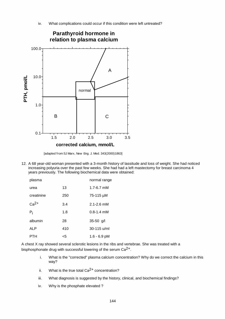
144
iv. What complications could occur if this condition were left untreated?
Parathyroid hormone inrelation to plasma calcium
1.5 2.0 2.5 3.0 3.50.1
1.0
10.0
100.0
normal
[adapted from SJ Marx, New Eng. J. Med. 343(2000)1863]
corrected calcium, mmol/L
PTH
, pm
ol/L
A
B C
12. A 68 year-old woman presented with a 3-month history of lassitude and loss of weight. She had noticed increasing polyuria over the past few weeks. She had had a left mastectomy for breast carcinoma 4 years previously. The following biochemical data were obtained:
plasma normal range
urea 13 1.7-6.7 mM
creatinine 250 75-115 µM
Ca2+ 3.4 2.1-2.6 mM
Pi 1.8 0.8-1.4 mM
albumin 28 35-50 g/l
ALP 410 30-115 u/ml
PTH <5 1.6 - 6.9 pM
A chest X ray showed several sclerotic lesions in the ribs and vertebrae. She was treated with a bisphosphonate drug with successful lowering of the serum Ca2+.
i. What is the "corrected" plasma calcium concentration? Why do we correct the calcium in this way?
ii. What is the true total Ca2+ concentration?
iii. What diagnosis is suggested by the history, clinical, and biochemical findings?
iv. Why is the phosphate elevated ?
145
v. Briefly describe the similarities and differences in (a) structure and (b) function of PTH and PTH-related peptide.
vi. Suggest why she had polyuria.
vii. How do bisphosphonates work?
13. A 15 year old boy presented with pain and stiffness in his wrists and fingers. Investigations to determine the cause of the arthritis were undertaken. A chest X ray showed bilateral enlarged hilar nodes.
Plasma results
Na+ 138, K+ 4.1, urea 4.2, creatinine 90, albumin 42, Ca2+ 3.1, Pi 1.1, alk. phos. normal
PTH undetectable.
A hydrocortisone suppression test was performed. The serum Ca2+ dropped to 2.5 mmol/l after 2 days of hydrocortisone administration. The serum angiotensin converting enzyme (SACE) level was elevated.
i. Comment on the biochemical findings. Which causes of hypercalcemia are EXCLUDED by the PTH result, and which are compatible with it?
ii. What diagnosis is suggested, taking into account the patient's symptoms, X ray features and other lab results?
iii. Explain the mechanism of the hypercalcaemia in this disorder.
14. A medical student awaiting a Chem Path oral exam had an attack during which she felt strange and had muscle spasms of her hands. This was associated with a feeling of numbness and tingling (parasthesia) around her lips, tachycardia, sweating and tremor. During the attack a blood sample was obtained by sympathetic lecturing staff, and the following results were obtained:
Plasma: Na+ 135, K+ 4.0, urea 4.5, glucose 5.3, Ca2+ 2.2, Pi 1.2, albumin 40
Acid-base: pH 7.68, pCO2 2.8 kPa, STD BIC 24 mmol/L, pO2 14.0 kPa
i. What type of acid-base disturbance is present?
ii. Suggest a diagnosis.
iii. What form of calcium is routinely measured in plasma (as in this case)?
iv. What is the cause of the muscle spasms and parasthesia in this case?
v. What treatment would you suggest?
15. A 30 year old woman complained of cramps in her arms and legs. She had a thyroidectomy 10 years previously for a thyroid disorder, and had been on medication for epilepsy for five years. On examination Chvostek's and Trousseau's signs were positive, and no other abnormalities were found. The following results were obtained:
Plasma: Ca2+ 1.70 mM,albumin 38, phosphate 1.8, alk. phos.105 (30-115),creatinine 55(75-115), Mg2+ 0.9 (0.7-1.1),PTH 2.2 pmol/L (1.6 - 6.9)
i. What diagnosis is suggested by the history, clinical signs and laboratory results?
ii. How can anticonvulsant drugs affect Ca2+ metabolism? Are the results in this case consistent with such a mechanism?
iii. How could the history of epilepsy be explained?
16. A 4-year old black girl presented with bowing of the legs. On examination she was well below expected weight and height for age, had oedema and other signs of kwashiorkor. Both parents were unemployed and the family was poverty stricken. X rays of the wrists and legs showed widened epipyses, characteristic of rickets.
plasma results
146
Ca2+ 1.5 mmol/L (2.1 - 2.6), albumin 24 g/L (35-45), Pi 0.9 (1.2-1.7 mM; note higher range in children), ALK PHOS 700 (60-180 units) PTH 52 pmol/L (1.6 - 6.9)
i. Is the low Ca2+ simply a reflection of the low albumin?
ii. Comment on these findings in relation to the clinical picture.
iii. Explain the likely pathogenesis of rickets in this child. Do you think that a deficiency of sun exposure was a likely contributory factor in this case?
17. A boy aged 6 presented with growth retardation, bowed tibiae and a radiological picture of rickets. Social circumstances were good, and there was no evidence of malnutrition. The following data were obtained:
Plasma: Na+ 135 (135-145 mM), K+ 3.0 (3.5-5.5 mM), Cl- 109 (97-107 mM),creat 75 (75-115 µM), Ca2+ 2.1 (2.1-2.6 mM), Pi 0.7(1.2-1.8 mM) (note higher range in children) TRP 53% (>85%), albumin 38 (35-50 g/l), ALP 400 (60-180 u/ml)
Acid-base:pH 7.18, pCO2 4.2 kPA, STD BIC 16 mM
urine: pH 6.8
i. What type of acid-base disturbance is present?
ii. Is the anion gap increased?
iii. Comment on the urine pH
iv. What diagnosis is suggested by this combination of biochemical findings in a patient with rickets?
v. Suggest other biochemical tests which might be of value in confirming the diagnosis.
18. Mr. F.J. aged 45 was on haemodialysis for several years for end-stage renal failure due to glomerulonephritis. He developed bone pain, and X rays showed marked osteopaenia and multiple bone cysts. Biochemistry was as follows:
Na+ 140 mM, K+ 5.3 mM, urea 35mM, creatinine 700 uM, Ca2+ 2.5 mM, albumin 37 g/L, Pi 3.5mM (N 0.8-1.4), alk phos 400 units (30-115), PTH 65 pmol/L (1.6 - 6.9)
i. Comment on the Ca2+ level: Is this typical in chronic renal failure?
ii. What factors are contributing to the pathogenesis of this man's bone disease?
iii. What treatment options may be of benefit?
iv. What is metastatic calcification? Do you think it might occur in this case? Why?
19. A 6-year old girl presented with generalized oedema. Her urine was frothy and contained large amounts of protein.
Plasma: Ca2+ 1.7 mM (2.1-2.6), Pi 1.4 mM (0.8-1.4), albumin 18 (35-50 g/l)
What diagnosis explains the clinical and biochemical findings?
i.What further investigations of calcium/phosphate metabolism are required?
20. An elderly woman presented with increasing bone pain, resulting in inability to walk. X rays showed osteomalacia. There was a history of weight loss and chronic diarrhoea for several years. A xylose absorption test indicated malabsorption due to intestinal disease, and a biopsy of the small bowel mucosa revealed amyloidosis. She was treated with calcium supplementation and parenteral vitamin D. However, she presented again some weeks later with frank tetany, and the following results were found:
Plasma: Ca2+ 1.1, albumin 34, Pi 0.8, Mg2+ 0.40 (0.7-1.2 mmol/l)
Urine: Ca2+ 0.6 mmol/day, Mg2+ 0.3 mmol/day

147
i. How is Mg2+ distributed in the body? How is Mg2+ balance regulated?
ii. In what ways can Mg2+ status affect Ca2+ metabolism?
iii. What is the value of knowing the urinary Mg2+ in a patient with hypomagnesemia?
SMALL GROUP TEACHING. LECTURE 7: CALCIUM, PHOSPHATE AND MAGNESIUM
ANSWERS
1. Calcium occurs in plasma in three forms namely, protein bound - 46%, complexed to citrate and phosphate - 7%, and as free ions - 47%. Ionized calcium represents the latter two.
2. Protons displace Calcium from albumin and increase the ionized fraction, hence in acidosis ionized calcium increases and vice versa.
3. Total calcium is routinely measured and together with albumin used to calculate a corrected calcium level. Ionized calcium is less frequently measured. Advantages of measuring ionized Ca2+: it is the physiologically active component; clinical interpretation of the result is not confused by changes is albumin, pH or chelators such as citrate. Ionized calcium. Disadvantage – ionized calcium must be collected into a sealed heparin tube on ice and processed quickly for a reliable result as it is influenced by free [H+]
4. Hypercalcaemia - muscle weakness, depression, psychosis, anorexia, abdominal pain, polyuria, dehydration , renal calculi and nephrocalcinosis, cardiac arrythmias, hypertension, increased QT interval.
Hypocalcaemia - Paraesthesiae, tetany, laryngeal stridor, convulsions, increased QT time , +ve Chvostek and Trosseau signs
5. Alk. Phos
6. Parathyroids and renal tubule
7.
DISORDER SERUM CALCIUM SERUM PHOSPHATE
ALK. PHOS.
hypoparathyroidism ↓ ↑ N
sarcoidosis ↑ N N
Pagets disease N (↑ with immobilization)
N ↑↑↑
osteoporosis N N N
primary hyperparathyroidism ↑ ↓ ↑
nutritional rickets N early, ↓late ↓ ↑
8. A defective Ca2+ sensing receptor leads to Familial Hypocalciuric Hypercalcemia (FHH). The receptor is less sensitive to Ca2+, so that parathyroid cell “set point“ is set at a higher level of extracellular Ca2+. Consequently, there is hypercalcemia with inappropriately normal PTH levels (i.e. non-suppressed PTH). The Ca2+ -sensing receptors in the kidney also fail to recognise the hypercalcemic state, accounting for the absence of hypercalciuria (which occurs in other forms of hypercalcemia) . The homozygous state of the FHH mutation results in Severe Neonatal Hypercalcemia - fatal unless parathyroidectomy is performed. The “opposite” condition also has been reported: A constitutively active Ca2+ receptor leads to Familial Hypercalciuric Hypocalcemia - this presents with renal ca2+ stones. There is hypercalciuria despite low or low-normal plasma Ca2+.
9. Citrate used as anticoagulant causes lowering of the free ionized [Ca2+].
148
10. Osteomalacia = defective mineralization of bone, with increased osteioid matrix. Osteoporosis = both organic and mineral phases diminished in amount. Osteopaenia = radiological finding of decreased bone mineral density (may be found in both osteoporosis and osteomalacia).
11.
i. A=primary hyperparathyroidism (and tertiary) ; B=hypoparathyroidism; C=hypercalcemia of malignancy, sarcoid, myeloma, Vit D excess, Milk-alkali syndrome
ii. primary hyperparathyroidism.
iii. surgical removal of parathyroid adenoma.
iv. renal damage, more stones, bone fractures or deformity, pancreatitis.
12. .
i. corr. Ca2+ = measured Ca2+ + (.025 x {alb - 40}). Answer: 3.7 mM
ii. The true total Ca2+ is 3.4. There is nothing "incorrect" about the Ca2+ measurement - the "correction" is simply a device to enable the same normal range for Ca2+ to be used irrespective of albumin level.
iii. Humoral hypercalcemia of malignancy due to PTH-related peptide.
iv. The peptides are similar in sequence at their N termini, and both activate the PTH receptor. However only PTH is "seen" by the RIA for PTH.
v. Hypercalcemia per se causes polyuria.
vi. Analogs of pyrophosphate. Taken up by bone and inhibit bone resorption.
13. .
i. EXCLUDED are primary or tertiary hyperparathyroidism because of the high PTH. COMPATIBLE are sarcoid, malignancy, myleoma, vit D excess, milk-alkali syndrome
ii. Sarcoidosis
iii. The granuloma tissue in sarcoid contains a high level of 1-hydroxylase activity leading to excessive production of 1,25-dihydroxycholecalciferol (calcitriol).
14. .
i. Respiratory alkalosis
ii. Anxiety attack with hyperventilation.
iii. Total calcium
iv. Decreased plasma ionized Ca2+
v. Breathe into a bag.
15.
i. hypoparathyroidism resulting from the thyroid surgery
ii. Anticonvulsants can cause hypocalcemia and osteomalacia by increasing vitamin D inactivation in the liver. The features would be those of vitamin D deficiency i.e. marked secondary hyperparathyroidism and low plasma Pi - not present in this case.
iii. Epilepsy in this case may have resulted from cerebral calcification (a feature of hypoparathyroidism) or from hypocalcemia per se.
16.
i. Corrected Ca2+ = 1.5 + {(40 - 28) x .025} = 1.8 , indicating that the low albumin does not account completely for the hypocalcemia.
149
ii. Findings indicative of nutritional rickets, with secondary hyperparathyroidism.
iii. Nutritional rickets in the S.A context is probably mainly due to dietary calcium deficiency, rather than vitamin D deficiency.
17. .
i. metabolic acidosis
ii. normal anion gap
iii. inappropriately alkaline urine indicates a renal tubular acidosis
iv. Fanconi syndrome. There is marked phosphaturia with hypophosphatemia, with a low-normal plasma calcium, consistent with phosphaturic rickets i.e. a phosphate "leak" as the primary lesion.
v. A generalized aminoaciduria, and/or renal glycosuria would confirm Fanconi syndrome.
18.
i. No, hypocalcemia is more usual in CRF. In this case tertiary hyperparathyroidism is developing, and hypercalcaemia would be the next stage.
ii. the marked hyperparathyroidism - the lack of active vitamin D (due to loss of kidney tissue, as well as hyperphosphatemia which inhibits 1α-hydroxylation) - the chronic acidosis (promotes bone resorption)
iii. calcitriol administration is used in CRF (although difficult in this case in view of the high calcium) - oral CaCO3 to decrease serum phosphate (binds phosphate in the gut) - parathyroidectomy - renal transplantation
iv. Metastatic calcification is a danger in renal failure with tertiary hyperparathyroidism because of the combination of elevated Ca2+ and phosphate.
19.
i. Nephrotic syndrome.
ii. The ionized Ca2+ is normal - there is no abnormality of calcium metabolism, and no further investigations for hypocalcemia are needed.
20. .
i. Mg2+ is mainly intracellular. Balance is regulated by the kidney.
ii. Mg2+ depletion can cause hypocalcemia by (a) impairing PTH secretion and/or (b) inducing a state of PTH resistance.
iii. If the kidneys are functioning normally, renal conservation of Mg2+ should be very efficient (urine Mg2+ < 0.5 mM). Urinary Mg2+ in excess of this suggests that the site of Mg2+ loss is renal rather than G.I.T.

150
Lecture 8: Iron Metabolism
PROF E.H.HARLEY,Updated by Dr David Haarburger 2007.
INTRODUCTION
Units of measurement: MW of iron = 56g/mol, therefore 1 µM = 56 µg/l
FUNCTIONS OF IRON
1. Binding of O2 (haemoglobin and myoglobin)
2. Redox reactions ( ferrous iron Fe2+ ↔Fe3+ ferric iron) as part of haem or iron-sulphur clusters in various
enzymes e.g. cytochromes
An unwanted toxic effect of excess free iron is that ferrous (Fe2+) iron can damage tissues by catalysing the
conversion of hydrogen peroxide (produced by activated macrophages) to free radicals such as the hydroxyl
radical OH • Fe2+ + H2O2 Fe3+ + OH • + OH –
Proteins have therefore evolved to bind iron with very high avidity to minimise this problem.
IRON RELATED PROTEINS
TRANSFERRIN
Transferrin is a plasma glycoprotein responsible for transporting iron in the circulation. It is synthesised in
the liver and has a molecular weight of 76,500kDa (∴not filtered by kidney). Each transferrin molecule has
two iron-binding sites where it is able to bind ferric iron. Iron-bound transferrin attaches itself to transferrin
receptors where the complex is internalised and the iron released in the cell. The rate of transferrin synthesis
is inversely related to iron stores. Transferrin is a negative acute phase protein so low levels are found in
inflammatory states despite iron deficiency. Carbohydrate-deficient variants of transferrin provide a useful
indicator of chronic alcoholism, since alcohol inhibits full glycosylation of this protein. Another function of
transferrin is its antimicrobial action: bacteria require iron. Tight binding to transferrin decreases free iron
available to pathogens, & confers resistance to infections. Antibacterial activity of plasma is inversely
proportional to transferrin saturation.
FERRITIN
Ferritin is a hollow protein shell or sphere composed of 24 subunits. One sphere can hold up to 5000 iron
atoms. Ferritin is mainly an intracellular protein where its function is iron storage. However, it is also present
in plasma - its function here is not understood. The plasma ferritin is almost empty of iron and does not
contribute to iron exchange or to the plasma iron level (an exception is acute liver cell damage, when iron-
containing ferritin is released). Plasma ferritin generally correlates well with iron stores but ferritin is an acute
phase reactant so rises with inflammation independent of the iron stores.
151
HAEMOSIDERIN
Haemosiderin is an insoluble iron containing pigment which is only found intracellularly. The molecular
structure is poorly understood but it is probably composed of degraded ferritin molecules and iron
oxyhydroxide particles.
HEPCIDIN
• Hepcidin is a peptide hormone, produced by the liver, which is involved in iron transport regulation.
Hepcidin works by inhibiting (internalising and degrading) ferroportin, an Fe2+ exporter found in
hepatocytes, macrophages and on the basolateral surface of enterocytes. Interleukin-6 and cytokine
interleukin-1 stimulate hepcidin production and interferon-β inhibits it.
• Synthesis and metabolism: Hepcidin is encoded by the hepcidin anti-micriobial peptide gene which is
found on chromosome 19q13. The product is an 84 amino acid pre-prohepcidin which is cleaved in
the endoplasmic reticulum to a 60 amino acid prohepcidin. In the golgi network further cleavage
takes place to the active 25 amino acid peptide. Hepcidin undergoes further processing to a lower
activity 22 amino acid form and a 20 amino acid inactive form.
• Antimicrobial activity: Hepcidin is most active against gram-positive bacteria, but also inhibits the
growth of certain yeast and gram-negative species. It works directly by disrupting bacterial cell
membranes but also through macrophages by restricting access to Fe by organisms and thus
preventing their growth. No immune dysfunction is present in hepcidin deficiency.
CELLULAR HANDLING OF IRON
IRON HANDLING BY THE ENTEROCYTE
Fe3+ in soluble iron complexes is reduced to Fe2+ by DcytB in the brush border and is transported into the
duodenal enterocytes by the divalent metal transporter-1. Haem enters the enterocyte, after enzymatic
digestion of haemoglobin and myoglobin, through a haem cell transport protein. Within the enterocyte, haem
is degraded by haem oxygenase, and Fe2+ is released. From there, iron is either stored as ferritin or
transported across the basolateral membrane to enter the circulation. This transport across the basolateral
membrane is mediated by the iron transporter ferroportin, which transports Fe2+ to the plasma, where it is
oxidized to Fe3+ by hephaestin, a membrane-resident multicopper oxidase very similar to caeruloplasmin,
facilitating binding to transferrin.
IRON HANDLING BY THE RETICULO-ENDOLETHILIAL SYSTEM
Reticuloendothelial macrophages carry out iron recycling. They ingest senescent erythrocytes and lyse them
in a phagolysosomal compartment. Haemoglobin is degraded by haem oxygenase, and iron is liberated from
haem. Iron is then either stored as ferritin or exported out of the cell by ferroportin and ceruloplasmin. A
considerable amount of iron is released as ferritin or haemoglobin.
152
IRON HANDLING BY THE HEPATOCYTE
As in macrophages, iron in hepatocytes is either stored as ferritin and hemosiderin or exported out of the cell
by ferroportin and subsequently oxidized by caeruloplasmin before binding to transferrin. Iron may also enter
hepatocytes as ferritin, haem or haemoglobin but these pathways have not been elucidated yet.
IRON REGULATION
IRON REQUIREMENTS
Iron requirement = iron loss. Normal iron losses are obligatory and largely unregulated.
• in males and non-menstruating females ± 1 mg lost daily:
mostly via GIT (red cells, exfoliated mucosal cells), remainder via skin and urine
• in menstruating females:
as above + another 1 mg/day (averaged over the whole month) from blood loss
• lactation : 0.5 - 1 mg/day
• pregnancy : 2 mg/day
MAJOR IMPORTANCE OF BLOOD LOSS: 1 litre blood = 400 mg iron. On average diet (max. absorption 3-4
mg/day) it takes several months to replace the iron lost in a 1 litre haemorrhage. Hence the importance of
iron stores which can be mobilized much more rapidly than absorption of new iron: 20 - 50 mg/day can be
mobilised from stores in the event of blood loss.
No pathway exists for efficient excretion of excess iron. To excrete 1 g of excess iron would take an adult
male ± 2 years. Iron balance is regulated only at the absorption stage.
IRON ABSORPTION occurs in the proximal duodenum. There are complex regulatory mechanisms:
1. Dietary regulator - iron content and availability in the diet
Haem iron (meat, fish) is well absorbed (± 35%). Absorption is unaffected by other dietary
constituents. Non-haem iron is poorly absorbed (± 5%) and availability depends on the composition
of diet:
↓ by tannates (tea, coffee), bran & fibre, oxalates, phosphates, EDTA (preservative), which form
unavailable complexes with iron.
↑ by ascorbate, amino acids (meat & fish) and citrate.
For several days after an iron bolus absorptive enterocytes become resistant to further iron
absorption (“mucosal block”), even if there is systemic iron deficiency.
2. Iron stores regulator – absorption can be increased 2-3 fold in iron deficiency
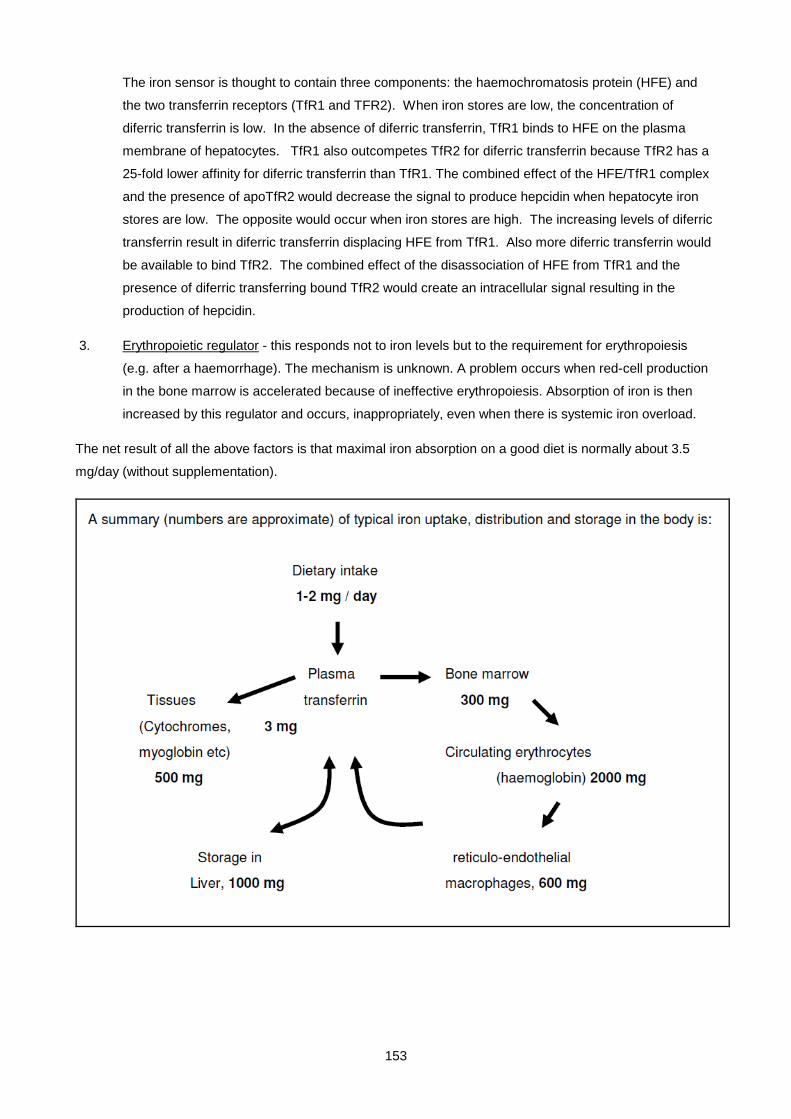
153
The iron sensor is thought to contain three components: the haemochromatosis protein (HFE) and
the two transferrin receptors (TfR1 and TFR2). When iron stores are low, the concentration of
diferric transferrin is low. In the absence of diferric transferrin, TfR1 binds to HFE on the plasma
membrane of hepatocytes. TfR1 also outcompetes TfR2 for diferric transferrin because TfR2 has a
25-fold lower affinity for diferric transferrin than TfR1. The combined effect of the HFE/TfR1 complex
and the presence of apoTfR2 would decrease the signal to produce hepcidin when hepatocyte iron
stores are low. The opposite would occur when iron stores are high. The increasing levels of diferric
transferrin result in diferric transferrin displacing HFE from TfR1. Also more diferric transferrin would
be available to bind TfR2. The combined effect of the disassociation of HFE from TfR1 and the
presence of diferric transferring bound TfR2 would create an intracellular signal resulting in the
production of hepcidin.
3. Erythropoietic regulator - this responds not to iron levels but to the requirement for erythropoiesis
(e.g. after a haemorrhage). The mechanism is unknown. A problem occurs when red-cell production
in the bone marrow is accelerated because of ineffective erythropoiesis. Absorption of iron is then
increased by this regulator and occurs, inappropriately, even when there is systemic iron overload.
The net result of all the above factors is that maximal iron absorption on a good diet is normally about 3.5
mg/day (without supplementation).

154
CLINICAL TESTS OF IRON STATUS
Routine tests:
1. plasma iron (least informative test)
2. Transferrin / Total Iron Binding capacity (TIBC)
3. % saturation
4. soluble transferrin receptor
5. plasma ferritin
PLASMA IRON
Normal range: 9 - 30 µmol/L (50 - 170 ug/dL) but subject to diurnal variation (highest at night), also there can
be as much as 20% change over even a few minutes, and 100% from one day to the next.
More than 95% of plasma iron is bound tightly to transferrin. The remaining 5% comprises a mixture of other
bound forms. The free iron concentration is extremely low (<1 pmol/L) - this is necessary to prevent toxic free
radical generation.
TRANSFERRIN
Transferrin is measured clinically both as a negative marker of iron stores and to calculate % saturation.
Each transferrin molecule of 80kDa is able to bind 2 molecules of iron and the normal range for transferring is
2 – 3.6g/L or 25 – 45umoll. Total iron-binding capacity is an indirect method of assessing plasma
transferrin levels (normal 45-70 µmol/l) and indicates the amount of iron that can be bound by available
transferring assuming a ratio of two iron atoms to each transferring molecule.
% SATURATION is another way of expressing the ratio or plasma iron to transferrin
% saturation is given by:
plasma iron (µmol/l)
% saturation = ______________________________
2 X transferrin (µmol/l) (normal range 20 - 55%)
Iron uptake into tissue cells is by receptor-mediated endocytosis. The rate of iron uptake is determined by
the number of transferrin receptors on the cell surface: cells requiring much iron (e.g. normoblasts) have
many receptors. Iron is very tightly bound (Kd = 1019 M-1) to transferrin at neutral pH, but is released in the
acidic environment of the endosome. Uptake of iron by cells is critically dependent on % saturation. When
the % saturation falls below 16%, iron delivery fails to meet requirement for erythropoiesis, leading to iron
deficiency anaemia. Thus the % saturation is a better index of iron deficiency than plasma iron. Example:
the 2 patients below both have low plasma iron, yet the first one does not develop anaemia, while the second
does:

155
plasma iron Transferrin % saturation clinical condition
6µM 9µM 33% patient with nephrotic syndrome; not iron deficient
6µM 30µM 10% iron deficiency
SOLUBLE TRANSFERRIN RECEPTOR
The transferrin receptor (TfR) is the major mediator of iron uptake by cells. The TfR is a transmembrane,
disulfide-linked dimer of two identical subunits that bind and internalize diferric transferrin, thereby delivering
iron to the cell cytosol. When a cell needs iron, TfR expression is increased to facilitate iron uptake. Since the
major use of iron is for haemoglobin synthesis, about 80% of total TfR is on erythroid progenitor cells. Soluble
transferrin receptor arises from proteolysis of the intact protein on the cell surface, leading to monomers that
can be measured in plasma and serum. Thus, the concentration of sTfR in plasma or serum is an indirect
measure of total TfR. The serum level of sTfR reflects either the cellular need for iron or the rate of
erythropoiesis. The concentration of sTfR in plasma or serum is elevated in iron deficiency but is not
appreciably affected by chronic disease. sTfR is elevated in subjects with hyperplastic erythropoiesis (eg,
hemolytic anemia, beta-thalassemia, polycythemia, etc) and depressed in subjects with hypoplastic
erythropoiesis (eg, chronic renal failure, aplastic anemia, or post-transplant anemia).
The result of the sTfR is often interpreted together with ferritin and the reticulocyte haemoglobin content
(CHr) as shown in the below diagram.
INTERPRETATION OF RESULTS
DECREASED plasma iron is found in
• Iron deficiency (<6 µM)
• Inflammatory states (e.g. bacterial infections, rheumatoid arthritis, malignancy); due to a block in the
release of iron by the RES initiated by high hepcidin levels.
• Ascorbic acid deficiency (scurvy). Also caused by a block in the release of iron by the RES.

156
• Nephrotic syndrome and malnutrition: low transferrin levels due to loss of transferrin in urine, or
failure of synthesis. The % saturation is normal.
INCREASED plasma iron (more reliable than decreased levels) is found in
• iron overload
• liver damage (e.g. hepatitis) where iron is released by damaged hepatocytes.
DECREASED transferrin may be due to ↓ synthesis in
• malnutrition, liver disease
• iron overload (↑iron inhibits transferrin synthesis)
• chronic inflammatory states (e.g. infections, rheumatoid arthritis, SLE, malignancy)
• or ↑ loss in nephrotic syndrome or protein-losing enteropathies
INCREASED transferrin is found in
• iron deficiency (stimulated by ↓ iron)
• High oestrogen states
o pregnancy (independent of iron deficiency)
o oestrogen-containing oral contraceptives
STORAGE IRON
Function of iron stores: to allow mobilisation of large amounts of iron for maximal erythropoiesis in case of
blood loss. There are two forms of stored iron:
1. ferritin
2. haemosiderin
There are two sites of storage:
(a) RES (mainly as haemosiderin)
(b) hepatocytes (called "parenchymal" storage) (mainly as ferritin)
Normal iron stores would be typically about a gram or two,
in iron deficiency: zero stores,
in iron overload: ± 5-50 g
ASSESSMENT OF IRON STORES
1. Bone marrow or liver biopsy, with staining for iron, is the definitive method, but invasive.

157
2. Plasma ferritin level correlates with iron stores. Normal range 15 - 200 µg/L. Ferritin synthesis is
regulated by cytoplasmic iron-regulatory proteins (IRPs) which bind iron and stimulate ferritin
synthesis when iron level is high.
3. Chelator-induced iron excretion: give desferrioxamine and measure urinary iron excretion over
next 6-24 hours. Iron excreted is proportional to iron stores.
However, as a complication to interpretation, high plasma ferritin levels are also found in:
1. chronic inflammatory states (infections, autoimmune diseases, neoplasms) because ferritin synthesis
is stimulated by cytokines
2. liver disease (acute or chronic): ferritin released from hepatocytes
3. leukaemias and other neoplasms (ferritin sometimes produced by tumour cells)
IRON DEFICIENCY
High risk groups:
1. Infancy: Rapid growth imposes high iron requirements. Iron stores are mobilised during this period.
Low birth weight infants are at highest risk because of their smaller stores. Breast -feeding provides
adequate iron. Infant formulas should be fortified to 4-12 mg of iron per litre.
2. Females during reproductive years: Additional blood, and therefore iron, loss is caused by
o Menstruation. Many women are close to iron deficiency much of the time. Menorrhagia due
to fibroids or IUCD’s increases the loss.
o Pregnancy. (equivalent to 1500 ml blood loss). Iron deficiency is common in pregnancy,
since the placenta transfers iron to the foetus at the mother’s expense, even if she is iron
deficient. Iron tablets should therefore be given routinely to all pregnant women.
In non high-risk patients, iron deficiency should always lead to a search for
• Occult blood loss, common causes of which are:
1. GIT blood loss - aspirin and related drugs
- peptic ulcer disease
- tumours
- hookworm infestation (450 million people worldwide)
2. other blood loss - urinary tract
- blood donors
• Malabsorption due to:
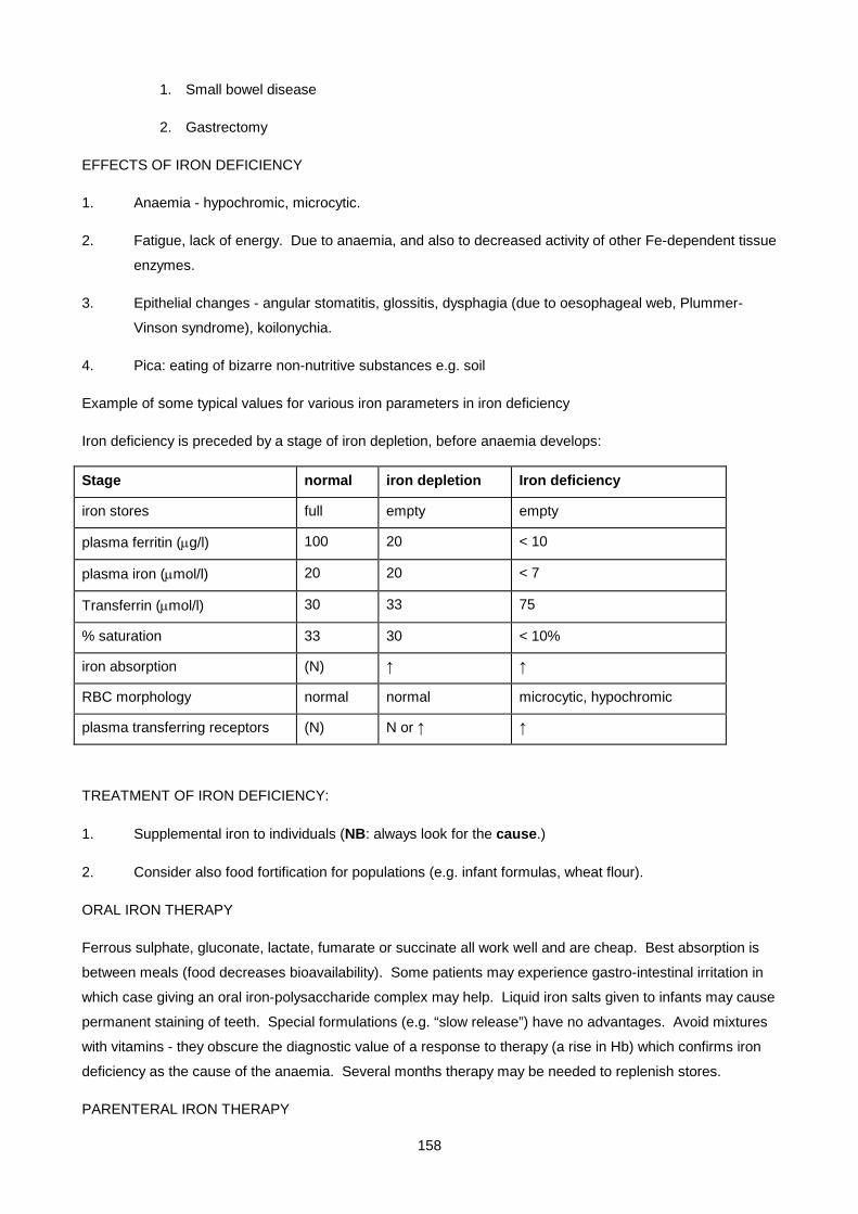
158
1. Small bowel disease
2. Gastrectomy
EFFECTS OF IRON DEFICIENCY
1. Anaemia - hypochromic, microcytic.
2. Fatigue, lack of energy. Due to anaemia, and also to decreased activity of other Fe-dependent tissue
enzymes.
3. Epithelial changes - angular stomatitis, glossitis, dysphagia (due to oesophageal web, Plummer-
Vinson syndrome), koilonychia.
4. Pica: eating of bizarre non-nutritive substances e.g. soil
Example of some typical values for various iron parameters in iron deficiency
Iron deficiency is preceded by a stage of iron depletion, before anaemia develops:
Stage normal iron depletion Iron deficiency
iron stores full empty empty
plasma ferritin (µg/l) 100 20 < 10
plasma iron (µmol/l) 20 20 < 7
Transferrin (µmol/l) 30 33 75
% saturation 33 30 < 10%
iron absorption (N) ↑ ↑
RBC morphology normal normal microcytic, hypochromic
plasma transferring receptors (N) N or ↑ ↑
TREATMENT OF IRON DEFICIENCY:
1. Supplemental iron to individuals (NB: always look for the cause.)
2. Consider also food fortification for populations (e.g. infant formulas, wheat flour).
ORAL IRON THERAPY
Ferrous sulphate, gluconate, lactate, fumarate or succinate all work well and are cheap. Best absorption is
between meals (food decreases bioavailability). Some patients may experience gastro-intestinal irritation in
which case giving an oral iron-polysaccharide complex may help. Liquid iron salts given to infants may cause
permanent staining of teeth. Special formulations (e.g. “slow release”) have no advantages. Avoid mixtures
with vitamins - they obscure the diagnostic value of a response to therapy (a rise in Hb) which confirms iron
deficiency as the cause of the anaemia. Several months therapy may be needed to replenish stores.
PARENTERAL IRON THERAPY
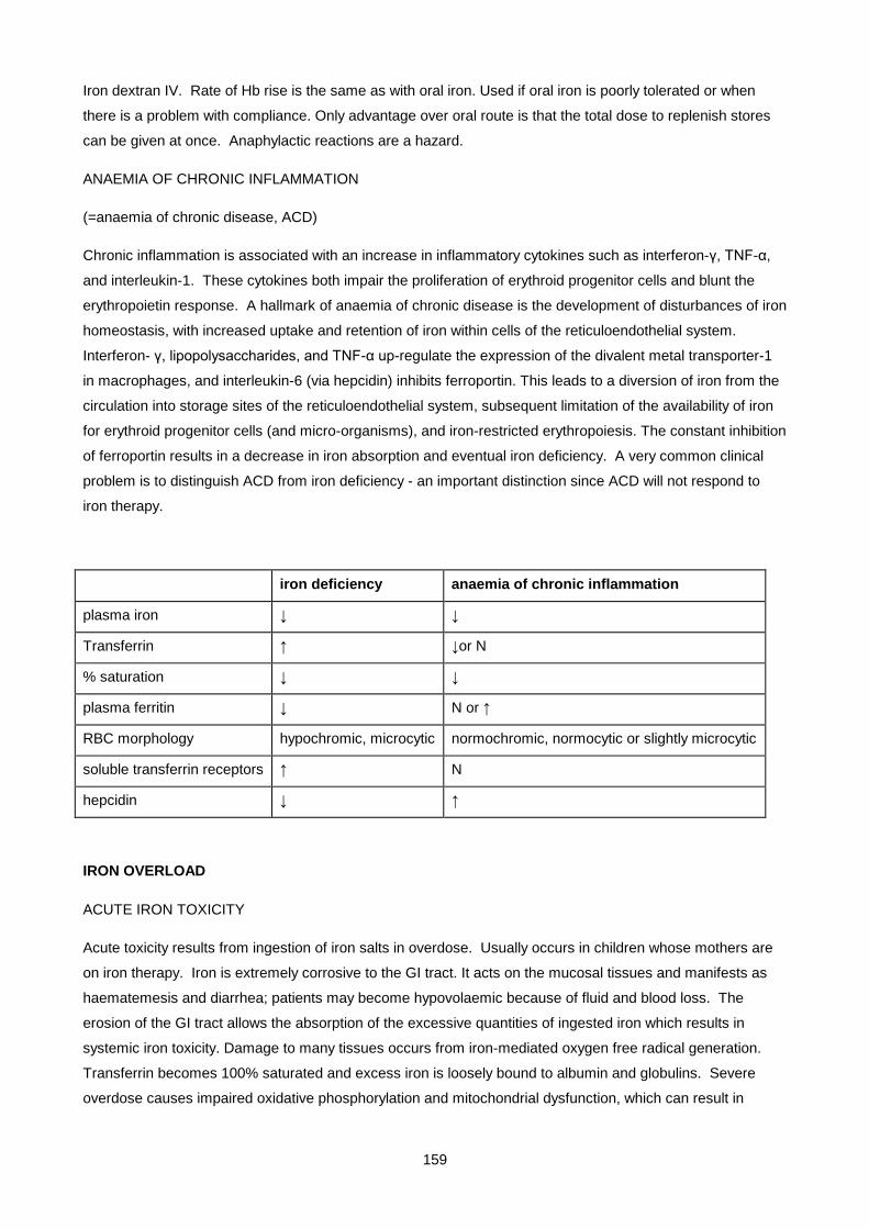
159
Iron dextran IV. Rate of Hb rise is the same as with oral iron. Used if oral iron is poorly tolerated or when
there is a problem with compliance. Only advantage over oral route is that the total dose to replenish stores
can be given at once. Anaphylactic reactions are a hazard.
ANAEMIA OF CHRONIC INFLAMMATION
(=anaemia of chronic disease, ACD)
Chronic inflammation is associated with an increase in inflammatory cytokines such as interferon-γ, TNF-α,
and interleukin-1. These cytokines both impair the proliferation of erythroid progenitor cells and blunt the
erythropoietin response. A hallmark of anaemia of chronic disease is the development of disturbances of iron
homeostasis, with increased uptake and retention of iron within cells of the reticuloendothelial system.
Interferon- γ, lipopolysaccharides, and TNF-α up-regulate the expression of the divalent metal transporter-1
in macrophages, and interleukin-6 (via hepcidin) inhibits ferroportin. This leads to a diversion of iron from the
circulation into storage sites of the reticuloendothelial system, subsequent limitation of the availability of iron
for erythroid progenitor cells (and micro-organisms), and iron-restricted erythropoiesis. The constant inhibition
of ferroportin results in a decrease in iron absorption and eventual iron deficiency. A very common clinical
problem is to distinguish ACD from iron deficiency - an important distinction since ACD will not respond to
iron therapy.
iron deficiency anaemia of chronic inflammation
plasma iron ↓ ↓
Transferrin ↑ ↓or N
% saturation ↓ ↓
plasma ferritin ↓ N or ↑
RBC morphology hypochromic, microcytic normochromic, normocytic or slightly microcytic
soluble transferrin receptors ↑ N
hepcidin ↓ ↑
IRON OVERLOAD
ACUTE IRON TOXICITY
Acute toxicity results from ingestion of iron salts in overdose. Usually occurs in children whose mothers are
on iron therapy. Iron is extremely corrosive to the GI tract. It acts on the mucosal tissues and manifests as
haematemesis and diarrhea; patients may become hypovolaemic because of fluid and blood loss. The
erosion of the GI tract allows the absorption of the excessive quantities of ingested iron which results in
systemic iron toxicity. Damage to many tissues occurs from iron-mediated oxygen free radical generation.
Transferrin becomes 100% saturated and excess iron is loosely bound to albumin and globulins. Severe
overdose causes impaired oxidative phosphorylation and mitochondrial dysfunction, which can result in
160
cellular death. The liver is one of the organs most affected by iron toxicity, but other organs such as the heart,
kidneys, lungs, and the haematologic systems also may be impaired.
FEATURES OF ACUTE IRON POISONING:
1) Damage to GIT mucosa: vomiting, diarrhoea ± blood
2) Shock due to vasodilatation and capillary damage
3) Drowsiness progressing to coma
4) Lactic acidosis - due to interference with mitochondrial respiration by iron
5) Liver damage
6) Myocardial toxicity - circulatory failure
Biochemical findings
Serum iron and % saturation can be markedly elevated. Transferrin and ferritin are usually normal. A
metabolic acidosis and hyperglycaemia are often present. A plasma iron level of more than 90µmol/l
indicates severe poisoning and complications are likely.
TREATMENT
As in all cases of overdose, the first step is to ensure the patency of the airway and that breathing and
circulation is adequate. IV fluid is often required to treat hypovolaemia. Since activated charcoal does not
bind iron, various methods of gastric decontamination may be tried (emesis, gastric lavage, whole bowel
irrigation). Chelation therapy with deferoxamine* if hypovolaemia, shock, lethargy, persistent vomiting,
diarrhea, positive anion gap metabolic acidosis, large number of pills on abdominal radiograph, or a serum
iron level greater than 90 µmol/l is present.
* Desferrioxamine is a water soluble organic compound with a high affinity for iron, binding 8.5 mg of iron for
every 100 mg desferrioxamine. It is colourless in solution but its complex with ferric iron (ferrioxamine) has a
pink colour and is freely excreted in the urine, giving it the colour of rosé wine. This colour change can be
used as a diagnostic test for iron overload.
Terminology: "iron overload" refers to chronic overload, not acute poisoning.
Haemosiderosis (or just 'siderosis'): histopathological picture in iron overload. Tissue damage only late.
Haemochromatosis = iron overload with tissue damage
HEREDITARY HAEMOCHROMATOSIS (Idiopathic haemochromatosis) (“Bronze diabetes”)
A genetic defect causing an inappropriate increase in iron absorption in the face of increased iron stores.
Clinical expression is highly variable: depends on the amount of iron in the diet and is potentiated by alcohol.
A large amount of stored iron (15-40 g) may accumulate over many years before symptoms manifest.
Commoner in males (10:1 :: m:f); females are protected by menstruation and pregnancy.
161
CLINICAL FEATURES
• cirrhosis: ↑ risk of hepatocellular carcinoma
• pigmentation (↑ melanin as well as hemosiderin)
• cardiac failure (cardiomyopathy)
• diabetes mellitus ("bronze diabetes")
• hypogonadism (due to pituitary dysfunction)
• arthritis
• susceptibility to infections
The genes involved in haemochromatosis have been discovered as shown in the table below.
Haemochromatosis
OMIM classification Aetiology Inheritence
Haemochromatosis Type 1 (Adult)
HFE gene mutations Autosomal recessive
Haemochromatosis Type 2A (Juvenile)
HJV gene mutations Autosomal recessive
Haemochromatosis Type 2B (Juvenile)
Hepcidin gene mutations Autosomal recessive
Haemochromatosis Type 3 Transferrin receptor 2 inactivation Autosomal recessive
Haemochromatosis Type 4 Ferroportin gene mutations Autosomal dominant
• HAEMOCHROMATOSIS TYPE I is by far the most common type of haemochromatosis. The
abnormal gene encodes a membrane protein called HFE which binds with TfR1. Mutations in HFE
prevent the intracellular signal that produces hepcidin. Haemochromatosis I is caused most
commonly by the C282Y mutation of HFE which occurs with high gene frequency in white
populations, having originated in a Celtic ancestor. It may have conferred an advantage during times
when iron nutrition was poor. This mutation is absent or very rare in African populations. Another
common mutation is the H63D mutation. Genetic screening is offered for both these mutations but
penetrance of the disease is only 80%.
• JUVENILE (TYPE II) HAEMOCHROMATOSIS presents in the teen years and is more likely to
present with endocrine and cardiac abnormalities than liver cirrhosis. It is caused either by a
defective hepcidin or by a mutation in a gene coding for haemojuvelin. Muscle normally sheds
haemojuvelin in low iron states which interacts with a receptor on hepatocytes and down regulates
hepcidin production
162
• HAEMOCHROMATOSIS TYPE III is extremely rare and is caused by a mutation in TfR2 which
disrupts the intracellular pathway which normally results in hepcidin production.
• HAEMOCHROMATOSIS TYPE IV is the only autosomal dominant haemochromatosis. It caused by
a mutated ferroportin which is not inactivated by hepcidin. It is also the only haemochromatosis
where high hepcidin levels are found.
IRON PARAMETERS
• plasma iron ↑
• TIBC ↓
• % saturation >55%
• plasma ferritin ↑↑ (700-8000 μg/L)
Site of iron storage is parenchymal (i.e. in hepatocytes). Definitive diagnosis by liver biopsy or DNA analysis.
TREATMENT:
• Repeated venesections (therapeutic phlebotomy). eg. weekly bleeding (500 ml) for 2 years will remove
20 g iron. Hepatic and cardiac function may improve. However, diabetes, hypogonadism, and arthritis
remain unchanged. The blood taken by phlebotomy can be donated.
• Screen other family members (% sat., serum ferritin, or DNA analysis) as complications are preventable.
AFRICAN IRON OVERLOAD
Caused by excessive dietary intake of iron – traditional beer brewed in ungalvanized steel drums. But also
has a genetic component – probably related to the ferroportin gene. Iron deposition in Kupfer cells and RES
(like in transfusional siderosis) suggests a defect in erythroid iron recycling. Tissue damage is less
pronounced than in idiopathic hemochromatosis but cirrhosis and hepatic carcinoma can occur, and
occasionally diabetes and cardiomyopathy. Ferritin is always elevated but % saturation does not always
reflect the true extent of overload.
Plasma ferritin ↑↑. Plasma iron ↑ and % saturation .
Treatment: venesection
TRANSFUSIONAL SIDEROSIS
Repeated blood transfusions for intractable anaemias causes iron overload as there is no pathway for
excretion of the excess iron. Seen in thalassaemias (globin gene defects), sideroblastic anaemias (defects in
haem synthesis) and marrow failure. Reticuloendothelial macrophages become iron loaded first, and later
the other typical tissues, cardiomyopathy being an especial problem
163
Treatment: Intensive chelator therapy with desferrioxamine, administered by infusion. (Venesection cannot
be used, as the primary problem is anaemia.)
PORPHYRIA CUTANEA TARDA
A disorder of porphyrin metabolism caused by a combination of factors, one of which is iron overload. The
ALA synthase gene is up-regulated by excess iron, leading to increased uroporphyrin excretion in urine, and
skin lesions (vesicles, ulcers, hyperpigmentation) due to excess porphyrins which cause photosensitivity.
Unlike other porphyrias, PCT is usually sporadic (acquired), and acute porphyric attacks do not occur.
Strongly associated with (1) excessive alcohol intake, and (2) mild to moderate iron overload. Probable
pathogenesis : genetic predisposition + alcohol + iron overload → PCT
Treatment : responds to venesection.
See also the porphyria lecture notes.
COPPER METABOLISM
Copper is an essential cofactor for several redox enzymes, where it functions as an electron acceptor/donor
(Cu+ ↔ Cu++ + e-). Approximately 5mg of copper is consumed a day. The copper is absorbed through the
small intestine where it is complexed with albumin and amino acids (mostly histidine). The copper is then
transported through the portal system to the liver where it is taken up by the hepatocytes. Within the
hepatocyte, copper can either be stored as metallothionein (the ‘ferritin’ of copper) or be incorporated into
copper containing enzymes. Any excess copper not used is excreted into the bile and released in the faeces.
Examples of copper containing enzymes
Enzyme Function Deficiency
Ceruloplasmin Ferroxidase Diabetes, retinal and basal ganglia degeneration
Copper/Zinc superoxide dismutase
Disproportion of superoxide anions
Sensitivity to oxygen. Neuronal degeneration.
Cytochrome c oxidase Electron transport in respiratory chain
Mitochondrial myopathies. Encephalomyelopathy
Dopamine β-hydroxylase Converts dopamine to noradrenalin
Hypotension, hypoglycaemia, hypothermia, in utero demise.
Peptidylglycine α–amidating monoxygenase
Peptide amidation Lethality in larval stage
Tyrosinase Converts tyrosine to melanin Absence of pigment in hair, skin, eyes, nystagmus, photopobia
Lysyl Oxidase Oxidative deamination of lysines in collagen and elastin
Laxity of skin and joints, wormian bones, hydroureter

164
CAERULOPLASMIN
Caeruloplasmin is an α2-gylcoprotein which contains 95% of plasma copper. It is a ferroxidase involved in
iron metabolism and does not exchange copper with that in the serum. It is a single polypeptide with six
atoms of copper. Caeruloplasmin occurs in a copper bound form (halo-caeruloplasmin) and a copper-free
(apo-caeruloplasmin). The copper content of the liver determines the ratio of apo to halo-caeruloplasmin.
The apo-form only has a half life of five hours compared to that of five and half days for halo-caeruloplasmin,
thus the caeruloplasmin level indicates the steady state sum of these two caeruloplasmin forms.
WILSON DISEASE
Wilson disease (also called "HEPATOLENTICULAR DEGENERATION") is characterized by copper overload
of the tissues. Mainly involved are:
- liver: cirrhosis, and sometimes a fulminant form of hepatitis
- brain: neurological signs, especially abnormal movements due to basal ganglia dysfunction, psychiatric
symptoms – personality changes, depression, schizophrenia
- cornea: copper deposition is visible as a brown Kayser-Fleischer ring
- kidney: renal tubular dysfunction.
Wilson’s disease shows autosomal recessive inheritance, and is caused by a defect in the Wilson ATPase
Cu2+ pump. This pump, which is exclusively found in the liver, pumps copper from the cytoplasm into the
endoplasmic reticulum so it can be incorporated into copper containing proteins (like caeruloplasmin) or be
targeted for excretion into the bile ducts. The main disturbances are therefore defective incorporation of
copper into caeruloplasmin, and decreased biliary excretion of copper with resultant copper-mediated
oxidative damage, activation of cell-death pathways, leakage of copper into the plasma, and eventual copper
overload in most tissues.
BIOCHEMICAL-FINDINGS:
- decreased total plasma copper, decreased plasma caeruloplasmin, increased urinary copper (reflecting
increased free copper in plasma). The gold standard for diagnosis is liver biopsy which shows increased
tissue copper.
TREATMENT:
Dietary restriction: Nuts, liver, chocolate, shellfish are all high in copper
Chelation: Penicillamine and triethylenetetramine dihydrochloride (trientine) forms complexes with Cu which
are then excreted in urine.
Zinc induces metallothionein within the enterocyte and thus causes intracellular chelation of copper and
decreased systemic absorption. Thiomolybdate forms non-toxic complexes with copper in the diet.
Liver transplant is curative and resolves neurologic and psychiatric symptoms.
165
MENKES DISEASE
Menkes Disease is an X-linked copper-related inherited disease causing copper deficiency. The disease
affects the kidneys, placenta, brain, gut and the vascular system.
The disease presents between one and three months of life with
• neurological
o mental retardation
o loss of developmental milestones
o seizures
o truncal hypotonia
• connective tissue (collagen) abnormalities
o soft bones and cartilage
o weakened artery walls
o abnormal sparse kinky hair and characteristic cherubic faces
The disease is caused by a mutation in the Menkes ATPase Cu2+ pump. This pump which is found in non-
hepatic tissues has the same function as the Wilson ATPase Cu2+ pump ie to transport copper from the
cytoplasm to the endoplasmic reticulum where it can be packed into copper requiring proteins or to be
package for copper export. When copper arrives at the intestine or placenta, copper is absorbed but can not
be exported out of the cells. Thus the placental and intestinal tissues become saturated in copper but the
rest of the body remains copper deficient.
FINDINGS
Low serum copper, low serum caeruloplasmin. Unfortunately these findings are not helpful until after early
infancy. The characteristic pili torti (twisted hairs) can be seen after a few months of life but this is not
specific for Menkes. The gold standard diagnosis is measurement of copper accumulation in cultured
fibroblasts.
TREATMENT: Using parenteral copper preparations (copper-histidine) to bypass the intestinal block,
normalises copper serum and caeruloplasmin. Seizures and irritability also improve but no improvement is
seen in neurological milestones.

166
SMALL GROUP TEACHING: LECTURE 8: IRON METABOLISM
QUESTIONS
1. A 63-year old woman complained of intermittent diarrhoea, weakness, weight loss and fainting attacks. On examination she appeared pale, there was no jaundice, clubbing or lymphadenopathy. The pulse rate was 104 with a normal BP. The cardiac apex was hyperkinetic, but there were no signs of cardiac failure.
FBC: Hb 7.2 g/dL, MCV - low, MCHC - low
Iron studies Plasma Fe 6 µM (9-30)
Plasma Transferrin 6g/L (2 – 3.6)
% saturation ........ (20-55%) work this out for yourselves
ferritin 8 ug/L (15-300)
i. What is the cause of the anemia, and what is the evidence for this conclusion?
ii. Are any further investigations required?
iii. What other signs or symptoms may be present in this disorder?
iv. What treatment would you advise? How long should it be continued?
v. What side effects may this treatment cause?
vi. What haematological response is expected?
2. A 46-year old man presented with weight loss, loss of appetite, and tiredness. On examination he was pale and wasted. The liver was felt 2 cm below the costal margin. The following laboratory results were obtained:
FBC: Hb 8.5, MCV low-normal, MCHC low-normal
Iron studies Plasma Fe 6 µM (9-30)
TIBC 50 µM (45-72)
% saturation ...........% (20-55) work this out yourselves
ferritin 360 ug/L (15-300)
Stool occult blood positive
ESR 35 mm/h (< 11)
i. Which two possible types of anaemia need to be considered in this patient?
ii. Weigh up the evidence for each of these diagnoses
iii. Are there any further tests which could help in distinguishing between these 2 types of anemia?
iv. What is the mechanism for the low plasma Fe in ACD?
3. A 45 year old diabetic man presented with a 2-week history of anorexia and pain in the right hip and left knee joints. Examination showed hyperpigmentation and enlarged tender liver. No jaundice,clubbing, lymphadenopthy or oedema. Diabetes mellitus was diagnosed 4 years previously. This had been initially controlled on oral hypoglycemic agents, but these had to be changed to insulin 6 months previously.
Na 142, K 4.2, Urea 4.6, Creat 105, glucose (non-fasting) 15.2 mmol/L, HbA1c 8.5% (N< 4.6), ALT 234 (<35), ALk. phos 187 (30-120), GGT 205 (<45),
i. What diagnosis fits with all these features ?
167
ii. Which cause of hyperpigmentation is excluded by the biochemical results?
Iron studies were requested, with the following results:
plasma Fe 36 µM (9-30)
TIBC 40 µM (45-70)
% saturation ......... (20-55%)
Ferritin >1500 ug/L (15-300)
iii. How can the diagnosis be confirmed definitively?
iv. What is the molecular defect in this disorder?
v. Comment on the mode of inheritance and demographic distribution of the abnormal gene.
vi. Outline the factors which affect the penetrance (expression) of the abnormal gene.
vii. What is the mechanism for the low TIBC ?
viii. What therapy would you advise , and how would you monitor the course of therapy?
4. An 18-year old male presented with pubertal delay. He had been diagnosed with Thalassaemia in childhood and had received regular blood transfusions for intractable anamia. On examination he had an unbroken voice, bronze coloured skin, and prepubertal genitalia (small penis, absence of pubic hair). Both testes were prepubertal in size. Iron studies and a plasma testosterone level were performed.
i. What values would you expect to find in the iron studies?
ii. What is thalassaemia? How does it lead to iron overload?
iii. Why was puberty delayed, and what testosterone result might you expect?
iv. How does the treatment of the iron overload differ from the previous case?
v. What other aspects of investigation and treatment need to be considered in this patient?
5. Copper
i. What is the basic in Wilson's disease?
ii. How does it present clinically?
iii. What biochemical abnormalities of copper metabolism are found?
iv. What drugs may be used in treatment and how do they work?
v. What inherited disease results in copper deficiency, what is its mode of inheritance, and how may it be treated?
6. True or False?
i. Spinach is a good source of iron.
ii. Iron tablets, like vitamins, are relatively harmless in excess.
iii. Venesection therapy may be helpful in porphyria cutanea tarda.
SMALL GROUP TEACHING: LECTURE 8: IRON METABOLISM
ANSWERS
168
1. Transferrin MW = 80 000 g/mole which equals 0.08g / umol. Therefore a plasma transferring of 6g/l = 6/0.08 = 75 umol/l transferrin. Given that each transferrin is able to bind 2 iron molecules – 6/(2X 75) = 4% transferrin saturation.
i. Iron deficiency. Evidence: hypochromic microcytic anemia, low plasma iron, high transferrin, very low % saturation, low ferritin (all the classical findings).
ii. Yes - establish the cause of iron deficiency: Simple dietary deficiency or malabsorption is possible (take detailed history), but blood loss must be excluded ( stool occult blood test and urine dipstick for blood) . Plasma B12 and folate levels should also be done ideally.
iii. Koilonychia, dysphagia (Plummer-Vinson syndrome), pica.
iv. Oral Ferrous sulphate tablets (200 mg TDS) (or other ferrous salts) . Continue for 3 months after normalization of Hb to replenish stores
v. Constipation, black stools, +ve occult blood test.
vi. Hb should rise by 1 g/dL per week, accompanied by ↑ reticulocyte count
2. Plasma Fe (6 µM)/TIBC (50 µM) X 100 = % saturation = 12% (normal is 20 -55%)
i. Anemia of Chronic disease (ACD) and Fe deficiency - distinguishing these is a common and difficult problem
ii. Weigh up the evidence for each of these diagnoses. In favour of ACD: normal TIBC, ↑ ferritin. The weight loss and ↑ ESR suggest a chronic illness. A GIT malignancy is suggested by the stool occult blood and enlarged liver. In favour of Fe deficiency: stool occult blood ?. Findings which are compatible with either ACD or Fe deficiency: FBC results, low plasma iron, low % saturation.Overall, the ↑ ferritin biases one towards ACD.
iii. Plasma transferrin receptor level: ↑ in iron deficiency, not in ACD.
iv. In chronic inflammation there is a block in release of iron from the RES (cytokine-mediated).
3.
i. The features of diabetes mellitus, pigmentation, liver disease and arthritis suggest hemochromatosis ("bronze diabetes").
ii. Addison's disease
iii. a) liver biopsy with staining for Fe and direct measurement of liver Fe b) DNA analysis.
iv. A mutation in the HFE protein. HFE functions as a regulator of Fe absorption
v. Autosomal recessive. Common in Caucasian populations.
vi. Expressed less frequently in females due to menstruation etc. Potentiated by alcohol.
vii. High intracellular hepatocyte iron inhibits transferrin synthesis (mediated by an iron-binding regulatory protein (IRP) which is a transcriptional regulator)
viii. Venesection therapy (e.g. 500 mL weekly). Monitor plasma Fe, TIBC and ferritin
4.
i. Same as in case 3.
ii. A defect (usually a deletion) in the globin gene leading to reduced production of (mostly) normal haemoglobin. Mechanisms of iron overload: a) multiple blood transfusions (no pathway exists for the excretion of excess iron). b) dietary Fe absorption is inappropriately increased in the "iron loading anemias" , due to the putative "erythroid regulator" of iron absorption
iii. Low testosterone due to hypopituitarism and/or testicular dysfunction
169
iv. Chelator therapy (Venesection not an option). Daily desferrioxamine given parenterally
v. Investigation of other pituitary functions (e.g. thyroid) - dont go into detail: it will be covered fully in Endocrinology tuts and lectures. Testosterone replacement therapy will be required.
5.
i. Defect in a copper pump
ii. Cirrhosis, neurological damage, Kayser-Fleischer rings, renal tubular dysfunction
iii. ↑ biliary excretion of copper, defective incorporation of copper into caeruloplasmin, ↓ total plasma copper (free copper ↑ ), ↓ plasma caeruloplasmin, ↑ urinary copper (reflecting ↑ free copper in plasma), ↑ tissue copper visible on liver biopsy
iv. Penicillamine - chelator for Copper, Zinc acetate - blocks intestinal absorption.
v. Menke's disease, X-linked, treat with subcutaneous copper-histidine.
6. True or False?
i. false : Popeye was a propaganda stunt to raise British morale during WW2
ii. false
iii. true
170
Lecture 9: Lipid And Lipoprotein Metabolism And Dyslipidaemias
Dr H. VREEDE & E.H.HARLEY 2004, UPDATED DECEMBER 2006 A/PROF H HENDERSON)
ABBREVIATIONS:
Chol Cholesterol
CE Cholesterol ester
TG / TAG Triglyceride / Triacylglycerol
PL Phospholipid
Chylo Chylomicrons
VLDL Very low density lipoproteins
LDL Low density lipoproteins
IDL Intermediate density lipoproteins
HDL High density lipoproteins
Apo LP Apolipoprotein
Apo A-I Apolipoprotein A-I(pronounced apolipoprotein "A" one)
Lp(a) Lipoprotein (a)(pronounced lipoprotein little "A")
LPL Lipoprotein lipase
LCAT Lecithin cholesterol acyl-transferase
LRP LDL receptor-related protein
CETP Cholesterol ester transfer protein
CAD / CHD / IHD Coronary artery disease / Coronary heart disease / Ischaemic heart disease
FH Familial hypercholesterolaemia
PDGF Platelet derived growth factor
IMPORTANCE OF LIPIDS
Lipids (cholesterol, triglyceride and phospholipids) are important physiologically for a number of reasons:
- cholesterol and phospholipids are vital structural components of cellular membranes
- cholesterol is the precursor of steroid hormones and bile acids
- triglyceride is an important storage and transport form of energy
- absorption of dietary lipid is essential for the absorption of fat-soluble vitamins.
Lipids are also important pathophysiologically:

171
• several rare inherited defects of lipid metabolism occur, causing significant morbidity and
mortality
• hypercholesterolaemia (and to a lesser extent hypertriglyceridaemia) is associated with an
increased risk of developing atherosclerosis, chiefly of the coronary arteries, which may
eventually compromise the circulation to the heart (coronary artery disease)
• hypertriglyceridaemia predisposes patients to pancreatitis
An understanding of the normal metabolism of lipids is essential to an understanding of abnormal
metabolism. These lectures will present:
- an overview of normal lipid metabolism
- the role of hyperlipidaemia in atherosclerosis
- an approach to investigating lipid abnormalities
- a review of disorders of lipid metabolism
- treatment of hyperlipidaemias
NORMAL LIPID METABOLISM
BIOCHEMISTRY OF LIPOPROTEINS
Cholesterol can be synthesised de novo from acetyl Co-A in the liver. The rate-limiting step is catalysed by
HMG-CoA reductase . This enzyme is regulated by intracellular cholesterol levels by means of feedback
inhibition.
Triglyceride is derived from dietary fatty acids (exogenous source) and from the liver (endogenous source)
which utilises fatty acids synthesised de novo and from fatty acids derived from adipose tissue. In both
cases, TGs are transported to target tissues in plasma.
The lipoprotein system evolved to solve the problem of transporting lipids in the aqueous environment of the
plasma. Lipids are generally hydrophobic and therefore insoluble in water. Only the charged phosphate
group of phospholipids (PL) and the hydroxyl group of free cholesterol (CHOL) are hydrophilic. Lipids are
therefore transported in plasma as lipoproteins, complex spherical structures that have a hydrophobic core
wrapped in a hydrophilic coating. The core contains triglyceride and cholesterol esters, while the surface
contains phospholipids, free cholesterol and proteins (the apolipoproteins).
Iceberg-sea model of plasma lipoprotein. The polar head groups are exposed to the aqueous environment,
and the neutral triglycerides and cholesterol esters are localised primarily in the core of the particle.
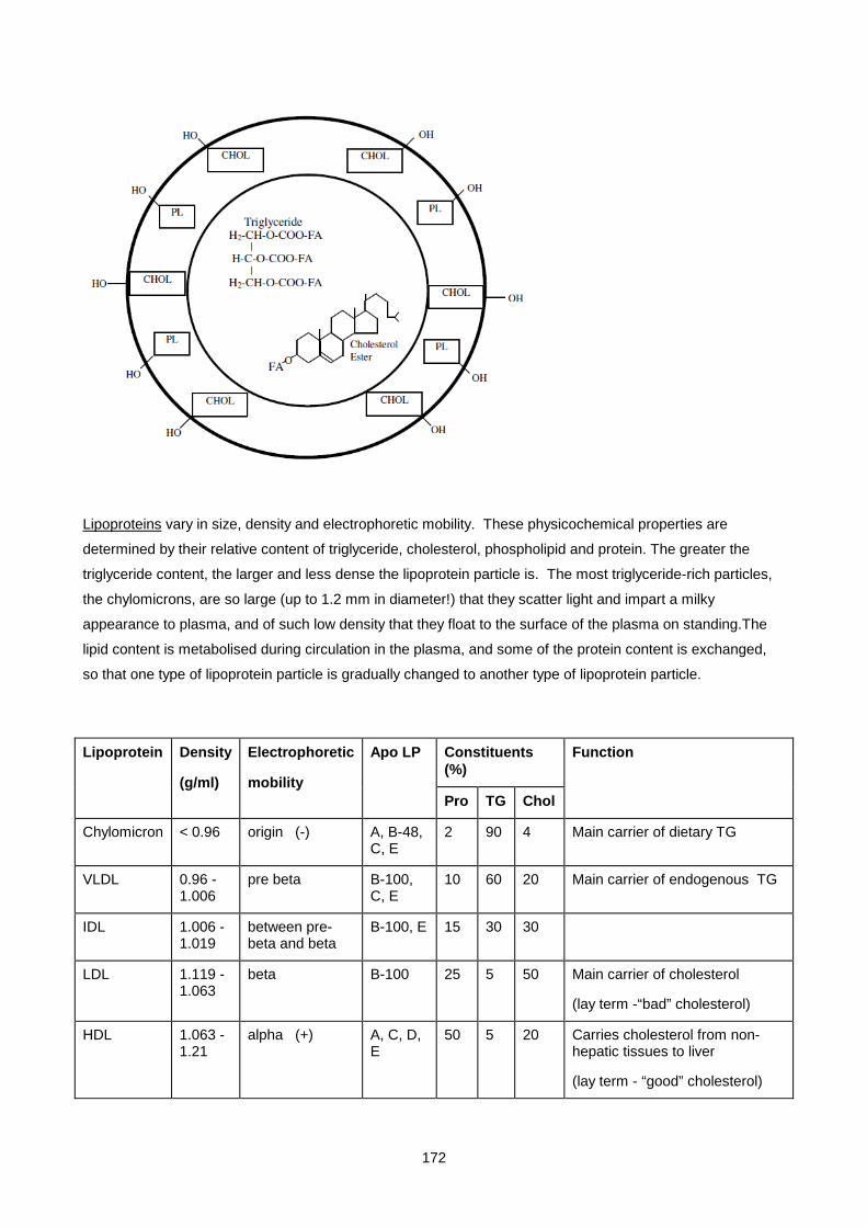
172
Lipoproteins vary in size, density and electrophoretic mobility. These physicochemical properties are
determined by their relative content of triglyceride, cholesterol, phospholipid and protein. The greater the
triglyceride content, the larger and less dense the lipoprotein particle is. The most triglyceride-rich particles,
the chylomicrons, are so large (up to 1.2 mm in diameter!) that they scatter light and impart a milky
appearance to plasma, and of such low density that they float to the surface of the plasma on standing.The
lipid content is metabolised during circulation in the plasma, and some of the protein content is exchanged,
so that one type of lipoprotein particle is gradually changed to another type of lipoprotein particle.
Lipoprotein Density
(g/ml)
Electrophoretic
mobility
Apo LP Constituents (%)
Function
Pro TG Chol
Chylomicron < 0.96 origin (-) A, B-48, C, E
2 90 4 Main carrier of dietary TG
VLDL 0.96 - 1.006
pre beta B-100, C, E
10 60 20 Main carrier of endogenous TG
IDL 1.006 - 1.019
between pre-beta and beta
B-100, E 15 30 30
LDL 1.119 - 1.063
beta B-100 25 5 50 Main carrier of cholesterol
(lay term -“bad” cholesterol)
HDL 1.063 - 1.21
alpha (+) A, C, D, E
50 5 20 Carries cholesterol from non-hepatic tissues to liver
(lay term - “good” cholesterol)

173
Apolipoproteins are the proteins which are found in association with plasma lipids. (Together, apolipoproteins
and lipids form the plasma lipoproteins). Some of the proteins are structural and are necessary for the
synthesis and release of the lipoproteins, while others perform functions as co-factors for various lipid-
metabolising enzymes or as receptor-binding proteins. Apolipoproteins vary in the extent to which they are
anchored to the lipoprotein. Most apolipoproteins can transfer from one lipoprotein to another, except
apolipoprotein B which remains with the lipoprotein in which it was initially assembled and secreted into the
plasma.
Apolipoprotein Site of synthesis
Found in lipoproteins
Functions
(a) liver LP(a) Remains unclear
A-I GIT, liver Chylo, HDL Structural protein of HDL
Activator of LCAT
A-II GIT, liver HDL Structural protein of HDL
B-48 GIT Chylo Structural protein of chylomicrons
B-100 Liver VLDL, IDL, LDL Structural protein of VLDL, IDL, LDL
Binds to B/E (LDL) receptor
C-I Liver Chylo, VLDL, HDL Activator of LCAT
Modulates the clearance of VLDL from plasma
C-II Liver Chylo, VLDL, HDL Activator of LPL
C-III Liver Chylo, VLDL, HDL Inhibitor of LPL
Inhibits binding of chylo and VLDL to B/E receptor
D Liver, CNS HDL Carrier of small hydrophobic molecules eg bilirubin, arachidonic acid
E Liver, macrophages
Chylo, VLDL, IDL, HDL
Binds to E (remnant) receptor
Binds to B/E (LDL) receptor
LIPID TRANSPORT AND METABOLISM IN PLASMA
The main function of the plasma lipoproteins is the transport of lipids from their site of synthesis to their sites
of storage or utilisation. During circulation in the plasma a number of processes take place which change the
composition, shape and size of the originally secreted lipoproteins and result in the formation of new
lipoproteins. These processes include:
• transfer of apolipoproteins between lipoproteins
• transfer of lipids between lipoproteins eg cholesterol ester transfer protein (CETP)
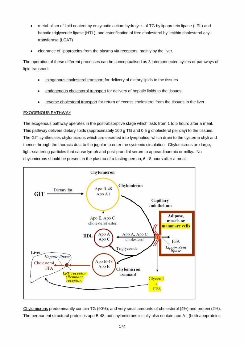
174
• metabolism of lipid content by enzymatic action: hydrolysis of TG by lipoprotein lipase (LPL) and
hepatic triglyceride lipase (HTL), and esterification of free cholesterol by lecithin cholesterol acyl-
transferase (LCAT)
• clearance of lipoproteins from the plasma via receptors, mainly by the liver.
The operation of these different processes can be conceptualised as 3 interconnected cycles or pathways of
lipid transport:
• exogenous cholesterol transport for delivery of dietary lipids to the tissues
• endogenous cholesterol transport for delivery of hepatic lipids to the tissues
• reverse cholesterol transport for return of excess cholesterol from the tissues to the liver.
EXOGENOUS PATHWAY
The exogenous pathway operates in the post-absorptive stage which lasts from 1 to 5 hours after a meal.
This pathway delivers dietary lipids (approximately 100 g TG and 0.5 g cholesterol per day) to the tissues.
The GIT synthesises chylomicrons which are secreted into lymphatics, which drain to the cysterna chyli and
thence through the thoracic duct to the jugular to enter the systemic circulation. Chylomicrons are large,
light-scattering particles that cause lymph and post-prandial serum to appear lipaemic or milky. No
chylomicrons should be present in the plasma of a fasting person, 6 - 8 hours after a meal.
Chylomicrons predominantly contain TG (90%), and very small amounts of cholesterol (4%) and protein (2%).
The permanent structural protein is apo B-48, but chylomicrons initially also contain apo A-I (both apoproteins
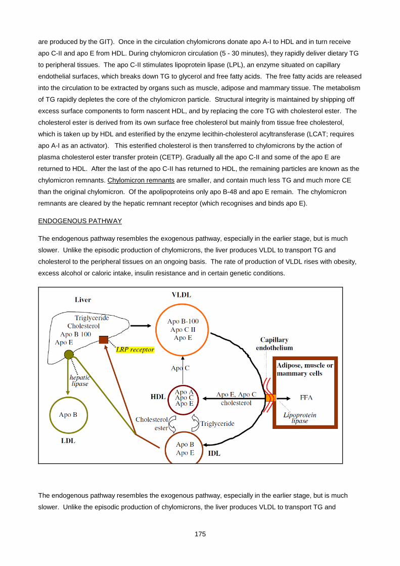
175
are produced by the GIT). Once in the circulation chylomicrons donate apo A-I to HDL and in turn receive
apo C-II and apo E from HDL. During chylomicron circulation (5 - 30 minutes), they rapidly deliver dietary TG
to peripheral tissues. The apo C-II stimulates lipoprotein lipase (LPL), an enzyme situated on capillary
endothelial surfaces, which breaks down TG to glycerol and free fatty acids. The free fatty acids are released
into the circulation to be extracted by organs such as muscle, adipose and mammary tissue. The metabolism
of TG rapidly depletes the core of the chylomicron particle. Structural integrity is maintained by shipping off
excess surface components to form nascent HDL, and by replacing the core TG with cholesterol ester. The
cholesterol ester is derived from its own surface free cholesterol but mainly from tissue free cholesterol,
which is taken up by HDL and esterified by the enzyme lecithin-cholesterol acyltransferase (LCAT; requires
apo A-I as an activator). This esterified cholesterol is then transferred to chylomicrons by the action of
plasma cholesterol ester transfer protein (CETP). Gradually all the apo C-II and some of the apo E are
returned to HDL. After the last of the apo C-II has returned to HDL, the remaining particles are known as the
chylomicron remnants. Chylomicron remnants are smaller, and contain much less TG and much more CE
than the original chylomicron. Of the apolipoproteins only apo B-48 and apo E remain. The chylomicron
remnants are cleared by the hepatic remnant receptor (which recognises and binds apo E).
ENDOGENOUS PATHWAY
The endogenous pathway resembles the exogenous pathway, especially in the earlier stage, but is much
slower. Unlike the episodic production of chylomicrons, the liver produces VLDL to transport TG and
cholesterol to the peripheral tissues on an ongoing basis. The rate of production of VLDL rises with obesity,
excess alcohol or caloric intake, insulin resistance and in certain genetic conditions.
The endogenous pathway resembles the exogenous pathway, especially in the earlier stage, but is much
slower. Unlike the episodic production of chylomicrons, the liver produces VLDL to transport TG and

176
cholesterol to the peripheral tissues on an ongoing basis. The rate of production of VLDL rises with obesity,
excess alcohol or caloric intake, insulin resistance and in certain genetic conditions.
VLDL are large, although not as large as chylomicrons. They predominantly contain TG (60%), and smaller
amounts of cholesterol (20%) and protein (10%). The permanent structural protein is apo B-100, and initially
the VLDL also contain apo C-II and apo E. Interaction with HDL contributes more apo C-II and apo E. During
VLDL circulation (t1/2 12 hours), TG are metabolised by LPL and free fatty acids are delivered to peripheral
tissues. As for chylomicrons, the VLDL free cholesterol is taken up by HDL, esterified by LCAT, and returned
as cholesterol ester along with the cholesterol ester derived from tissue cholesterol taken up by HDL.
Gradually all of the apo C-II and some of the apo E are returned to HDL. After the last of the apo C-II has
returned to HDL, the remaining particles are known as IDL.
IDL are smaller than VLDL. They contain equal amounts of TG and cholesterol (30% TG, 30% cholesterol)
and a moderate amount of protein (15%). The permanent structural protein is apo B-100, and IDL also still
contain apo E. IDL are very rapidly cleared, and therefore IDL is undetectable in normal plasma. More than
half of the IDL is taken up by the LDL receptor, which recognises apo B-100 and apo E. The rest is
converted to LDL by the action of hepatic TG lipase (HTGL), located in the vascular endothelium of the liver,
which removes some of the TG. After the last of the apo E has returned to HDL, the remaining particles are
known as LDL.
LDL are even smaller than IDL. They contain predominantly cholesterol (50%) and protein (25%) and a very
small amount of TG (5%). The permanent structural protein is apo B-100 which is the only protein in LDL.
LDL have a long half-life of 3 days. They deliver cholesterol to peripheral tissues, and are eventually taken
up by the LDL receptor (at low LDL levels) or by the macrophage scavenger receptor (at high LDL levels).
REVERSE CHOLESTEROL TRANSPORT
Excess cholesterol in living and dead cells can be returned to the liver by the reverse cholesterol transport
pathway. Endogenous cholesterol synthesis can then be down-regulated by feedback inhibition. Only HDL
have been shown to take up cellular cholesterol.

177
HDL (nascent) are produced in the liver and GIT, and also originate from excess chylomicron surface
membranes, liberated during the metabolism of chylomicrons and VLDL. Newly-formed (nascent) HDL
consist of bilamellar phospholipid discs with no lipid core. During their circulation, nascent HDL acquire a
lipid core of variable composition, hence HDL exists in various subclasses.
In general, mature HDL predominantly contain protein (50%) and cholesterol (20%) with a small amount of
TG (5%). The permanent structural proteins are apo A-I and A-II which also act as cofactors. HDL also
contain apo D and apo C-II and E (which are transferred to and from chylomicrons and VLDL). Newly-formed
HDL accept free cholesterol from chylomicrons, VLDL and peripheral tissues forming small spherical
particles (HDL3). The free cholesterol is rapidly esterified by LCAT and is packed into a lipid core, forming
larger spherical particles (HDL2). Some of the HDL2 is cleared by the liver via the scavenger receptor class
BI (SR-BI) delivering CE directly to the liver. Most of the HDL2 is however cycled back to HDL3, by
transferring the CE back to the TG-rich chylomicron remnants and VLDL in exchange for TG via CETP.
These lipoproteins are eventually cleared by the liver LDL receptor, thereby delivering CE to the liver.
IMPORTANT FUNCTIONS OF HDL:
• promotes efflux and uptake of cholesterol from peripheral tissues, facilitates the conversion to
cholesterol ester and subsequent delivery to the liver, either directly or via other lipoproteins (reverse
cholesterol transport)
• facilitate lipolysis of chylomicron and VLDL TGs, by LPL, through the provision of apo CII (activator of
LPL)
• facilitate clearance of chylomicron remnants and IDL (by supplying apo E)
• inflammatory modulation by inhibition of LDL oxidation, removal of lipid peroxides through PON
(paraoxonase) activity, minimising membrane damage through provision of Clusterin (complement
lysis inhibitor), and platelet aggregation through PAFAH (platelet activating factor acetyl-hydrolase).
LIPOPROTEIN RECEPTORS
Various cell surface receptors mediate the removal of lipoproteins from the circulation.
• The VLDL receptor is a member of the LDL receptor family but does not bind LDL. It is a peripheral
lipoprotein receptor and is expressed in fatty acid active tissues such as heart, skeletal muscle and
adipose tissue. It binds VLDL via its Apo E and LPL components.
• The LDL receptor is located on the cell surfaces of all cells that require cholesterol. The bulk of
circulating LDL (75%) is taken up by the liver. The LDL receptor recognises and binds apo B-100 or
apo E. It therefore binds and internalises LDL, IDL and some HDL. The receptor is recycled
repeatedly. Synthesis and expression of the receptor are up or down regulated by decreased or
increased levels of hepatic cholesterol respectively, thereby controlling intracellular levels of
cholesterol. Mutations in the gene coding for the LDL receptor cause the most serious type of
hereditary hypercholesterolaemia, called "familial hypercholesterolaemia" or FH.
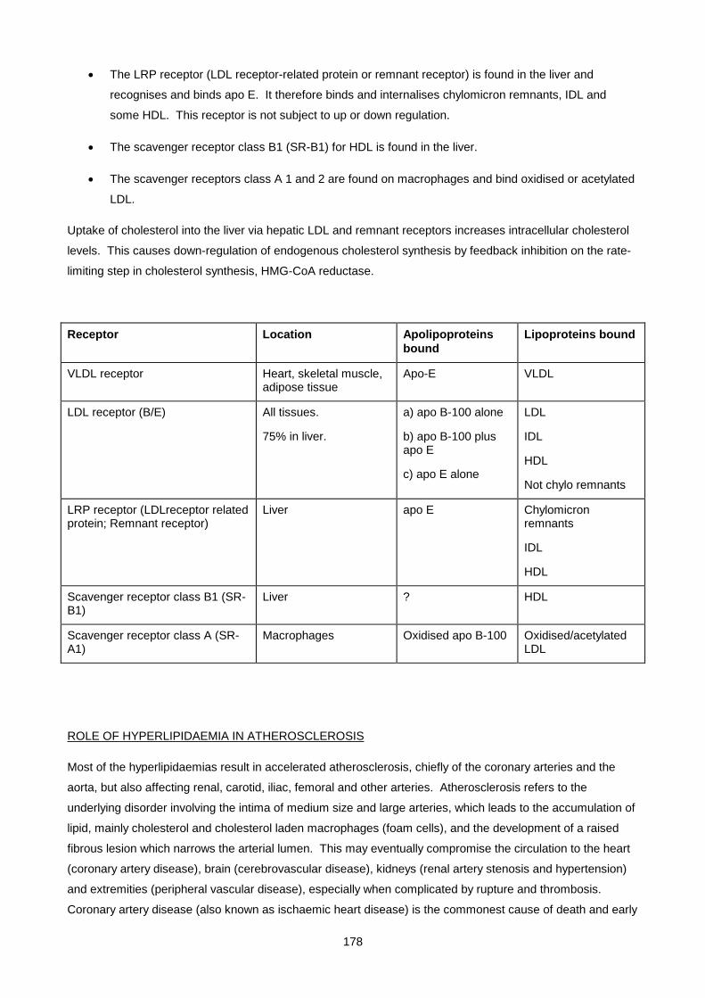
178
• The LRP receptor (LDL receptor-related protein or remnant receptor) is found in the liver and
recognises and binds apo E. It therefore binds and internalises chylomicron remnants, IDL and
some HDL. This receptor is not subject to up or down regulation.
• The scavenger receptor class B1 (SR-B1) for HDL is found in the liver.
• The scavenger receptors class A 1 and 2 are found on macrophages and bind oxidised or acetylated
LDL.
Uptake of cholesterol into the liver via hepatic LDL and remnant receptors increases intracellular cholesterol
levels. This causes down-regulation of endogenous cholesterol synthesis by feedback inhibition on the rate-
limiting step in cholesterol synthesis, HMG-CoA reductase.
Receptor Location Apolipoproteins bound
Lipoproteins bound
VLDL receptor Heart, skeletal muscle, adipose tissue
Apo-E VLDL
LDL receptor (B/E) All tissues.
75% in liver.
a) apo B-100 alone
b) apo B-100 plus apo E
c) apo E alone
LDL
IDL
HDL
Not chylo remnants
LRP receptor (LDLreceptor related protein; Remnant receptor)
Liver apo E
Chylomicron remnants
IDL
HDL
Scavenger receptor class B1 (SR-B1)
Liver ? HDL
Scavenger receptor class A (SR-A1)
Macrophages Oxidised apo B-100 Oxidised/acetylated LDL
ROLE OF HYPERLIPIDAEMIA IN ATHEROSCLEROSIS
Most of the hyperlipidaemias result in accelerated atherosclerosis, chiefly of the coronary arteries and the
aorta, but also affecting renal, carotid, iliac, femoral and other arteries. Atherosclerosis refers to the
underlying disorder involving the intima of medium size and large arteries, which leads to the accumulation of
lipid, mainly cholesterol and cholesterol laden macrophages (foam cells), and the development of a raised
fibrous lesion which narrows the arterial lumen. This may eventually compromise the circulation to the heart
(coronary artery disease), brain (cerebrovascular disease), kidneys (renal artery stenosis and hypertension)
and extremities (peripheral vascular disease), especially when complicated by rupture and thrombosis.
Coronary artery disease (also known as ischaemic heart disease) is the commonest cause of death and early

179
invalidism in Western society. A number of risk factors for the development of coronary artery disease have
been identified in long-term studies. These include:
i. increasing age
ii. male gender
iii. family history of premature CAD (indicating genetic factors)
iv. hypercholesterolaemia *
v. cigarette smoking *
vi. hypertension *
vii. diabetes mellitus
viii. obesity
ix. sedentary lifestyle
x. others (hyperuricaemia, oral contraceptives, abnormal fibrinogen levels).
Risk factors i) to iii) cannot be altered, while the remaining risk factors are "influenceable".
Risk factors iv) to vi) are primary risk factors (can themselves be linked to CAD).
Risk factors vii) to ix) are secondary risk factors (worsen pre-existing CAD).
The presence of a risk factor does not mean that CAD will occur (risk factors are not causes), but increases
the likelihood of developing CAD. Most risk factors are continuously related to the development of CAD i.e.
there is no threshold value which is normal and below which there is no risk. However, smoking and
hypertension approximately double the risk of developing CAD, while hypercholesterolaemia trebles it. The
hypercholesterolaemia of FH (Familial Hypercholesterolaemia) increases the risk 100-fold or more.
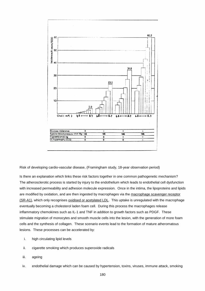
180
Risk of developing cardio-vascular disease, (Framingham study, 18-year observation period)
Is there an explanation which links these risk factors together in one common pathogenetic mechanism?
The atherosclerotic process is started by injury to the endothelium which leads to endothelial cell dysfunction
with increased permeability and adhesion molecule expression. Once in the intima, the lipoproteins and lipids
are modified by oxidation, and are then ingested by macrophages via the macrophage scavenger receptor
(SR-A1), which only recognises oxidised or acetylated LDL. This uptake is unregulated with the macrophage
eventually becoming a cholesterol laden foam cell. During this process the macrophages release
inflammatory chemokines such as IL-1 and TNF in addition to growth factors such as PDGF. These
stimulate migration of monocytes and smooth muscle cells into the lesion, with the generation of more foam
cells and the synthesis of collagen. These scenario events lead to the formation of mature atheromatous
lesions. These processes can be accelerated by:
i. high circulating lipid levels
ii. cigarette smoking which produces superoxide radicals
iii. ageing
iv. endothelial damage which can be caused by hypertension, toxins, viruses, immune attack, smoking
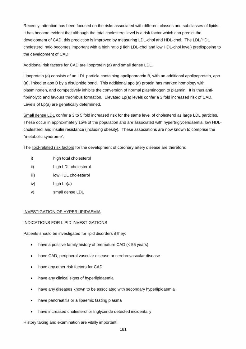
181
Recently, attention has been focused on the risks associated with different classes and subclasses of lipids.
It has become evident that although the total cholesterol level is a risk factor which can predict the
development of CAD, this prediction is improved by measuring LDL-chol and HDL-chol. The LDL/HDL
cholesterol ratio becomes important with a high ratio (High LDL-chol and low HDL-chol level) predisposing to
the development of CAD.
Additional risk factors for CAD are lipoprotein (a) and small dense LDL.
Lipoprotein (a) consists of an LDL particle containing apolipoprotein B, with an additional apolipoprotein, apo
(a), linked to apo B by a disulphide bond. This additional apo (a) protein has marked homology with
plasminogen, and competitively inhibits the conversion of normal plasminogen to plasmin. It is thus anti-
fibrinolytic and favours thrombus formation. Elevated Lp(a) levels confer a 3 fold increased risk of CAD.
Levels of Lp(a) are genetically determined.
Small dense LDL confer a 3 to 5 fold increased risk for the same level of cholesterol as large LDL particles.
These occur in approximately 15% of the population and are associated with hypertriglyceridaemia, low HDL-
cholesterol and insulin resistance (including obesity). These associations are now known to comprise the
“metabolic syndrome”.
The lipid-related risk factors for the development of coronary artery disease are therefore:
i) high total cholesterol
ii) high LDL cholesterol
iii) low HDL cholesterol
iv) high Lp(a)
v) small dense LDL
INVESTIGATION OF HYPERLIPIDAEMIA
INDICATIONS FOR LIPID INVESTIGATIONS
Patients should be investigated for lipid disorders if they:
• have a positive family history of premature CAD (< 55 years)
• have CAD, peripheral vascular disease or cerebrovascular disease
• have any other risk factors for CAD
• have any clinical signs of hyperlipidaemia
• have any diseases known to be associated with secondary hyperlipidaemia
• have pancreatitis or a lipaemic fasting plasma
• have increased cholesterol or triglyceride detected incidentally
History taking and examination are vitally important!

182
LABORATORY INVESTIGATIONS AVAILABLE
EDTA plasma sample after overnight fast (fasting not essential for cholesterol, but is for TG), taken without
excessive venostasis, preferably on a normal diet, no alcohol, no recent change in exercise/weight/diet, no
recent illness/injury. Be aware that recent acute myocardial infarction or other events evoking an acute
phase response (within the preceding 6 weeks) causes falsely low cholesterol levels.
Be aware that biological variability of cholesterol is 5-10% and of triglyceride is about 25% (more if non-
fasting). Diagnosis and management should therefore preferably be based on repeat measurements 2
weeks apart.
Lipid profile (TChol, TG, HDL-C, LDL-C):
Total cholesterol and triglyceride are measured enzymatically.
HDL cholesterol is measured enzymatically after all non-HDL lipoproteins are immunologically rendered non-
reactive.
LDL cholesterol is calculated using Friedewald's formula:
LDL cholesterol = total cholesterol - HDL cholesterol - TG / 2.2
Not valid if TG > 4.5 mmol/l
Apolipoprotein measurement:
Apo A-I, apo B-100 and Lp(a) are measured by protein measuring technique (RIA, nephelometry, or
turbidimetry)
Appearance of plasma after overnight refrigeration:
If creamy layer present - increased chylomicrons (Type I)
If creamy layer and turbid infranatant (Type V pattern)
If infranatant turbid - increased VLDL or IDL (Type IIb or IV pattern)
If infranatant clear - normal VLDL and IDL (Type IIa pattern)
Appearance of serum after refrigeration does not assess levels of cholesterol-rich LDL or HDL.
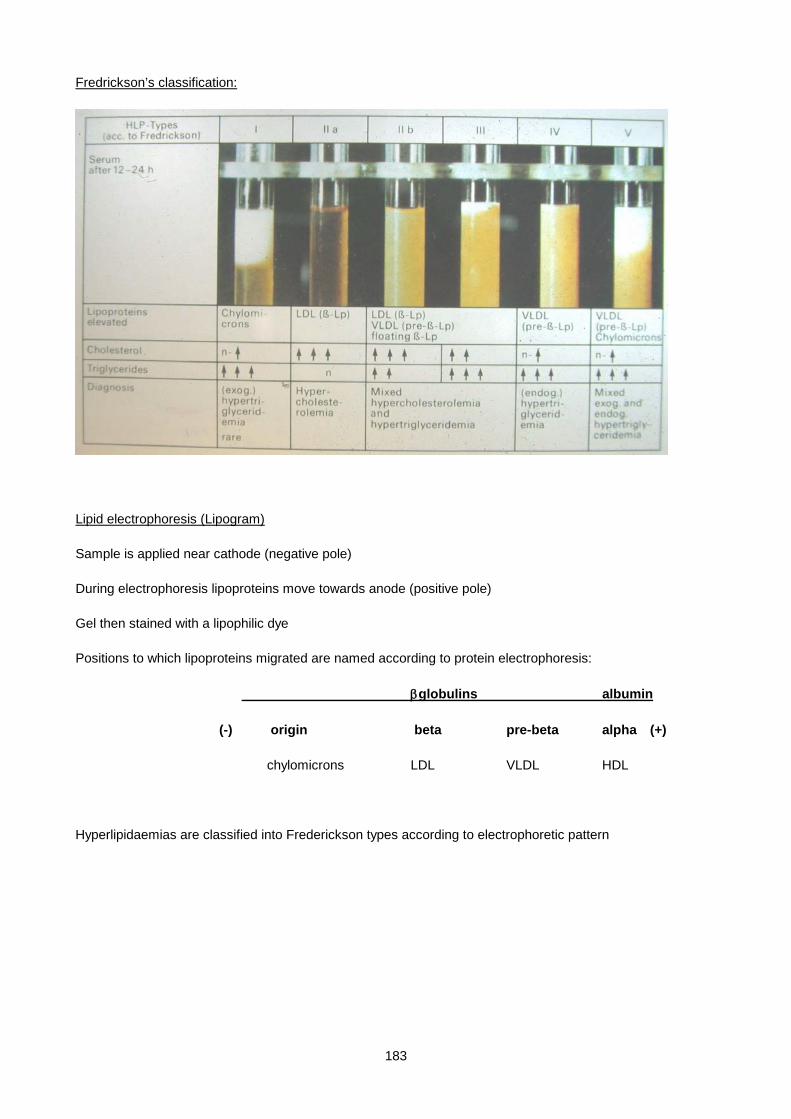
183
Fredrickson’s classification:
Lipid electrophoresis (Lipogram)
Sample is applied near cathode (negative pole)
During electrophoresis lipoproteins move towards anode (positive pole)
Gel then stained with a lipophilic dye
Positions to which lipoproteins migrated are named according to protein electrophoresis:
βglobulins albumin
(-) origin beta pre-beta alpha (+)
chylomicrons LDL VLDL HDL
Hyperlipidaemias are classified into Frederickson types according to electrophoretic pattern

184
A typical example of a Lipogram:
Lane 1: Type III
Lane 2: Type IIa
Lane 3: Type IV
Lane 4: Type IIb
Lane 5: Type IIb
Lane 6: Type IIa
Lane 7: Control plasma
* chylomicrons are not seen in fasting samples, unless in Types I and IV
FORMULATING A DIAGNOSIS
The lipid investigations performed should answer the following questions:
i. Is hyperlipidaemia present, and if so, how severe?
ii. What type of hyperlipidaemia is present (phenotype)?
iii. What risk factors for CAD are present (risk profile)?
iv. What is the cause of the hyperlipidaemia (aetiology)?
+ve
Chylomicrons
VLDL
HDL
LDL
185
i. IS HYPERLIPIDAEMIA PRESENT, AND IF SO, HOW SEVERE?
Statistically speaking, hyperlipidaemia is any lipid value above the 97.5th percentile (mean + 2 SD), but
clinically speaking we use the term hyperlipidaemia to mean any lipid value with an adverse clinical effect.
• Cholesterol Ideal level < 5.2 mmol/l
Mild to moderate hypercholesterolaemia 5.2 – 7.5 mmol/l
Severe hypercholesterolaemia 7.5 – 15.0 mmol/l
Very severe hypercholesterolaemia > 15 mmol/l * usually homozygous FH
• Triglyceride Ideal level < 2.5 mmol/l
Mild hypertriglyceridaemia 2.5 – 5.0 mmol/l
Moderate hypertriglyceridaemia 5.0 – 11 mmol/l
Severe hypertriglyceridaemia 11 – 150 mmol/l
• LDL cholesterol Ideal level < 3.5 mmol/l
• HDL cholesterol Ideal level > 1.2 mmol/l * low HDL is non-ideal
• apolipoprotein B Ideal level < 100 mg/dl
• apolipoprotein A Ideal level > 120 mg/dl * low apo A is non-ideal
• lipoprotein (a) Ideal level < 30 mg/dl
Quick guide to predicting phenotype from results of laboratory tests:
If plasma TG is elevated, chylomicrons or VLDL or both must be raised.
If plasma cholesterol is elevated, LDL or HDL or both must be raised.
If plasma TG and cholesterol are both elevated, either TG-rich and cholesterol-rich particles are both raised,
or IDL is raised.
Levels of apo B reflect predominantly LDL levels
Levels of apo A1 reflect HDL levels
186
ii. WHAT TYPE OF HYPERLIPIDAEMIA IS PRESENT (PHENOTYPE)?
The simplest phenotypic classification of hyperlipidaemia is in terms of serum cholesterol and triglyceride
levels. Thus one may have mild, moderate or severe grades of :
• pure hypercholesterolaemia
• pure or predominant hypertriglyceridaemia
• mixed hyperlipidaemia
A more complex phenotypic classification of hyperlipidaemia is on the basis of the pattern of elevation of
lipoproteins, as seen on lipid electrophoresis. This classification is the W.H.O. or Fredrickson classification.
The Fredrickson classification is very old and therefore well known and widely accepted. It is useful in
identifying some of the inherited (primary) hyperlipidaemias, and is the only readily available method of
identifying elevated VLDL and IDL levels. It however has the disadvantages that only a few patterns are
specific to a single disease entity,
relatives with the same disease (e.g. FH) can have different patterns, patterns can change over time or with
treatment and diet, and no account is taken of HDL levels
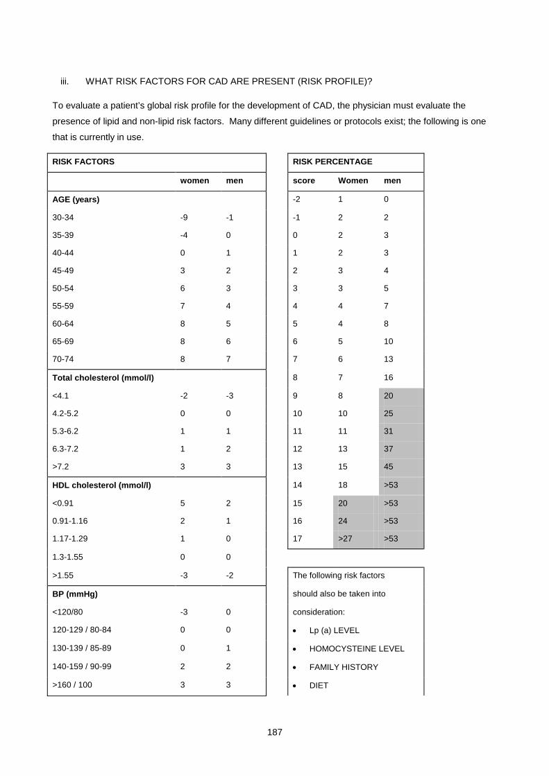
187
iii. WHAT RISK FACTORS FOR CAD ARE PRESENT (RISK PROFILE)?
To evaluate a patient’s global risk profile for the development of CAD, the physician must evaluate the
presence of lipid and non-lipid risk factors. Many different guidelines or protocols exist; the following is one
that is currently in use.
RISK FACTORS RISK PERCENTAGE
women men score Women men
AGE (years) -2 1 0
30-34 -9 -1 -1 2 2
35-39 -4 0 0 2 3
40-44 0 1 1 2 3
45-49 3 2 2 3 4
50-54 6 3 3 3 5
55-59 7 4 4 4 7
60-64 8 5 5 4 8
65-69 8 6 6 5 10
70-74 8 7 7 6 13
Total cholesterol (mmol/l) 8 7 16
<4.1 -2 -3 9 8 20
4.2-5.2 0 0 10 10 25
5.3-6.2 1 1 11 11 31
6.3-7.2 1 2 12 13 37
>7.2 3 3 13 15 45
HDL cholesterol (mmol/l) 14 18 >53
<0.91 5 2 15 20 >53
0.91-1.16 2 1 16 24 >53
1.17-1.29 1 0 17 >27 >53
1.3-1.55 0 0
>1.55 -3 -2 The following risk factors
BP (mmHg) should also be taken into
<120/80 -3 0 consideration:
120-129 / 80-84 0 0 • Lp (a) LEVEL
130-139 / 85-89 0 1 • HOMOCYSTEINE LEVEL
140-159 / 90-99 2 2 • FAMILY HISTORY
>160 / 100 3 3 • DIET

188
SMOKER 2 2 • OBESITY
DIABETIC 4 2 • SEDENTARY LIFESTYLE
Risk factors are evaluated and a score assigned for each risk factor. The total score is converted to a risk
percentage, which is the risk of developing a myocardial infarction within the next 10 years. When the risk
percentage is >20%, lipid-lowering drug treatment is instituted, with the goal of achieving a Total cholesterol
of < 5.2 mmol/l, and an LDL cholesterol of < 3 mmol/l. For patients with Familial Hypercholesterolaemia or a
previous history of CAD, lipid-lowering drug treatment is instituted irrespective of the risk percentage.
iv. WHAT IS THE CAUSE OF THE HYPERLIPIDAEMIA (AETIOLOGY)?
The simplest aetiological classification of hyperlipidaemias divides them into 2 categories:
i. Primary hyperlipidaemia – where the hyperlipidaemia is not due to an identifiable underlying disease,
but due to an inherited disorder of lipoprotein metabolism.
Primary hyperlipidaemias are caused by:
• An inherited disorder of lipoprotein metabolism for which specific gene defects have been identified,
e.g., familial hypercholesterolaemia
• An inherited disorder of lipoprotein metabolism for which specific gene defects have not yet been
identified.
• Certain genetic predispositions to hyperlipidaemia, which requires exposure to other factors before
the disease becomes manifest.
ii. Secondary hyperlipidaemias – where the hyperlipidaemia is caused by an identifiable underlying
disease or drug regimen which interferes with normal lipid metabolism.
A combination of history, examination, family history, lipid levels, lipid electrophoresis, other laboratory
tests, and genetic tests is used to arrive at a final aetiological diagnosis. The features of the main known
hyperlipidaemias are discussed in detail below.
189
DISORDERS OF LIPID METABOLISM
HYPERLIPIDAEMIA (much more common than hypolipidaemia)
PRIMARY HYPERLIPIDAEMIAS
FAMILIAL HYPERCHOLESTEROLAEMIA (FH)
Autosomal dominant, inherited defect of the LDL receptor. Frequency: heterozygotes 1 in 500, homozygotes
1 in 1000000. Commoner in Afrikaners with a heterozygote frequency of ≈1/100. Homozygotes have very
high cholesterol levels (> 15 mM) and usually die of CAD before 20 years of age. Heterozygotes have
intermediate levels of cholesterol (8 mM - 11 mM) and develop CAD in their forties. Heterozygotes constitute
+ 5% of all myocardial infarcts. Clinically patients usually have positive family histories of CAD, and often
have tendon xanthomata, especially on the Achilles tendon (cholesterol ester deposition in macrophages).
Arcus cornealis (deposition of cholesterol in the cornea) and xanthelasmata (deposition of cholesterol in the
eyelids) often occur. Arcus and xanthelasmata can also be found in patients with normal lipid levels. To date,
the LDLR gene mutation database lists 771 mutant alleles coding for LDL receptor defects, which can be
divided into receptor-negative (non-functional gene product), receptor-defective (binding of LDL reduced to
1-10% of normal) and internalisation-defective (binds normally but unable to transport LDL into the cell).
Phenotypic homozygotes have 2 abnormal alleles in any combination. Failure of receptor-mediated
internalisation of LDL results in failure to down regulate HMG-CoA reductase at normal plasma cholesterol
levels. Down regulation only occurs at high levels when non-receptor mediated uptake is effective. The
result is that in the steady state cholesterol is synthesised at a normal rate, but plasma levels are very high
with increased plasma half-life. This causes increased macrophage uptake of oxidised LDL, in the intima, via
the scavenger receptor, and formation of foam cells resulting in xanthomata and initiation of atheroma.
Plasma LDL levels are inversely proportional to LDL receptor activity in these patients due to impaired uptake
and catabolism of LDL. LDL production is also increased in homozygotes as IDL is not cleared by the LDL
receptor and thus more is converted to LDL. Patients with FH usually have an isolated increase in
cholesterol with increased LDL (type IIa pattern) but may also have an increase in triglyceride with increased
VLDL (type IIb pattern) in 10% cases.
Treatment: homozygotes - low fat diet and drugs, but also require plasma exchange/apheresis (removal of
LDL by filtering plasma through affinity columns) or liver transplant. Heterozygotes - rigorous diet (low
cholesterol, low saturated fat) plus lipid lowering drugs. Most effective are HMG-CoA reductase inhibitors
[the Statins eg simvastatin, atorvostatin(Lipitor) ] which lower cholesterol 30-50%.
FAMILIAL BINDING DEFECTIVE APOLIPOPROTEIN B-100
The condition is co-dominantly inherited and is not as common as FH in South Africa. A single base change
in the cDNA for apo B-100 decreases binding to the LDL receptor. This decreases the rate of clearance of
LDL from plasma and thus causes hypercholesterolaemia. Clinically this may appear similar to FH, but
should be differentiated as response to lipid lowering drugs differs. Requires specialist lipid laboratory
investigation.
190
AUTOSOMAL RECESSIVE HYPERCHOLESTEROLAEMIA (ARH)
This condition is very rare and has a phenotype that mimics closely Homozygous FH. A critical difference
between ARH and FH is that the parents of ARH patients will have normal blood lipid levels and there is also
unlikely to be a family history of premature coronary artery disease. This condition arises from an absence of
an adaptor protein called ARH that is required for LDL receptor function. These patients thus present with a
functional abnormality of the LDL receptor, with a resultant massive increase in plasma LDL levels.
POLYGENIC HYPERCHOLESTEROLAEMIA
This category accounts for the majority (85%) of patients with high LDL cholesterol. The pattern of
inheritance suggests that this is not a single gene defect, but that several genes are involved. Only 10% of
first degree relatives have hypercholesterolaemia, and relatives display a continuous distribution of serum
cholesterol levels, whereas in FH there is a clear distinction between normals, heterozygotes and
homozygotes.
Cholesterol levels are elevated, but usually not more than 10 mM. Cholesterol levels are largely diet
dependent. Lipid electrophoresis shows a type IIa or IIb pattern. Patients have an increased risk of CAD, but
xanthomata are not a feature. Treatment by diet + lipid lowering agents is successful in lowering cholesterol
into the "desirable" range.
FAMILIAL COMBINED HYPERLIPIDAEMIA (FCHL) (Multiple lipoprotein-type hyperlipidaemia)
Encountered in 1-2% of people in the Western World. This category accounts for 10% of cases of
hypercholesterolaemia, and can also be a cause of hypertriglyceridaemia or mixed hyperlipidaemia. Within a
family, 50% of first degree relatives will have hyperlipidaemia which is multiple in nature i.e. some have
increased cholesterol, some increased TG and some both. The inheritance pattern is usually autosomal
dominant and several different genes are known to be involved. Clarity on the exact function of these gene
products is awaited. A subgroup have overproduction of apo B causing increased VLDL secretion and hence
increased LDL.
Cholesterol and/or TG may be elevated causing a IIa (30%), IIb (30%) or IV (30%) pattern. Patients have a
significantly increased risk of CAD, possibly associated with the increased apo B in LDL even though LDL
cholesterol may be normal. Xanthomata are not present. Treatment is by diet + lipid lowering agents.
FAMILIAL DYSBETALIPOPROTEINAEMIA (Broad beta disease / Familial type III hyperlipidaemia)
These patients are homozygous for an apo E variant, known as apo E2. The usual apo E phenotype is
E3/E3, with other phenotypes e.g. E4 also found (E4 is associated with an increased risk of Alzheimer's
Disease). The homozygous apo E2/E2 state is present in 1% of the normal population, but alone does not

191
cause hyperlipidaemia. The homozygous apo E2/E2 state coupled with another inherited hyperlipidaemia or
alcoholism, obesity, diabetes or hypothyroidism results in the characteristic hyperlipidaemia which only has a
frequency of 0.01%. Apo E phenotypes other than E2/E2 bind with high affinity to the remnant receptor and
LDL receptor, thus facilitating clearance of chylomicron remnants and IDL. In the apo E2/E2 state, impaired
binding results in impaired clearance of these particles. Patients present in adulthood with characteristic
yellow fat deposits in palmar creases, and orange/yellow tubero-eruptive xanthomata over bony prominences
(not always present). There is an increased risk of CAD, cerebro-vascular disease and peripheral vascular
disease. On electrophoresis they present with a typical "broad beta" band due to excess IDL and chylomicron
remnants. Consequently cholesterol and TG levels are elevated. Diagnosis is confirmed by apo E
phenotyping. DNA diagnosis is now available. The hyperlipidaemia is usually very responsive to diet and
lipid lowering agents especially clofibrate. Secondary causes of hyperlipidaemia especially hypothyroidism
(TSH) should always be sought and treated in these patients.
FAMILIAL HYPERTRIGLYCERIDAEMIA
This is a common autosomal dominant defect causing increased VLDL in the plasma. The basic defect may
be decreased VLDL catabolism in some cases, or excess TG synthesis by the liver and increased VLDL
secretion in others. Chylomicrons may also rise as VLDL competes for lipoprotein lipase. Patients thus have
increased TG with type IV or V pattern. Patients only become hyperlipidaemic post puberty and are often
asymptomatic. The elevated TG are not a risk factor for CAD, but the slightly increased cholesterol and low
HDL are. Xanthomata are not present. Clinically these patients are often obese, hyperglycaemic and
hyperinsulinaemic and may have hyperuricaemia and hypertension. 50% of first degree adult relatives have
elevated TG with little or no elevation of cholesterol. Treatment is by controlling diabetes, obesity, alcohol
intake and hypothyroidism, and avoiding oestrogen preparations. Fish oils (omega-3 fatty acids) have a
dramatic effect on lowering the TG probably by decreasing hepatic VLDL synthesis.
FAMILIAL LIPOPROTEIN LIPASE DEFICIENCY
This is due to an autosomal recessively inherited defect of the enzyme lipoprotein lipase. Chylomicrons are
not cleared from the blood resulting in very high TG levels, which causes plasma to have a thick layer of
"cream" above a clear infranatant on standing at 4°C and a type I pattern on electrophoresis. VLDL do not
build up as they are cleared by an alternative ill-understood pathway possibly macrophage related, and also,
impaired influx of Fatty Acids into the liver may hinder VLDL secretion. Presentation is usually in childhood
with eruptive xanthomata or recurrent abdominal pain due to pancreatitis (leaked pancreatic lipase).
Occasionally hepatosplenomegaly is present due to RES uptake of TG. There is no increase in CAD risk.
Diagnosis is by failure to record an increase in LPL activity after heparin infusion, which normally releases
LPL from the capillary endothelium into the plasma. DNA diagnosis is possible if the likely gene defect is
known. Therapy is by giving low fat diets with TG given as medium chain fatty acids which are absorbed
directly into the blood without being formed into chylomicrons. Fat soluble vitamins should be given as
supplements.
192
FAMILIAL APO C-II DEFICIENCY
This is due to an autosomal recessive inherited defect in apo C-II, which is an essential cofactor for LPL.
This deficiency thus causes a similar picture to LPL deficiency. Chylomicrons accumulate in plasma (type I
pattern), and sometimes VLDL also accumulate (type V pattern). Diagnosis is made by showing absent apo
C-II on gel electrophoresis of VLDL apolipoproteins. Transfusion of normal plasma will cause a rapid drop of
plasma TG (unlike LPL deficiency). This may be useful in treating pancreatitis.
FAMILIAL HYPERALPHALIPOPROTEINAEMIA
Elevated HDL level is HDL > 2.1 mmol/L. This condition may be autosomal dominant or polygenic. Total
cholesterol may be elevated due to an increase in HDL cholesterol (and apo A1). There is an association
with a decreased incidence of CAD. Affected patients have no symptoms or signs. No treatment is necessary
HOMOZYGOUS CETP DEFICIENCY
Homozygous CETP deficiency causes the formation of very large "HDL" particles that may become
atherogenic, as heart disease seems more prevalent. Cholesterol accumulates in these HDL because it
cannot be exchanged for reverse transport. Affected patients display no symptoms or signs but have HDLs >
3.9mmol/L.
SECONDARY HYPERLIPIDAEMIA
These account for ±40% of hyperlipidaemias and affect ±5% of adults. Should always be excluded if
hyperlipidaemia is present. Treatment of the underlying disorder corrects the hyperlipidaemia. A secondary
cause of hyperlipidaemia will precipitate or worsen a coexistent inherited hyperlipidaemia.
HYPOTHYROIDISM
Decreased catabolism of VLDL, IDL and LDL. Predominantly hypercholesterolaemia with normal or slightly
raised triglyceride levels. Type IIa or IIb pattern. Hypothyroidism can also expose an underlying familial
dysbetalipoproteinaemia and therefore present with a type III pattern.
RENAL DISEASE
Nephrotic syndrome - protein synthesis (including apolipoprotein synthesis) is stimulated as a consequence
of proteinuria. Being large molecules lipoproteins are not lost in the urine at the same rate as other proteins,
thus patients become hypercholesterolaemic, with normal or raised triglyceride levels. Type IIa or IIb pattern.
All nephrotics have hyperlipidaemia. Chronic renal failure and dialysis patients - roughly 1/3 of these patients
have hyperlipidaemia due to decreased lipoprotein lipase activity causing hypertriglyceridaemia. Type IV or V
pattern.
193
LIVER DISEASE
In cholestasis diversion of biliary cholesterol and phospholipids into the blood-stream occurs, leading to
severe hypercholesterolaemia and variable hypertriglyceridaemia. An abnormal cholesterol-rich lipoprotein
(lipoprotein X) is found in plasma. Acute hepatic dysfunction can impair synthesis of apolipoproteins, HDL
and LCAT. Levels of HDL reflect the course of acute disease, while levels of other lipoproteins only decrease
in severe disease. The function of hepatic TG lipase is also impaired, causing an accumulation of TG-rich
lipoproteins (type IIb or IV pattern).
DIABETES
Diabetes is characterised by an over-production of VLDL, leading to hypertriglyceridaemia. Prolonged insulin
deficiency also decreases LPL activity, and if VLDL rise markedly, chylomicrons accumulate due to
competition for LPL. In well-controlled diabetes there may thus be a mild hypertriglyceridaemia, while in
poorly controlled diabetes there is both severe hypertriglyceridaemia and chylomicronaemia which may lead
to acute pancreatitis. In addition HDL cholesterol levels are low. Type IV and V patterns. Diabetes can also
expose an underlying familial dysbetalipoproteinaemia and therefore present with a type III pattern.
ALCOHOL
Usually causes hypertriglyceridaemia due to increased VLDL synthesis. Hepatic triglyceride synthesis is
increased due to increased fatty acid synthesis and decreased fatty acid oxidation. Increased fatty acid
synthesis is due to increased acetyl CoA from metabolism of ethanol. The usual pattern seen is type IV. A
small subgroup have massive elevations of TG due to ↑ VLDL and ↑ chylos (type V). These patients may
have eruptive xanthomata and pancreatitis. On withdrawing alcohol these patients revert to a moderate type
IV hyperlipidaemia, suggesting that they may have underlying familial hypertriglyceridaemia or familial
combined hyperlipidaemia. Alcohol can also expose an underlying familial dysbetalipoproteinaemia and
therefore present with a type III pattern. Moderate alcohol intake increases HDL cholesterol, which is
beneficial!
OBESITY
Increased fatty acid delivery to the liver (dietary excess or insulin resistance) results in increased VLDL
synthesis which leads to hypertriglyceridaemia of a Type IV pattern. Occasionally increased conversion to
LDL causes a type IIb pattern. Obesity is associated with low HDL levels. All abnormalities are reversible
by weight reduction. Obesity can also expose an underlying familial dysbetalipoproteinaemia and
therefore present with a type III pattern.
SUMMARY:
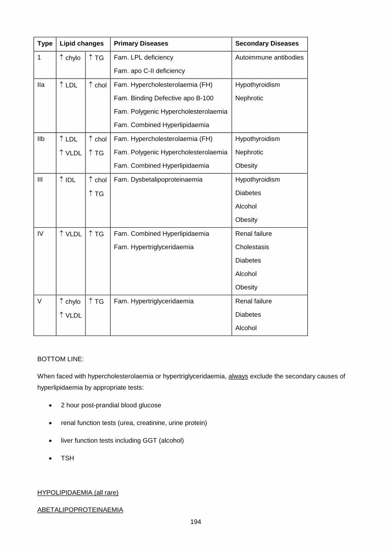
194
Type Lipid changes Primary Diseases Secondary Diseases
1 ↑ chylo ↑ TG Fam. LPL deficiency
Fam. apo C-II deficiency
Autoimmune antibodies
IIa ↑ LDL ↑ chol Fam. Hypercholesterolaemia (FH)
Fam. Binding Defective apo B-100
Fam. Polygenic Hypercholesterolaemia
Fam. Combined Hyperlipidaemia
Hypothyroidism
Nephrotic
IIb ↑ LDL
↑ VLDL
↑ chol
↑ TG
Fam. Hypercholesterolaemia (FH)
Fam. Polygenic Hypercholesterolaemia
Fam. Combined Hyperlipidaemia
Hypothyroidism
Nephrotic
Obesity
III ↑ IDL ↑ chol
↑ TG
Fam. Dysbetalipoproteinaemia Hypothyroidism
Diabetes
Alcohol
Obesity
IV ↑ VLDL ↑ TG Fam. Combined Hyperlipidaemia
Fam. Hypertriglyceridaemia
Renal failure
Cholestasis
Diabetes
Alcohol
Obesity
V ↑ chylo
↑ VLDL
↑ TG Fam. Hypertriglyceridaemia Renal failure
Diabetes
Alcohol
BOTTOM LINE:
When faced with hypercholesterolaemia or hypertriglyceridaemia, always exclude the secondary causes of
hyperlipidaemia by appropriate tests:
• 2 hour post-prandial blood glucose
• renal function tests (urea, creatinine, urine protein)
• liver function tests including GGT (alcohol)
• TSH
HYPOLIPIDAEMIA (all rare)
ABETALIPOPROTEINAEMIA
195
Defect in synthesis of apo B. The mutation may be homozygous mutated MTTP (microsomal TG transfer
protein) which adds neutral lipid to apo B during the synthesis of chylomicrons and VLDL, or due to truncation
of apo B which cannot act as a template for the formation of chylomicrons and VLDL. Chylomicrons, VLDL
and LDL are absent, with very low plasma cholesterol and TG. Thus fat malabsorption (steatorrhoea) and a
failure of lipid delivery to cells occur, causing membrane abnormalities manifesting as acanthocytosis (red
cells), retinitis pigmentosa and ataxic neuropathy.
FAMILIAL HYPOBETALIPOROTEINAEMIA
Hypopbetalipoproteinemia is defined by plasma levels of total cholesterol or LDL cholesterol, or total apo B
being below the 5th percentile. Signs and symptoms are similar to Abetalipoproteinaemia but less severe.
This condition arises mainly from truncation mutants of apoB which are poor carriers of intracellular
triglycerides; VLDL synthesis is thus markedly impaired but not entirely absent
ANALPHALIPOPROTEINAEMIA (Tangier Disease)
Absent HDL. The defect is in the ABC1 transporter protein which brings cholesterol from within a cell to the
surface for transfer to apo A1 and the generation of HDL. Cholesterol poor Apo A1 is rapidly cleared from the
circulation, much of it by the kidney. Patients present with large orange tonsils, corneal opacities and
neuropathy.
LCAT DEFICIENCY
The inability to esterify free cholesterol results in excess unesterified cholesterol in plasma and tissues. All
lipoproteins have abnormal structure, VLDL is elevated. Patients have haemolytic anaemia, premature
atherosclerosis and renal insufficiency.
TREATMENT
Does treatment aimed at improving lipid profiles improve the risk profile for CAD, i.e. does it decrease the
incidence, the severity and the mortality of CAD?
In hereditary hyperlipidaemias there is no doubt that improving lipid profiles significantly reduces the
incidence and mortality of CAD. In acquired hyperlipidaemias there have been multiple long-term studies
which point to some benefits - reduced incidence of CAD, and possibly regression of pre-existing
atherosclerotic lesions. Mortality due to CAD has also been shown to be significantly reduced.
Current recommendations are to attempt to reduce cholesterol and TG levels to more desirable ranges,
especially if other risk factors for CAD are present, by diet or drugs if necessary.
i) TREAT UNDERLYING CAUSE (if present)
ii) DIET (also improves other risk factors)
Advise patient to:
- reduce total caloric intake
- reduce total fat content of diet (no more than 30% of total calories)
196
- reduce saturated fats (no more than 30% of total fat intake) and increase mono- and polyunsaturated fats
- reduce dietary cholesterol
- reduce excess body weight
- avoid excess alcohol
- avoid excess salt
- increase dietary fibre
Assess efficacy 3 monthly.
iii) DRUGS
Consider drug treatment for:
- moderate hyperlipidaemia unresponsive to diet after 3 to 6 months
- severe hyperlipidaemia
- any degree of hyperlipidaemia if CAD known to be present
- inherited hyperlipidaemia
“Statins” - HMG-CoA reductase inhibitors, (e.g. simvastatin, atorvastatin), which inhibit rate-limiting. Step in hepatic synthesis of cholesterol, lower total and LDL cholesterol
cholestyramine - bile acid sequestrant , bind bile acids in GIT and reduce enterohepatic circulation of cholesterol, lower total and LDL cholesterol
nicotinic acid inhibits lipolysis of adipose TG, lowers total and LDL cholesterol, TG and Lp(a), raises HDL cholesterol
Probucol – lipid soluble anti-oxidant , reduces oxidation of LDL, stimulates CETP, lowers LDL and HDL cholesterol, reduces CAD risk
“fibrates” (e.g. gemfibrozil), reduce synthesis of VLDL, increases LPL activity, lowers TG, total and LDL cholesterol, may raise HDL cholesterol
APPROACH TO TREATMENT:
Hypercholesterolaemia 1. Statin alone
2. Statin plus bile acid sequestrant (increases efficacy in resistant patients or to keep statin dose low)
3. Bile acid sequestrant alone (if resistant to statin or in children)
4. Fibrate, preferably combined with statin or bile acid sequestrant
5. Nicotinic acid
Hypertriglyceridaemia 1. Fibrate
2. Nicotinic acid
3. Statin
197
SMALL GROUP TEACHING: LECTURE 9: LIPID AND LIPOPROTEIN METABOLISM AND DYSLIPIDAEMIAS
QUESTIONS
1. You are referred a 49 year old male for on-going care after having suffered an AMI. The notes from the cardiologist states the patient is apparently not hypercholesterolaemic but is somewhat obese and his father died of a ‘heart attack’ at 59 years of age. On clinical examination you confirm the obesity (BMI = 31), the patients BP is 165 /105 mmHg, he admits to smoking between 20 and 30 cigarettes a day until his AMI, but no overt evidence of hyperlipidaemia is detected. Other than his father there is no clear history of premature CAD in his family. A fasting cholesterol is 5.2 mmol/l, HDL-C 0.8 mmol/l, trigs 2.7 mmol/l.
i. Discuss the LP profile in terms of its risk potential and causation.
ii. Discuss the overall risk of the patient.
iii. Discuss what other laboratory determination(s) may be useful in assessing this patient.
iv. Outline your approach to treatment.
2. A mother brings her 3 month infant who she says is somewhat restless and ‘colicky’. She recently noticed a fine rash on his cheeks and trunk. You confirm a papular rash and note that the child has enlarged liver and readily palpable spleen. There is no evidence of jaundice and the child appears well. On taking blood you notice an unusual ‘raspberry milkshake’ appearance. The laboratory reports marked turbidity and a creamy layer on standing. The triglyceride level was 25 mmol/l, cholesterol 17 mmol/l but HDL cholesterol and LDL cholesterol were not reported.
i. What type of hyperlipidaemia is present?
ii. What is the most likely cause?
iii. Do you think the lipid results reported are necessarily accurate? Explain your answer. Is it important to obtain the LDL cholesterol and HDL cholesterol Levels? Why was the LDL cholesterol not reported?
iv. Could you have made the correct diagnosis in this case without the laboratory results?
v. What is the appropriate clinical response in this patient?
3. A young male is referred to you for evaluation because of a ‘high cholesterol level’ by an insurance company. He is otherwise healthy, fit and non-obese. You perform a randomcholesterol which yields a value of 9.5 mmol/l.
i. Do you think a random total cholesterol level is a reasonable reflection of his fasting level?
ii. Name the possible causes of the patient’s cholesterol level.
iii. In view of your answers to question 2 above, outline specifically what you would look for in the clinical history, physical examination and laboratory tests.
No signs of secondary hyperlipidaemia were found in 3 above. The patient’s fasting lipid profile was:
Cholesterol 9.3 mmol/l
LDL-C 7.5 mmol/l
HDL-C 1.1 mmol/l
TG 1.5 mmol/l
iv. What diagnosis do you now strongly suspect?
v. What could you do to absolutely confirm the diagnosis? Is it necessary?
vi. Outline your approach to Mx.

198
4. 41 yr-old male complaining of a rash on his back, legs, and soles of the feet for 11 months. Past history of diabetes on oral agents for 6 yrs, and hypertension on treatment. Drinking: 1½ bottles brandy + 5-6 beers/week. No family history of note.
On examination, eruptive xanthomata on arms, face, back, abdomen, chest, soles of feet. No tendon xanthomata, no arcus cornealis. Fundi: lipaemia retinalis. Slightly enlarged (2 cm) tender liver.
Lab results:
Na 116
K 3.4
Cl 78 extremely lipaemic sample
glucose 13.5
urea 3.5
creat 119
Chol 35
HDL 0.5
TG 121 (0.7 - 2.0)
Amylase 40
Urine : 4 + glucose and 3 + ketones
After starting Rx with insulin, his blood sugar was rapidly controlled, total cholesterol 8.3mmol/l and T.G. 33 mmol/l. The xanthomata started regressing.
i. What is your diagnosis and what is the pathogenesis of the hyperlipidaemia?
ii. Explain the origin of the rash, hepatomegally and fundoscopy findings.
iii. Explain the hyponatraemia. How could you prove your explanation.
iv. Outline therapy and prognosis for this patient.
v. What is the danger of T.G. >20 mM?
SMALL GROUP TEACHING: LECTURE 9: LIPID AND LIPOPROTEIN METABOLISM AND DYSLIPIDAEMIAS
ANSWERS
1.
i. Total cholesterol is borderline normal, HDL cholesterol is low and TG is elevated. Calculated LDL cholesterol = 5.2 – (0.8 + 2.7/2.2) = 3.17 mmol/l which is normal. This combination of normal total cholesterol, low HDL cholesterol and elevated TG is a classic “atherogenic” profile which carries a high risk of CAD despite normal total cholesterol and LDL cholesterol levels. Often apolipoprotein B is elevated more than usual.
ii. Adding up the score for the risk factors of gender, age, hypertension, smoking and lipid levels gives a 10 year risk percentage of 20%. Adding to this the risk factors of obesity, positive family history and previous AMI increases his risk further. The co-existence of obesity, hypertension and his lipid profile suggest the possibility that he may have diabetes mellitus, which would increase the risk even more.
iii. Further lipid investigations – apo B and Lp(a). Other investigations – fasting glucose or GTT, TSH, renal function tests
199
iv. Cessation of smoking. Weight reduction and exercise. Good diet. Rx of hypertension. Rx of lipid profile – probably statin.
2.
i. Severe hypertriglyceridaemia caused by hyperchylomicronaemia (evidence – eruptive xanthomata, “raspberry milkshake” blood, enlarged liver and spleen, creamy chylomicron layer on standing serum). Fredrickson type I.
ii. Familial LPL deficiency (more common) or apo C2 deficiency (rarer).
iii. Total cholesterol result can be an overestimation, due to interference in the assay from high TG. TG result can be an underestimation, due to the fact that a large dilution would be required to measure such high levels, and chylomicrons are difficult to pipette accurately. LDL and HDL cholesterol are always low in such severe hypertriglyceridaemias. LDL cholesterol cannot be calculated accurately when the TG are this high.
iv. Yes. The clinical features plus appearance of the blood are sufficient.
v. Urgent referral to a lipid specialist. Urgent institution of a very fat-restricted diet (remember to supplement fat-soluble vitamins).
3.
i. Yes. Random and fasting cholesterol levels are very similar, unlike TG.
ii. Genetic causes (primary) – FH, familial binding defective, type III hyperlipidaemia. FH most common in SA. Acquired causes (secondary) – hypothyroidism, nephrotic syndrome, cholestasis. All unlikely in view of patient’s age and good health.
iii. Clinical history – family history of hyperlipidaemia / hypercholesterolaemia / CAD, especially of one side of the family. Clinical features of hypothyroidism, nephrotic syndrome, liver disease. Alcohol, smoking, diet and drug intake.
Physical examination – xanthomas especially Achilles tendon, features of hypothyroidism (skin, mental functioning, reflexes, etc), nephrotic syndrome (oedema), liver disease (jaundice, abdominal examination).
Laboratory tests – fasting lipid profile with apoprotein measurement and lipid electrophoresis. TSH, urea and creatinine, albumin, bilirubin.
iv. FH
v. Clinically examine and perform lipid profiles on family members. Can make a DNA based diagnosis since there are only a few mutations common in SA, but unnecessary in a clear-cut case.
vi. Usual lifestyle modification. Treat hypercholesterolaemia aggressively – use statins vigorously, if necessary in high doses or in combination with bile acid sequestrants, fibrates or nicotinic acid. Homozygotes with very high cholesterol levels may benefit from plasmaphoresis or LDL apharesis, and eventually liver transplantation. Treat affected family members. Prenatal counseling and if necessary ante-natal diagnosis of potentially affected pregnancies.
Hyperlipidaemia secondary to alcohol/diabetes possibly with underlying familial hypertriglyceridaemia or familial combined hyperlipidaemia.
TG increase.
Pseudohyponatraemia. Ultracentrifuge sample or use Na ion-selective electrode without prior sample dilution.
Control alcohol and diabetes.
Pancreatitis.

200
Lecture 10: Obesity
DR PETER BERMAN
BACKGROUND
Obesity is a common problem wherever food is in plentiful supply, and has reached epidemic proportions in
the US, where, depending on definition, approximately 1 person in 4 is abnormally obese. And it is not
confined to 1st World; in SA, incidence of obesity in Black African women >45y is reported as 56%. High
prevalence of obesity should not be too surprising, since, during most of human evolutionary history, a
tendency to deposit fat conferred obvious survival value (“Thrifty genes”), whereas the same tendency in a
modern affluent society becomes a liability.
Obesity has serious long-term medical sequelae, which include:
• Type 2 /Non-insulin-dependent diabetes
• Hypertension
• Hyperlipidaemia & coronary heart disease
• Osteoarthritis of weight-bearing joints
• Cancer (esp endometrium, breast, colon)
• Earlier onset of puberty
BODY MASS INDEX
BMI Is defined as body mass (in kg) divided by the square of the height (in metres)
eg a 2 metre man weighing 100 kg would have a BMI of 25 kg.m-2.
A BMI of 20-25 is described as ideal. 25 is “overweight”; >30 is obese and >40 is ‘morbidly’ obese. It should
be realised that these definitions are based on Western European populations. If one were to study
populations in the Far East, for example, the BMI cut-offs would be lower because people from these regions
tend to be leaner and therefore average weights are lower.
Fat distribution is also important i.e. for the same BMI, visceral (abdominal) obesity has more serious health
implications than peripheral obesity (fat around hips & thighs). For this reason, additional parameters, such
as waist/hip ratio and absolute abdominal girth are useful in assessing severity of obesity. Earlier data,
suggesting an increased mortality among individuals below an ‘ideal’ body weight, may have been skewed by
including those with pre-existing serious disease (eg malignancy) or who smoked, both of which are
associated with leanness and decreased life expectancy. When these are excluded, there does not appear
to be a lower limit to ‘healthy’ body mass. In fact, among many species, from mice to fruitflies and worms, a
negative correlation exists between life expectancy and nutritional excess.

201
CAUSES OF OBESITY
Essentially obesity results from an inbalance between calorie intake and expenditure
1. Increased intake of calories
2. Energy expenditure: Some individuals undoubtedly metabolize at a more rapid rate than others. This may
be due to increased expression of ‘uncoupling protein’ in mitochondria, and is certainly increased by
thyroid hormone. Such energy squanderers would be leaner than those with efficient energy metabolism
(the ‘thrifty’ phenotype), despite ingesting the same quantity of food.
3. Low birth weight Metabolic imprinting of maternal nutritional status during fetal life influences tendency to
develop obesity in later life. Adults who were low birth weight babies tend to develop a ‘thrifty’
phenotype’, depositing ingested calories as fat rather than burning them up in uncoupled mitochondria.
4. Society Certain (non-Western) societies regard fat as attractive eg existence of ‘fattening clinics’ to make
young women more ‘marriageable’. In societies with high prevalence of AIDS, obesity may be
synonymous with health.
5. Emotional Eating releases mood-elevating neurotransmitters eg serotonin, in the brain.
6. Genetic Rare causes of obesity, but they teach us valuable lessons about normal control of fat mass.
CONTROL OF APETITE
The appetite centre located in hypothalamus, where various influences are integrated to regulate food intake.
These include central (brain-derived) signals, such as:
1. Neuropeptide-Y (NPY)
2. Melanocyte stimulating hormone (MSH)
3. Cocaine and amphetamine-related transcript (CART)
4. Orexin,
5. as well as peripheral (body-derived) signals, including:
6. Leptin (ex-adipose tissue)
7. Insulin (ex-β-cells)
8. Cholecystokinin (CCK) (ex-small bowel)
9. Ghrelin (ex-stomach)
ENDOCRINE FACTORS INFLUENCING OBESITY
1. Testosterone potentiates the action of growth hormone to enhance muscle mass and decrease fat
deposition. This accounts for the higher muscle/fat ratio of young men compared to women, and

202
slow decline of growth hormone from young adulthood explains the gradual replacement of muscle
with fat as individuals age – to so-called ‘somatopause’. For those that can afford it, GH replacement
will slow this process.
2. Cortisol is a well-described enhancer of fat deposition, particularly visceral fat (consider the typical
appearance of someone with Cushings syndrome). It has been suggested that local production of
cortisol from inactive cortisone by the enzyme 11β-hydroxysteroid dehydrogenase (11β-HSD) in
visceral fat explains the tendency of certain people to develop visceral obesity without any
biochemical or other physical features of Cushings.
HOW DOES OBESITY CAUSE INSULIN RESISTANCE AND GLUCOSE INTOLERANCE?
1. In obesity, abundance of circulating fatty acids and liver-derived triglyceride (VLDL) provide an
excellent fuel for muscle, decreasing their requirement for glucose (the ‘Randle’ cycle).
2. Exercise stimulates glucose transport into skeletal muscle (via induction of the glucose transporter
GLUT-4). Obese subject tend to be sedentary, and thus muscle consumes less glucose
3. Increased delivery of fatty acids to liver (as in visceral obesity) enhances gluconeogenesis i.e. hepatic
glucose output. In lean individuals, this only happens in during starvation, where it is appropriate.
4. Increased FFAs cause insulin resistance directly by activating enzymes that decrease the response
to insulin, thereby aggravating the pre-existing insulin resistance
5. There is recent evidence showing that. rather than simply a passive fuel store, adipose tissue is an
active endocrine organ, secreting peptide hormones than can either impair (TNF-α) or enhance
(adiponectin) insulin sensitivity in liver and muscle. An imbalance in secretion of such hormones in
obesity may explain the insulin resistance. In this context, lipodystrophic individuals, who have no
adipose tissue at all, are profoundly insulin resistant and develop hypertriglyceridaemia. Interestingly,
this condition is remarkably responsive to leptin therary and probably forms the major clinical use for
leptin at present.
DIFFERENTIAL DIAGNOSIS OF OBESITY
In any patient who develops obesity, the following possibilities should be considered:
1. Hypothalamic damage (trauma, post-meningitis, tumor)
2. Cushing’s syndrome
3. Hypothyroidism
4. Polycystic ovarian syndrome (PCOS)
5. Drugs (anti-psychotics, steroids, anti-diabetics including insulin, sulphonylureas & PPAR-γ agonists)
6. Quitting smoking
7. Rare, genetic forms, such as a defect in leptin or its receptor, in the POMC gene that encodes MSH
(fat, fair, red- haired and hypoadrenal), or in the MSH receptor (MCR-4).
203
PRINCIPLES OF TREATMENT
Behavioral i.e. what one eats, how much, and when. Also exercise.
Drugs
1. serotonin agonists, such as amphetamines (highly addictive because of their mood-elevating
properties)
2. Serotonin reuptake inhibitors (Sibutramine)
3. pancreatic lipase inhibitors to reduce GIT fat absorption (Orlistat)
4. Endocannabinoid receptor blockers eg Rimonobant ( Acomplia). Those students who have
smoked cannibis may be aware of its appetite stimulating effects!!
5. GITsurgery – gastric stapling or jejunal bypass. Unexpected side-effect of the latter is calcium
oxalate kidney stones.
THE LEPTIN STORY
The finding that body weight in any individual remains remarkably constant over a long period suggested a
homeostatic mechanism that tightly matches energy intake to energy expenditure. ‘Parabiosis’ experiments
done by Coleman in the ‘60s on mice, shed some light on this mechanism: Two strains of mouse suffer from
a monogenic form of obesity – these are the ob mouse and the db mouse. When a normal mouse had its
circulation connected to an ob mouse (a parabiotic experiment), the obese mouse ate less & got thinner. In
contrast, when a normal mouse was connected to a db mouse, the db mouse stayed just as fat, while the
normal mouse stopped eating & starved to death. Finally, when an ob mouse was connected to a db mouse,
again the db mouse stayed fat, while the ob mouse starved to death. This suggested the presence of a
‘satiety’ hormone, which the ob mouse lacked, and to which the db mouse failed to respond; i.e. feeding →
satiety→ hormone→ receptor→eat less
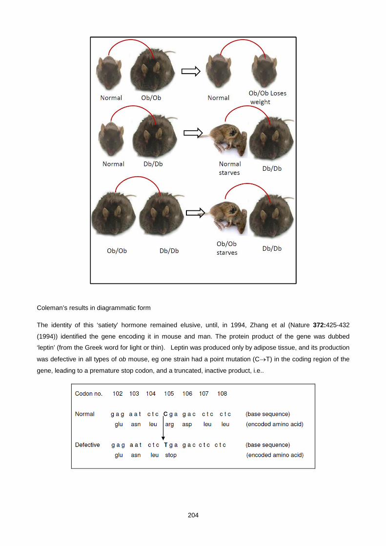
204
Coleman’s results in diagrammatic form
The identity of this ‘satiety’ hormone remained elusive, until, in 1994, Zhang et al (Nature 372:425-432
(1994)) identified the gene encoding it in mouse and man. The protein product of the gene was dubbed
‘leptin’ (from the Greek word for light or thin). Leptin was produced only by adipose tissue, and its production
was defective in all types of ob mouse, eg one strain had a point mutation (C→T) in the coding region of the
gene, leading to a premature stop codon, and a truncated, inactive product, i.e..
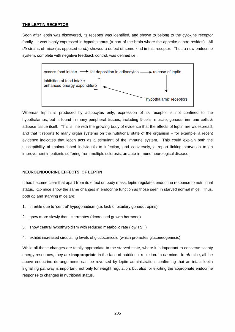
205
THE LEPTIN RECEPTOR
Soon after leptin was discovered, its receptor was identified, and shown to belong to the cytokine receptor
family. It was highly expressed in hypothalamus (a part of the brain where the appetite centre resides). All
db strains of mice (as opposed to ob) showed a defect of some kind in this receptor. Thus a new endocrine
system, complete with negative feedback control, was defined i.e.
Whereas leptin is produced by adipocytes only, expression of its receptor is not confined to the
hypothalamus, but is found in many peripheral tissues, including β-cells, muscle, gonads, immune cells &
adipose tissue itself. This is line with the growing body of evidence that the effects of leptin are widespread,
and that it reports to many organ systems on the nutritional state of the organism – for example, a recent
evidence indicates that leptin acts as a stimulant of the immune system. This could explain both the
susceptibility of malnourished individuals to infection, and conversely, a report linking starvation to an
improvement in patients suffering from multiple sclerosis, an auto-immune neurological disease.
NEUROENDOCRINE EFFECTS OF LEPTIN
It has become clear that apart from its effect on body mass, leptin regulates endocrine response to nutritional
status. Ob mice show the same changes in endocrine function as those seen in starved normal mice. Thus,
both ob and starving mice are:
1. infertile due to ‘central’ hypogonadism (i.e. lack of pituitary gonadotropins)
2. grow more slowly than litterrmates (decreased growth hormone)
3. show central hypothyroidism with reduced metabolic rate (low TSH)
4. exhibit increased circulating levels of glucocorticoid (which promotes gluconeogenesis)
While all these changes are totally appropriate to the starved state, where it is important to conserve scanty
energy resources, they are inappropriate in the face of nutritional repletion. In ob mice. In ob mice, all the
above endocrine derangements can be reversed by leptin administration, confirming that an intact leptin
signalling pathway is important, not only for weight regulation, but also for eliciting the appropriate endocrine
response to changes in nutritional status.

206
DEFECTS OF THE LEPTIN SIGNALLING PATHWAY IN HUMANS
Discovery of the leptin signalling pathway in rodents unleashed a flurry of activity, to determine whether
human obesity could be ascribed to defects in leptin or its receptor. Case studies have recently been
published, describing defects in leptin itself, or in its receptor, in human familial obesity.
DEFECTIVE LEPTIN IN HUMANS
Stephen O”Rahilly & colleagues described 2 siblings, a girl of 8 and a boy of 2, who had normal birth weight,
but developed severe obesity from an early age (Nature, 387:903-908 (1997)). Neither parents nor other
siblings were morbidly obese. The mutation turned out to be in the coding region of the leptin gene. Deletion
of a single guanine in codon 133 of the leptin gene caused a frame-shift, and gave rise to a truncated,
inactive protein product. Serum leptin levels were very low, particularly in the context of their gross obesity.
DEFECTIVE LEPTIN RECEPTOR IN HUMANS
A year later, a French group led by Phillipe Froguel described another family in which 3 daughters were born
normal, but rapidly became morbidly obese (Nature, 1998, 392:398-401). They were also the product of a
consanguinous marriage, in which both parents & other siblings had a normal appearance. In this case, it
turned out that the obesity was due to a mutation in the leptin receptor gene; a point mutation in the first base
of intron 16 (G→A) gave rise to erroneous splicing of the primary RNA transcript, so that exon 16 was omitted
from the mRNA. This led to a truncated receptor protein product, missing its intracellular signalling domain,
which was therefore non-functional.

207
Serum leptin levels in affected sisters was very high (>500ng/ml (normal range 10-30 ng/ml)). In the 2
surviving sisters of 14 and 19 yr, there were no signs of pubertal development. This emphasizes the role of
leptin in signalling adequate nutritional status to the brain before
reproductive function – a high energy demanding activity in females – is initiated.
TREATMENT OF OBESITY WITH LEPTIN
Although these cases confirm that human obesity can arise from mutations in leptin or its receptor, they are
extremely rare. Typically, human obesity is associated with appropriately elevated serum leptin, indicating a
degree of leptin resistance (analogous to diabetes caused by insulin resistance, rather than by insulin
deficiency). Consistent with this, a recent controlled study in obese subjects involving administration of big
doses of leptin led to a modest weight loss in only a small subset of subjects. Patients with leptin deficiency
respond extremely well to leptin replacement. In fact, the two index patients have had their weights and
eating habits normalised by leptin administration.
Although these findings were initially disappointing, it has stimulated research into ‘downstream’ mediators of
leptin action, and a whole array of leptin’s ‘partners’ has emerged. Defective regulation of any of these
‘partners’ could cause obesity, and modulating their activity could provide potential targets for obesity
treatment. These partners are all hypothalamic peptides controlled by leptin. They can be grouped in
appetite stimulators that leptin suppresses, or appetite inhibitors that it activates.
TREATMENT OF LIPODYSTROPHY WITH LEPTIN
Leptin can also be used to treat generalised lipodystrophy. Several clinical trials have demonstrated the
efficacy of leptin treatment
HYPOTHALAMIC APPETITE-STIMULATING PEPTIDES INHIBITED BY LEPTIN
1. Neuropeptide-Y (NPY) is powerful inducer of feeding behavior. An OB mouse crossed with an NPY-
knockout mouse is thinner than a plain OB, but is still not normal. This indicates that leptin does not
inhibit feeding solely by suppressing NPY.
2. Agouti-related peptide (AgRP) exerts an appetite stimulating effect by directly antagonizing α-MSH action
on its receptor. AgRP is closely related to agouti protein that is locally produced in hair follicles and
208
accounts for the ‘ticked’ fur of many animal species eg German shepherds. Mutant mice overexpressing
agouti leads to a grossly obese, bright yellow phenotype (the agouti mouse).
3. Melanin concentrating hormone (MCH). This peptide was initially discovered as a factor causing
lightening of fish scales, but has now also been shown to stimulate appetite. MCH knockout mice are
20% leaner than littermates. MCH-producing neurones are enervated by neurones from the olfactory
cortex, which raises the possibility that appetite can be stimulated by smell even when one is actually
satiated – the pizza effect.
4. Orexins. Newly discovered peptides. Unexpectedly, orexin knockout mice develop narcolepsy (a sleep
disorder).
HYPOTHALAMIC APPETITE-SUPPRESSING PEPTIDES ACTIVATED BY LEPTIN
1. .α-MSH, a product of POMC cleavage, exerts a powerful appetite suppressing action via the
melanocortin-4 (MC-4) receptor in the brain (distinct from the MC-1 receptor in skin that mediates MSH-
induced pigmentation). An estimated 2% of human obesity is believed to be due to inactivating mutations
of the MC-4 receptor, while a mutation in the POMC gene has been described that gives rise to a rare
syndrome of obesity, red hair and adrenal insufficiency.
2. Cocaine & amphetamine-related transcript (CART) is an appetite suppressing peptide. It accounts for
the anorexia induced by amphetamines and cocaine. Particularly amphetamines have been used as
appetite suppressants for many years.
UNCOUPLING PROTEIN (UCP)
Uncoupling protein traverses the inner mitochondrial membrane, where it dissipates the proton gradient
created by electron transport. Thus oxidation of fuel is no longer coupled to ATP synthesis, but simply
squandered as heat (analogous to a short circuit on an electrical appliance). Three members of the family
have been described: UCP-1 in brown adipose tissue, UCP-2 present in all tissues, and UCP-3 in skeletal
muscle. By indirectly activating UCP’s via the sympathetic nervous system, leptin increases fuel oxidation
and thereby promotes weight loss.
CONCLUSIONS
Clearly, appetite regulation is a complex process, with many interactive players, both within and outside the
CNS. And the story is far from complete. For example, a desert weed from South Africa (Kalahari desert)
known as hoodia (Hoodia gordonii), used by the San people to combat hunger during lean times, contains a
powerful appetite suppressant. Its mechanism of action and where it fits into what we already know about
appetite regulation, remains to be determined. The active ingredient in the Hoodia has been isolated and is
currently undergoing clinical trials
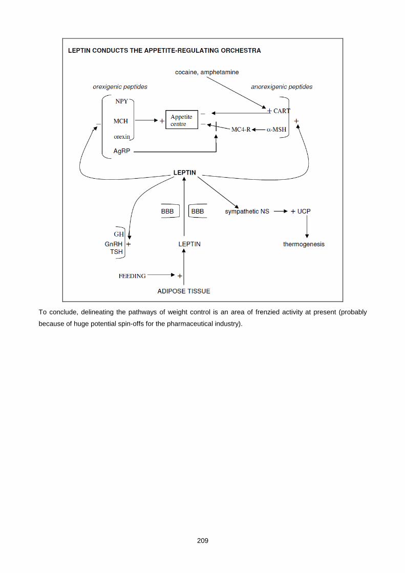
209
To conclude, delineating the pathways of weight control is an area of frenzied activity at present (probably
because of huge potential spin-offs for the pharmaceutical industry).

210
SMALL GROUP TEACHING: LECTURE 10: OBESITY
QUESTIONS:
1. What effect can obesity have on respiratory function? How may this manifest biochemically? What is the Pickwickian syndrome?
2. From your height and weight, calculate your BMI. What is ideal in terms of long-term health?
3. What are serum leptin levels in obese subjects, compared to non-obese controls? What does that tell you about the involvement of leptin in common forms of obesity?
4. Under certain circumstances, a low serum leptin results in an appropriate physiological response. What are the circumstances and what is the response? Indicate another metabolic disease where hormone deficiency results in a response that can occur normally, but is inappropriate under the circumstances. Both conditions lead to a distortion of metabolic reality.
5. List the downstream targets of leptin whereby it mediates weight loss.
6. Explain how a loss of function mutation of the POMC gene leads to a syndrome of morbid obesity, red hair and adrenal failure.
7. What is uncoupling protein? Where is it found? How is its expression affected by leptin?
8. How does obesity aggravate type 2 diabetes? What new class of drugs are useful to manage this problem, & how do they work?
SMALL GROUP TEACHING: LECTURE 10: OBESITY
ANSWERS
1. Reduces tidal volume for mechanical reasons. Rising pCO2. Attacks of severe somnolence during the day - possibly due to chronically raised pCO2, or disturbed sleep from nocturnal apnoeic episodes - from a Dickensian character of that name.
2. weight/height2 eg if weight = 67.5kg and height = 1.5m, then BMI = 30KG/M2
3. High. Common form of obesity due to leptin resistance, rather than deficiency.
4. Circumstances = starvation. Response = ⇑ appetite, ⇓ heat generation, neuroendocrine response (⇓reproduction, ⇓growth, ⇓thyroid function, ⇑cortisol). Type 1 diabetes exhibits a starvation response (gluconeogenesis & ketogenesis) in the face of caloric excess.
5.
i. Inhibits orexigenic peptide release (neuropeptide-Y (NPY), agouti-related peptide (AgRP), orexin, melanin concentrating hormone (MCH).
ii. Activates anorexigenic peptide release (α-MSH, cocaine and amphetamine-related transcript (CART)).
iii. Enhances fat combustion via induction of uncoupling protein (UCP-1) in mitochondria of brown adipose tissue.
6. ↓ α-MSH action in skin via MC1-R (no eumelanin, only phaeomelanin ⇒ red hair) ↓ hypothalamic satiety signalling via MC4-R ⇒ obesity ↓ ACTH ⇒ central hypoadrenalism
7. A protein in the inner mitochondrial membrane that discharges the proton gradient, thus increasing electron transport and ultimately energy expenditure. In adipose tissue - mainly brown but also white. Enhanced by leptin via sympathetic innervation of adipocytes acting through specific β3 adrenergic receptors.
8. Yes. By aggravating insulin resistance. Activators of the adipocyte transcription factor, PPARγ, the thiazolidinediones, eg troglitazone. Stimulate adipocyte differentiation and insulin receptor expression

211
Lecture 11: Biochemistry Of Alcohol Abuse
DR PETER BERMAN 2007
The damage to liver, pancreas, brain, heart, and fetus caused by ethanol is well recognised. Ethanol excess
can also give rise to certain acute metabolic derangements that include hypoglycaemia, metabolic acidosis
(lactate or keto-), hyperlipidaemia, hypophosphataemia and hyperuricaemia. To understand their
pathogenesis, one needs to grasp the basics of ethanol metabolism.
METABOLISM OF ETHANOL
Ethanol is rapidly absorbed from the upper GIT, and distributed in the total body water. Thus ingestion of a
litre of beer (3 standard cans) containing 4% ethanol taken up in a total body water volume of 40 litres would
give an ethanol concentration of 40g/40litre = 1 gram/litre (cf legal limit of 0,8 gram/litre). Ethanol excretion
via the breath or urine is negligible. Its elimination depends primarily on hepatic metabolism, since the
enzymes catalysing the first two steps of ethanol oxidation are confined to the liver.
The first step involves oxidation of ethanol to acetaldehyde. This step can be accomplished by 3 different
systems, namely alcohol dehydrogenase in the cytosol, microsomal ethanol oxidizing system of the
endoplasmic reticulum, and catalase in the peroxisomes. Of these, only the first two are of significance.
1. Alcohol dehydrogenase (AD) is a cytosolic enzyme with a low Km for ethanol (+ 1 mM); hence it is
saturated at low blood ethanol levels, and eliminates ethanol at a constant rate. It is constitutively present
(i.e. not induced). Two hydrogen atoms are transferred from ethanol to NAD, forming NADH and
acetaldehyde. The enzyme is not specific for ethanol, but is capable of metabolizing certain ethanol
homologues, including isopropanol (but not methanol).
2. Microsomal ethanol oxidizing system (MEOS) is a cytochrome P-450 containing enzyme in the
endoplasmic reticulum, and uses NADPH and molecular oxygen to convert ethanol to acetaldehyde. Its
synthesis is induced by chronic exposure to ethanol. Since it has a high Km for ethanol (+ 10 mM), it
assumes increasing importance at high ethanol levels
3. Catalase. This enzyme is located in peroxisomes. It is of little importance in ethanol metabolism, but
plays a major role in methanol oxidation.
In the second step, acetaldehyde, a highly reactive and toxic compound, enters the mitochondria, where it is
oxidized to acetic acid by aldehyde dehydrogenase (ALD). The high activity of this enzyme ensures that
acetaldehyde levels are kept to a minimum. As with AD, this step also generates NADH from NAD.
Disulfiram (antabuse) is an inhibitor of ALD, and is used deliberately to help alcoholics abstain; ingestion of
any ethanol by subjects taking disulfiram results in the acute discomfort of acetaldehyde toxicity. Flagyl
(metronidazole), an anti-microbial drug, has a similar effect. A common genetic polymorphism in Orientals,
resulting in low ALD activity, explains the low ethanol tolerance and low incidence of alcoholism among these
populations.
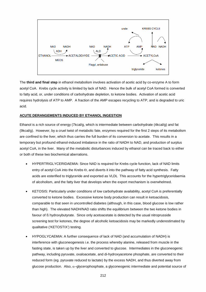
212
The third and final step in ethanol metabolism involves activation of acetic acid by co-enzyme A to form
acetyl CoA. Krebs cycle activity is limited by lack of NAD. Hence the bulk of acetyl CoA formed is converted
to fatty acid, or, under conditions of carbohydrate depletion, to ketone bodies. Activation of acetic acid
requires hydrolysis of ATP to AMP. A fraction of the AMP escapes recycling to ATP, and is degraded to uric
acid.
ACUTE DERANGEMENTS INDUCED BY ETHANOL INGESTION
Ethanol is a rich source of energy (7kcal/g, which is intermediate between carbohydrate (4kcal/g) and fat
(9kcal/g). However, by a cruel twist of metabolic fate, enzymes required for the first 2 steps of its metabolism
are confined to the liver, which thus carries the full burden of its conversion to acetate. This results in a
temporary but profound ethanol-induced imbalance in the ratio of NADH to NAD, and production of surplus
acetyl CoA, in the liver. Many of the metabolic disturbances induced by ethanol can be traced back to either
or both of these two biochemical aberrations.
• HYPERTRIGLYCERIDAEMIA: Since NAD is required for Krebs cycle function, lack of NAD limits
entry of acetyl CoA into the Krebs Θ, and diverts it into the pathway of fatty acid synthesis. Fatty
acids are esterified to triglyceride and exported as VLDL This accounts for the hypertriglyceridaemia
of alcoholism, and the fatty liver that develops when the export mechanism is overwhelmed.
• KETOSIS: Particularly under conditions of low carbohydrate availability, acetyl CoA is preferentially
converted to ketone bodies. Excessive ketone body production can result in ketoacidosis,
comparable to that seen in uncontrolled diabetes (although, in this case, blood glucose is low rather
than high). The elevated NADH/NAD ratio shifts the equilibrium between the two ketone bodies in
favour of ß hydroxybutyrate. Since only acetoacetate is detected by the usual nitroprusside
screening test for ketones, the degree of alcoholic ketoacidosis may be markedly underestimated by
qualitative (‘KETOSTIX’) testing.
• HYPOGLYCAEMIA: A further consequence of lack of NAD (and accumulation of NADH) is
interference with gluconeogenesis i.e. the process whereby alanine, released from muscle in the
fasting state, is taken up by the liver and converted to glucose. Intermediates in the gluconeogenic
pathway, including pyruvate, oxaloacetate, and di-hydroxyacetone phosphate, are converted to their
reduced form (eg. pyruvate reduced to lactate) by the excess NADH, and thus diverted away from
glucose production. Also, α–glycerophosphate, a glyconeogenic intermediate and potential source of
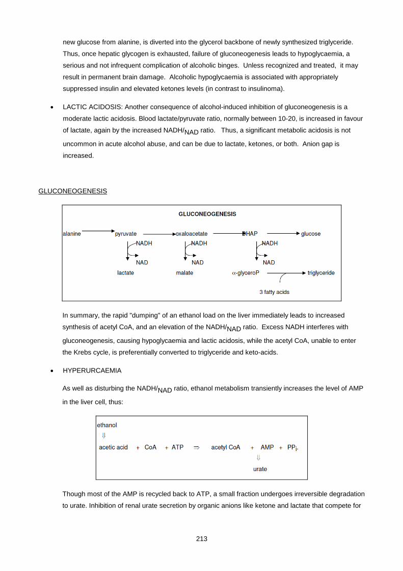
213
new glucose from alanine, is diverted into the glycerol backbone of newly synthesized triglyceride.
Thus, once hepatic glycogen is exhausted, failure of gluconeogenesis leads to hypoglycaemia, a
serious and not infrequent complication of alcoholic binges. Unless recognized and treated, it may
result in permanent brain damage. Alcoholic hypoglycaemia is associated with appropriately
suppressed insulin and elevated ketones levels (in contrast to insulinoma).
• LACTIC ACIDOSIS: Another consequence of alcohol-induced inhibition of gluconeogenesis is a
moderate lactic acidosis. Blood lactate/pyruvate ratio, normally between 10-20, is increased in favour
of lactate, again by the increased NADH/NAD ratio. Thus, a significant metabolic acidosis is not
uncommon in acute alcohol abuse, and can be due to lactate, ketones, or both. Anion gap is
increased.
GLUCONEOGENESIS
In summary, the rapid "dumping" of an ethanol load on the liver immediately leads to increased
synthesis of acetyl CoA, and an elevation of the NADH/NAD ratio. Excess NADH interferes with
gluconeogenesis, causing hypoglycaemia and lactic acidosis, while the acetyl CoA, unable to enter
the Krebs cycle, is preferentially converted to triglyceride and keto-acids.
• HYPERURCAEMIA
As well as disturbing the NADH/NAD ratio, ethanol metabolism transiently increases the level of AMP
in the liver cell, thus:
Though most of the AMP is recycled back to ATP, a small fraction undergoes irreversible degradation
to urate. Inhibition of renal urate secretion by organic anions like ketone and lactate that compete for

214
the same transporter in renal tubules, further increases plasma urate levels, and accounts for the
hyperuricaemia and attacks of gout that are a feature of alcohol abuse.
• OSMOLALITY: Being a small molecule (molecular weight 46), ethanol contributes significantly to
plasma osmolality. For example, the legal limit of .08% (80 mg/100 ml) corresponds to an ethanol
concentration of 80 x 10/46 = 18 mmol/l, which increases plasma osmolality by 18 mosmol/kg. In
severe alcohol intoxication, ethanol can contribute in excess of 50 mosmol/kg to plasma osmolality.
Thus, whenever an obvious disparity exists between measured osmolality and that estimated from
Na, glucose and urea (calculated osmolarity), consider the presence of ethanol. Note that because
ethanol, like urea, is freely diffusable across cell membranes, it cannot establish osmotic gradients
and shift water across cells membranes, as does glucose or sodium.
• ABNORMAL WATER AND NA BALANCE: Alcohol may disturb water homeostasis in a number of
ways. It inhibits release of ADH, resulting in diuresis and mild dehydration (the familiar ‘na-dors’
syndrome). On the other hand, ingestion of vast quantities of water in the form of beer can lead to
dilutional hyponatraemia, where fluid excretion cannot keep pace with intake. In fact, drunk
behaviour in this situation may be due, in part, to acute cerebral oedema rather than ethanol per se.
Over-hydration and hyponatraemia can be distinguished from that due to SIADH by the low urine
osmolality.
LONG-TERM CONSEQUENCES OF ETHANOL INGESTION
POTASSIUM, MAGNESIUM AND PHOSPHATE
Plasma levels of these 3 electrolytes are typically normal at presentation in chronic alcoholics admitted to
hospital, but fall precipitously during treatment to potentially dangerous levels. Why? When ethanol replaces
carbohydrate as major source of calories, intracellular levels of glycolytic intermediates fall (similar to the
situation in starvation and uncontrolled diabetes). Since glycolytic intermediates are all phosphorylated (eg.
glucose-6-phosphate, fructose 1,6 bis-phosphate), their decline is accompanied by efflux of phosphate from the
cell, along with associated cations K+ and Mg2+, which are then lost into the urine. When these patients are
treated with glucose, this process reverses. Glucose taken up by cells becomes phosphorylated, and these
phosphorylated metabolites bind K+ and Mg2+. As phosphate, potassium and magnesium move back into the
cell, there is a rapid fall in their plasma levels. Low ECF K+ and Mg2+ predispose to cardiac arrythmias, while
phosphate depletion can lead to muscle weakness and rhabdomyolysis. Thus, plasma levels of these ions
should be monitored and adequately replaced. A similar situation arises in management of diabetic
ketoacidosis.
VITAMINS
Functional vitamin deficiencies are common in alcohol abusers, and involve mainly folate, thiamine,
pyridoxine and vitamin A. The way in which ethanol affects vitamin utilization varies; pyridoxine (B6) is
inactivated by chemical reaction between acetaldehyde and the pyridoxamine form of the vitamin, while
ethanol competes directly in the conversion of retinol to retinal by retinol dehydrogenase (hence ‘blind’ drunk).

215
The large red cells (MCV > 100 fl), typical of ethanol abuse, are due to impaired folate metabolism, while the
mechanism whereby ethanol potentiates beri-beri (caused by thiamine deficiency) is unknown.
ENDOCRINE EFFECTS
Male alcoholics often show hypogonadism and features of feminization, including gynaecomastia, which has
been attributed to impaired hepatic metabolism of oestrogens. Plasma testosterone levels are also
sometimes reduced. Alcoholism may present with clinical and biochemical features of Cushings syndrome,
including elevated midnight cortisol, loss of diurnal rhythm, and even failure to suppress on low dose
dexamethazone (termed pseudoCushings). All abnormalities, however, disappear on alcohol withdrawal.
PLASMA LIPIDS
Ethanol increases hepatic VLDL synthesis, so that elevated plasma triglycerides are a feature of excessive
alcohol intake. Marked hypertriglyceridaemia (type IV or type V pattern) predisposes to acute pancreatitis
and to the development of eruptive xanthomata. However, the ‘up’ side is that moderate drinkers have a
lower incidence of ischaemic heart disease than do teetotalers. This has been attributed to their higher HDL
cholesterol levels, particularly HDL2.
ALTERED DRUG METABOLISM Many drugs, including barbiturates, are metabolized in the liver by the same cytochrome P-450 system as
ethanol (MEOS). Competition between ethanol and drugs for hepatic metabolism explains why many drugs
are potentiated by simultaneous ingestion of ethanol. For example, ingestion of barbiturates by intoxicated
individuals can lead to barbiturate-induced respiratory arrest. However, once MEOS has been induced by
chronic ethanol intake, hepatic clearance of certain drugs is increased. This accounts for the tolerance
exhibited by alcoholics towards many drugs, including alcohol itself (eg. ‘I can drink you under the table’ really
means ‘my MEOS is more induced than your MEOS’). In certain cases, induction of MEOS can enhance
drug toxicity. This occurs when the metabolite is more toxic than the parent drug - for example, paracetamol,
itself harmless, is metabolized by hepatic microsomes to a highly toxic quinone-imine intermediate that
damages the liver cell. Alcoholics with induced MEOS are, therefore, more prone to paracetamol toxicity.
BIOCHEMICAL MARKERS OF ETHANOL ABUSE
A number of biochemical markers of alcohol abuse have been proposed, which include:
1. AST/ALT ratio: Elevated serum transaminases, with a characteristically high AST/ALT ratio as compared
to non-alcoholic forms of hepatitis.
2. γ-GT: Elevation of serum γ-glutamyl transferase, out of proportion to the alkaline phosphatase and other
markers of cholestasis, is characteristic of chronic ethanol exposure, and is due to induction of this
hepatic microsomal enzyme by ethanol.
3. Carbohydrate-deficient transferring: Alcohol abuse results in defective glycosylation of transferrin by
the liver (carbohydrate-deficient transferrin (CDT)), which can be demonstrated electrophoretically.

216
4. MCV: Although not a strictly biochemical marker, increased red cell size (mean corpuscular volume) is a
useful marker of ongoing alcohol abuse, often exceeding 100fl (normally 80-90fl).
FOOTNOTE
Despite this litany of ills caused by ethanol, one should acknowledge the euphoria it undoubtedly confers on
countless people every day - statistics show that moderate drinkers have a longer lifespan than teetotallers.
For example, the French have the highest per capita intake of alcohol (in the form of red wine), as well as the
largest percentage of old people, in Europe. Makes you think….
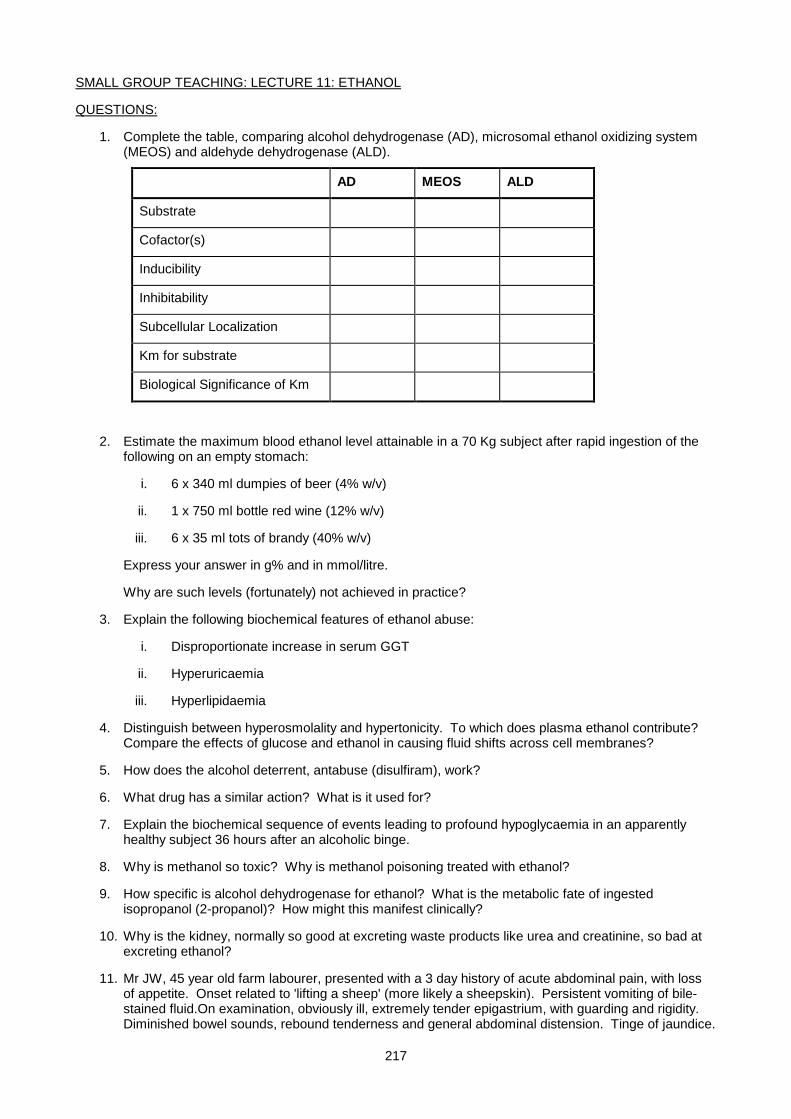
217
SMALL GROUP TEACHING: LECTURE 11: ETHANOL
QUESTIONS:
1. Complete the table, comparing alcohol dehydrogenase (AD), microsomal ethanol oxidizing system (MEOS) and aldehyde dehydrogenase (ALD).
AD MEOS ALD
Substrate
Cofactor(s)
Inducibility
Inhibitability
Subcellular Localization
Km for substrate
Biological Significance of Km
2. Estimate the maximum blood ethanol level attainable in a 70 Kg subject after rapid ingestion of the following on an empty stomach:
i. 6 x 340 ml dumpies of beer (4% w/v)
ii. 1 x 750 ml bottle red wine (12% w/v)
iii. 6 x 35 ml tots of brandy (40% w/v)
Express your answer in g% and in mmol/litre.
Why are such levels (fortunately) not achieved in practice?
3. Explain the following biochemical features of ethanol abuse:
i. Disproportionate increase in serum GGT
ii. Hyperuricaemia
iii. Hyperlipidaemia
4. Distinguish between hyperosmolality and hypertonicity. To which does plasma ethanol contribute? Compare the effects of glucose and ethanol in causing fluid shifts across cell membranes?
5. How does the alcohol deterrent, antabuse (disulfiram), work?
6. What drug has a similar action? What is it used for?
7. Explain the biochemical sequence of events leading to profound hypoglycaemia in an apparently healthy subject 36 hours after an alcoholic binge.
8. Why is methanol so toxic? Why is methanol poisoning treated with ethanol?
9. How specific is alcohol dehydrogenase for ethanol? What is the metabolic fate of ingested isopropanol (2-propanol)? How might this manifest clinically?
10. Why is the kidney, normally so good at excreting waste products like urea and creatinine, so bad at excreting ethanol?
11. Mr JW, 45 year old farm labourer, presented with a 3 day history of acute abdominal pain, with loss of appetite. Onset related to 'lifting a sheep' (more likely a sheepskin). Persistent vomiting of bile-stained fluid.On examination, obviously ill, extremely tender epigastrium, with guarding and rigidity. Diminished bowel sounds, rebound tenderness and general abdominal distension. Tinge of jaundice.
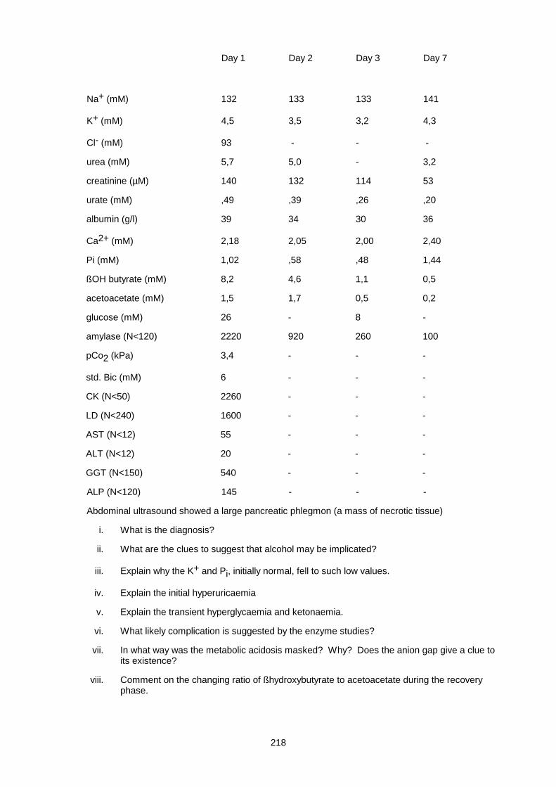
218
Day 1 Day 2 Day 3 Day 7
Na+ (mM) 132 133 133 141
K+ (mM) 4,5 3,5 3,2 4,3
Cl- (mM) 93 - - -
urea (mM) 5,7 5,0 - 3,2
creatinine (µM) 140 132 114 53
urate (mM) ,49 ,39 ,26 ,20
albumin (g/l) 39 34 30 36
Ca2+ (mM) 2,18 2,05 2,00 2,40
Pi (mM) 1,02 ,58 ,48 1,44
ßOH butyrate (mM) 8,2 4,6 1,1 0,5
acetoacetate (mM) 1,5 1,7 0,5 0,2
glucose (mM) 26 - 8 -
amylase (N<120) 2220 920 260 100
pCo2 (kPa) 3,4 - - -
std. Bic (mM) 6 - - -
CK (N<50) 2260 - - -
LD (N<240) 1600 - - -
AST (N<12) 55 - - -
ALT (N<12) 20 - - -
GGT (N<150) 540 - - -
ALP (N<120) 145 - - -
Abdominal ultrasound showed a large pancreatic phlegmon (a mass of necrotic tissue)
i. What is the diagnosis?
ii. What are the clues to suggest that alcohol may be implicated?
iii. Explain why the K+ and Pi, initially normal, fell to such low values.
iv. Explain the initial hyperuricaemia
v. Explain the transient hyperglycaemia and ketonaemia.
vi. What likely complication is suggested by the enzyme studies?
vii. In what way was the metabolic acidosis masked? Why? Does the anion gap give a clue to its existence?
viii. Comment on the changing ratio of ßhydroxybutyrate to acetoacetate during the recovery phase.
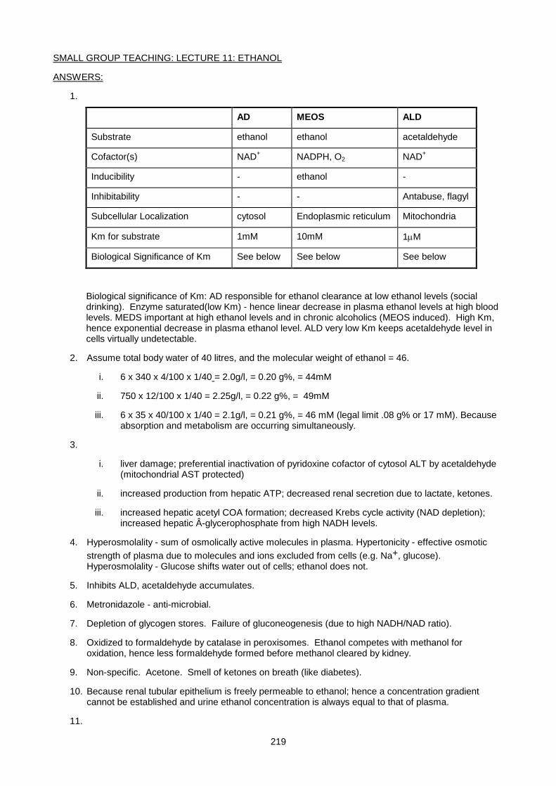
219
SMALL GROUP TEACHING: LECTURE 11: ETHANOL
ANSWERS:
1.
AD MEOS ALD
Substrate ethanol ethanol acetaldehyde
Cofactor(s) NAD+ NADPH, O2 NAD+
Inducibility - ethanol -
Inhibitability - - Antabuse, flagyl
Subcellular Localization cytosol Endoplasmic reticulum Mitochondria
Km for substrate 1mM 10mM 1µM
Biological Significance of Km See below See below See below
Biological significance of Km: AD responsible for ethanol clearance at low ethanol levels (social drinking). Enzyme saturated(low Km) - hence linear decrease in plasma ethanol levels at high blood levels. MEDS important at high ethanol levels and in chronic alcoholics (MEOS induced). High Km, hence exponential decrease in plasma ethanol level. ALD very low Km keeps acetaldehyde level in cells virtually undetectable.
2. Assume total body water of 40 litres, and the molecular weight of ethanol = 46.
i. 6 x 340 x 4/100 x 1/40 = 2.0g/l, = 0.20 g%, = 44mM
ii. 750 x 12/100 x 1/40 = 2.25g/l, = 0.22 g%, = 49mM
iii. 6 x 35 x 40/100 x 1/40 = 2.1g/l, = 0.21 g%, = 46 mM (legal limit .08 g% or 17 mM). Because absorption and metabolism are occurring simultaneously.
3.
i. liver damage; preferential inactivation of pyridoxine cofactor of cytosol ALT by acetaldehyde (mitochondrial AST protected)
ii. increased production from hepatic ATP; decreased renal secretion due to lactate, ketones.
iii. increased hepatic acetyl COA formation; decreased Krebs cycle activity (NAD depletion); increased hepatic Â-glycerophosphate from high NADH levels.
4. Hyperosmolality - sum of osmolically active molecules in plasma. Hypertonicity - effective osmotic strength of plasma due to molecules and ions excluded from cells (e.g. Na+, glucose). Hyperosmolality - Glucose shifts water out of cells; ethanol does not.
5. Inhibits ALD, acetaldehyde accumulates.
6. Metronidazole - anti-microbial.
7. Depletion of glycogen stores. Failure of gluconeogenesis (due to high NADH/NAD ratio).
8. Oxidized to formaldehyde by catalase in peroxisomes. Ethanol competes with methanol for oxidation, hence less formaldehyde formed before methanol cleared by kidney.
9. Non-specific. Acetone. Smell of ketones on breath (like diabetes).
10. Because renal tubular epithelium is freely permeable to ethanol; hence a concentration gradient cannot be established and urine ethanol concentration is always equal to that of plasma.
11.
220
i. Acute pancreatitis, alcohol induced.
ii. Alcohol commonest cause of acute pancreatitis in W. Cape, especially in wine farm labourers. High urate, high GGT. Possibly high AST/ALT ratio.
iii. Uptake of glucose into cells during treatment.
iv. Increased production/decreased secretion.
v. Hyperglycaemia - transient ßcell dysfunction, acute stress (cortisol, adrenalin).
vi. Hyperketonaemia - ßcell dysfunction as well as synthesis from ethanol.
vii. Alcoholic rhabdomyolysis.
viii. Concomitant respiratory alkalosis. Stress of acute pancreatitis. Anion gap = 27 (increased).
ix. ßhydroxy/acetoacetate ratio decreases as hepatic NADH/NAD ratio normalizes.
221
Lecture 12: Disorders Of Protein Metabolism
PROF E.H. HARLEY, UPDATED DR F OMAR 2007
PLASMA PROTEINS COMPARED TO TISSUE PROTEINS
Plasma proteins differ from proteins in cells because they need to last longer (be more stable) and be more
soluble (many intra-cellular proteins are membrane-bound or part of multi-enzyme complexes). To effect
these properties they are
1) more oxidised than intra-cellular proteins: i.e. there are more proteins with -S-S- bonds than with free -SH
groups.
2) they have a high carbohydrate (CHO) content, and are therefore mostly glycoproteins.
Tissue Proteins in contrast, being less long lived and more insoluble, are more reduced (more -SH groups
than -S-S-), and have a low CHO content. CHO therefore:
• Increases solubility
• " stability
• Specific CHO moieties are also obligatory for secretion into the plasma.
DISTRIBUTION in a (typical) 70 kg man:
Plasma (intravascular): 75g/l = 250g total. Extravascular Pool = 350g Total = 600g
60% of Albumin is extravascular compared with only 20% for fibrinogen. However, the concentration of
albumin is still much higher in the plasma than in interstitial fluids, thereby maintaining the oncotic pressure
and preventing fluid leaking out of the circulation.
Equilibration is SLOW between intravascular and extravascular pools therefore:
For acute changes, e.g. haemorrhage, consider IV losses only.
For chronic changes consider changes in volume of the extravascular pool as well.
The half life of most plasma proteins (t½) varies between 10 hours and 24 days.
Total daily turnover is ± 25g plasma protein (compared with ± 150g for tissue proteins)
SYNTHESIS:
Liver - most plasma proteins
Plasma cells and lymphocytes - immunoglobulins
Gut - lipoproteins
Synthesis on ribosomes takes about 1-2 mins:- Extracellular Secretion takes 20-40 min
CATABOLISM:
222
Takes place in
• capillary endothelium
• hepatocyte
• kidney - normally only low M.W. proteins, which are filtered, reabsorbed and degraded by proximal tubule cells e.g. B2 microglobulin, Bence Jones Protein (BJP)
FUNCTIONS (examples in parentheses)
• Oncotic pressure (all)
• Repair, complement activation (CRP)
• Buffering (all)
• Clotting (fibrinogen, clotting factors)
• Carrier, of metals, hormones, vitamins etc
• Immune system (immunoglobulins)
• Anti-oxidant (albumin)
• Enzymes (renin, Angiotensin Converting Enz.)
• Hormonal and signalling (insulin)
• Anti-enzymes (α1-antitrypsin, α2 macroglobulin)
CHANGES IN TOTAL PROTEIN
Raised in
1. Water loss - dehydration
2. Some chronic diseases - generalised increase in γ-globulins
3. Paraproteinaemias - increase in a specific γ-globulin
Lowered in
1. Water overload - overhydration, artifactual if sample taken from "drip arm"
2. Excessive protein loss - renal (nephrotic syndrome), skin (burns), intestine (PLE)
3. Decreased synthesis - kwashiorkor, cirrhosis, malabsorption
PROTEIN DEFICIENT DIETS:
• Kwashiorkor - diet of high carbohydrate, but low protein - plump looking child, but much of this may be oedema from low oncotic pressure.
• Marasmus - diet deficient in calories and proteins - thin, emaciated appearance.
Effects of a period of total starvation:
• 24 hrs - glycogen depletion
• 2-5 days - protein and (mostly) fat catabolism begins

223
• Eventually fat stores are completely depleted leading to a rise in protein catabolism pre-mortem.
Albumin is a reasonable index of medium to long term protein depletion (due to long t½) but note opposing
effects of dehydration.
Hyperglobulinaemia can coexist with depletion of other proteins if there is superadded infection, so total
protein may be normal in malnutrition, where there is often chronic infection with high IGs.
Prealbumin and retinol binding protein are short half-life proteins which can be used to measure short term
protein status.
Nomalizing protein status before elective surgery by good nutrition improves outcome.
The blood urea concentration is a useful index of protein metabolism, normally = 3-7 mmol/l. but higher at
birth and in the elderly. It is decreased in pregnancy and during anabolic states, and with low protein diets. It
is raised in renal disease and by high protein diets
SERUM PROTEIN ELECTROPHORESIS
Because of their amino acid composition, all proteins have a net negative or positive charge at a given pH.
This property is exploited during the process of protein electrophoresis, to separate proteins into 5 distinct
groups seen in the electrophoretogam below (Albumin, Alpha 1 globulins, Alpha 2 globulins, Beta (1 &
2)globulins and Gamma globulins). In agarose gel electrophoresis, serum sample is applied close to the
cathode end of the support medium within a buffer (pH 8.6). A voltage (100V) is applied through the medium
via two electrodes. The most negatively charged proteins will migrate rapidly towards the anode (eg albumin),
while the more positively charged proteins will migrate slower, remaining closer to the cathode (eg
immunoglobulins). After separation, the protein is fixed and then stained with an appropriate protein stain e.g.
Coomassie blue. The strip can then be either visually inspected, or the bands can be scanned
densitometrically (the absorbance of the dye is measured and the protein concentration is calculated as a
percentage of the total protein).
Another method which is now more commonly used is capillary zone electropheresis. This method makes
use of thin glass capillaries, with mobile cations associated with the negatively charged inner walls. The
sample is introduced into a capillary and a voltage applied (10-30kV). This causes the cations in the alkaline
buffer to move towards the cathode (negative electrode), forcing the movement of the buffer and its contents
cathodally (endosmosis). A detector (measures absorbance of peptide bonds at 220nm) is present at the
cathode, and the endosmotic action forces the movement of the different proteins past the detector, the more
positively charged proteins arriving first, followed by the more negatively charged proteins (thus the most
negatively charged proteins (anodal) take the longest to move past the detector). The graph obtained is one
of absorbance (dependent on protein concentration) vs time. It consists of 5 zones, namely from anodally:
albumin, alpha 1, alpha 2, beta and gamma.
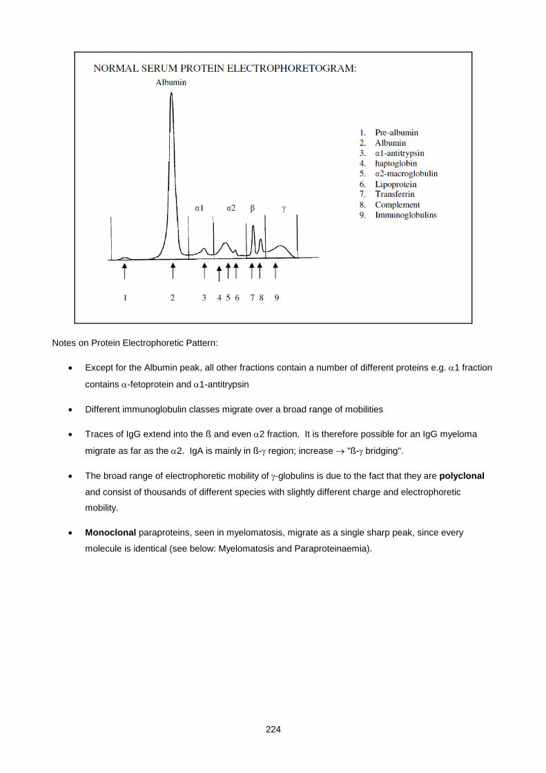
224
Notes on Protein Electrophoretic Pattern:
• Except for the Albumin peak, all other fractions contain a number of different proteins e.g. α1 fraction
contains α-fetoprotein and α1-antitrypsin
• Different immunoglobulin classes migrate over a broad range of mobilities
• Traces of IgG extend into the ß and even α2 fraction. It is therefore possible for an IgG myeloma
migrate as far as the α2. IgA is mainly in ß-γ region; increase → "ß-γ bridging".
• The broad range of electrophoretic mobility of γ-globulins is due to the fact that they are polyclonal
and consist of thousands of different species with slightly different charge and electrophoretic
mobility.
• Monoclonal paraproteins, seen in myelomatosis, migrate as a single sharp peak, since every
molecule is identical (see below: Myelomatosis and Paraproteinaemia).
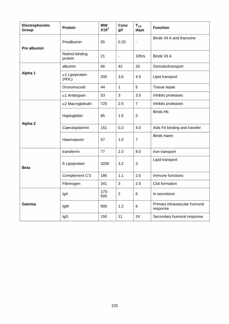
225
Electrophoretic Group Protein MW
X103 Conc g/l
T1/2 days Function
Pre albumin Prealbumin 55 0.25 -
Binds Vit A and thyroxine
Retinol binding protein 21 - 10hrs Binds Vit A
Alpha 1
albumin 66 42 20 Osmotic/transport
α1 Lipoprotein (HDL) 200 3.6 4.5 Lipid transport
Orosomucoid 44 1 5 Tissue repair
α1 Antitrypsin 53 3 3.5 Inhibits proteases
Alpha 2
α2 Macroglobulin 725 2.5 7 Inhibits proteases
Haptoglobin 85 1.5 2 Binds Hb
Caeruloplasmin 151 0.3 4.5 Aids Fe binding and transfer
Haemopexin 57 1.0 7 Binds haem
Beta
transferrin 77 2.3 8.5 Iron transport
ß Lipoprotein 3200 3.2 3 Lipid transport
Complement C'3 185 1.1 2.5 Immune functions
Fibrinogen 341 3 2.5 Clot formation
Gamma
IgA 175-500 2 6 In secretions
IgM 900 1.2 6 Primary intravascular humoral response
IgG 150 11 24 Secondary humoral response
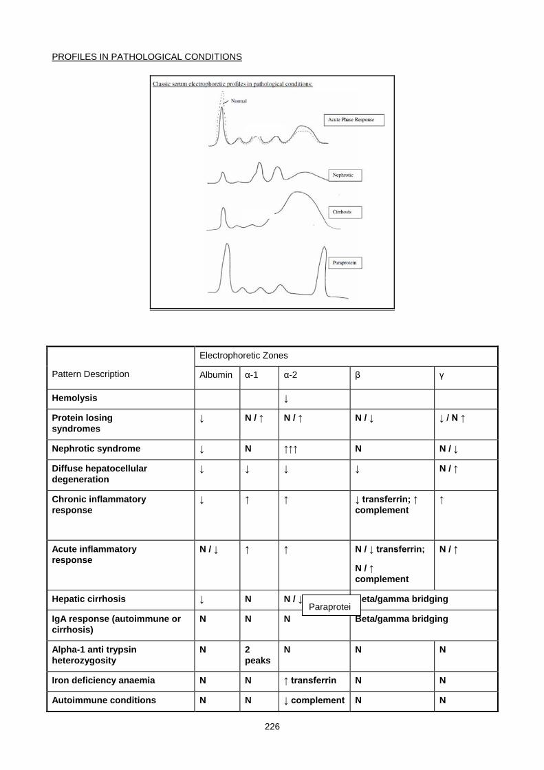
226
PROFILES IN PATHOLOGICAL CONDITIONS
Pattern Description
Electrophoretic Zones
Albumin α-1 α-2 β γ
Hemolysis ↓
Protein losing syndromes
↓ N / ↑ N / ↑ N / ↓ ↓ / N ↑
Nephrotic syndrome ↓ N ↑↑↑ N N / ↓
Diffuse hepatocellular degeneration
↓ ↓ ↓ ↓ N / ↑
Chronic inflammatory response
↓ ↑ ↑ ↓ transferrin; ↑ complement
↑
Acute inflammatory response
N / ↓ ↑ ↑ N / ↓ transferrin;
N / ↑ complement
N / ↑
Hepatic cirrhosis ↓ N N / ↓ Beta/gamma bridging
IgA response (autoimmune or cirrhosis)
N N N Beta/gamma bridging
Alpha-1 anti trypsin heterozygosity
N 2 peaks
N N N
Iron deficiency anaemia N N ↑ transferrin N N
Autoimmune conditions N N ↓ complement N N
Paraprotei
227
Other patterns:
• Broad increase in gamma region = polyclonal hypergammaglobulinaemia as in chronic infections.
• Oligoclonal banding is occasionally seen in chronic aggressive hepatitis, chronic infections and
certain malignancies.
• A narrow peak in the gamma region (or, more rarely, in more anodal regions), often accompanied by
a general decrease in the normal gamma globulins = suggestive of monoclonal paraprotein,
commonly due to multiple myeloma.
ALBUMIN AND PRE-ALBUMIN
PREALBUMIN
This region is anodal to albumin, consisting mainly of transthyretin (transports thyroxine) and retinol binding
protein (tranports vitamin A).
Transthyretin complexes with retinol binding protein, preventing filtration by glomeruli.
Prealbumin decreased in hepatic damage, acute phase inflammatory response and tissue necrosis.
Low prealbumin is a sensitive marker of poor protein nutritional status (short half life – 2d). Elevated levels
are seen in patients on steroids, in alcoholism and chronic renal failure.
ALBUMIN
Gene on chromosome 4q (linked to α-feto protein and vitamin D binding globulin).
Most abundant protein in serum. Synthesised in liver.
Two well-known functions:
• maintaining intravascular colloid osmotic pressure
• Binds and transports various substances:
o Bilirubin
o FFA (high affinity)
o Ca++ (& other metals) low affinity and high capacity
o Hormones, e.g. thyroxine and T3, cortisol, aldosterone
o Many drugs
Abnormal forms found include:
o Analbuminaemia – congenital, autosomal codominant inheritance. Mild oedema and altered lipid metabolism.
228
o Bisalbuminaemia – altered electrophoretic pattern (2 peaks)
o Alb B – migrates more cathodally on electrophoresis (glu →lys substitution)
o Familial dysalbuminaemic hyperthyroxinaemia (decreased or increased affinity for thyroxine – euthyroid but elevated serum total T4 with normal free T4)
CAUSES OF INCREASED ALBUMIN:
Increased only in dehydration
CAUSES OF DECREASED ALBUMIN:
• Decreased synthesis
o Inherited: analbuminaemia
o Inadequate dietary protein
o Liver disease
• Increased catabolism
o Starvation
o Infections
o Burns
o Malignancy
• Increased losses
o acute phase - ↑ vascular permeability (shift into ECF)
o nephrotic syndrome
o protein losing enteropathy (PLE)
o burns
o haemorrhage
• Volume changes
o posture (higher when standing than recumbent)
o pregnancy
o haemorrhage
o overhydration, drip arm contamination
CONSEQUENCES OF HYPOALBUMINAEMIA
• Oedema* and secondary hyperaldosteronism
• Decreased binding by albumin leads to increased toxicity (metals, bilirubin etc)
• Decreased total serum calcium
* More generally, oedema can be caused by decreased oncotic pressure, increased capillary hydrostatic pressure, or an increase in capillary permeability.
229
CLINICAL USES OF ALBUMIN MEASUREMENT:
• Assessment of long term nutritional status (t½ 20 days)
• Calculation of corrected calcium
• Liver function test
• Assessment of risk of kernicterus
• Assessment of hydration status
•
* More generally, oedema can be caused by decreased oncotic pressure, increased capillary hydrostatic
pressure, or an increase in capillary permeability.
ANALYTICAL METHODOLOGY:
Serum levels: dye binding methods including bromcresol green or purple (higher affinity for albumin than
other protein)
Urine and cerebrospinal fluid require more sensitive methods: immunoturbidimetry and
immunonephelometry.
ACUTE PHASE PROTEINS:
These are proteins which are increased or decreased in any acute inflammatory process. They are part of
the body's response to limit further tissue damage (by proteases, free radicals etc), and to initiate repair.
Surgery, trauma, myocardial infarction, baterial infections and tumours are conditions eliciting an
inflammatory response.
Positive acute phase reponders Negative acute phase responders
Fibrinogen (clotting)
Haptoglobin (Hb salvage)
Caeruloplasmin (Hb synthesis)
α1-antitrypsin (anti-protease)
α1-antichymoptrypsin (anti-protease)
α1 acid glycoprotein (repair)
C-reactive protein (aids phagocytosis)
Complement (C’3)
Ferritin
Transthyretin
Albumin
Transferrin
Elevated first: CRP, serum amyloid A, α1- anti-chymotrypsin
After 12hours: α1-acid glycoprotein
230
After 24 – 48 hours: α1-antitrypsin, haptoglobin, fibrinogen and finally, C3 and caeruloplasmin
1. ALPHA 1-ANTITRYPSIN (AAT)
General serine protease inhibitor (serpin) - of elastase, collagenase, trypsin, chymotrypsin, thrombin,
plasmin [Other serpins incl. α1-anti-chymotrypsin, α2-antiplasmin, antithrombin III]
AAT has a small molecular weight (51kDa) and can thus be found in all bodily fluids. It is the most important
inhibitor of leukocyte elastase (released during phagocytosis by PMN) found in vascular endothelium as well
as in the tracheobronchial tree. Uninhibited elastase action leads to emphysema
Increased levels occur during the acute phase response (synthesis stimulated by cytokines) and with
increased estrogen (increased synthesis).
Decreased levels are seen in:
o Increased utilisation: Secondarily lowerd in neonatal distress syndrome, severe neonatal hepatitis
and preterminal pancreatic disease
o Urinary and GIT loss: normally diffuses into glomerular urine (small size) but usually reabsorbed and
degraded by proximal tubular cells and thus not seen in urine unless there is proximal tubular
damage or overflow as in nephrotic syndrome. Patients with nephrotic syndrome will have lowered
serum levels.
o Protein losing enteropathies.
Variants of α1 antitrypsin are encountered in the population :
Normal form is : Pi MM - M is the normal allele (Pi stands for Proteinase inhibitor)
Abnormal forms : Pi SS ~60% normal activity
Pi ZZ~15% normal activity: a point mutation causes a single amino acid substitution preventing glycosylation
and thus secretion.
Compound heterozygotes are also common, e.g. Pi SZ (38% normal activity)
Deficiency causes:
o lung disease - due to failure to inhibit elastase, resulting in emphysema. Smoking aggravates the
problem by recruitment and activation of leukocytes, and by generating free radicals. These oxidize a
methionine at the α1 antitrypsin active site, and thereby inactivate it.
o liver disease – neonatal cholestasis (may need to distinguish from biliary atresia), cirrhosis (in ZZ
only) - ZZ not secreted, and builds up in hepatocytes, hepatocellular carcinoma.
α1 antitrypsin should be measured in chronic lung disease and paediatric liver disease.
231
Deficiency should be suspected when the α1 band is decreased on electrophoresis, α1 antitrypsin being the
main constituent of the α1 region.
Confirmation of α1 antitrypsin deficiency requires accurate measurement of serum levels using specific
immunological techniques.
Analytical methodology: major constituent of the α1 globulin region on serum protein electrophoresis.
,Immunoturbidimetry, immunonephelometry
2. HAPTOGLOBIN
An alpha-2 glycoprotein; acute phase reactant.
Functions:
o NB binds irreversibly to free (oxy)haemoglobin liberated into the plasma during intravascular
haemolysis. The haptoglobin-haemoglobin complex is removed rapidly by the reticulo-endothelial
system, this prevents loss of haemoglobin in urine, & conserves iron. This complexing also prevents
lipid peroxidation by haemoglobin.
o Anti-inflammatory role (potent peroxidase; inhibits cathepsin B released by phagocytes;
bacteriostatic)
Low levels are a diagnostic indicator of intravascular haemolysis (but may be low in liver disease or with
endogenous or exogenous estrogen).
Elevated levels are associated with acute phase response, nephrotic syndrome and with corticosteroids.
Analytical methodology: alpha 2 region on SPE; immunoturbidimetry
3. COMPLEMENT
Many components (at least 20 proteins): Major quantitatively is C3 (migrates as a β-globulin on
electrophoresis).
Increased in acute phase reactions. Acts with some immunoglobulins to destroy bacteria.
Decreased in active auto-immune disease e.g. active Systemic Lupus Erythematosus (SLE).
Also decreased with increased consumption e.g. post streptococcoal glomerulonephritis and other severe
infections such as Meningococcal septicaemia.
Analytical methodology: migrates to β2 region on serum protein electrophoresis; immunoturbidimetry.
4. C-REACTIVE PROTEIN
Migrates anywhere in the beta and gamma zones (on electrophoresis) depending on buffer. Normal levels
very low (6 mg/l).
Binds the C-polysaccharide of Strep. pneumoniae and many other anions.
232
Acts like a primitive antibody, and when complexed activates the complement cascade.
A (very) acute phase reactant (increases up to 2000 x normal) e.g. after myocardial infarct, trauma, infection,
surgery, or neoplastic proliferation. Therefore used as a very sensitive index of the inflammatory response
e.g. for monitoring therapeutic response in rheumatoid arthritis.
High Sensitivity CRP also independent risk predictor in cardiovascular disease.
Analytical methodology: migrates between the β and γ regions on serum protein electrophoresis;
immunoturbidimetry
OTHER PLASMA PROTEINS
ALPHA 2-MACROGLOBULIN
Very large (mw ~800,000). Not an acute phase protein since levels don't rise in acute inflammation.
Protease inhibitor - binds to all proteases and inhibits by enfolding them and preventing substrate access.
The protease-α2 macroglobulin complex is rapidly cleared by the reticulo-endothelial system. Causes a
marked increase in the alpha 2 region of the plasma electrophoretic profile in nephrotic syndrome, since low
albumin induces a generalized increase in hepatic protein synthesis - the small proteins are lost in the urine
but large (α2 macro and ß lipoprotein i.e. LDL) accumulate.
ß2 MICROGLOBULIN (MW 11800)
This small protein is part of the histocompatibility HLA antigen found on the cell membrane of nearly all cells,
but especially lymphocytes. It is freely filtered and is normally reabsorbed and degraded in the proximal
convoluted tubule. Urine levels increase in renal tubular damage and one of its main uses is in monitoring of
nephrotoxic drugs. Blood levels rise in renal failure (due to decreased GFR) and in increased tissue turnover
states (tumours, inflammatory states).
CAERULOPLASMIN
• Binds 8 Cu atoms per molecule
• Although 95% of plasma Cu is associated with caeruloplasmin it is not a Cu transport protein
• Oxidizes Fe2+ to Fe3+ and thus facilitates iron binding to transferrin, facilitating Hb synthesis.
• Wilson's disease : inherited disease with failure of normal liver excretion of Cu into the bile, resulting
in hepatic Cu overload with cirrhosis and deposition of Cu in brain (encephalopathy) and pupil
(Kayser Fleischer ring). Increased Cu stores decrease caeruloplasmin synthesis. Diagnosis : low
plasma caeruloplasmin and increased hepatic copper as well as increased urinary excretion of
copper
TRANSFERRIN
• Transferrin is the principle transport protein for iron (Fe3+), one molecule binding two ferric ions and
an associated anion (usually bicarbonate).
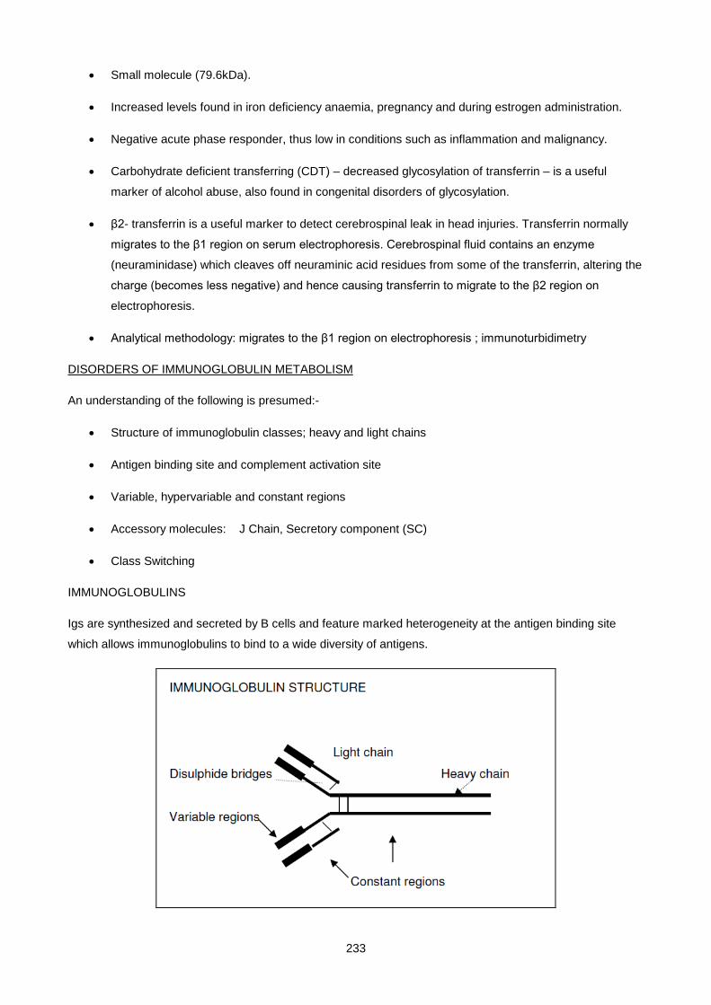
233
• Small molecule (79.6kDa).
• Increased levels found in iron deficiency anaemia, pregnancy and during estrogen administration.
• Negative acute phase responder, thus low in conditions such as inflammation and malignancy.
• Carbohydrate deficient transferring (CDT) – decreased glycosylation of transferrin – is a useful
marker of alcohol abuse, also found in congenital disorders of glycosylation.
• β2- transferrin is a useful marker to detect cerebrospinal leak in head injuries. Transferrin normally
migrates to the β1 region on serum electrophoresis. Cerebrospinal fluid contains an enzyme
(neuraminidase) which cleaves off neuraminic acid residues from some of the transferrin, altering the
charge (becomes less negative) and hence causing transferrin to migrate to the β2 region on
electrophoresis.
• Analytical methodology: migrates to the β1 region on electrophoresis ; immunoturbidimetry
DISORDERS OF IMMUNOGLOBULIN METABOLISM
An understanding of the following is presumed:-
• Structure of immunoglobulin classes; heavy and light chains
• Antigen binding site and complement activation site
• Variable, hypervariable and constant regions
• Accessory molecules: J Chain, Secretory component (SC)
• Class Switching
IMMUNOGLOBULINS
Igs are synthesized and secreted by B cells and feature marked heterogeneity at the antigen binding site
which allows immunoglobulins to bind to a wide diversity of antigens.
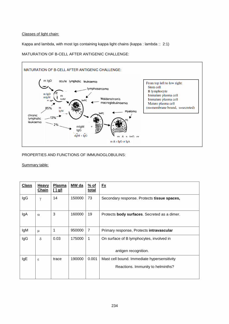
234
Classes of light chain:
Kappa and lambda, with most Igs containing kappa light chains (kappa : lambda :: 2:1)
MATURATION OF B-CELL AFTER ANTIGENIC CHALLENGE:
PROPERTIES AND FUNCTIONS OF IMMUNOGLOBULINS:
Summary table:
Class Heavy Chain
Plasma [ ] g/l
MW da % of total
Fx
IgG γ 14 150000 73 Secondary response. Protects tissue spaces,
IgA α 3 160000 19 Protects body surfaces. Secreted as a dimer.
IgM µ 1 950000 7 Primary response, Protects intravascular
IgG δ 0.03 175000 1 On surface of B lymphocytes, involved in
antigen recognition.
IgE ε trace 190000 0.001 Mast cell bound. Immediate hypersensitivity
Reactions. Immunity to helminths?
m & s IgG or
m IgM
mIgM + IgG
m IgD
235
Further Details of functions:
IgG
• Function: Protection of tissue spaces and bloodstream
• Binds complement
• Crosses placenta - Full term neonate has normal adult levels
IgA
• Function: Protection of body mucosal surfaces
• Formed by plasma cells beneath mucosal surfaces
• IgA enters the mucosal cells which synthesize and attach a secretory piece that binds two IgA molecules to form dimers which are then secreted onto the luminal surface of the mucosa.
• Deficiency of IgA causes upper respiratory tract infections and gastroenteritis
IgM
• Pentamer (five of the above basic units joined at the base to form a five pointed "star"
• Functions: Protection of bloodstream. First antibody to be synthesized in immune response.
• Binds complement
• Does not cross placenta but can be synthesized by the foetus if there is intra-uterine infection;
therefore elevated levels at birth implies intrauterine infection
IgE
• Binds to plasma membranes of mast cells and basophils.
• Binding of antigen (Ag) causes cell degranulation with release of histamine, kinins etc. which cause asthma and hay-fever.
• Elevated in extrinsic asthma. Measurement of Ag specific IgE (RAST test) identifies allergen to which the patient is allergic.
IMMUNOGLOBULIN DEFICIENCY STATES
Primary - suspect these in cases of recurrent or atypical infections.
Exclude by specific immunoglobulin measurements using immunological techniques.
e.g. i) Severe combined immunodeficiency : Immunoglobulin and T cell deficiency
ii) x-linked (Bruton) : Ig deficiency, T cell function normal
iii) selective IgA is common - occurs 1 in 500 of the population. Patients have increased GIT, respiratory and renal infections.
Secondary - far more common than primary - accompanied by increased risk of infection.
• Lymphoid neoplasia
• Toxic (uraemia, steroids, cytotoxic drugs, diabetes mellitus)
• Delayed Maturation in neonate (occurs in 4% of full-term babies)
• Exogenous loss (nephrotic syndrome, burns, protein losing enteropathy)
• Endogenous hypercatabolism (malnutrition, exogenous protein loss)
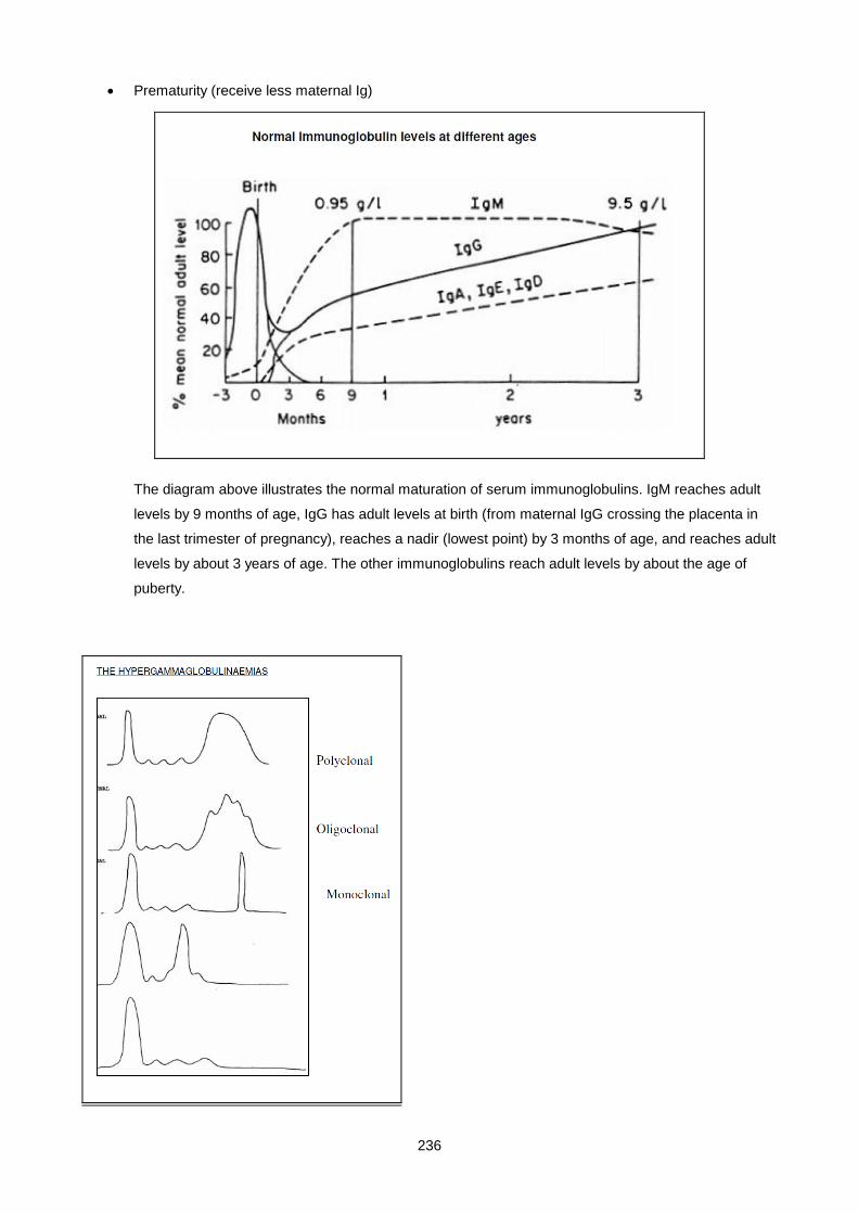
236
• Prematurity (receive less maternal Ig)
The diagram above illustrates the normal maturation of serum immunoglobulins. IgM reaches adult
levels by 9 months of age, IgG has adult levels at birth (from maternal IgG crossing the placenta in
the last trimester of pregnancy), reaches a nadir (lowest point) by 3 months of age, and reaches adult
levels by about 3 years of age. The other immunoglobulins reach adult levels by about the age of
puberty.
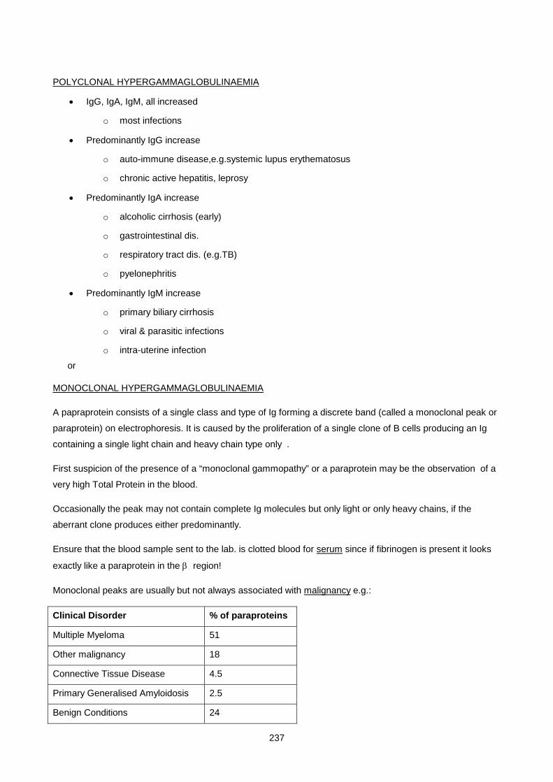
237
POLYCLONAL HYPERGAMMAGLOBULINAEMIA
• IgG, IgA, IgM, all increased
o most infections
• Predominantly IgG increase
o auto-immune disease,e.g.systemic lupus erythematosus
o chronic active hepatitis, leprosy
• Predominantly IgA increase
o alcoholic cirrhosis (early)
o gastrointestinal dis.
o respiratory tract dis. (e.g.TB)
o pyelonephritis
• Predominantly IgM increase
o primary biliary cirrhosis
o viral & parasitic infections
o intra-uterine infection
MONOCLONAL HYPERGAMMAGLOBULINAEMIA
A papraprotein consists of a single class and type of Ig forming a discrete band (called a monoclonal peak or
paraprotein) on electrophoresis. It is caused by the proliferation of a single clone of B cells producing an Ig
containing a single light chain and heavy chain type only .
First suspicion of the presence of a “monoclonal gammopathy” or a paraprotein may be the observation of a
very high Total Protein in the blood.
Occasionally the peak may not contain complete Ig molecules but only light or only heavy chains, if the
aberrant clone produces either predominantly.
Ensure that the blood sample sent to the lab. is clotted blood for serum since if fibrinogen is present it looks
exactly like a paraprotein in the β region!
Monoclonal peaks are usually but not always associated with malignancy e.g.:
Clinical Disorder % of paraproteins
Multiple Myeloma 51
Other malignancy 18
Connective Tissue Disease 4.5
Primary Generalised Amyloidosis 2.5
Benign Conditions 24
or

238
(i.e. 51% of paraproteins are associated with myeloma)
CRITERIA FOR DISTINGUISHING BENIGN PARAPROTEINS FROM MALIGNANT:
Frequency in benign disease Frequency in malignant disease
Bence Jones Protein in urine Very rare Common
Immunoparesis uncommon Very common
Paraprotein > 10g/l Uncommon Very common
Rapid increase in [ ] over time uncommon Very common
MALIGNANT PARAPROTEINAEMIAS:
Disease Paraprotein constituent
1. Myelomatosis IgG, IgA usually
2. Waldenstroms Macroglobulinaemia IgM
3. Heavy chain disease γ,α, or µ
4. Lymphoreticular system
5. Rarely - other malignancies
BENCE-JONES PROTEIN
• Presence indicates malignancy with a high probability
• Present in 50% of myelomas
• 20% of myelomas produce only light chains. These are freely filtered due to their low M.W. These
patients thus have no serum paraprotein. All suspected myelomas should have urine tested for
B.J.P. - Only found in the plasma when severe renal failure decreases the amount filtered
sufficiently.
• B.J.P. is reabsorbed by proximal convoluted tubule cells, causing damage in the process
• Unusual heat properties : precipitates maximally at ± 60°C, resolubilizes when hotter or cooler
• Deposition in tissues is one cause of Amyloid disease
• The light chains consist of either kappa or lambda classes, not both in the same patient
MULTIPLE MYELOMA
Multiple myeloma is a plasma cell malignancy of the bone marrow.

239
The annual incidence in European countries is 50 per million, with a median age at presentation of 70 years
(at GSH median age is 59 years; with 50% of patients <60years of age). (Unpublished data: M. Stein).
Myeloma is characterised by the production and secretion (excl. non-secretors/producers) of a monoclonal
immunoglobulin (also called paraprotein, M-protein).
This M-protein is found in the serum or urine of 99% of myeloma patients. IgG (50%), IgA (20%), light chains
(15%) are the more common while IgD (2%), IgE (<1%) and IgM myelomas are rare.
Clinical features are due to:
• Marrow infiltration:
o Bone pain (especially backache) due to osteolytic (“punched out”) lesions
o Anaemia, bleeding tendency (either due to inhibition of fibrin monomer polymerisation;
acquired von Willebrand’s disease; heparin-like anticoagulants or factor V deficiency in AL
amyloidosis), recurrent infection (hypogammaglobulinaemia, T-cell dysfunction)
o Hypercalcaemia (osteoclast activation due to increased RANKL expression /
Osteoprotegerin suppression).
o Cord compression (5%) – extramedullary foci
• Paraprotein production
o Renal failure due to B.J.P
Renal failure can be caused by the following in myeloma: dehydration,
hypercalcaemia, light chain-caused proximal tubular damage, AL amyloidosis, light
chain deposition disease, renal cell infiltration, hyperuricaemia, infection and use of
nephrotoxic drugs
o AL Amyloidosis (15% of myeloma patients) – see later in notes for more details
o Pseudohyponatraemia (analytical error due to elevated protein)
o Hyperviscosity syndrome: epistaxis, dizziness, transient paresis, visual disturbances
May develop with high serum paraprotein concentrations (IgA, IgG3)
IgM myeloma (very rare myeloma)
o Persistently elevated ESR or plasma viscosity (usually incidental finding)
o Peripheral neuropathy – rare, associated with amyloidosis or osteosclerotic myeloma
WHO diagnostic criteria for multiple myeloma (2001)
Requires a minimum of 1 major and 1 minor criteria

240
Or 3 minor criteria (must include (1) and (2).
These criteria must manifest in a symptomatic patient with progressive disease.
Major criteria Minor criteria
1. Marrow plasmacytosis >30%
2. Plasmacytoma on biopsy
3. M-component:
o Serum: IgG> 35g/L, IgA > 20g/L
o Urine > 1g/ 24hr of BJP
1. Marrow plasmacytosis 10 – 30%
2. M-component: present but less than major criteria
3. Lytic bone lesions
4. Reduced normal immunoglobulins (<50% normal: IgG < 6g/L, IgA < 1g/L,IgM < 0.5g/L
The above-mentioned criteria are currently in use at GSH. However, the most current myeloma classification
of MGUS and myeloma (by the International Myeloma Working Group 2003) is simpler and is based on the
M-protein concentration, percentage of bone marrow plasma cells and the presence or absence of myeloma-
related organ or tissue impairment (ROTI). It defines defines MGUS and asymptomatic and symptomatic
myeloma, the distinction between the latter two depending on the presence of ROTI.
Diagnostic criteria for MGUS, asymptomatic myeloma and symptomatic myeloma (International Myeloma
Working Group, 2003)
MGUS Asymptomatic Myeloma
Symptomatic Myeloma
M-protein <30g/L serum >30g/L serum Present in serum &/or urine (any amount)
Bone marrow clonal plasma cells
<10% >10% Present; or biopsy proven plasmacytoma
ROTI (incl. bone lesions or symptoms)
Absent * Absent Any ROTI present
*No evidence of other B-cell proliferative disorders or light-chain associated amyloidosis or other light-chains,
heavy-chain or immunoglobulin-associated tissue damage.
Myeloma related organ or tissue impairment (ROTI) (International Myeloma Working Group, 2003)
↑ Calcium levels Corr. Calcium > 0.25mmol/l above upper limit
Or > 2.75mmol/l
Renal insufficiency Attributable to myeloma
Anaemia Hb 2g/dL below lower limit of normal
Or Hb <10g/dL
Bone lesions Lytic lesions or osteoporosis with compression fractures
Other Symptomatic hyperviscosity, amyloidosis, recurrent bacterial infections (>2 episodes in 12m)
241
Diagnosis:
Initial investigation of a suspected myeloma patient should include the following:
• FBC (↓Hb), ESR (↑)
• Serum electrolytes, urea (↑), creatinine (↑), calcium (↑), total protein (↑), albumin (↓) and uric acid (↑)
• Serum and urine protein electrophoresis
• Serum immunoglobulin quantification
Confirm diagnosis with
• Bone marrow aspirate / trephine biopsy
• Serum and urine immunofixation
Diagnosis is confirmed with demonstration of M-protein in serum / urine &/or bone lesions with increased plasma cells in BM.
Assessment of tumour burden (prognostic factors)
• Β2 microglobulin (increase associated with poorer prognosis)
• CRP (increase associated with poorer prognosis)
• Albumin (decrease associated with poorer prognosis)
• Cytogenetics (del/monosomy chromosome 13, certain translocations associated with poorer prognosis)
Monitoring of disease and treatment:
• Asymptomatic myeloma patients should be monitored 3 monthly, indefinitely (clinically and serum and urinary paraprotein)
• Symptomatic myeloma: measuring response to treatment is based on changes in serum and/or urine M-protein levels.
o Complete Response: no detectable paraprotein by immunofixation (IF) for at least 6weeks
Patients with no paraprotein by electrophoresis, but detectable by (IF): poorer prognosis than afore-mentioned. Thus IF must be performed following a negative routine electrophoresis
o Partial Response: >50% decrease in serum M protein level &/or >90% decrease in BJP for at least 6 weeks
o Minimal Response: 25 – 49% decrease in serum M protein &/or 50 – 89% decrease in BJP for at least 6 weeks
o No change: less than minimal response, but not progressive
o Plateau: <25% drop in serum M protein & BJP for 3 months; no evidence of continuing ROTI
o Progressive disease: ROTI continuing / reappearing despite treatment
>25% increase in serum (>5g/L) &/or urine (>0.2g/24hr) M-protein
>25% increase in BM plasma cells
o Relapse: Reappearance in previously complete response
M-protein detectable by immunofixation
Plasmacytoma:Clonal plasma cell tumour with localised osseous (solitary lytic lesion on X-ray) or
extraosseous growth pattern.
242
• Bone tumour typically shows no M-protein in serum, although some may have low levels.
• 80% of extraosseous plasmacytomas occur in the respiratory tract. 15-20% of these patients have M-proteins. No anaemia, hypercalcaemia or renal impairment.
WALDENSTRÖMS MACROGLOBULINAEMIA
o Lymphocytes rather than plasma cells
o Monoclonal IgM
o Hyperviscosity predominates - Transient paresis, dizziness, epistaxis, headache and thromboses
o Anaemia
o Lymphadenopathy
o Bence Jones Proteinuria may be present
o Plasmaphoresis helps by removing IgM and normalising blood viscosity
HEAVY CHAIN DISEASE
These are rare B cell neoplasms producing monoclonal heavy chains only (no accompanying light chains). Composed of either IgG, IgA or IgM.
γ (in generalised lymphoma) - Franklin's disease (Gamma heavy chain disease) – rare, older
patients (60yr)
α (in intestinal lymphoma - MALT) - commonest form. Can present with malabsorption.
Younger (2nd to 3rd decade).
µ (in chronic lymphocytic leukaemia)
BENIGN PARAPROTEINAEMIAS
This is where a serum monoclonal protein is present but in the absence of lytic bone lesions, anaemia,
hypercalcaemia, and renal insufficiency. i.e. no evidence of myeloma, Waldenstrom’s macroglobulinaemia,
amyloidosis or other related B cell malignancy.Can only be called benign after being shown to be stable for 5
years (IgG & IgA) and for 10 years (IgM). Often associated with autoimmune disease (esp. ankylosing
spondylitis), chronic infections or cirrhosis.
Monoclonal gammopathy of unknown significance (MGUS) is the term used to describe paraproteinaemia
without a definitive classification. These patients have circulating M-protein levels of less than the 30g/L
required for a diagnosis of myeloma, with a bone marrow plasma cell count of less than 10%. Also, there are
no clinical manifestations related to the monoclonal gammopathy, and malignany disorders such as
myeloma, Waldernstrom’s, primary amyloidosis, B-cell lymphoma and chronic lymphocytic leukaemia have
been excluded.
Features:

243
• prevalence increases with age, 3.2% >50year; 5.3% >70years (Minnesota study, NEJM 2006)
• higher in men than in women; blacks more than whites (US)
• cause unknown
• M-protein: IgG most common (69%), then IgM (17%) and IgA (11%)
• Plasma-cell clone and associated M-protein is stable for years, with progression to malignancy (such
as myeloma) about 1% per year post diagnosis (i.e. 25% after 25y). The risk of progression in
10years is approximately numerically equal to the M-protein in g/L (e.g. 20g/L = 20% risk).
• 25% develop myeloma, primary amyloidosis, macroglobulinaemia or other lymphoproliferative
disease after more than 20 years
• < 1% spontaneously disappear. May become oligoclonal (band with normal kappa:lambda)
• Corresponding decrease in uninvolved immunoglobulins in 25% of patients
Assessment and management:
Diagnosis:
• History & examination
• Hb, Calcium, Creatinine
• Protein studies (total protein, serum and urine protein electrophoresis and immunofixation)
Patients with low M-protein levels, without clinical evidence of myeloma-related organ or tissue impairment,
need not be subjected to bone marrow aspirates or extensive skeletal surveys. These are not mandatory for
MGUS diagnosis, but are recommended in younger patients and in older patients with levels higher than
20g/L. Clinical evaluation and paraprotein levels should be reviewed at 3 and 6 months to establish diagnosis.
Monitoring:
Monitoring should occur 6 – 12monthly, indefinitely (increased frequency depending on M-protein level and
age) and this should include clinical evaluation as well as paraprotein levels.
CRYOGLOBULINS
These are immunoglobulins that precipitate at temperatures lower than the normal body temperature.
Cryoglobulins can be polyclonal or monoclonal (usually IgM). Aggregated immunoglobulins (associated with
RF) can activate complement causing symptoms due to vasculitis e.g. Raynaud’s phenomenon, proteinuria
and microinfarcts Cryoglobulins include:
- Paraproteins (this can be an artefactual cause of an apparent "non-producer")
- Ig/anti-Ig complexes e.g. Rheumatoid factor which is an IgM antibody against IgG !
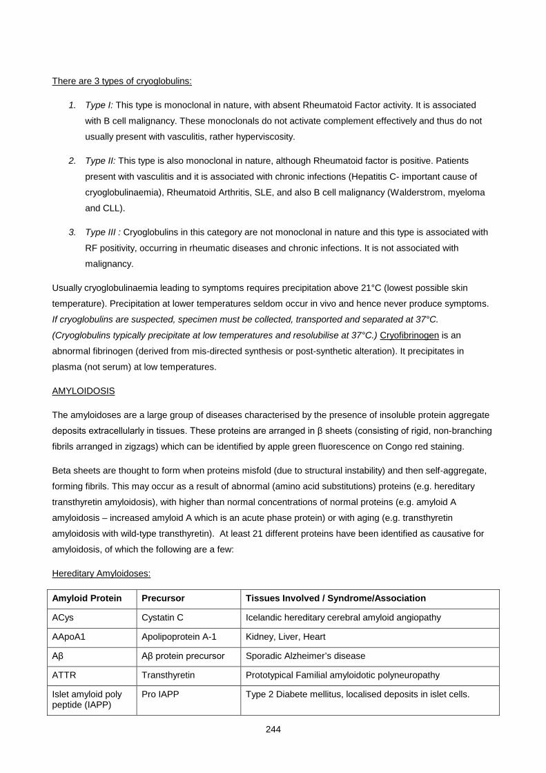
244
There are 3 types of cryoglobulins:
1. Type I: This type is monoclonal in nature, with absent Rheumatoid Factor activity. It is associated
with B cell malignancy. These monoclonals do not activate complement effectively and thus do not
usually present with vasculitis, rather hyperviscosity.
2. Type II: This type is also monoclonal in nature, although Rheumatoid factor is positive. Patients
present with vasculitis and it is associated with chronic infections (Hepatitis C- important cause of
cryoglobulinaemia), Rheumatoid Arthritis, SLE, and also B cell malignancy (Walderstrom, myeloma
and CLL).
3. Type III : Cryoglobulins in this category are not monoclonal in nature and this type is associated with
RF positivity, occurring in rheumatic diseases and chronic infections. It is not associated with
malignancy.
Usually cryoglobulinaemia leading to symptoms requires precipitation above 21°C (lowest possible skin
temperature). Precipitation at lower temperatures seldom occur in vivo and hence never produce symptoms.
If cryoglobulins are suspected, specimen must be collected, transported and separated at 37°C.
(Cryoglobulins typically precipitate at low temperatures and resolubilise at 37°C.) Cryofibrinogen is an
abnormal fibrinogen (derived from mis-directed synthesis or post-synthetic alteration). It precipitates in
plasma (not serum) at low temperatures.
AMYLOIDOSIS
The amyloidoses are a large group of diseases characterised by the presence of insoluble protein aggregate
deposits extracellularly in tissues. These proteins are arranged in β sheets (consisting of rigid, non-branching
fibrils arranged in zigzags) which can be identified by apple green fluorescence on Congo red staining.
Beta sheets are thought to form when proteins misfold (due to structural instability) and then self-aggregate,
forming fibrils. This may occur as a result of abnormal (amino acid substitutions) proteins (e.g. hereditary
transthyretin amyloidosis), with higher than normal concentrations of normal proteins (e.g. amyloid A
amyloidosis – increased amyloid A which is an acute phase protein) or with aging (e.g. transthyretin
amyloidosis with wild-type transthyretin). At least 21 different proteins have been identified as causative for
amyloidosis, of which the following are a few:
Hereditary Amyloidoses:
Amyloid Protein Precursor Tissues Involved / Syndrome/Association
ACys Cystatin C Icelandic hereditary cerebral amyloid angiopathy
AApoA1 Apolipoprotein A-1 Kidney, Liver, Heart
Aβ Aβ protein precursor Sporadic Alzheimer’s disease
ATTR Transthyretin Prototypical Familial amyloidotic polyneuropathy
Islet amyloid poly peptide (IAPP)
Pro IAPP Type 2 Diabete mellitus, localised deposits in islet cells.

245
Acquired Amyloidoses:
Amyloid Protein Precursor Tissues Involved / Syndrome/Association
Aβ2M Β2 Microglobulin Chronic Haemodialysis; joints
AL Ig light chain Primary amyloidosis, myeloma
AA Amyloid A Secondary amyloidosis, reactive to chronic infection / inflammation
APrP Prion protein Sporadic CJD
As can be seen from above, amyloidoses can be divided into hereditary and acquired (primary and
secondary). Specific amyloidoses involve specific organs (specific proteins target defined organs) e.g. β2
microglobulin (joints), transthyretin (peripheral nerves), light chains (virtually any organ). The three most
common forms are AL, ATTR and AA, which all differ in their pathogenesis.
AL Amyloidosis : Immunoglobulin light chain is the causative protein in this type, with the variable region of
the lambda light chain most often identified in the amyloid deposits. AL amyloidosis has a wide spectrum of
organ involvement, the most commonly involved being the kidneys (nephrotic syndrome (massive
proteinuria), with normal or mildly altered renal function; progressive renal failure and subsequent
hypertension is very rare) and heart (congestive heart failure – usually right sided; myocardial infarction
without coronary artery disease; asymptomatic abnormal ECG findings may precede). No CNS involvement,
although peripheral involvement is common with autonomic and sensory neuropathy being relatively common
(carpal tunnel syndrome, postural hypotension, impotence, GIT motility). Hepatomegaly (hard, with elevated
ALP) and macroglossia (sleep apnoea) are common. Adrenal and thyroid (10 – 20% of patients) involvement
may occur, with hypoadrenalism and hypothyroidism.
ATTR Amyloidosis: This hereditary (autosomal dominant) disease involves aggregation of a tranthyretin
mutant, with more than 80 mutations having been identified thus far. Different mutations tend to have
different prominent manifestations. Peripheral sensorimotor and autonomic neuropathy (GIT symptoms incl.
diarrhoea and weight loss) is common. Renal disease is much less prominent than in AL amyloidosis. No
macroglossia. Depending on mutation, may have cardiac conduction abnormalities often requiring pacing, or
myocardial infiltration; cardiac failure not as common as in AL amyloidosis. There is often a family history of
an “unknown neurologic disease” or “motor neuron disease”.
AA Amyloidosis: This amyloidosis is secondary to chronically elevated levels of serum amyloid A, an acute
phase protein. This may accur in conditions of chronic inflammation e.g. tuberculosis, rheumatoid arthritis
and inflammatory bowel disease. Patients most commonly present with renal disease, but may also present
with hepatomegaly and/or splenomegaly. Cardiac involvement is rare.
Diagnosis relies heavily on clinical suspicion, confirmed by Congo red staining of tissue biopsy
(subcutaneous fat biopsy positive in 85% of AL). The type of amyloidosis must be determined (defines
treatment). Plasma dyscrasias should be excluded. Serum and urine protein electrophoresis and
immunofixation should be done to detect monoclonal immunoglobulins / light chains (positive in 90% of AL
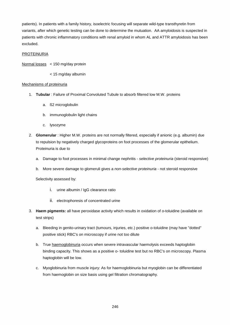
246
patients). In patients with a family history, isoelectric focusing will separate wild-type transthyretin from
variants, after which genetic testing can be done to determine the mutuation. AA amyloidosis is suspected in
patients with chronic inflammatory conditions with renal amyloid in whom AL and ATTR amyloidosis has been
excluded.
PROTEINURIA
Normal losses < 150 mg/day protein
< 15 mg/day albumin
Mechanisms of proteinuria
1. Tubular : Failure of Proximal Convoluted Tubule to absorb filtered low M.W. proteins
a. ß2 microglobulin
b. immunoglobulin light chains
c. lysozyme
2. Glomerular : Higher M.W. proteins are not normally filtered, especially if anionic (e.g. albumin) due
to repulsion by negatively charged glycoproteins on foot processes of the glomerular epithelium.
Proteinuria is due to
a. Damage to foot processes in minimal change nephritis - selective proteinuria (steroid responsive)
b. More severe damage to glomeruli gives a non-selective proteinuria - not steroid responsive
Selectivity assessed by:
i. urine albumin / IgG clearance ratio
ii. electrophoresis of concentrated urine
3. Haem pigments: all have peroxidase activity which results in oxidation of o-toluidine (available on
test strips)
a. Bleeding in genito-urinary tract (tumours, injuries, etc.) positive o-toluidine (may have "dotted"
positive stick) RBC's on microscopy if urine not too dilute
b. True haemoglobinuria occurs when severe intravascular haemolysis exceeds haptoglobin
binding capacity. This shows as a positive o- toluidine test but no RBC's on microscopy. Plasma
haptoglobin will be low.
c. Myoglobinuria from muscle injury: As for haemoglobinuria but myoglobin can be differentiated
from haemoglobin on size basis using gel filtration chromatography.
247
Lecture 13: Cerebrospinal Fluid
DR GEORGE VAN DER WATT, REVISED BY DR JD DEETLEFS 2006, PROF TS PILLAY 2008
LECTURE OUTLINE
1. Review of Physiology
2. Sample collection
3. CSF analysis and CNS diseases
• Meningitis
• SAH
• Head injury
• Demyelinating disorder
• Other
LEARNING OBJECTIVES
• Know the difference in composition between CSF and plasma
• Understand the function of the blood-brain-barrier
• Outline the typical CSF findings encountered with different types of meningitis
• Know the definition and significance of xanthochromia
• List different analytes that can be measured in the CSF, their clinical application and significance
1. BASIC PHYSIOLOGY
• Total volume of 150ml and daily production of 550 ml gives a daily turnover of 3.7 times (about 15% is
produced per hour; 0.38 ml/min). CSF is essentially a strictly controlledultra filtrate of plasma produced
by the choroid plexuses in the ventricles and around blood vessels in the brain. It is in direct
communication with, and actually the same as brain ECF. The function of CSF is to provide
mechanical support and cushioning for the brain and spinal cord and transport biological compounds,
waste removal and to provide a chemical environment.
• Together the tight junctions between the epithelial cells of the capillaries and those of the choroid
plexus form the blood-brain barrier preventing the free movement of large and/or hydrophilic molecules
from plasma to CSF and facilitating movement of essential molecules like glucose ( via GLUT 1
transporter) into the CSF. Fat soluble molecules cross easily, as do O2 and CO2 . In contrast HCO3- and
H+ do not cross easily and rely on Carbonic Anhydrase in the choroid plexus to regenerate them from
CO2 & H2O, the [H+] in CSF being essential in regulating respiration.
• The blood brain barrier is there to ensure a tightly controlled brain ECF environment for optimum
neuronal function. The rate of CSF production is largely driven by hydrostatic pressure and does not
vary within a wide range of blood pressure fluctuations.
248
• As a result of this, normal CSF is crystal clear with a low protein content, osmolality equal to plasma
and glucose 60% that of plasma (Table 1 ).
• CSF circulates from the brain tissue and choroid plexuses into the ventricles and out into the
subarachnoid space via the lateral and medial foramens of Luschka and Magendie leading out from
the fourth ventricle. From here it is readily absorbed into the blood of the venous sinuses via the so
called arachnoid villi, these being projections of fused arachnoid and capillary endothelium into the
venous sinuses across which the transport of CSF and its constituents occurs freely and is again
pressure dependent. Here however the pressure gradient is lower with a CSF pressure of + 12cm H2O
and venous sinus pressure roughly equal to the jugular venous pressure.
The chief functions of the CSF are therefore to regulate the composition of the brain ECF and to suspend the
brain thereby cushioning it in the rigid skull and acting as a shock absorber.
CSF Plasma CSF/Plasma %
Na mmol/l 140 140 100 (takes an hour ro equilibrate)
K mmol/l 3 4.5 65%
Cl mmol/l 118 99 114%
Protein g/l 0.2 60 0.3% (CSF is 1/200 of serum)
Glucose mmol/l 3 5 60%
Table 1. Chemistry of normal CSF vs plasma.
2. SAMPLE COLLECTION
• CSF is usually aspirated by Lumbar Puncture (LP) or direct aspiration during open neuro surgery. (It
should be remembered that the different sources of CSF will have different reference range from that
taken by lumbar puncture-see below). Approximately 10-12 ml can be taken from an adult and less
than 3 ml from a neonate. Care must be taken not to contaminate the sample with blood. The fluid is
collected into 3 tubes via free passive flow through the needle. Up to four tubes of CSF can be
collected aseptically. Each tube should contain 2-4 mL of fluid. The tubes should be labelled with a
number indicating the order in which they were collected. Each tube goes to a different section of the
laboratory
for testing
i. Tube #1 Chemistry
ii. Tube #2 Haematology, Flow Cytometry, Cytology
249
iii. Tube #3 Microbiology
iv. Tube #4 Haematology, Molecular & Reference Lab
• If the plan is to examine CSF Chloride, then no fluoride should be used with CSF so transport to the
lab for glucose measurement should be done stat. At the same time blood should be drawn
simultaneously for serum glucose measurement as well. The 1st 1/3rd of the fluid collected is often
contaminated with cells and should be used for chemistry, the rest for MCS and cytology.
* In many clinical conditions, performing a lumber puncture is strongly contraindicated. A LP
should never be performed in the presence of raised intracranial pressure or infection over
the puncture site.
• CSF in the ventricles is different from CSF in the lumbar region. Protein levels in the ventricles are
66% lower (mean 0.15g/L in ventricles versus 0.45g/L in lumbar area) and glucose levels are 10%
higher in ventricles. Clumps of choroidal cells are observed more commonly in VP shunt fluid.
Normal CSF from both regions should contain less than 5 WBCs/ μl.
•
3. CSF ANALYSIS AND CNS DISEASE
CSF is investigated to aid in the diagnosis of SAH (subarachnoid hemorrhage), Meningitis, Demyelinating
disorders, CNS malignancy, Head injury with CSF leak and other less common tests, the majority of the
investigations involve cytology and microbiology with chemistry adding a small but important contribution.
MENINGITIS
• Meningitis can be bacterial ( pyogenic or tuberculous), viral or fungal ( Cryptococcus). Apart from
direct organism identification, biochemistry and cell counts can help distinguish between the types of
meningitis. In classical pyogenic meningitis ( eg Strep Cryptococcal) CSF protein levels are high >
0.8 g/L, glucose is much lower than 60% of blood value, high counts of PMNL’ s ( polymorphonuclear
lymphocytes) > 500x106 occur and the CSF is turbid. TBM (tuberculous meningitis) can be
differentiated by a mixed PMNL/lymphocyte high cell count and a low CSF chloride. Viral meningitis
often has a lymphocytosis with a normal glucose, normal protein levels and a clear CSF. Fungal
infections present with CSF lymphocytosis, a high protein count and normal glucose.
• Often however the picture is indistinct due to partial treatment with antibiotics or an early presentation
and further tests are required to confirm the diagnosis eg Latex agglutination for common bacterial
antigens ( Strep pneumoneae, Neisseria, Cryptococcus & Haemophilus), Indian ink staining for
Cryptococcus and VDRL and RPR serology for neurosyphylis . A high CSF ADA ( adenosine
deaminase) would indicate Tuberculous meningitis whereas a high CSF lactate and CRP (C- reactive
protein) would indicate a bacterial meningitis.
• Low CSF protein levels can occur in conditions such as repeated lumbar puncture or a chronic leak,
in which CSF is lost at a higher than normal rate. Low CSF protein levels also are seen in some

250
children between the ages of six months and two years, in acute water intoxication, and in a minority
of patients with idiopathic intracranial hypertension.
TABLE 2
Typical Cerebrospinal Fluid Findings in Various Types of Meningitis
• Lactate levels are used mostly to distinguish early bacterial meningitis from viral meningitis. Lactate
levels are usually >3.9 mmol/l in bacterial meningitis and < 2.8 mmol/l in viral meningitis. Levels
between 2.9-3.37 mmol/l are equivocal.
• Mycoplasma meningitis will usually give elevated lactate levels, even if cultures are negative. All
xanthochromic fluids will have elevated lactate levels, so it is best not to measure lactate in these
fluids. CSF Lactate Dehydrogenase(LDH)
• LDH is normally present in CSF with 40 U/L being the upper limit of normal for adults and 70 U/L for
neonates. LDH values may help in differentiating traumatic tap from intracranial hemorrhage because
fresh traumatic taps with intact RBC's do not significantly elevate the LD level.
• LD activity is significantly higher in bacterial meningitis than in aseptic meningitis. Elevated LD levels
76 hours following resuscitation predict a poor outcome in patients with hypoxic brain injury.
CSF GLUCOSE
• Glucose enters the CSF from the plasma by 2 mechanisms, diffusion and active transport. Glucose
is the primary source of energy for the brain. Brain is 5 % body mass but uses 20% glucose

251
• The level of CSF glucose is influenced by the concentration and duration of the plasma glucose level.
CSF glucose levels are roughly two-thirds of plasma glucose levels (exeptions to this are: First 6
months of life - CSF glucose can equal plasma glucose; Applies to glucose levels < 20 mmol/l; i.e.
with severe hyperglycaemia transporters are saturated and ratio can appear low; Diabetic patient who
has recently taken insulin or oral hypoglycaemic)
• The normal fasting CSF glucose is 2.2-3.9 mmol/l. In children the nonfasting reference range is 2.5-
5.6 mmol/l. The CSF glucose level lags behind the plasma level by approximately 30-90 minutes. An
increase in CSF glucose means the patient was hyperglycaemic 30 to 90 minutes before and has no
special clinical significance.
• Therefore, CSF glucose can only be interpreted reliably with a blood sample taken within 15 min
before or after an LP. CSF glucose can take up to 2-4 hours to equilibrate with plasma
CSF CHLORIDE
• The diagnosis of tuberculous meningitis is notoriously difficult to make, particularly in the setting of
HIV infection. In the western Cape, a low CSF chloride (less than 110mmol/1) is used to distinguish
tuberculous from viral and bacterial meningitis. A CSF chloride of less than 100mmol/l is thought to
be virtually diagnostic of TBM. The normal range of CSF chloride is 116 to 130 mmol/litre, compared
to a normal range in serum of 95 - 105 mmol/litre.
CSF PROTEIN
• Spinal fluid is an ultrafiltrate of plasma that lacks high molecular weight proteins such as beta
lipoprotein, alpha-2 macroglobulin, IgM, and polymeric haptoglobins.
• Unlike plasma, on electrophoresis CSF has a prominent prealbumin peak and a split beta peak,
which contains transferrin (tau transferrin) and m2-transferrin, which is also called tau-protein. The
protein concentration of spinal fluid is less than 1% of plasma proteins. The normal range for lumbar
puncture CSF varies with age. Infants have higher protein levels (0.6-1.5 g/L) due to increased blood
brain barrier permeability. Cisternal and ventricular fluids have lower total protein concentration than
fluid obtained by lumbar puncture.
SUBARACHNOID HAEMORRHAGE
• In subarachnoid haemorrhage red cells are a diagnostic feature in the CSF so careful LP is essential.
Red cells that have been in the CSF for longer than 4 hours cause a yellow staining of the CSF called
xanthochromia and the presence of this in fresh CSF confirms bleeding into the subarachnoid space
from a source other than contamination during the LP. Keep in mind that there are other causes of
xanthochromia. Xanthochromia refers to a yellow, orange or pink colour. Two to four hours after
haemorrhage, RBC's lyse and release oxyhaemoglobin giving a pink to orange color. By 24 hours
haemoglobin is metabolized to bilirubin, resulting in a yellow color. Bilirubin concentration peaks at 36
hours, but persists for several weeks.
252
• Other causes of Xanthochromia
o Very high CSF protein concentration/ Increased protein (>1.50 g/L)
o Elevated serum bilirubin/Jaundice
o Meningeal melanoma
o Hypercarotenaemia
o Rifampicin therapy
o Previous traumatic tap
• Xanthochromia is a visual diagnosis and rather subjective. A test referred to as the net bilirubin
absorbance, is sometimes requested by neurosurgeons. This test is designed to pick up minute
quantities of CSF bilirubin causes by small sub-arachnoid bleeds. Also, traumatic taps and SAH may
co-exist confounding the problem of distinguishing between the two.
• Early diagnosis of subarachnoid haemorrhage is important such that timely surgical intervention can
be undertaken. CT is positive in 98% of patients presenting within 12 h but only positive in 50%
presenting within 1 week. CSF bilirubin is used to determine the need for angiography. You must
protect the CSF sample from light to prevent decay of the bilirubin. This can be positive 2 weeks after
the event. It is recommended that the LP only be performed > 12 hours after the onset of symptoms.
Always ensure that the last tube is sent for CSF bilirubin scanning. The determination of net bilirubin
absorbance is performed by scanning the sample at different wavelengths to determine if there is
bilirubin present or whether there is haemoglobin. These can be distinguished by the wavelengths at
which they show maximal absorbance.
HEAD INJURY
• In patients with rhinorrhoea or otorrhoea, post head injury or spontaneously, it is important to
ascertain whether CSF is present in the fluid, which would confirm a CSF leak. This is done by
identifying beta-2-transferrin (a desialylated form of transferrin that only occurs in CSF and not in
plasma; often call tau-transferrrin but this risks confusion with other tau proteins. The preferred term
is Asialotransferrin) in the fluid leak sample, using electrophoresis and immunofixation.
DEMYELINATING DISORDERS
• In Multiple sclerosis the presence of elevated levels of myelin basic protein in the CSF derived from
damaged myelin sheaths together with high CSF protein levels due to immunoglobulins (often with an
oligoclonal pattern) can help confirm the diagnosis, following the usual MRI scan. Demyelinating
disorders release myelin basic protein into the CSF. Levels can be used to follow the activity of
demyelination, diagnose early MS before oligoclonal bands form, and assist in the diagnosis of the
10% of MS patients who never form oligoclonal bands. The reference range is < 1.5 ng/mL.
Alternatively the IgG/Albumin index or IgG synthesis rate can be calculated by measuring the albumin
and IgG concentration in both the serum and CSF. This is because multiple sclerosis is associated
with increased synthesis of immunoglobulins within the CNS, reflecting a local immune response.
253
Oligoclonal banding is visualized on electrophoresis of CSF. 90% of MS patients have these bands in
CSF, although this is not specific for this condition. This can be used to rule out suspected MS.
However, this must be done in parallel with serum electrophoresis to ensure that the banding seen is
only present in CSF. Oligoclonal bands may also be seen in the CSF of patients with lymphoma,
CLL, malignancies, chronic active hepatitis, rheumatoid factor, and viral illnesses.
Protein index:
• This assesses the amount of intrathecal protein synthesis that may occur in an inflammatory disease
and assesses the permeability of the blood-brain barrier in relation to increased intrathecal synthesis
(eg IgG). Albumin is used as a reference protein. A normal CSF Albumin/Serum Albumin ratio is less
than 9. CSF IgG index = CSF IgG/Serum IgG ÷ CSF Albumin/Serum Albumin Usually the CSF IgG
index will be 0.3-0.8. If > 0.8, this indicates increased intrathecal synthesis such as may be seen in
multiple sclerosis.
OTHER
• CSF can be examined for the presence of various other substances that can aid in the diagnosis of
Alzheimer's disease, Cerebral infarctions, Creutzfeld-Jacob disease, various malignancies and
psychiatric disorders: for example in Major Depressive Disorder the morning CSF levels of 5-
Hydroxyindoleacetic acid ( 5-HIAA) can be measured and if low indicate a deficiency in serotonin. In
addition, MHPG (3-methoxy-4-hydroxyphenylglycol, a metabolite of noradrenaline) can also be low in
depression and can predict responses to such antidepressants in patients with depression.
• Recently two new markers have emerged that contribute to the diagnosis of Alzheimer's disease.
Tau protein (abundantly present in neurons where it serves to stabilize the microtubular network in
the axons) demonstrates significantly elevated CSF levels. Bamyloid, a sticky protein fragment
abnormally cleaved from a larger precursor protein called amyloid precursor protein, concentrations
are significantly lower in csf samples.
• HIV: HIV can infect the CNS and the most common AIDS-related neurological disorder is HIV
encephalitis/AIDS dementia complex. CSF findings include a moderate increase in mononuclear
cells and oligoclonal banding. Viral DNA can be detected using PCR. In general, a wide variety of
CSF abnormalities may be seen including increased IgG indices, oligoclonal banding and increased
lymphocytes. Serious opportunistic fungal infections may exist with minimal CSF changes.
• Guillain-Barré syndrome: CSF can show a very high protein concentration.
• Inherited metabolic disease: High CSF lactate may be found in disorders of the respiratory chain
even when plasma lactate is normal or only slightly increased.. CSF pyruvate may also be high in
these conditions. CSF amino acid analysis may be useful in disorders of amino acid metabolism.
• Sarcoidosis: CSF may show nonspecific elevation of protein and a lymphocytosis. This may occur in
neurosarcoidosis. Other markers that may be raised include angiotensinconverting enzyme;
lysozyme and β2-microglobulin.

254
• The CSF VDRL test has a sensitivity of only 50-60% but is highly specific. A positive test rules in
neurosyphilis. CSF VDRL is inappropriate as a screening test for neurosyphilis; it should be
performed only if serum FTA-ABS tests are positive. The RPR test is unsuitable for CSF because it
has more false positives than the VDRL. CSF FTA-ABS is essentially 100% sensitive, but false
positives may be caused by increased blood-brain barrier permeability due to inflammation, or as little
as 0.8 mL of blood per mL of CSF (4000 CSF RBC's). A negative serum FTA-ABS test rules out any
syphilitic infection, whereas a positive serum FTA-ABS with a nonreactive CSF FTA-ABS excludes
the diagnosis of active neurosyphilis.
• Lyme disease: This is an infection caused by Borrelia burgdorferi and can infect the CNS. CSF
findings include modestly elevated protein, mononuclear cells, normal CSF glucose. Antibodies to the
organism will confirm the diagnosis.
• Inborn errors of metabolism: CSF biochemistry is useful in several disorders.
Important points:
1. Acute bacterial meningitis is associated with high CSF neutrophil count, increase CSF total protein
and a low CSF glucose;
2. In a patient with suspected subarachnoid haemmorrhage and negative CT scan, CSF
spectrophotometry may help determine the need for angiography
3. CSF oligoclonal bands are seen in 90% of patients with MS, but is not specific for this conditions. The
absence of oligoclonal banding can rule out MS.
4. Very high CSF protein concentrations can occur in non-infectious conditions including Guillain-Barré
syndrome and blocked CSF flow (Froin’ s syndrome).
255
Lecture 14: Chemical Pathology Of Liver Disease
DR H. VREEDE & E.H.HARLEY, 2007
LIVER STRUCTURE
DUAL BLOOD SUPPLY
• Hepatic artery (25% of liver’s blood flow) – Oxygenated
• Portal vein (75%). Drains:
o spleen (bilirubin)
o GI (nutrients, toxins, bacteria)
o pancreas (insulin, glucagon)
The hepatic artery and portal vein enter the liver at the porta hepatis, and each then divides into 2 branches.
These further subdivide to provide hepatic arterioles and portal venules which run in portal tracts together
with bile canaliculi, lymphatics and nerves.
Anastomotic connections between the systemic and the portal circulation exist at 3 sites :
• paraumbilical veins
• lower oesophagus
• rectal veins
Varices and portal-systemic shunting occur at these sites in portal hypertension.
VENOUS DRAINAGE
All the liver’s blood drains into hepatic venules which join to form three hepatic veins. The hepatic veins drain
into the inferior vena cava.
ARCHITECTURE
Structural unit = hepatic lobule.
Each hepatic lobule is an hexagonal unit surrounded by connective tissue septae. Three to four portal tracts
lie perpendicularly in the septae at the periphery of each hepatic lobule. A terminal hepatic venule lies in the
centre of each hepatic lobule.
Functional unit = acinus.
The acinus is the liver tissue lying between two centrilobular venules. It is centrally divided by the connective
tissue septum in which runs a small branch of the portal tract containing terminal branches of the hepatic
arteriole and portal venule. The parenchymal liver cells (hepatocytes) are arranged in columns (plates)

256
extending from the septum to the centrilobular venule, with vascular spaces (sinusoids) between them. The
sinusoids are separated from the hepatocytes by the space of Disse.
Blood flows from the portal tract, through the sinusoids, and drains into the centrilobular venule. The acinus
can therefore be divided into 3 functional zones on the basis of different oxygen content in these areas:
• periportal highest oxygen content, least susceptible to ischaemic damage
• mediolobular intermediate
• centrilobular lowest oxygen content, most susceptible to ischaemic damagemost involved in drug
metabolism, most susceptible to drug-induced damage
METABOLIC FUNCTIONS OF THE LIVER
The liver plays a major role in carbohydrate, lipid and protein homeostasis, with the processes of glycolysis,
the Krebs cycle, gluconeogenesis, glycogen synthesis and glycogenolysis, lipogenesis, ketogenesis, amino
acid synthesis and degradation, and protein synthesis all taking place in the hepatocytes. Hepatocytes also
metabolise and detoxify endogenous (haem) and exogenous products (drugs), which are then excreted via
the biliary tree.
BILIRUBIN METABOLISM
SOURCES OF BILIRUBIN:
• Haemoglobin
o RBC breakdown. 90% of RBC breakdown occurs within RES cells (mainly in spleen)
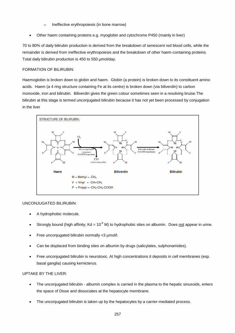
257
o Ineffective erythropoiesis (in bone marrow)
• Other haem containing proteins e.g. myoglobin and cytochrome P450 (mainly in liver)
70 to 80% of daily bilirubin production is derived from the breakdown of senescent red blood cells, while the
remainder is derived from ineffective erythropoiesis and the breakdown of other haem-containing proteins.
Total daily bilirubin production is 450 to 550 µmol/day.
FORMATION OF BILIRUBIN:
Haemoglobin is broken down to globin and haem. Globin (a protein) is broken down to its constituent amino
acids. Haem (a 4 ring structure containing Fe at its centre) is broken down (via biliverdin) to carbon
monoxide, iron and bilirubin. Biliverdin gives the green colour sometimes seen in a resolving bruise.The
bilirubin at this stage is termed unconjugated bilirubin because it has not yet been processed by conjugation
in the liver
UNCONJUGATED BILIRUBIN:
• A hydrophobic molecule.
• Strongly bound (high affinity; Kd = 10-8 M) to hydrophobic sites on albumin. Does not appear in urine.
• Free unconjugated bilirubin normally <3 µmol/l.
• Can be displaced from binding sites on albumin by drugs (salicylates, sulphonamides).
• Free unconjugated bilirubin is neurotoxic. At high concentrations it deposits in cell membranes (esp.
basal ganglia) causing kernicterus.
UPTAKE BY THE LIVER:
• The unconjugated bilirubin - albumin complex is carried in the plasma to the hepatic sinusoids, enters
the space of Disse and dissociates at the hepatocyte membrane.
• The unconjugated bilirubin is taken up by the hepatocytes by a carrier-mediated process.

258
• Within the hepatocyte the unconjugated bilirubin is bound to ligandin
CONJUGATION BY THE LIVER:
• The bilirubin is then conjugated with glucuronic acid by UDP-glucuronyl transferase (UDPGT I) to
bilirubin monoglucuronide (BMG) and by UDPGT II to bilirubin diglucuronide (BDG).
• Conjugated bilirubin is more water soluble and can be excreted in bile or urine.
• Under normal circumstances there is no conjugated bilirubin present in plasma
EXCRETION INTO BILE:
• Conjugated bilirubin is transported out of the liver cells into the bile canaliculi by an energy-dependant
carrier-mediated process which is sensitive to cell injury. This canalicular excretion step rather than
conjugation is thought to be the rate-limiting step in bilirubin metabolism.
• Bile flows through the canaliculi, into the bile ducts, and finally into the duodenum.
UROBILINOGEN:
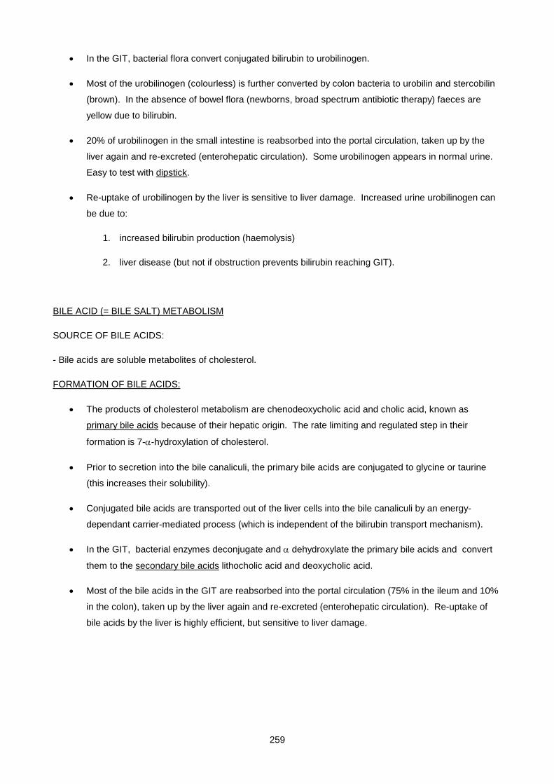
259
• In the GIT, bacterial flora convert conjugated bilirubin to urobilinogen.
• Most of the urobilinogen (colourless) is further converted by colon bacteria to urobilin and stercobilin
(brown). In the absence of bowel flora (newborns, broad spectrum antibiotic therapy) faeces are
yellow due to bilirubin.
• 20% of urobilinogen in the small intestine is reabsorbed into the portal circulation, taken up by the
liver again and re-excreted (enterohepatic circulation). Some urobilinogen appears in normal urine.
Easy to test with dipstick.
• Re-uptake of urobilinogen by the liver is sensitive to liver damage. Increased urine urobilinogen can
be due to:
1. increased bilirubin production (haemolysis)
2. liver disease (but not if obstruction prevents bilirubin reaching GIT).
BILE ACID (= BILE SALT) METABOLISM
SOURCE OF BILE ACIDS:
- Bile acids are soluble metabolites of cholesterol.
FORMATION OF BILE ACIDS:
• The products of cholesterol metabolism are chenodeoxycholic acid and cholic acid, known as
primary bile acids because of their hepatic origin. The rate limiting and regulated step in their
formation is 7-α-hydroxylation of cholesterol.
• Prior to secretion into the bile canaliculi, the primary bile acids are conjugated to glycine or taurine
(this increases their solubility).
• Conjugated bile acids are transported out of the liver cells into the bile canaliculi by an energy-
dependant carrier-mediated process (which is independent of the bilirubin transport mechanism).
• In the GIT, bacterial enzymes deconjugate and α dehydroxylate the primary bile acids and convert
them to the secondary bile acids lithocholic acid and deoxycholic acid.
• Most of the bile acids in the GIT are reabsorbed into the portal circulation (75% in the ileum and 10%
in the colon), taken up by the liver again and re-excreted (enterohepatic circulation). Re-uptake of
bile acids by the liver is highly efficient, but sensitive to liver damage.
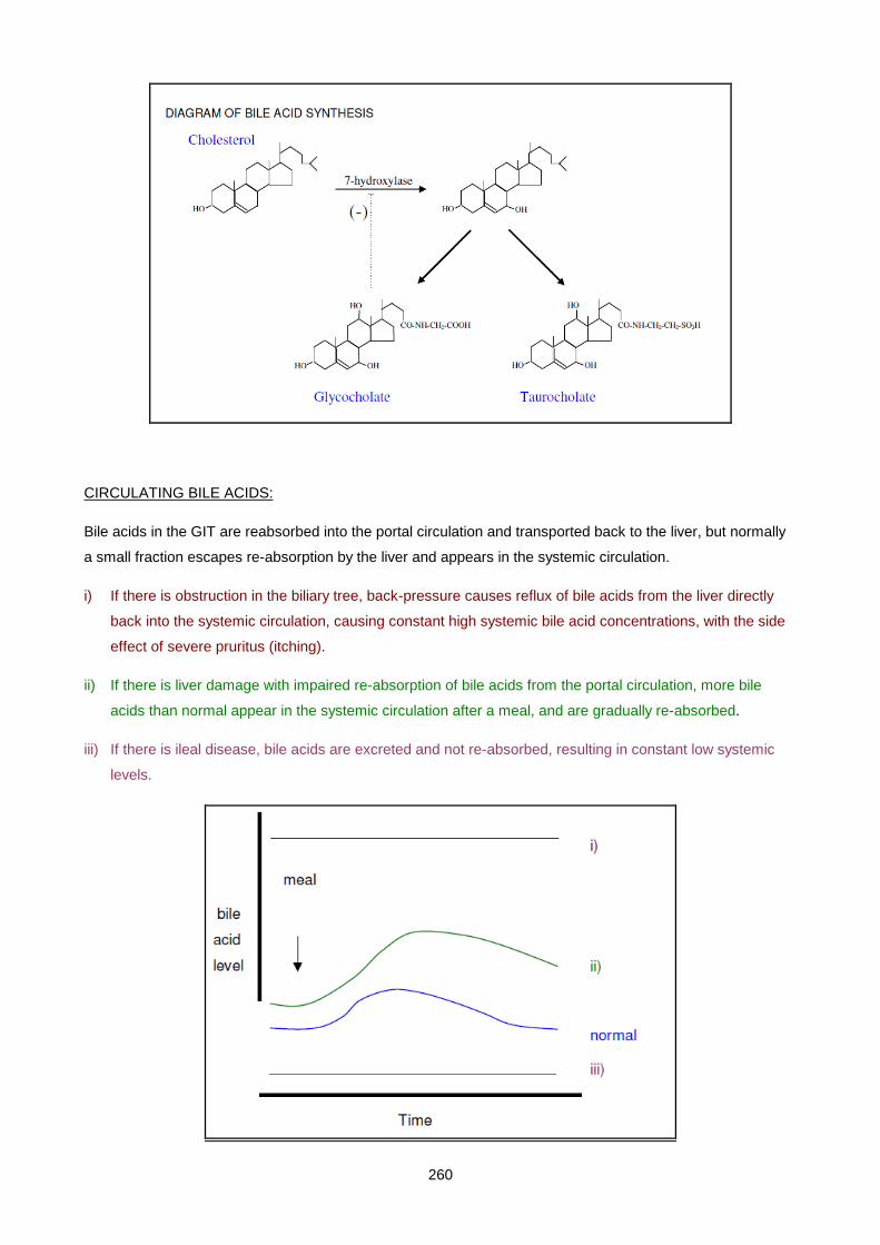
260
CIRCULATING BILE ACIDS:
Bile acids in the GIT are reabsorbed into the portal circulation and transported back to the liver, but normally
a small fraction escapes re-absorption by the liver and appears in the systemic circulation.
i) If there is obstruction in the biliary tree, back-pressure causes reflux of bile acids from the liver directly
back into the systemic circulation, causing constant high systemic bile acid concentrations, with the side
effect of severe pruritus (itching).
ii) If there is liver damage with impaired re-absorption of bile acids from the portal circulation, more bile
acids than normal appear in the systemic circulation after a meal, and are gradually re-absorbed.
iii) If there is ileal disease, bile acids are excreted and not re-absorbed, resulting in constant low systemic
levels.
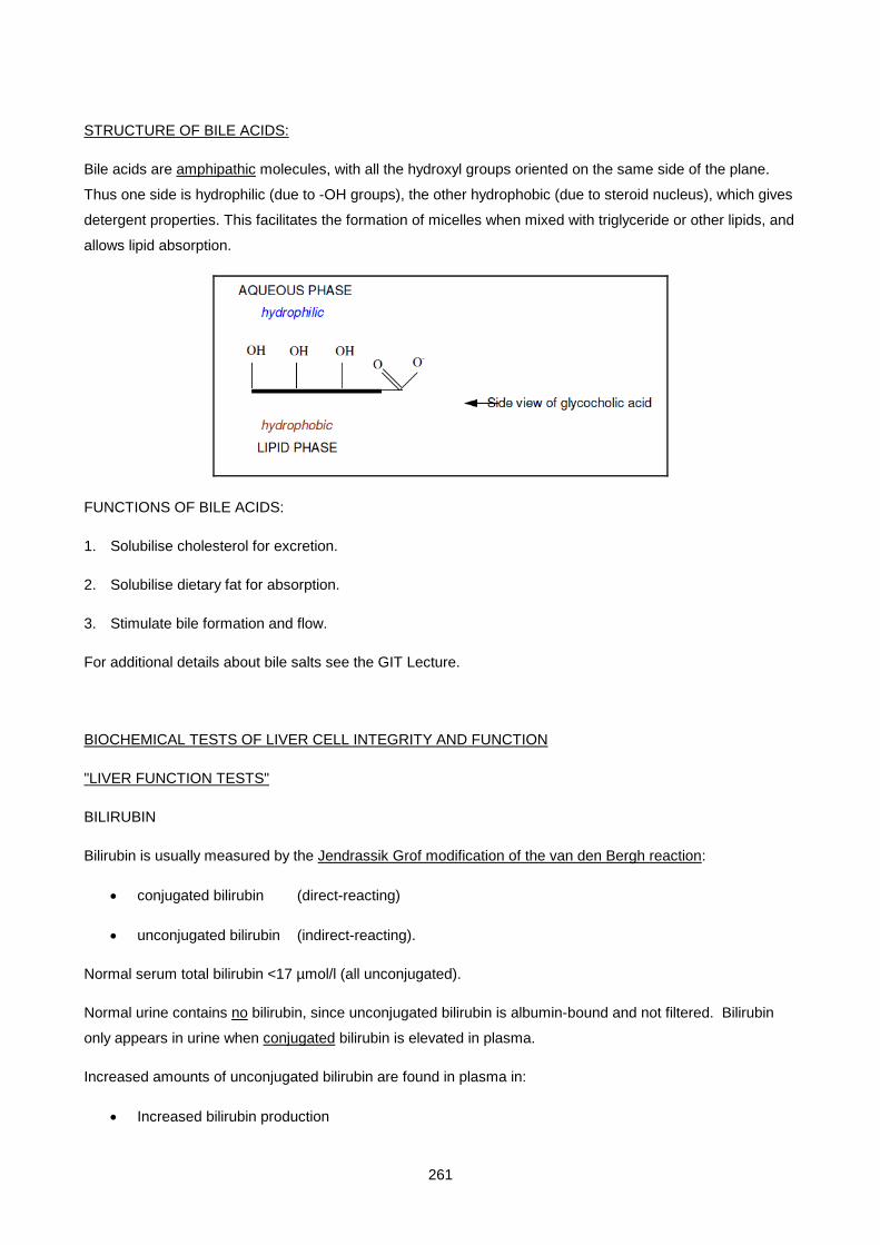
261
STRUCTURE OF BILE ACIDS:
Bile acids are amphipathic molecules, with all the hydroxyl groups oriented on the same side of the plane.
Thus one side is hydrophilic (due to -OH groups), the other hydrophobic (due to steroid nucleus), which gives
detergent properties. This facilitates the formation of micelles when mixed with triglyceride or other lipids, and
allows lipid absorption.
FUNCTIONS OF BILE ACIDS:
1. Solubilise cholesterol for excretion.
2. Solubilise dietary fat for absorption.
3. Stimulate bile formation and flow.
For additional details about bile salts see the GIT Lecture.
BIOCHEMICAL TESTS OF LIVER CELL INTEGRITY AND FUNCTION
"LIVER FUNCTION TESTS"
BILIRUBIN
Bilirubin is usually measured by the Jendrassik Grof modification of the van den Bergh reaction:
• conjugated bilirubin (direct-reacting)
• unconjugated bilirubin (indirect-reacting).
Normal serum total bilirubin <17 µmol/l (all unconjugated).
Normal urine contains no bilirubin, since unconjugated bilirubin is albumin-bound and not filtered. Bilirubin
only appears in urine when conjugated bilirubin is elevated in plasma.
Increased amounts of unconjugated bilirubin are found in plasma in:
• Increased bilirubin production
262
• Decreased uptake or conjugation of bilirubin
• in generalised hepatocellular dysfunction
• in specific rare inherited syndromes (Gilbert’s and Criggler-Najjar syndromes).
Increased amounts of conjugated bilirubin are found in the plasma (and urine) in:
Decreased excretion of bilirubin
o in obstructive liver disease
o in specific rare inherited syndromes (Rotor’s and Dubin-Johnson syndromes).
(In prolonged obstructive jaundice delta-bilirubin is gradually formed, which is conjugated bilirubin covalently
linked to albumin. Not filtered by kidney. This explains why conjugated hyperbilirubinaemia may occasionally
persist even after bilirubinuria has disappeared).
URINE UROBILINOGEN
Urine urobilinogen is measured qualitatively using a dipstick.
Normal urine contains some urobilinogen.
Increased amounts of urobilinogen are found in the urine in:
• Increased bilirubin production.
• Decreased re-uptake into liver due to hepatocellular dysfunction (but not if obstruction prevents
bilirubin reaching GIT).
LIVER ENZYMES
(Also see Enzymology lectures).
The usefulness of serum enzymes as markers of liver disease is limited by the fact that they are also found in
and released from other tissues. It is therefore more useful to look at all the parameters of liver function (or
at least all of the enzymes) rather than at one single enzyme.
ENZYMES REFLECTING LIVER CELL DAMAGE:
These enzymes are released from damaged cells, due to increased cell membrane permeability or cell
necrosis.
• Transaminases
263
o Aspartate transaminase (AST) has widespread tissue distribution including liver, red blood
cells, skeletal and cardiac muscle. No tissue-specific isoenzymes. Cytosolic and
mitochondrial.
o Alanine transaminase (ALT) is more liver-specific. Cytosolic only.
• Lactate Dehydrogenase (LD or LDH) has widespread tissue distribution including liver, red blood
cells, skeletal and cardiac muscle. Tissue-specific isoenzymes can be separated by electrophoresis.
The LD5 isoenzyme is found in liver and skeletal muscle only.
ENZYMES REFLECTING CHOLESTASIS:
• Alkaline phosphatase (ALP) has widespread tissue distribution including liver, bone, placenta and
GIT. The liver isoenzyme can be identified by electrophoresis or heat inactivation. The natural
function of this enzyme is unknown. It is an ecto-enzyme, located on the outside of the cell
membrane (canalicular side of liver cell). It is released into plasma in cholestasis.
• Gamma-glutamyl transpeptidase (GGT) is more liver-specific. It is involved in the transport of amino
acids across the liver cell plasma membrane. Serum level increased by cholestasis or chronic
ingestion of alcohol, barbiturates, phenytoin and other drugs which induce the enzyme.
PLASMA PROTEINS
(Also see Plasma Protein lectures)
• The liver synthesises all plasma proteins except immunoglobulins.
• Albumin is decreased in chronic liver disease, but is insensitive as an index of liver function. Not
useful in acute liver disease because of its long half-life (18 days).
• Clotting factors have short half-lives, e.g. factor VII t½ = 4h. The prothrombin time (INR) and partial
thromboplastin time (PTT) may be prolonged in liver disease due to decreased synthesis of clotting
factors or secondary vitamin K deficiency resulting from fat malabsorption.
• Immunoglobulins show a generalised increase (polyclonal) in chronic liver disease, especially
cirrhosis. This results from increased antigenic stimulation due to shunting of blood from GIT directly
into systemic circulation.
• In primary biliary cirrhosis IgM is characteristically increased.
• In alcoholic cirrhosis IgA is characteristically increased and this tends to cause "beta-gamma
bridging" on serum electrophoresis.
• In autoimmune chronic active hepatitis IgG is particularly increased.
264
• Alpha-foetoprotein (AFP) is the embryonic form of albumin, normally absent from plasma. It is
increased markedly in primary liver cell carcinoma (hepatoma) as a result of reversion of the
malignant cells to a de-differentiated state. Also produced by some germ cell tumours (e.g.
teratoma). Moderate elevations may occur when liver tissue is regenerating, such as in the recovery
stage after hepatitis or in cirrhosis.
• Alpha-1 antitrypsin deficiency is a genetic disorder which presents with childhood cirrhosis and
severe emphysema in early adulthood.
• Ferritin is the form in which iron is stored in the liver. Increased levels reflect increased iron stores or
liver cell necrosis. (See Iron lectures).
PLASMA AMMONIA
Normal level <40 µmol/l. Measurement of ammonia is most useful in cases of altered levels of
consciousness, since elevated ammonia levels are responsible for the neurological signs of hepatic
encephalopathy.
Hyperammonaemia occurs in :
• Generalised liver disease with hepatic failure.
• Transient Hyperammonaemia of the Newborn, due to liver immaturity.
• Urea cycle enzyme deficiencies. These mostly present in childhood, with neurological symptoms.
Diagnosis is made on the basis of hyperammonaemia and specific amino acid abnormalities in
plasma and urine (see Inborn Errors of Metabolism lectures).
Plasma ammonia rises rapidly after blood sampling due to glutaminase activity in plasma and RBCs - test
must therefore be pre-arranged with lab., blood sent on ice, spun immediately, test done within 1 hr after
taking blood.
CSF GLUTAMINE
Increased in hepatic encephalopathy.
LIVER DISORDERS AND JAUNDICE
JAUNDICE or icterus is the yellow appearance of skin and sclerae due to the presence of an excessive
amount of bilirubin (jaundice becomes clinically visible when serum bilirubin is >40 µmol/l). The liver has a
large reserve capacity - jaundice only appears with severe impairment of liver function.
Classification of causes of jaundice:
265
A. PREHEPATIC
• Excess bilirubin production.
B. INTRAHEPATIC
• Decreased uptake of bilirubin into liver cells.
• Decreased conjugation of bilirubin by liver cells.
• Decreased excretion of bilirubin into bile canaliculi.
C. POSTHEPATIC
• Biliary obstruction.
A. PREHEPATIC JAUNDICE (increased production of bilirubin)
HAEMOLYTIC DISORDERS
• Abnormal haemoglobins (e.g. sickle cell anaemia).
• RBC membrane defects (e.g. hereditary spherocytosis).
• RBC enzyme defects (e.g. G6PD deficiency).
• Autoimmune haemolytic anaemias.
• Blood group incompatibility
o foetal / maternal (newborns)
o transfusion.
• Bacterial toxins causing haemolysis.
• Malaria.
INEFFECTIVE ERYTHROPOIESIS
• Megaloblastic anaemias.
RESULTS OF LABORATORY TESTS in cases of excess bilirubin production:
• Increased unconjugated bilirubin (rate of bilirubin production exceeds maximum rate of conjugation). Haemolytic jaundice is never deep (except in newborns).
• Normal conjugated bilirubin (no obstruction).
• Urine bilirubin negative.
• Urine urobilinogen increased (increased flux through pathway).
• Other liver function tests normal.
• Haematological tests:
o increased reticulocyte count
o tests to identify cause of haemolysis (RBC morphology, abnormal haemoglobin, Coombs test, etc.)
B. INTRAHEPATIC JAUNDICE (decreased handling of bilirubin by the liver)

266
SPECIFIC INHERITED DEFECTS IN BILIRUBIN METABOLISM (rare)
DEFECT IN UPTAKE / CONJUGATION OF BILIRUBIN
GILBERT’S SYNDROME :
• Inherited disorder (AR) - decreased expression of UDPGT I due to a TA insertion in the TATA box.
• Mildly increased levels of unconjugated bilirubin (levels up to 85 µmol/l), made worse by viral infections and fasting.
CRIGGLER-NAJJAR SYNDROMES :
• Criggler-Najjar type I syndrome
o Inherited disorder (AR) - severe mutation of UDPGT I gene leading to absent UDPGT I activity.
o Severe disorder with very high levels of unconjugated bilirubin.
• Criggler-Najjar type II syndrome (Arias syndrome)
o Inherited disorder (AR) - milder mutation of UDPGT I gene leading to reduced UDPGT I activity.
o Milder than type I.
DEFECT IN EXCRETION OF BILIRUBIN INTO BILE CANALICULI
DUBIN-JOHNSON and ROTOR’S SYNDROMES :
• Benign inherited disorders.
• Increased levels of conjugated bilirubin in serum and urine.
• Due to impaired excretion.
ACUTE VIRAL HEPATITIS
IMPORTANCE:
• Common infectious disease worldwide.
• Clinical severity varies from asymptomatic, through mild hepatitis, to severe fulminant hepatitis which can be fatal.
• Responsible for the majority of chronic liver disease, which in turn is associated with hepatocellular carcinoma.
CAUSES:
• Hepatitis A and B (commonest).
• Non-A, Non-B hepatitis
• Hepatitis C, D and E viruses (serologic tests available).
• Other hepatitis viruses (no serologic tests available yet).
• Other viruses e.g. EBV, CMV.
CLINICAL FEATURES:
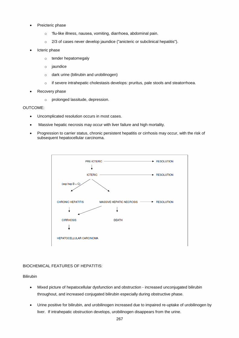
267
• Preicteric phase
o 'flu-like illness, nausea, vomiting, diarrhoea, abdominal pain.
o 2/3 of cases never develop jaundice ("anicteric or subclinical hepatitis").
• Icteric phase
o tender hepatomegaly
o jaundice
o dark urine (bilirubin and urobilinogen)
o if severe intrahepatic cholestasis develops: pruritus, pale stools and steatorrhoea.
• Recovery phase
o prolonged lassitude, depression.
OUTCOME:
• Uncomplicated resolution occurs in most cases.
• Massive hepatic necrosis may occur with liver failure and high mortality.
• Progression to carrier status, chronic persistent hepatitis or cirrhosis may occur, with the risk of subsequent hepatocellular carcinoma.
BIOCHEMICAL FEATURES OF HEPATITIS:
Bilirubin
• Mixed picture of hepatocellular dysfunction and obstruction - increased unconjugated bilirubin
throughout, and increased conjugated bilirubin especially during obstructive phase.
• Urine positive for bilirubin, and urobilinogen increased due to impaired re-uptake of urobilinogen by
liver. If intrahepatic obstruction develops, urobilinogen disappears from the urine.

268
ALT and AST
• Early and dramatic rise in serum transaminases reflecting hepatocyte damage. Start to rise before
onset of jaundice. Reach 20 to 100 times upper normal limit. Also increased in anicteric cases.
Useful in screening contacts.
• In massive hepatic necrosis transaminase levels may suddenly decrease due to total destruction of
liver cells - this is a grave prognostic sign
ALP and GGT
• Not greatly elevated early. Later marked increases as intrahepatic cholestasis develops due to
swelling of cells.
Plasma proteins
• Albumin only slightly decreased due to long half-life.
• Immunoglobulins: early increase in IgM, later IgG. Smooth muscle auto-antibodies may appear.
• Decreased prothrombin index due to impaired synthesis of clotting factors. Not restored by vitamin K.
• Transient mild rise in alpha-foetoprotein during regeneration phase.
EPIDEMIOLOGY:
• Hepatitis A
o “infectious hepatitis”
o spread by faecal-oral route
o mild infection, often subclinical, complications and fatalities are rare
o no progression to carrier status or chronic liver disease
o diagnosis :
HAV antigen = current infection
HAV IgM antibody = current infection
HAV IgG antibody = current or previous exposure
• Hepatitis B
o “serum hepatitis”
o spread by blood products, sexual transmission, transmission to infants in the first years of life
o variable clinical features, from subclinical to massive hepatic necrosis
o 10% progress to carrier status (more frequently in children), which includes :- asymptomatic carrie
o mild chronic hepatitis with little or no progression to cirrhosis
o severe chronic hepatitis with 50% progressing to cirrhosis and/or hepatocellular carcinoma
o diagnosis :
269
HBsAg = current infection (if it persists for longer than 6 months = carrier status)
HBeAg = current infection (if it persists for longer than 3 months = active carrier status)
HBcAb IgM = current infection (present in the window between HBsAg disappearing and HBsAb appearing)
HBsAb, HBeAb, HBcAb IgG = previous exposure and non-carrier status.
• Hepatitis C
o 80% of NANB transfusion hepatitis and 50% of community acquired NANB hepatitis
o spread by blood products, sexual transmission, and ? close contact with carrier
o mild infection, but 50% progress to cirrhosis and/or hepatocellular carcinoma
o diagnosis :
HCV antibodies which often only become positive after a long delay.
TOXIC HEPATITIS
The liver is the site of metabolism of most drugs. Many drugs are hepatotoxic. Some are idiosyncratic reactions, others affect all individuals. Drugs may cause either:
• Toxic hepatitis (e.g. alcohol, paracetamol). Biochemical changes similar to acute viral hepatitis.
• Intrahepatic cholestasis. (See below).
One syndrome in which drugs may be implicated is Reye's syndrome:
• An acute disorder of unknown aetiology in children
• Drugs (usually salicylates) in combination with viral infection have been implicated.
• Characterised by
o enlarged liver with fatty change
o encephalopathy, hyperammonaemia
o elevated transaminases
o high mortality.
INTRAHEPATIC CHOLESTASIS
CAUSES:
• Drugs
• Pregnancy ? sensitivity to oestrogen or progesterone.
• Benign recurrent intrahepatic cholestasis. Precipitated by viral infections.
BIOCHEMICAL FEATURES:
(Same as extrahepatic cholestasis)
• Increased conjugated bilirubin in serum and urine.
270
• Increased ALP and GGT. Transaminases only elevated if cholestasis is severe enough to cause secondary hepatocellular damage.
• No urobilinogen in urine.
CHRONIC HEPATITIS / CHRONIC LIVER DISEASE
Chronic hepatitis is defined as hepatic inflammation (documented histologically or biochemically) due to any cause, persisting for more than 6 months.
The commonest antecedent causes are viral, toxic or autoimmune hepatitis. Irrespective of aetiology, the classification, prognosis and treatment are based on the histological features. Two categories are commonly used:
• Chronic persistent hepatitis
• Mildly elevated transaminases. Not associated with extrahepatic manifestations and with a good prognosis.
• Chronic active hepatitis
• Autoimmune disorder - may be associated with autoimmune disease of other organs (thyroiditis, arthritis, colitis).
• Episodes of cell necrosis, leading to cirrhosis. Histologic picture is characteristic: "piecemeal necrosis".
• Elevated serum IgG, smooth-muscle antibodies and antinuclear factor are characteristic. Some patients may have elevated IgM and mitochondrial antibodies (resembling primary biliary cirrhosis).
Other specific chronic liver disease entities include Alcoholic Liver Disease and Non-Alcoholic Steatohepatitis.
ALCOHOLIC LIVER DISEASE
A range of liver pathology may occur in alcoholic liver disease, ranging from fatty liver, to alcoholic hepatitis, to fullblown alcoholic cirrhosis.
Biochemical features include:
• raised GGT because of induction as well as cholestasis (since GGT levels decline with abstention, GGT is used to monitor alcohol intake)
• mild disease - few additional biochemical indicators are present
• severe disease- transaminases are elevated, especially AST (therefore the ALT/AST ratio is less than 1)
• in cirrhosis
o hyperlipidaemia
o increased immunoglobulins esp. IgA producing "beta-gamma bridging" on serum electrophoresis
(NON-ALCOHOLIC STEATOHEPATITIS (NASH)
A form of chronic hepatitis which is similar histologically to alcoholic hepatitis, but which occurs in non-
alcoholic patients.
271
The diagnosis may only be made if the histological picture resembles that of alcoholic hepatitis
(macrovesicular steatosis, parenchymal inflammation, hepatic “ballooning” with/without Mallory bodies and/or
fibrosis) and alcohol and other causes of chronic liver disease have been excluded.
The pathogenesis is not yet known, but there is experimental evidence to suggest that peroxisomal oxidation
of excess free fatty acids or ketones in the liver may be involved, and endotoxin-mediated injury may also
play a role.
Although the cause has not yet been definitively determined, risk factors associated with the condition
include:
• obesity (present in the majority of patients)
• NIDDM (present in the majority of patients)
• jejenal-ileal bypass
• small bowel resection and small bowel bacterial contamination
• drugs such as amiodarone, calcium channel blockers and others.
Biochemical features include:
• raised transaminases, especially ALT
• hyperlipidaemia
• NIDDM
Treatment includes gradual weight loss (not rapid) and small bowel sterilisation where indicated. Control of
underlying NIDDM and hyperlipidaemia and avoidance of alcohol appear sensible, but may not influence the
existing hepatic pathology. Liver transplantation has been used.
CIRRHOSIS
IMPORTANCE:
• Final common pathway of many chronic liver diseases, leading eventually to hepatic insufficiency
(hepatic failure).
• May also follow acute massive necrosis.
PATHOLOGIC (HISTOLOGICAL) FEATURES:
• Fibrosis.
• Regenerating nodules. Alcoholic, cardiac cirrhosis are micronodular. Post-hepatitic is macronodular.
272
• Derangement of microvasculature leading to portal hypertension and shunting of portal blood to
systemic circulation.
CAUSES:
Drugs / toxins: Alcohol, Methotrexate, Methyldopa, Isoniazid,
Infections: Hepatitis B, Hepatitis C, Schistosomiasis
Autoimmune : Chronic active hepatitis, Primary biliary cirrhosis
Metabolic : Wilson's disease, Haemochromatosis, Alpha-1-antitrypsin deficiency, Glycogen storage diseases
Biliary obstruction: Atresia, Gallstones Strictures
Vascular: Chronic right heart failure, Budd-Chiari syndrome, Veno-occlusive disease
Miscellaneous: Sarcoidosis Indian childhood cirrhosis, Cryptogenic
COURSE:
• Cirrhosis may remain "compensated" for many years before hepatic failure is precipitated by some event, often a GIT bleed from oesophageal varices.
• Hepatic failure occurs in two settings:
o Terminal phase of cirrhosis.
o Acute massive hepatic necrosis.
FUNCTIONAL DERANGEMENTS:
The manifestations of cirrhosis and hepatic failure are due to a combination of mechanisms:
• The specific disorder causing the cirrhosis.
• Hepatocellular failure due to loss of hepatic tissue and shunting of blood past the remaining tissue.
• Cholestasis and its consequences (e.g. vitamin K deficiency).
• Portal hypertension leading to hypersplenism and portal-systemic anastomoses.
CLINICAL AND BIOCHEMICAL MANIFESTATIONS OF CIRRHOSIS AND HEPATIC FAILURE:
• Jaundice
o Due to
Shunting of blood past the liver.
Impaired conjugation ability due to hepatocellular dysfunction.
Obstruction.
Mixed picture of hepatocellular dysfunction and obstruction: increased unconjugated and conjugated bilirubin
273
• Liver enzymes
o No changes which are characteristic of cirrhosis per se.
o Mild increases of ALT and AST indicate some liver cell damage, while mild increases in ALP and GGT indicate some obstruction).
• Hepatic encephalopathy
o Early symptoms of irritability, personality or behavioural changes.
o Later features of tremor (typically a "flapping" tremor), ataxia, confusion, delirium and coma.
o Biochemical basis for hepatic encephalopathy is complex but includes:
Hyperammonaemia.
Hypoglycaemia due to failure of glycogenolysis/gluconeogenesis. Patients may need to be maintained on a constant glucose infusion.
False neurotransmitters (e.g. octopamine) produced by GIT bacteria which are normally degraded by the liver.
Accumulation of short chain fatty acids (e.g. butyrate, valerate) which interfere with neurotransmission.
• Abnormal amino acid metabolism. In serum, tyrosine and methionine are characteristically elevated.
• Sterilisation of the GIT with neomycin and lactulose (laxative) is often employed therapeutically.
• Ascites due to
Portal hypertension.
Low plasma albumin due to hepatocellular dysfunction.
Elevated aldosterone and salt retention. Secondary hyperaldosteronism is present in cirrhosis as a result of a low "effective arterial volume" due to low albumin.
• Bleeding tendency due to
Impaired synthesis of clotting factors due to hepatocellular dysfunction.
Vitamin K deficiency due to fat malabsorption.
Thrombocytopaenia due to hypersplenism.
o Oesophageal varices are the commonest site for bleeding.
• Anaemia due to
Disturbances in vitamin B12 and folate metabolism.
Bleeding.
Hypersplenism.
• Endocrine changes
o Failure to conjugate oestrogens leads to gynaecomastia, loss of body hair, and testicular atrophy. Palmar erythema and spider naevi have also been attributed to excess oestrogens.
• Malnutrition and vitamin deficiencies are often present due to
Alcoholism.
Loss of appetite.
Malabsorption.
274
• Hepatoma may develop in a regenerating liver nodule in any form of cirrhosis.
SPECIFIC FORMS OF CIRRHOSIS:
POSTHEPATITIC CIRRHOSIS
• Shrunken, fibrotic liver (macronodular).
• May be carriers of HBsAg.
• IgG is the predominantly elevated immunoglobulin.
(PRIMARY BILIARY CIRRHOSIS
• Probably autoimmune pathogenesis.
• Proliferation of epithelium of small bile ductules, with inflammatory infiltration and fibrosis.
• Presents with manifestations of cholestasis.
• Hypercholesterolaemia is marked, with cutaneous xanthomata.
• IgM is the predominantly elevated immunoglobulin - a useful diagnostic point.
• Mitochondrial antibodies are characteristic, but not entirely specific for primary biliary cirrhosis.
SECONDARY BILIARY CIRRHOSIS
• Due to chronic extrahepatic obstruction from any cause.
• The immunologic abnormalities of primary biliary cirrhosis are absent.
POSTHEPATIC JAUNDICE
CAUSES OF EXTRAHEPATIC OBSTRUCTION (“CHOLESTASIS”):
• In the lumen (gallstones, worms).
• In the wall (bile duct carcinoma, stricture, atresia).
• Outside the wall (carcinoma of head of pancreas, lymph node enlargement at porta hepatis).
CLINICAL AND BIOCHEMICAL FEATURES OF OBSTRUCTION (INTRAHEPATIC OR EXTRAHEPATIC):
• Jaundice, dark urine, pale stools. The jaundice may be very deep, despite a generally well patient.
• Increased conjugated bilirubin in serum and urine.
TYPES OF GALLSTONES:
• Cholesterol stones.
275
o The common gallstone.
o Predisposing factors:
Obesity, increasing age, female sex, diabetes, hyperlipidaemia
Chronic cholecystitis.
Relative deficiency of tri- and dihydroxy bile salts leading to "lithogenic bile" . Oral chenodeoxycholic acid has been used therapeutically.
• Pigment stones.
o Excess bilirubin production in haemolytic anaemias.
PRESENTATION:
• Intermittent passage of gallstone:
• Severe right upper quadrant abdominal pain (biliary colic).
• Intermittent jaundice and bilirubinuria.
• Intermittent elevations of ALP, GGT.
• Prolonged painless obstructive jaundice.
• Acute cholecystitis.
MISCELLANEOUS TOPICS
SPACE-OCCUPYING LESIONS OF THE LIVER
CAUSES:
• Primary liver cell carcinoma (hepatoma).
• Marked increase in alpha-foetoprotein.
• Predisposing factors:
• Cirrhosis.
• Previous hepatitis B infection.
• Secondary (metastatic).
• Primary can be anywhere, often GIT.
• Hydatid cysts.
• Amoebic liver abscess.
FEATURES:
• Enlarged firm liver.
• Increased ALP, GGT due to local obstruction
• However jaundice is rarely present and serum bilirubin is normal or only slightly increased, since remaining normal tissue has large reserve capacity for excreting bilirubin.
• AST, ALT normal/slightly increased.
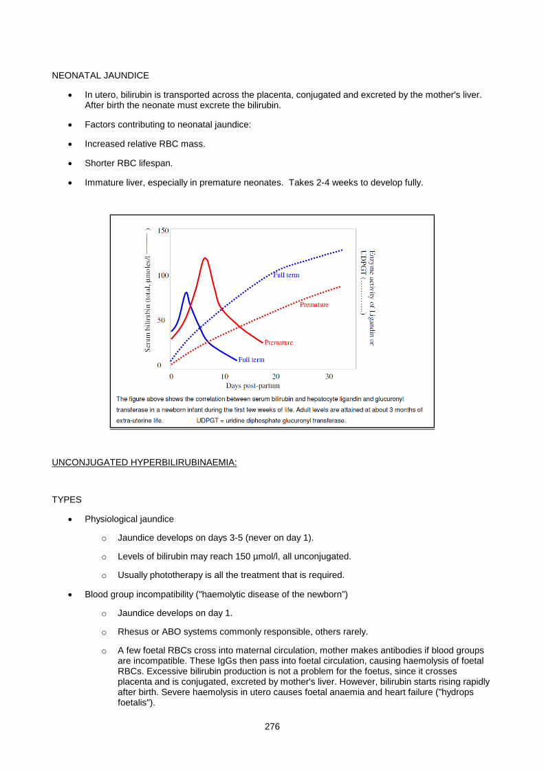
276
NEONATAL JAUNDICE
• In utero, bilirubin is transported across the placenta, conjugated and excreted by the mother's liver. After birth the neonate must excrete the bilirubin.
• Factors contributing to neonatal jaundice:
• Increased relative RBC mass.
• Shorter RBC lifespan.
• Immature liver, especially in premature neonates. Takes 2-4 weeks to develop fully.
UNCONJUGATED HYPERBILIRUBINAEMIA:
TYPES
• Physiological jaundice
o Jaundice develops on days 3-5 (never on day 1).
o Levels of bilirubin may reach 150 µmol/l, all unconjugated.
o Usually phototherapy is all the treatment that is required.
• Blood group incompatibility ("haemolytic disease of the newborn”)
o Jaundice develops on day 1.
o Rhesus or ABO systems commonly responsible, others rarely.
o A few foetal RBCs cross into maternal circulation, mother makes antibodies if blood groups are incompatible. These IgGs then pass into foetal circulation, causing haemolysis of foetal RBCs. Excessive bilirubin production is not a problem for the foetus, since it crosses placenta and is conjugated, excreted by mother's liver. However, bilirubin starts rising rapidly after birth. Severe haemolysis in utero causes foetal anaemia and heart failure ("hydrops foetalis").
277
• Inherited haemolytic disorders (as in adults).
COMPLICATION
Risk of kernicterus when unconjugated bilirubin > 200 µmol/l. Risk is increased by other factors:
• Level of unconjugated bilirubin.
• Low albumin.
• Drugs (displace bilirubin from albumin).
• Acidosis (promotes dissociation of bilirubin from albumin).
• Heparin administration (causes liberation of free fatty acids which displace bilirubin from albumin).
• Hypoxia, hypoglycaemia, hypothermia, sepsis (increase bilirubin transport into the brain).
Features: - Lethargy, hypotonia, poor feeding, spasticity and death.
TREATMENT
• Phototherapy
o Light at 440-470 nm causes formation in skin of isomers of unconjugated bilirubin which are more soluble, excreted in urine.
• Exchange transfusion
o Required when bilirubin > 305 µmol/l (term babies) or > 200 µmol/l in premature babies.
CONJUGATED HYPERBILIRUBINAEMIA:
TYPES
• Neonatal hepatitis
o Many infective agents can cause hepatitis in neonates (e.g. rubella, CMV, herpes simplex, toxoplasmosis).
• Biliary atresia
o Cause of biliary atresia is unclear: may be toxic degeneration of bile duct rather than a congenital malformation.
• Metabolic disorders
TREATMENT
- Treat the underlying condition.

278
SMALL GROUP TEACHING: LECTURE 14: CHEMICAL PATHOLOGY OF LIVER DISEASE
QUESTIONS
1. The following results were obtained from a patient who presented with mild jaundice:
Albumin 45g/l (35-50), total bilirubin 43µmol/l (<17), conj. bilirubin 2µmol/l (<4), Alk. Pho 102 U/l (39-117), LDH 720U/l (240-480), AST 72U/l (1-38), ALT 25U/l(1-41)
i) What type of disorder is indicated?
ii) What biochemical abnormalities are likely to be present in this patient's urine?
iii) What LDH isoenzyme pattern would you expect to find?
iv) What further investigations are required?
v) List the causes of this type of disorder.
2. A 63-year old man presented with haematemesis. He had lost 12 kg of weight over the preceding 6 months. Gastroscopy showed a carcinoma of the stomach. The following results were reported:
urea 5.0 mmol/l (1.7-6.7) creatinine 66 µmol/l (60-115) total protein 55 g/l (60-80) albumin 23 g/l (35-50) calcium 1.86 mmol/l (2.1-2.6) total bilirubin 15 µmol/l (<17) conj. bilirubin 3 µmol/l (<4) GGT 373 U/l (7-49)
Alk. Phos. 443 U/l (39-117) LDH 344 U/l (240-480) AST 18 U/l (1-38) ALT 22 U/l (1-41)
aFP 20 ng/ml (<10)
i) What do the liver function tests indicate? Comment on the fact that the bilirubin is not elevated.
ii) Comment on the calcium level.
3. A twenty-year old student developed a ‘flu-like illness with loss of appetite, nausea, and pain in the right hypochondrium. On examination, the liver was just palpable and tender. Two days later he developed jaudice, dark urine and pale stools.
on presentation 1 week later
serum:
total bili. 38 230 µmol/l (<17)
conj. bili. 25 200µmol/l (<4)
albumin 40 38g/l (35-50)
ALT 950 760U/l (1-41)
AST 624 580U/l (1-38)
Alk. Phos. 79 230U/l (39-117)
GGT 20 215U/l (7-49)
urine (dipstick):

279
bilirubin + +++
urobilinogen +++ +
i) Suggest the most likely diagnosis. Explain the changes in biochemical profile during the course of the illness.
4. A 55-year old woman presented to Casualty in a coma. On examination, she was noted to be jaundiced, and multiple spider naevi were present on her trunk. Her husband said that she was a heavy drinker, and had previously had "liver trouble". She had begun to vomit blood the previous day. Blood was taken for emergency investigations which showed:
Na+ 129mmol/l (135-145), K+ 4.5mmol/l (3.5-5.0), urea 7.1mmol/l(1.7-6.7),creatinine 120µmol/l (50-100), glucose 1.5mmol/l (3.9-5.6)
acid-base: pH 7.54, pCO2 6.5kPa, SBC 35mmol/l
ammonia 240µmol/l (<40)
total protein 80g/l (60-80)
albumin 23g/l (35-50)
total bilirubin 345 µmol/l (<17)
conj. bilirubin 290 µmol/l(<4)
ALT 60U/l (1-41)
Alk.Phos 445U/l (39-117)
GGT 190U/l (7-49)
i) suggest a diagnosis.
ii) Explain the hypoglycaemia. Does this require treatment?
iii) Comment on the other biochemical findings.
iv) Would a lumbar puncture be informative?
v) ist the main approaches to management.
vi) What life-threatening complications may occur?
5. A medical student recovering from a bout of 'flu noticed he was slightly jaundiced. These results were found:
total bilirubin 60 µmol/l (<17)
conj. bilirubin 2 µmol/l (<4)
ALT 15 U/l (1-41)
AST 25 U/l (1-38)
Alk. Phos. 65 U/l (39-117)
Urine bilirubin was negative.
Hemoglobin was 16 g/dl (normal) and the retic. count was not elevated.
i) Suggest a diagnosis, and treatment.
6. A 65 year old presented with visible jaundice which he had noticed was deepening in colour. He had no pain, but noticed some weight loss and his stools were pale. He was a moderate drinker, and not on any drugs.
total bilirubin 250µmol/l(<17), ALT 92U/l(1-41), AST 87U/l(1-38), Alk. Phos .850U/l(39-117)
280
i) What is the differential diagnosis
ii) What other investigations would be helpful in making the diagnosis?
SMALL GROUP TEACHING: LECTURE 14: CHEMICAL PATHOLOGY OF LIVER DISEASE
ANSWERS
1. Sickle cell disease. i) haemolytic anaemia ii) +++ urobilinogen, negative for bilirubin
iii) elevation of LD1 and LD2 (equally)
iv) hematological investigation of the cause of the haemolysis
v) RBC enzyme defects (e.g. G6PD def.), RBC membrane defects (hereditary spherocytosis), immune haemolysis (autoimmune, Rhesus disease), mechanical (artificial heart valve)
2. Carcinoma of the stomach with liver metastases.
i) Picture of localized obstruction - increased ALP and GGT, normal bilirubin.
ii) Normal after correction for low albumin.
3. Infectious Hepatitis. Early: Dramatic rise in serum transaminases reflecting hepatocyte damage. Bilirubin mixed picture of hepatocellular dysfunction and obstruction. ALP and GGT - initially normal. Urine positive for bilirubin, and urobilinogen increased due to impaired re-uptake of urobilinogen by liver. Later: Transaminases persist high till resolution. Bilirubin obstructive picture predominates. ALP and GGT - marked increases as intrahepatic cholestasis develops. Urine urobilinogen disappears as obstruction develops.
4. Decompensated cirrhosis with hepatic encephalopathy
i) Hepatic encephalopathy in decompensated cirrhosis with a GIT bleed.
ii) Decreased gluconeogenesis and glycogen storage. Yes of course.
iii) Hyponatraemia often seen in cirrhosis. Mechanism unclear, possibly increased ADH due to decreased effective arterial volume due to hypoalbuminaemia. Metabolic alkalosis due to vomiting. Low albumin - decreased liver synthetic function. Increased globulins - polyclonal, due to shunting of portal blood containing GIT-derived antigens and chronic stimulation of immune system.
iv) LP would show increased CSF glutamine - provides no additional information.
v) Treat bleeding varices as matter of urgency. Monitor and treat hypoglycaemia. Treat bleeding tendency - fresh frozen plasma, vitamin K. Limit protein, sterilize the GIT with lactulose/neomycin.
vi) Hypoglycaemia. Bleeding. Coma.
5. Gilbert’s syndrome. No treatment required.
6. Ca pancreas with obstructive jaundice
i) Obstructive jaundice (pale stools, high ALP), and signs suggestive of malignancy (deep painless jaundice) suggests ca head pancreas.
ii) Imaging of vicinity of head of pancreas and common bile duct.
281
Lecture 15: Enzymology
DR H. VREEDE 2007
“Clinical Enzymology is the application of the Science of Enzymes to the diagnosis and treatment of disease”.
(Moss and Henderson, 1994 in Tietz Textbook of Clinical Chemistry, 2nd ed.)
• 20-25% of workload of Chemical Pathology Lab.
• 12 to 15 enzymes are routinely measured.
HISTORICAL
1908 Wolgemuth - Amylase in urine
1920-1930 King and Bodansky - Alkaline phosphatase
1930’s Acid phosphatase for Ca Prostate
1955 La Due, Wroblewski and Karmen - Transaminases in serum of Acute Myocardial Infarction
GENERAL PROPERTIES OF ENZYMES RELEVANT FOR DIAGNOSIS
1. Proteins having catalytic properties.
Being proteins, enzymes are susceptible to inactivation and denaturation at extremes of temperature,
pH and protein precipitants.
2. High specificity for substrates.
• NOTE: Vmax - Activity of enzyme when saturated with substrate.
• Km - Concentration of substrate which gives half-maximal activity.
o The lower the Km, the higher the affinity of enzyme for substrate.
e.g.,Glucokinase in liver - Km for glucose is 10 Mm
Hexokinase in muscle - Km for glucose is < 0.1 mM,
Therefore hexokinase has greater affinity for glucose than glucokinase.
3. Isoenzymes are multiple forms of the same enzyme which catalyse the same reaction, but are
differentiated by their physical or chemical properties e.g., electrophoretic mobility, heat stability,
substrate specificity, sensitivity to inhibitors, antigenicity. Usually the product of two or more distinct
genes e.g., B and M subunits of creatine kinase, but may also arise from different post-translational
modification of the same gene product, e.g., bone and liver isoenzymes of alkaline phosphatase.
NOTE: Isoenzymes may have different Km’s.
282
4. Enzymes may require coenzymes for activity. Coenzymes are low molecular weight cofactors, e.g.,
thiamine (vitamin B1) is required for pyruvate dehydrogenase activity, and pyridoxine (vitamin B6) for
transaminase activity.
5. Enzymes are also antigens e.g., the M and B subunits of CK can be recognised antigenically.
6. High tissue : plasma activity ratio, e.g., activity of transaminases in liver cells, or creatine kinase in
muscle cells, is more than 10 000 times the normal plasma levels.
7. Enzymes are measured in terms of their activity. The unit of measurement is the International Unit
per Litre (IU/L). One IU = one µmol of substrate consumed per minute. One can measure rate of
substrate consumption or rate of product formation.
IMPORTANCE OF ENZYME ASSAYS
1. Detection of enzyme deficiencies within cells - either inherited (inborn errors) or acquired (vitamin
deficiency, poisoning).
2. Detection of tissue damage by measuring extracellular enzymes.
INBORN ERRORS
These include urea cycle defects, defects of carbohydrate metabolism (glycogen storage diseases,
galactosaemia, fructose intolerance), lysosomal storage diseases. Liver biopsy may be required for
diagnosis. Sometimes the enzyme of interest is expressed in cultured fibroblasts or peripheral leucocytes.
Red cell enzyme defects often present with haemolytic anaemia e.g., glucose-6-phosphate dehydrogenase
(G6PD), pyruvate kinase, pyrimidine 5' nucleotidase.
ACQUIRED DEFICIENCIES
Red cell transketolase activity is low in thiamine deficiency, red cell dALA dehydratase is low in lead toxicity,
plasma pseudocholinesterase is low in organophosphate poisoning.
TISSUE DAMAGE
Sample - usually serum; also urine, serous fluid, CSF, amniotic fluid, even aqueous humour. Enzyme assay
gives information about the state of cells in contact with that fluid.
Classify serum enzymes into:-
1. Plasma specific enzymes : pseudocholinesterase, clotting factors (proteolytic enzymes) - synthesised
by liver.
2. Secreted enzymes : pancreatic digestive enzymes (amylase, lipase), prostatic acid phosphatase.
3. Cellular enzymes : normally intracellular - leak out when tissue damaged. Advantages as markers of
damage:-
283
i) Sensitivity : There are often enormous differences in enzyme concentration between cells
and plasma. Minor damage results in noticeable increase in plasma activity - hence an
inherently sensitive test.
ii) Ease of assay - most enzymes are highly specific for the reaction catalysed - no need for
purification - merely provide the substrate.
iii) Tissue specificity - depends on using:
o Relatively organ specific enzymes (creatine kinase in muscle, ornithine
transcarbamylase in liver).
o Isoenzymes, which often have a different distribution between organs, and
sometimes also have a different distribution during development.
o A number of enzymes, to establish an organ specific pattern in the serum reflecting
the enzyme distribution in the organ of origin - e.g., after myocardial infarction, serum
creatine kinase, lactate dehydrogenase and aspartate transaminase rise and fall in a
definite sequence.
Serum enzymes increases may be due to:-
1. Cell death - this results in a small short-lived increase (e.g., following myocardial infarction).
2. Increased cell membrane permeability in living cells (due to hypoxia, inflammation, drugs/poisons,
cellular swelling) gives rise to a large protracted increase in serum enzymes as there is ongoing
enzyme synthesis (e.g., Duchenne muscular dystrophy, acute viral hepatitis).
3. Increased synthesis in a specific cell type (e.g., gamma glutamyl transferase in liver cells is induced
by alcohol or anticonvulsant drugs, alkaline phophatase in liver cells is induced by obstruction, lactate
dehydrogenase is induced in neoplastic tissues).
Raised enzyme levels are not synonymous with organ failure - highest levels are often seen in acute injury to
previously healthy tissue. Conversely organ failure is associated with unimpressive enzyme increases, e.g.,
end-stage liver disease, muscular dystrophy, chronic pancreatitis.
CAUSES OF CELL DAMAGE OR DEATH
Mechanism Example
Hypoxia Atheromatosis in myocardium, shock, anaemia
Drugs and Chemicals Alcohol, paracetamol, lead, mercury, carbon tetrachloride
Physical agents Trauma, radiation
Microbiological Bacteria, viruses, fungi, protozoa, helminths
Immune mechanisms Anaphylaxis, hypersensitivity, cytotoxicity, immunocomplexes (damage lysosomes)
Genetic Polygenic diseases, e.g., diabetes, gout; Mendelian disorders (XL, AR, AD)
Nutritional Malnutrition, vitamin deficiencies, mineral deficiencies
284
SPECIFIC COMMONLY MEASURED ENZYMES
CREATINE KINASE (CK) Creatine-P + ADP -----> creatine + ATP
• Important in tissues where significant metabolic energy is stored as creatine phosphate.
• Distribution : skeletal muscle, heart, brain.
• Isoenzyme composition : CK is a dimer consisting of sub-units M or B coded by two distinct genes.
• Thus 3 possible isoenzymes - M M ; M B ; B B
• Skeletal muscle - all MM; Heart - 80% MM, 20% MB; Brain - all BB.
• Separation of isoenzymes used to be performed by electrophoresis. Now CK-MB is more commonly
measured enzymatically or by mass determination.
• In the enzymatic assay, the M subunit (in MM and MB isoenzymes) is inhibited, after which the enzyme
activity of the remaining B subunit (in MB and BB isoenzymes) is measured. False positive results due to
raised BB isoenzyme can occur. A result of CK-MB >6% of total CK activity is regarded as positive.
• In the mass determination assay, the M subunit (in MM and MB isoenzymes) is bound by means of an
antibody, after which the concentration of the remaining B subunit (in the MB isoenzyme only) is
measured by means of a specific antibody reaction. False positives are less frequent, but more
specialised instrumentation is required for this assay.
o Normal pattern in serum - predominantly MM present, with MB < 6% of total CK.
Uses:
1. Myocardial infarction – early increase of total CK, specifically the MB isoenzyme. Total CK levels
start to rise at about 6 hours , peak at 18 to 30 hours, and return to normal by 3 days. If total CK
raised, measurement of CK-MB is indicated. CK-MB levels may be raised by 4 hours, are almost
certain to be raised by 12 hours, and may return to normal by 24 hours after MI. Used as a
diagnostic test before 24 hours, and as a prognostic indicator (amount of increase reflects extent of
cardiac damage).
2. Skeletal muscle damage e.g., trauma, surgery (especially in cardiac surgery), over-exercise,
convulsions, ischaemia, inflammation (myositis), malignant hyperthermia, congenital Duchenne
muscular dystrophy - increase of total CK, but MB isoenzyme not increased. In neurogenic muscle
disease, e.g., poliomyelitis and Parkinsonism, CK levels are normal. In Duchenne Muscular
Dystrophy, CK elevation precedes onset of symptoms by years, and falls as disease progresses. In
chronic muscle disease there is reversion to the foetal isoenzyme pattern (MB appears in skeletal
muscle and serum).
3. Hypothyroidism is associated with high total CK levels.
285
LACTATE DEHYDROGENASE (LD) Lactate + NAD+ -----> pyruvate + NADH + H+
Important in the disposal of glycolytically generated NADH, particularly when mitochondrial disposal is
impaired by hypoxia. Distribution: ubiquitous, including heart, skeletal muscle, liver and rbc’s - specificity is
improved by isoenzyme analysis. LD is a tetramer consisting of sub-units H or M coded by two different
genes.
Thus 5 possible isoenzymes : H H H H H H H M H H M M H M M M M M M M
LD1 LD2 LD3 LD4 LD5
Anoxic tissues (liver, muscle) express M subunit, thus LD5 predominates. Erythrocyte precursors and heart
express the H subunit, hence LD1 predominates in these tissues.
LDex - a tetramer of third type subunit found exclusively in spermatozoa.
Normal pattern in serum - predominantly LD2, with slightly less LD1, and even less LD3, LD4 and LD5.
Uses:
1. Myocardial infarction - late and long-lasting increase in total LD. Total LD levels start to rise at about
8 to 12 hours, peak at 24 to 48 hours and return to normal by 10 days. The predominant isoenzyme
is LD1, which is present in greater concentration than the normal LD2 (flipped pattern).
2. Liver damage, including viral or toxic hepatitis (ethanol, paracetamol overdose, carbon tetrachloride),
cardiac failure (liver congestion) - increase in total LD, exclusively due to LD5.
3. Haematological disorders - often isolated elevation in total LD. Due to breakdown of circulating red
cells or red cell precursors in bone marrow.
• Intra-vascular (or in vitro) haemolysis, e.g., due to an auto-immune disorder, prosthetic heart
valve, inherited enzyme deficiency (G6PD, PK) - both LD1 and LD2 increased. Associated
features include elevated serum unconjugated bilirubin, increased urine and stool
urobilinogen, and decreased haptoglobin.
• Megaloblastic anaemia due to folate or vitamin B12 deficiency : failure of cell division leads to
cell lysis and enzyme release from the bone marrow - predominant increase in LD1 (as in
myocardial infarction). Extremely high levels can be achieved.
4. Malignant tumours may manifest an isolated increase in serum LD due to enhanced synthesis of
glycolytic enzymes by a wide variety of neoplasms (even measured in aqueous humour to diagnose
retinoblastoma). Typically centripetal isoenzyme pattern (LD2, LD3 and LD4) due to expression of
both subunits (H and M). An exception is seen in germ cell tumours which show an increase in LD1.

286
Serum LD patterns obtained with agarose electrophoresis.
• Pattern A above, and 1 below: normal serum
• Pattern B above and 8 below: acute myocardial infarction showing a “flipped” LD1.
• Pattern C above and 4 below: centripetal pattern of malignancy with predominance of LD3
• Pattern D above and 5 below: congestive cardiac failure with elevated LD5 due to hepatic congestion
and hypoxia.
•

287
TRANSAMINASES
ASPARTATE TRANSAMINASE (AST) (aspartate +α-ketoglutarate -----> oxaloacetate + glutamate)
ALANINE TRANSAMINASE (ALT) (alanine + α-ketoglutarate -----> pyruvate + glutamate)
Wide distribution in tissues, including liver, skeletal muscle, heart, kidney and rbc’s.
Cofactor vitamin B6 (pyridoxine) - carries amino acid intermediates.
Intracellular function of transaminases:
• ALT required for amino acid catabolism for gluconeogenesis (cytosol)
• AST required to transfer reducing equivalents into mitochondria (cytosol and mitochondria).
AST exists as cytosolic and mitochondrial isoenzymes, but these cannot be distinguished in the laboratory.
Uses:
1. Acute hepatitis - high, sustained elevations of both ALT and AST (up to 100 x normal).
ALT higher especially in mild injury; precedes onset of jaundice (allows early diagnosis); decreasing
levels herald recovery (prognostic guide).
Disproportionate AST elevation indicates liver cell necrosis (involving mitochondria) or ethanol
induced damage (acetaldehyde formed during ethanol metabolism depletes cytosolic pyridoxine,
hence ALT activity selectively lost).
2. Myocardial infarction - AST elevation of 5 - 10 x normal. AST levels start to rise at about 6 to 8
hours, peak at 18 to 24 hours and return to normal by 4 to 5 days. ALT hardly increases at all. AST
always greater than ALT (de Ritis quotient AST/ALT > 5. In liver disease this ratio is usually < 1).
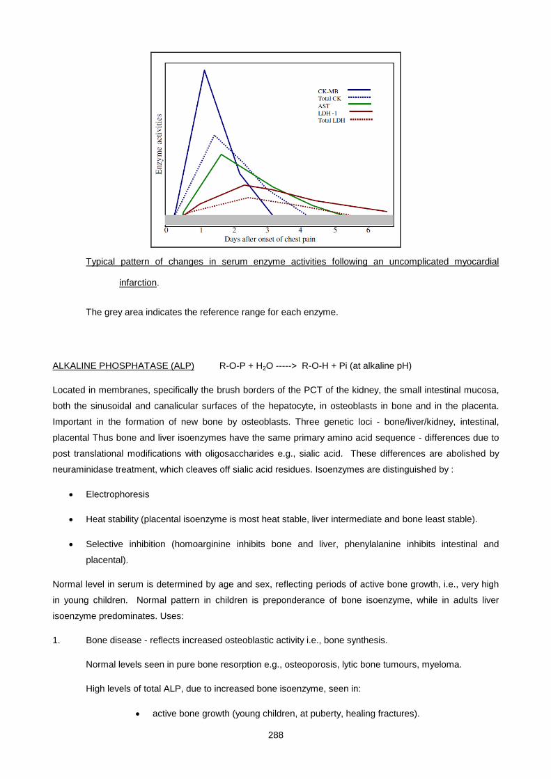
288
Typical pattern of changes in serum enzyme activities following an uncomplicated myocardial
infarction.
The grey area indicates the reference range for each enzyme.
ALKALINE PHOSPHATASE (ALP) R-O-P + H2O -----> R-O-H + Pi (at alkaline pH)
Located in membranes, specifically the brush borders of the PCT of the kidney, the small intestinal mucosa,
both the sinusoidal and canalicular surfaces of the hepatocyte, in osteoblasts in bone and in the placenta.
Important in the formation of new bone by osteoblasts. Three genetic loci - bone/liver/kidney, intestinal,
placental Thus bone and liver isoenzymes have the same primary amino acid sequence - differences due to
post translational modifications with oligosaccharides e.g., sialic acid. These differences are abolished by
neuraminidase treatment, which cleaves off sialic acid residues. Isoenzymes are distinguished by :
• Electrophoresis
• Heat stability (placental isoenzyme is most heat stable, liver intermediate and bone least stable).
• Selective inhibition (homoarginine inhibits bone and liver, phenylalanine inhibits intestinal and
placental).
Normal level in serum is determined by age and sex, reflecting periods of active bone growth, i.e., very high
in young children. Normal pattern in children is preponderance of bone isoenzyme, while in adults liver
isoenzyme predominates. Uses:
1. Bone disease - reflects increased osteoblastic activity i.e., bone synthesis.
Normal levels seen in pure bone resorption e.g., osteoporosis, lytic bone tumours, myeloma.
High levels of total ALP, due to increased bone isoenzyme, seen in:
• active bone growth (young children, at puberty, healing fractures).

289
• primary bone tumours (osteogenic sarcoma).
• secondary tumours evoking a sclerotic response e.g., prostatic and breast
metastases.
• rickets (children) and osteomalacia (adults).
• Paget’s disease of bone (especially elderly men)
• long-standing primary or secondary hyperparathyroidism (e.g., chronic renal disease
where calcium is resorbed from bone, leading to renal osteodystrophy)
2. Liver disease - classic marker of cholestasis due to extra-hepatic (pancreatic CA, gallstones) or intra-
hepatic (drugs, inflammation) obstruction. Synthesis of liver isoenzyme induced by biliary obstruction
- differentiates obstruction from hepatocellular damage. Elevated liver ALP without jaundice
suggests :
• Intermittent or incomplete obstruction (gallstone).
• Intra-hepatic space-occupying mass (tumour).
3. Placental isoenzyme is found in the serum in late pregnancy and remains elevated a week or two
after delivery. Ectopic production occurs in some tumours e.g., CA lung (Regan isoenzyme)
GAMMA GLUTAMYL TRANSFERASE (GGT) γ-glutamyl-N-donor + acceptor --> γ-glutamyl-N-acceptor +
donor. Distribution : present in all cells except muscle. Located in cell membranes and endoplasmic
reticulum of hepatocytes.Role: Synthesis of reduced glutathione (GSH) required for drug detoxification e.g.,
paracetamol. Normal level in serum derived from liver. Uses:
1. Liver disease - hepatic synthesis of GGT is induced by biliary obstruction. Serum levels correlate
with liver ALP and 5’ nucleotidase. Very sensitive and specific marker of liver disease.
2. Hepatic synthesis of GGT is also induced by drugs (especially barbiturates, antidepressants and
anticonvulsants) and alcohol. Serum GGT is increased not just in patients with alcoholic liver
disease, but also in people who are heavy drinkers.
ACID PHOSPHATASE (ACP) R-O-P + H2O ------> R-O-H + Pi (at acid pH)
Distribution : prostate, lysosomes of all cells, red blood cells (avoid haemolysis). Number and genetics of
isoenzymes not known, but at least prostatic and red cell isoenzymes exist. Identify prostatic isoenzyme by L-
tartrate inhibitable activity. ACP is notoriously temperature and pH labile. Specimens for enzyme assay
should be submitted on ice. Uses:
1. Prostatic CA. High activity of ACP, especially the prostatic isoenzyme, indicates disseminated
disease (usually spreads to bone). CA-in-situ and benign hypertrophy of the prostate usually have
290
normal levels of ACP. Recently, ACP assays have been largely replaced by measurement of
prostate-specific antigen.
2. Gaucher’s disease, bone destruction by infection and neoplasia. Lysosomal isoenzyme - tartrate
insensitive, normal RIA result.
AMYLASE polyglucans (e.g., starch) ------> dextrins, maltose and glucose
Distribution : secreted by the pancreas and salivary glands; also present in Fallopian tubes and small
intestine. Two common isoenzymes occur, a pancreatic (P) and salivary (S) type, which can be differentiated
using a wheat germ lectin which selectively inhibits the S isoenzyme. This is however seldom necessary.
Normal pattern in serum : both P and S isoenzymes are present. Uses :
1. Acute pancreatitis - an increase in total amylase, in the appropriate clinical setting, is suggestive of
acute pancreatitis. In cases of an acute abdomen, determination of isoenzyme type is seldom helpful
- P isoenzyme is increased not only in acute pancreatitis, but also in perforated peptic ulcer and
intestinal obstruction/ infarction; however, S isoenzyme is increased in ruptured ectopic pregnancies.
Serum lipase activity is more specific for pancreatic disease, but is more difficult to measure
(requires hydrophobic substrate). Amylase only remains elevated in the serum for 3 to 4 days, but
remains elevated in the urine for longer (6 days). Levels of both amylase and lipase are normal in
chronic pancreatitis.
2. Amylase in serous effusions (peritoneal, pleural) indicates pancreatic cyst formation.
PSEUDOCHOLINESTERASE (PChE) (CH3)3N+-CH2-CH2-O-R + H2O -----> (CH3)3N+-CH2-CH2-OH + R-
OH
Acetylcholinesterase or “true” cholinesterase is found in nerve endings, in the grey matter of the brain and in
red blood cells - its function is to hydrolyse acetylcholine at nerve endings to allow transmission of neural
impulses.Benzoyl cholinesterase or “pseudo” cholinesterase is found in liver, in the white matter of the brain
and in serum - its function is unknown, but its activity can be measured in the laboratory. Both
cholinesterases are irreversibly inhibited by some organic phosphorus compounds. Both are reversibly
inhibited by a large variety of other compounds, including dibucaine and fluoride. Multiple isoenzymes of
pseudocholinesterase occur in the population (not in individuals), due to at least 2 genes, and 4 allelic forms
of the one gene. Only one isoenzyme (UU) has normal affinity, activity and inhibition characteristics. Other
isoenzymes (including AA and FF) have decreased affinity and activity, and different inhibition characteristics.
Uses:
1. Reduced activity in organophosphate poisoning (e.g., Parathion) - due to irreversible binding of toxin
to the active site of the enzyme. Symptoms due to inactivation of true cholinesterase, which causes
acetylcholine accumulation at neuromuscular junctions (paralysis) and parasympathetic nerve
endings (excess bronchial secretions, hypotension, bradycardia, diarrhoea, cramps). Treatment
291
atropine, assisted ventilation, and cholinesterase re-activators (oxime derivatives) which displace the
phosphate group from the active site of the enzyme.
2. A genetically determined abnormal pseudocholinesterase results in scoline apnoea, which is
prolonged respiratory paralysis following administration of the ultra-short acting muscle relaxant,
scoline (an analogue of acetylcholine). Defective enzyme is unable to hydrolyse scoline, and
manifests as a low serum ChE activity which is unaffected by an inhibitor of the normal enzyme,
Dibucaine. Dibucaine number is the % inhibition of enzyme activity. Normal - 80% inhibited, atypical
enzyme - 20% inhibited.
3. Low levels are also seen in end-stage liver failure.
ADENOSINE DEAMINASE (ADA) Adenosine + H2O -----> inosine + NH3
Measured in serous fluids as a marker of underlying TB infection. Rationale: synthesis of ADA is enhanced in
T and B lymphocytes responding to tuberculous infection - non-specific but usually excludes malignancies
and viral infections which show low activity. Uses: ADA is a useful laboratory aid for the diagnosis of
pulmonary TB by measuring enzyme activity in pleural effusions. It is also used for the diagnosis of
Tuberculous ascites and pericarditis.
COMMONLY REQUESTED GROUPS (“PANELS”) OF ENZYMES
Cardiac enzymes CK, LD, AST
Liver enzymes LD, AST, ALT, ALP, GGT
Pancreatic enzymes Amylase, Lipase
Tumour markers LDH (many tumours), ALP (bone or liver), ACP (prostrate)
DIAGNOSIS OF MYOCARDIAL INFARCTION
New and different diagnostic tests for the diagnosis of myocardial infarction (none of which are enzymes), are
emerging.
MYOGLOBIN: Present in both cardiac and skeletal muscle.Becomes positive very early after MI – may be
positive by 1 hour, almost certain to be positive by 4 hours.Used as a “rule out“ test - if negative 4 hours
after onset of chest pain, MI is highly unlikely, and an early discharge can be considered. If positive, and if
the clinical picture or ECG support the diagnosis of MI, this is an indication to start thrombolytic therapy,
which must commence within 4 hours. Not very helpful in the absence of convincing clinical evidence, since
a positive result may be due to cardiac or skeletal muscle damage and requires additional diagnostic tests.
TROPONIN T: One of the complex of proteins making up the cardiac muscle fibre.Becomes raised early after
MI – may be raised by 4 hours, almost certain to be raised by 15 hours, remains raised for 5+ days. Troponin
T rises not only after MI, but also in patients with severe unstable angina pectoris. Patients with UAP who
have a positive Troponin T have a much worse prognosis (i.e., more likely to go on to develop a full-blown
292
MI), than patients with UAP who have a negative Troponin T.False positives may occur with renal failure,
myositis and sarcoidosis. Measured on unspun heparinised blood. Fairly costly, and not available
eveywhere.Used as a prognostic indicator in UAP, and as a diagnostic test for MI especially after 24 hours.
TROPONIN IAlso one of the complex of proteins making up the cardiac muscle fibre. Slightly more cardiac
specific than Troponin T. Not yet widely available in laboratories. Similar time scale of increase and
indications as for Troponin T.
ALGORITHM OF INVESTIGATION FOR SUSPECTED MI
Clinical picture and ECG typical of MI:
• Diagnostic tests : none needed
• Therapeutic tests: myoglobin (if thrombolytic therapy is being considered)
• Prognostic tests : CK and CK-MB; Troponin T or I
Clinical picture and ECG not typical, therefore diagnosis uncertain:
• Diagnostic tests depending on time since chest pain
<4 hours – myoglobin
if negative before 4 hours, repeat after 4 hours
if negative after 4 hours, MI excluded
positivity at any time is not diagnostic of MI
4-24 hours - CK and CK-MB (preferably by mass determination(
Troponin T or I - preferable, if available
>24 hours - Troponin T or I
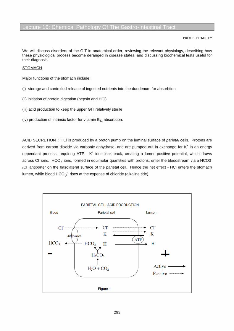
293
Lecture 16: Chemical Pathology Of The Gastro-Intestinal Tract
PROF E. H HARLEY
We will discuss disorders of the GIT in anatomical order, reviewing the relevant physiology, describing how these physiological process become deranged in disease states, and discussing biochemical tests useful for their diagnosis.
STOMACH
Major functions of the stomach include:
(i) storage and controlled release of ingested nutrients into the duodenum for absorbtion
(ii) initiation of protein digestion (pepsin and HCl)
(iii) acid production to keep the upper GIT relatively sterile
(iv) production of intrinsic factor for vitamin B12 absorbtion.
ACID SECRETION : HCl is produced by a proton pump on the luminal surface of parietal cells. Protons are
derived from carbon dioxide via carbonic anhydrase, and are pumped out in exchange for K+ in an energy
dependant process, requiring ATP. K+ ions leak back, creating a lumen-positive potential, which draws
across Cl- ions. HCO3- ions, formed in equimolar quantities with protons, enter the bloodstream via a HCO3-
/Cl- antiporter on the basolateral surface of the parietal cell. Hence the net effect - HCl enters the stomach
lumen, while blood HCO3- rises at the expense of chloride (alkaline tide).
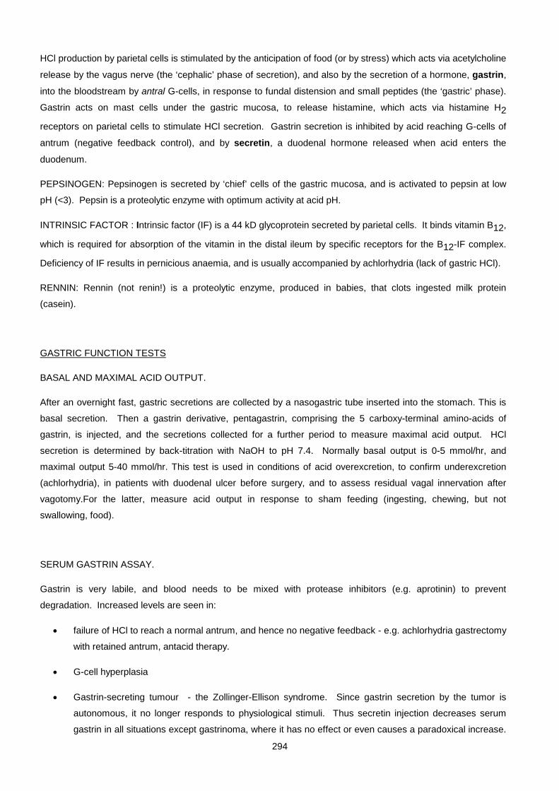
294
HCl production by parietal cells is stimulated by the anticipation of food (or by stress) which acts via acetylcholine
release by the vagus nerve (the ‘cephalic’ phase of secretion), and also by the secretion of a hormone, gastrin,
into the bloodstream by antral G-cells, in response to fundal distension and small peptides (the ‘gastric’ phase).
Gastrin acts on mast cells under the gastric mucosa, to release histamine, which acts via histamine H2
receptors on parietal cells to stimulate HCl secretion. Gastrin secretion is inhibited by acid reaching G-cells of
antrum (negative feedback control), and by secretin, a duodenal hormone released when acid enters the
duodenum.
PEPSINOGEN: Pepsinogen is secreted by ‘chief’ cells of the gastric mucosa, and is activated to pepsin at low
pH (<3). Pepsin is a proteolytic enzyme with optimum activity at acid pH.
INTRINSIC FACTOR : Intrinsic factor (IF) is a 44 kD glycoprotein secreted by parietal cells. It binds vitamin B12,
which is required for absorption of the vitamin in the distal ileum by specific receptors for the B12-IF complex.
Deficiency of IF results in pernicious anaemia, and is usually accompanied by achlorhydria (lack of gastric HCl).
RENNIN: Rennin (not renin!) is a proteolytic enzyme, produced in babies, that clots ingested milk protein
(casein).
GASTRIC FUNCTION TESTS
BASAL AND MAXIMAL ACID OUTPUT.
After an overnight fast, gastric secretions are collected by a nasogastric tube inserted into the stomach. This is
basal secretion. Then a gastrin derivative, pentagastrin, comprising the 5 carboxy-terminal amino-acids of
gastrin, is injected, and the secretions collected for a further period to measure maximal acid output. HCl
secretion is determined by back-titration with NaOH to pH 7.4. Normally basal output is 0-5 mmol/hr, and
maximal output 5-40 mmol/hr. This test is used in conditions of acid overexcretion, to confirm underexcretion
(achlorhydria), in patients with duodenal ulcer before surgery, and to assess residual vagal innervation after
vagotomy.For the latter, measure acid output in response to sham feeding (ingesting, chewing, but not
swallowing, food).
SERUM GASTRIN ASSAY.
Gastrin is very labile, and blood needs to be mixed with protease inhibitors (e.g. aprotinin) to prevent
degradation. Increased levels are seen in:
• failure of HCl to reach a normal antrum, and hence no negative feedback - e.g. achlorhydria gastrectomy
with retained antrum, antacid therapy.
• G-cell hyperplasia
• Gastrin-secreting tumour - the Zollinger-Ellison syndrome. Since gastrin secretion by the tumor is
autonomous, it no longer responds to physiological stimuli. Thus secretin injection decreases serum
gastrin in all situations except gastrinoma, where it has no effect or even causes a paradoxical increase.

295
Similarly a test meal containing protein normally increases serum gastrin, whereas in gastrinoma, there
is no change.
ZOLLINGER ELLISON SYNDROME
Caused by a gastrin secreting tumour (gastrinoma), usually pancreatic, but occasionally located in the bowel
wall. It presents with severe duodenal ulceration (recurrent and multiple, extending to distal duodenum or
even the jejunum), and with diarrhoea (excess acid interferes with the action of pancreatic enzymes & bile
salts). Gastrinomas are often malignant, and are sometimes associated with the Multiple Endocrine
Neoplasia syndrome type 1 (MEN I). If hyperparathyroidism is present as part of MEN I, hypercalcaemia may
exacerbate the symptoms. Basal acid output is increased - often equal to maximal output. Serum gastrin
level is very high. Treatment is surgical, or, if operable, with H2 receptor antagonists .
GASTRIC ULCER
Often associated with below-normal acid output. There is believed to be a disturbed gastric mucosal barrier
to HCl. May be secondary to ethanol, aspirin or glucocorticoids. However, recent evidence has implicated a
Gram -ve, urease-producing organism, Helicobacter pylori, as perhaps the most common factor in the
pathogenesis, and in consequence the condition often responds to antibiotic therapy.
DUODENAL ULCER
Often associated with high acid output, e.g. due to G-cell hyperplasia. Gastric acid output may be reduced by
vagotomy or partial gastrectomy, or by drugs, such as ranitidine (Zantac), a histamine H2 receptor antagonist,
or omeprazole (Losec), a proton-pump inhibitor, which selectively becomes activated at acid pH, and
irreversibly oxidizes -SH groups at the active site of the pump. Helicobacter pylori has also been implicated
in causation of duodenal ulcers, since some respond to broad spectrum antibiotics.
Although measurement of acid output and serum gastrin can be useful, direct endoscopic visualization is the
method of choice for diagnosis of gastric & duodenal ulcers.
PANCREAS
Apart from its endocrine function (insulin, glucagon), the pancreas is an important exocrine gland. It
produces 2 types of exocrine secretion, from different cell types and under separate control.
• A watery HCO3--rich fluid (pH 8), produced by an inwardly-directed proton pump in ductular cells in
response to secretin released from the duodenum. Its effect is to neutralize gastric acidity, so as to
provide an optimum pH for pancreatic enzymes.
• A low volume, viscid secretion of enzyme-rich fluid from acinar cells. Enzymes include amylase,
lipase, ribonucleases, and a variety of proteases (trypsin, chymotrypsin, elastase, carboxypeptidase).
Proteases are secreted as inactive precursors, to prevent autodigestion of the pancreas. Once in the
duodenum, trypsinogen is activated by duodenal enterokinase. The resultant trypsin in turn activates
residual trypsinogen & other proteases by proteolytic cleavage.

296
Amylase converts starch and glycogen to maltose Pancreatic lipase completes hydrolysis of triglyceride to fatty
acids and monoglycerides that was initiated by lingual lipase. Proteases split peptide bonds at specific sites in
the interior (endopeptidases) or ends (exopeptidase) of proteins, to yield small peptides. Stimulus for secretion
of this enzyme-rich fluid is the hormone, cholecystokinin (CCK), released from duodenum in response to entry of
fatty acids and peptides. CCK has a dual action - it also causes gall bladder contraction with expulsion of bile to
aid fat digestion, hence its name.
ACUTE PANCREATITIS
A medical emergency. Frequent association with alcohol, gallstones, hyperlipidaemia. Probable common
mechanism is premature activation of proteases within the pancreas.
Presentation - acute abdominal pain and shock. Leakage of activated pancreatic enzymes into the
pancreas and surrounding tissues with acute inflammation and haemorrhage. Treatment is generally
conservative- naso-gastric suction and IV fluids. Laparotomy is contra-indicated, as this would facilitate the
intra-abdominal spread of active enzymes. Since its presentation is similar to abdominal crises that need to
be treated surgically, it is important to have a reliable diagnostic test. Serum amylase is the most widely
used, but returns to normal relatively rapidly (+ 72 hours) because of renal clearance. Amylase is not entirely
specific - increases (usually smaller) are seen in other acute upper GIT pathology e.g. perforating duodenal
ulcer or bowel infarction, which, unlike acute pancreatitis, require urgent surgery. Other useful tests include
urine amylase, amylase/creatinine clearance ratio (increased in acute pancreatitis, decreased in macro-
amylaseaemia), serum lipase, and amylase iso-enzymes.
Associated biochemical features:

297
methaemalbuminaemia (massive extravascular hemolysis- a mega-bruise!), hyperglycaemia (transient β-cell
dysfunction), hypocalcaemia (deposition of insoluble calcium soaps at sites of fat necrosis). Jaundice and a
lipaemic serum suggest that gallstones and hyperlipidaemia, respectively, may be the predisposing cause of
the acute pancreatitis..
CHRONIC PANCREATITIS
Destruction of pancreatic parenchyma with replacement by fibrous tissue and cysts. Presents with
malabsorption (particularly of fat), and sometimes with secondary diabetes. Usually there is a strong ethanol
association.
Useful biochemical tests include:
1. Analysis of pancreatic fluid secreted in response to hormonal stimulation. A double-lumen tube is
passed - the orifice of one is positioned opposite the opening of the pancreatic duct, the other
aspirates gastric contents to prevent contamination of the sample by gastric acid. A basal sample is
collected, then secretin and CCK are infused (usually separately), while duodenal contents continue
to be aspirated. Fluid is analysed for (i) volume (ii) HCO3- concentration (normal > 90 mM), and (iii)
enzyme activity- usually only a single enzyme eg. amylase or trypsin, is chosen. In chronic
pancreatitis, all parameters are affected, whereas a low flow rate with normal HCO3- & enzyme
concentration suggests partial obstruction- eg a carcinoma.
2. Lundh test meal. Standard meal ingested, containing CHO, fat and protein. Duodenal contents are
aspirated and tryptic activity assayed. A rather messy procedure. In addition to testing pancreatic
function, it relies on an intact duodenum for release of CCK.
3. Synthetic substrate for chymotrypsin given orally, cleaved to p-amino benzoate and excreted in urine.
4. Glucose tolerance test.
5. Faecal fat measurement (see p 10)
Other useful tests include abdominal X-ray (for calcification), ultrasound (for cysts), endoscopic retrograde
cholangio-pancreatography (ERCP).
Summary of GIT hormones
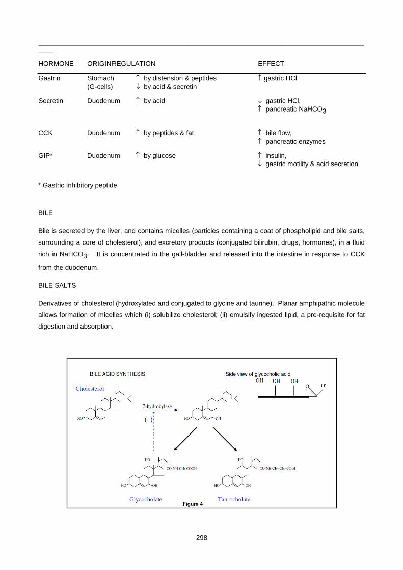
298
___________________________________________________________________________________________
HORMONE ORIGIN REGULATION EFFECT
Gastrin Stomach ↑ by distension & peptides ↑ gastric HCl (G-cells) ↓ by acid & secretin
Secretin Duodenum ↑ by acid ↓ gastric HCl, ↑ pancreatic NaHCO3
CCK Duodenum ↑ by peptides & fat ↑ bile flow, ↑ pancreatic enzymes
GIP* Duodenum ↑ by glucose ↑ insulin, ↓ gastric motility & acid secretion
* Gastric Inhibitory peptide
BILE
Bile is secreted by the liver, and contains micelles (particles containing a coat of phospholipid and bile salts,
surrounding a core of cholesterol), and excretory products (conjugated bilirubin, drugs, hormones), in a fluid
rich in NaHCO3. It is concentrated in the gall-bladder and released into the intestine in response to CCK
from the duodenum.
BILE SALTS
Derivatives of cholesterol (hydroxylated and conjugated to glycine and taurine). Planar amphipathic molecule
allows formation of micelles which (i) solubilize cholesterol; (ii) emulsify ingested lipid, a pre-requisite for fat
digestion and absorption.
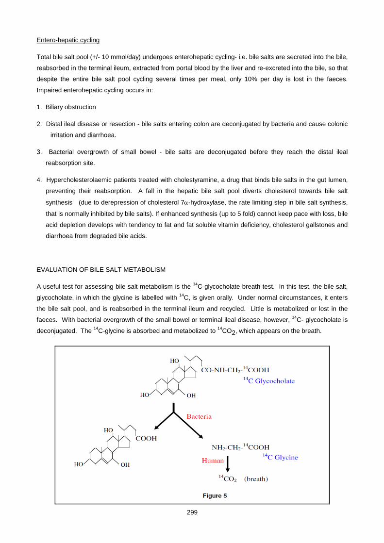
299
Entero-hepatic cycling
Total bile salt pool (+/- 10 mmol/day) undergoes enterohepatic cycling- i.e. bile salts are secreted into the bile,
reabsorbed in the terminal ileum, extracted from portal blood by the liver and re-excreted into the bile, so that
despite the entire bile salt pool cycling several times per meal, only 10% per day is lost in the faeces.
Impaired enterohepatic cycling occurs in:
1. Biliary obstruction
2. Distal ileal disease or resection - bile salts entering colon are deconjugated by bacteria and cause colonic
irritation and diarrhoea.
3. Bacterial overgrowth of small bowel - bile salts are deconjugated before they reach the distal ileal
reabsorption site.
4. Hypercholesterolaemic patients treated with cholestyramine, a drug that binds bile salts in the gut lumen,
preventing their reabsorption. A fall in the hepatic bile salt pool diverts cholesterol towards bile salt
synthesis (due to derepression of cholesterol 7α-hydroxylase, the rate limiting step in bile salt synthesis,
that is normally inhibited by bile salts). If enhanced synthesis (up to 5 fold) cannot keep pace with loss, bile
acid depletion develops with tendency to fat and fat soluble vitamin deficiency, cholesterol gallstones and
diarrhoea from degraded bile acids.
EVALUATION OF BILE SALT METABOLISM
A useful test for assessing bile salt metabolism is the 14C-glycocholate breath test. In this test, the bile salt,
glycocholate, in which the glycine is labelled with 14C, is given orally. Under normal circumstances, it enters
the bile salt pool, and is reabsorbed in the terminal ileum and recycled. Little is metabolized or lost in the
faeces. With bacterial overgrowth of the small bowel or terminal ileal disease, however, 14C- glycocholate is
deconjugated. The 14C-glycine is absorbed and metabolized to 14CO2, which appears on the breath.
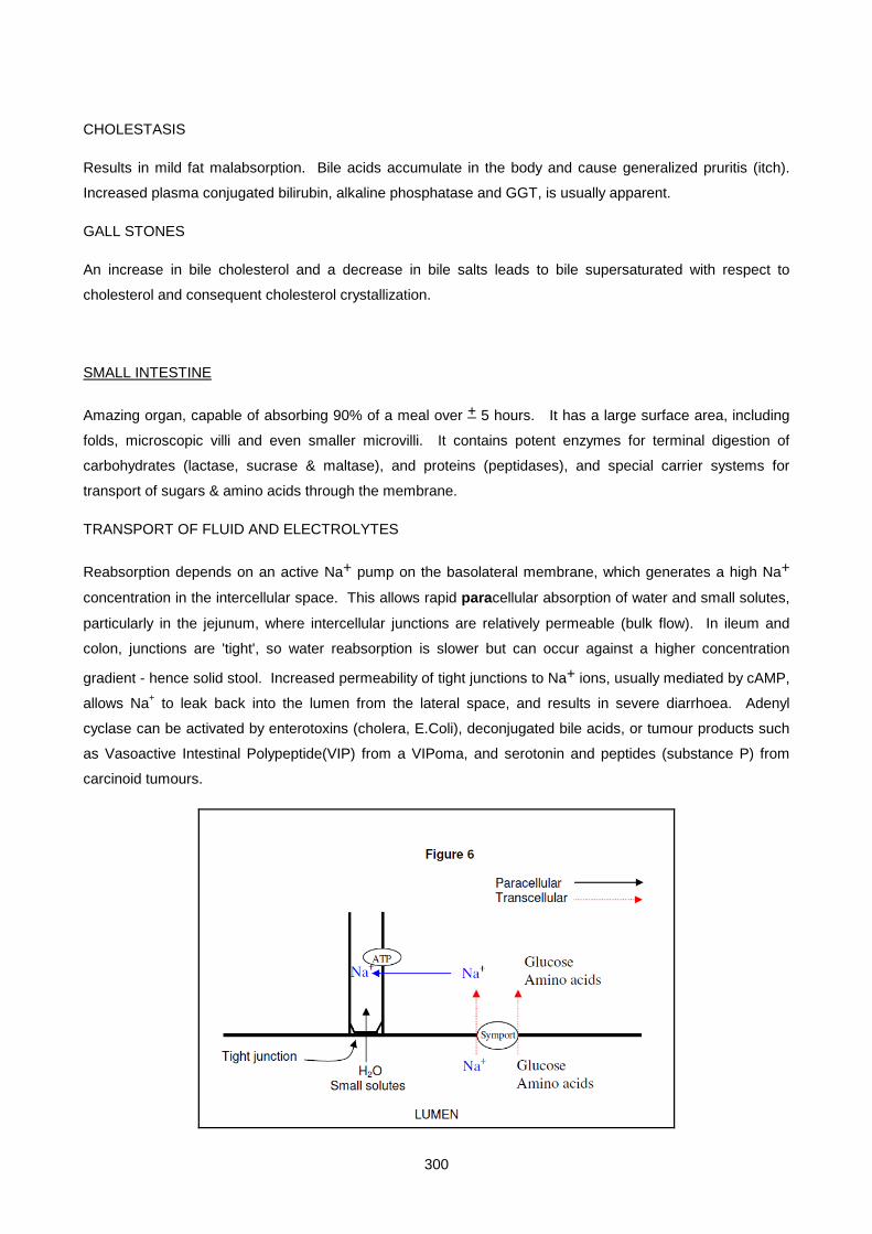
300
CHOLESTASIS
Results in mild fat malabsorption. Bile acids accumulate in the body and cause generalized pruritis (itch).
Increased plasma conjugated bilirubin, alkaline phosphatase and GGT, is usually apparent.
GALL STONES
An increase in bile cholesterol and a decrease in bile salts leads to bile supersaturated with respect to
cholesterol and consequent cholesterol crystallization.
SMALL INTESTINE
Amazing organ, capable of absorbing 90% of a meal over + 5 hours. It has a large surface area, including
folds, microscopic villi and even smaller microvilli. It contains potent enzymes for terminal digestion of
carbohydrates (lactase, sucrase & maltase), and proteins (peptidases), and special carrier systems for
transport of sugars & amino acids through the membrane.
TRANSPORT OF FLUID AND ELECTROLYTES
Reabsorption depends on an active Na+ pump on the basolateral membrane, which generates a high Na+
concentration in the intercellular space. This allows rapid paracellular absorption of water and small solutes,
particularly in the jejunum, where intercellular junctions are relatively permeable (bulk flow). In ileum and
colon, junctions are 'tight', so water reabsorption is slower but can occur against a higher concentration
gradient - hence solid stool. Increased permeability of tight junctions to Na+ ions, usually mediated by cAMP,
allows Na+ to leak back into the lumen from the lateral space, and results in severe diarrhoea. Adenyl
cyclase can be activated by enterotoxins (cholera, E.Coli), deconjugated bile acids, or tumour products such
as Vasoactive Intestinal Polypeptide(VIP) from a VIPoma, and serotonin and peptides (substance P) from
carcinoid tumours.

301
Low intracellular Na+ concentration allows absorption of nutrients (glucose, aminoacids) by a symport
mechanism, against the nutrient’s concentration gradient. This is the transcellular route.
ABSORPTION OF CARBOHYDRATE
Starch is broken down to maltose and isomaltose oligosaccharides by amylase. Maltose, isomaltose,
sucrose, and lactose hydrolysed by brush border disaccharidases (maltase, isomaltase, sucrase and lactase)
to yield monosaccharides.Monosaccharides (glucose, fructose, galactose) absorbed into enterocytes against
concentration gradient by carrier mediated co-transport with Na+. Exits into capillaries by diffusion down its
concentration gradient.
ABSORPTION OF PROTEIN
Digestion of dietary (+100 gram/day) and endogenous protein (mucus, enzymes, often >100 gram/day !)) by
pancreatic proteases yields mainly small peptides which are hydrolysed by brush border peptidases to amino
acids and taken up by a Na+ dependent symport mechanism.
FAT DIGESTION & ABSORPTION
Dietary fat is water insoluble. Emulsified into small lipid droplets (micelles) by mechanical and chemical
forces (bile salts and phospholipids). Colipase binds and stabilizes attachment of lipase which hydrolyses
triglyceride to monoglyceride and two fatty acids. Monoglycerides are amphipathic and help to emulsify the
residual dietary fat. Fatty acids and monoglycerides diffuse across the mucosal cell membrane, micelles
remain in the lumen. Within the cell, triglyceride is resynthesized, coated with phospholipid and
apolipoprotein (apo B-48) and enters lymphatics as chylomicrons. In abetalipoproteinaemia, lipid
accumulates as droplets within intestinal cells. Abnormal connections between bowel lymphatics and serous
cavities or urinary tract results in chylous effusion or chyluria, respectively.
Medium chain-length triglycerides (MCT) are not as dependant on bile salts and lipase for digestion. They
are NOT re-esterified but are transported directly to the liver in portal blood. MCT used therapeutically as
source of dietary lipid in chronic pancreatitis, biliary or lymphatic obstruction.
CALCIUM ABSORPTION
Transcellular mechanism. Ca2+ is actively extruded at the basolateral pole of the mucosal cell, while passive
carrier mediated entry takes place at the brush border. 1,25-di(OH) vitamin D enhances absorption by
stimulating synthesis of a calcium binding protein.
PHOSPHATE ABSORPTION
Transcellular mechanism. Neutral symport with Na+. Also stimulated by 1,25 di-OH-D3.
IRON ABSORPTION
Iron is absorbed in the upper duodenum, where the pH is still relatively acid, since Fe+++ is highly insoluble at
alkaline pH

302
VITAMIN B12 ABSORPTION
Vitamin B12 only found in animal products. Require + 1 µg/day. Hepatic stores last several years.
Absorption is complex. Vit B12 binds to salivary and dietary R-proteins. R-proteins must be digested by
pancreatic proteases which liberates B12, allowing it to bind to intrinsic factor (IF), a glycoprotein synthesized
by gastric parietal cells. This B12-IF complex traverses the length of the small bowel and is absorbed in the
terminal ileum by interaction with a specific receptor.
B12absorption impaired by: (a) lack of intrinsic factor (IF); (b) lack of pancreatic proteases; (c) bacterial
overgrowth of small intestine; (d) terminal ileal disease.
INVESTIGATION OF MALABSORPTION
CLINICAL PRESENTATION: weight loss, anaemia, signs of vitamin deficiency, despite adequate food intake;
chronic diarrhoea/ steatorrhoea.
HAEMATOLOGICAL AND BIOCHEMICAL SCREENING TESTS:
• Haemoglobin.
• Mean red cell volume (MCV) : ↓ in Fe deficiency, ↑ in folate or B12 deficiency.
• Reticulocyte response to appropriate therapy
• Serum albumin
• Serum cholesterol, carotene, vitamin A, prothrombin time (in fat malabsorption)
303
• Serum Ca, Mg, phosphate, iron, zinc, (for electrolyte malabsorption)
• Serum B12, red cell folate (for water soluble vitamin malabsorption)
SPECIAL TESTS:
ASSESSMENT OF CARBOHYDRATE ABSORPTION
LACTOSE TOLERANCE TEST
: Indicated where a history of milk intolerance suggests deficiency of the brush border enzyme, lactase. 50
gram of lactose is administered orally, and blood glucose measured over the next 2 hours. Normal
individuals tolerate the test well, and blood glucose rises by > 1.1mmol/l. In lactase deficiency, glucose rises
by less than 1mM, and the patient develops abdominal distension, cramps, borborygmi and diarrhoea. An
abnormal result should be followed up by giving equivalent amount of glucose and galactose, which
differentiates generalised disease of the small bowel mucosa from a specific lactase deficiency.
An alternative approach is to measure breath hydrogen after a lactose load, since lactose that is not
absorbed in the upper small bowel, is fermented by colonic bacteria to hydrogen, which appears in expired
air. Thus after 50 gram lactose, breath hydrogen is inversely proportional to lactase activity. A false positive
result may occur in rapid intestinal transit and bacterial overgrowth of small bowel.
In infants, lactase deficiency may be detected by low faecal pH, and/or reducing sugars in the stool, after a
milk feed.To confirm, one may resort to a lactase assay performed directly on a jejunal biopsy.
D-XYLOSE EXCRETION TEST:
D-xylose is ideally suited for testing small intestinal integrity i.e. it is not normally present in the diet, no
digestion is required, it is actively absorbed by the small bowel, it cannot be metabolized, and is excreted by
the kidneys. After a 5 gram oral dose of D-xylose, >25% of the dose is excreted in the urine within 5 hours
(and half of that within the first 2 hours). In infants, where urine collection may be problematic, blood levels
may be measured, and should be >1 & >2 mmol/l after 30 & 60 minutes, respectively. This is a specific test
of intestinal malabsorption, in that it is unaffected by biliary or pancreatic disease. Positive in intrinsic
mucosal damage (coeliac disease, Crohn's disease) or intra-luminal disease, ( bacterial overgrowth of the
small bowel). False positive results may be seen in gastric outlet obstruction.
ASSESSMENT OF PROTEIN ABSORPTION
It is useless to measure faecal protein directly, since what is not absorbed is degraded by colonic bacteria.
Methods are available, however, to detect leakage of serum proteins into the bowel, due to, say, mucosal
inflammation or lymphatic obstruction (protein-losing enteropathy):
• Intravenous administration of 51Cr labelled albumin. If labelled albumin exudes into the bowel lumen,
it is degraded, the 51Cr is retained in the lumen and is excreted in the stool. Faecal radioactivity in
normal subjects is +1%/day; in protein losing enteropathy it can increase to >30%/day.

304
• An alternative approach is to measure faecal alpha-l antitrypsin, this protein being resistant to
proteolytic digestion. One can calculate the α-1 anti-trypsin clearance from blood & faecal levels of
the protein, which is numerically equal to the volume of plasma oozing into the bowel lumen.
ASSESSMENT OF FAT ABSORPTION
• Faecal fat excretion: Normal faecal output of fat is up to 6 gram/day, on a normal diet of + 100 gram
fat per day. Increased excretion is seen in generalized small bowel disease, pancreatic insufficiency,
biliary obstruction, or terminal ileal disease/resection. Gross steatorrhoea (>40 gram/day) is usually
associated with chronic pancreatitis. Collect stool for 3 days, to ensure a reasonably accurate
collection. Although an unpleasant test to perform, faecal fat assay remains the best test for
evaluation of fat malabsorption.
• In an attempt to circumvent unpleasant faecal fat assays, various ‘fat tolerance tests’ have been
devised, whereby plasma triglyceride (or simply plasma turbidity) is measured after a fat load.
Another approach has been to feed 14C-labelled triglyceride (usually 14C-triolein), then measure
breath 14CO2. Individuals with fat malabsorption show decreased excretion of 14CO2. However,
these tests have not supplanted the faecal fat assay in terms of reliability.
• Increased urinary excretion of oxalic acid (hyperoxaluria) may be associated with fat malabsorption.
By binding calcium in the bowel lumen, unabsorbed fatty acids render dietary oxalate soluble (oxalate
normally occurs as an extremely insoluble calcium complex). Soluble oxalate is absorbed by the
colon and excreted in the urine, which carries the risk calcium oxalate kidney stone formation.
ASSESSMENT OF VITAMIN ABSORPTION
• Serum carotene and vitamin A levels are decreased in fat malabsorption, as is the vitamin K-
dependant prothrombin index.
• Vitamin B12 - the Schilling test:
o This test is performed whenever vitamin B12 malabsorption is suspected. 1ug 58Co labelled
vitamin B12 is given orally, together with a large 1mg intramuscular dose of unlabelled B12 to
saturate body stores. The 58Co labelled B12 is normally absorbed and at least 5-8% of the
label is excreted in the urine within 24 hours. Low values indicate (see Fig 7 above) :
(a) Lack of intrinsic factor (pernicious anaemia) – check by repeating with intrinsic factor.
(b) Lack of pancreatic proteases to degrade R-proteins and allow intrinsic factor to bind B12 -
excretion improves on administration of pancreatic enzyme supplements.
(c) Small bowel bacterial overgrowth - improves with antibiotic therapy.
(d) Ileal disease/resection - no improvement with any manipulation.

305
Note: Vit. B12 deficiency for any reason, impairs mucosal cell maturation and can result in transient intestinal
malabsorption, and a Schilling test result that suggests ileal disease.
ASSESSMENT OF SMALL BOWEL BACTERIAL OVERGROWTH
Usually due to some anatomical abnormality, eg. a congenital or surgically-produced blind loop, diverticular
disease, a stricture or a fistula, that provides a stagnant segment of small bowel in which bacteria can
proliferate.
Biochemical confirmation can be done as follows:
1. Increased urinary indican excretion. Bacteria degrade dietary tryptophan to indole, which is
absorbed, conjugated with sulphate, and excreted in urine as indican.
2. 14C glycocholate breath test. Glycocholate, in which the glycine part is labelled with 14C, is
given orally, and breath 14CO2 monitored. Under normal circumstances, this labelled
glycocholate joins the bile salt pool and undergoes enterohepatic cycling. Little is degraded.
However, if bacteria are present proximal to its absorption site (the distal ileum), they
deconjugate the labelled bile salt, i.e. remove 14Cglycine, which is metabolized to 14CO2 and
appears in the breath. A positive result is also obtained in distal ileal disease. However,
here, faecal excretion of 14C is increased. i.e.
Normal Bacterial contamination Ileal disease
Breath 14CO2 0 +++ +++
Faecal 14C 0 0 ++
3. Breath hydrogen following xylose ingestion - also elevated with intestinal malabsorption.
4. Abnormal Schilling test, or steatorrhoea, responsive to antibiotics.
5. Culture of small intestinal juice for bacteria (and also for the protozoan parasite,Giardia).
ASSESSMENT OF SMALL INTESTINAL TRANSIT TIME
In a condition known as ‘irritable bowel syndrome’, there is a rapid transit of food through the GIT, sometimes
to the point of affecting absorption. For confirmation, measure the time lag between an oral dose of lactulose
and appearance of hydrogen in the breath. Lactulose, a fructose-galactose disaccharide, cannot be digested
or absorbed, hence passes through the small intestine to the colon where it is rapidly fermented by bacteria
to yield hydrogen. Appearance of hydrogen in the breath indicates arrival in the colon. Spuriously shortened
306
(apparent) transit times occur with small bowel bacterial overgrowth where H2 is produced earlier than
normal. h
MISCELLANEOUS TESTS
• Increased sweat NaCl concentration in cystic fibrosis. Cystic fibrosis is an inherited disease, where a
defect in a chloride channel results in abnormally viscous mucous, which blocks exocrine glands and
predisposes to infections. Children with this condition often develop malabsorption due to pancreatic
and bowel involvement, and recurrent respiratory infections. The same defect in sweat glands leads
to high sweat NaCl levels which is valuable diagnostically.
• Some intrinsic bowel disorders, such as Crohn’s disease & ulcerative colitis, may cause bleeding into
the bowel lumen. Presence of blood in stool can be readily detected, using the principle that haem
exhibits potent peroxidase activity - this forms the basis of the ‘occult blood test’ -
haem
Hydrogen peroxide (H2O2) + dye oxidized dye (coloured) + H2O
This is a very sensitive test, but false positives occur with ingestion of plant peroxidases (which can
be destroyed by boiling- the stool, not the plant!) and meat. Confirmed by tagging patient’s own red
cells with 51Cr and measuring stool radio-activity.
• Jejunal biopsy for histology. This is particularly useful in coeliac disease, where sensitivity to the
wheat protein, gluten, damages the intestinal mucosa (probably via an immune mechanism). The
mucosa becomes flattened, with villous atrophy, and infiltrated with lymphocytes. These changes are
readily observable microscopically. In ‘sprue’, a disorder confined to the tropics, the same clinical &
histological changes are present, but gluten sensitivity is not a feature. A bacterial aetiology for this
condition is suggested by its response to broad-spectrum antibiotics.
COLON
The colon may be unflatteringly defined as a commodious stagnant reservoir containing unabsorbed food
residues and teeming with bacteria. It however has an important role in NaCl and water reabsorption. For
this, it uses the same mechanism as the small bowel i.e. a basolateral sodium pump, with high osmolality in
the lateral space. Tight junctions are less permeable, hence water absorption is slow but occurs against a
higher osmotic gradient, efficiently removing sodium and water and rendering faeces solid. Na+ is absorbed
in exchange for K+ or H+ (as in the distal renal tubule), while Cl- is absorbed in exchange for HCO3-. This
obligatory mechanism for NaCl resorption explains the normal anion gap metabolic acidosis and
hypokalaemia seen when colon is used as an artificial urinary bladder. An unfortunate consequence of a
colon filled with bacteria is that any undigested protein, including a GIT haemorrhage, is rapidly degraded to
ammonia, which is absorbed in the portal blood and returned to the liver for conversion into urea. When the
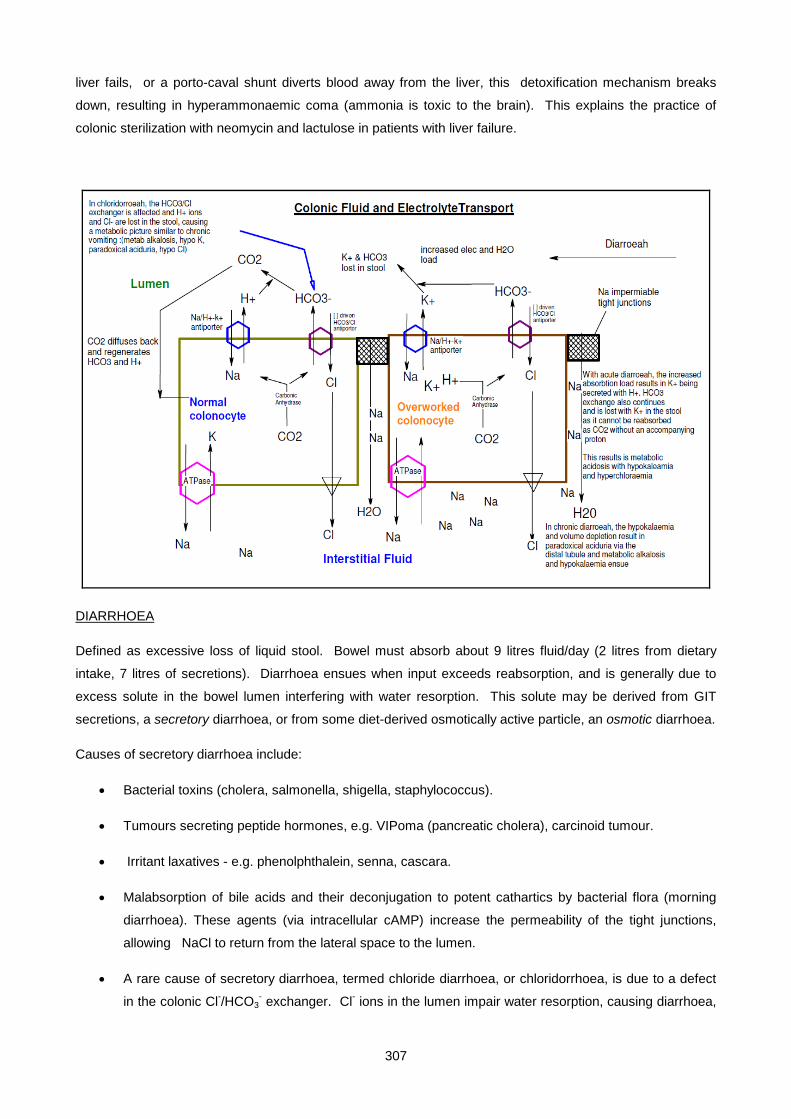
307
liver fails, or a porto-caval shunt diverts blood away from the liver, this detoxification mechanism breaks
down, resulting in hyperammonaemic coma (ammonia is toxic to the brain). This explains the practice of
colonic sterilization with neomycin and lactulose in patients with liver failure.
DIARRHOEA
Defined as excessive loss of liquid stool. Bowel must absorb about 9 litres fluid/day (2 litres from dietary
intake, 7 litres of secretions). Diarrhoea ensues when input exceeds reabsorption, and is generally due to
excess solute in the bowel lumen interfering with water resorption. This solute may be derived from GIT
secretions, a secretory diarrhoea, or from some diet-derived osmotically active particle, an osmotic diarrhoea.
Causes of secretory diarrhoea include:
• Bacterial toxins (cholera, salmonella, shigella, staphylococcus).
• Tumours secreting peptide hormones, e.g. VIPoma (pancreatic cholera), carcinoid tumour.
• Irritant laxatives - e.g. phenolphthalein, senna, cascara.
• Malabsorption of bile acids and their deconjugation to potent cathartics by bacterial flora (morning
diarrhoea). These agents (via intracellular cAMP) increase the permeability of the tight junctions,
allowing NaCl to return from the lateral space to the lumen.
• A rare cause of secretory diarrhoea, termed chloride diarrhoea, or chloridorrhoea, is due to a defect
in the colonic Cl-/HCO3- exchanger. Cl- ions in the lumen impair water resorption, causing diarrhoea,
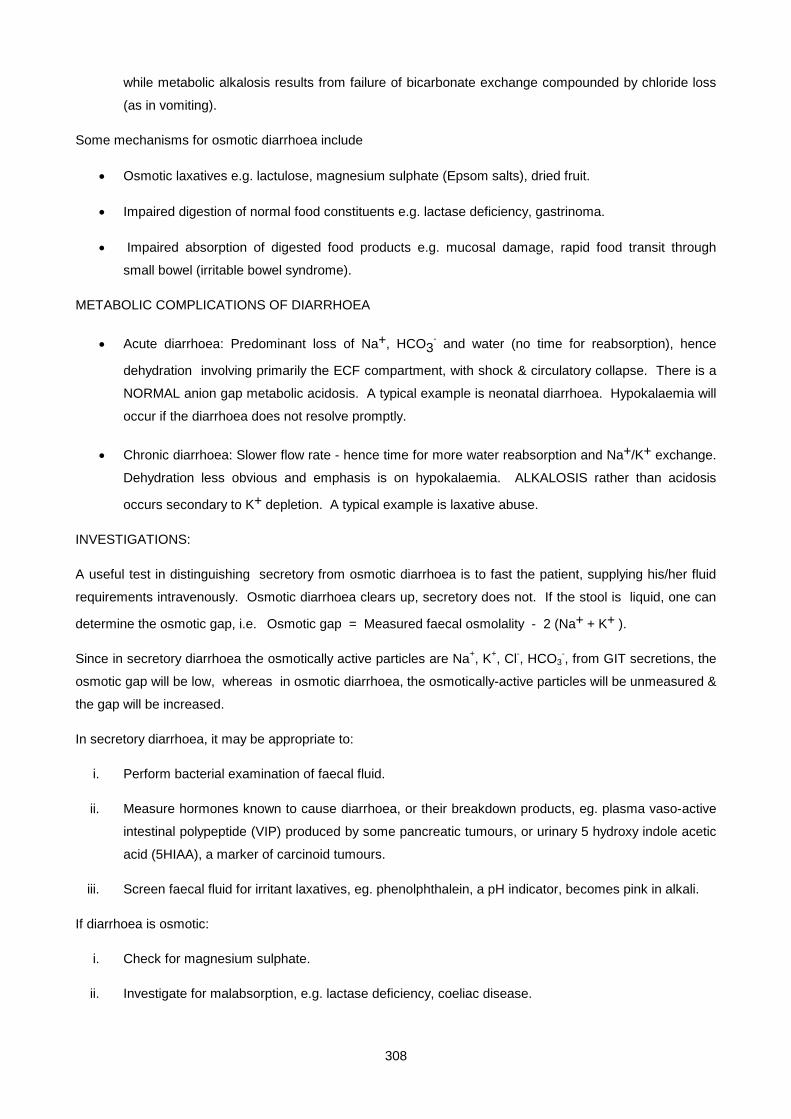
308
while metabolic alkalosis results from failure of bicarbonate exchange compounded by chloride loss
(as in vomiting).
Some mechanisms for osmotic diarrhoea include
• Osmotic laxatives e.g. lactulose, magnesium sulphate (Epsom salts), dried fruit.
• Impaired digestion of normal food constituents e.g. lactase deficiency, gastrinoma.
• Impaired absorption of digested food products e.g. mucosal damage, rapid food transit through
small bowel (irritable bowel syndrome).
METABOLIC COMPLICATIONS OF DIARRHOEA
• Acute diarrhoea: Predominant loss of Na+, HCO3- and water (no time for reabsorption), hence
dehydration involving primarily the ECF compartment, with shock & circulatory collapse. There is a
NORMAL anion gap metabolic acidosis. A typical example is neonatal diarrhoea. Hypokalaemia will
occur if the diarrhoea does not resolve promptly.
• Chronic diarrhoea: Slower flow rate - hence time for more water reabsorption and Na+/K+ exchange.
Dehydration less obvious and emphasis is on hypokalaemia. ALKALOSIS rather than acidosis
occurs secondary to K+ depletion. A typical example is laxative abuse.
INVESTIGATIONS:
A useful test in distinguishing secretory from osmotic diarrhoea is to fast the patient, supplying his/her fluid
requirements intravenously. Osmotic diarrhoea clears up, secretory does not. If the stool is liquid, one can
determine the osmotic gap, i.e. Osmotic gap = Measured faecal osmolality - 2 (Na+ + K+ ).
Since in secretory diarrhoea the osmotically active particles are Na+, K+, Cl-, HCO3-, from GIT secretions, the
osmotic gap will be low, whereas in osmotic diarrhoea, the osmotically-active particles will be unmeasured &
the gap will be increased.
In secretory diarrhoea, it may be appropriate to:
i. Perform bacterial examination of faecal fluid.
ii. Measure hormones known to cause diarrhoea, or their breakdown products, eg. plasma vaso-active
intestinal polypeptide (VIP) produced by some pancreatic tumours, or urinary 5 hydroxy indole acetic
acid (5HIAA), a marker of carcinoid tumours.
iii. Screen faecal fluid for irritant laxatives, eg. phenolphthalein, a pH indicator, becomes pink in alkali.
If diarrhoea is osmotic:
i. Check for magnesium sulphate.
ii. Investigate for malabsorption, e.g. lactase deficiency, coeliac disease.
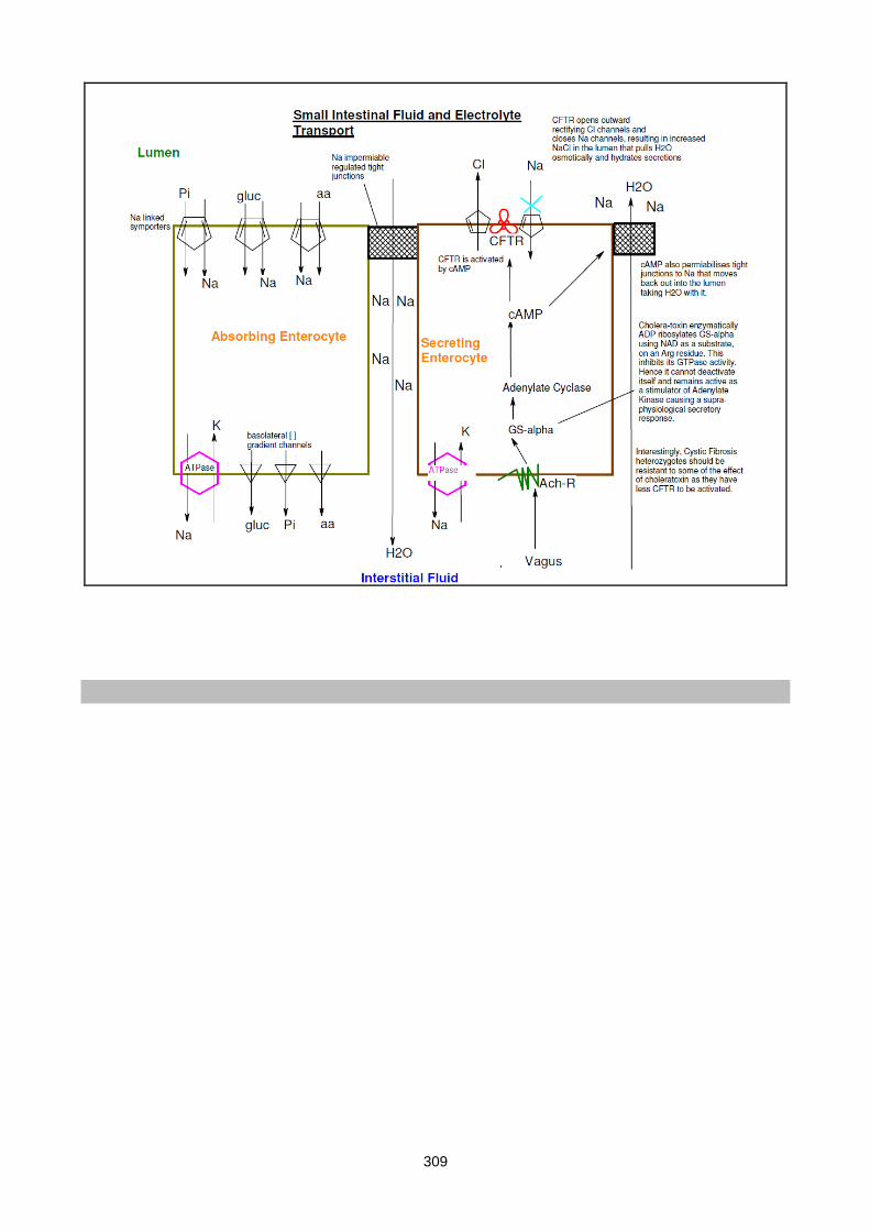
309

310
Lecture 17: Endocrinology 1: Pituitary And Hypothalamus
DR PETER BERMAN
INTRODUCTION:
The hypothalamus, located on the ventral surface of the brain, around the third ventricle, is responsible for
controlling pituitary hormone secretion. It secretes its peptide hormones, also known as hypothalamic
releasing factors, either:
• into the short private portal system to directly impinge on the anterior pituitary (eg. TRH, GnRH), or
• directly into the general circulation (eg. oxytocin, vasopressin)
MAJOR CONCEPTS:
In order to understand the regulation of pituitary function in normal and pathological states, the following
concepts must be appreciated:
a) Stress can alter pituitary hormone secretion and be used to test for abnormality
eg. insulin hypoglycaemic ↑CRF ↑ACTH
stress ↑GHRH ↑GH
b) Feed-back by products of end-organ endocrine glands (eg. steroids) or tissues (eg. IGF-I) act
back,usually inhibiting, or in rare cases, enhancing, pituitary response to hypothalamic factors. Feed-
back is thus negative or positive, and either long-loop or short-loop, as shown below:
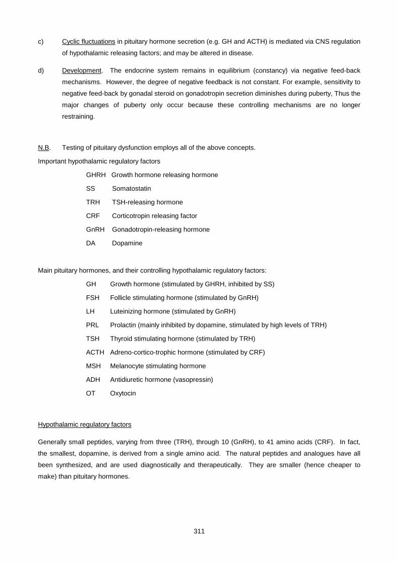
311
c) Cyclic fluctuations in pituitary hormone secretion (e.g. GH and ACTH) is mediated via CNS regulation
of hypothalamic releasing factors; and may be altered in disease.
d) Development. The endocrine system remains in equilibrium (constancy) via negative feed-back
mechanisms. However, the degree of negative feedback is not constant. For example, sensitivity to
negative feed-back by gonadal steroid on gonadotropin secretion diminishes during puberty, Thus the
major changes of puberty only occur because these controlling mechanisms are no longer
restraining.
N.B. Testing of pituitary dysfunction employs all of the above concepts.
Important hypothalamic regulatory factors
GHRH Growth hormone releasing hormone
SS Somatostatin
TRH TSH-releasing hormone
CRF Corticotropin releasing factor
GnRH Gonadotropin-releasing hormone
DA Dopamine
Main pituitary hormones, and their controlling hypothalamic regulatory factors:
GH Growth hormone (stimulated by GHRH, inhibited by SS)
FSH Follicle stimulating hormone (stimulated by GnRH)
LH Luteinizing hormone (stimulated by GnRH)
PRL Prolactin (mainly inhibited by dopamine, stimulated by high levels of TRH)
TSH Thyroid stimulating hormone (stimulated by TRH)
ACTH Adreno-cortico-trophic hormone (stimulated by CRF)
MSH Melanocyte stimulating hormone
ADH Antidiuretic hormone (vasopressin)
OT Oxytocin
Hypothalamic regulatory factors
Generally small peptides, varying from three (TRH), through 10 (GnRH), to 41 amino acids (CRF). In fact,
the smallest, dopamine, is derived from a single amino acid. The natural peptides and analogues have all
been synthesized, and are used diagnostically and therapeutically. They are smaller (hence cheaper to
make) than pituitary hormones.
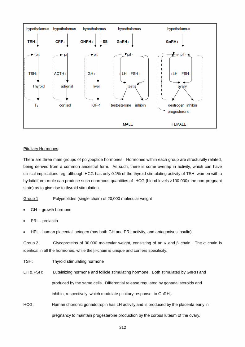
312
Pituitary Hormones:
There are three main groups of polypeptide hormones. Hormones within each group are structurally related,
being derived from a common ancestral form. As such, there is some overlap in activity, which can have
clinical implications eg. although HCG has only 0.1% of the thyroid stimulating activity of TSH, women with a
hydatidiform mole can produce such enormous quantities of HCG (blood levels >100 000x the non-pregnant
state) as to give rise to thyroid stimulation.
Group 1 Polypeptides (single chain) of 20,000 molecular weight
• GH - growth hormone
• PRL - prolactin
• HPL - human placental lactogen (has both GH and PRL activity, and antagonises insulin)
Group 2 Glycoproteins of 30,000 molecular weight, consisting of an α and β chain. The α chain is
identical in all the hormones, while the β-chain is unique and confers specificity.
TSH: Thyroid stimulating hormone
LH & FSH: Luteinizing hormone and follicle stimulating hormone. Both stimulated by GnRH and
produced by the same cells. Differential release regulated by gonadal steroids and
inhibin, respectively, which modulate pituitary response to GnRH,.
HCG: Human chorionic gonadotropin has LH activity and is produced by the placenta early in
pregnancy to maintain progesterone production by the corpus luteum of the ovary.

313
Group 3 A series of smaller peptides derived from a single large precursor, pro-opio-melano-cortin
(POMC) that is cleaved specifically to produce a family of biologically active peptides. The most important
product of the anterior lobe is ACTH (adrenocorticotrophic hormone). More extensive processing of POMC
by other cell types yields biologically active peptides including melanocyte-stimulating hormone (MSH) and
endorphins, which play a role in appetite control and pain suppression, respectively.
Gonadotropin releasing-hormone (GnRH) stimulates the secretion of both luteinising hormone (LH) and
follicle-stimulating hormone (FSH). It is secreted under physiological conditions as pulses with a frerquency
of about 1 hour. Hence pulsatile administration is required to treat infertility due to hypothalamic disease
(hypogonadotropic hypogonadism) in both sexes, and for delayed puberty. Continuous, high-dose
administration of GnRH in fact down-regulates secretion of FSH & LH, thereby inhibiting the reproductive
system. This is useful in certain gynaecological disorders (polycystic ovarian syndrome, endometriosis,
uterine fibroids, ovarian and breast cancer), as well as for precocious puberty in children and prostatic cancer
in men. There is currently interest in using GnRH analogues as contraceptive agents in men and women
(with appropriate low dose sex steroid replacement). This approach may reduce incidence of breast and
ovarian cancer, which are much commoner among women who experience large numbers of menstrual
cycles (about 500 in a life-time for the average Western woman), compared with women in Third World
countries, who have far fewer (about 50 cycles, because of early and frequent pregnancies and prolonged
breast-feeding and lactation).
Somatostatin was originally isolated as a hypothalamic peptide which inhibits growth hormone (GH)
secretion. It has since been found to be widely distributed in the gastrointestinal tract, where it inhibits just
about everything, including gut motility, gastric, biliary and pancreatic juice secretion, and hormones including
insulin, glucagon, and gastrin, Hence somatostatin has found use in treating many disorders, including
acromegaly, pancreatitis, gastrinoma, glucagonomas and nesidioblastosis. Understandably, given the
diversity of somatostatin’s actions, it has certain undesirable side-effects, such as gallstones and
malabsorption.
Growth hormone-releasing hormone (GHRH) is a much less expensive alternative to GH in the treatment
of GH-deficient children, where the defect generally resides at hypothalamic rather than pituitary level.
Continuous exposure does not cause receptor desensitisation as in the case of GnRH, which simplifies its
therapeutic application.
Corticotropin releasing-factor (CRF) is a 41-amino acid peptide which stimulates adrenocorticotropin
(ACTH) secretion. It is used diagnostically to establish hypocortisolism of pituitary origin as part of the
combined pituitary test, as well as to distinguish Cushing’s syndrome due to pituitary adenoma (good ACTH
response) from ectopic ACTH-producing tumours (no response).

314
Posterior pituitary peptides. Unlike the anterior pituitary, which is of endodermal origin, the posterior
pituitary is comprised of neuronal tissue and is really a part of the hypothalamus.
It is therefore not surprising that its hormones, antidiuretic hormone (vasopressin) and oxytocin, are
simple 9-amino acid cyclic peptides like the peptides of the hypothalamus. The difference is mainly one of
quantity. The hypothalamic peptides are present in tiny amounts which are active because they are secreted
directly into the hypophyseal portal system to travel a few millimeters to the anterior pituitary. Whereas the
posterior pituitary peptides are produced in much larger amounts, to enable them to reach their targets
(kidney, breast and uterus) at an adequate concentration despite being secreted into the general circulation.
Analogues of oxytocin and vasopressin have found useful application in the stimulation of uterine contraction
pre- and post-partum, in facilitating milk ejection, and in diabetes insipidus.
315
Lecture 18: Endocrinology 2: Pituitary Diseases
Peter Berman 2007
First we will list and describe hormones secreted by the pituitary, then indicate how we investigate both hyper-
and hypo-function of this gland.
Growth hormone is a polypeptide of about 200 amino acids, and is essential for normal growth. It has 2
major actions. The first is to stimulate production of insulin-like growth factor 1 (IGF-1), mainly by the liver but
to some extent by all tissues. This explains growth hormone’s anabolic action, namely to promote growth
and inhibit protein breakdown (which is insulin-like). Its second action is direct, to promote lipolysis and
antagonise insulin-mediated glucose uptake, which is appropriate to the fasting state. GH is mainly regulated
by 2 hypothalamic hormones; GH releasing hormone (GHRH) which stimulates release, and somatostatin,
that inhibits it. IGF-1 feeds back, blocking GHRH stimulation of the pituitary, and enhancing release of
somatostatin by the hypothalamus. Recently, a new hormone has been described, ghrelin, which is secreted
by the (empty) stomach, and stimulates both GH release and appetite, via a specific receptor. GH secretion
occurs in bursts, particularly intense after onset of sleep. Thus blood GH levels vary widely during the course
of 24h, and stimulation tests are required to prove existence of GH deficiency. These include the adrenergic
stimulant drug clonidine, insulin-induced hypoglycaemia, fasting, or exercise. On the other hand, GH
secretion is inhibited by a rise in blood glucose. Thus failure to suppress GH with an oral glucose load
indicates pathological GH secretion. Excessive GH secretion causes gigantism in children and acromegaly in
adults. GH deficiency usually presents as growth failure in children, while adults the effects are subtle, such
as a decrease in muscle mass and increased adiposity.
Prolactin, is similar in size and sequence to GH, but has a totally different function, namely to initiate and
sustain lactation. Secretion increases during pregnancy, but declines promptly after childbirth unless
breastfeeding occurs. The major regulator of prolactin (PRL) is dopamine from the hypothalamus, which
inhibits its secretion. Though TRH stimulates PRL release, this is not physiologically important (but does
explain the high PRL levels seen in primary hypothyroidism). Increased blood PRL can be caused by a
prolactin-secreting tumor, or by any process that obstructs blood flow from the hypothalamus (so-called ‘stalk’
effect). Stress and dopamine antagonists used to treat major psychiatric illness are other causes of elevated
PRL levels. The only clinical manifestation of a PRL deficiency is failure to lactate (eg in Sheehan’s
syndrome).
Thyroid stimulating hormone (TSH) is a large glycoprotein hormone composed of an α and β subunit. The
α-subunit is common to all the glycoprotein hormones (FSH, LH, hCG), while the β-subunit is unique to
TSH.Secretion of TSH is controlled by stimulation by the hypothalamic tri-peptideTRH, while the action and
secretion of TRH is inhibited by thyroid hormone (classic negative feedback).Measuring TSH is a widely-used
and valuable diagnostic test in thyroid disease; low blood levels of TSH indicates excess thyroid hormone
(hyperthyroidism); conversely, high TSH indicates primary hypothyroidism. Failure of TSH secretion can
cause a secondary hypothyroidism, whereas hyperthyroidism due to a TSH-secreting tumor is extremely rare.

316
Gonadotrophins, comprising follicle stimulating hormone (FSH) and luteinizing hormone (LH), are
glycoprotein hormones, similar in size and structure to TSH.They contain a common α-subunit with a unique
β-subunit that determines their specificity. Synthesis and release of both peptides is stimulated by the
pulsatile secretion of hypothalamic gonadotropin-releasing hormone (GnRH), and modulated by feedback
inhibition of gonadal steroids and inhibin, which allows for their independent regulation.In males, LH
stimulates testosterone production by the Leydig cells, which feedback inhibits the action of GnRH on LH
secretion. FSH stimulates spermatogenesis, and production by the testis of a peptide, inhibin, which
feedback inhibits further FSH release. The situation in women is a little more complex.FSH secreted in the
early part of the menstrual cycle stimulates growth of the Graafian follicle that secretes oestrogen. Oestrogen
and inhibin feedback inhibit FSH release (thereby avoiding multiple follicle development). At about mid-cycle,
the increasing oestrogen leads to explosive release of LH by a positive feedback mechanism. This LH surge
triggers ovulation, and subsequent formation of a corpus luteum, which secretion of oestrogen and
progesterone that both prepares the uterus for implantation and suppresses further FSH and LH secretion.
Failing conception, the corpus luteum regresses due to declining LH. Steroid hormone levels fall, which
triggers both endometrial shedding (menstruation) and derepression of FSH secretion, which heralds onset of
a new cycle. Before puberty, both FSH and LH are very low, and unresponsive to GnRH. With onset of
puberty, gonadotrophin secretion increases, FSH before LH. Increased level of gonadotropins are seen in
ovarian failure, whether premature or at onset of the menopause (loss of negative feedback). In men with
azoospermia (failure of sperm production), FSH is increased, while LH increases in response to a fall in
testosterone. Gonadotropin-secreting tumors of the pituitary are rare, whereas gonadal failure secondary to
decreased gonadotropin secretion is much commoner. Hypogonadotropic hypogonadism can be isolated
due to hypothalamic dysfunction, or be a part of generalized pituitary failure.
Adrenocorticotrophic hormone (ACTH) is a single polypeptide of 39 amino acids that stimulates adrenal
glucocorticoid (but not minerallocorticoid) secretion. It is derived from the much larger pro-opio-melanocortin
(POMC), and its release is positively regulated by corticotrophin releasing peptide (CRP) from the
hypothalamus. ACTH secretion exhibits diurnal variation, highest at 08h00, lowest at midnight. Secretion is
greatly increased by stress, and inhibited by cortisol in a classic negative feedback manner. Excessive
secretion of ACTH occurs with some pituitary tumors (Cushing’s disease) and in primary adrenal failure
(Addison’s disease). It is also occasionally produced ectopically by non-pituitary tumors (eg. small cell lung
CA) in large amounts. Since ACTH has a melanocyte stimulating action, hyperpigmentation is a feature of
high circulating ACTH levels, as occurs in Addison’s disease. Decreased ACTH leading to secondary
adrenal failure is usually part of generalised pituitary insufficiency, though it can occur in isolation.
Testing anterior pituitary function
Pulsatility of secretion of many pituitary hormones makes it inappropriate to rely on a single measurement for
diagnostic purposes. For example, an IGF-1 measurement is a better reflection of growth hormone status
than a single GH level, which can fluctuate widely over a 24h period. It is often useful to measure the pituitary
hormone in conjunction with its target organ product; eg a TSH in the normal range associated with a low
serum thyroxine signifies pituitary insufficiency causing secondary hypothyroidism i.e. the TSH is
317
inappropriately normal, whereas it should be high. Dynamic tests are useful tools for investigating pituitary
function. Stimulatory tests are employed in cases of suspected insufficiency, and suppression tests for
hyperfunction. In assessing anterior pituitary reserve, it is often useful to assess all hormones in a single
procedure, hence the use of the combined pituitary function (triple bolus) test. A single intravenous injection
of TRH, GnRH and insulin is given, then all 6 anterior pituitary hormones are measured serially over about
90min. TRH should evoke a TSH and PRL response, GnRH a FSH and LH response, and the
hypoglycaemic stress a GH, ACTH and PRL response. For technical reasons, cortisol assay is often
substituted for ACTH. During the test, it is important to document adequate hypoglycaemia by blood glucose
measurements, since failure of an ACTH response might well be due to an inadequate stimulus. On the
other hand, blood glucose might fall dangerously low (particularly in a subject with an impaired insulin
counterregulatory hormone capacity), hence the test should be done under close supervision, with injectable
glucose solution at hand. To avoid the danger of hypoglycaemia, the new trend is to substitute GHRH and
CRF for insulin. A recent test currently popular in assessment of pituitary reserve is the metyrapone
stimulation test. Metyrapone blocks the final step in cortisol synthesis (11-hydroxylase). In normal subjects,
the resulting fall in blood cortisol relieves the feedback inhibition on ACTH, which in turn stimulates the
adrenal. Unable to make cortisol, the adrenal secretes 11 deoxycortisol, the cortisol synthesis intermediate
just proximal to the block. Hypopituitary individuals exhibit a subnormal or absent 11 deoxycortisol response
to metyrapone.
HYPOPITUITARISM
Clinical presentation varies, largely depending on age. Growth failure is a common presentation in children,
whereas adults often present with sexual and/or reproductive dysfunction. Symptoms depend on which
hormone(s) are affected; GH and gonadotrophins (FSH and LH) are usually the first to go, followed by ACTH
and TSH. A table of the clinical features (from Marshall ‘Clinical Chemistry, 4th Ed)) is shown below.
Hormone Features of deficiency
Growth hormone adults:decreased muscle bulk
children:growth retardation, any tendency to hypoglycaemia may be accentuated
Prolactin
failure of lactation
Gonadotropins
children:delayed puberty
females:oligomenorrhoea, infertility, atophy of breasts and genitalia
males: impotence, azoospermia, testicular atrophy
both sexes:decreased libido, loss of body hair, fine wrinkling of skin
ACTH weight loss, weakness, hypotension, hypoglycaemia, decreased pigmentation, insidious onset with acute exacerbation when stressed
TSH weight gain, cold intolerance, fatigue, slow cerebration eg speech
ADH thirst, polyuria

318
An isolated deficiency of a single hormone is usually congenital and due to a deficiency of the relevant
hypothalamic releasing factor. A good example of this is Kallman’s syndrome that presents with primary
infertility and anosmia, and is due to defective migration of GnRH-secreting and olfactory neurones to their
correct location during embryogenesis. While the posterior pituitary is often spared in hypopituitarism ,
presence of diabetes insipidis (DI) should always be considered. Posterior pituitary involvement is usually
due to an invasive tumor or surgical damage. DI can be masked by a concurrent ACTH deficiency. Since
cortisol is required to excrete a water load, dehydration may only become a problem once glucocorticoid
replacement is started.
Causes of hypopituitarism include:
Tumors - eg non-functioning (chromophobe adenoma, craniopharyngioma) or functioning pituitary tumors, hypothalamic tumors,
Post traumatic - eg fracture base of skull
Post-infection - eg after bacterial or TB meningitis
Vascular – eg necrosis following post partum haemorrhage (Sheehan’s syndrome)
• Infiltration – eg haemochromatosis, sarcoidosis
• Iatrogenic – eg post pituitary surgery, radiation
Treatment of hypopituitarism generally entails replacing glucocorticoid and thyroxine, and either sex
steroids themselves if fertility is not desired, or GnRH/gonadotrophins, if it is.
Anorexia nervosa a disorder of self-imposed starvation, may mimic hypopituitarism (amenorrhoea, weight
loss), but loss of body hair does not occur, and GH and cortisol levels are increased as an appropriate
response to starvation, rather than decreased in pituitary disease.
Growth Hormone deficiency
This an uncommon but important and treatable cause of growth retardation. A random GH level above
10ng/ml excludes the condition, but since GH may be undetectable in serum of normal children at a
particular time, a low result is not diagnostic. Certain stimulation tests can be done, using vigorous exercise,
clonidine (an adrenergic agonist), GHRH, or insulin. Another option is to perform multiple GH
measurements during sleep via an indwelling catheter. Sex steroids enhance the GH response. Thus
equivocal responses in pre-pubertal children may need to be repeated after priming with testosterone or
oestrogens (in boys & girls, respectively). Treatment is based on GH replacement – originally obtained from
human cadavers (with the risk of Creutzveld-Jacob disease) – but now made by recombinant DNA
technology. Since most cases of isolated GH deficiency are actually due to lack of hypothalamic GHRH,
treatment with the latter is becoming an option. In GH deficient adults, GH is reported to improve vigor and
muscle mass, and decrease fat accumulation. GH is used illicitly by weightlifters for this purpose.

319
Laron dwarfism
A distinct form of childhood dwarfism is due to a genetic defect in the GH receptor. Since the growth
promoting effects of GH operate via IGF-1, these children clinically resemble those with GH deficiency,
except that their blood GH levels are high, IGF-1 is low, and they do not of course respond to GH
therapy.They are called ‘Laron’ dwarfs after the Israeli who first described the condition 20 years ago. Much
commoner forms of functional GH deficiency include emotionally-deprived children, who fail to grow at the
normal rate, and severely malnourished children, eg kwashiorkor, where GH secretion is diminished,
probably by low circulating leptin (a logical response to starvation).
PITUITARY TUMORS
Pituitary tumors can be non-functional (eg. chromophobe adenomas, craniopharyngiomas), but often produce
an excess of a particular hormone. PRL-secreting tumors (prolactinomas) are commonest, followed by GH,
ACTH, gonadotrophin and, rarest of all, TSH producing tumors. Apart from specific symptoms due to
hypersecretion of a particular pituitary hormone eg galactorrhoea, acromegaly), any tumor can give rise to:
• Hypopituitarism, by encroaching on space occupied by normal pituitary cells
• Local effects eg raised intracranial pressure (headache, vomiting, papilloedema), visual field defects due
to its proximity to the optic chiasma (typically bitemporal hemianopia)
Acromegaly and gigantism is caused 95% of the time by a GH-secreting pituitary tumor (the remainder are
due to ectopic production of GH and GHRH by tumors elsewhere). Whether gigantism or acromegaly
develops depends entirely on whether the tumor arises before long bone epiphyses have fused.
Many of the tumors show mutations in the Gs protein that links GHRH stimulation to GH secretion in a
gonadotrope. These mutations abolish the GTPase activity of the Gs protein, permanently switching it on,
thereby driving the production of GH independent of receptor activation by GHRH. The clinical features of
acromegaly are shown below:
Somatic Metabolic Local effects of tumor
Increased growth of: skin, subcutaneous tissues, skull and jaw, hands, feet, long bones, if before fusion of epiphyses
excessive sweating, coarse, greasy skin
cardiomegaly, hypertension
elevated, non-suppressible plasma GH level glucose intolerance, hypercalcaemia,
hyperphosphataemia
Headache
visual field defects
hypopituitarism
diabetes insipidis
Confirmation of acromegaly GH in a random serum sample is usually raised, but since GH secretion in
normal subjects is episodic, one should confirm by a failure of GH to suppress following a glucose tolerance
test. In acromegaly, glucose tolerance is frequently impaired, and may even be frankly diabetic.
320
Other confirmatory evidence for tumor-derived GH secretion is:
• Stimulation of GH by TRH, which does not normally affect GH secretion
• Elevated levels of IGF-1, which give a more integrated view of GH secretion, since it is not subject to
such wide fluctuations. IGF-1 levels are also useful for following response to treatment.
Treatment of acromegaly involves reducing GH levels, preventing or treating other pituitary hormone
deficiencies, and preventing damage to nearby structures, like the optic nerve. First line of treatment is
surgical – transphenoidal for small tumors, but by frontal craniotomy for large tumors with suprasellar
extension. External radiation can be used, and many tumors respond to medical treatment with somatostatin,
or, less frequently, dopamine. Appropriate hormone replacement is instituted, and patients are followed up
regularly to check for tumor regrowth and further loss of normal pituitary function.
Hyperprolactinaemia
Elevated serum prolactin is a common disorder, causing impotence in males, amenorrhoea in females, and
infertility and/or galactorrhoea in either sex. PRL down-regulates the entire hypothalamic-pituitary-gonadal
axis by disrupting the normal pulsatile secretion of GnRH (this occurs physiologically during lactation). High
PRL can be due to a pituitary tumor that secretes PRL directly (prolactinoma) or any pituitary lesion that
disrupts flow of portal blood carrying dopamine from the hypothalamus (so-called ‘stalk’ effect ).
Increased PRL secretion may also be due to:
• Anti-dopaminergic drugs (commonly used to treat psychosis or hypertension)
• Primary hypothyroidism, where it is due to stimulation by hypothalamic TRH
• Stress
Blood levels of PRL are generally highest in PRL-secreting tumors, particularly among invasive macro-
adenomas, as opposed to smaller (<1cm) microadenomas. Regarding treatment, most prolactinomas are
exquisitely sensitive to dopamine agonists – eg bromocryptine, which both lowers PRL levels and results in
significant tumor shrinkage. Microadenomas may require no further treatment, though macroadenomas
often ultimately still require surgical removal.
ACTH secreting tumors will be discussed more fully in the adrenal section. Just to note here that
adrenalectomy as treatment for the excessive cortisol secretion in such patients carries the risk of
accelerating tumor expansion and causing hyperpigmentation (Nelson’s syndrome)This is due to removal of
the restraining influence of cortisol on tumor cell growth and ACTH secretion.
.
321
SMALL GROUP TEACHING: LECTURE 18: ENDOCRINOLOGY 2 PITUITARY DISEASE
QUESTIONS
Endocrine disease presents in 3 ways: 1. HYPOFUNCTION 2. HYPERFUNCTION 3. LOCAL EFFECTS e.g. a pituitary tumor may present with acromegaly (hyperfunction), hypogonadism and hypothyroidism due to compression of the pituitary (hypofunction), and headaches or bitemporal hemianopia (local effects).
1. An 11-year old boy was brought by his parents to the doctor because of his short stature. He was below the 3rd centile for height and weight. The doctor took a history and examined the boy.
i. What causes of short stature was the doctor looking for in his history and examination?
ii. The doctor sent a blood sample for a GH level. Plasma GH was < 0.25 ng/mL (ref. range 0-10). Describe the normal pattern of GH secretion. Why is a random GH level not of much diagnostic value in investigating short stature?
The child was referred to an endocrinologist who performed further investigations.
Insulin hypoglycemia test (insulin given at time 0)
Time:min plasma glucose mmol/L plasma GH ng/mL plasma ACTH pg/mL
0 4.8 <0.25 35
30 2.0 <0.25 56
60 3.6 1.3 168
90 4.7 1.4 153
120 4.6 <0.25 55
Note that adequate hypoglycemia was achieved (glucose <2.2). Normal response: peak GH > 10 ng/mL. Result indicates inadequate GH response. Prolactin, TSH and fT4 were normal CT scan of pituitary was normal
iii. What precautions must be observed during this test?
iv. Could this be a case of Laron dwarfism?
2. A 54-year old farmer consulted his GP because of poor vision. The doctor noted the man had coarse facial features, a protruding jaw, thick fingers, and a sweaty, greasy skin. The following test was performed by an endocrinologist:
Glucose suppression test (75g glucose orally):
time (min) plasma glucose mmol/L plasma GH ng/mL
0 8.2 36
60 13.5 32
120 9.6 38
Normal range: randomly sampled plasma GH varies from <0.25 to >20 ng/mL because of its pulsatile secretion pattern. Normal response to a glucose load: GH supresses to <2 ng/mL.
322
i. What type of visual disturbance was this man likely to have? (Perhaps this explains why David was able to slay Goliath!)
ii. Why is the basal plasma glucose (pre-glucose load) elevated?
iii. What symptom common to many pituitary tumors did this man not have?
iv. What other endocrine abnormalities should be looked for in this patient?
A metyrapone (MTP) test was performed:
pre-MTP post-MTP normal response
plasma 11-deoxycortisol 16 123 > 200 nmol/L
plasma ACTH 12 56 > 100 pg/mL
v. How does the metyprapone test work?
vi. What other clinical symptoms/signs may be present in acromegaly?
vii. Outline the treatment options which are available for this condition.
3. A 25 year old woman had been on an oral contraceptive pill for 4 years, then stopped taking the pill. 13 months later her periods had still not returned. A pregnancy test was negative. The following investigations were performed:
prolactin 346 ng/mL (N < 29)
TSH 1.2 (0.35-5.5)
fT4 17.0 (10-24)
9 a.m. plasma cortisol 560 nmol/L (140-700)
i. What diagnosis is likely?
ii. Which drugs can cause a raised PRL level?
iii. What is meant by the term "stalk effect", and how does it arise?
MRI scan of the pituitary showed a small tumour. The PRL level returned to normal and the menstrual cycle was restored following treatment with bromocriptine.
iv. What symptom of this condition did this patient NOT have?
v. How can the normal thyroid and cortisol results in this patient be reconciled with the other abnormal findings?
vi. How does bromocriptine work?
4. A 34 year old woman consulted her gynaecologist 8 months after the birth of her third baby because her periods had not returned since the birth. The pregnancy had been normal, but the birth was complicated by a severe post-partum haemorrhage for which a transfusion had been necessary. The baby was bottle-fed because her breast milk had been insufficient. This was in contrast to her 2 previous infants, whom she had breast fed successfully for 9 months each.
The following results were obtained:
normal range
Prolactin <3 ng/mL <29
FSH 1.3U/L 3-18
LH <1U/L 1.4-47
323
TSH 0.25U/L 0.35-5.5
free T4 5.6pmol/L 11-24
cortisol (9am) 86nmol/L 140-700
i. What diagnosis is indicated ? Explain the pathophysiology of this disorder.
ii. Which hormones does this patient require as replacement therapy? What clinical manifestations might occur if no hormone replacements were given?
iii. Would you expect to find hyperkalemia and acidosis in this patient, as occurs in Addisons disease?
5. Plot the plasma concentrations of the following substances on the logarithmic scale below: sodium, potassium, calcium, urea, creatinine, glucose, lactate (0.2 mM), iron (20 µM), bilirubin (10 µM), albumin (0.6 mM), alanine (0.1 mM), cortisol (400 nM), testosterone (male 15 nM), estradiol (female 400 pM), aldosterone (500 pM), PTH (5 pM), ACTH (10 pM).
1 M, 100 mM, 10 mM, 1 mM, 100 µM, 10 µM,1 µM,100 nM,10 nM,1 nM, 100 pM,10 pM,1 pM
You will notice all hormones are in the nmol/l to pmol/l range.
i. What implications does this have a) for their measurement and b) for the affinity of hormone for its receptor?
6. Briefly tabulate the main differences between steroid and peptide hormones under the following headings:
• Solubility and transport in plasma
• Stability
• Biological half-life and mode of inactivation
• Duration of onset of action
• Mode of action on target cells
• Storage in endocrine cells
7. What special precautions may be necessary when collecting a specimen of plasma for assay of a peptide hormone?
8. Explain the principle of the competitive radioimmunoassay.
9. Can you suggest why Mother Nature has evolved CASCADE systems for the regulation of hormones? (HYPOTHALAMUS -----> PITUITARY -----> ADRENAL/GONAD/THYROID)
10. Which hormones in the cascade are measurable in clinical practice?
11. Which of the anterior pituitary hormones are related in structure? Which are glycoproteins?
12. Draw simple diagrams to illustrate the regulation (including feedback loops) of the release of ACTH, GH, PRL, LH and TSH. Why is it often necessary to do DYNAMIC TESTING in endocrinology, rather than simply measuring random plasma hormone levels?
13. How does panhypopituitarism present CLINICALLY
a) in childhood?
b) in an adult?
14. What is the defect in Laron dwarfism? Do they respond to GH therapy? A child presents with the clinical and laboratory features of Laron dwarfism (high plasma GH, low IGF-1), yet responds to administration of exogenous GH. Postulate the gene defect.
324
SMALL GROUP TEACHING: LECTURE 18: ENDOCRINOLOGY 2: PITUITARY DISEASE
ANSWERS
1.
i. Nutritional deficiency, emotional deprivation, chronic illness, rickets, achondroplasia (short limbs), hypopituitarism or hypothyroidism.
ii. GH secreted in pulsatile fashion, mostly secreted during sleep. The basal level between pulses is very low and often undetectable.
iii. Monitor hypoglycemia carefully & give glucose if symptomatic.
iv. The results show that this is an isolated GH deficiency, as the other pituitary functions are intact. It is most commonly due to a hypothalamic defect in GRH secretion. The treatment is daily injections of recombinant human GH which is very effective but very expensive. Laron dwarfism is due to a GH receptor defect - would have high GH levels.
2.
i. Bitemporal hemianopia
ii. Excess GH has a diabetogenic effect
iii. Headaches
iv. Hypofunction of other pituitary hormones due to compression of the gland. More rarely, acromegaly may occur as part of the MEN1 syndrome - e.g. pancreatic islet cell tumors, parathyroids.
v. MTP blocks cortisol synthesis at the 11-hydroxylase step. The drop in cortisol causes ↑ ACTH secretion, resulting in ↑ 11-deoxycortisol accumulation (proximal to the block). MTP is administered orally in the evening, and blood is sampled for ACTH and 11-deoxycortisol the following morning. The results here show an impaired ACTH response.
vi. (a) Effects of ↑GH: Cardiac failure (cardiomyopathy), hypertension, increasing shoe size, gigantism if before fusion of epiphyses, acne.
(b) Effects of hypofunction of other hormones (features of hypopituitarism)
vii. (a) Surgery - often not curative in acromegaly as these tumors are often large, with extension out of the pit. fossa.
(b) Somatostatin anaolgue (e.g. Octreotide) is effective in inhibiting GH secretion, but is expensive
(c) radiotherapy
3.
i. Prolactinoma
ii. Phenothiazines, methyldopa, metoiclopramide, reserpine
iii. Compression of the pituitary stalk by a space occupying lesion leads to ↑ PRL, by preventing hypothalamic dopamine from exerting its normal inhibition of PRL release.
iv. Galactorrhoea
v. The tumor in this case is small and has not damaged the pituitary gland. High levels of PRL inhibit GnRH secretion, and hence LH, FSH and sex steroids are low and non-cycling.
vi. Bromocriptine is a long-acting dopamine agonist, which crosses the BBB and inhibits PRL secretion.
4.

325
i. The endocrinological findings are indicative of panhypopituitarism. This is Sheehan's syndrome (pituitary infarction). The pituitary is highly vascular during pregnancy and is susceptible to infarction , typically after a post-partum hemorrhage.
ii. Cortisol (hydrocortisone), thyroxine, estrogen. Symptoms: hypoglycemia (ACTH + GH deficiency), hypothyroid symptoms, osteoporosis (estrogen deficiency).
iii. No, because renin-angiotension system is intact. Hyponatremia may occur in ACTH deficiency, since a) cortisol is required to excrete a water load and b) cortisol sensitises blood vessels to catecholamines; poor vascular tone triggers ADH release.
5. This is an exercise to familiarize students with units of concentration and the vast differences in concentration encountered in physiology.
a) Need sensitive (and therefore expensive) RIA or similar immunoassay for measurement rather than simpler & cheaper colorimetric assays
b) Very high affinity of hormone for its receptor (Kd 10-9 to 10-12)
6.
STEROID HORMONE FAMILY PEPTIDE HORMONES
Solubility lipophilic, transported by binding proteins
hydrophilic, generally not bound
Stability stable in plasma susceptible to protease degradation
Half-life long (hours to days).
aldosterone 20 min. (shortest)
cortisol 2hours
Thyroxine 7 days
Inactivated by metabolism (e.g hydoxylation), usually in liver
short (minutes).
Inactivated by uptake & degradation by peripheral cells
onset and mode of action
slow onset. Activate intracellular receptors, acting at transcriptional level
seconds. Act via rapid mechanisms e.g. generation of second messengers followed by phosphorylation of existing proteins
Storage not stored (except thyroid Hs which are stored in precursor form bound to thyroglobulin)
stored in secretory vesicles
7. The problem is proteolytic degradation of the hormone. Keep cold, separate plasma rapidy, freeze plasma, additives to prevent proteolysis (EDTA, Trasylol).
8. Unknown amt. of unlabelled analyte in plasma competes with labelled analyte (tracer) for binding to a limiting number of binding sites (usually an antibody). Bound tracer is separated from free tracer (e.g. by a second antibody coupled to a bead) and radioactivity is counted.
9. Reasons for cascade systems:
o Amplification of small signals (e.g. neuronal release of hormones like TRH, GnRH) to allow dilution into general circulation and effects at a distance
o Integration of central (neuronal) information with signals from the periphery (usually by negative feedback)
o Multiple levels of control allow highly flexible & subtle regulation.
326
10. Hypothalamic releasing factors NOT measurable. Others are.
11. TSH, LH, FSH and HCG are heterodimeric glycoprotein hormones, sharing a common α subunit, while having specific β subunits. PRL and GH are related polypeptide hormones (not glycosylated). ACTH unrelated to anything else.
12. Hormone levels fluctuate continuously because their function is to control other systems; therefore normal ranges are very wide and often not useful in separating disease from normal states. Dynamic tests measure hormonal responses under defined conditions.
13.
a) ADULT: hypothyroidism (TSH), infertility, impotence, ↓libido, osteoporosis (LH, FSH, sex steroids), hypoglycemia (ACTH , cortisol and GH deficiency), hyponatremia and hypotension (cortisol deficiency), failure to lactate (PRL).
b) CHILD: ↓ growth (GH, TSH), cretinism (TSH), failure of puberty (LH, FSH), hypoglycemia (ACTH, GH)
14. Defect in GH receptor. No point mutation in GH-gene, abolishing GH bio-actvnity but not immunogenicity in the GH assay. GH-receptor intact.

327
Lecture 19: Endocrinology 3: Adrenal Diseases
DR PETER BERMAN
Function of adrenal steroids
Cortisol - antagonizes insulin in carbohydrate and protein metabolism. Causes proteolysis (muscle,
connective tissue) resulting in the production of amino acids employed in gluconeogenesis.
Excess cortisol results in muscle wasting and hyperglycaemia. Also causes fat deposition in
specific sites, namely face, neck, and trunk (central or visceral obesity).
-anti-inflammatory
-suppresses immune system
-antagonizes Vit D to reduce Ca2+ absorption. Combined with its proteolytic activity this
results in decreased bone formation (osteoporosis).
Aldosterone - promotes Na+ and water retention, and K+ and H+ excretion, by the renal DCT
DHEA - is a weak androgen but important for prepubertal growth and in secondary sexual
development, particularly for girls..
Steroid hormone biosynthesis
Steroid hormone are all produced by chemical modifications of cholesterol. These include trimming off the
side chain (side chain cleavage), shifting a double bond, followed by a series of hydroxylations (introducing
an –OH group). These hydroxylations involve a common mechanism, i.e.:
(Steroid Hormone) S-H + NADPH + H+ + O2 S-OH + NADP+ + H2O
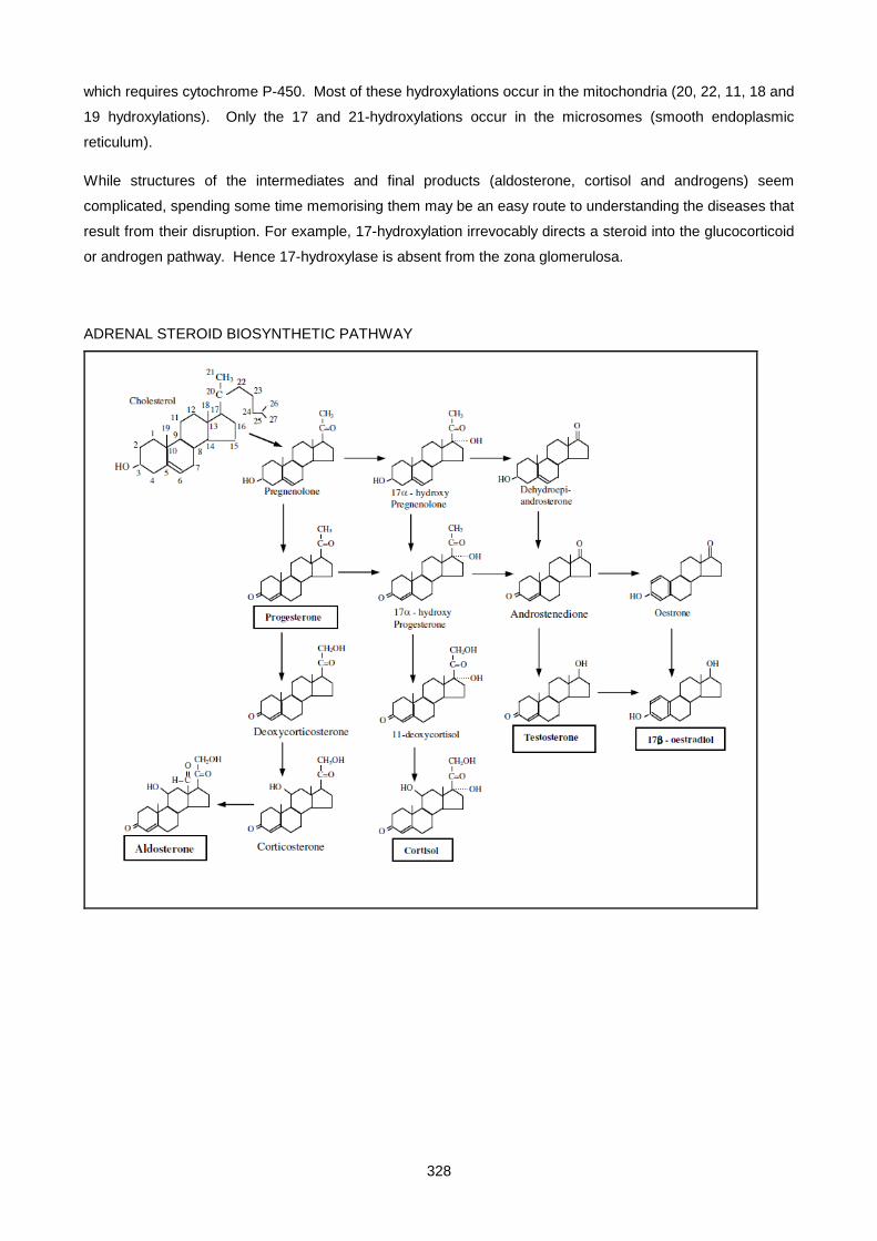
328
which requires cytochrome P-450. Most of these hydroxylations occur in the mitochondria (20, 22, 11, 18 and
19 hydroxylations). Only the 17 and 21-hydroxylations occur in the microsomes (smooth endoplasmic
reticulum).
While structures of the intermediates and final products (aldosterone, cortisol and androgens) seem
complicated, spending some time memorising them may be an easy route to understanding the diseases that
result from their disruption. For example, 17-hydroxylation irrevocably directs a steroid into the glucocorticoid
or androgen pathway. Hence 17-hydroxylase is absent from the zona glomerulosa.
ADRENAL STEROID BIOSYNTHETIC PATHWAY

329
REGULATION OF STEROID
BIOSYNTHESIS
ACTH stimulates cortisol production by promoting early steps in steroidogenesis, namely the transport of
stored cholesterol into mitochondria, and activation of the side chain cleavage enzyme to form ∆5-
pregnenolone. The increased availability of this precursor stimulates production of all adrenal steroids
including aldosterone. Subsequent increase in cortisol then inhibits pituitary ACTH response to hypothalamic
CRF (negative feedback). Although aldosterone is stimulated by ACTH, its major regulation is by angiotensin
II and K+. The total flux of steroid to aldosterone is less than 100th of that through the cortisol pathway.
Thus, even when ACTH levels are low, there is sufficient precursor for aldosterone synthesis. The relative
independence of aldosterone synthesis on ACTH is demonstrated by the fact that patients with ACTH
deficiency suffer from a lack of cortisol but not of aldosterone. ACTH and other peptide and amine hormones
and neurotransmitters, being hydrophilic, do not penetrate the cell and bind membrane receptors. ACTH
stimulates a cascade process within the adrenal cell, involving cAMP which activates protein kinase A (PKA).
This, in turn, activates specific gene sequences, such as that encoding cholesterol side chain cleavage
enzyme, the first and rate-limiting step of steroidogenesis. This is in sharp contrast to steroid hormones
themselves, that are hydrophobic and therefore easily penetrate the cell membrane, to interact with nuclear
receptors present only in target tissues. This leads to transcription of specific gene DNA, mRNA, and, finally,
protein. Thyroid hormones act in a similar way
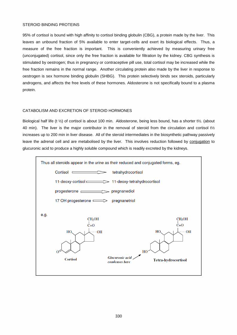
330
STEROID BINDING PROTEINS
95% of cortisol is bound with high affinity to cortisol binding globulin (CBG), a protein made by the liver. This
leaves an unbound fraction of 5% available to enter target-cells and exert its biological effects. Thus, a
measure of the free fraction is important. This is conveniently achieved by measuring urinary free
(unconjugated) cortisol, since only the free fraction is available for filtration by the kidney. CBG synthesis is
stimulated by oestrogen; thus in pregnancy or contraceptive pill use, total cortisol may be increased while the
free fraction remains in the normal range. Another circulating protein also made by the liver in response to
oestrogen is sex hormone binding globulin (SHBG). This protein selectively binds sex steroids, particularly
androgens, and affects the free levels of these hormones. Aldosterone is not specifically bound to a plasma
protein.
CATABOLISM AND EXCRETION OF STEROID HORMONES
Biological half life (t ½) of cortisol is about 100 min. Aldosterone, being less bound, has a shorter t½. (about
40 min). The liver is the major contributor in the removal of steroid from the circulation and cortisol t½
increases up to 200 min in liver disease. All of the steroid intermediates in the biosynthetic pathway passively
leave the adrenal cell and are metabolised by the liver. This involves reduction followed by conjugation to
glucuronic acid to produce a highly soluble compound which is readily excreted by the kidneys.
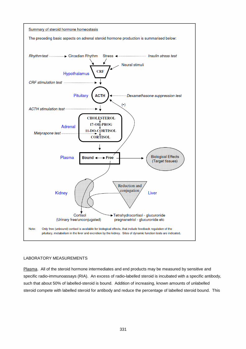
331
LABORATORY MEASUREMENTS
Plasma. All of the steroid hormone intermediates and end products may be measured by sensitive and
specific radio-immunoassays (RIA). An excess of radio-labelled steroid is incubated with a specific antibody,
such that about 50% of labelled-steroid is bound. Addition of increasing, known amounts of unlabelled
steroid compete with labelled steroid for antibody and reduce the percentage of labelled steroid bound. This
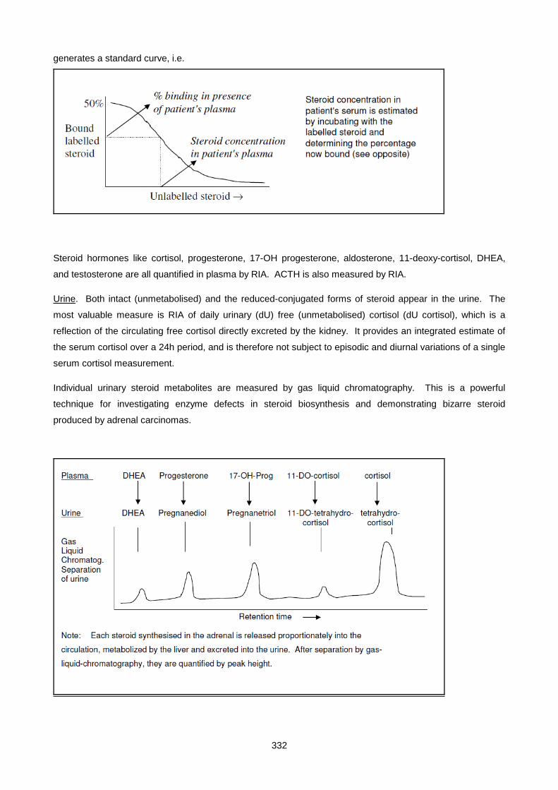
332
generates a standard curve, i.e.
Steroid hormones like cortisol, progesterone, 17-OH progesterone, aldosterone, 11-deoxy-cortisol, DHEA,
and testosterone are all quantified in plasma by RIA. ACTH is also measured by RIA.
Urine. Both intact (unmetabolised) and the reduced-conjugated forms of steroid appear in the urine. The
most valuable measure is RIA of daily urinary (dU) free (unmetabolised) cortisol (dU cortisol), which is a
reflection of the circulating free cortisol directly excreted by the kidney. It provides an integrated estimate of
the serum cortisol over a 24h period, and is therefore not subject to episodic and diurnal variations of a single
serum cortisol measurement.
Individual urinary steroid metabolites are measured by gas liquid chromatography. This is a powerful
technique for investigating enzyme defects in steroid biosynthesis and demonstrating bizarre steroid
produced by adrenal carcinomas.

333
DYNAMIC FUNCTION TESTS (see figure)
• DIURNAL RHYTHM OF PLASMA CORTISOL (12 midnight and 9 am). Lost early in Cushing’s
disease.
• DEXAMETHAZONE SUPPRESSION TEST (for Cushing's)
Dexamethazone is a potent analogue of cortisol, which is not measured by RIA. Both low dose and
high dose tests are used; low dose to confirm the presence of Cushing’s syndrome, high dose to
identify the cause.
o low dose: give 0.5 mg Dex q.i.d. (2mg/day) for 2 days - measure du cortisol on day 2, and
9am plasma cortisol on day 3.
o high dose: give 2.0 mg q.i.d. (8mg/day) for 2 days – measure as above.
These are long forms of the test. The corresponding short test is to give 1 mg or 8mg, respectively,
before retiring, then measure 9am plasma cortisol next morning
• ACTH STIMULATION TEST: Inject ACTH, then measure plasma cortisol response. Used for
Addison's disease,
• METYRAPONE STIMULATION TEST 750 mg q.i.d. for 2 days is based on the inhibition of the 11-
hydroxylase, leading to diminished cortisol, increased ACTH and build-up of precursor steroid, 11-
deoxy-cortisol, which is measured. Useful for assessing residual pituitary function in suspected
secondary hypoadrenalism (pituitary disease).
• INSULIN TOLERANCE TEST relies on insulin (0.15 U/kg) inducing hypoglycaemia, resulting in CRF
and ACTH release and measurement of cortisol response. Useful in suspected secondary
hypoadrenalism, but potentially hazardous.
• CRF STIMULATION for the differential diagnosis of Cushing's syndrome. Give CRF, measure ACTH
response (ACTH rises in pituitary Cushings, no change in adrenal tumours and ectopic ACTH-
producing tumours). Diagnostic value enhanced by selective inferior petrosal sinus sampling, where
ACTH is measured in both L and R inf. petrosal sinuses, and in peripheral blood, following CRF
stimulation. This can also help in pre-operative tumor localization

334
Lecture 20: Endocrinology 4: Adrenal Dysfunction: Hypercortisolism And Hyperaldosteronism
DR PETER BERMAN 2007
HYPERCORTISOLISM (CORTISOL OVERPRODUCTION/CUSHING'S SYNDROME)
Cushing's syndrome is due to overproduction, primarily of glucocorticoids (cortisol), though excess
minerallocorticoid and androgen may also be present. The clinical manifestations are mainly due to the
biological effects of cortisol, and include:
• Increased, typically centripetal, fat deposition (face (‘moon’ face), neck (‘buffalo hump’, trunk and
abdomen – limbs are noticeably spared). 90% of patients exhibit this feature. Cortisol is a powerful
stimulant of the appetite centre. The visceral obesity is strongly associated with insulin resistance
(see below)
• Protein wasting, giving rise to reduced muscle bulk and strength (esp proximal muscles), thinning of
skin, easy bruising, purple striae, etc
• Osteoporosis, leading to back pain and possible vertebral collapse
• Hypertension, hypernatraemia, hypokalaemic alkalosis results from mineralocorticoid activity of
excessive cortisol and/or associated steroids
• Stimulation of gluconeogenesis leads to hyperglycaemia without ketosis. Masquerades as type 2
diabetes
• Menstrual irregularity, hirsutism, due to increased androgen, may occur in females
• Psychiatric disturbances, eg euphoria, mania
• Increased pigmentation (only if ACTH markedly elevated)
Clinical features are, however, highly variable and also depend on the type of Cushing's (see below).
Moreover, depressive, alcoholic and obese patients can present with features of Cushing's and must be
excluded.
CAUSES OF CUSHING'S SYNDROME
a. Pituitary tumors producing excess ACTH – account for 80% of Cushings syndrome
b. Ectopic ACTH production by non-pituitary tumors – eg CA bronchus, carcinoid tumors
c. Adrenal tumors, either adenoma or carcinoma
d. Iatrogenic (glucocorticoid therapy for allergic or autoimmune disorders)
INVESTIGATION APPROACH
The three objectives in investigating Cushing's syndrome are:
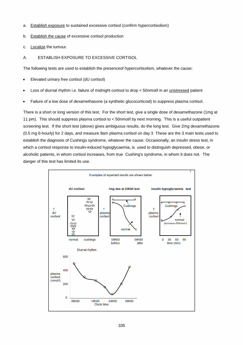
335
a. Establish exposure to sustained excessive cortisol (confirm hypercortisolism)
b. Establish the cause of excessive cortisol production
c. Localize the tumour.
A. ESTABLISH EXPOSURE TO EXCESSIVE CORTISOL
The following tests are used to establish the presenceof hypercortisolism, whatever the cause:
• Elevated urinary free cortisol (dU cortisol)
• Loss of diurnal rhythm i.e. failure of midnight cortisol to drop < 50nmol/l in an unstressed patient
• Failure of a low dose of dexamethasone (a synthetic glucocorticoid) to suppress plasma cortisol.
There is a short or long version of this test. For the short test, give a single dose of dexamethazone (1mg at
11 pm). This should suppress plasma cortisol to < 50nmol/l by next morning. This is a useful outpatient
screening test. If the short test (above) gives ambiguous results, do the long test. Give 2mg dexamethazone
(0.5 mg 6-hourly) for 2 days, and measure 9am plasma cortisol on day 3 These are the 3 main tests used to
establish the diagnosis of Cushings syndrome, whatever the cause. Occasionally, an insulin stress test, in
which a cortisol response to insulin-induced hypoglycaemia, is used to distinguish depressed, obese, or
alcoholic patients, in whom cortisol increases, from true Cushing's syndrome, in whom it does not. The
danger of this test has limited its use.

336
B. DETERMINE THE CAUSE OF EXCESSIVE CORTISOL PRODUCTION.
1. High dose dexamethazone suppression test.
Give 8mg (2.0 mg 6-hourly) for 2 days, and measure 9am cortisol on day 3. An alternative overnight version
of the high dose test involves giving 4mg dexamethasone at 11pm and measuring cortisol at 9am next
morning.
• Most cases of pituitary Cushing's do suppress to < ½ the pretreatment value (since ACTH-secreting
tumors are not totally autonomous)
• Most cases of ectopic ACTH-producing tumors do not suppress; in those that do, the tumor is
probably producing CRF.
• All adrenal tumours do not suppress (they are not ACTH-dependent in the first place).
2. Plasma ACTH
• undetectable in adrenal tumors
• in the upper normal range or slightly above normal in pituitary Cushing's
• highly elevated (>40 pmol/l) in ectopic ACTH-producing tumors
ACTH assays are of great diagnostic value, but since the hormone is extremely labile, blood must be
collected into tubes containing protease inhibitors, the plasma rapidly separated, and stored frozen until
assayed.

337
3. CRF stimulation test
Pituitary ACTH-producing tumors generally retain responsiveness to CRF, whereas in ectopic ACTH
producing and adrenal tumors, ACTH does not change in response.to injected CRF. A recent
refinement of the test is to place sampling catheters into both petrosal sinuses (the venous drainage
of the pituitary), which is technically quite tricky. Then CRF is administered, and ACTH levels in both
petrosal sinuses, and a peripheral site is measured. Typically in a pituitary tumor, ACTH levels from
one of the two sinuses increases markedly (more than 3-fold), relative to the other and to the
peripheral sample. This can also be a useful (though not infallible) indication of which side of the
pituitary harbours the tumor. In ectopic ACTH-producing tumors, ACTH levels from all three sites are
high, equally elevated and do not change appreciably in response to CRF.
4. Radiological studies for pituitary and ectopic tumours as well as adrenal tumours. These include
CAT and MRI scans, and/or radioactive iodo-cholesterol uptake and scanning for adrenal tumors,
These techniques are particularly useful for tumor localization. Indiscriminate use results in a high
incidence of ‘incidentalomas’.
DIAGNOSTIC PITFALLS
Many patients with Cushingoid features are merely obese. They may have slightly raised cortisol but usually
suppress on low dose dexamethazone and have a normal circadian rhythm. They always respond to the
insulin stress test, unlike true Cushing's patients. Alcoholism and severe depressive disease have clinical and
biochemical pictures akin to pituitary Cushing's and are best distinguished by their positive cortisol response
to insulin-induced hypoglycaemia.
HYPOKALAEMIC ALKALOSIS
Hypokalaemic alkalosis can occur in any form of Cushings, it is particularly common in ectopic ACTH-
producing tumors, possibly because the extremely high levels of ACTH stimulate adrenal production of
steroids with mineralocorticoid action. The Cushing’s syndrome caused by such tumors is also unusual in
that it typically has a short history, presents with weight loss (rather than gain) and increased skin
pigmentation.
TREATMENT OF CUSHING'S
• Pituitary - transphenoidal hypophysectomy and irradiation. Large tumors may require a frontal
approach

338
• Ectopic ACTH - surgery (prognosis usually very poor).
• Adrenal adenoma - surgery (prognosis good).
• Adrenal carcinoma - surgery (prognosis very poor). Tumours characterised by rapid growth and
inefficient steroid production, so that it has often metastasized before clinical symptoms occur.
Metastases can be controlled in the short term by the toxic adrenolytic agent o,p-DDD (related to the
insecticide, DDT).
Metyrapone, which blocks cortisol synthesis by inhibiting steroid 11 hydroxylase, can be used prior to surgery
to decrease cortisol production, thereby improving wound healing, and for symptomatic relief when surgery is
not possible. Following repeated unsuccessful pituitary surgery, pituitary Cushing's may need to be treated
by bilateral adrenalectomy. Although this controls the problematical hypercortisolism, the complete removal
of any restraining feedback influence on the pituitary may lead to growth of the pituitary tumour, erosion of
the pituitary fossa and severe pigmentation due to high ACTH. This condition is referred to as Nelson's
syndrome and can be avoided by pituitary irradiation soon after adrenalectomy. Needless to say, after total
hypophysectomy or bilateral adrenalectomy, patients need lifelong glucocorticoid and, in the latter case,
minerallocorticoid, replacement.
PRIMARY HYPERALDOSTERONISM (CONN'S SYNDROME)
DEFINITION
This condition is characterized by the excessive, inappropriate secretion of aldosterone. Most often (80%) it
is due to an autonomous aldosterone-secreting tumor in the zona glomerulosa. Less commonly it is caused
by bilateral diffuse hyperplasia of cells in the zona glomerulosa.
CLINICAL AND BIOCHEMICAL FEATURES
Clinically it presents as hypertension due to excessive sodium and water reabsorbtion, with renal potassium
and H+ ion wasting. Typical biochemical findings are:
Na+ > 140mmol/l K+ < 3.5 mmol/l
HCO3- > 28mmol/l pH > 7.42
24h urinary K+ > 30mmol (despite presence of hypokalaemia)
Hypokalaemia leads to muscle weakness and polyuria/polydipsia, while the alkalosis can manifest as
parasthesia and tetany. Although not a common cause of hypertension (∼1% of cases), it is important to
recognise, as it is potentially curable by surgery.

339
THE LICORICE CONNECTION
Primary hyperaldosteronism may be mimicked by carbenoxolone (an ulcer treatment) or licorice ingestion,
since these substances inhibit an enzyme (11β hydroxysteroid dehydrogenase typeII - 11β HSD-2) that
inactivates cortisol in the DCT cell by converting it to cortisone. This is important because the aldosterone
receptor is ‘promiscuous’, in that it responds equally well to cortisol as to aldosterone, and circulating levels of
cortisol are ∼ 1000x > aldosterone.
PRIMARY VS SECONDARY HYPERALDOSTERONISM
Primary hyperaldosteronism, in which aldo secretion is autonomous, must be clearly differentiate from
secondary hyperaldosteronism, where aldosterone hypersecretion is secondary to renin stimulation. A
hallmark of primary hyperaldosteronism is that renin levels are low (usually unmeasureable), while in
secondary hyperaldosteronism, they are elevated. Whereas most forms of secondary hyperaldosteronism
(cardiac failure, cirrhosis, nephrotic syndrome) are obvious from clinical examination, absence of
hypertension, low (<135) rather than normal to high (>140) serum Na+, some forms of secondary
hyperaldosteronism may mimic the hypertension and biochemical abnormalities seen in Conn’s syndrome.
Such conditions include reno-vascular hypertension (eg renal artery stenosis), and rarer renin-secreting
tumors. The only definitive way to differentiate them from Conn’s syndrome is by plasma renin assay.
DIAGNOSIS
Primary hyperaldosteronism should be suspected in any hypertensive patient with a low plasma K+
concentration who is not on diuretic treatment. Ongoing renal K+ wasting in the face of hypokalaemia in a
patient not on diuretics is highly suggestive of Conn’s syndrome. In equivocal cases, the diagnostic accuracy
of this finding can be enhanced by salt loading – say 200mmol/day (though this doesn’t do their hypertension
much good!). Renin and aldosterone levels are normally affected by posture; both increase on assuming the
upright posture (decreases renal blood flow). Hence basal measurements should be made with the patient
remaining recumbent (lying down) after waking. A totally suppressed renin, which remains suppressed after
30min ambulation or lasix treatment, is typical of primary hyperaldosteronism. Another useful diagnostic
feature of Conn’s syndrome is failure to suppress the high aldosterone levels by salt loading, synthetic
minerallocorticoid or ACE inhibitors (autonomous tumor is no longer dependant on angio II stimulation) Once
diagnosis is confirmed by low renin and unsuppressible high aldosterone, imaging is used to localize the
tumor.

340
TREATMENT
Surgical removal is the treatment of choice for Conn’s syndrome due to a tumor. Bilateral hyperplasia can be
treated medically with spironolactone, an aldosterone antagonist. This drug can also be used to control blood
pressure in tumor patients prior to surgery.
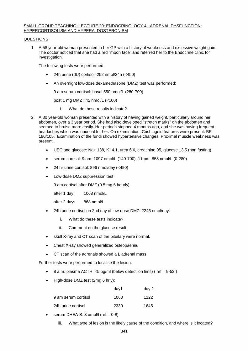
341
SMALL GROUP TEACHING: LECTURE 20: ENDOCRINOLOGY 4: ADRENAL DYSFUNCTION: HYPERCORTISOLISM AND HYPERALDOSTERONISM
QUESTIONS
1. A 58 year-old woman presented to her GP with a history of weakness and excessive weight gain. The doctor noticed that she had a red "moon face" and referred her to the Endocrine clinic for investigation.
The following tests were performed
• 24h urine (dU) cortisol: 252 nmol/24h (<450)
• An overnight low-dose dexamethasone (DMZ) test was performed:
9 am serum cortisol: basal 550 nmol/L (280-700)
post 1 mg DMZ : 45 nmol/L (<100)
i. What do these results indicate?
2. A 30 year-old woman presented with a history of having gained weight, particularly around her abdomen, over a 3 year period. She had also developed "stretch marks" on the abdomen and seemed to bruise more easily. Her periods stopped 4 months ago, and she was having frequent headaches which was unusual for her. On examination, Cushingoid features were present. BP 180/105. Examination of the fundi showed hypertensive changes. Proximal muscle weakness was present.
• UEC and glucose: Na+ 138, K+ 4.1, urea 6.6, creatinine 95, glucose 13.5 (non fasting)
• serum cortisol: 9 am: 1097 nmol/L (140-700), 11 pm: 858 nmol/L (0-280)
• 24 hr urine cortisol: 896 nmol/day (<450)
• Low-dose DMZ suppression test :
9 am cortisol after DMZ (0.5 mg 6 hourly):
after 1 day 1068 nmol/L
after 2 days 868 nmol/L
• 24h urine cortisol on 2nd day of low-dose DMZ: 2245 nmol/day.
i. What do these tests indicate?
ii. Comment on the glucose result.
• skull X-ray and CT scan of the pituitary were normal.
• Chest X-ray showed generalized osteopaenia.
• CT scan of the adrenals showed a L adrenal mass.
Further tests were performed to localise the lesion:
• 8 a.m. plasma ACTH: <5 pg/ml (below detectiion limit) ( ref = 9-52 )
• High-dose DMZ test (2mg 6 hrly):
day1 day 2
9 am serum cortisol 1060 1122
24h urine cortisol 2330 1645
• serum DHEA-S: 3 umol/l (ref = 0-8)
iii. What type of lesion is the likely cause of the condition, and where is it located?
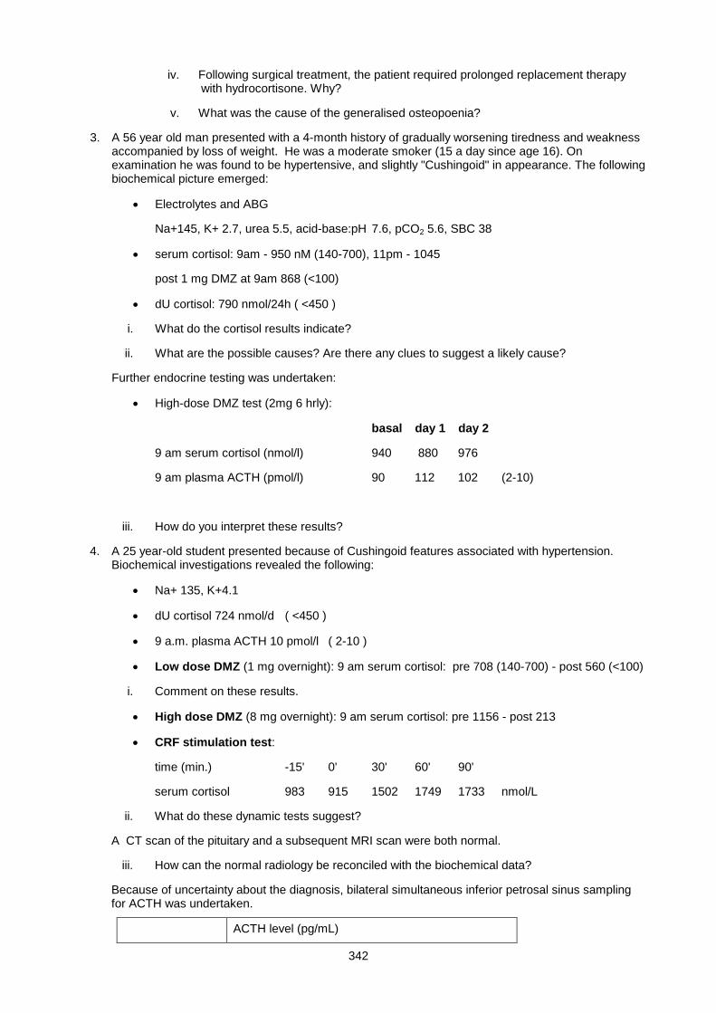
342
iv. Following surgical treatment, the patient required prolonged replacement therapy with hydrocortisone. Why?
v. What was the cause of the generalised osteopoenia?
3. A 56 year old man presented with a 4-month history of gradually worsening tiredness and weakness accompanied by loss of weight. He was a moderate smoker (15 a day since age 16). On examination he was found to be hypertensive, and slightly "Cushingoid" in appearance. The following biochemical picture emerged:
• Electrolytes and ABG
Na+145, K+ 2.7, urea 5.5, acid-base:pH 7.6, pCO2 5.6, SBC 38
• serum cortisol: 9am - 950 nM (140-700), 11pm - 1045
post 1 mg DMZ at 9am 868 (<100)
• dU cortisol: 790 nmol/24h ( <450 )
i. What do the cortisol results indicate?
ii. What are the possible causes? Are there any clues to suggest a likely cause?
Further endocrine testing was undertaken:
• High-dose DMZ test (2mg 6 hrly):
basal day 1 day 2
9 am serum cortisol (nmol/l) 940 880 976
9 am plasma ACTH (pmol/l) 90 112 102 (2-10)
iii. How do you interpret these results?
4. A 25 year-old student presented because of Cushingoid features associated with hypertension. Biochemical investigations revealed the following:
• Na+ 135, K+4.1
• dU cortisol 724 nmol/d ( <450 )
• 9 a.m. plasma ACTH 10 pmol/l ( 2-10 )
• Low dose DMZ (1 mg overnight): 9 am serum cortisol: pre 708 (140-700) - post 560 (<100)
i. Comment on these results.
• High dose DMZ (8 mg overnight): 9 am serum cortisol: pre 1156 - post 213
• CRF stimulation test:
time (min.) -15' 0' 30' 60' 90'
serum cortisol 983 915 1502 1749 1733 nmol/L
ii. What do these dynamic tests suggest?
A CT scan of the pituitary and a subsequent MRI scan were both normal.
iii. How can the normal radiology be reconciled with the biochemical data?
Because of uncertainty about the diagnosis, bilateral simultaneous inferior petrosal sinus sampling for ACTH was undertaken.
ACTH level (pg/mL)
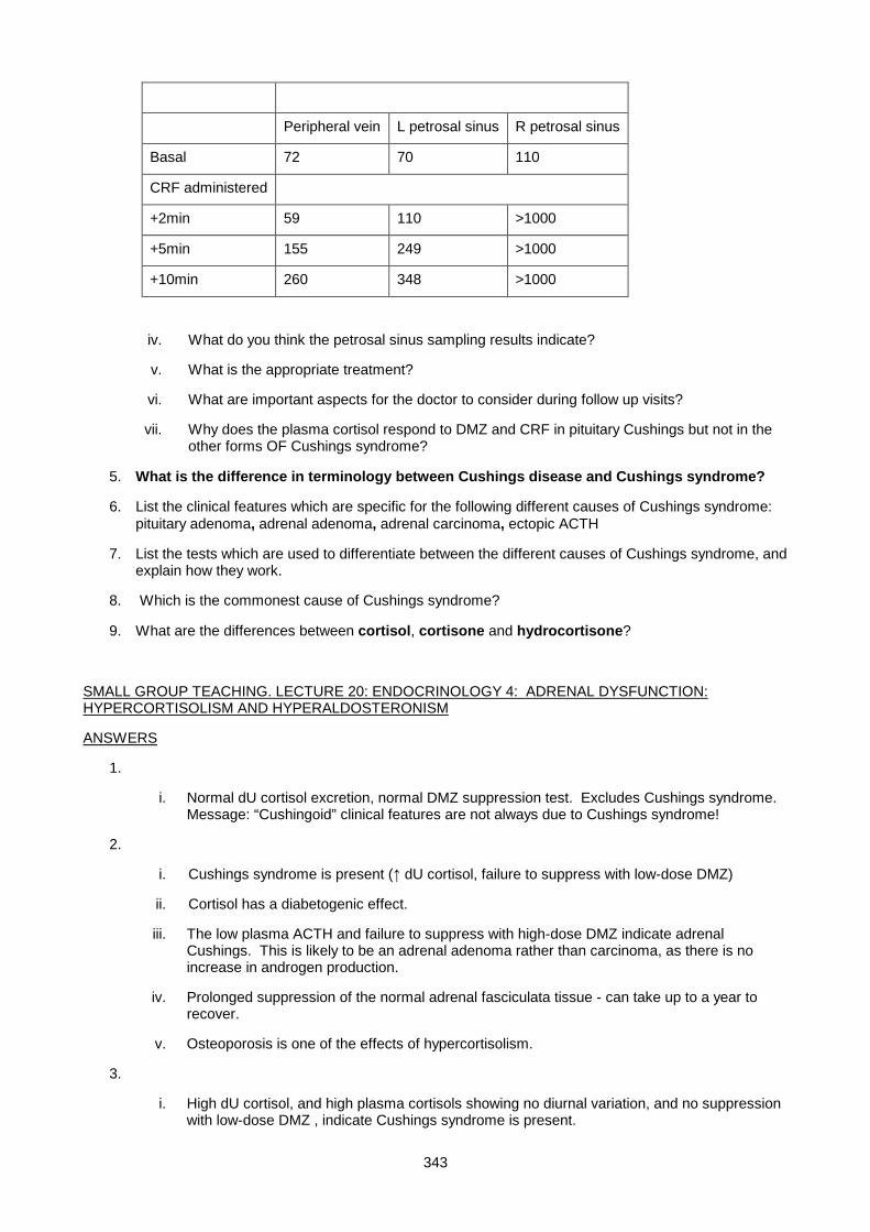
343
Peripheral vein L petrosal sinus R petrosal sinus
Basal 72 70 110
CRF administered
+2min 59 110 >1000
+5min 155 249 >1000
+10min 260 348 >1000
iv. What do you think the petrosal sinus sampling results indicate?
v. What is the appropriate treatment?
vi. What are important aspects for the doctor to consider during follow up visits?
vii. Why does the plasma cortisol respond to DMZ and CRF in pituitary Cushings but not in the other forms OF Cushings syndrome?
5. What is the difference in terminology between Cushings disease and Cushings syndrome?
6. List the clinical features which are specific for the following different causes of Cushings syndrome: pituitary adenoma, adrenal adenoma, adrenal carcinoma, ectopic ACTH
7. List the tests which are used to differentiate between the different causes of Cushings syndrome, and explain how they work.
8. Which is the commonest cause of Cushings syndrome?
9. What are the differences between cortisol, cortisone and hydrocortisone?
SMALL GROUP TEACHING. LECTURE 20: ENDOCRINOLOGY 4: ADRENAL DYSFUNCTION: HYPERCORTISOLISM AND HYPERALDOSTERONISM
ANSWERS
1.
i. Normal dU cortisol excretion, normal DMZ suppression test. Excludes Cushings syndrome. Message: “Cushingoid” clinical features are not always due to Cushings syndrome!
2.
i. Cushings syndrome is present (↑ dU cortisol, failure to suppress with low-dose DMZ)
ii. Cortisol has a diabetogenic effect.
iii. The low plasma ACTH and failure to suppress with high-dose DMZ indicate adrenal Cushings. This is likely to be an adrenal adenoma rather than carcinoma, as there is no increase in androgen production.
iv. Prolonged suppression of the normal adrenal fasciculata tissue - can take up to a year to recover.
v. Osteoporosis is one of the effects of hypercortisolism.
3.
i. High dU cortisol, and high plasma cortisols showing no diurnal variation, and no suppression with low-dose DMZ , indicate Cushings syndrome is present.
344
ii. Pituitary tumor, adrenal tumor or ectopic ACTH. The history of smoking, weight loss and the hypokalemic alkalosis point to lung cancer as a likely cause.
iii. Plasma ACTH levels are very high. There is no suppression of ACTH or cortisol on high-dose DMZ. These features indicate ectopic ACTH syndrome.
Chest X-ray showed a mass lesion, which was resected and proved to be a bronchial carcinoma. The patient died 6 months later from metastatic disease.
4.
i. The ↑ dU cortisol, and failure to adequately suppress on the overnight low-dose DMZ test indicate Cushings syndrome. Plasma ACTH is (inappropriately) within the normal range, excluding adrenal Cushings.
ii. High-dose DMZ resulted in >80% suppression of plasma cortisol, which makes ectopic ACTH a very unlikely cause. There was a definite response to CRF, which supports the diagnosis of a pituitary tumour.
iii. An ACTH-secreting microadenoma may not be visible using imaging techniques.
iv. The petrosal sinus sampling results prove that the origin of the ACTH is pituitary, and suggest that the adenoma is on the right (but does not prove this as venous drainage is frequently asymmetrical.
v. Surgical removal of the tumor
At transsphenoidal surgery, the surgeon removed what appeared to be a small tumor. Histology showed a 5 mm diameter adenoma. On the morning after surgery, the patients plasma cortisol was 55 nmol/L (normal 140-700) and hydrocortisone therapy was started (the remainder of the corticotroph cell population has been suppressed and may take many months to recover).
vi. Monitor pituitary functions as the operation carries the risk of pituitary damage. Gradual weaning off the hydrocortisone. Monitoring for recurrence.
vii. The pituitary adenoma is well-differentiated and retains the control mechanisms (CRF receptors, and glucocorticoid negative feedback mechanism) of the parent cell largely intact.
5. Cushings syndrome = clinical syndrome due to hypercortisolemia from any cause. Cushings Disease refers specifically to an ACTH-secterting pituitary tumour
6. PATHOLOGY CLINICAL FEATURES
pituitary adenoma headaches, visual loss
adrenal adenoma no specific features
adrenal carcinoma androgenic effects
ectopic ACTH hypokalemic alkalosis
7.
• Plasma ACTH level
• High-dose DMZ test
• CRF stimulation test
• plasma DHEAS level
8. Pituitary tumour (about 80% of cases)
9. Cortisol and hydrocortisone are the same compound. Cortisone is cortisol in which the 11-OH group has been oxidised to a keto group. Cortisone is biologically inactive except by conversion to cortisol.
345
Lecture 21: Endocrinology 5: Adrenal Dysfunction, Hypoadrenalism And Congenital Adrenal Hyperplasia (CAH)
DR PETER BERMAN
HYPOADRENALISM
Adrenal cortex insufficiency can either be a primary abnormality within the gland that impairs its capacity to
secrete cortisol and aldosterone, or secondary to a lack of ACTH stimulation, which leads to a selective
cortisol deficiency, leaving aldosterone secretion intact .
CAUSES OF PRIMARY HYPOADRENALISM (ADDISON’S DISEASE)
1. Adrenal destruction by TB, autoimmune disease, metastatic tumor, haemochromatosis or haemorrhage.
2. Inherited enzyme deficiency in steroidogenesis (see CAH below)
CAUSES OF SECONDARY HYPOADRENALISM
1. ACTH deficiency, either isolated, or more commonly, part of generalized hypopituitarism
2. Iatrogenic; due to rapid withdrawal of prolonged steroid therapy.
ADDISON'S DISEASE (PRIMARY HYPOADRENALISM)
CLINICAL FEATURES
Lack of cortisol:
• Non-specific symptoms, including tiredness, weakness, lethargy
• GIT symptoms, including anorexia, nausea, vomiting, diarrhoea
• Weight loss (cortisol is a powerful appetite stimulant)
• Hypoglycaemia
• Depression
Lack of aldosterone (but also of cortisol, which maintains vasomotor tone):
• Hypotension, which can progress to frank hypovolaemic shock
• Salt craving is experienced by some patients
Lack of androgens:
• Loss of body hair (mainly in women)
Excess ACTH:
• Pigmentation (in naturally dark-skinned people, this is easily seen on the palmar creases, over the
knuckles and inside the mouth)
346
Associated auto-immune destruction of other endocrine organs, including parathyroid, thyroid, gonads, and
even non-endocrine tissue, eg gastric parietal cells, resulting in pernicious anaemia; (only seen in
autoimmune Addison’s disease).
RAPIDITY OF ONSET
Onset is generally gradual, with patients feeling relatively well providing they maintain a high salt intake. An
acute presentation, termed an ‘Addisonian crisis’, is usually due to an episode of isotonic fluid loss
(diarrhoea, vomiting) or acute stress (trauma, infection, surgery) in someone with incipient adrenal failure.
Adrenal haemorrhage (eg in meningococcal septicaemia) is another cause of acute adrenal failure; the
ensuing hypovolaemic shock and profound hypoglycaemia constitutes a medical emergency.
BIOCHEMICAL FEATURES
Lack of aldosterone results in Na+ and water loss in the urine. Retention of K+ and H+, leads to
hyperkalaemia and mild acidosis. The hyponatraemia is not directly due to urinary sodium loss, since Na+ is
lost along with an equivalent volume of water (isotonic loss). However, replacement of depleted extracellular
fluid by pure water intake (depleted ECF volume triggers thirst and ADH release) dilutes serum Na+. Also,
since cortisol is required to excrete a water load (increases GFR), cortisol deficiency aggravates the
dilutional hyponatraemia.Thus, the typical biochemical findings in Addison's disease include:
• low plasma Na+
• increased plasma K+
• mild metabolic acidosis (↓ plasma HCO3-)
• elevated plasma urea and creatinine (once sigificant ECF volume depletion occurs)
• fasting hypoglycaemia
• urine Na+ wasting, despite ECF volume depletion (inappropriately high urine Na+)
DIAGNOSIS
Definitive diagnosis established by measuring cortisol and aldosterone. A 9am cortisol < 50nmol/l confirms
the diagnosis (provided the patient is not taking synthetic glucocorticoids), whereas a value of > 550nmol/l
excludes it. Most patients with adrenal failure have values between these extremes, and require an ACTH
stimulation test. Synacthen (synthetic ACTH) is injected, and blood is sampled at 0, 30 and 60min. A blunted
cortisol response is seen in both primary and secondary adrenal failure. To differentiate, the ‘long’ ACTH
stimulation test is performed; ACTH is given for 3 days, and the test repeated on day 4. In primary adrenal
failure, cortisol response remains absent or blunted; whereas in secondary, it normalises (a normal adrenal
chronically deprived of ACTH needs a few days stimulation to recover secretory ability).
Other useful findings to confirm Addison’s disease include:
• Low plasma aldosterone with a high plasma renin activity
347
• Greatly elevated plasma ACTH – this test is excellent for discriminating primary from secondary adrenal
failure, since in the latter condition, ACTH levels are low.
TREATMENT
Treatment of Addison’s disease involves lifelong replacement with cortisol and fludrocortisone (a synthetic
minerallocorticoid). Cortisol is given in twice a day in unequal doses (bigger dose in the morning). Some
milder cases can be treated with cortisol alone, particularly if they maintain a high salt intake, since cortisol
possesses some intrinsic minerallocorticoid activity. Plasma renin activity is useful for assessing adequacy of
(or indicating excessive) minerallocorticoid replacement . Addisonian crisis requires blood taken for basal
hormone measurements, then immediate rehydration with isotonic (0.9%) saline until adequate blood
pressure is restored. Glucocorticoid replacement with synthetic glucocorticoid (eg dexamethasone) is
commenced to prevent hypoglycaemia. This will not invalidate the results of a synacthen test done within the
next few days, since adrenal atrophy from lack of ACTH stimulation takes a while.
SECONDARY HYPOADRENALISM
Secondary hypoadrenalism is hypocortisolism due to a deficient adrenal stimulation by ACTH. It is similar
clinically and biochemically to Addison’s disease, but there are some important differences: Hypopit patients
are not salt-losing and do not require minerallocorticoid replacement (aldosterone production is not ACTH
dependent), Thus hyperkalaemia is not a problem. However, although they are not salt-losing, they can still
be hypotensive and hyponatraemic;
• Hypotensive, because cortisol is required to maintain vasomotor tone in response to catecholamines,
• hyponatraemic because of:an ADH response to the hypotension and a requirement for cortisol to excrete
a water load
Hyperpigmentation is not a feature of secondary hypoadrenalism, since ACTH levels are low. The possibility
of other pituitary hormone deficiencies should be investigated, as isolated ACTH deficiency is rare.
BIOCHEMICAL DIAGNOSIS OF SECONDARY HYPOADRENALISM
1. Hyponatraemia, hypoglycaemia, ↓ cortisol, normal aldosterone, ↓ ACTH (Fig. 1)
2. Confirmation by demonstrating a response to repetitive ACTH stimulation (see Fig. 2)
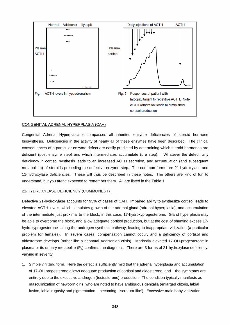
348
CONGENITAL ADRENAL HYPERPLASIA (CAH)
Congenital Adrenal Hyperplasia encompasses all inherited enzyme deficiencies of steroid hormone
biosynthesis. Deficiencies in the activity of nearly all of these enzymes have been described. The clinical
consequences of a particular enzyme defect are easily predicted by determining which steroid hormones are
deficient (post enzyme step) and which intermediates accumulate (pre step). Whatever the defect, any
deficiency in cortisol synthesis leads to an increased ACTH secretion, and accumulation (and subsequent
metabolism) of steroids preceding the defective enzyme step. The common forms are 21-hydroxylase and
11-hydroxylase deficiencies. These will thus be described in these notes. The others are kind of fun to
understand, but you aren’t expected to remember them. All are listed in the Table 1.
21-HYDROXYLASE DEFICIENCY (COMMONEST)
Defective 21-hydroxylase accounts for 95% of cases of CAH. Impaired ability to synthesize cortisol leads to
elevated ACTH levels, which stimulates growth of the adrenal gland (adrenal hyperplasia), and accumulation
of the intermediate just proximal to the block, in this case, 17-hydroxyprogesterone. Gland hyperplasia may
be able to overcome the block, and allow adequate cortisol production, but at the cost of shunting excess 17-
hydroxyprogesterone along the androgen synthetic pathway, leading to inappropriate virilization (a particular
problem for females). In severe cases, compensation cannot occur, and a deficiency of cortisol and
aldosterone develops (rather like a neonatal Addisonian crisis). Markedly elevated 17-OH-progesterone in
plasma or its urinary metabolite (P3) confirms the diagnosis. There are 3 forms of 21-hydroxylase deficiency,
varying in severity:
1. Simple virilizing form. Here the defect is sufficiently mild that the adrenal hyperplasia and accumulation
of 17-OH progesterone allows adequate production of cortisol and aldosterone, and the symptoms are
entirely due to the excessive androgen (testosterone) production. The condition typically manifests as
masculinization of newborn girls, who are noted to have ambiguous genitalia (enlarged clitoris, labial
fusion, labial rugosity and pigmentation – becoming ‘scrotum-like’). Excessive male baby virilization
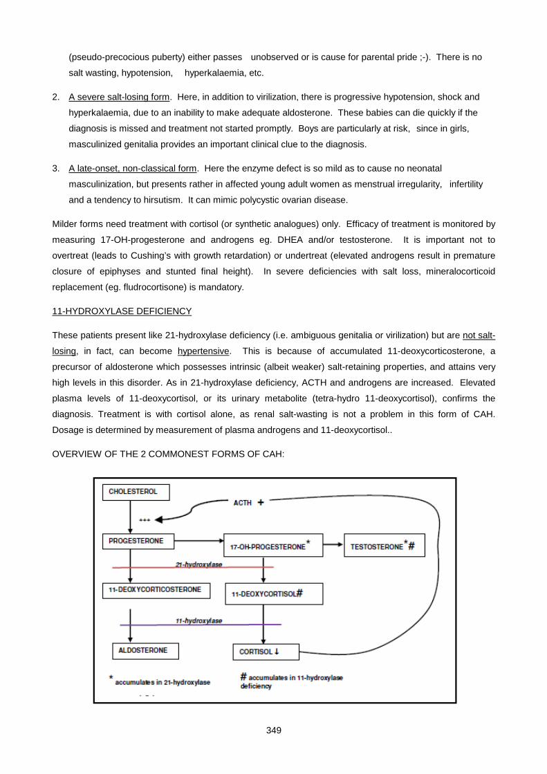
349
(pseudo-precocious puberty) either passes unobserved or is cause for parental pride ;-). There is no
salt wasting, hypotension, hyperkalaemia, etc.
2. A severe salt-losing form. Here, in addition to virilization, there is progressive hypotension, shock and
hyperkalaemia, due to an inability to make adequate aldosterone. These babies can die quickly if the
diagnosis is missed and treatment not started promptly. Boys are particularly at risk, since in girls,
masculinized genitalia provides an important clinical clue to the diagnosis.
3. A late-onset, non-classical form. Here the enzyme defect is so mild as to cause no neonatal
masculinization, but presents rather in affected young adult women as menstrual irregularity, infertility
and a tendency to hirsutism. It can mimic polycystic ovarian disease.
Milder forms need treatment with cortisol (or synthetic analogues) only. Efficacy of treatment is monitored by
measuring 17-OH-progesterone and androgens eg. DHEA and/or testosterone. It is important not to
overtreat (leads to Cushing’s with growth retardation) or undertreat (elevated androgens result in premature
closure of epiphyses and stunted final height). In severe deficiencies with salt loss, mineralocorticoid
replacement (eg. fludrocortisone) is mandatory.
11-HYDROXYLASE DEFICIENCY
These patients present like 21-hydroxylase deficiency (i.e. ambiguous genitalia or virilization) but are not salt-
losing, in fact, can become hypertensive. This is because of accumulated 11-deoxycorticosterone, a
precursor of aldosterone which possesses intrinsic (albeit weaker) salt-retaining properties, and attains very
high levels in this disorder. As in 21-hydroxylase deficiency, ACTH and androgens are increased. Elevated
plasma levels of 11-deoxycortisol, or its urinary metabolite (tetra-hydro 11-deoxycortisol), confirms the
diagnosis. Treatment is with cortisol alone, as renal salt-wasting is not a problem in this form of CAH.
Dosage is determined by measurement of plasma androgens and 11-deoxycortisol..
OVERVIEW OF THE 2 COMMONEST FORMS OF CAH:
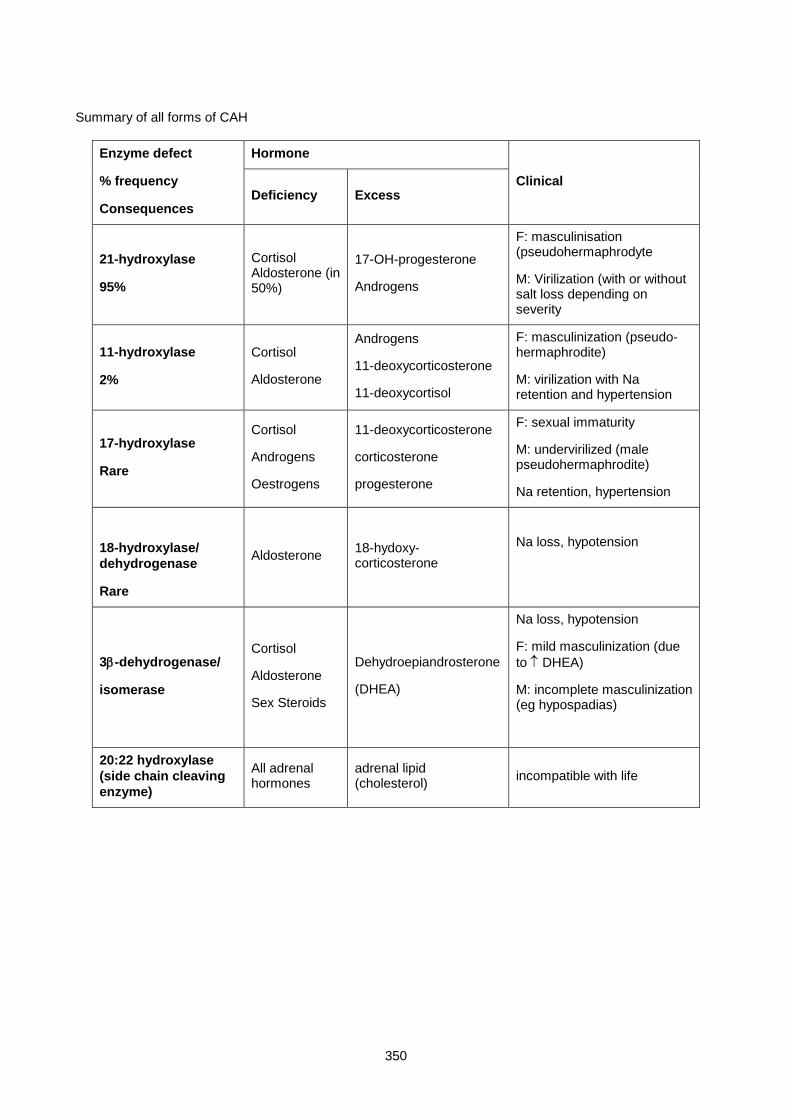
350
Summary of all forms of CAH
Enzyme defect
% frequency
Consequences
Hormone
Clinical Deficiency Excess
21-hydroxylase
95%
Cortisol Aldosterone (in 50%)
17-OH-progesterone
Androgens
F: masculinisation (pseudohermaphrodyte
M: Virilization (with or without salt loss depending on severity
11-hydroxylase
2%
Cortisol
Aldosterone
Androgens
11-deoxycorticosterone
11-deoxycortisol
F: masculinization (pseudo-hermaphrodite)
M: virilization with Na retention and hypertension
17-hydroxylase
Rare
Cortisol
Androgens
Oestrogens
11-deoxycorticosterone
corticosterone
progesterone
F: sexual immaturity
M: undervirilized (male pseudohermaphrodite)
Na retention, hypertension
18-hydroxylase/ dehydrogenase
Rare
Aldosterone 18-hydoxy-corticosterone
Na loss, hypotension
3β-dehydrogenase/
isomerase
Cortisol
Aldosterone
Sex Steroids
Dehydroepiandrosterone
(DHEA)
Na loss, hypotension
F: mild masculinization (due to ↑ DHEA)
M: incomplete masculinization (eg hypospadias)
20:22 hydroxylase (side chain cleaving enzyme)
All adrenal hormones
adrenal lipid (cholesterol) incompatible with life

351
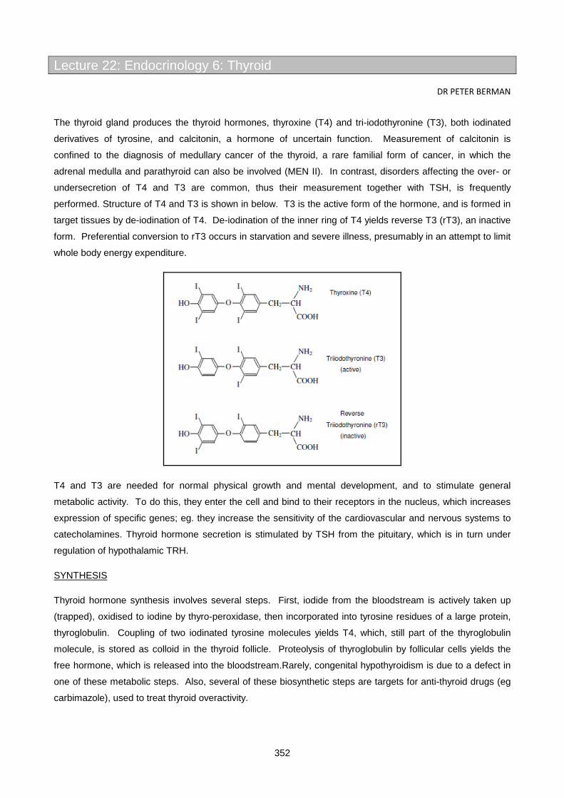
352
Lecture 22: Endocrinology 6: Thyroid
DR PETER BERMAN
The thyroid gland produces the thyroid hormones, thyroxine (T4) and tri-iodothyronine (T3), both iodinated
derivatives of tyrosine, and calcitonin, a hormone of uncertain function. Measurement of calcitonin is
confined to the diagnosis of medullary cancer of the thyroid, a rare familial form of cancer, in which the
adrenal medulla and parathyroid can also be involved (MEN II). In contrast, disorders affecting the over- or
undersecretion of T4 and T3 are common, thus their measurement together with TSH, is frequently
performed. Structure of T4 and T3 is shown in below. T3 is the active form of the hormone, and is formed in
target tissues by de-iodination of T4. De-iodination of the inner ring of T4 yields reverse T3 (rT3), an inactive
form. Preferential conversion to rT3 occurs in starvation and severe illness, presumably in an attempt to limit
whole body energy expenditure.
T4 and T3 are needed for normal physical growth and mental development, and to stimulate general
metabolic activity. To do this, they enter the cell and bind to their receptors in the nucleus, which increases
expression of specific genes; eg. they increase the sensitivity of the cardiovascular and nervous systems to
catecholamines. Thyroid hormone secretion is stimulated by TSH from the pituitary, which is in turn under
regulation of hypothalamic TRH.
SYNTHESIS
Thyroid hormone synthesis involves several steps. First, iodide from the bloodstream is actively taken up
(trapped), oxidised to iodine by thyro-peroxidase, then incorporated into tyrosine residues of a large protein,
thyroglobulin. Coupling of two iodinated tyrosine molecules yields T4, which, still part of the thyroglobulin
molecule, is stored as colloid in the thyroid follicle. Proteolysis of thyroglobulin by follicular cells yields the
free hormone, which is released into the bloodstream.Rarely, congenital hypothyroidism is due to a defect in
one of these metabolic steps. Also, several of these biosynthetic steps are targets for anti-thyroid drugs (eg
carbimazole), used to treat thyroid overactivity.

353
THYROID HORMONES IN BLOOD
Both T3 and T4 are extensively protein bound in the bloodstream, principally to thyroxine-binding globulin
(TBG). T4, for example, is 99.98% protein bound, which means that only one molecule in 5000 is free, and
hence biologically active. Because it is the free, biologically active, form of the hormone that is regulated by
TSH, factors that increase (oestrogens) or decrease (protein malnutrition, nephrotic syndrome) TBG will lead
to an increased or decreased total T4, respectively, while the patient remains euthyroid with a normal level of
freeT4. Certain drugs produce a similar effect by competing with thyroid hormones for binding sites on TBG,
thereby lowering effective TBG concentration. In such cases, measurement of total thyroid hormone alone
can yield misleading information. Early methods sought to ‘correct’ total T4 for changes in TBG by measuring
unoccupied binding sites on TBG (radioactive T3 resin uptake assay); however, sensitive methods are now
available that are designed to measure free hormone levels directly (freeT4 and freeT3). TSH assay also
helps by providing a useful independent marker of free thyroid hormone status at the tissue level.
The reason for such extensive protein binding of thyroid hormones is uncertain; possibly it maintains a ‘buffer’
of circulating thyroid hormone, which explains the long half-life of T4 (∼1 week), and also minimizes losses
via the kidneys. It does, however, create major problems for laboratory assessment of thyroid status.
TESTS OF THYROID FUNCTION
A plasma TSH and freeT4 is generally adequate to diagnose most forms of thyroid disease i.e.
Plasma free T4
High Normal Low
Plasma TSH High TSH secreting tumour
Thyroid Hormone resistance
Early Hypothyroid Primary Hypothyroid
Recovery from sick euthyroid
Normal Euthyroid Sick euthyroid (low fT3)
Early hypopituitrism
Low Hyperthyroid T3-toxicosis Severe hypopituitrism
Severe sick euthyroid
To save money, some labs (like ours) use a sensitive TSH assay as a screening test; low values indicating
thyrotoxicosis, and high values hypothyroidism. This works quite well, but can miss early secondary
hypothyroidism and sick euthyroid syndrome, where TSH can be normal in the presence of a sub-normal free
T4. Also, TSH changes slowly in response to therapy; for example, a patient with Graves disease treated
with radio-iodine or antithyroid drugs may become overtly hypothyroid with a low fT4, despite a still-
suppressed TSH, giving the erroneous impression of persistent thyrotoxicosis. On the other hand, a newly
diagnosed hypothyroid patient put onto perfectly adequate thyroxine replacement might take weeks to
normalise their high TSH. Thus, anyone known to have, or have had, thyroid disease, particularly on
treatment, or anyone with suspected pituitary disease, must have a fT4, in addition to a TSH.
Plasma fT3 assay is not routinely used as a thyroid function test. It may be useful
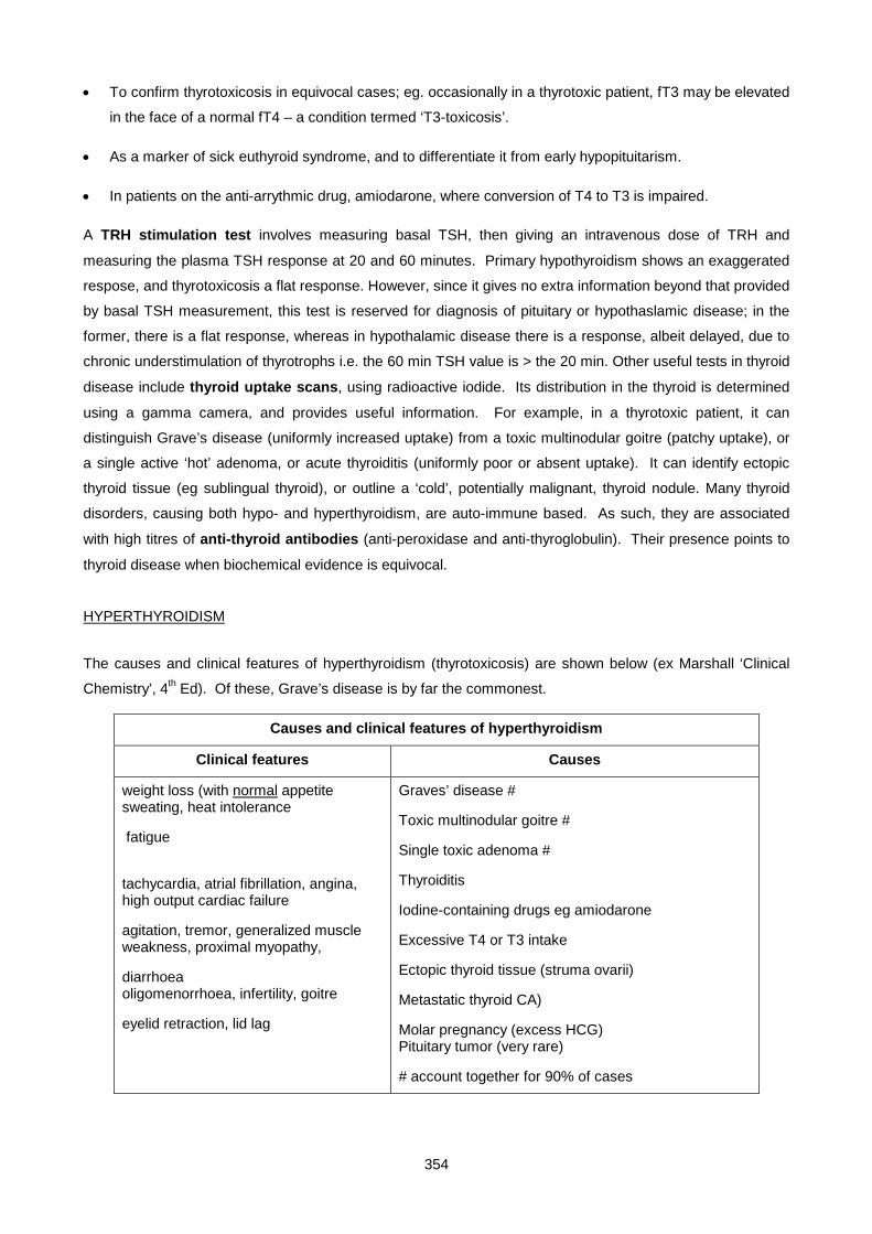
354
• To confirm thyrotoxicosis in equivocal cases; eg. occasionally in a thyrotoxic patient, fT3 may be elevated
in the face of a normal fT4 – a condition termed ‘T3-toxicosis’.
• As a marker of sick euthyroid syndrome, and to differentiate it from early hypopituitarism.
• In patients on the anti-arrythmic drug, amiodarone, where conversion of T4 to T3 is impaired.
A TRH stimulation test involves measuring basal TSH, then giving an intravenous dose of TRH and
measuring the plasma TSH response at 20 and 60 minutes. Primary hypothyroidism shows an exaggerated
respose, and thyrotoxicosis a flat response. However, since it gives no extra information beyond that provided
by basal TSH measurement, this test is reserved for diagnosis of pituitary or hypothaslamic disease; in the
former, there is a flat response, whereas in hypothalamic disease there is a response, albeit delayed, due to
chronic understimulation of thyrotrophs i.e. the 60 min TSH value is > the 20 min. Other useful tests in thyroid
disease include thyroid uptake scans, using radioactive iodide. Its distribution in the thyroid is determined
using a gamma camera, and provides useful information. For example, in a thyrotoxic patient, it can
distinguish Grave’s disease (uniformly increased uptake) from a toxic multinodular goitre (patchy uptake), or
a single active ‘hot’ adenoma, or acute thyroiditis (uniformly poor or absent uptake). It can identify ectopic
thyroid tissue (eg sublingual thyroid), or outline a ‘cold’, potentially malignant, thyroid nodule. Many thyroid
disorders, causing both hypo- and hyperthyroidism, are auto-immune based. As such, they are associated
with high titres of anti-thyroid antibodies (anti-peroxidase and anti-thyroglobulin). Their presence points to
thyroid disease when biochemical evidence is equivocal.
HYPERTHYROIDISM
The causes and clinical features of hyperthyroidism (thyrotoxicosis) are shown below (ex Marshall ‘Clinical
Chemistry’, 4th Ed). Of these, Grave’s disease is by far the commonest.
Causes and clinical features of hyperthyroidism
Clinical features Causes
weight loss (with normal appetite sweating, heat intolerance
fatigue
tachycardia, atrial fibrillation, angina, high output cardiac failure
agitation, tremor, generalized muscle weakness, proximal myopathy,
diarrhoea oligomenorrhoea, infertility, goitre
eyelid retraction, lid lag
Graves’ disease #
Toxic multinodular goitre #
Single toxic adenoma #
Thyroiditis
Iodine-containing drugs eg amiodarone
Excessive T4 or T3 intake
Ectopic thyroid tissue (struma ovarii)
Metastatic thyroid CA)
Molar pregnancy (excess HCG) Pituitary tumor (very rare)
# account together for 90% of cases

355
Graves’ disease is an autoimmune condition, in which antibodies to the TSH receptor bind it in such a way as
to mimic TSH and lock the receptor in its active conformation. This causes constant stimulation of the gland
to grow and produce thyroxine. Pituitary TSH secretion is suppressed. A unique feature of Graves’ (as
opposed to other causes of toxicosis) is the presence of proptosis (bulging eyes) due to thickening of retro-
orbital muscles, and swelling of soft tissue in the legs and feet (pre-tibial myxoedema). These phenomena
are also auto-antibody mediated, and while they usually co-exist with hyperthyroidism, they can occur in
isolation. Laboratory diagnosis of Grave’s disease depends on demonstration of high plasma concentration of
T3 and (usually) T4, with totally suppressed TSH. Three options of treatment are available:
• Anti-thyroid drugs
• Radio-active iodine
• Surgery (sub-total thyroidectomy)
Treatment with β-adrenergic blocking drugs provides prompt symptomatic relief, but has no effect on
overproduction of thyroxine. They are nevertheless useful since antithyroid drugs take some time to work
(they suppress new hormone synthesis but do not affect release of pre-formed hormone); similar logic
dictates use of β-blockers, rather than anti-thyroid drugs, to treat the transient hyperthyroidism of thyroiditis.
Since there is a reasonable chance that Graves’ disease may go into permanent remission, a reasonable
approach in a younger person without a large thyroid swelling (goitre) is to try anti-thyroid drugs for a period of
1-2 years. The disease will remit spontaneously in 30-40% of cases. Once the drugs have rendered the
patient euthyroid (normal T3 and T4), one either reduces the dose or maintains the high dose and adds
thyroxine replacement. TSH can remain suppressed for some time – thus a low TSH by itself does not
indicate persisting hyperthyroidism in the short term. Thyroid function is monitored regularly for 1-2 years,
then treatment is stopped. If patient relapses, a further course of drug treatment or radio-iodine is indicated.
Radio-iodine ablation of the thyroid is a complication-free and definitive treatment for Grave’s disease. The
only problem is that most patients become permanently hypothyroid and need lifelong thyroxine replacement.
Because of the safety and efficacy of radio-iodine for Grave’s disease in all age groups (except pregnancy),
surgery is currently reserved for large goitres compressing structures in the neck. Whatever modality of
treatment is employed, patients need long term hormone measurement to check for:
• Relapse of thyrotoxicosis
• Development of hypothyroidism
THYROID STORM refers to an acute presentation of thyrotoxicosis, with hyperpyrexia, dehydration and
cardiac failure. Although very rare, this is a medical emergency associated with significant mortality, and
requires urgent treatment.
NEONATAL HYPERTHYROIDISM occurs in infants born to mothers who have or have had Graves’ disease.
It is due to thyroid stimulating antibodies (IgG) crossing the placenta and stimulating the fetal thyroid. It is
transient since these immunoglobulins are gradually cleared, but may need short term treatment. The
diagnosis is often overlooked when the mother’s Graves’ disease was treated many years previously and she
is currently euthyroid, but still has circulating thyroid-stimulating antibodies.

356
HYPOTHYROIDISM (also termed ‘myxoedema’)
There are many causes of hypothyroidism, but autoimmune destruction of the gland or previous radio-iodine
or surgical treatment of thyrotoxicosis (iatrogenic) are the commonest. Rarer causes include congenital
hypothyroidism (cretinism) and secondary to pituitary or hypothalamic disease.
Causes and clinical features are tabulated below:
Causes and clinical features of hypothyroidism
Clinical features Causes
lethargy, tiredness
cold intolerance
dryness and coarsening of skin & hair
weight gain
slow relaxation of muscles and tendon reflexes
depression, dementia, psychosis
constipation
bradycardia, angina pericardial effusions
muscle stiffness
carpal tunnel syndrome
menorrhagia, infertility, galactorrhoea
Atrophic hypothyroidism #
Autoimmune hypothyroidism (Hashimoto’s D.) #
Post surgery, radio-iodine, antithyroid drugs #
Enzyme defect in thyroxine synthesis
Pituitary or hypothalamic disease
Iodine deficiency
# accounts for >90% of cases
Apart from the clinical features listed, any unexplained hypercholesterolaemia or elevated creatine kinase
should raise the suspicion of hypothyroidism. Biochemical diagnosis is quite straightforward – a decreased
fT4 with a markedly increased TSH. The disease often begins insidiously; in its early stages, the only
abnormality may be an increased TSH, to which the failing gland responds by secreting enough T4 to keep
the fT4 within the normal range (usually close to the lower end). In such cases, high titres of anti-thyroid
antibodies provide supportive evidence of auto-immune thyroid disease. Treatment is also simple and cheap
– lifelong administration of sufficient thyroxine to normalise the plasma fT4 and TSH. Abnormally high TSH
indicates inadequate replacement or erratic compliance, whereas suppressed TSH suggests excessive
replacement.T3 treatment is used in situations where rapid onset of action is desired (eg. treatment of
myxoedema coma) or where rapid reversal of overdosage is important (eg. treating hypothyroid patients with
ischaemic heart disease). This is because of the rapid onset of action and shorter half-life of T3 compared to
T4.Occasionally hypothyroid patients may present with hypothermia and stupor or coma, a condition termed
‘myxoedema coma’. This is a medical emergency that carries a high risk of mortality. Apart from T3
replacement, one needs to restore body temperature and correct electrolyte imbalance (typically
hyponatraemia from water retention).
THYROIDITIS is acute inflammation of the thyroid, usually on a viral or autoimmune basis. Release of pre-
formed thyroxine may result in mild, transient hyperthyroidism (for about 6 weeks), often followed by a period
357
of hypothyroidism. Normal function is generally returns, though in autoimmune thyroiditis, hypothyroidism
may be permanent and require thyroxine replacement. The thyrotoxic phase is generally clinically and
biochemically mild, requiring.only reassurance and possibly β-blockers. Anti-thyroid drugs are
contraindicated. The condition is clearly distinguished from Graves’ disease by the low, rather than
increased, uptake of radio-active iodide.
SICK EUTHYROID SYNDROME
Typically, during the acute phase of a serious illness, fT3 and, less often, fT4 concentration drops below
normal. TSH is usually normal but may be low in the severely ill. Possible explanations include decreased
peripheral conversion of T4 to T3, and inhibition of TSH secretion by cortisol. Certain drugs, including β-
blockers and the iodine-containing anti-arrythmic drug, amiodarone, can inhibit conversion of T4 to T3.
GOITRE is a thyroid enlargement, that can be part of hyperthyroidism (Graves’ disease, toxic multinodular
goitre, single toxic adenoma), hypothyroidism (Hashimoto’s disease, iodine deficiency) or occur in euthyroid
individuals bearing benign or malignant thyroid tumors. Thus any patient with a goitre should have their
thyroid hormone status checked as a clue to diagnosis.
THYROID CANCER is a fairly common malignancy occurring in young people, particularly women. In fact,
thyroid disease in general is much commoner in women. Incidence of cancer spiked in Europe in the
aftermath of the Chernobyl debacle, due to release of radio-iodine into the atmosphere (in doses insufficient
to cause thyroid ablation). Following total thyroidectomy and ablative radio-iodine treatment, patients are
maintained on doses thyroxine sufficient to render them mildly toxic. The logic here is to suppress TSH
secretion in the hope of preventing growth of residual tumor. A useful tumor marker for thyroid cancer is
plasma thyroglobulin. Trace amounts of this protein normally leak out of the thyroid and can be measured in
the blood. After ablative thyroid surgery and/or radiotherapy however, circulating thyroglobulin should
disappear. Its persistence indicates presence of residual tumor tissue or tumor recurrence.Calcitonin-
secreting medullary cancer of the thyroid is a rare form of inherited cancer, and forms part of the multiple
endocrine neoplasia type II (MEN II) syndrome, where it occurs in association with tumors of the adrenal
medulla and parathyroid. It can be detected early in susceptible individuals by regular plasma calcitonin
measurements.
CONGENITAL HYPOTHYROIDISM (cretinism) is a sufficiently common and devastating condition to make
neonatal screening programs worthwhile. Untreated affected children exhibit progressive growth and mental
retardation. Provided the diagnosis is made soon after birth, life-long T4 replacement provides simple, cheap
and effective therapy. Screening normally entails measurement of TSH at one week of age.

358
SMALL GROUP TEACHING: LECTURE 22: ENDOCRINOLOGY 6: THYROID
QUESTIONS
1. A 30 year old woman presented with a 3 month history of palpitations and 5 kg weight loss. She had not had any symptoms of neck pain. On examination, she had a fine tremor, warm moist hands, lid retraction and lid lag. The thyroid was diffusely enlarged; no bruit was heard. BP 140/80, pulse 108, regular.
• fT4: 56 pmol/L (10-24), TSH: < 0.01 µu/mL (0.35-5.5) i. What do the clinical findings and biochemical tests indicate? ii. List the possible causes, in order of frequency. iii. What clinical signs are found in Graves disease and not in other causes of thyrotoxicosis?
What causes these clinical effects? iv. A thyroid 99Tc uptake scan was performed which showed uniformly increased uptake.
What diagnosis does this indicate? v. What is the natural history of Graves disease? vi. Outline the treatment options available fot his patient, and discuss their advantages and
disadvantages. vii. Name 2 antithyroid drugs. viii. At what level do they work (hypothalamus, pituitary or thyroid gland)? ix. 131I therapy is associated with an increased risk of thyroid cancer (T / F ?) x. 131I therapy is safe in pregnancy (T / F ?)
The patient agreed to have 131I therapy, and was seen at a follow up visit 2 months later, with the following thyroid function tests:
• fT4: 7.2 (10-24) , TSH :10.8 (0.35-5.5) xi. What would you say to the patient now? xii. What is the half-life of thyroxine?
2. A 65 year-old man is referred from a country hospital with a history of experiencing disturbing palpitations. He has noted a decreased effort tolerance, but is otherwise well, and has not lost weight. His pulse is 76, irregular. An ECG shows atrial fibrillation. He has no tremor or abnormal eye signs, and his thyroid feels normal.
i. Could this patient be hyperthyroid? ii. Which test would you use as a first screen for hyperthyroidism ?
TFTs were performed: fT4 :36.8, TSH < 0.01
iii. Is a free T3 level needed? iv. What is a "thyroid storm"? When does it occur?
3. A 65 year-old woman was admitted to hospital with a 1 week history of shortness of breath. Examination revealed an obese, uncommunicative woman. The skin was dry, with scaling over the upper arms. Decreased breath sounds were found over her left lung posteriorly, with dullness to percussion. The pulse was 56, regular, the JVP was not raised and heart sounds were normal. Delayed relaxation of the knee and ankle jerks were observed.
A chest X ray showed a left pleural effusion.
i. Suggest a likely diagnosis based on these clinical features. ii. What laboratory tests are required to confirm this diagnosis?
TFTs were performed
• fT4: 5.6 , TSH: 67 iii. What are the common causes of this condition? iv. What do you understand by the terms "myxoedema" and "pretibial myxoedema"? v. What is the significance of the pleural effusion? vi. What additional biochemical abnormalities are common in this condition? vii. Discuss the treatment.
4. A 68 year old man with metastatic carcinoma of the prostate complained of having palpitations. Thyroid function tests were requested and showed:
359
• fT4: 12 pmol/l (10-24), fT3: 1.5 pmol/l (3.4-7.2), TSH: 0.24 µu/ml (0.35-5.5) i. What do these results indicate? ii. Where and how is T3 produced? iii. What is rT3? When is it increased, and what are its actions? iv. Discuss appropriate therapy for this case.
5. A 45 year-old woman complained of fatigue, lethargy, attacks of giddiness and sweating. Her periods had become irregular and infrequent. Her doctor attributed the symptoms to the menopause, and started her on estrogen therapy. However, 3 weeks later she collapsed and was taken to the Emergency Unit where her plasma glucose level was found to be 2.2 mmol/L. Thyroid function tests showed:
• fT4: 4.1, TSH: 0.78 , fT3: 2.8 i. What do the results indicate? ii. Suggest a reason for the hypoglycemia. iii. Which additional endocrine tests are required?
A metyrapone test showed a subnormal ACTH response.LH and FSH were low. A CT scan showed a pituitary mass. This was removed and showed the histological features of a chromophobe adenoma (nonsecreting pituitary tumor).
iv. Comment on the pitfalls of using TSH as the primary screening test for hypothyroidism. 6. A 26 year old woman who was 6 months pregnant, with her first child, came to the doctor when she
noticed a swelling in her neck. On examination, she had a diffusely enlarged thyroid gland, which was non-tender. There were no signs of hyper- or hypothyroidism. The following results were obtained:
• TSH: 1.3, fT4: 17 i. When examining a neck swelling, what feature suggests that it is a goitre (enlarged thyroid)? ii. In what ways can pregnancy affect the thyroid gland and its function? iii. What percentage of T3 and T4 are free and bound in plasma? To what are they bound? iv. Total T4 is no longer routinely measured in clinical practice. If it had been measured, what would
you expect the level to be in this case? 7. Draw diagrams of HCG and TSH, the TSH receptor, and its signalling pathway 8. To which families of receptors do (a) the T3 receptor, and (b) the TSH receptor belong?
SMALL GROUP TEACHING: LECTURE 22: ENDOCRINOLOGY 6: THYROID
ANSWERS
1. i. Hyperthyroidism ii. Common causes: Graves disease, toxic multinodular goitre Uncommon causes: toxic
adenoma, exogenous thyroid hormone administration, trophoblastic disease. Rare: struma ovarii iii. Signs specific to Graves: Exopthalmos, periorbital edema, pretibial myxoedema, Graves
acropachy: Caused by autoimmune inflammatory process. Increased thyroid activity and bruit caused by antibodies stimulating the TSH receptor.
iv. Unifornly increased uptake indicates Graves disease. The scan would show in other conditions: multinodular goitre - patchy uptake; adenoma - single "hot" spot; thyroiditis or exogenous thyroxine - decreased uptake.
v. One third of cases remit within a year. Of these, some relapse at some time. The other 2/3 have a progressive course.
vi. Treatment options: antithyroid drugs, beta blockers, 131I therapy,surgery (rarely done). vii. Carbimazole (Neomercazole); propylthiouracil (PTU) viii. At thyroid gland level. ix. False x. False - pregnancy is an absolute contraindication. xi. Your thyroid hormone levels are now too low and you need replacement therapy with thyroxine. xii. 7 days.

360
2. i. Yes . AF may be the only sign of hyperthyroidism, especially in the elderly. ii. TSH iii. No, a fT3 is not needed as the diagnosis is clear - a free T3 is only needed when the other
results are equivocal or contradictory. iv. Thyroid storm is an acute exacerbation of hyperthyroidism, which may be precipitated by illness
or trauma. Characterized by hyperpyrexia, dehydration, cardiac failure. 3.
i. Hypothyroidism. ii. TSH (a raised TSH is all that is needed), although a free T4 is usually also done to confirm the
diagnosis. iii. Autoimmune (idiopathic), post-surgical, post 131I therapy. iv. This causes great confusion: Myxoedema refers to the clinical syndrome of hypothyroidism,
pretibial myxoedema is a manifestation of Graves disease (an autoimmune dermopathy). v. Pleural and pericardial effusions are features of hypothyroidism. The fluid is high in protein - due
to increased permeability of pleural/pericardial membranes. vi. Raised plasma cholesterol (secondary hyperlipidemia); raised plasma CK (myopathy) vii. Thyroxine treatment must be started at a low dose, gradually increasing over several weeks.
Too rapid correction may precipitate MI if coronary artery disease is present. 4.
i. Sick euthyroid syndrome. ii. T3 produced from T4 in peripheral tissues by 5'-deiodinase. This enyme is downregulated in
non-thyroidal illnesses. TSH is due to neural downregulation of TRH secretion. iii. Reverse T3 is an inactive metabolite, formed from T4 in peripheral tissues. Increased in many
non-thyroidal illnesses. iv. This is a "normal" response to severe illness, and does not require, or benefit from, thyroxine or
T3 treatment. 5.
i. Secondary hypothyroidism (pituitary disease). The TSH, while still in the normal range, is inappropriate for the low fT4.
ii. ACTH and GH deficiency. iii. Assesment of other aspects of pituitary function: ACTH / cortisol axis, gonadotropins, GH. iv. Using TSH as the sole test would have missed this diagnosis. The message is: if hypopituitarism
is a possibility, both TSH and fT4 need to be done. 6.
i. It moves on swallowing. ii. (a) Pregnancy (or estrogen administration) causes ↑ TBG resulting in ↑ total T4 with normal free
T4 as a rule (b) Free T4 is occasionally ↑ in the first trimester, due to HCG stimulation of the TSH receptor. This occurs especially in trophoblastic disease. (c) An enlargement of the thyroid gland (goitre) is common in pregnancy and does not indicate hyper- or hypothyroidism (this case)
iii. Both T3 and T4 are more than 99% bound, mainly to thyroid-binding globulin (TBG). iv. Total T4 would have been increased. 7. HCG and TSH both consist of alpha and beta subunits. The alpha subunit consists of the identical
gene product and both molecules only differ at the beta subunits. The TSH receptor is a classical G-protein coupled receptor with 7 trans-membrane loops and when activated by TSH it interacts with G-s protein that releases GDP and binds GTP. The alpha subunit of G-s, bound to GTP then dissociates and activates adenylyl cyclise that increases intracellular cAMP and triggers downstream events.
8. a) T3 binds to a nuclear receptor b) – TSH = a G Protein Coupled membrane Recepto

361
Lecture 23: Endocrinology 7: Disorders Of Gonadal Function
DR PETER BERMAN 2003
BACKGROUND PHYSIOLOGY
The role of the testis is simple; it must:
• Produce spermatozoa
• Secrete testosterone
Spermatogenesis requires stimulation by FSH. In response to FSH, Sertoli cells secrete the peptide
hormone inhibin, which inhibits further FSH secretion (negative feedback). Spermatogenesis also depends
on adequate testosterone production by Leydig cells in the testis (a paracrine effect).
Testosterone is production by Leydig cells is controlled by the pituitary LH stimulation; LH release is inhibited
by testosterone via a negative feedback mechanism. Testosterone is a powerful anabolic hormone (hence its
abuse by certain athletes) and is responsible for development of male secondary sexual characteristics at
puberty.Testosterone is also present in females, albeit at ten-fold lower levels (2 vs 20nmol/l); some is
produced directly by the ovary, but most is derived from peripheral conversion of adrenal androgens, such as
DHEA. Adrenal androgens are important in females, where they are responsible for growth of musculature
and pubic and axillary hair at puberty (as well as for less desirable features like adolescent acne). In males,
testosterone needs to be converted to a more potent derivative, dihydrotestosterone (DHT) by the enzyme,
5α-reductase, to exert its full effect. Thus while testosterone is adequate for development of male internal
genitalia, DHT is required for full masculinization of external genitalia – eg. penile growth.Two causes
(admittedly rare) of male undervirilization include 5α-reductase deficiency and a defective testosterone
receptor (termed ‘androgen insensitivity syndrome’). Testosterone in plasma is, like other steroid hormones,
strongly protein bound, principally to sex hormone binding globulin (SHBG).SHBG synthesis by the liver is
(like that of TBG and CBG) enhanced by oestrogens. Thus for a given level of total plasma testosterone, the
greater the SHBG, the less testosterone is free and hence less biologically active.Normal women have on
average twice the SHBG levels of men (due to their oestrogens); thus as well as having 10x less circulating
total testosterone than men, the little that women do have is more extensively protein-bound, and hence
inactive. In women, SHBG is important in regulating exposure of target tissues to androgens. For example,
polycystic ovarian disease, a disorder characterised by irregular, anovulatory cycles and male-pattern hair
growth (hirsutism), is often associated with a decreased SHBG.Since free testosterone assays are not freely
available, the ratio of total testosterone/SHBG provides an index of free testosterone status.
OESTROGENS AND THE OVARY
Oestradiol is the principal oestrogen secreted by the ovary, and is responsible for the appearance of female
secondary sexual characteristics at puberty. It also maintains the function of the reproductive system – eg.
stimulates endometrial growth in the first half of the menstrual cycle, maintains a normal vaginal epithelium
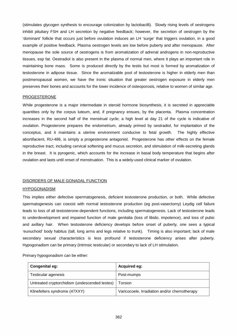
362
(stimulates glycogen synthesis to encourage colonization by lactobacilli). Slowly rising levels of oestrogens
inhibit pituitary FSH and LH secretion by negative feedback; however, the secretion of oestrogen by the
‘dominant’ follicle that occurs just before ovulation induces an LH ‘surge’ that triggers ovulation, in a good
example of positive feedback. Plasma oestrogen levels are low before puberty and after menopause. After
menopause the sole source of oestrogens is from aromatization of adrenal androgens in non-reproductive
tissues, esp fat. Oestradiol is also present in the plasma of normal men, where it plays an important role in
maintaining bone mass. Some is produced directly by the testis but most is formed by aromatization of
testosterone in adipose tissue. Since the aromatizable pool of testosterone is higher in elderly men than
postmenopausal women, we have the ironic situation that greater oestrogen exposure in elderly men
preserves their bones and accounts for the lower incidence of osteoporosis, relative to women of similar age.
PROGESTERONE
While progesterone is a major intermediate in steroid hormone biosynthesis, it is secreted in appreciable
quantities only by the corpus luteum, and, if pregnancy ensues, by the placenta. Plasma concentration
increases in the second half of the menstrual cycle; a high level at day 21 of the cycle is indicative of
ovulation. Progesterone prepares the endometrium, already primed by oestradiol, for implantation of the
conceptus, and it maintains a uterine environment conducive to fetal growth. The highly effective
abortifacient, RU-486, is simply a progesterone antagonist. Progesterone has other effects on the female
reproductive tract, including cervical softening and mucus secretion, and stimulation of milk-secreting glands
in the breast. It is pyrogenic, which accounts for the increase in basal body temperature that begins after
ovulation and lasts until onset of menstruation. This is a widely-used clinical marker of ovulation.
DISORDERS OF MALE GONADAL FUNCTION
HYPOGONADISM
This implies either defective spermatogenesis, deficient testosterone production, or both. While defective
spermatogenesis can coexist with normal testosterone production (eg post-vasectomy) Leydig cell failure
leads to loss of all testosterone-dependent functions, including spermatogenesis. Lack of testosterone leads
to underdevelopment and impaired function of male genitalia (loss of libido, impotence), and loss of pubic
and axillary hair. When testosterone deficiency develops before onset of puberty, one sees a typical
‘eunuchoid’ body habitus (tall, long arms and legs relative to trunk). Timing is also important; lack of male
secondary sexual characteristics is less profound if testosterone deficiency arises after puberty.
Hypogonadism can be primary (intrinsic testicular) or secondary to lack of LH stimulation.
Primary hypogonadism can be either:
Congenital eg: Acquired eg:
Testicular agenesis Post-mumps
Untreated cryptorchidism (undescended testes) Torsion
Klinefelters syndrome (47XXY) Varicocoele, Irradiation and/or chemotherapy
363
Secondary hypogonadism (hypogonadotropic hypogonadism) may be due to pituitary disease
(panhypopituitarism, prolactinoma) or a hypothalamic disorder of GnRH secretion (Kallman’s syndrome).
Laboratory diagnosis of male hypogonadism rests with the demonstration of a low sperm count and/or a
persistently low plasma testosterone.
In primary hypogonadism, gonadotropins are elevated; in most cases, both FSH and LH are raised. If the
defect is confined to spermatogenesis (eg post-irradiation), only FSH may be increased. In primary
hypogonadism there is no plasma testosterone response to injected hCG (has similar action to LH), as
opposed to secondary hypogonadism, where initial response to hCG may be poor but improves after serial
hCG injections.
Semen analysis is useful, and testicular biopsy may occasionally be necessary (ouch!).
Low levels of FSH & LH in the presence of low testosterone suggests secondary (pituitary or hypothalamic)
hypogonadism. Since their secretion is normally pulsatile, provocative tests are employed. Such tests
include clomiphene and GnRH stimulation; Clomiphene is a gonadal steroid antagonist that relieves
feedback inhibition of gonadotropin secretion, while GnRH stimulates gonadotropin secretion directly. Failure
of an FSH & LH response confirms pituitary or hypothalamic dysfunction. A normal response after repeated
daily injections of GnRH suggests a hypothalamic, rather than a pituitary, problem.
GYNAECOMASTIA
This is breast development in males, and is due to a disturbance in the balance of oestrogens to androgens.
It can be physiological in neonates (exposure to maternal oestrogens), puberty (temporary increase in
testicular oestrogen production) and the elderly (declining testosterone), but at other times requires
investigation. Causes include:
1. Increased circulating oestrogens (chronic liver disease, oestrogen-producing tumors, true
hermaphrodites)
2. Conversion of androgens to oestrogens (weight-lifters on steroids, hCG-producing tumors, refeeding
after starvation, gross obesity).
3. Decreased androgen production (Klinefelter’s syndrome (XXY), prolactinoma)
Androgen insensitivity (male pseudohermaphrodite)
4. Drugs, like oestrogen itself (topical or systemic), digoxin, or spironolactone (androgen antagonists)
DISORDERS OF FEMALE GONADAL FUNCTION
MENOPAUSE
Menopause is physiological ovarian failure that occurs around age 50, associated with cessation of
menstruation and symptoms of oestrogen deficiency. In younger women it is termed ‘premature ovarian
failure’. Though plasma oestrogen level gradually falls with declining ovarian function, a more reliable
indication is greatly increased levels of plasma gonadotropins, FSH increasing earlier than LH. The test can
be made more sensitive by giving clomifene, and monitoring FSH response; an exaggerated, more
prolonged increase in FSH indicates ovarian failure (oestrogen production by a normal ovary rapidly restores
364
FSH levels to normal). Metabolic consequences of oestrogen deficiency include increased LDL cholesterol,
with risk of atherosclerosis, and osteoporosis; these complications can be satisfactorily treated by statins and
bisphosphonates, respectively. They do not necessarily require long-term oestrogen replacement, which
carries its own risks of inducing breast and uterine cancer. Oestrogen deficiency, as a risk factor for
development of senile dementia, is still controversial.
AMENORRHOEA
Amenorrhoea can be primary (never had periods eg. androgen insensitivity syndrome) or secondary (had
periods, but they stopped eg. prolactinoma) Exclude:Pregnancy (measure hCG – note that since hCG can
cross-react in some LH assays, high LH with normal FSH raises the suspicion of pregnancy).
a) Weight loss (serious dieters, athletes, dancers); once body mass falls to < 75% of ideal, periods invariably cease.
Reversible.
b) Excessive androgens. Suggested by hirsutism and/or virilization, confirmed by elevated testosterone and DHEA.
Investigate for 21-hydroxylase deficiency, polycystic ovarian syndrome (PCOS), ovarian/adrenal tumor.
c) Endocrine disease. Cushing’s syndrome and thyrotoxicosis are less common causes of oligo- or amenorrhoea.
d) Uterine dysfunction. Do a progesterone challenge test. Progesterone is given for 5 days, then stopped. A
withdrawal bleed should occur. Failure to bleed may indicate insufficient oestrogenization of the endometrium; in
this case, priming the endometrium for 21 days with oestrogen then giving progesterone should cause a withdrawal
bleed (mimicking a menstrual cycle). Absence of bleeding confirms uterine disease – usually loss of endometrium
and obliteration of the uterine cavity by overzealous D&C (Ashermann’s syndrome).
e) If all the above are negative, measure FSH, LH and prolactin.
• Hyperprolactinaemia induces amenorrhoea. It is mostly likely due to a prolactinoma, but may be due to
unsuspected primary hypothyroidism (TRH stimulated PRL release) or drugs (dopamine antagonists).
• High FSH (usually with high LH) indicates premature ovarian failure (eg. auto-immune destruction, Turner’s
syndrome).
• High LH with normal FSH suggests polycystic ovaries (PCO)
• Normal to low FSH and LH indicates a pituitary or hypothalamic cause for amenorrhoea.
TREATMENT OF AMENORRHOEA
If fertility is required, gonadotropin stimulation of the ovary must occur. If the problem lies at hypothalamic
level, clomiphene (blocks oestrogen receptors) or pulsatile GnRH may induce gonadotropin release.
Whereas pituitary dysfunction requires FSH (to stimulate ovum development) and LH (or hCG) to induce
ovulation (mimics the LH surge). Careful monitoring of plasma oestradiol is necessary to avoid
hyperstimulation, which carries the risk of multiple pregnancy and a toxic response to excessive follicular fluid
into the peritoneal cavity (abdominal pain, ascites, shock, thrombosis, renal failure). Fertility treatment is not
possible in primary ovarian failure. If fertility is not required, all that is needed is oestrogen replacement to
prevent osteoporosis - given with progesterone (if the patient has a uterus) to allow regular shedding and
avoid the risk of uterine carcinoma from unopposed oestrogenic stimulation of the endometrium.
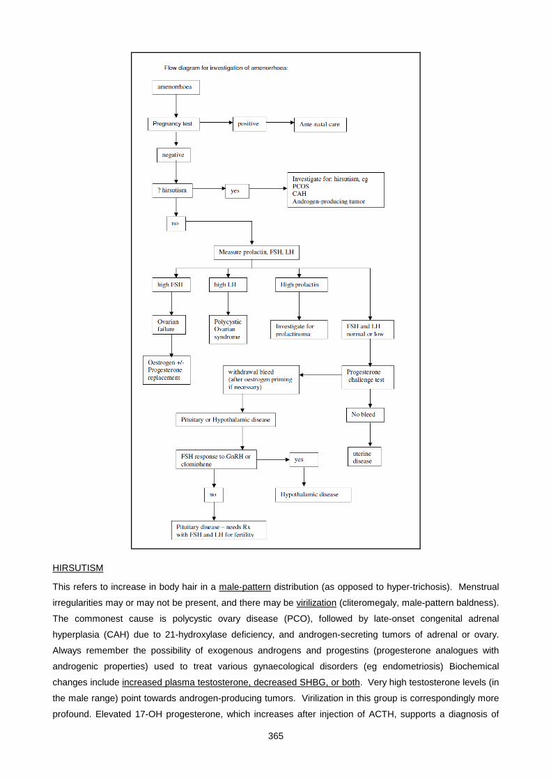
365
HIRSUTISM
This refers to increase in body hair in a male-pattern distribution (as opposed to hyper-trichosis). Menstrual
irregularities may or may not be present, and there may be virilization (cliteromegaly, male-pattern baldness).
The commonest cause is polycystic ovary disease (PCO), followed by late-onset congenital adrenal
hyperplasia (CAH) due to 21-hydroxylase deficiency, and androgen-secreting tumors of adrenal or ovary.
Always remember the possibility of exogenous androgens and progestins (progesterone analogues with
androgenic properties) used to treat various gynaecological disorders (eg endometriosis) Biochemical
changes include increased plasma testosterone, decreased SHBG, or both. Very high testosterone levels (in
the male range) point towards androgen-producing tumors. Virilization in this group is correspondingly more
profound. Elevated 17-OH progesterone, which increases after injection of ACTH, supports a diagnosis of
366
late onset CAH. Cushing’s syndrome is confirmed by elevated midnight plasma cortisol, elevated urinary free
(dU) cortisol, and failure of low-dose dexamethasone suppression. Treatment of hirsutism entails identifying
those conditions where specific treatment is available (surgery for tumors, cortisol replacement for CAH).
POLYCYSTIC OVARIAN SYNDROME (PCOS) is a disorder characterised by multiple ovarian cysts and
irregular, anovulatory menstrual cycles. Its causation remains obscure, but it has very characteristic clinical
features and biochemical abnormalities. It often begins in young women (in their 20s). A dominant follicle
fails to develop, hence ovulation does not occur and there is continuous oestrogenic stimulation of the
endometrium, without any progesterone. This causes breakthrough bleeding at irregular intervals, while
overproduction of ovarian androgens causes mild virilization (excessive body hair, acne). SHBG is typically
low, which enhances the effect of circulating testosterone. Plasma LH is high relative to FSH. There is
usually marked insulin resistance (↑↑ insulin with or without frank diabetes), central obesity, and acanthosis
nigricans (darkening and velvety thickening of the skin over the neck and armpits). Multiple ovarian cysts are
often present on abdominal ultrasound examination. In mild PCO, excessive hirsutism can be successfully
treated cosmetically, while in severe cases, anti-androgen drugs (eg cyproterone) are used. Do not use anti-
androgens if fertility is desired (can cause permanent undermasculinization of a male fetus). Clomiphene or
FSH stimulation may be useful in restoring fertility.
Confirmation of ovulation
In investigating amenorrhoea/infertility, it is important to establish whether or not the woman concerned is
ovulating. While regular periods suggest ovulation, a very useful test is measurement of plasma
progesterone at 21 of the cycle (mid-lueal phase). A high value (>30nmol/l) confirms ovulation, while a low
result (<10nmol/l) negates it.
PRECOCIOUS PUBERTY
This is defined as signs of puberty before age of 8. Either sex can be affected. It can either be central (true
precocious puberty), where gonadotropin levels are high (appropriate to that stage of puberty) or pseudo-
precocious puberty, where the appearance of secondary sexual characteristics is due to autonomous sex
hormone production. Typical causes of true precocious puberty include developmental or neoplastic
abnormalities in or around the hypothalamus, whereas pseudo precocious puberty is usually due to a gonadal
sex-steroid producing tumor. The latter shows suppressed plasma gonadotropins (and small gonads, apart
from the tumor itself).
BIOCHEMICAL ALTERATIONS IN PREGNANCY
• HORMONES: hCG production by the embryonic trophoblast cells (developing placenta) commences
with a few days of fertilization. The β-subunit can be detected in maternal blood 7-9 days post-
conception – well before the first missed period, and is a highly specific and sensitive test of
pregnancy. Plasma levels peak at 10-12 weeks, but remain elevated throughout pregnancy. hCG
assays are also useful in diagnosis and management of complications of pregnancy, including
incomplete abortion, ectopic pregnancy and trophoblastic tumors (hydatidiform mole,
choriocarcinoma). Oestriol is a unique steroid produced by the feto-placental unit, and excreted in
367
maternal urine. It has been used as a test of fetal wellbeing. Total plasma thyroxine and cortisol
increase in pregnancy (due to enhanced synthesis of their binding proteins, TBG and CBG,
respectively). Free levels of these hormones remain normal.
• RENAL FUNCTION: GFR increases by about 30% in pregnancy – this accounts for the lower plasma
urea, creatinine and urate (the anabolic milieu of pregnancy contributes to the low urea). An abrupt
increase in serum urate during pregnancy is a marker of pre-eclamptic toxaemia. The increased GFR
also lowers the renal threshold for glucose, predisposing to renal glycosuria.
• GLUCOSE METABOLISM: Preferential use of maternal glucose and aminoacids by the growing fetus
leads to a state of ‘accelerated starvation’ in pregnancy, with a lower fasting blood glucose and
greater tendency to develop ketosis compared to the non-pregnant state. In late pregnancy, the
placenta produces HPL (human placental lactogen), a member of the growth hormone/prolactin
family that antagonizes insulin. This accounts for the higher postprandial glucose levels that occur in
pregnancy, and the entity of ‘gestational diabetes’ i.e. appearance of diabetes during pregnancy that
resolves after delivery (though it may recur some years later).
• LIPIDS: Plasma triglycerides and/or cholesterol can increase in pregnancy, due to the effect of
oestrogen. HDL cholesterol is particularly increased. Amniotic fluid lecithin/sphingomyelin ratios are
sometimes used to assess fetal lung maturity.
• MISCELLANEOUS: Serum alkaline phosphatase is moderately elevated in pregnancy and is due to
placental production. The placental isoenzyme is characteristically heat stable, which enables it to be
easily distinguished from the bone and liver iso-forms. α-Feto protein is produced by the fetal liver,
and is the major circulating plasma protein during early fetal life. Production declines in late
pregnancy, and, by birth, α-FP has largely been replaced by albumin. Leakage of α-feto protein into
amniotic fluid, and from there into maternal blood, occurs with various fetal abnormalities, particularly
open neural tube defects.
368
SMALL GROUP TEACHING: LECTURE 23 ENDOCRINOLOGY: DISORDERS OF SEXUAL DISORIENTATION AND THE GONADS
QUESTIONS
1. A 20-year old man presented with impotence. No abnormality could be found on examination. The serum testosterone was 25 nmol/L (normal range 8.4-30). What is the likely cause of this problem?
2. A 45 year old woman presented with complaints of irritability, depression and hot flushes. Her periods had become irregular over the past 6 months. She had been on treatment for hypertension with a beta-blocker and an ACE inhibitor for 6 years.
i. What is the most likely explanation for her symptoms?
ii. What tests can be used to confirm this?
iii. What do the results indicate?
iv. What treatment options can be offered to control her symptoms?
v. Besides alleviating the symptoms, what additional benefits does HRT confer?
3. A 20-year old man presented with pubertal delay. On examination he was of normal height and had a eunuchoid body shape. Pubic and axillary hair was sparse, the genitalia were pre-pubertal, and both testes were present in the scrotum.
Plasma testosterone: 3.0 nmol/L (N 8.4-30), LH: 0.3 U/L (N 0.4 - 5.5), FSH: 1.2 U/L (N 0 - 16)
i. Define the meaning of the terms primary hypogonadism and secondary hypogonadism. What type of hypogonadism is present in this patient? (i.e. at what level is the lesion?)
ii. Suggest some possible causes of male primary (hypergonadotropic) hypogonadism.
iii. Suggest some possible causes of male secondary (hypogonadotropic) hypogonadism.
iv. Questioning of the patient revealed that he had no sense of smell, and had never had one.
v. Does the finding of anosmia have any special significance in this context?
4. A one-month old girl was investigated for ambiguous genitalia. The pregnancy and birth had been normal, and the baby had gained weight normally and had shown no signs of illness. Ambiguous genitalia were noted at birth. The clitoris was enlarged and there was partial fusion of the labia. An ultrasound scan showed the presence of a uterus. An XX karyotype was found in peripheral blood. Biochemical tests on plasma revealed the following (reference ranges in brackets):
Na+: 135mmol/L, K+: 4.3mmol//L
Cortisol (11 am): 313nmol/L ( 9 am : 140-700), (11 pm: 0- 138)
Testosterone: 6.6nmol/L (<1 prepubertal) (8.4-30 adult male) (<2.7 adult female)
17-OH-progesterone: 356nmol/L (<6)
Androstenedione: 19.4nmol/L (<12)
i. What is the diagnosis? (work it out from the steroid synthetic pathway).
ii. Explain the reason for the overproduction of androgens.
iii. Would you expect the level of plasma ACTH to be elevated, decreased or normal?
iv. Comment on the cortisol level.
v. Is it necessary to treat this condition? Why?
vi. Explain the principles of therapy in this disorder.
vii. What are the genetics of the condition? What advice would you give the parents regarding future children?
369
After 4 weeks of therapy (hydrocortisone) the following plasma results were obtained:
17-OH-progesterone: 14 nmol/L, Testosterone: 1.2 nmol/L
5. A 3 week old boy was admitted for investigation of his failure to gain weight. There was no history of diarrhoea or vomiting, and examination revealed nothing of diagnostic significance. On the third day of admission the baby became clinically dehydrated.
Biochemical tests on plasma revealed the following:
Na+ 106 mmol/L, K+ 6.2 mmol/L, Cl- 80 mmol/L, HCO3 14 mmol/L, Urea 6.5 mmol/L, Creatinine 50 µmol/L, Glucose 4.5 mmol/L
i. Calculate the plasma osmolality and anion gap.
ii. What type of acid-base disturbance is present?
iii. Suggest one or more possible diagnoses.
Further blood and urine samples were taken for endocrine investigations. Treatment comprising IV saline, glucocorticoid (cortisone acetate) and mineralocorticoid (fluorocortisone) was commenced. Plasma electrolytes, osmolality and hydration state normalized over the next 9 days. Results of endocrine investigations were as follows:
Plasma 17-OH-progesterone 2240 nmol/L (N <6)
i. What is the final diagnosis?
ii. Would you expect the plasma renin level to be low, normal or high?
iii. Which adrenal enzyme deficiency presents with hypertension? Explain the mechanism.
6. A 19 year old woman presented with primary amenorrhoea. She was normal in appearance, except for the almost complete absence of pubic and axillary hair. Gynaecological examination revealed a short, blind-ended vagina, and no cervix was palpable. Radiology showed absence of the uterus. Blood karyotyping showed XY chromosomes. Plasma testosterone 29 nmol/L (adult female 1-2.8; adult male 8.4-30) The diagnosis is that of the testicular feminization syndrome (also called androgen insensitivity syndrome). This is due to a defect in the androgen receptor, leading to androgen resistance, complete in this case. Other mutations cause incomplete androgen resistance, giving rise to a spectrum of genital abnormalities ranging from hypospadias to completely female external phenotype. All are X-linked, since the androgen receptor gene is located on the X chromosome. The testes are inguinal or abdominal. The essential biochemical feature is a high level of testosterone in the presence of incomplete masculinization. Adrenal androgen precursors are not elevated as seen in CAH.
i. Why is there an absence of internal genital organs in this disorder?
ii. What is the inheritance pattern and typical family history of testicular feminisation syndrome (male pseudo hermaphroditism)?
iii. List the 3 zones of the adrenal cortex and the hormones produced there.
iv. What is meant by the terms (a) adrenarche (b) thelarche (c) menarche?
v. Which structures are derived from (a) the Wolffian ducts (b) the Mullerian ducts? How is the fate of these primitive structures determined?
vi. What is meant by “hypospadias” and “cryptorchidism”?
370
LECTURE 23: ENDOCRINOLOGY -DISORDERS OF SEXUAL DIFFERNTIATION AND THE GONADS
ANSWERS
1. (psychogenic sexual dysfunction): Sexual dysfunction in young, normal males is common, and has a psychological, not an endocrine basis. Vascular (venous or arterial), or neurogenic (eg diabetic neuropathy) should be excluded.
2. (menopause)
i. Menopause
ii. Plasma FSH
iii. She is menopausal
iv. Estrogen is the only therapy which will control menopausal symptoms. As she has an intact uterus, estrogen must be given combined with a progestogen (unopposed estrogen therapy carries an increased risk of endometrial carcinoma). This can be given either in cyclical form, which will produce withdrawal bleeds, or continuously.
v. Protection against osteoporosis, prevention of atrophy of vaginal mucosa, improved libido.
3. (Kallmann's syndrome)
i. primary (hypergonadotropic) hypogonadism : deficiency at the gonadal level (gonadotropins are Secondary (hypogonadotropic) hypogonadism: lesion at pituitary or hypothalamic level (gonadotropins low or inappropriately "normal"). This patient has secondary hypogonadism.
ii. Causes of male primary hypogonadism: trauma or infections of testes (mumps); bilateral torsion; irradiation; Klinefelters syndrome; castration
iii. Causes of male secondary hypogonadism: hypopituitarism; Kallmann's syndrome
iv. Anosmia is a feature of Kallmann's syndrome, a developmental abnormality where there is failure of the GnRH neurons to migrate from the olfactory tracts to the hypothalamus during embryogenesis
4. (21-hydroxylase deficiency, simple virilizing form)
i. Diagnosis:21-hydroxylase deficiency, simple virilizing form.
ii. Block in cortisol production causes increased ACTH production that in turn stimulates synthesis of cortisol precursors which are shunted into the androgen pathway.
iii. ACTH would be elevated
iv. Perhaps surprisingly, the cortisol level is usually within the normal range. Evidently, the ACTH drive is able to sustain cortisol production, at the expense of a high level of precursors.
v. Untreated, the virilization will get worse, and the androgen will cause premature fusion of the epiphyses, resulting in short stature.
vi. Glucocorticoid therapy (hydrocortisone) switches off the excessive ACTH drive, decreasing androgen production. Overtreatment with glucocorticoid also causes stunting of growth, however, so the glucocorticoid dose must be carefully adjusted, using the 17-OHP and testosterone levels as a guide.
vii. autosomal recessive, therefore a 25% risk in each future child. The gene is linked to the HLA locus, so HLA typing can be used for prenatal diagnosis.
5. (21-hydroxylase deficiency, salt – losing form)
i. Osmoloality = 2 x Na + glucose + urea = 212 + 11 = 223; anion gap = Na – Cl – HCO3 = 12
ii. metabolic acidosis
iii. The biochemical findings strongly suggest mineralocorticoid deficiency (or resistance). Salt-losing 21-hydroxylase deficiency is the most likely, but other adrenal enzyme blocks are possible

371
(eg 18-hydroxylase deficiency). Aldosterone resistance (psuedohypoaldosteronism) is a rare cause.
iv. Salt-losing form of 21-hydroxylase deficiency . A more severe degree of enzyme block than the simple virilizing form.
v. The renin would be very high.
vi. 11-hydroxylase deficiency. 11-deoxycorticosterone, a substrate for the defective enzyme has mineralocorticoid activity, hence produces the effects of excess mineralocorticoid (as in Conn's syndrome).
6. (testicular feminization syndrome)
i. The female internal genital organs (uterus, upper third of vagina & Fallopian tubes) are derived from the Mullerian ducts. Their development is suppressed in the male embryo by MIF (mullerian inhibiting factor), secreted by the testis. There is no defect in MIF secretion or action in this disorder. Male internal genitalia development requires testosterone (and its receptor, of course)
ii. X-linked recessive. Infertile maiden aunts (not uncles)
iii. Zona glomerulosa : aldosterone. Zona fascilculata and reticularis: cortisol & androgens
iv. adrenarche: the increase in adrenal steroid production that occurs at puberty thelarche: the onset of breast development menarche: onset of menstruation
v. Wolffian ducts → male internal genital structures (vas deferens & seminal vesicles) Mullerian ducts → female internal structures (uterus, upper 1/3 vagina, fallopian tubes)
vi. Hypospadias: urethral opening not at the end of the penis Cryptorchidism: undescended testis
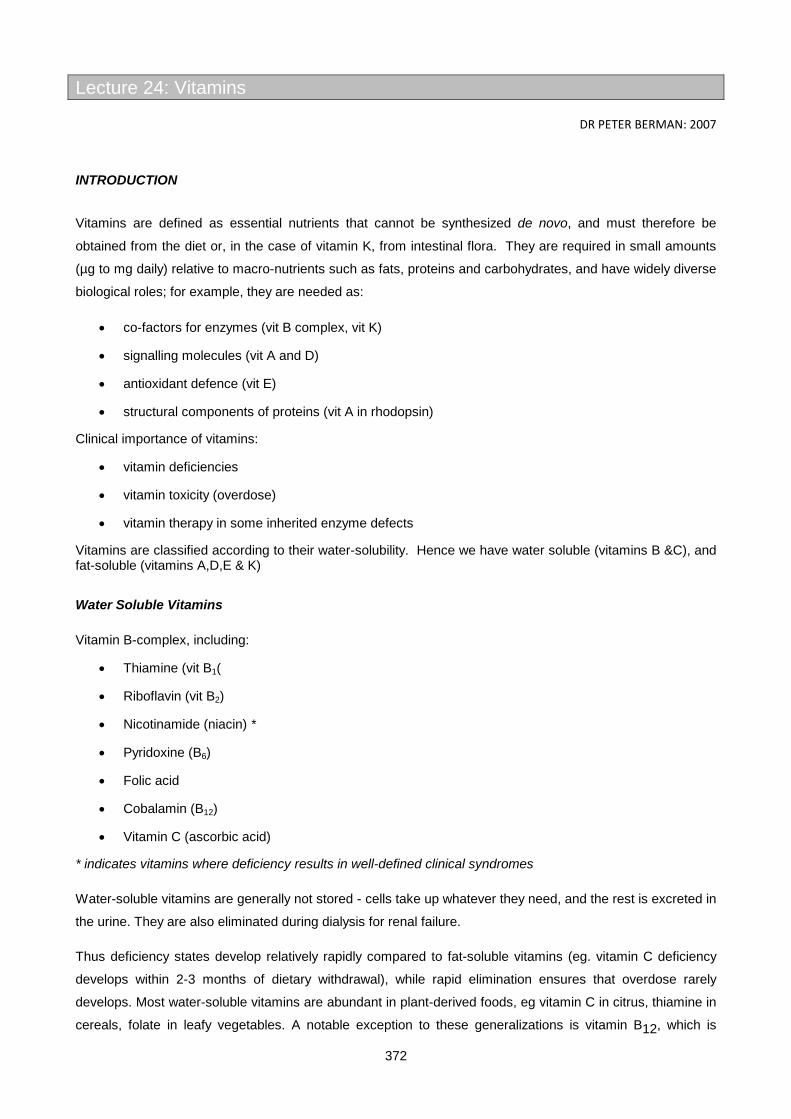
372
Lecture 24: Vitamins
DR PETER BERMAN: 2007
INTRODUCTION
Vitamins are defined as essential nutrients that cannot be synthesized de novo, and must therefore be
obtained from the diet or, in the case of vitamin K, from intestinal flora. They are required in small amounts
(µg to mg daily) relative to macro-nutrients such as fats, proteins and carbohydrates, and have widely diverse
biological roles; for example, they are needed as:
• co-factors for enzymes (vit B complex, vit K)
• signalling molecules (vit A and D)
• antioxidant defence (vit E)
• structural components of proteins (vit A in rhodopsin)
Clinical importance of vitamins:
• vitamin deficiencies
• vitamin toxicity (overdose)
• vitamin therapy in some inherited enzyme defects
Vitamins are classified according to their water-solubility. Hence we have water soluble (vitamins B &C), and fat-soluble (vitamins A,D,E & K)
Water Soluble Vitamins
Vitamin B-complex, including:
• Thiamine (vit B1(
• Riboflavin (vit B2)
• Nicotinamide (niacin) *
• Pyridoxine (B6)
• Folic acid
• Cobalamin (B12)
• Vitamin C (ascorbic acid)
* indicates vitamins where deficiency results in well-defined clinical syndromes
Water-soluble vitamins are generally not stored - cells take up whatever they need, and the rest is excreted in
the urine. They are also eliminated during dialysis for renal failure.
Thus deficiency states develop relatively rapidly compared to fat-soluble vitamins (eg. vitamin C deficiency
develops within 2-3 months of dietary withdrawal), while rapid elimination ensures that overdose rarely
develops. Most water-soluble vitamins are abundant in plant-derived foods, eg vitamin C in citrus, thiamine in
cereals, folate in leafy vegetables. A notable exception to these generalizations is vitamin B12, which is
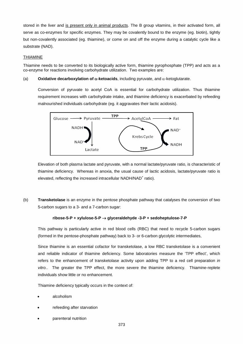
373
stored in the liver and is present only in animal products. The B group vitamins, in their activated form, all
serve as co-enzymes for specific enzymes. They may be covalently bound to the enzyme (eg. biotin), tightly
but non-covalently associated (eg. thiamine), or come on and off the enzyme during a catalytic cycle like a
substrate (NAD).
THIAMINE
Thiamine needs to be converted to its biologically active form, thiamine pyrophosphate (TPP) and acts as a co-enzyme for reactions involving carbohydrate utilization. Two examples are:
(a) Oxidative decarboxylation of α-ketoacids, including pyruvate, and α-ketoglutarate.
Conversion of pyruvate to acetyl CoA is essential for carbohydrate utilization. Thus thiamine
requirement increases with carbohydrate intake, and thiamine deficiency is exacerbated by refeeding
malnourished individuals carbohydrate (eg. it aggravates their lactic acidosis).
Elevation of both plasma lactate and pyruvate, with a normal lactate/pyruvate ratio, is characteristic of
thiamine deficiency. Whereas in anoxia, the usual cause of lactic acidosis, lactate/pyruvate ratio is
elevated, reflecting the increased intracellular NADH/NAD+ ratio).
(b) Transketolase is an enzyme in the pentose phosphate pathway that catalyses the conversion of two
5-carbon sugars to a 3- and a 7-carbon sugar:
ribose-5-P + xylulose-5-P → glyceraldehyde -3-P + sedoheptulose-7-P
This pathway is particularly active in red blood cells (RBC) that need to recycle 5-carbon sugars
(formed in the pentose-phosphate pathway) back to 3- or 6-carbon glycolytic intermediates,
Since thiamine is an essential cofactor for transketolase, a low RBC transketolase is a convenient
and reliable indicator of thiamine deficiency. Some laboratories measure the ‘TPP effect’, which
refers to the enhancement of transketolase activity upon adding TPP to a red cell preparation in
vitro.. The greater the TPP effect, the more severe the thiamine deficiency. Thiamine-replete
individuals show little or no enhancement.
Thiamine deficiency typically occurs in the context of:
• alcoholism
• refeeding after starvation
• parenteral nutrition
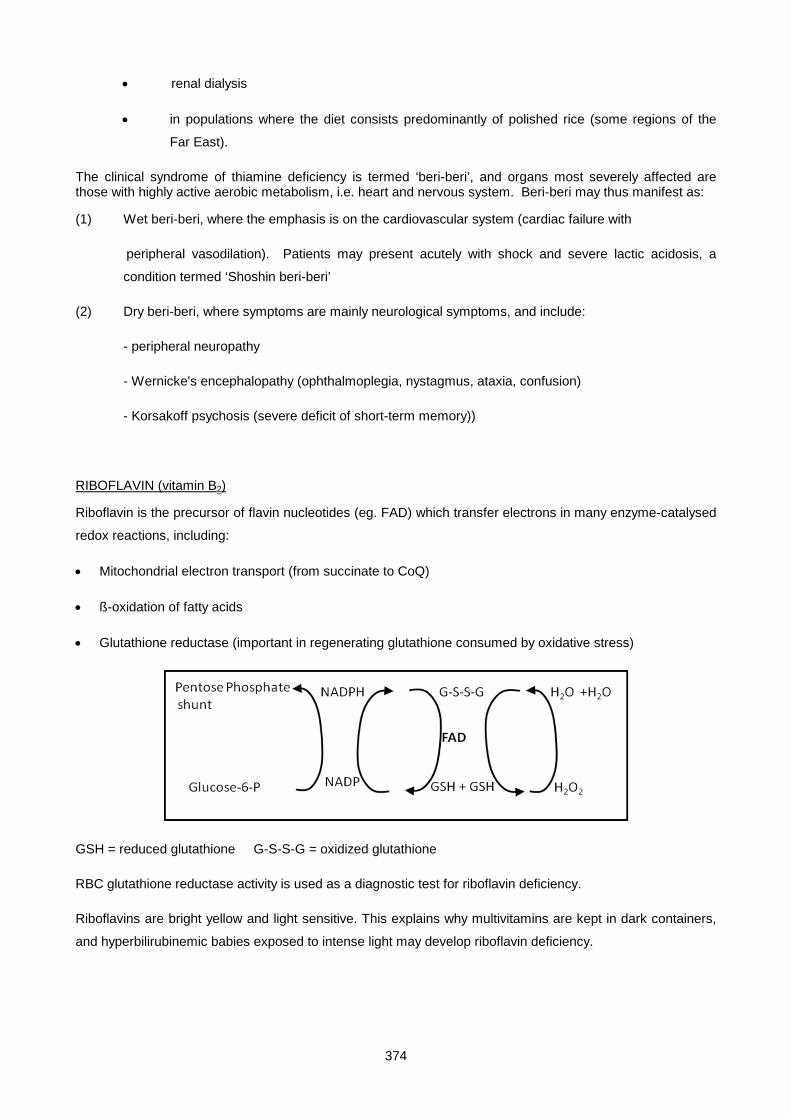
374
• renal dialysis
• in populations where the diet consists predominantly of polished rice (some regions of the
Far East).
The clinical syndrome of thiamine deficiency is termed ‘beri-beri’, and organs most severely affected are those with highly active aerobic metabolism, i.e. heart and nervous system. Beri-beri may thus manifest as:
(1) Wet beri-beri, where the emphasis is on the cardiovascular system (cardiac failure with
peripheral vasodilation). Patients may present acutely with shock and severe lactic acidosis, a
condition termed ‘Shoshin beri-beri’
(2) Dry beri-beri, where symptoms are mainly neurological symptoms, and include:
- peripheral neuropathy
- Wernicke's encephalopathy (ophthalmoplegia, nystagmus, ataxia, confusion)
- Korsakoff psychosis (severe deficit of short-term memory))
RIBOFLAVIN (vitamin B2)
Riboflavin is the precursor of flavin nucleotides (eg. FAD) which transfer electrons in many enzyme-catalysed
redox reactions, including:
• Mitochondrial electron transport (from succinate to CoQ)
• ß-oxidation of fatty acids
• Glutathione reductase (important in regenerating glutathione consumed by oxidative stress)
GSH = reduced glutathione G-S-S-G = oxidized glutathione
RBC glutathione reductase activity is used as a diagnostic test for riboflavin deficiency.
Riboflavins are bright yellow and light sensitive. This explains why multivitamins are kept in dark containers,
and hyperbilirubinemic babies exposed to intense light may develop riboflavin deficiency.
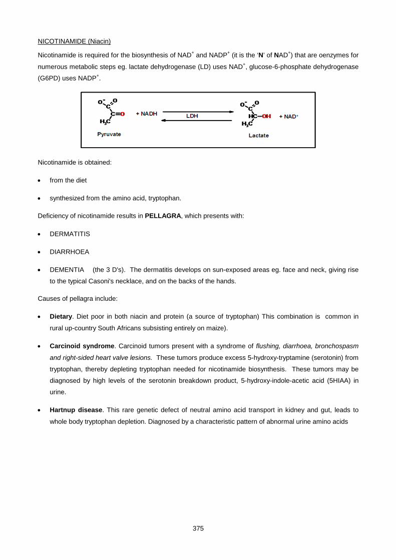
375
NICOTINAMIDE (Niacin)
Nicotinamide is required for the biosynthesis of NAD+ and NADP+ (it is the ‘N’ of NAD+) that are oenzymes for
numerous metabolic steps eg. lactate dehydrogenase (LD) uses NAD+, glucose-6-phosphate dehydrogenase
(G6PD) uses NADP+.
Nicotinamide is obtained:
• from the diet
• synthesized from the amino acid, tryptophan.
Deficiency of nicotinamide results in PELLAGRA, which presents with:
• DERMATITIS
• DIARRHOEA
• DEMENTIA (the 3 D's). The dermatitis develops on sun-exposed areas eg. face and neck, giving rise
to the typical Casoni's necklace, and on the backs of the hands.
Causes of pellagra include:
• Dietary. Diet poor in both niacin and protein (a source of tryptophan) This combination is common in
rural up-country South Africans subsisting entirely on maize).
• Carcinoid syndrome. Carcinoid tumors present with a syndrome of flushing, diarrhoea, bronchospasm
and right-sided heart valve lesions. These tumors produce excess 5-hydroxy-tryptamine (serotonin) from
tryptophan, thereby depleting tryptophan needed for nicotinamide biosynthesis. These tumors may be
diagnosed by high levels of the serotonin breakdown product, 5-hydroxy-indole-acetic acid (5HIAA) in
urine.
• Hartnup disease. This rare genetic defect of neutral amino acid transport in kidney and gut, leads to
whole body tryptophan depletion. Diagnosed by a characteristic pattern of abnormal urine amino acids
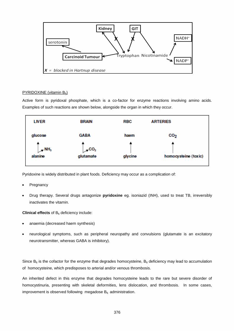
376
PYRIDOXINE (vitamin B6)
Active form is pyridoxal phosphate, which is a co-factor for enzyme reactions involving amino acids.
Examples of such reactions are shown below, alongside the organ in which they occur.
Pyridoxine is widely distributed in plant foods. Deficiency may occur as a complication of:
• Pregnancy
• Drug therapy. Several drugs antagonize pyridoxine eg. isoniazid (INH), used to treat TB, irreversibly
inactivates the vitamin.
Clinical effects of B6 deficiency include:
• anaemia (decreased haem synthesis)
• neurological symptoms, such as peripheral neuropathy and convulsions (glutamate is an excitatory
neurotransmitter, whereas GABA is inhibitory).
Since B6 is the cofactor for the enzyme that degrades homocysteine, B6 deficiency may lead to accumulation
of homocysteine, which predisposes to arterial and/or venous thrombosis.
An inherited defect in this enzyme that degrades homocysteine leads to the rare but severe disorder of
homocystinuria, presenting with skeletal deformities, lens dislocation, and thrombosis. In some cases,
improvement is observed following megadose B6 administration.

377
BIOTIN
Biotin is a cofactor for carboxylase enzymes that carboxylate (add CO2 to) various organic acids, including pyruvic acid, acetic acid and propionic acid. Inherited deficiencies of these enzymes present in childhood with severe metabolic acidosis (eg. lactic or propionic acidosis), that may benefit from megadose biotin therapy.
The main source of biotin is the intestinal flora. Egg white contains a protein called avidin which binds biotin avidly - ingestion of large quantities of raw egg can result in biotin deficiency.
FOLATE
Folate (folic acid) is required for DNA synthesis. Hence rapidly replicating cells are most affected by its
deficiency. Such cells include:
• blood cell precursors in the bone marrow
• mucosal cells of the intestine
• the fetus during early development
Thus, features of folate deficiency include:
• Megaloblastic anemia, characterized by abnormally large circulating red cells (macrocytes) with a mean
corpuscular volume (MCV) > 100 fl (normal 80-90 fl) and large nucleated red cell precursors
(megaloblasts) in the bone marrow. Hypersegmented polymorphs are a feature.
• Intestinal malabsorption
• Fetal abnormalities.
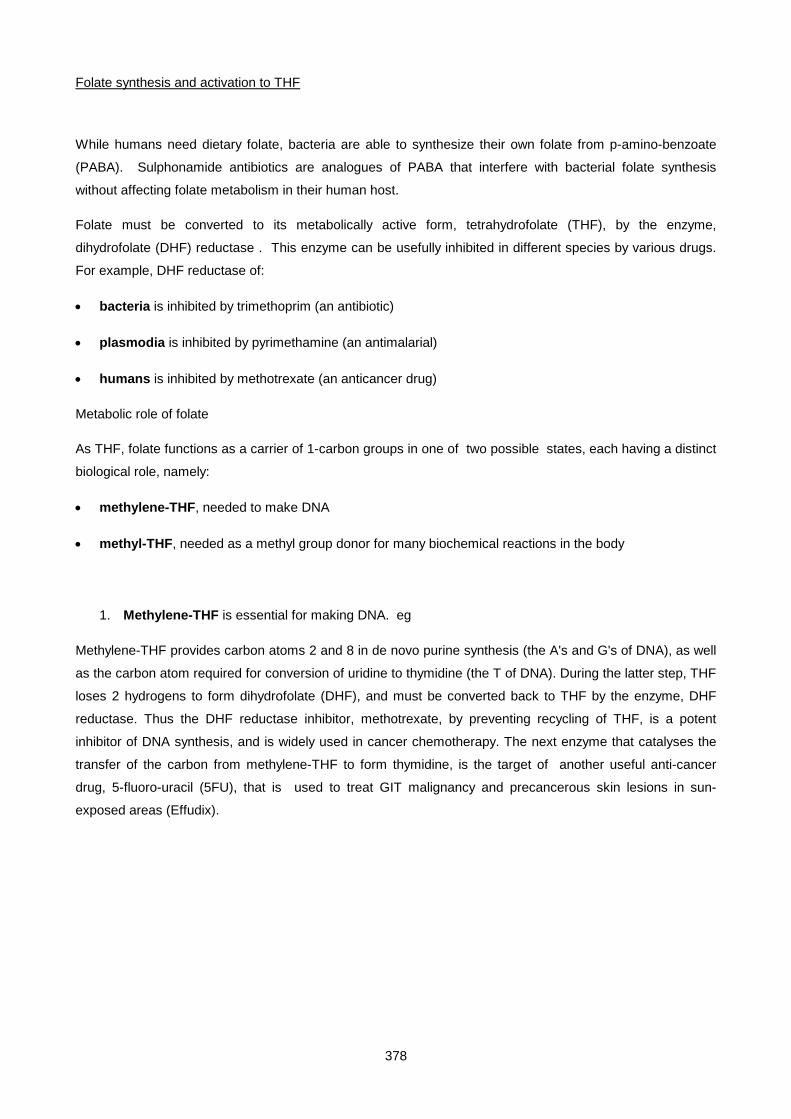
378
Folate synthesis and activation to THF
While humans need dietary folate, bacteria are able to synthesize their own folate from p-amino-benzoate
(PABA). Sulphonamide antibiotics are analogues of PABA that interfere with bacterial folate synthesis
without affecting folate metabolism in their human host.
Folate must be converted to its metabolically active form, tetrahydrofolate (THF), by the enzyme,
dihydrofolate (DHF) reductase . This enzyme can be usefully inhibited in different species by various drugs.
For example, DHF reductase of:
• bacteria is inhibited by trimethoprim (an antibiotic)
• plasmodia is inhibited by pyrimethamine (an antimalarial)
• humans is inhibited by methotrexate (an anticancer drug)
Metabolic role of folate
As THF, folate functions as a carrier of 1-carbon groups in one of two possible states, each having a distinct
biological role, namely:
• methylene-THF, needed to make DNA
• methyl-THF, needed as a methyl group donor for many biochemical reactions in the body
1. Methylene-THF is essential for making DNA. eg
Methylene-THF provides carbon atoms 2 and 8 in de novo purine synthesis (the A's and G's of DNA), as well
as the carbon atom required for conversion of uridine to thymidine (the T of DNA). During the latter step, THF
loses 2 hydrogens to form dihydrofolate (DHF), and must be converted back to THF by the enzyme, DHF
reductase. Thus the DHF reductase inhibitor, methotrexate, by preventing recycling of THF, is a potent
inhibitor of DNA synthesis, and is widely used in cancer chemotherapy. The next enzyme that catalyses the
transfer of the carbon from methylene-THF to form thymidine, is the target of another useful anti-cancer
drug, 5-fluoro-uracil (5FU), that is used to treat GIT malignancy and precancerous skin lesions in sun-
exposed areas (Effudix).
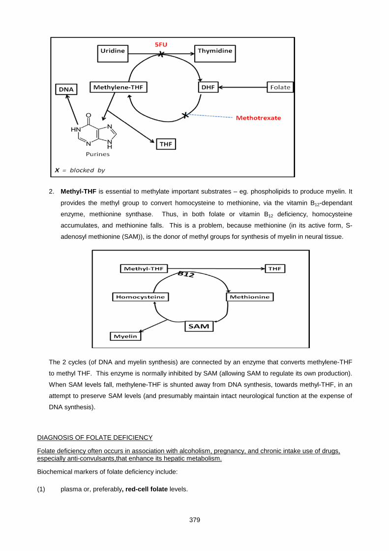
379
2. Methyl-THF is essential to methylate important substrates – eg. phospholipids to produce myelin. It
provides the methyl group to convert homocysteine to methionine, via the vitamin B12-dependant
enzyme, methionine synthase. Thus, in both folate or vitamin B12 deficiency, homocysteine
accumulates, and methionine falls. This is a problem, because methionine (in its active form, S-
adenosyl methionine (SAM)), is the donor of methyl groups for synthesis of myelin in neural tissue.
The 2 cycles (of DNA and myelin synthesis) are connected by an enzyme that converts methylene-THF
to methyl THF. This enzyme is normally inhibited by SAM (allowing SAM to regulate its own production).
When SAM levels fall, methylene-THF is shunted away from DNA synthesis, towards methyl-THF, in an
attempt to preserve SAM levels (and presumably maintain intact neurological function at the expense of
DNA synthesis).
DIAGNOSIS OF FOLATE DEFICIENCY
Folate deficiency often occurs in association with alcoholism, pregnancy, and chronic intake use of drugs, especially anti-convulsants,that enhance its hepatic metabolism.
Biochemical markers of folate deficiency include:
(1) plasma or, preferably, red-cell folate levels.
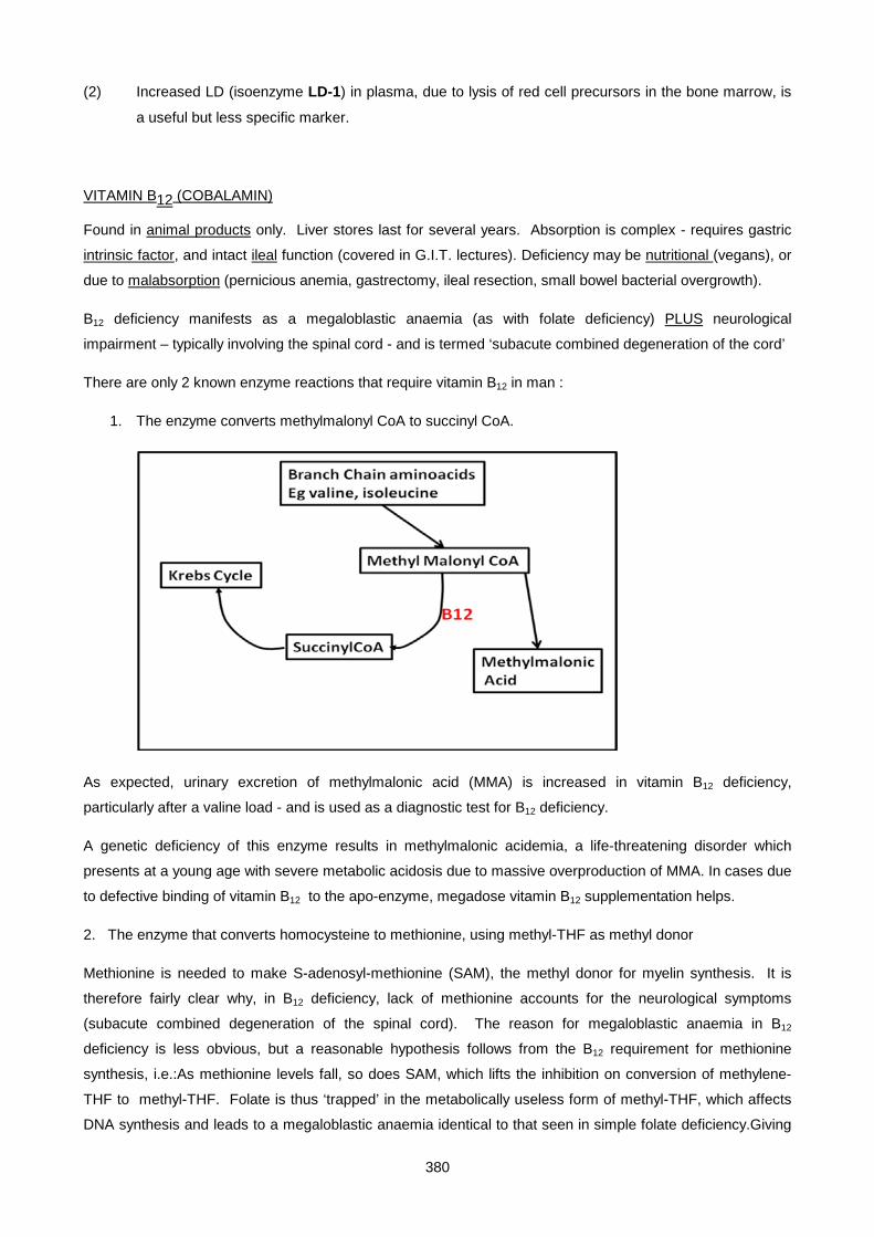
380
(2) Increased LD (isoenzyme LD-1) in plasma, due to lysis of red cell precursors in the bone marrow, is
a useful but less specific marker.
VITAMIN B12 (COBALAMIN)
Found in animal products only. Liver stores last for several years. Absorption is complex - requires gastric
intrinsic factor, and intact ileal function (covered in G.I.T. lectures). Deficiency may be nutritional (vegans), or
due to malabsorption (pernicious anemia, gastrectomy, ileal resection, small bowel bacterial overgrowth).
B12 deficiency manifests as a megaloblastic anaemia (as with folate deficiency) PLUS neurological
impairment – typically involving the spinal cord - and is termed ‘subacute combined degeneration of the cord’
There are only 2 known enzyme reactions that require vitamin B12 in man :
1. The enzyme converts methylmalonyl CoA to succinyl CoA.
As expected, urinary excretion of methylmalonic acid (MMA) is increased in vitamin B12 deficiency,
particularly after a valine load - and is used as a diagnostic test for B12 deficiency.
A genetic deficiency of this enzyme results in methylmalonic acidemia, a life-threatening disorder which
presents at a young age with severe metabolic acidosis due to massive overproduction of MMA. In cases due
to defective binding of vitamin B12 to the apo-enzyme, megadose vitamin B12 supplementation helps.
2. The enzyme that converts homocysteine to methionine, using methyl-THF as methyl donor
Methionine is needed to make S-adenosyl-methionine (SAM), the methyl donor for myelin synthesis. It is
therefore fairly clear why, in B12 deficiency, lack of methionine accounts for the neurological symptoms
(subacute combined degeneration of the spinal cord). The reason for megaloblastic anaemia in B12
deficiency is less obvious, but a reasonable hypothesis follows from the B12 requirement for methionine
synthesis, i.e.:As methionine levels fall, so does SAM, which lifts the inhibition on conversion of methylene-
THF to methyl-THF. Folate is thus ‘trapped’ in the metabolically useless form of methyl-THF, which affects
DNA synthesis and leads to a megaloblastic anaemia identical to that seen in simple folate deficiency.Giving
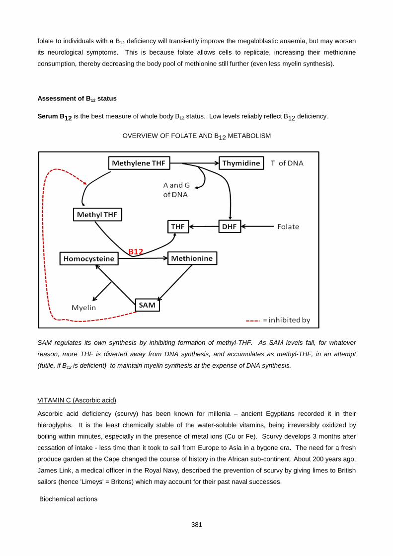
381
folate to individuals with a B12 deficiency will transiently improve the megaloblastic anaemia, but may worsen
its neurological symptoms. This is because folate allows cells to replicate, increasing their methionine
consumption, thereby decreasing the body pool of methionine still further (even less myelin synthesis).
Assessment of B12 status
Serum B12 is the best measure of whole body B12 status. Low levels reliably reflect B12 deficiency.
OVERVIEW OF FOLATE AND B12 METABOLISM
SAM regulates its own synthesis by inhibiting formation of methyl-THF. As SAM levels fall, for whatever
reason, more THF is diverted away from DNA synthesis, and accumulates as methyl-THF, in an attempt
(futile, if B12 is deficient) to maintain myelin synthesis at the expense of DNA synthesis.
VITAMIN C (Ascorbic acid)
Ascorbic acid deficiency (scurvy) has been known for millenia – ancient Egyptians recorded it in their
hieroglyphs. It is the least chemically stable of the water-soluble vitamins, being irreversibly oxidized by
boiling within minutes, especially in the presence of metal ions (Cu or Fe). Scurvy develops 3 months after
cessation of intake - less time than it took to sail from Europe to Asia in a bygone era. The need for a fresh
produce garden at the Cape changed the course of history in the African sub-continent. About 200 years ago,
James Link, a medical officer in the Royal Navy, described the prevention of scurvy by giving limes to British
sailors (hence 'Limeys' = Britons) which may account for their past naval successes.
Biochemical actions
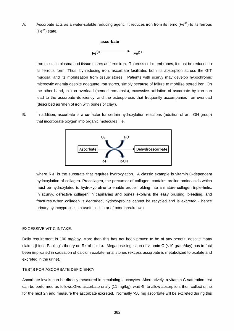
382
A. Ascorbate acts as a water-soluble reducing agent. It reduces iron from its ferric (Fe3+) to its ferrous
(Fe2+) state.
ascorbate
Fe3+ Fe2+
Iron exists in plasma and tissue stores as ferric iron. To cross cell membranes, it must be reduced to
its ferrous form. Thus, by reducing iron, ascorbate facilitates both its absorption across the GIT
mucosa, and its mobilisation from tissue stores. Patients with scurvy may develop hypochromic
microcytic anemia despite adequate iron stores, simply because of failure to mobilize stored iron. On
the other hand, in iron overload (hemochromatosis), excessive oxidation of ascorbate by iron can
lead to the ascorbate deficiency, and the osteoporosis that frequently accompanies iron overload
(described as ‘men of iron with bones of clay’).
B. In addition, ascorbate is a co-factor for certain hydroxylation reactions (addition of an –OH group)
that incorporate oxygen into organic molecules, i.e.
where R-H is the substrate that requires hydroxylation. A classic example is vitamin C-dependent
hydroxylation of collagen. Procollagen, the precursor of collagen, contains proline aminoacids which
must be hydroxylated to hydroxyproline to enable proper folding into a mature collagen triple-helix.
In scurvy, defective collagen in capillaries and bones explains the easy bruising, bleeding, and
fractures.When collagen is degraded, hydroxyproline cannot be recycled and is excreted - hence
urinary hydroxyproline is a useful indicator of bone breakdown.
EXCESSIVE VIT C INTAKE.
Daily requirement is 100 mg/day. More than this has not been proven to be of any benefit, despite many
claims (Linus Pauling’s theory on Rx of colds). Megadose ingestion of vitamin C (<10 gram/day) has in fact
been implicated in causation of calcium oxalate renal stones (excess ascorbate is metabolized to oxalate and
excreted in the urine).
TESTS FOR ASCORBATE DEFICIENCY
Ascorbate levels can be directly measured in circulating leucocytes. Alternatively, a vitamin C saturation test
can be performed as follows:Give ascorbate orally (11 mg/kg), wait 4h to allow absorption, then collect urine
for the next 2h and measure the ascorbate excreted. Normally >50 mg ascorbate will be excreted during this
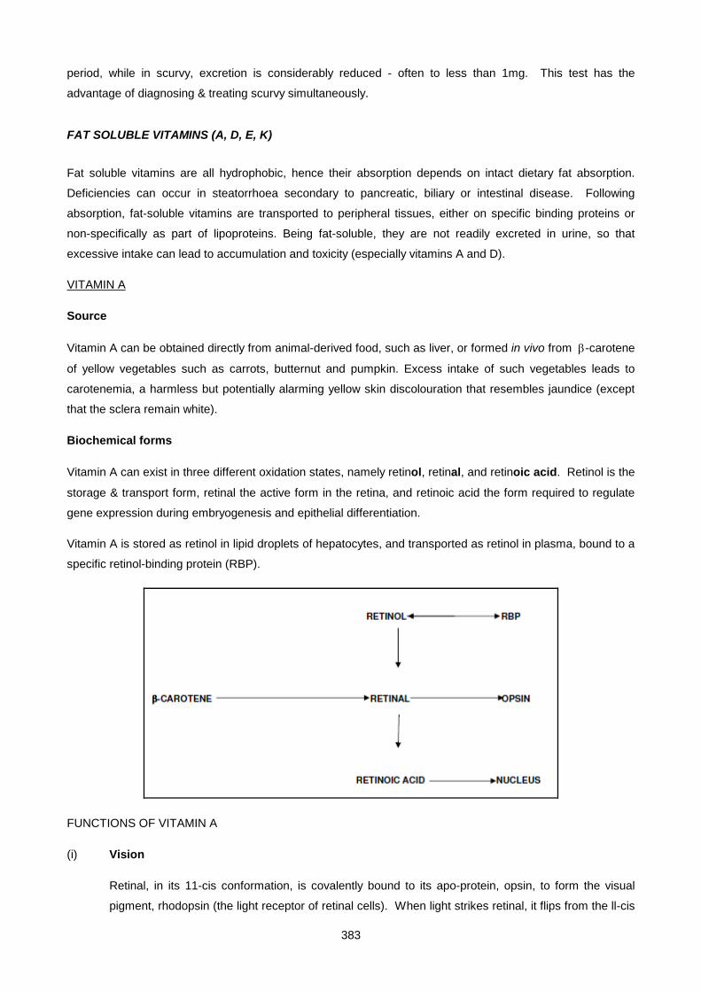
383
period, while in scurvy, excretion is considerably reduced - often to less than 1mg. This test has the
advantage of diagnosing & treating scurvy simultaneously.
FAT SOLUBLE VITAMINS (A, D, E, K)
Fat soluble vitamins are all hydrophobic, hence their absorption depends on intact dietary fat absorption.
Deficiencies can occur in steatorrhoea secondary to pancreatic, biliary or intestinal disease. Following
absorption, fat-soluble vitamins are transported to peripheral tissues, either on specific binding proteins or
non-specifically as part of lipoproteins. Being fat-soluble, they are not readily excreted in urine, so that
excessive intake can lead to accumulation and toxicity (especially vitamins A and D).
VITAMIN A
Source
Vitamin A can be obtained directly from animal-derived food, such as liver, or formed in vivo from β-carotene
of yellow vegetables such as carrots, butternut and pumpkin. Excess intake of such vegetables leads to
carotenemia, a harmless but potentially alarming yellow skin discolouration that resembles jaundice (except
that the sclera remain white).
Biochemical forms
Vitamin A can exist in three different oxidation states, namely retinol, retinal, and retinoic acid. Retinol is the
storage & transport form, retinal the active form in the retina, and retinoic acid the form required to regulate
gene expression during embryogenesis and epithelial differentiation.
Vitamin A is stored as retinol in lipid droplets of hepatocytes, and transported as retinol in plasma, bound to a
specific retinol-binding protein (RBP).
FUNCTIONS OF VITAMIN A
(i) Vision
Retinal, in its 11-cis conformation, is covalently bound to its apo-protein, opsin, to form the visual
pigment, rhodopsin (the light receptor of retinal cells). When light strikes retinal, it flips from the ll-cis
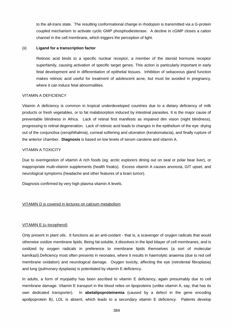
384
to the all-trans state. The resulting conformational change in rhodopsin is transmitted via a G-protein
coupled mechanism to activate cyclic GMP phosphodiesterase. A decline in cGMP closes a cation
channel in the cell membrane, which triggers the perception of light.
(ii) Ligand for a transcription factor
Retinoic acid binds to a specific nuclear receptor, a member of the steroid hormone receptor
superfamily, causing activation of specific target genes. This action is particularly important in early
fetal development and in differentiation of epithelial tissues. Inhibition of sebaceous gland function
makes retinoic acid useful for treatment of adolescent acne, but must be avoided in pregnancy,
where it can induce fetal abnormalities.
VITAMIN A DEFICIENCY
Vitamin A deficiency is common in tropical underdeveloped countries due to a dietary deficiency of milk
products or fresh vegetables, or to fat malabsorption induced by intestinal parasites. It is the major cause of
preventable blindness in Africa. Lack of retinal first manifests as impaired dim vision (night blindness),
progressing to retinal degeneration. Lack of retinoic acid leads to changes in the epithelium of the eye: drying
out of the conjunctiva (xerophthalmia), corneal softening and ulceration (keratomalacia), and finally rupture of
the anterior chamber. Diagnosis is based on low levels of serum carotene and vitamin A.
VITAMIN A TOXICITY
Due to overingestion of vitamin A rich foods (eg. arctic explorers dining out on seal or polar bear liver), or
inappropriate multi-vitamin supplements (health freaks). Excess vitamin A causes anorexia, GIT upset, and
neurological symptoms (headache and other features of a brain tumor).
Diagnosis confirmed by very high plasma vitamin A levels.
VITAMIN D is covered in lectures on calcium metabolism
VITAMIN E (α-tocopherol)
Only present in plant oils. It functions as an anti-oxidant - that is, a scavenger of oxygen radicals that would
otherwise oxidize membrane lipids. Being fat-soluble, it dissolves in the lipid bilayer of cell membranes, and is
oxidized by oxygen radicals in preference to membrane lipids themselves (a sort of molecular
kamikazi).Deficiency most often presents in neonates, where it results in haemolytic anaemia (due to red cell
membrane oxidation) and neurological damage. Oxygen toxicity, affecting the eye (retrolental fibroplasia)
and lung (pulmonary dysplasia) is potentiated by vitamin E deficiency.
In adults, a form of myopathy has been ascribed to vitamin E deficiency, again presumably due to cell
membrane damage. Vitamin E transport in the blood relies on lipoproteins (unlike vitamin A, say, that has its
own dedicated transporter). In abetalipoproteinemia (caused by a defect in the gene encoding
apolipoprotein B), LDL is absent, which leads to a secondary vitamin E deficiency. Patients develop
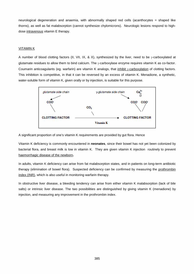
385
neurological degeneration and anaemia, with abnormally shaped red cells (acanthocytes = shaped like
thorns), as well as fat malabsorption (cannot synthesize chylomicrons). Neurologic lesions respond to high-
dose intravenous vitamin E therapy.
VITAMIN K
A number of blood clotting factors (II, VII, IX, & X), synthesized by the liver, need to be γ-carboxylated at
glutamate residues to allow them to bind calcium. The γ-carboxylase enzyme requires vitamin K as co-factor.
Coumarin anticoagulants (eg. warfarin) are vitamin K analogs, that inhibit γ-carboxylation of clotting factors.
This inhibition is competitive, in that it can be reversed by an excess of vitamin K. Menadione, a synthetic,
water-soluble form of vitamin K, given orally or by injectiion, is suitable for this purpose.
A significant proportion of one’s vitamin K requirements are provided by gut flora. Hence
Vitamin K deficiency is commonly encountered in neonates, since their bowel has not yet been colonized by
bacterial flora, and breast milk is low in vitamin K. They are given vitamin K injection routinely to prevent
haemorrhagic disease of the newborn.
In adults, vitamin K deficiency can arise from fat malabsorption states, and in patients on long-term antibiotic
therapy (elimination of bowel flora). Suspected deficiency can be confirmed by measuring the prothrombin
index (INR), which is also useful in monitoring warfarin therapy.
In obstructive liver disease, a bleeding tendency can arise from either vitamin K malabsorption (lack of bile
salts) or intrinsic liver disease. The two possibilities are distinguished by giving vitamin K (menadione) by
injection, and measuring any improvement in the prothrombin index.

386
SMALL GROUP TEACHING: LECTURE 24: VITAMINS
QUESTIONS
1. Which vitamin deficiency causes beri-beri? What is its active form, what is its biochemical function, and why is its deficiency aggravated by carbohydrate refeeding?
2. What are the symptoms of pellagra? How would you confirm that they were due to an underlying carcinoid tumor in a particular patient?
3. What is a potential danger of an elevated plasma homocysteine? Why is combination treatment
with vitamin B6, vitamin B12 and folate effective in reducing homocysteine levels?
4. Why is vitamin C required for normal collagen synthesis? List the clinical features of scurvy.
5. Indicate the origin of urine hydroxyproline and deoxypyridinolinium (DPD) cross-links. Name a condition in which their excretion is markedly increased.
6. Explain how B12 deficiency leads to megaloblastic anaemia. Can folate substitute for B12 in treating it? Why/why not?
7. Why may a deeply jaundiced patient have an abnormal prothrombin index? What information does the response to intramuscular vitamin K provide?
8. Red cells serve as a readily available source of vitamin B-dependent enzymes that are used for diagnostic purposes. List the enzymes, giving their vitamin-derived cofactor and physiological function in the RBC.
9. Distinguish enteral from parenteral nutrition. What trace element frequently needs to be replaced in artificial nutrition? How does its deficiency manifest?
10. What are peri-follicular haemorrhages?
11. What is the earliest visual symptom of vitamin A deficiency?
12. List uses of measuring the prothrombin index (INR).
13. Which vitamin should be given early in pregnancy to reduce risk of developing neural tube defect?
14. Why is selenium an important trace element in combatting oxidant stress? How may selenium status be assessed?
15. 31 year old man. No fixed abode, a "gentleman of the road". Heavy ethanol intake.
History: 3 week history of dyspnoea on effort, ankle swelling, parasthesiae of feet.
Examination: Bilateral pedal oedema. CVS: pulse 150; BP 90/0 (diastolic unrecordable); JVP 8 cm. Warm periphery. Hyperkinetic apex beat, laterally displaced. No murmurs. RS: bilateral basal crepitations. ABD: 6 cm soft hepatomegaly. No spleen or ascites. CNS: Fully oriented. Higher functions intact. Cranial nerves intact. Abnormal unsteady gait. No cerebellar signs or nystagmus. Bilateral sensory loss to light touch and pinprick over feet and lower legs. Absent ankle jerks. Other reflexes normal. Motor power normal.
Investigations:
Na 135 mM (135 – 145), K 5.0 mM (3.5 - 5.5), urea 1.2 mM (2-6), creatinine 100 µM (70-110), glucose 6.5 mM (fasting < 7.0), albumin 25 g/l (30 – 50), Ca2+ 2.0 mM (2.1 - 2.6)
total bili 15 µM < 17
AST 10 U/l < 40
ALT 20 U/l < 53
LDH 1100 U/l < 350
ALP 100 U/l < 115

387
gamma GT 120 U/l < 50
• Haematology: Hb 13 g%, MCV 125 fl (normal ~ 90 fl), macrocytes and hypersegmented polymorphs seen on smear.
• LDH isoenzymes: mainly LD1 elevated.
• RBC transketolase: decreased
• Serum folate and RBC folate: decreased. Serum B12: normal
i. What is "high output" cardiac failure? What is the evidence for it in this case?
ii. The plasma HCO3 was not measured. Why may it be relevant in this case? Would you expect an anion gap?
iii. Why is the gamma GT slightly increased, with a normal ALP?
iv. Comment on the albumin and urea levels. Is there a connection?
v. What other conditions may also elevate LD 1? How are they excluded in this case?
vi. Why is the Ca2+ reduced?
vii. What conclusions can you draw about the patient's dietary history?
viii. Why were the polymorphs hypersegmented?
16. A young ballet dancer presented at an Emergency unit with a history of having collapsed suddenly during a performance. (She was doing a ‘grande jetee’ at the time). She was shocked, with a pulse rate of 150/min and BP 70/0. Signs of severe ischaemia with incipient gangrene of the right leg prompted the surgeon to perform an exploratory laparotomy, where a torn iliac artery was identified and repaired. Repair was hampered by the friable nature of the arterial wall - described as "wet blotting paper" in the operation notes. Healing of the surgical wound was slow. The alert surgeon performed a vitamin C saturation test, which showed an ascorbic acid excretion of < 2 mg/2 hours (normal > 50 mg/2 hours).
i. What was the diagnosis?
ii. Explain the basis of the vitamin C saturation test.
iii. Why do you think a ballet dancer may be prone to this disorder?
iv. Why is plasma ascorbate not measured to assess adequacy of replacement? What test(s) are better?
17. 46 year old woman from Transkei. Diet consisted chiefly of maize. 8 week history of:
Erythema of sunexposed areas of skin (well demarcated), Diarrhoea. Anorexia and nausea. Painful tongue. Anxiety and depression. Response to treatment was dramatic. Within 24h the skin erythema faded, and diarrhoea subsided. The patient's behaviour and mental state improved.
i. What is the diagnosis?
ii. What is the relevance of the dietary history?
iii. What is the treatment?
iv. What is the biochemical role of the vitamin?
18. What vitamin deficiencies are suggested by the following histories?
i. A commercial shortage of multivitamins for parenteral use resulted in the omission of vitamins from the total parenteral nutrition (TPN) mixture for several patients unable to consume oral feeds. This catastrophe resulted in at least six deaths due to vitamin deficiency. Patients exhibited a severe lactic acidosis, followed in 8 to 24 h by congestive heart failure. Several of the patients died within days of the onset of symptoms.

388
ii. A 4-month-old black infant who was exclusively breast-fed was hospitalized because of poor growth, swollen and tender joints, and a large open fontanel.
iii. A 47-year-old woman with progressive obstructive jaundice due to sclerosing cholangitis for the past 10 y developed a limp. Physical examination indicated weakness in both lower limbs and loss of deep tendon reflexes in both upper and lower limbs.
iv. A 23-year-old was hospitalized because of a 3-wk history of progressive jaundice and chalk-coloured stools. During subsequent tests to determine the cause of complete obstruction of bile flow, blood in the urine and feces was noted.
v. A 14-day-old, white, exclusively breast-fed premature infant with persistent jaundice was treated for hyperbilirubinemia by exposure to standard blue fluorescent lights. During the next 2 weeks, he showed a progressive decrease in bilirubin but began to feed poorly and developed a rash around his mouth and nose.
vi. An infant, born 6 weeks prematurely, developed generalized tonic seizures at 18 days of life. The pregnancy had been complicated by the discovery of active pulmonary tuberculosis in the mother 2 months prior to delivery, at which time treatment with streptomycin and isonicotinic acid hydrazide (INH) was initiated.
SMALL GROUP TEACHING: LECTURE 24: VITAMINS
ANSWERS
1. Thiamine; thiamine pyrophosphate; cofactor in oxidative decarboxylation, transketolase; needed to metabolize pyruvate generated from glucose.
2. solar dermatitis, diarrhoea, dementia. Measure urinary 5-HIAA.
3. Thrombosis (particularly venous). B6 a cofactor for cystathionine β-synthase, B12 and folate for methionine synthase, both of which metabolize homocysteine.
4. Maintains the Fe atom in proline hydroxylase in the reduced Fe2+ state.
Haemorrhage into skin, soft tissue, GIT, periosteum. Anaemia. Spongy gums. Perifollicular haemorrhages.
5. Degraded collagen. Paget’s Disease.
6. Diverts folate away from methylene THF to methyl THF i.e. ↓ B12 ⇒ ↓ methionine synthetase ⇒ ↓ methionine ⇒ ↓ S-adenosylmethionine (SAM) ⇒ derepression of methylene THF reductase ⇒ diversion of folate intermediates to 5-methyl THF, which is then unavailable for purine and thymidine synthesis (methyl folate trap hypothesis).
7. Liver failure/vit K deficiency due to bile duct obstruction → fat malabsorption.
Which of the two.
8.
Enzyme co-factor Role
Transketolase thiamine-PP Interconversion of 5-C sugars with 6- and 3-C sugars of glycolysis
Glutathione reductase FAD Maintaining GSH in reduced form using NADPH
AST Pyridoxal-P ?
9. Enteral = GIT route (oral or gastrostomy/jejunostomy). Parenteral = IV.
10. Bleeding into the skin immediately surrounding a hair follicle.
389
11. Night blindness
12. Liver function test; vit K deficiency due to fat malabsorption; monitoring anti-coagulant Rx.
13. Folate
14. Integral component of glutathione peroxidase that degrades hydrogen peroxide.
By measuring glutathione peroxidase in red cells.
15.
i. Increased pulse pressure, warm periphery, hyperkinetic apex beat.
ii. Pyruvate/lactate acidosis, yes.
iii. Alcohol.
iv. Poor protein intake, chronic liver disease.
v. Myocardial infarction or skeletal muscle damage, hepatitis. Normal AST.
vi. Low albumin.
vii. Poor intake of fresh vegetables and unrefined cereals (thiamine and folate deficiency). Animal protein intake not so bad (vit B12 spared - ? meat sticks in the bar)
viii. Impaired DNA replication → delayed cell division → "older" cells (right shift).
16.
i. Scurvy.
ii. Vit C replete tissues do not take up ascorbate, hence renal excretion.
iii. Drastic diet to reduce weight for a performance.
iv. Because it is not as accurate a reflection of tissue stores. Saturation test or leucocyte levels is better.
17.
i. Pellagra.
ii. Maize protein is poor in lysine and tryptophan; special preparation of maize (steeping it in lime) needed to release the nicotinamide - this was known to indigenous Mexicans, where maize was first cultivated.
iii. Nicotinamide.
iv. Essential component of NAD and NADP, electron acceptors for numerous redox reactions (e.g. couples citrate cycle to respiratory chain, LD, G6PD etc.)
18.
i. Thiamine
ii. Vitamin D
iii. Vitamin E
iv. Vitamin K
v. Riboflavin
vi. Pyridoxine (vitamin B6)
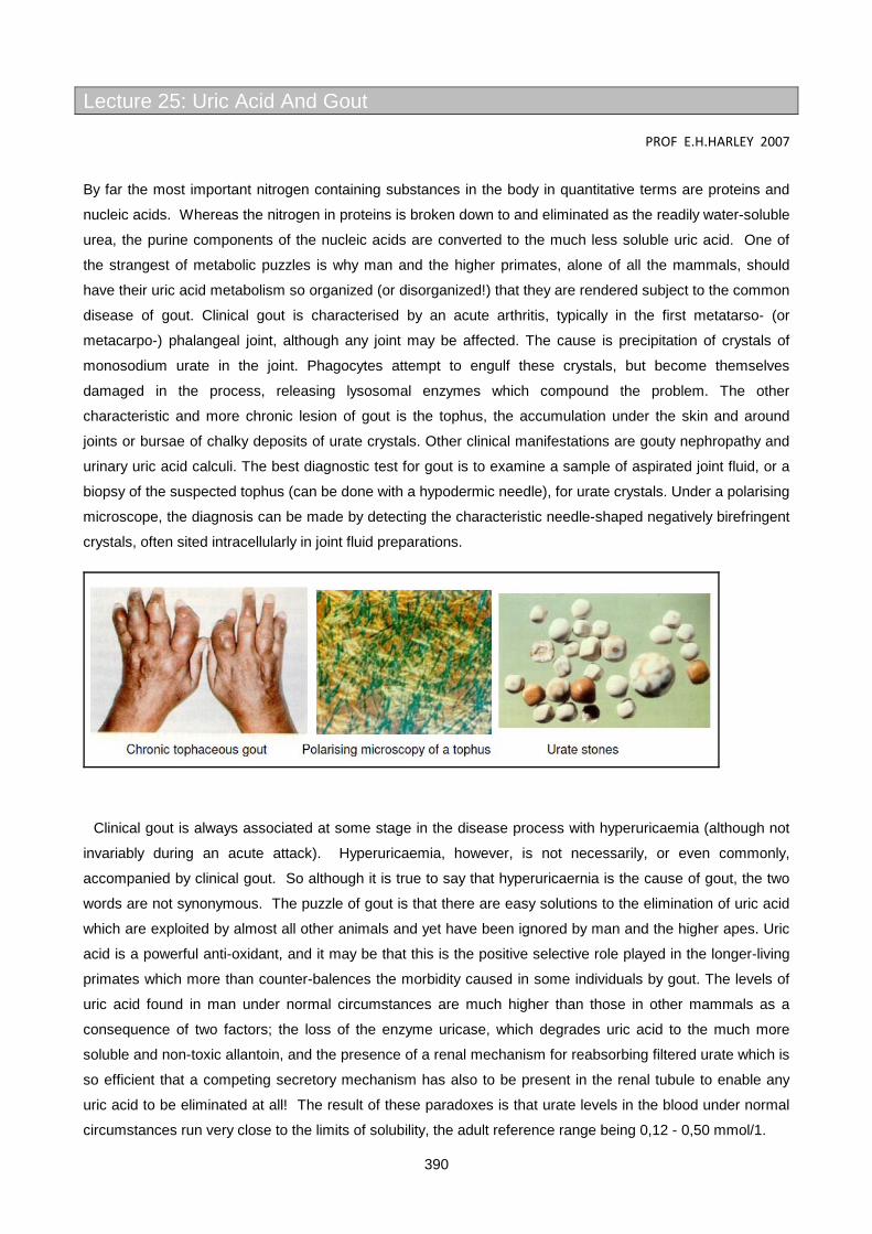
390
Lecture 25: Uric Acid And Gout
PROF E.H.HARLEY 2007
By far the most important nitrogen containing substances in the body in quantitative terms are proteins and
nucleic acids. Whereas the nitrogen in proteins is broken down to and eliminated as the readily water-soluble
urea, the purine components of the nucleic acids are converted to the much less soluble uric acid. One of
the strangest of metabolic puzzles is why man and the higher primates, alone of all the mammals, should
have their uric acid metabolism so organized (or disorganized!) that they are rendered subject to the common
disease of gout. Clinical gout is characterised by an acute arthritis, typically in the first metatarso- (or
metacarpo-) phalangeal joint, although any joint may be affected. The cause is precipitation of crystals of
monosodium urate in the joint. Phagocytes attempt to engulf these crystals, but become themselves
damaged in the process, releasing lysosomal enzymes which compound the problem. The other
characteristic and more chronic lesion of gout is the tophus, the accumulation under the skin and around
joints or bursae of chalky deposits of urate crystals. Other clinical manifestations are gouty nephropathy and
urinary uric acid calculi. The best diagnostic test for gout is to examine a sample of aspirated joint fluid, or a
biopsy of the suspected tophus (can be done with a hypodermic needle), for urate crystals. Under a polarising
microscope, the diagnosis can be made by detecting the characteristic needle-shaped negatively birefringent
crystals, often sited intracellularly in joint fluid preparations.
Clinical gout is always associated at some stage in the disease process with hyperuricaemia (although not
invariably during an acute attack). Hyperuricaemia, however, is not necessarily, or even commonly,
accompanied by clinical gout. So although it is true to say that hyperuricaernia is the cause of gout, the two
words are not synonymous. The puzzle of gout is that there are easy solutions to the elimination of uric acid
which are exploited by almost all other animals and yet have been ignored by man and the higher apes. Uric
acid is a powerful anti-oxidant, and it may be that this is the positive selective role played in the longer-living
primates which more than counter-balences the morbidity caused in some individuals by gout. The levels of
uric acid found in man under normal circumstances are much higher than those in other mammals as a
consequence of two factors; the loss of the enzyme uricase, which degrades uric acid to the much more
soluble and non-toxic allantoin, and the presence of a renal mechanism for reabsorbing filtered urate which is
so efficient that a competing secretory mechanism has also to be present in the renal tubule to enable any
uric acid to be eliminated at all! The result of these paradoxes is that urate levels in the blood under normal
circumstances run very close to the limits of solubility, the adult reference range being 0,12 - 0,50 mmol/1.

391
Two major population studies conducted in the USA, one in Framingham, the other in Tecumseh, give useful
illustrative demographic statistics on prevalance, etc. :
Prevalence of Gout in different age cohorts
No of subjects Mean age (years) Prevalence, %
5127 44 0.2
5127 58 1.5 (0.28% in men) (0.4% in women)
Ethnic and social variations in Serum urate values (mmoles/l)
Males Females
Population No Mean No Mean
Tecumseh 2,987 0.29 3,013 0.25
Framingham 2,283 0.30 3,013 0.25
Social Class
U.S. craftsman 532 0.28
University Professors
113 0.34
Edinburgh executives
100 0.36
Ethnic group
Indians 141 0.22
Maori 366 0.42 381 0.35
Pokapukans 188 0.42 191 0.37
At least 95% of urate in plasma exists as the free form, with a small and uncertain proportion bound to
albumin and to a specific α-globulin; as a consequence, the solubility product of sodium urate (sodium being
the.pre-dominant cation available) is exceeded once the urate level rises above about 0.42 mmol/1, a value
which is less than the upper limit of the reference range given above. At higher levels than this therefore, the
plasma and interstitial fluids are supersaturated with sodium urate and the potential for precipitation exists.
That precipitation does not occur more frequently than it does is surprising, and may point to some factor
which normally helps keep the urate in solution even though supersaturated. The urate solubility is markedly
dependent on temperature and this may help to explain the predilection for urate precipitation in the colder
extremities of the body.
Serum urate concentrations in adults are unimodally distributed but skewed to the right. Values are low in
both sexes before puberty and then rise - markedly so in men, rapidly reaching a mean of about 0.35
mmol/1 sustained with little change until age 70 or over. In women, levels comparable to those in men are
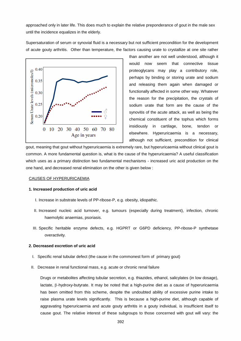
392
approached only in later life. This does much to explain the relative preponderance of gout in the male sex
until the incidence equalizes in the elderly.
Supersaturation of serum or synovial fluid is a necessary but not sufficient precondition for the development
of acute gouty arthritis. Other than temperature, the factors causing urate to crystallize at one site rather
than another are not well understood, although it
would now seem that connective tissue
proteoglycans may play a contributory role,
perhaps by binding or storing urate and sodium
and releasing them again when damaged or
functionally affected in some other way. Whatever
the reason for the precipitation, the crystals of
sodium urate that form are the cause of the
synovitis of the acute attack, as well as being the
chemical constituent of the tophus which forms
insidiously in cartilage, bone, tendon or
elsewhere. Hyperuricaemia is a necessary,
although not sufficient, precondition for clinical
gout, meaning that gout without hyperuricaemia is extremely rare, but hyperuricaemia without clinical gout is
common. A more fundamental question is, what is the cause of the hyperuricaemia? A useful classification
which uses as a primary distinction two fundamental mechanisms - increased uric acid production on the
one hand, and decreased renal elimination on the other is given below :
CAUSES OF HYPERURICAEMIA
1. Increased production of uric acid
I. Increase in substrate levels of PP-ribose-P, e.g. obesity, idiopathic.
II. Increased nucleic acid turnover, e.g. tumours (especially during treatment), infection, chronic
haemolytic anaemias, psoriasis.
III. Specific heritable enzyme defects, e.g. HGPRT or G6PD deficiency, PP-ribose-P synthetase
overactivity.
2. Decreased excretion of uric acid
I. Specific renal tubular defect (the cause in the commonest form of primary gout)
II. Decrease in renal functional mass, e.g. acute or chronic renal failure
Drugs or metabolites affecting tubular secretion, e.g. thiazides, ethanol, salicylates (in low dosage),
lactate, β-hydroxy-butyrate. It may be noted that a high-purine diet as a cause of hyperuricaemia
has been omitted from this scheme, despite the undoubted ability of excessive purine intake to
raise plasma urate levels significantly. This is because a high-purine diet, although capable of
aggravating hyperuricaemia and acute gouty arthritis in a gouty individual, is insufficient itself to
cause gout. The relative interest of these subgroups to those concerned with gout will vary: the
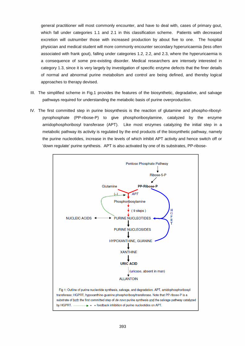
393
general practitioner will most commonly encounter, and have to deal with, cases of primary gout,
which fall under categories 1.1 and 2.1 in this classification scheme. Patients with decreased
excretion will outnumber those with increased production by about five to one. The hospital
physician and medical student will more commonly encounter secondary hyperuricaemia (less often
associated with frank gout), falling under categories 1.2, 2.2, and 2.3, where the hyperuricaemia is
a consequence of some pre-existing disorder. Medical researchers are intensely interested in
category 1.3, since it is very largely by investigation of specific enzyme defects that the finer details
of normal and abnormal purine metabolism and control are being defined, and thereby logical
approaches to therapy devised.
III. The simplified scheme in Fig.1 provides the features of the biosynthetic, degradative, and salvage
pathways required for understanding the metabolic basis of purine overproduction.
IV. The first committed step in purine biosynthesis is the reaction of glutamine and phospho-ribosyl-
pyrophosphate (PP-ribose-P) to give phosphoribosylamine, catalyzed by the enzyme
amidophosphoribosyl transferase (APT). Like most enzymes catalyzing the initial step in a
metabolic pathway its activity is regulated by the end products of the biosynthetic pathway, namely
the purine nucleotides, increase in the levels of which inhibit APT activity and hence switch off or
'down regulate' purine synthesis. APT is also activated by one of its substrates, PP-ribose-
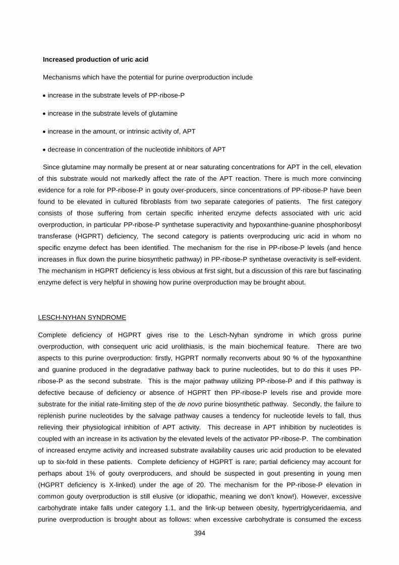
394
Increased production of uric acid
Mechanisms which have the potential for purine overproduction include
• increase in the substrate levels of PP-ribose-P
• increase in the substrate levels of glutamine
• increase in the amount, or intrinsic activity of, APT
• decrease in concentration of the nucleotide inhibitors of APT
Since glutamine may normally be present at or near saturating concentrations for APT in the cell, elevation
of this substrate would not markedly affect the rate of the APT reaction. There is much more convincing
evidence for a role for PP-ribose-P in gouty over-producers, since concentrations of PP-ribose-P have been
found to be elevated in cultured fibroblasts from two separate categories of patients. The first category
consists of those suffering from certain specific inherited enzyme defects associated with uric acid
overproduction, in particular PP-ribose-P synthetase superactivity and hypoxanthine-guanine phosphoribosyl
transferase (HGPRT) deficiency, The second category is patients overproducing uric acid in whom no
specific enzyme defect has been identified. The mechanism for the rise in PP-ribose-P levels (and hence
increases in flux down the purine biosynthetic pathway) in PP-ribose-P synthetase overactivity is self-evident.
The mechanism in HGPRT deficiency is less obvious at first sight, but a discussion of this rare but fascinating
enzyme defect is very helpful in showing how purine overproduction may be brought about.
LESCH-NYHAN SYNDROME
Complete deficiency of HGPRT gives rise to the Lesch-Nyhan syndrome in which gross purine
overproduction, with consequent uric acid urolithiasis, is the main biochemical feature. There are two
aspects to this purine overproduction: firstly, HGPRT normally reconverts about 90 % of the hypoxanthine
and guanine produced in the degradative pathway back to purine nucleotides, but to do this it uses PP-
ribose-P as the second substrate. This is the major pathway utilizing PP-ribose-P and if this pathway is
defective because of deficiency or absence of HGPRT then PP-ribose-P levels rise and provide more
substrate for the initial rate-limiting step of the de novo purine biosynthetic pathway. Secondly, the failure to
replenish purine nucleotides by the salvage pathway causes a tendency for nucleotide levels to fall, thus
relieving their physiological inhibition of APT activity. This decrease in APT inhibition by nucleotides is
coupled with an increase in its activation by the elevated levels of the activator PP-ribose-P. The combination
of increased enzyme activity and increased substrate availability causes uric acid production to be elevated
up to six-fold in these patients. Complete deficiency of HGPRT is rare; partial deficiency may account for
perhaps about 1% of gouty overproducers, and should be suspected in gout presenting in young men
(HGPRT deficiency is X-linked) under the age of 20. The mechanism for the PP-ribose-P elevation in
common gouty overproduction is still elusive (or idiopathic, meaning we don’t know!). However, excessive
carbohydrate intake falls under category 1.1, and the link-up between obesity, hypertriglyceridaemia, and
purine overproduction is brought about as follows: when excessive carbohydrate is consumed the excess
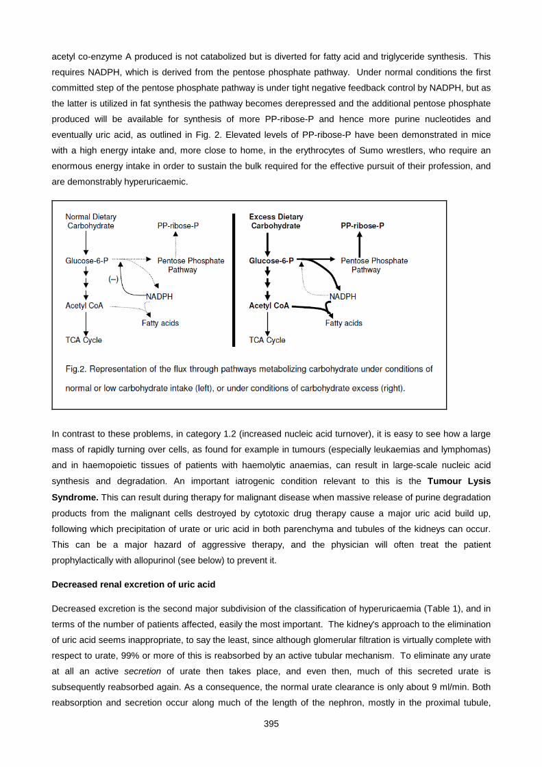
395
acetyl co-enzyme A produced is not catabolized but is diverted for fatty acid and triglyceride synthesis. This
requires NADPH, which is derived from the pentose phosphate pathway. Under normal conditions the first
committed step of the pentose phosphate pathway is under tight negative feedback control by NADPH, but as
the latter is utilized in fat synthesis the pathway becomes derepressed and the additional pentose phosphate
produced will be available for synthesis of more PP-ribose-P and hence more purine nucleotides and
eventually uric acid, as outlined in Fig. 2. Elevated levels of PP-ribose-P have been demonstrated in mice
with a high energy intake and, more close to home, in the erythrocytes of Sumo wrestlers, who require an
enormous energy intake in order to sustain the bulk required for the effective pursuit of their profession, and
are demonstrably hyperuricaemic.
In contrast to these problems, in category 1.2 (increased nucleic acid turnover), it is easy to see how a large
mass of rapidly turning over cells, as found for example in tumours (especially leukaemias and lymphomas)
and in haemopoietic tissues of patients with haemolytic anaemias, can result in large-scale nucleic acid
synthesis and degradation. An important iatrogenic condition relevant to this is the Tumour Lysis
Syndrome. This can result during therapy for malignant disease when massive release of purine degradation
products from the malignant cells destroyed by cytotoxic drug therapy cause a major uric acid build up,
following which precipitation of urate or uric acid in both parenchyma and tubules of the kidneys can occur.
This can be a major hazard of aggressive therapy, and the physician will often treat the patient
prophylactically with allopurinol (see below) to prevent it.
Decreased renal excretion of uric acid
Decreased excretion is the second major subdivision of the classification of hyperuricaemia (Table 1), and in
terms of the number of patients affected, easily the most important. The kidney's approach to the elimination
of uric acid seems inappropriate, to say the least, since although glomerular filtration is virtually complete with
respect to urate, 99% or more of this is reabsorbed by an active tubular mechanism. To eliminate any urate
at all an active secretion of urate then takes place, and even then, much of this secreted urate is
subsequently reabsorbed again. As a consequence, the normal urate clearance is only about 9 ml/min. Both
reabsorption and secretion occur along much of the length of the nephron, mostly in the proximal tubule,
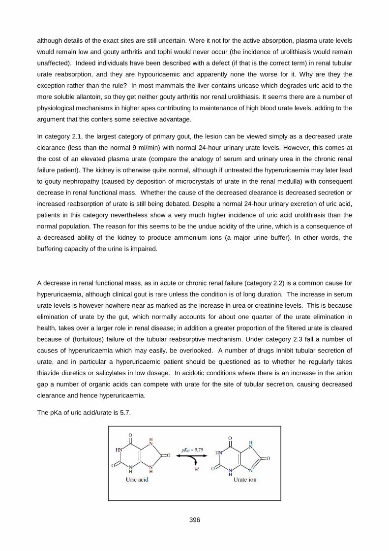
396
although details of the exact sites are still uncertain. Were it not for the active absorption, plasma urate levels
would remain low and gouty arthritis and tophi would never occur (the incidence of urolithiasis would remain
unaffected). Indeed individuals have been described with a defect (if that is the correct term) in renal tubular
urate reabsorption, and they are hypouricaemic and apparently none the worse for it. Why are they the
exception rather than the rule? In most mammals the liver contains uricase which degrades uric acid to the more soluble allantoin, so they get neither gouty arthritis nor renal urolithiasis. It seems there are a number of
physiological mechanisms in higher apes contributing to maintenance of high blood urate levels, adding to the
argument that this confers some selective advantage.
In category 2.1, the largest category of primary gout, the lesion can be viewed simply as a decreased urate
clearance (less than the normal 9 ml/min) with normal 24-hour urinary urate levels. However, this comes at
the cost of an elevated plasma urate (compare the analogy of serum and urinary urea in the chronic renal
failure patient). The kidney is otherwise quite normal, although if untreated the hyperuricaemia may later lead
to gouty nephropathy (caused by deposition of microcrystals of urate in the renal medulla) with consequent
decrease in renal functional mass. Whether the cause of the decreased clearance is decreased secretion or
increased reabsorption of urate is still being debated. Despite a normal 24-hour urinary excretion of uric acid,
patients in this category nevertheless show a very much higher incidence of uric acid urolithiasis than the
normal population. The reason for this seems to be the undue acidity of the urine, which is a consequence of
a decreased ability of the kidney to produce ammonium ions (a major urine buffer). In other words, the
buffering capacity of the urine is impaired.
A decrease in renal functional mass, as in acute or chronic renal failure (category 2.2) is a common cause for
hyperuricaemia, although clinical gout is rare unless the condition is of long duration. The increase in serum
urate levels is however nowhere near as marked as the increase in urea or creatinine levels. This is because
elimination of urate by the gut, which normally accounts for about one quarter of the urate elimination in
health, takes over a larger role in renal disease; in addition a greater proportion of the filtered urate is cleared
because of (fortuitous) failure of the tubular reabsorptive mechanism. Under category 2.3 fall a number of
causes of hyperuricaemia which may easily. be overlooked. A number of drugs inhibit tubular secretion of
urate, and in particular a hyperuricaemic patient should be questioned as to whether he regularly takes
thiazide diuretics or salicylates in low dosage. In acidotic conditions where there is an increase in the anion
gap a number of organic acids can compete with urate for the site of tubular secretion, causing decreased
clearance and hence hyperuricaemia.
The pKa of uric acid/urate is 5.7.
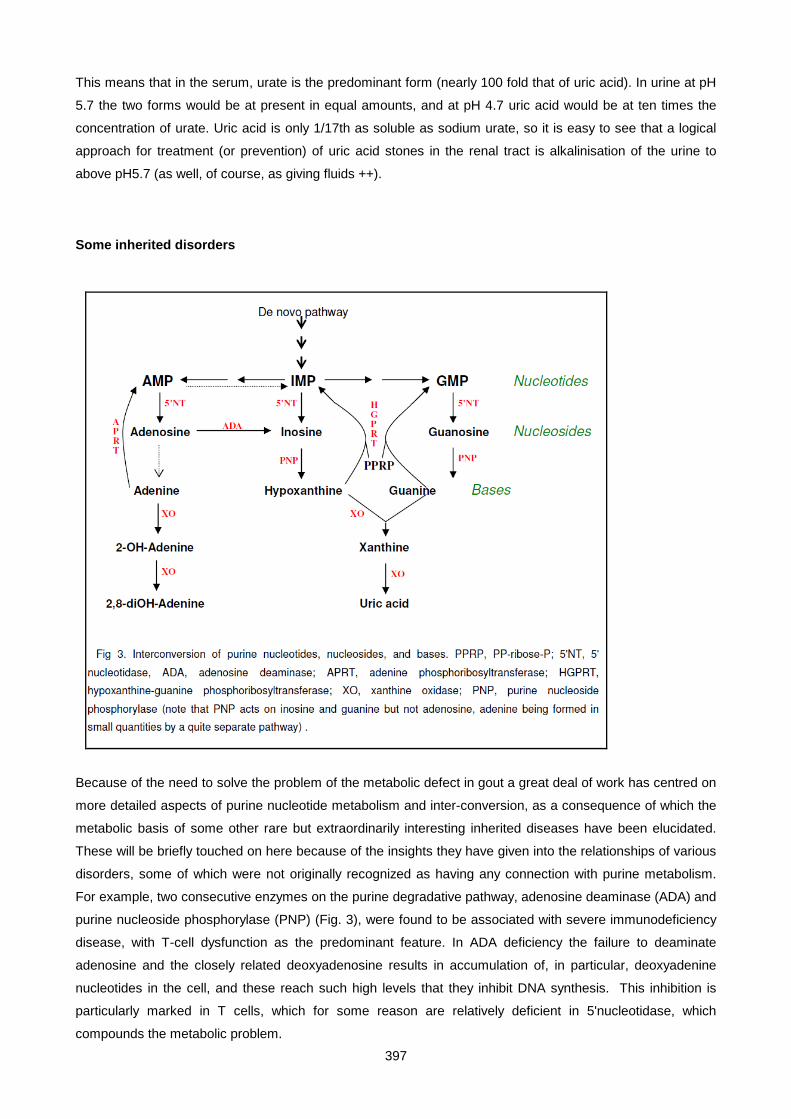
397
This means that in the serum, urate is the predominant form (nearly 100 fold that of uric acid). In urine at pH
5.7 the two forms would be at present in equal amounts, and at pH 4.7 uric acid would be at ten times the
concentration of urate. Uric acid is only 1/17th as soluble as sodium urate, so it is easy to see that a logical
approach for treatment (or prevention) of uric acid stones in the renal tract is alkalinisation of the urine to
above pH5.7 (as well, of course, as giving fluids ++).
Some inherited disorders
Because of the need to solve the problem of the metabolic defect in gout a great deal of work has centred on
more detailed aspects of purine nucleotide metabolism and inter-conversion, as a consequence of which the
metabolic basis of some other rare but extraordinarily interesting inherited diseases have been elucidated.
These will be briefly touched on here because of the insights they have given into the relationships of various
disorders, some of which were not originally recognized as having any connection with purine metabolism.
For example, two consecutive enzymes on the purine degradative pathway, adenosine deaminase (ADA) and
purine nucleoside phosphorylase (PNP) (Fig. 3), were found to be associated with severe immunodeficiency
disease, with T-cell dysfunction as the predominant feature. In ADA deficiency the failure to deaminate
adenosine and the closely related deoxyadenosine results in accumulation of, in particular, deoxyadenine
nucleotides in the cell, and these reach such high levels that they inhibit DNA synthesis. This inhibition is
particularly marked in T cells, which for some reason are relatively deficient in 5'nucleotidase, which
compounds the metabolic problem.
398
A similar mechanism in principle occurs in PNP deficiency, but in this condition it is the disproportional
accumulation of deoxyguanine nucleotides which inhibits DNA synthesis and therefore cell division, again
predominantly in T lymphocytes. PNP deficiency in addition gives rise to the surprising combination of
hypouricaemia, yet with purine overproduction. Inosine and guanosine are the purine products excreted in
the urine. Purine overproduction is caused by PP-ribose-P accumulation since the salvage pathway
catalyzed by HGPRT is not operating. Whereas in the Lesch-Nyhan syndrome this is due to lack of HGPRT,
in PNP deficiency it is due to lack of the substrates (hypoxanthine and guanine). A consequence of the
elucidation of these biochemical mechanisms was the realization that this knowledge could be used for
devising novel treatments of quite unrelated disorders. Coformycin and deoxycoformycin specifically inhibit
ADA, and these agents have been used for the treatment of certain leukaemias, exploiting the mechanisms
described above to produce a therapeutic ADA deficiency in the patient.
Two unusual types of renal calculus, which can be easily mistaken for uric acid calculi, are caused by
deficiency of two other enzymes shown in Fig. 3. In xanthine oxidase (X0) deficiency the stones are of
xanthine, and the patient is also hypouricaemic. In adenine phosphoribosyl transferase (APRT) deficiency
the stones consist of 2,8-dihydroxyadenine, and the condition is worth remembering, since alkalinization of
the urine, appropriate for the dissolution of uric acid stones, is contraindicated here, since the stones, unlike
uric acid, are not more soluble in alkali.
Glucose-6-phosphatase deficiency (Von Gierke's disease) is characterized by hyperuricaemia. In this
condition there is increased flux down the pentose phosphate pathway with increased availability of ribose-5-
phosphate for PP-ribose-P synthesis. In addition, the high lactate levels found in this disease compete for
urate secretion in the renal tubule and therefore compound the hyperuricaemia by decreasing renal excretion.
It is paradoxical that the less common the cause of hyperuricaemia the better is its mechanism understood.
When the definitive mechanisms for the common forms of primary gout are finally established, then new and
logical approaches to therapy may become apparent. An understanding of the pathophysiology underlying
these various disorders of purine metabolism provides a guide for the investigation of patients with such
disorders and a basis for appropriate choice of therapy, i.e. whether in gout to use a uricosuric agent or
allopurinol, a metabolic inhibitor of XO. If the patient is an overproducer, then it is logical to use allopurinol to
reduce production. If an underexcreter, it is logical to use a uricosuric to decrease reabsorbtion. If there is any
element of renal failure it is best to prescribe allopurinol. Treatment in all cases, once initiated, should be for
life. The acute attack is best treated with an anti-inflammatory drug, with allopurinol or a uricosuric instituted
later. In the initial stages of such treatment acute gouty attacks often still occur, and it may take some time
before the body mass of urate in tophi or micro-tophi is totally eliminated. Gout used to be a crippling and
eventually fatal disease. Treatment nowadays is so effective that the condition is considered little more than a
nuisance, and taking a couple of tablets a day for the rest of their life is a price most patients are only too glad
to pay.
399
SMALL GROUP TEACHING: LECTURE 25: URIC ACID AND GOUT
QUESTIONS:
1. What is the difference between gout, and hyperuricaemia?
2. How much more soluble is uric acid in plasma than in urine and why is it useful to know what the pKa (5.75) of uric acid is?
3. Draw a rough graph of the relative susceptibilities to gout of males and females with respect to age.
4. (a)Draw out the pathways of purine anabolism, inter-conversion and catabolism, and give the mechanism whereby HPRT deficiency causes purine overproduction.
(b)See if by simple logic, you can work out what might happen to purine excretion in purine nucleotide phosphorylase (PNP, nucleosides --> bases) deficiency.
5. What does the normal renal clearance value for uric acid (typically 9 ml/min) tell you about the way the kidney handles uric acid?
6. Why, in renal failure, do blood urea levels rise more markedly than blood urate levels?
7. A 54-year old man with no previous history presented with an acutely inflamed 1st metacarpo-phalangeal joint of his left foot on the 27 December.
Investigations:
Blood urea 5.4 mmol/l (1.7-6.7)
Blood urate 0.55 mmoles/l (0.10-0.50)
Urinary urate 3.1 mmol/day (1.5-4.4)
i. What type of gout is this man likely to have?
ii. A diagnostic procedure was performed on the inflamed joint: What was this likely to be?
iii. Why is this joint in particular so susceptable to gouty arthritis?
iv. Small nodules were found in the right pinna and over the olecranon. What might these be?
v. How would you treat him?

400
Lecture 26: Inherited Metabolic Diseases
PROF E H HARLEY, UPDATED BY PROF H HENDERSON 2007
INTRODUCTION
Inherited metabolic diseases (IMD's), sometimes referred to as inborn errors of metabolism, are monogenic
disorders, meaning that they are caused by the absence or dysfunction of a single gene product, either a
structural protein or an enzyme. They are in a quite separate category from chromosomal disorders such as
Down's syndrome in which there are major changes affecting the chromosome number or architecture,
visible on light microscopy of chromosome preparations and involving the duplication or deletion of large
numbers of genes.
Since any of the thousands of enzymes present in an organism can be defective, there are a corresponding
number, theoretically, of inheritable metabolic disorders, and this is the key problem in addressing this
category of diseases: They are each individually rare, although as a general category, they are quite
common - constituting 6-8% of paediatric admissions in the USA, for example. As a consequence, it is
impossible for the student, or the practising clinician, to have a detailed knowledge of all of them. The best
compromise is to know the general principle of the genetics, diagnosis, and treatment possibilities, with some
examples of individual disorders studied in more detail where these are either locally common or illustrate
useful or interesting principles.
GENETICS
Most serious inherited disorders are autosomal recessive. Thus disease only results if 2 defective alleles
come together in an individual. For example, if the frequency of a recessive lethal gene in a population is 1 in
100 (ie. n = 50), the incidence of unaffected carrier heterozygotes is 1/50 (two chances of having the gene)
and the incidence of affected homozygotes would be 1 in 50 x 1 in 50 x 1 in 4 = 1 in 10,000 births. Obviously,
the incidence of autosomal recessive disorders is higher in consanguinous matings. Some other important
genetic diseases are inherited as X-linked recessive. These include haemophilia, deficiencies of G6PD
(causes a haemolytic anaemia), HPRT (Lesch-Nyhan syndrome), OTC (the urea-cycle enzyme, ornithine
transcarbamoylase, a cause of hyperammonaemia), and Duchenne muscular dystrophy. In X-linked
recessive disorders only the male manifests the disease, and female carriers pass on the gene to 50% of
their children – ½ of their male children will therefore be affected and ½ their daughters will be carriers. Less
often, the inheritance of a genetic defect is autosomal dominant, in which case both allelic genes need to be
functioning normally to avoid overt disease. Variegate porphyria (South African genetic porphyria) is one
such example, and familial hypercholesterolaemia is another; both are especially relevant in South Africa.
Even if only one parent carried a defective allele, in dominantly inherited disorders, that parent will manifest
the disease and pass it on to 50% of his/her children. Sometimes, individuals carrying a dominant gene in a
family are affected to very varying extents (varying penetrance), e.g. in variegate or acute intermittent
porphyria, even to the extent of the disease remaining latent for the lifetime of the individual. Recently, a new
class of inherited disease has become recognised characterised by a strict maternal mode of inheritance, in
401
which all the offspring of an affected mother are affected, but none of an affected father. Leber’s Hereditary
Optic Atrophy (LHON), common in the local Cape Town community, causes blindness in middle life, and is
one such example. In these disorders the mutation is in the small piece (only 16000 base pairs in size) of
DNA found in the mitochondrion, which mostly codes for enzyme subunits of the oxidative phosphorylation
pathway, and mitochondria are only passed on to the next generation in the cytoplasm of the ovum. The
mitochondrial genome is haploid and undergoes no recombination. It is present in several hundred copies per
cell.
PATHOLOGY
1. The defective enzyme may be a Km or Vmax mutant. A Km mutant shows decreased affinity for
substrate, a Vmax mutant shows decreased overall activity, and this is often equated with decreased
quantities of an otherwise normal enzyme (often due to a decreased enzyme lifetime, or to a
mutation in the promoter region of the gene).
2. The defect may lie in the protein part of the enzyme (apoenzyme), or in the ability to synthesize active
co-factor (co-enzyme). Co-factors are usually vitamin B derivatives and include biopterin (defects
cause a form of phenylketonuria), vitamin B12 (methylmalonic acidaemia), and biotin (propionic
acidaemia). Cofactor defects may respond to megadose vitamin supplements.
3. Symptoms may be due either to deficiency of enzyme product, or to accumulation of enzyme
substrate. An example in the first category is deficiency of pyrimidines in orotic aciduria (caused by a
defect in orotate phosphoribosyl transferase, OPRT) which leads to megaloblastic anaemia (failure of
DNA and RNA synthesis) and responds to uridine therapy, which provides pyrimidines by an
alternate salvage pathway.
The accumulated substrate may not be toxic in itself, but may be converted to a toxic or damaging
metabolite via a side pathway. For example, the galactose that accumulates in galactosemia is
converted to galactitol in the lens, which results in cataract formation, the adenine that accumulates
in APRT (adenine phosphoribosyl transferase) deficiency is converted to 2.8 dihydroxy adenine,
which causes renal stones, while 17-hydroxyprogesterone is converted to androgens in congenital
adrenal hyperplasia.
There is a very real need for the student, general practitioner, physician and paediatrician to be aware of
progress in the area of IMDs for several reasons:
• IMDs as a group are commoner than is generally appreciated, and especially common as a cause of
acute illness and death in the neonatal period and early childhood.
• As knowledge of the biochemical mechanisms underlying individual disorders accumulates, effective
therapeutic measures are becoming available for previously untreatable conditions.
• Even if the disease is not amenable to treatment, genetic counselling by a specialist aware of the
prognosis and the risk of recurrence is essential to advise the family regarding management of future
pregnancies. Advances in antenatal diagnosis are enabling many disorders giving rise to lethal
disease to be detected before the 20th week of gestation. Termination of the pregnancy, if ethically
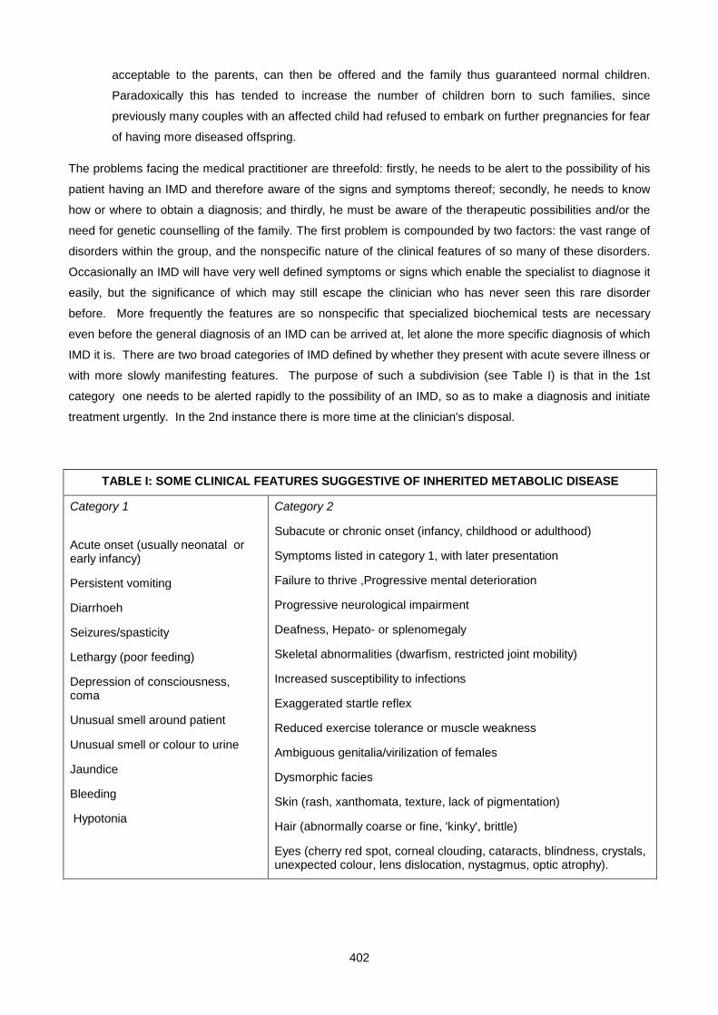
402
acceptable to the parents, can then be offered and the family thus guaranteed normal children.
Paradoxically this has tended to increase the number of children born to such families, since
previously many couples with an affected child had refused to embark on further pregnancies for fear
of having more diseased offspring.
The problems facing the medical practitioner are threefold: firstly, he needs to be alert to the possibility of his
patient having an IMD and therefore aware of the signs and symptoms thereof; secondly, he needs to know
how or where to obtain a diagnosis; and thirdly, he must be aware of the therapeutic possibilities and/or the
need for genetic counselling of the family. The first problem is compounded by two factors: the vast range of
disorders within the group, and the nonspecific nature of the clinical features of so many of these disorders.
Occasionally an IMD will have very well defined symptoms or signs which enable the specialist to diagnose it
easily, but the significance of which may still escape the clinician who has never seen this rare disorder
before. More frequently the features are so nonspecific that specialized biochemical tests are necessary
even before the general diagnosis of an IMD can be arrived at, let alone the more specific diagnosis of which
IMD it is. There are two broad categories of IMD defined by whether they present with acute severe illness or
with more slowly manifesting features. The purpose of such a subdivision (see Table I) is that in the 1st
category one needs to be alerted rapidly to the possibility of an IMD, so as to make a diagnosis and initiate
treatment urgently. In the 2nd instance there is more time at the clinician's disposal.
TABLE I: SOME CLINICAL FEATURES SUGGESTIVE OF INHERITED METABOLIC DISEASE
Category 1
Acute onset (usually neonatal or early infancy)
Persistent vomiting
Diarrhoeh
Seizures/spasticity
Lethargy (poor feeding)
Depression of consciousness, coma
Unusual smell around patient
Unusual smell or colour to urine
Jaundice
Bleeding
Hypotonia
Category 2
Subacute or chronic onset (infancy, childhood or adulthood)
Symptoms listed in category 1, with later presentation
Failure to thrive ,Progressive mental deterioration
Progressive neurological impairment
Deafness, Hepato- or splenomegaly
Skeletal abnormalities (dwarfism, restricted joint mobility)
Increased susceptibility to infections
Exaggerated startle reflex
Reduced exercise tolerance or muscle weakness
Ambiguous genitalia/virilization of females
Dysmorphic facies
Skin (rash, xanthomata, texture, lack of pigmentation)
Hair (abnormally coarse or fine, 'kinky', brittle)
Eyes (cherry red spot, corneal clouding, cataracts, blindness, crystals, unexpected colour, lens dislocation, nystagmus, optic atrophy).
403
Table I summarises those symptoms and signs in these two categories which should arouse suspicion of
inherited metabolic disease, especially if common acquired diseases have been excluded and the patient
fails to respond to conventional treatment. The acute disorders giving rise to the features in the 1st category
virtually always manifest in the neonatal period or early childhood and so are paediatric problems. Those in
the 2nd category are predominantly paediatric but may manifest in adult life.
Having considered the possibility of an IMD the clinician can now address the main problem: how to confirm
that his patient is indeed suffering from an IMD, and if so, which specific one it is. Even the first part of this
problem is by no means trivial, but a few simple investigations will lend support to the suspicion. Firstly, re-
examine the family history for any suggestion of a similar disorder in a relative. In particular, have any other
siblings died in early life, and is there any degree of consanguinity? Also bear in mind that some IMDs have
an increased frequency in certain race groups, for example, Gaucher's and Tay-Sachs diseases among
Ashkenazi Jews, thalassaemia among Greeks and some races of tropical origin, cystic fibrosis among
Europeans, and familial hypercholesterolaemia among the Afrikaners. After a thorough physical examination,
paying special attention to skin, hair, eyes, genitalia and facial characteristics in addition to standard
neurological, cardiovascular and abdominal organ examination, a few simple biochemical tests should be
performed. These include urine examination for abnormal colour, smell and reducing substances, as well as
microscopy for abnormal cells or for crystals, and blood sampling for estimation of glucose, bilirubin, sodium
and potassium, urate, urea, calcium and, especially important, acid/base studies, since metabolic acidosis is
a feature of many acute metabolic disorders. Standard haematological tests are also necessary to detect
neutropenia or red cell abnormalities
A number of rapid (spot) tests can also be performed on urine to identify abnormal metabolites, these include
Ferric chloride for keto-acids, cyanide-nitroprusside for cystine or homocystine, CTAB or toluidine blue for
mucopolysaccharides (glycosaminoglycans), Ehrlichs aldehyde for urobilinogen and porphobilinogen. More
elaborate methods requiring more sophisticated technologies can quantitatively assay amino acids and
organic acids in blood or urine.
It is interesting to consider the various levels at which diagnosis of IMDs can be made. These are:-
I. Clinical: If the enzymatic defect is unknown and there are no characteristic biochemical markers
then the diagnosis may be made solely on the characteristic presentation and evidence of
(mendelian) inheritance e.g. Marfan syndrome.
II. Secondary biochemical disturbance: affected metabolic pathway identified by detection of
abnormal metabolite(s) e.g. PKU (phenylketones), methylmalonic aciduria.
III. Protein: Identification of precise enzyme or protein affected e.g. one can now differentiate between
the various types of hyperphenylalaninaemia: PKU type I - phenylalanine hydroxylase deficiency,
PKU type II - dihydropteridine reductase deficiency, PKU type V - dihydrobiopterin synthetase
deficiency (see below).
IV. Gene: Identification of the precise mutational event in the DNA.
404
Often it is only feasible to go as far as level 2, but the extra time and effort in going to level 3 is justified
because there may be several different enzyme defects giving the same type of secondary biochemical
disturbance (e.g. hyperphenylalaninaemia, methylmalonic aciduria) but with each having different treatment
regimes, prognosis, or genetic modes of inheritance. Identification at level 4 is becoming common place with
the use of methods such as the Polymerase Chain Reaction (PCR), and gene chip technologies becoming
the method of choice in many instances. Whereas most chemical pathology investigations are performed on
readily accessable body fluids such as blood, urine, or CSF, investigations at level 3 may require using body
cells such as red cells, white cells, skin fibroblast cell cultures, amnion cell cultures or chorionic villus biopsies
(useful for anti-natal diagnosis), and biopsy material (e.g. liver) if the defect is only expressed in a
differentiated tissue as opposed to all cells. Paradoxically, diagnostic proceedures based on DNA (level 4)
can be performed on any cell preparation, since the same DNA (and the mutation) is present in every cell
(with a few rare exceptions). The white cells from 1-5 ml of blood is often sufficient to provide enough DNA
for PCR-based proceedures. Some PCR methods start by generating cDNA by reverse transcribing mRNA,
which is expressed to a different extent in different tissues. This approach has the advantage of avoiding the
large intronic regions found in genes (these are spliced out in mRNA copy of the gene).
Criteria for an effective population-wide screening test (e.g. on all newborns):
I. Availability of a test that is a) cheap and b) reliable
II. Condition is treatable and fatal (or leads to chronic disability) if untreated, and reasonably common.
Criteria required before performing ante-natal diagnostic tests:
I. A reliable and safe diagnostic test is available
II. There is a significant risk of the foetus having the suspected IMD.
III. Condition is serious enough to warrant offering termination, and is not treatable.
IV. Parents are willing to consider termination of pregnancy if foetus is shown to be affected.
PRINCIPLES OF TREATMENT
Treatment is generally disappointing, emphasizing the need for effective preventative methods such as
genetic councelling and anti-natal diagnosis. However, with increasing knowledge of detailed
pathophysiology, the situation is improving. At present available therapy normalises life span in about 15% of
patients, reproductive capacity in 11%, but social adaptation in only 6%.
Strategies for treatment or prevention:
i. Avoidance of precipitating environmental factors – e.g. porphyria, albinism, G6PD.
ii. Dietary restriction of substrate - PKU, galactosaemia.
iii. Exploit alternative pathway - benzoate in urea cycle disorders, penicillamine in cystinuria.
iv. Metabolic inhibition - allopurinol in gout or Lesch-Nyhan syndrome.

405
v. Replacement of missing metabolite or cofactor - uridine in orotic aciduria, thyroxine in familial goitre,
biotin in biotinidase deficiency.
vi. Protein replacement - Factor VIII in haemophilia.
vii. Organ transplantation - liver in OTC def., kidney in cystinosis.
viii. Gene therapy.
Dietary and medical treatments remain the mainstay of therapy, one of the most remarkable advances in this
area being the use of a new drug, NTBC, for the effective treatment of Tyrosinaemia type I. NTBC was
originally used as a herbicide and its efficacy was discovered accidentally . Tyrosinaemia type I is caused by
deficiency of fumarylacetoacetase, and NTBC acts by inhibiting 4-hydroxyphenylpyruvate dioxygenase,
thereby preventing the accumulation of toxic metabolites, and is remarkably effective in preventing the
Fanconi-type syndrome typical of this disorder. PKU and haemophilia B (Xmas disease) are two candidate
conditions for gene therapy, the former because of its high incidence and because of neuropsychiatric
sequelae if treatment lapses, the latter because of the expense of treatment and risk of viral pathogens
transmitted in blood transfusions. These two conditions also have animal models : promising results have
been obtained using retroviral vectors in haemophilia Band in dogs there were long term significant
reductions in clotting times although only .1 to 1% of normal gene product levels were achieved (the
downside is the requirement for partial hepatectomy to provide a milieu of dividing liver cells for the vector to
transfect). In the PKU mouse an adenovirus vector-based approach was effective in complete normalisation
of phenyl-alanine levels, with only about 10% of normal enzyme activity required. However, this satisfactory
state only lasted a week or two, and retreatment was unsuccessful because of the development of antibodies
to the gene product. Immune reactions against the viral vector are a major problem, and over-ambitious and
premature attempts at gene therapy in humans have already resulted in one death from an excessive
immune response.
EXAMPLES OF DIFFERENT IMD's
DISORDERS OF CARBOHYDRATE METABOLISM
• Glycogen storage disease Type 1, or von Gierke's disease, is due to deficient hepatic glucose-6-
phosphatase (G-6-Pase), which catalyses the final step in hepatic glycogenolysis and
gluconeogenesis. Maintenance of an adequate blood glucose in the fasting state is a problem, and
repeated attacks of hypoglycaemia in early childhood may lead to permanent brain damage. The
liver is grossly distended with glycogen, and physical growth is stunted. Apart from fasting
hypoglycaemia, other biochemical features include persistent lactate acidosis, hyper-triglyceridaemia
and hyperuricaemia. Lactate and triglyceride export provide an alternative means of removing
glucose-6-phosphate from the hepatocyte, and enhanced urate production follows activation of the
direct oxidative (pentose phosphate) pathway in the liver with excess production of PPRP.
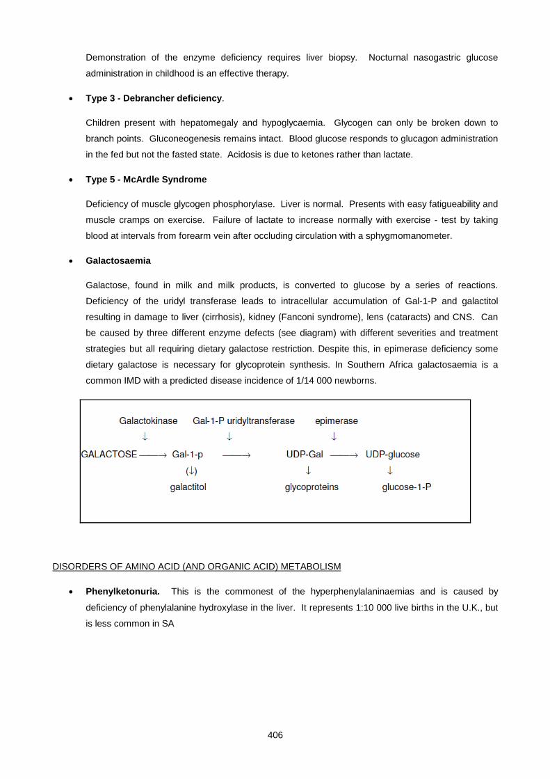
406
Demonstration of the enzyme deficiency requires liver biopsy. Nocturnal nasogastric glucose
administration in childhood is an effective therapy.
• Type 3 - Debrancher deficiency.
Children present with hepatomegaly and hypoglycaemia. Glycogen can only be broken down to
branch points. Gluconeogenesis remains intact. Blood glucose responds to glucagon administration
in the fed but not the fasted state. Acidosis is due to ketones rather than lactate.
• Type 5 - McArdle Syndrome
Deficiency of muscle glycogen phosphorylase. Liver is normal. Presents with easy fatigueability and
muscle cramps on exercise. Failure of lactate to increase normally with exercise - test by taking
blood at intervals from forearm vein after occluding circulation with a sphygmomanometer.
• Galactosaemia
Galactose, found in milk and milk products, is converted to glucose by a series of reactions.
Deficiency of the uridyl transferase leads to intracellular accumulation of Gal-1-P and galactitol
resulting in damage to liver (cirrhosis), kidney (Fanconi syndrome), lens (cataracts) and CNS. Can
be caused by three different enzyme defects (see diagram) with different severities and treatment
strategies but all requiring dietary galactose restriction. Despite this, in epimerase deficiency some
dietary galactose is necessary for glycoprotein synthesis. In Southern Africa galactosaemia is a
common IMD with a predicted disease incidence of 1/14 000 newborns.
DISORDERS OF AMINO ACID (AND ORGANIC ACID) METABOLISM
• Phenylketonuria. This is the commonest of the hyperphenylalaninaemias and is caused by
deficiency of phenylalanine hydroxylase in the liver. It represents 1:10 000 live births in the U.K., but
is less common in SA

407
Clinical presentation. Children are born normal, but within a few months developmental delay and
progressive mental retardation become evident. Children display agitated behaviour, tremors,
hyperreflexia and seizures. They are typically fair with blue eyes (failure of melanin synthesis),
develop an eczematous rash and a mousey odour. If detected before 1 month of age and treated on
phenylalanine restricted (not phenylalanine-free) diet, normal mental development follows. This
indicates that elevated levels of phenylalanine >1000 µM ( normal <200 µM) or its metabolic products
by side-pathways (e.g. phenyl pyruvic acid) are toxic to the developing brain.
Diagnosis. For a successful outcome, phenylketonuria must be detected in the first few weeks of
life. Blood levels of phenylalanine are elevated by the end of the first week, and may be detected by
screening tests (Guthrie microbiological assay) or quantitated by amino acid chromatography.
Excess excretion of organic acids derived from phenylalanine, including phenylpyruvic acid, may be
detected by screening tests, e.g. ferric chloride which gives a green colour on adding a few drops to
urine, or by special dipsticks (Phenistix).
Deficiency of enzymes involved either in the synthesis of, or the reduction of, the biopterin cofactor of
phenylalanine hydroxylase constitute variant forms of hyperphenylalaninaemia. They can develop
progressive neurological impairment which can respond to a combination of phenylalanine restriction
together with replacement of substrates (DOPA, 5-OH tryptophan) to make the neurotransmitters
whose synthesis depends on the normal activity of biopterin dependent tyrosine and tryptophan
hydroxylases (noradrenalin, dopamine, serotonin), as well as biopterin administration - which may be
sufficient in itself in biopterin synthesis deficiency. With the successful treatment of chronic
PKU, many women are reaching childbearing age and the management of pregnancy is becoming
important to prevent high levels of maternal phenylalanine from causing neurological damage to the
foetus.
• Alkaptonuria
This defect of amino acid catabolism is due to deficiency of homogentisic acid oxidase, which leads
to accumulation of homogentisic acid in the tissues and urine.

408
CNH2
O
O
CNH2
O
O
OH
C
O
O
OH
O
C
OHO
OH
OH
C
OO
OOH
OH O
O
O OH
O
O
OH O
OO
OOO
O
OH
O
OO
Tyrosine amino transaminase(Oculocutaneous TyrosinaemiaType II) Phe-OHase
(Phenylketonuria)
TyrPhe
P-hydroxyphenylpyruvate
P-hydroxyphenylpyruvatedioxygenase (Transient Tyrosinaemia)(Tyrosinaemia Type III)(Hawkinsinuria)(NTBC)
Homogentisic acid
Homogentisic acid oxidase(Alkaptonuria)
maleylacetoacetate
isomerase
fumaryl acetoacetate
Fumaryl acetoacetate hydrolase(Hepatorenal Tyrosinaemia Type I)
fumarate acetoacetate
CO2
CO2
Succinylacetone
On exposure to air, urine gradually darkens due to oxidation and polymerization of homogentisic acid
to form a dark brown/black pigment. This process also occurs in connective tissue, resulting in
darkening of cartilage of ears and nose, sclera, tendons and ligaments, termed ochronosis. Pigment
deposition in articular cartilage can lead to arthritis in later life, usually involving large joints (spine,
hips, knees). Also shown above is where NTBC inhibits to treat Tyrosinaemia type I (fumaryl-
acetoacetate hydrolase deficiency); the accumulation of succinylacetone in the latter inhibits ALA
synthase and can cause a porphyria-like peripheral neuropathy.
Diagnosis of alkaptonuria: Urine has a normal appearance when passed, but rapidly darkens,
particularly under alkaline conditions. Fresh urine gives a positive Benedict's test for reducing
substances. May be confirmed by specific tests for homogentisic acid.
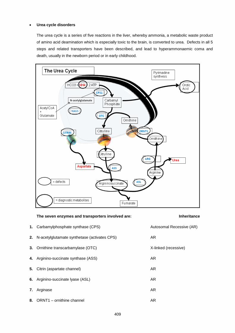
409
• Urea cycle disorders
The urea cycle is a series of five reactions in the liver, whereby ammonia, a metabolic waste product
of amino acid deamination which is especially toxic to the brain, is converted to urea. Defects in all 5
steps and related transporters have been described, and lead to hyperammonaemic coma and
death, usually in the newborn period or in early childhood.
The seven enzymes and transporters involved are: Inheritance
1. Carbamylphosphate synthase (CPS) Autosomal Recessive (AR)
2. N-acetylglutamate synthetase (activates CPS) AR
3. Ornithine transcarbamylase (OTC) X-linked (recessive)
4. Arginino-succinate synthase (ASS) AR
5. Citrin (aspartate channel) AR
6. Arginino-succinate lyase (ASL) AR
7. Arginase AR
8. ORNT1 – ornithine channel AR
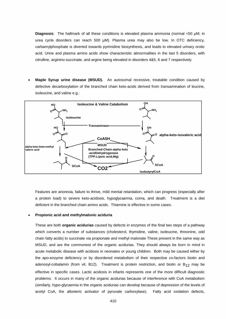
410
Diagnosis: The hallmark of all these conditions is elevated plasma ammonia (normal <50 µM; in
urea cycle disorders can reach 500 µM). Plasma urea may also be low. In OTC deficiency,
carbamylphosphate is diverted towards pyrimidine biosynthesis, and leads to elevated urinary orotic
acid. Urine and plasma amino acids show characteristic abnormalities in the last 5 disorders, with
citrulline, arginino-succinate, and argine being elevated in disorders 4&5, 6 and 7 respectively.
• Maple Syrup urine disease (MSUD). An autosomal recessive, treatable condition caused by
defective decarboxylation of the branched chain keto-acids derived from transamination of leucine,
isoleucine, and valine e.g.:
NH2O
OH
NH2O
OH
O
OH
O O
OH
O
O O
isoleucine
Transaminase
SCoA SCoA
Branched-Chain-alpha keto-acidDehydrogenase(TPP,Lipoic acid,Mg)
CoASH
CO2
MSUD
IsobutyrylCoA
Isoleucine & Valine Catabolism
alpha-keto-isovaleric acid
alpha-keto-beta-methylvaleric acid
Features are anorexia, failure to thrive, mild mental retardation, which can progress (especially after
a protein load) to severe keto-acidosis, hypoglycaemia, coma, and death. Treatment is a diet
deficient in the branched chain amino acids. Thiamine is effective in some cases.
• Propionic acid and methylmalonic aciduria
These are both organic acidurias caused by defects in enzymes of the final two steps of a pathway
which converts a number of substances (cholesterol, thymidine, valine, isoleucine, threonine, odd
chain fatty acids) to succinate via propionate and methyl malonate These present in the same way as
MSUD, and are the commonest of the organic acidurias. They should always be born in mind in
acute metabolic disease with acidosis in neonates or young children. Both may be caused either by
the apo-enzyme deficiency or by disordered metabolism of their respective co-factors biotin and
adenosyl-cobalamin (from vit. B12). Treatment is protein restriction, and biotin or B12 may be
effective in specific cases. Lactic acidosis in infants represents one of the more difficult diagnostic
problems. It occurs in many of the organic acidurias because of interference with CoA metabolism
(similarly, hypo-glycaemia in the organic acidurias can develop because of depression of the levels of
acetyl CoA, the allosteric activator of pyruvate carboxylase). Fatty acid oxidation defects,

411
mitochondrial disorders of the respiratory chain, and pyruvate dehydrogenase deficiency can all give
rise to lactic acidosis. The latter is interesting in that the appropriate treatment is to supply fats, and
avoid carbohydrates, as a source of acetyl CoA.
O
O
OH O
O
O
OHS
SCoA
ProprionylCoA
MethylmalonylCoA
SuccinylCoA
ProprionylCoA-Carboxylase(Proprionic Acidaemia)FT, vomiting, dehydration, ketosis, acidosis, neurological degeneration, coma, hepatomegaly.
SCoA
methyl-malonic acid
Methylmalonyl-CoA-mutase (methyl-malonic acidaemia)
deoxyadenosylcobalamin(B12)
CoACAC GNG
Biotin
Methionone, Odd chain FFA's and Threoninealso end up as proprionyl-SCoA
some MMA patients have a defectin adenosylcobalamin synthesis from cyanocobalamin
valineisoleucine
LYSOSOMAL STORAGE DISEASES:
These conditions develop as a result of deficiencies of specific acid hydrolases present in lysosomes and
responsible for degrading macromolecules such as membrane lipids and mucopoly-saccharides. The
lysosomes become grossly distended with accumulated substrate, and this eventually disturbs cell function.
These disorders present insidiously, with dysmorphic facial features, hepatosplenomegaly, progressive
mental retardation and growth failure, being frequent (but not invariant) features.
• Tay-Sachs disease (GM 2 gangliosidosis)
Progressive mental and motor deterioration with onset in early childhood and invariably fatal
outcome. Lysosomes, especially in neurones, accumulate GM2 ganglioside (a glycosphingolipid) due
to lack ofhexosaminidase A, one of the series of enzymes required for normal turnover of these
complex glycolipids. Early blindness is a feature with a characteristic "cherry-red" spot at the macula.
Hexosaminidase A is found in serum and amniotic fluid, so this provides for a simple screening test,
especially in high risk populations such as Ashkenazi Jews.
412
• Hurler's syndrome. This is caused by deficiency of alpha-iduronidase, one of a number of
lysosomal enzymes required to degrade dermatan and heparan sulphate, which are essential matrix
components (glycosaminoglycans) of connective tissue. Patients are clinically normal in infancy, with
subsequent progressive mental and physical deterioration with stiff joints, deformities, and dwarfism.
There is corneal clouding and a characteristic coarse facial appearance (gargoylism). Death results
from cardiac or respiratory failure. The glycosaminoglycans consist of a chain of modified sugar
residues - heparan sulphate, for example, is a chain of glucuronic and iduronic acid residues, some
of which are sulphated, alternating with α-linked glucosamine residues. Degradation therefore
requires a number of different enzymes and deficiencies in these generally give rise to a set of
diseases with similar but varying clinical pictures. Hunter, Sanfilippo, Morquio and Maroteaux-Lamy
are some of the names linked to specific lysosomal enzyme deficiencies in this group. Another
(older) name for this group of disorders is the mucopolysaccaridoses.
DISORDERS OF AMINO ACID TRANSPORT
In these disorders the defect lies in a membrane transport protein, rather than an enzyme, that is present in
both gut and renal tubular epithelium. Since several amino acids share a common transport mechanism,
characteristic patterns of elevated amino acids are seen in the urine. Note that, in contrast to some of the
enzyme deficiencies described earlier, plasma levels of the affected amino acids are normal or low.
• Cystinuria
This defect involves the transport of the dibasic amino acids, cystine, lysine, arginine and ornithine,
and this results in the excessive excretion of these 4 amino acids. Of the 4, cystine is least soluble,
and tends to precipitate out, particularly in acid urine, forming crystals and stones. In fact, the name
“cystine” derives from a bladder stone of (then) unknown composition, described over a century ago.
Cystine stones are radio-opaque, owing to the presence of the sulfur atom. Cystinuria is relatively
common for an inherited disorder (1 in 7000). A useful screening test is addition of cyanide
nitroprusside to urine. A red colour develops in the presence of free sulfhydryl groups. A positive
test is also observed in homocystinuria, a different condition arising from defective metabolism of the
amino acid, homocysteine. Cystine crystals may be present in urine, and appear as flat hexagonal
rings. Definitive diagnosis depends on the demonstration of increased urine levels of cystine, lysine,
arginine and ornithine. Any renal calculi should be specifically examined for the presence of cystine.
Do not confuse cystinuria with cystinosis, which is a lysosomal disorder due to defective
carrier-mediated transport of cystine across the lysosomal membrane. The major clinical
manifestation of cytinosis is renal failure at about 10 years of age, preceded by development of the
renal tubular Fanconi syndrome - a triad of glycosuria, phosphaturia, and generalised amino-aciduria.
Treatment with cysteamine helps by converting cystine (CS-SC) to cysteine (CSH) and a cysteine-
cysteamine mixed disulphide which can be transported by a different lysosomal transport system (the
lysine transporter)
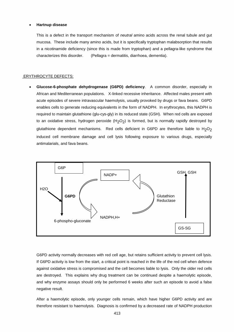
413
• Hartnup disease
This is a defect in the transport mechanism of neutral amino acids across the renal tubule and gut
mucosa. These include many amino acids, but it is specifically tryptophan malabsorption that results
in a nicotinamide deficiency (since this is made from tryptophan) and a pellagra-like syndrome that
characterizes this disorder. (Pellagra = dermatitis, diarrhoea, dementia).
ERYTHROCYTE DEFECTS:
• Glucose-6-phosphate dehydrogenase (G6PD) deficiency. A common disorder, especially in
African and Mediterranean populations. X-linked recessive inheritance. Affected males present with
acute episodes of severe intravascular haemolysis, usually provoked by drugs or fava beans. G6PD
enables cells to generate reducing equivalents in the form of NADPH. In erythrocytes, this NADPH is
required to maintain glutathione (glu-cys-gly) in its reduced state (GSH). When red cells are exposed
to an oxidative stress, hydrogen peroxide (H2O2) is formed, but is normally rapidly destroyed by
glutathione dependent mechanisms. Red cells deficient in G6PD are therefore liable to H2O2
induced cell membrane damage and cell lysis following exposure to various drugs, especially
antimalarials, and fava beans.
G6PD activity normally decreases with red cell age, but retains sufficient activity to prevent cell lysis.
If G6PD activity is low from the start, a critical point is reached in the life of the red cell when defence
against oxidative stress is compromised and the cell becomes liable to lysis. Only the older red cells
are destroyed. This explains why drug treatment can be continued despite a haemolytic episode,
and why enzyme assays should only be performed 6 weeks after such an episode to avoid a false
negative result.
After a haemolytic episode, only younger cells remain, which have higher G6PD activity and are
therefore resistant to haemolysis. Diagnosis is confirmed by a decreased rate of NADPH production
G6P
6-phospho-gluconate
H2O
NADP+
NADPH,H+
G6PD Glutathion Reductase
GS-SG
GSH GSH

414
when a red cell lysate is incubated with glucose-6-phosphate and NADP. In a useful screening test
(Motulsky's test) delay in reducing methylene blue to a colourless leuco form is observed in affected
intact erythrocytes.
Pyruvate kinase (PK) deficiency. Affected patients also show a lifelong tendency to haemolysis.
Decreased activity of this essential glycolytic enzyme impairs ATP synthesis which is required to
maintain red cell integrity. The chronic anaemia is ameliorated by the accumulation of 2,3-DPG,
which enhances oxygen delivery by red cells.
CH3
O P
OOH
CH3
O
OOH
CH3
OH
OOH
Pyruvate Kinase
ADP ATPLDH
NADH,H+
NAD+
PEP
PYRUVATE
LACTATE
340nm
• Methaemoglobin reductase deficiency. Haemoglobin in red cells undergoes slow autoxidation to
ferri- or met-haemoglobin, a dark brown derivative unable to carry oxygen. Methaemoglobin is
maintained in the reduced state by an NADH dependent methaemoglobin reductase. Deficiency of
this enzyme presents as chronic cyanosis and diminished exercise tolerance.
• Sickle cell anaemia (Haemoglobin S disease). A single valine to glutamate substitution in the ß
chain of haemoglobin (HbA HbS) causes it to become insoluble at low oxygen tension. This
precipitation results in deformation of affected erythrocytes (sickling) which aggregate and obstruct
blood flow in capillaries in the brain, joints, and kidney. Splenic infarcts are common. Intercurrent
infection or hypoxia provoke these acute attacks (sickling crises). Heterozygotes show a selective
advantage by having a degree of resistance to malaria infections, hence HbS is common in people of
African ancestry. HbS can be differentiated from normal haemoglobin (HbA1) by electrophoresis of a
red cell lysate.
• Thalassaemia. A genetic defect in α or ß globin production leads to a decrease in normal
haemoglobin (HbA1, α2ß2) and a microcytic hypochromic anaemia. Common amongst Greeks. ß
thalassaemia is more severe (the human genome contains 4 α genes, but only 2 ß genes) and
usually requires lifelong transfusions. Iron overload becomes a problem. Diagnosis depends on
demonstration of abnormal haemoglobins HbA2 (α2 δ2) and HbF(α2 γ2) and imbalance between
415
rates of α and ß globin synthesis. α and ß globin gene probes are also available for demonstrating
the defect at the DNA level.
MITOCHONDRIAL DEFECTS
The major function of the mitochondrion is the synthesis of ATP by the oxidative phosphorylation pathway.
Many of the disorders of energy metabolism can be traced to a defect in one of these enzyes. Presentation
can take a number of forms but include neurological signs, often of sudden onset, blindness, fatigue,
diabetes mellitus – the presentation often being influenced by the degree of mitochondrial deficiency in a
particular tissue.
Subunits of the enzymes of oxidative phosphorylation are encoded both by nuclear DNA and by mitochondrial
DNA (the residual genome of a long-ago captured bacterial symbiont). The degree of mitochondria deficiency
is affected by the nature and site of the mutation and by the proportion of mitochondria in the tissue which
carry the mutation – only in a few cases does the mutation become fixed in all mitochondria in the body. One
such case is Leber’s Hereditary Optic Neuropathy (LHON – see above), which is non-lethal, strictly maternally
inherited, and can affect many successive generations. Otherwise only sporadic individuals are typically
infected, with differing percentages of mitochondria affected in different tissues resulting in differing
presenatations. Examples are MERRF (Myoclonic Epilesy and Ragged Red Fibres) and MELAS
(Mitochondrial Encephalomyopathy and Lactic AcidoSis), the names illustrating some of the main clinical
features of the conditions.
MOLECULAR GENETICS AND INHERITED DISEASE
In the last twenty years the extraordinary advances in recombinant DNA technology (RDT) and bio-
informatics, including the Feb 2000 draft sequence of the whole human genome, have provided new
approaches to the understanding, diagnosis, and therapy of inherited diseases. The implications are
profound and beginning to be fully appreciated. This DNA revolution has relevance to IMDs in three ways:
Firstly, the complete sequence of the human genome has enabled the identification of all genes in the human
genome. The challenge now is to develop an understanding of the pathophysiology of each of these genes,
many of which bear little homology to genes of known function. This was also often the case in the pre-
human genome days when the defective gene (cystic fibrosis, Huntington's) was identified and characterised
before its protein product was found! Secondly, the technology is providing novel and powerful diagnostic
methods, some of which will be described below. Thirdly, the promise of gene therapy where defective genes
are replaced by a normal copy in patients cells, is at last being realised.
Gene Mutations:
These may be of several types:
i. Point mutations due to a single base change. When these occur in the coding region of genes they
can alter a single amino acid, and if this is at a critical position it can change the kinetic properties,

416
stability, transport properties, or processing of the product enzyme. It can also result in a premature
to a stop codon with a consequently shortened protein product.
ii. Loss or gain of bases in numbers not divisible by three (ie. Not 3 or multiples of 3!) causes a frame
shift with the rest of the codons now out of phase, making a nonsense protein product distal to the
shift.
iii. Splice junction mutations can cause an intron to be read as an exon, or vice versa. Often a stop
codon is then found in the transcribed intron, terminating the protein early.
iv. Major deletions or rearrangement of the DNA obviously will cause gross disruption of the gene, with
either no product or a grossly altered product being found.
v. Mutations in the transcriptional control region "upstream" of the coding region of the gene can cause
decreased transcription of mRNA and hence protein product, although the latter would be
qualitatively normal. It would present as a Vmax defect, with normal Km.
vi. Amplification of trinucleotide repeats. A surprising number of dominantly inherited neurological
disorders, of which Huntington’s disease is the classic example, have been found to have
amplification of a short repeat region. In Huntington’s a CAG repeat, normally present in 11-24
copies, is found in typically 40-90 copies in patients, and tends to increase in length with each
generation, with progressively earlier onset of symptoms in the ensuing generations (this is called
“anticipation”).
IDENTIFICATION OF GENE MUTATIONS
The PCR based approach.
The advent in the last few decades of the amazingly powerful polymerase chain reaction (PCR) technique
has relegated the earlier Southern Blotting technique to the history books (very seldom used today). PCR
provides an pproach that is very rapid and versatile and pleasingly, does not require the use of radioactive
probes. PCR uses oligonucleotide primers about 20 base pairs long which need to match the target
sequence exactly for the amplification stage to succeed. For known mutations, oligos can be made which
utilise the mutation itself and create restriction cutting sites within the PCR product which will enable the
distinction between the normal and mutant sequence. For example, the Q188R mutation in exon 6 of the
GALT gene of galactosaemia (AG) , PCR primers are designed to introduce a HpaII cutting site into the
PCR product of the mutant allele. Patients carrying this mutation will show a different PCR product banding
pattern on electrophoresis and can easily be recognised.
Here are the PCR primers used for this mutation detection assay
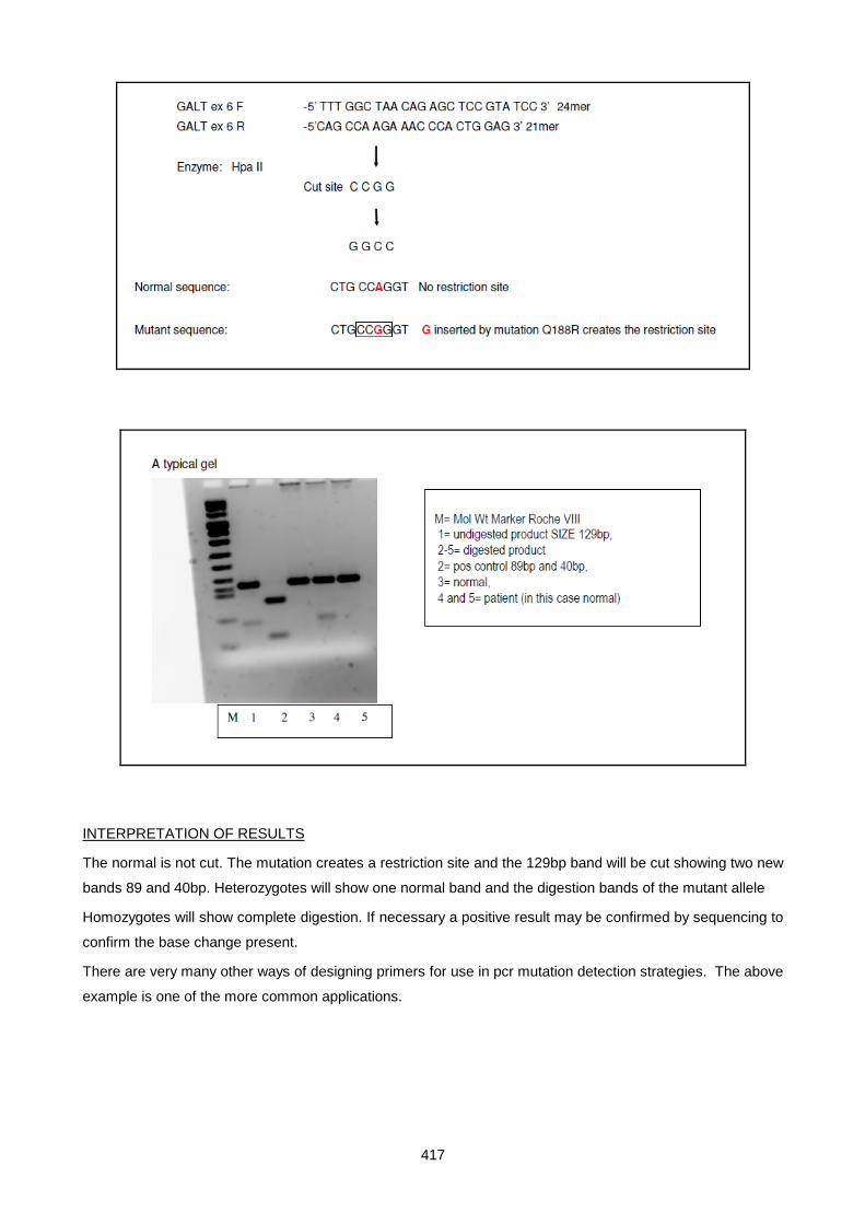
417
INTERPRETATION OF RESULTS
The normal is not cut. The mutation creates a restriction site and the 129bp band will be cut showing two new
bands 89 and 40bp. Heterozygotes will show one normal band and the digestion bands of the mutant allele
Homozygotes will show complete digestion. If necessary a positive result may be confirmed by sequencing to
confirm the base change present.
There are very many other ways of designing primers for use in pcr mutation detection strategies. The above
example is one of the more common applications.
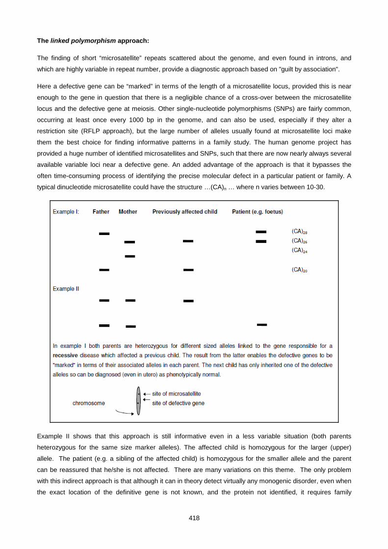
418
The linked polymorphism approach:
The finding of short “microsatellite” repeats scattered about the genome, and even found in introns, and
which are highly variable in repeat number, provide a diagnostic approach based on "guilt by association".
Here a defective gene can be “marked” in terms of the length of a microsatellite locus, provided this is near
enough to the gene in question that there is a negligible chance of a cross-over between the microsatellite
locus and the defective gene at meiosis. Other single-nucleotide polymorphisms (SNPs) are fairly common,
occurring at least once every 1000 bp in the genome, and can also be used, especially if they alter a
restriction site (RFLP approach), but the large number of alleles usually found at microsatellite loci make
them the best choice for finding informative patterns in a family study. The human genome project has
provided a huge number of identified microsatellites and SNPs, such that there are now nearly always several
available variable loci near a defective gene. An added advantage of the approach is that it bypasses the
often time-consuming process of identifying the precise molecular defect in a particular patient or family. A
typical dinucleotide microsatellite could have the structure …(CA)n … where n varies between 10-30.
Example II shows that this approach is still informative even in a less variable situation (both parents
heterozygous for the same size marker alleles). The affected child is homozygous for the larger (upper)
allele. The patient (e.g. a sibling of the affected child) is homozygous for the smaller allele and the parent
can be reassured that he/she is not affected. There are many variations on this theme. The only problem
with this indirect approach is that although it can in theory detect virtually any monogenic disorder, even when
the exact location of the definitive gene is not known, and the protein not identified, it requires family

419
members for study, especially a previously affected sibling, and may require examining a number of potential
marker loci before an informative situation can be found.
Microarrays.
It is now possible to mount large numbers of DNA fragments, each representing a different gene, on a mount
the size of a glass microscope slide. DNA (or even better, mRNA) from a normal reference individual and
from a patient can be differentially labelled with fluorescent dyes; when both are hybridised to the gene
microarray different coloured spots enable differences in gene structure or gene expression to be detected. In
this way it is becoming possible to screen for a huge array of possible defects. This technology is now well
developed and being used for example, to screen patients and populations for a battery of mutations in
cancer genes and cystic fibrosis and familial hypercholesterolemia
Gene Therapy
The technology to clone normal genes and introduce them into cells or the whole organism provide at least a
theoretical means of correcting monogenic disorders. An important principle is that the aim would be to treat
only the somatic tissues of the individual (somatic gene therapy) rather than attempting to correct germ line
DNA, which would carry over the transferred genes to the next generation. Somatic gene therapy is only
being considered for life threatening disorders for which there is no satisfactory alternative therapy. The main
problems have turned out to be getting the genes to the appropriate tissues, and getting controlled,
appropriate, and regulated expression. Candidate disorders are immunodefiencies, lysozomal storage
diseases, hepatic deficiencies (Phenylketonuria and α-1-anti-trypsin deficiency) and coagulation deficiencies
(haemophilia). In 2002 the first fully successful result was published (New Eng, J.Med. 346;1185-1193) in
which four out of five boys suffering from an X-linked form of Severe Combined Immunodeficiency (SCID)due
to a mutation in a cytokine gene necessary for the development of T cells and natural killer cells were
successfully treated by introducing the normal gene (coupled to a retroviral vector) into their bone marrow
cells. A key component of this success was that progenitor T cells which have taken up the gene have a
selective growth advantage, so increase in number. Correction of the immunodeficiency eradicated
established infections and allowed the patients, followed now over two years, to have a normal life. Similar
success has been had with adenosine deaminase deficieny, which is another one of the SCID disorders.
420
Lecture 27- Disorders Of Porphyrin Metabolism
DR P BERMAN 2007
INTRODUCTION
Porphyrias are a group of inherited disorders, each due to a defect in one of the 8 enzymes involved in haem
biosynthesis. Defects in all 8 enzymes have been described; each is associated with a distinct clinical
presentation and pattern of biochemical abnormalities. Their frequency varies enormously with geographical
location. For instance, one porphyria called variegate porphyria (VP), has its highest incidence in SA, and is
due to a so-called ‘founder effect’. The clinical symptoms of porphyria are due to accumulated pathway
intermediates, rather than to a deficiency of haem, the final product of the pathway. Two types of symptoms
occur; those due to acute attacks of nervous system involvement, and those due to chronic skin
photosensitivity. Acute attacks are caused by early pathway intermediates (ALA and PBG), while later
intermediates, the porphyrinogens (after spontaneous conversion to porphyrins), are responsible for
photosensitivity. Certain porphrias only manifest with acute attacks (acute intermittent porphyria), others only
with skin manifestations (porphyria cutanea tarda), while some have both (variegate porphyria). Porphyrias
can also be divided up according to whether the liver or bone marrow is the major source of intermediate
overproduction (hepatic or erythropoeitic porphyrias). Surprisingly, although the affected enzyme is deficient
in all tissues, only the liver or the bone marrow overproduce toxic intermediates.
CONTROL OF HAEM BIOSYNTHESIS
In common with many other biosynthetic pathways, the overall rate of haem biosynthesis is controlled by the
activity of the first enzyme of the pathway, ALA synthase. Particularly in liver, ALA synthase is potently
inhibited by haem, in a classic example of negative feedback.
Many drugs, for example anti-convulsants, induce hepatic synthesis of cytochromeP-450. Since cytP-450 is
a haem-containing enzyme, the demand for haem causes the level of free haem level in the liver cell to fall,
which upregulates ALA synthase, and increases flux down the pathway. If the pathway is partially blocked by
an enzyme deficiency, such drugs lead to a massive increase in intermediates immediately proximal to the
block.
GENETIC CONSIDERATIONS
Most porphyrias are inherited as autosomal dominant; thus enzyme activity in all cells is approximately 50%
of normal. Surprizingly, this modest reduction is sufficient to cause disease in some genetically susceptible
individuals, and accounts for many porphyrics who go through life without experiencing any acute attacks
(latent porphyria). An exception is the devastating congenital erythropoeitic porphyria (CEP), inherited as an
autosomal recessive disorder, and presenting from birth with progressive mutilation of sun-exposed parts of
the body.
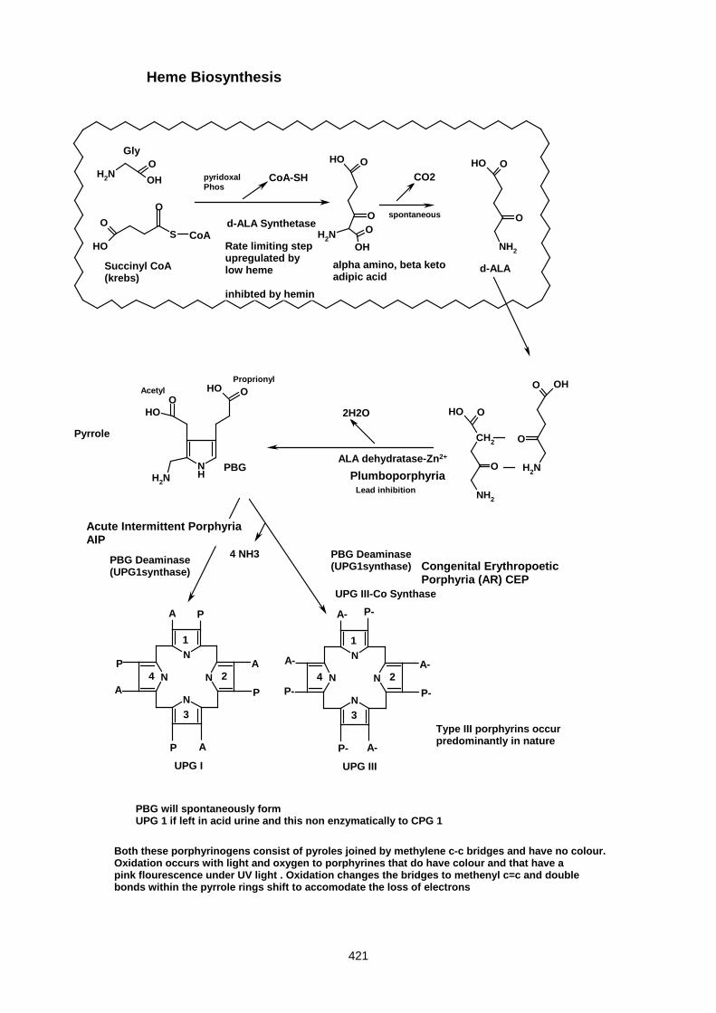
421
NHNH2
OOHO
OH
O
OHS
OO
NH2O
OH
OOH
NH2
OOH
O
NH2
OOH
CH2
NH2
OOH
O NH2
O OH
O
N
N
N
N N
N
N
N
Pyrrole
CoA
Gly
Succinyl CoA(krebs)
CoA-SH
d-ALA Synthetase
alpha amino, beta ketoadipic acid
CO2
spontaneous
d-ALA
pyridoxalPhos
ALA dehydratase-Zn2+
2H2O
PBG
Lead inhibition
PBG Deaminase(UPG1synthase)
4 NH3 PBG Deaminase(UPG1synthase)
1
2
3
4
A P
A
P
AP
A
P
UPG I
1
2
3
4
A- P-
A-
P-
A-P-
A-
P-
UPG III
PBG will spontaneously formUPG 1 if left in acid urine and this non enzymatically to CPG 1
UPG III-Co Synthase
Both these porphyrinogens consist of pyroles joined by methylene c-c bridges and have no colour.Oxidation occurs with light and oxygen to porphyrines that do have colour and that have apink flourescence under UV light . Oxidation changes the bridges to methenyl c=c and doublebonds within the pyrrole rings shift to accomodate the loss of electrons
Type III porphyrins occurpredominantly in nature
Rate limiting stepupregulated bylow heme
inhibted by hemin
AcetylProprionyl
Plumboporphyria
Acute Intermittent PorphyriaAIP
Congenital ErythropoeticPorphyria (AR) CEP
Heme Biosynthesis
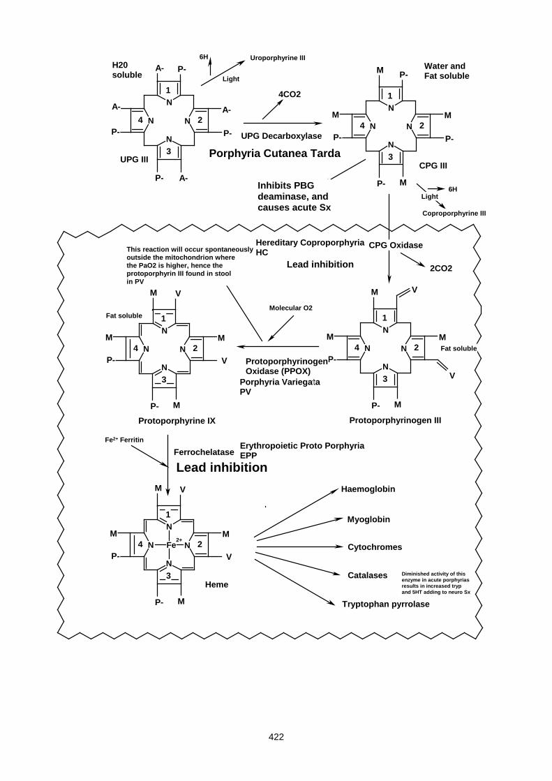
422
N
N
N
NN
N
N
N
N
N
N N N
N
N
N
N
N
N NFe2+
1
2
3
4
P-
A-
P-
A-
A-
P-
UPG III
P-
A-
1
2
3
4
P-
P-P-
P-
4CO2
UPG Decarboxylase
M
M
M
M
Porphyria Cutanea TardaCPG III
6H Uroporphyrine III
Light
Light6H
Coproporphyrine III
CPG Oxidase
1
2
3
4P-
P-
M
M
M
M
V
V
1
2
3
4P-
P-
M
M
M
M
2CO2
V
V
Protoporphyrinogen III
Hereditary CoproporphyriaHC
Inhibits PBGdeaminase, andcauses acute Sx
ProtoporphyrinogenOxidase (PPOX)
Porphyria VariegataPV
Molecular O2
This reaction will occur spontaneouslyoutside the mitochondrion wherethe PaO2 is higher, hence the protoporphyrin III found in stoolin PV
Protoporphyrine IX
1
2
3
4P-
P-
M
M
M
V
V
M
FerrochelataseErythropoietic Proto PorphyriaEPP
Heme
Fe2+ Ferritin
H20soluble
Water andFat soluble
Fat soluble
Fat soluble
Haemoglobin
Myoglobin
Cytochromes
Catalases
Tryptophan pyrrolase
Diminished activity of thisenzyme in acute porphyriasresults in increased trypand 5HT adding to neuro Sx
Lead inhibition
Lead inhibition

423
ACUTE ATTACK OF PORPHYRIA
Only 4 porphyrias present with acute attacks. These are acute intermittent porphyria (AIP), variegate
porphyria (VP) and the much rarer hereditary coproporphyria (HC) and plumboporphyria (PLP).
Attacks generally occur in young adults, almost always after puberty, and are commoner among women.
They are caused by the porphyrin precursors, ALA and PBG, that are overproduced by the liver, often in
response to certain precipitating factors. Indeed, increased urinary ALA and PBG is the diagnostic hallmark of
an acute porphyric attack. Precipitating factors include:
1. Many commonly prescribed drugs, eg. barbiturates, sulfonamides
2. Carbohydrate depletion (fasting, crash diets)
3. Certain steroid hormones and their metabolites; this explains the association with pregnancy, the
menstrual cycle, oral contraceptives, and the higher incidence in females generally.
Drugs induce an acute attack by depleting the hepatic free haem pool. Most (eg. barbiturates) do so by
inducing the hepatic cytochrome P450 system as described. Others directly inhibit one of the haem
biosynthetic enzymes, eg. sulphonamides inhibit PBG deaminase and griseofulvin inhibits ferrochelatase.
Fasting induces haem-containing gluconeogenic enzymes (eg tryptophan pyrrolase), while sex steroids,
particularly metabolites of progesterone and testosterone, directly activate ALA synthase.
Clinical features of an acute attack
I. Abdominal pain and vomiting (may mimic a surgical emergency)
II. Sensory neuropathy (limb pain, parasthesia, numbness)
III. Motor neuropathy (absent reflexes, weakness, which can progress to a bulbar palsy and respiratory
paralysis)
IV. Autonomic neuropathy (tachycardia, hypertension, cardiac arrythmias, constipation and urinary
retention)
V. Central nervous system involvement (anxiety, depression, psychosis, convulsions)
VI. Hyponatraemia – variously attributed to vomiting, renal salt wasting, or SIADH
MANAGEMENT OF ACUTE ATTACKS
Do not operate !!!
Treat symptomatically, eg physiotherapy for muscle palsies, artifcial ventilation for respiratory paralysis.
Only use drugs known not to induce porphyria, eg opiates for pain, chlorpromazine for nausea, β-blockers for
adrenergic overactivity.

424
Push carbohydrate intake, either orally or intravenously
Correct hyponatraemia if present, and maintain normal fluid balance.
Specific therapy is directed at maintaining intrahepatic haem levels to suppress ALA synthase; this is
achieved either by infusing haem directly, or giving an inhibitor of haem oxygenase, the enzyme that breaks
haem down to bilirubin.
Prevention of acute attacks
Any individuals at risk for acute attacks should be identified by appropriate biochemical tests, and educated
about predisposing factors, including drugs, steroid hormones and fasting. In some women, down-regulation
of the pituitary-ovarian axis with a GnRH analogue may be the only way to prevent debilitating pre-menstrual
attacks.
PHOTOSENSITIVITY IN PORPHYRIA
Cutaneous photosensitivity is a major problem in many porphyrias (excluding acute intermittent porphyria
(AIP)). Skin damage is caused by circulating porphyrins, formed by spontaneous oxidation of the
corresponding porphyrinogens. Damage always occurs in sun exposed areas such as the face and dorsum
of the hands, and begins with blisters, which ulcerate and heal slowly, often become secondarily infected, and
leave disfiguring scars. Old lesions can be hyper- and/or hypo-pigmented, and there is increased skin
fragility Facial hypertrichosis (hairiness) is a common feature (possibly an attempt to limit light exposure? -
PB).
The molecular mechanism of the skin injury is interesting. Porphyrins absorb long-wavelength UV light
(around 400 nm) and transmit the energy to molecular oxygen, forming reactive oxygen radicals that destroy
the lipids in cell membranes. Thus lipid-soluble oxygen free-radical scavengers, such as ß carotene, exert a
protective effect.
Management of photosensitivity includes:
• Avoidance of direct sunlight
• Sunscreens that block long-wavelength UV light (conventional sunscreens only absorb short UV)
• Administration of ß-carotene.
• Oral charcoal (rather unpalatable!) binds porphyrins in the bowel lumen and enhances their excretion.
BIOCHEMICAL DIAGNOSIS OF PORPHYRIA
Biochemical confirmation of porphyria requires demonstration of increased quantities of pathway
intermediates. Where does one look?
The early intermediates of haem biosynthesis namely ALA, PBG and the 8 carboxyl uroporphyrin, are highly
water soluble, and are therefore excreted in the urine. As porphyrins lose carboxyl groups, they become
progressively less water soluble, and come to depend on biliary excretion into the stool, i.e.
425
URINE STOOL
ALA & PBG coproporphyrin
uroporphyrin protoporphyrin
coproporphyrin
ALA & PBG:
A useful test for PBG is a pink colour that forms on the addition of Ehrlichs aldehyde to urine.
As other compounds (particularly urobilinogen) give a similar colour, the test can be made more specific by
extracting the coloured product with organic solvents, such as butanol. In the case of PBG, the pink colour
remains in the aqueous phase.
While no such simple screening test exists for ALA, it can be measured in most laboratories, and is useful for
confirming an acute porphyric attack or for diagnosing lead poisoning.
PORPHYRINS: Detection of porphyrins is based on the intense red fluorescence emit when illuminated with
long wavelength UV light (∼400nm). Since fluorescence can be masked in opaque fluids (blood or faeces), it
is customary to first extract porphyrins into organic solvents, then check for red fluorescence.
More detailed information can be obtained by seperating individual porphyrins by chromatography, which
enables us to identify which prophyrins are present. For example isocoproporphyrin in stool is indicative of
PCT, whereas protoporphyrin in plasma indicates VP.
Analysis of red cells can be useful. Elevated levels of porphyrins in red cells confirm one of the
erythropoeitic porphyrias (CEP or EPP), or an acquired defect of erythropoeisis, such as iron deficiency or
lead poisoning. Also, red cells provide a convenient source of certain haem biosynthetic enzymes eg. red cell
PBG deaminase and ALA dehydrase assays readily detect latent AIP and lead poisoning, respectively.
SPECIFIC PORPHYRIAS
o Acute intermittent porphyria (AIP)
This is a common form of porphyria world-wide, with highest incidence in Sweden, and is due to a
defect in PBG deaminase, leading to overproduction of ALA and PBG. It presents with acute attacks
only (no skin lesions). Diagnosis rests on demonstrating increased urine ALA and PBG, particularly
during attacks. Stool and blood porphyrins are normal. Urinary PBG may, however, polymerize non-
enzymatically to form porphyrin, which accounts for the gradually deepening red colour that develops
in the urine on exposure to air.
A 50% reduction in PBG deaminase activity of erythrocytes is a useful marker of AIP in the latent
phase, i.e. before puberty or between attacks, and for identifying family members at risk.
o Variegate porphyria (VP)

426
This form of porphyria is particularly common in South Africa among people of Afrikaner extraction,
and can be traced back to a single Dutch woman (Adriaantjie Adriaanse) who arrived at the Cape in
1688 (founder effect). It is due to a point mutation in the protoporphyrinogen oxidase gene, and can
present with either acute attacks, skin lesions, or both (hence variegate). The photosensitivity is due
to the accumulated protoporphyrin, while acute attacks are probably due to a secondary inhibition of
PBG deaminase by accumulated protoporphyrinogen.
A markedly increased stool content of porphyrin (proto > copro) is diagnostic of this condition. GIT
bleeding (removal of the Fe atom from haem by bowel flora) and dietary chlorophyll may give false
positive fluorescence.
Urine ALA and PBG are only increased during attacks. Stool protoporphyrin remains elevated
between attacks, and is the best marker of latent disease. A recently described test relies on the
presence of red fluorescence in plasma (due to accumulated protoporphyrin, which is less polar and
hence more protein bound than other porphyrins).
o Hereditary coproporphyria (HC)
This condition is much rarer than AIP or VP, and is due to a defect in coproporphyrinogen oxidase,
leading to accumulation of coproporphyrin. Patients suffer acute attacks and may develop skin
lesions that are generally milder than in VP. The condition is characterized by excess coproporphyrin
excretion in urine and stool. Urine ALA and PBG are increased only during acute attacks.
THE FOLLOWING 3 PORPHYRIAS DO NOT MANIFEST ACUTE ATTACKS.
o Porphyria cutanea tarda (PCT)
This is the commonest porphyria in global terms, and is due to a defect in hepatic uroporphyrinogen
decarboxylase. Uroporphyrin accumulates and causes skin lesions, often with a prominent fibrotic
response resembling scleroderma (pinched features, circumoral contracture). It never causes acute
attacks. The biochemical hallmark of PCT is massive increase in urine porphyrin, mainly
uroporphyrin. Stool characteristically contains isocoproporphyrin, a 4 carboxyl porphyrin with 1
acetate and 3 propionate side chains (as opposed to regular coproporphyrin, where all 4 carboxyls
are propionates). In many patients with PCT, no defect in the uro-decarboxylase gene can be
demonstrated, and a family history is not present; others are heterozygous for a defective uro-
decarboxylase gene. In either case, an additional environmental factor is essential for manifestation
of the disease phenotype. Such environmental factors include iron overload, alcoholism (particularly
with liver damage), or oestrogen administration. It is more frequent in males (more predisposed to
iron overload), and presents in late adulthood (hence "tarda", meaning slow onset).Treatment of PCT
includes iron depletion (repeated venesection) and alcohol withdrawal. Low dose chloroquine (an
antimalarial) binds to and enhances uroporphyrin excretion.Occasionally, a PCT-like syndrome arises
in people poisoned with chlorinated hydrocarbons such as hexachlorobenzene, that targets the same
enzyme (uro-decarboxylase). Such an outbreak of 'monkey' disease in Turkey (1956) due to
accidental ingestion of contaminated wheat seed. Severe hypertrichosis, due to photosensitivity,
resulted in a monkey-like appearance.
427
IN THE FOLLOWING 2 PORPHYRIAS, BONE MARROW, RATHER THAN LIVER, IS THE
SOURCE OF PORPHYRIN OVERPRODUCTION
o Congenital Erythropoetic Porphyria (CEP)
This (fortunately) rare disorder is inherited as autosomal recessive (most other porphyrias show
dominant inheritance) and is due to a defect in uroporphyrinogen III co-synthase. Severe
photosensitivity is the big problem here. Skin damage begins from infancy, and progresses to
mutilation of face and hands with marked hypertrichosis. Mothers may report red/brown staining of
nappies due to excessive urinary porphyrins. Teeth show a brown discoloration (erythrodontia), and
fluoresce red in UV light. It has been suggested that individuals with CEP gave rise to the legend of
‘werewolves’. Haemolytic anaemia is often present (red cells are damaged as they pass through skin
capillaries), and responds to splenectomy. Improvement of the anaemia helps to reduce the
haematopoetic drive and hence lessen porphyrin overproduction.
There is an enormous increase in red cell, plasma and urine porphyrins of the I series (uro-I and
copro-I). Urine ALA and PBG are normal. The diagnostic hallmark of CEP is the enormously
increased porphyrin content of urine AND red cells.
o Erythropoetic Protoporphyria (EPP)
This condition is due to a deficiency of ferro-chelatase, and presents in childhood. Skin damage is
much less severe than in CEP, and manifests as erythema, itching, and burning, immediately
following exposure to sunlight. It leaves little permanent scarring. The major risk is development of
gallstones and liver damage in later life due to accumulation of protoporphyrin in the liver.
Since the excess protoporphyrin is excreted in the faeces (as in VP), an increase in red cell and
faecal porphyrin, with normal urine porphyrin, is typical of EPP.
PORPHYRINURIA
Porphyrinuria is the excessive urinary excretion of porphyrins (mainly coproporphyrin) due to an acquired,
rather than inherited, disorder. Some of the commoner causes of porphyrinuria include:
o Hepatic disease
Since the liver is responsible for removing much of the normal production of coproporphyrin, failure of
biliary excretion in liver disease manifest with increased urinary coproporphyrin excretion.
In Dubin-Johnson syndrome, a specific defect in biliary excretion of anions, including conjugated
bilirubin and coproporphyrin, leads to jaundice and porphyrinuria, respectively.
Primary liver cancer (hepatoma) may also overproduce porphyrin and lead to porphyrinuria.
o Ineffective haematopoeisis
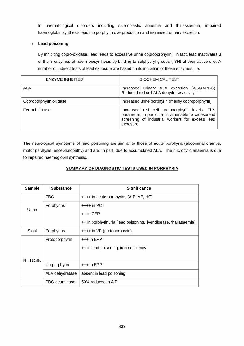
428
In haematological disorders including sideroblastic anaemia and thalassaemia, impaired
haemoglobin synthesis leads to porphyrin overproduction and increased urinary excretion.
o Lead poisoning
By inhibiting copro-oxidase, lead leads to excessive urine coproporphyrin. In fact, lead inactivates 3
of the 8 enzymes of haem biosynthesis by binding to sulphydryl groups (-SH) at their active site. A
number of indirect tests of lead exposure are based on its inhibition of these enzymes, i.e.
ENZYME INHBITED BIOCHEMICAL TEST
ALA Increased urinary ALA excretion (ALA>>PBG) Reduced red cell ALA dehydrase activity
Coproporphyrin oxidase Increased urine porphyrin (mainly coproporphyrin)
Ferrochelatase Increased red cell protoporphyrin levels. This parameter, in particular is amenable to widespread screening of industrial workers for excess lead exposure.
The neurological symptoms of lead poisoning are similar to those of acute porphyria (abdominal cramps,
motor paralysis, encephalopathy) and are, in part, due to accumulated ALA. The microcytic anaemia is due
to impaired haemoglobin synthesis.
SUMMARY OF DIAGNOSTIC TESTS USED IN PORPHYRIA
Sample Substance Significance
Urine
PBG ++++ in acute porphyrias (AIP, VP, HC)
Porphyrins ++++ in PCT
++ in CEP
++ in porphyrinuria (lead poisoning, liver disease, thallasaemia)
Stool Porphyrins ++++ in VP (protoporphyrin)
Red Cells
Protoporphyrin +++ in EPP
++ in lead poisoning, iron deficiency
Uroporphyrin +++ in EPP
ALA dehydratase absent in lead poisoning
PBG deaminase 50% reduced in AIP

429
SMALL GROUP TEACHING: LECTURE 27: PORPHYRIA
QUESTIONS
1. A nineteen-year-old girl was admitted to hospital with colicky abdominal pain, which had started suddenly twelve hours before. She had vomited several times but had not passed stool since the pain started. Her abdomen was tender on examination but was otherwise normal. Her pulse was 140/min and her blood pressure was 160/100mmHg. After she had been taken to the ward for observation, a nurse in the emergency room noticed that a specimen of patient's urine, which had been collected for routine testing, had become a deep red colour although it had been normal when first passed. On being informed of this, the admitting doctor questioned the patient further and examined her more carefully. She said that she had also noticed cramping pains in her arms and was found to have bilateral partial wrist drop.
Investigations
screening test for urinary porphobilinogen: strongly positive
quantitative analysis of urine:
porphobilinogen very high
d-aminolaevulinic acid very high
uroporphyrin slightly raised
coproporphyrin slightly raised
Comments
Acute porphyrias may present as an acute abdomen; systemic hypertension and sinus tachycardia are often present. They are of course a very uncommon cause of abdominal pain and the diagnosis may be missed, at least initially. In this case, the nurse's observation of the changed colour of the urine was crucial and led to the presumptive diagnosis of an acute porphyria being made, supported clinically by the evidence of neuropathy and also by the positive screening test for PBG. There was no evidence of photosensitivity and the very high urinary excretion of porphyrin precursors, with only slightly increased excretion of intact porphyrins, favours a diagnosis of acute intermittent porphyria rather than variegate or hereditary coproporphyria, both of which are anyway much less common (EXCEPT IN SA).
The patient's symptoms and signs resolved rapidly with appropriate treatment. It transpired that she had started taking an oral contraceptive pill a few days before. The diagnosis was later confirmed by the demonstration of a reduced red cell PBG deaminase activity; she was advised to use an alternative method of contraception and told which drugs she should avoid. She remained well thereafter and no problems arose when she underwent elective surgery (cholecystectomy) three years later, nor during a subsequent pregnancy.
i. Account for her acute constipation.
ii. Account for the change in colour of the patient's urine on standing.
iii. What is the screening test for urinary porphobilinogen?
iv. How low do you think her red cell PBG deaminase was, compared to normal controls? Why?
v. Based on your knowledge of the genetic basis of the disorder, what advice would you give family members?
vi. Why is particular mention made of no problems developing when she underwent elective surgery or became pregnant?
2. Fill in the table
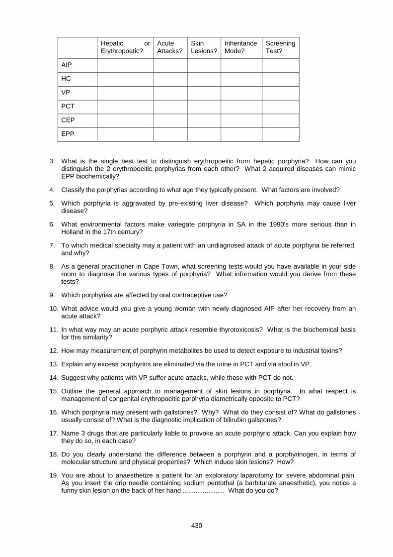
430
Hepatic or Erythropoetic?
Acute Attacks?
Skin Lesions?
Inheritance Mode?
Screening Test?
AIP
HC
VP
PCT
CEP
EPP
3. What is the single best test to distinguish erythropoeitic from hepatic porphyria? How can you distinguish the 2 erythropoeitic porphyrias from each other? What 2 acquired diseases can mimic EPP biochemically?
4. Classify the porphyrias according to what age they typically present. What factors are involved?
5. Which porphyria is aggravated by pre-existing liver disease? Which porphyria may cause liver disease?
6. What environmental factors make variegate porphyria in SA in the 1990's more serious than in Holland in the 17th century?
7. To which medical specialty may a patient with an undiagnosed attack of acute porphyria be referred, and why?
8. As a general practitioner in Cape Town, what screening tests would you have available in your side room to diagnose the various types of porphyria? What information would you derive from these tests?
9. Which porphyrias are affected by oral contraceptive use?
10. What advice would you give a young woman with newly diagnosed AIP after her recovery from an acute attack?
11. In what way may an acute porphyric attack resemble thyrotoxicosis? What is the biochemical basis for this similarity?
12. How may measurement of porphyrin metabolites be used to detect exposure to industrial toxins?
13. Explain why excess porphyrins are eliminated via the urine in PCT and via stool in VP.
14. Suggest why patients with VP suffer acute attacks, while those with PCT do not.
15. Outline the general approach to management of skin lesions in porphyria. In what respect is management of congenital erythropoeitic porphyria diametrically opposite to PCT?
16. Which porphyria may present with gallstones? Why? What do they consist of? What do gallstones usually consist of? What is the diagnostic implication of bilirubin gallstones?
17. Name 3 drugs that are particularly liable to provoke an acute porphyric attack. Can you explain how they do so, in each case?
18. Do you clearly understand the difference between a porphyrin and a porphyrinogen, in terms of molecular structure and physical properties? Which induce skin lesions? How?
19. You are about to anaesthetize a patient for an exploratory laparotomy for severe abdominal pain. As you insert the drip needle containing sodium pentothal (a barbiturate anaesthetic), you notice a funny skin lesion on the back of her hand ....................... What do you do?
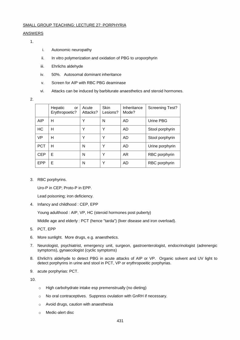
431
SMALL GROUP TEACHING: LECTURE 27: PORPHYRIA
ANSWERS
1.
i. Autonomic neuropathy
ii. In vitro polymerization and oxidation of PBG to uroporphyrin
iii. Ehrlichs aldehyde
iv. 50%. Autosomal dominant inheritance
v. Screen for AIP with RBC PBG deaminase
vi. Attacks can be induced by barbiturate anaesthetics and steroid hormones.
2.
Hepatic or Erythropoetic?
Acute Attacks?
Skin Lesions?
Inheritance Mode?
Screening Test?
AIP H Y N AD Urine PBG
HC H Y Y AD Stool porphyrin
VP H Y Y AD Stool porphyrin
PCT H N Y AD Urine porphyrin
CEP E N Y AR RBC porphyrin
EPP E N Y AD RBC porphyrin
3. RBC porphyrins.
Uro-P in CEP; Proto-P in EPP.
Lead poisoning; iron deficiency.
4. Infancy and childhood : CEP, EPP
Young adulthood : AIP, VP, HC (steroid hormones post puberty)
Middle age and elderly : PCT (hence "tarda") (liver disease and iron overload).
5. PCT, EPP
6. More sunlight. More drugs, e.g. anaesthetics.
7. Neurologist, psychiatrist, emergency unit, surgeon, gastroenterologist, endocrinologist (adrenergic symptoms), gynaecologist (cyclic symptoms)
8. Ehrlich's aldehyde to detect PBG in acute attacks of AIP or VP. Organic solvent and UV light to detect porphyrins in urine and stool in PCT, VP or erythropoeitic porphyrias.
9. acute porphyrias: PCT.
10.
o High carbohydrate intake esp premenstrually (no dieting)
o No oral contraceptives. Suppress ovulation with GnRH if necessary.
o Avoid drugs, caution with anaesthesia
o Medic-alert disc
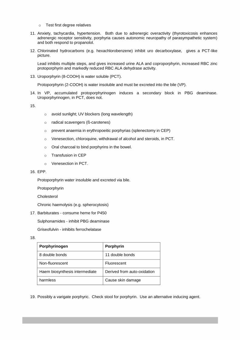
432
o Test first degree relatives
11. Anxiety, tachycardia, hypertension. Both due to adrenergic overactivity (thyrotoxicosis enhances adrenergic receptor sensitivity, porphyria causes autonomic neuropathy of parasympathetic system) and both respond to propanolol.
12. Chlorinated hydrocarbons (e.g. hexachlorobenzene) inhibit uro decarboxylase, gives a PCT-like picture.
Lead inhibits multiple steps, and gives increased urine ALA and coproporphyrin, increased RBC zinc protoporphyrin and markedly reduced RBC ALA dehydrase activity.
13. Uroporphyrin (8-COOH) is water soluble (PCT).
Protoporphyrin (2-COOH) is water insoluble and must be excreted into the bile (VP).
14. In VP, accumulated protoporphyrinogen induces a secondary block in PBG deaminase. Uroporphyrinogen, in PCT, does not.
15.
o avoid sunlight; UV blockers (long wavelength)
o radical scavengers (ß-carotenes)
o prevent anaemia in erythropoeitic porphyrias (splenectomy in CEP)
o Venesection, chloroquine, withdrawal of alcohol and steroids, in PCT.
o Oral charcoal to bind porphyrins in the bowel.
o Transfusion in CEP
o Venesection in PCT.
16. EPP.
Protoporphyrin water insoluble and excreted via bile.
Protoporphyrin
Cholesterol
Chronic haemolysis (e.g. spherocytosis)
17. Barbiturates - consume heme for P450
Sulphonamides - inhibit PBG deaminase
Griseofulvin - inhibits ferrochelatase
18.
Porphyrinogen Porphyrin
8 double bonds 11 double bonds
Non-fluorescent Fluorescent
Haem biosynthesis intermediate Derived from auto-oxidation
harmless Cause skin damage
19. Possibly a varigate porphyric. Check stool for porphyrin. Use an alternative inducing agent.

433
Lecture 28: Tumour Markers
PROF E H HARLEY, REVISED BY DR F. OMAR 2007
An IDEAL tumour marker
• Is normally absent, or present only at low levels in non-diseased individuals.
• Provides no false positives or false negatives (i.e. it is sensitive and specific), clearly separating
normal and diseased levels.
• Provides a useful “lead time” compared to the usual clinical presentation.
• Is specific for a particular tumour.
• Correlates with tumour mass and stage.
• Is prognostically useful i.e. a rise in level predicts recurrence / relapse.
• Is easily and inexpensively measured in readily available bodily fluids.
There are no tumour markers that meet all of these criteria. In addition, if a tumour marker is used to detect
early stages of cancer, a treatment must exist.
An ideal tumour marker would be useful for screening, diagnosis, prognosis and staging, monitoring
response to therapy, monitoring for recurrence or remission and tumour localisation. However, of all the
tumour markers in use currently, only PSA has been proven useful for screening. The other markers are
useful as diagnostic aids (e.g. CA 125); for treatment guidance (e.g. estrogen receptor) and monitoring (e.g.
CEA), as well as for staging and prognosticating.
Evaluating the clinical utility of a tumour marker
• The decision level (cut-off that seperates positive and negative results) for a specific marker / analyte
must be established.
• This is accomplished using the predictive value model (sensitivity, specificity and predictive value) of
the test for a positive diagnosis.
• The ROC curve (see diagram) provides visual display of performance for the entire range of decision
levels.
• The most optimal decision level (i.e. with most optimal sensitivity and specificity) can be pinpointed.
• Useful when comparing different markers for the same cancer.

434
Several biochemical tests are useful, either as primary tumour markers or as secondary tests for invasion or
metastases or cancer.
Classification of tumour markers:
1. Proteins:
a. Enzymes
b. Hormones
c. Other proteins
i. Monoclonal Immunoglobulins
ii. β2-microglobulin
iii. C-peptide
iv. Hormone receptors (estrogen and progesterone receptors)
2. Oncofoetal antigens
3. Carbohydrate markers
4. Genetic markers
a. Oncogenes
b. Tumour suppressor genes
5. Newer “markers”
a. Microarray
b. Proteomics
c. Cell free RNA
PROTEINS
A. ENZYMES
Plasma enzyme/iso-enzymes activities often increased due to secondary effects of tumour.
Levels correlate well with tumour burden. Usually non-specific (excl. PSA) and thus not useful in diagnosis
of tumour type or organ involved. Benefit in monitoring.
Decision
Figure: Receiver Operating Characteristic (ROC)
Curve:
This curve plots the sensitivity vs (1 –
specificity) over a series of decision levels.
The closer the curve gets to the top left
corner, the better is the test at distinguishing
diseased from non-diseased.
435
Iso-enzyme measurement can improve specificity. Foetal form (iso-enzymes) or ectopic production in
certain malignancies.
Examples of useful enzymes in malignancy:
i. Alkaline phosphatase
Useful in assessment of bone or liver metastases. Bone ALP elevated in osteoblastic lesions (e.g.
prostate carcinoma), not in osteolytic lesions (e.g. myeloma).Correlates better with extent of liver
metastases than other liver enzymes. Elevated GGT and isoenzyme determination (heat stability)
confirms liver origin. Regan iso-enzyme (placental ALP) elevated in malignancies such as ovarian,
lung, trophoblastic and GIT cancers.
ii. Lactate Dehydrogenase
Glycolytic enzyme released following cell damage.
Usually tumours that primarily utilise glycolysis rather than aerobic respiration for their energy
requirements. Elevated with many large tumours, including breast, colon and stomach tumours;
also in lymphoma, leukaemia and neuroblastoma.
Levels correlate well with tumour burden.
iii. Creatine kinase
Occasionally elevated in tumours such as prostate, prostate and small cell carcinoma of the lung.
Usually BB iso-enzyme. May cause false elevation in CK-MB level in certain CK-MB assays.
iv. Neuron specific enolase
Glycolytic enzyme found in neuronal tissue and neuroendocrine system. Elevated in small cell
carcinoma of the lung, neuroblastoma, phaechromocytoma, carcinoid, medullary carcinoma of the
thyroid and pancreatic endocrine tumours. Associated with poor prognosis.
v. Urokinase-plasminogen activator (uPA) system
Includes uPA, uPA receptor and uPA inhibitors (PAI-1 & -2). uPA activates plasmin, leading to
eventual degradation of extracellular matrix and activation of certain growth factors. Negative
prognosticator in breast, colorectal, gastric and oesophageal cancers. PAI-1 regulates uPA, also
inhibits apoptosis and promotes angiogenesis. High PAI-1 levels associated with aggressive
disease. Analytical methodology: ELISA in serum and tissue extracts; Immunohistochemistry
(semi-quantitative, but easier).
vi. Telomerase
Repairs telomeres (hexanucleotide repeats at the end of a chromosome; shortens with each
replication; sets finite life span to normal somatic cells). Telomerase suppressed in most somatic
cells at birth. Germline cells and other immortal cells have continued activity, preventing telomere
shortening. Found in many tumours incl. that of lung, breast, bladder and cervix. Correlates with
436
tumour progression and prognosis. Analytical methodology: Telomeric Repeat Amplification
Protocol (PCR, Elisa, RT-PCR)
MARKERS OF PROSTATE CANCER
vii. Prostatic acid phosphatase
Tartrate sensitive acid phosphatase produced by epithelial cells of prostate gland. No longer in
routine use, replaced by PSA. Clinical use restricted to confirmation of metastatic prostate cancer
and prostate cancer staging. Less likely to elevated in BPH than is PSA.
viii. PROSTATE SPECIFIC ANTIGEN (PSA)
Sensitive and stable
Expressed by normal, benign, hyperplastic and malignant prostatic tissue.
Thus specific for prostate, but not for malignancy. Single chain glycoprotein. Mw 28.4kDa. Serine
protease (similar action to trypsin and chymotrypsin), produced by epithelial cells of acini and ducts
of prostate gland. Secreted into seminal fluid. Function: cleaves seminogelin (after sexual
intercourse), liquefying seminal coagulum to release entrapped spermatozoa. Basement
membrane prevents PSA escape into circulation before inactivation in ducts. Disruption of this
barrier (as in cancer) causes PSA overspill directly into circulation before inhibition.
Two major forms of PSA exist i.e.
a. free = nicked inactive form (this is good PSA)
b. complexed = intact enzymes that form complexes with α1-antichymotrypsin and α2-
macroglobulin (this is bad PSA)
Clinical application:
Total PSA < 4 ug/L is considered normal.
4 – 10 ug/L “grey zone”
> 10 ug/L prostate cancer
PSA useful in detection, staging and monitoring of prostate cancer. Not specific for prostate
cancer. Also elevated in benign conditions such as:
BPH - overlaps with early organ-confined prostate carcinoma (4 – 10ug/L)
Prostatitis – returns to normal within 6 weeks (levels usually 4 – 10ug/L)
Also urethral catheterisation, acute urinary retention, prostate biopsy, prostatic massage, digital
rectal examination (2-fold increase), sexual intercourse, even cycling.
Approaches to improve specificity for early prostate CA:
437
a. age-adjusted reference intervals
40-49yrs 0-2.5ug/L
50-59yrs 0-3.5ug/L
60-69yrs 0-4.5ug/L
70-79yrs 0-6.5ug/L
b. PSA density-[PSA]/prostate volume by transrectal ultrasonography
c. PSA velocity – rate of PSA increase over time. Rate > 0.75ug/L/yr suggestive of prostate
cancer
d. Percentage fPSA- especially useful when tPSA falls in the grey zone. fPSA < 15% highly
suggestive of prostate CA.
e. Complexed PSA – elevated cPSA associated with prostate carcinoma
PSA levels correlate with stages of tumour extension and metastases, advanced stages associated
with higher PSA levels.
Organ confined usually < 50ug/L
Bone metastases seldom < 20ug/L
PSA in monitoring of treatment:
Almost exclusively produced by prostatic tissue. Levels should drop below detection limit after
prostatectomy – within 2 – 3 weeks (t ½ = 2-3 days).
Levels increase with cancer progression, decrease in remission and remain unchanged in stable
disease.
Analytical methodology: immunometric assay. Different assays yield different results due to
variations in antibody epitopes, assay calibration, assay reaction time, reagent matrices, assay
sensitivity and imprecision. Equimolar assays bind fPSA and cPSA equally (best). Non-equimolar
assays favour one over the other (unreliable). Ultrasensitive assays (detection limit 0.01 –
0.001ug/L) – facilitates detection of residual prostate cancer.
B. HORMONES
Can be elevated either due to:
• Overproduction by endocrine tissue
• Ectopic production (non-endocrine tissue)
Thus elevation of a specific hormone is not specific for a particular tumour.
438
Hormones used as tumour markers include:
i. ACTH
Produced by corticotrophic cells of anterior pituitary. Can be pituitary or ectopic production. Level
>200ug/L suggestive of ectopic production. Usually small cell carcinoma of lung (also pancreatic,
beast, stomach and colon cancer).
ii. Calcitonin
Produced by C-cells of thyroid. Marker for medullary carcinoma of thyroid. . Valuable in screening,
diagnosis, prognosticating and monitoring treatment in familial MEN II. Correlates with tumour
volume, invasion and metastases. Pentagastrin / calcium stimulation improves sensitivity.
iii. Human chorionic gonadotropin (HCG)
Glycoprotein, consisting of 2 subunits, encoded by separate genes.
Cancer associated with differential production of subunits. Most cancer patients produce both free
and intact molecules. HCG markedly elevated in trophoblastic tumours (> 1million IU/L); also in
70% of patients with non-seminomatous testicular tumours (along with AFP). Lower levels of
elevations in melanoma, breast, GIT, lung and ovarian cancer; also in benign conditions such as
cirrhosis, duodenal ulcer and inflammatory bowel disease. Levels correlate with tumour burden and
prognosis. Useful in monitoring of treatment and progression of trophoblastic disease. Decline
expected after removal of tumour (t ½ = 12-20hrs). Slow decline or persistent elevation implies
residual disease. Reference limit in men and non-pregnant women is < 5IU/L
Analytical methodology: sandwich immunometric method.
• Intact HCG: measures only intact HCG
• Total β HCG: measures both free and intact β subunits – preferred as tumour
marker (tumours produce large amounts of free β subunits)
iv. Other hormones used as tumour markers include:
• ADH: small cell carcinoma lung; adrenal cortex, pancreatic and duodenal cancer
• Gastrin: gastrinoma
• Growth Hormone: pituitary adenoma; renal, lung cancer
• Human placental: trophoblastic, gonads, lung, breast cancer
• Lactogen
• PTHrP: breast, lung cancer
v. Catecholamines and serotonin: see further down
439
C. OTHER PROTEINS
i. Paraproteins – see protein notes
ii. β2 microglobulin – see protein notes
iii. C-peptide – useful in insulinomas
iv. Hormone Receptors – Estrogen (ER) and Progesterone (PR) receptors
Nuclear receptors.Sensitive indicator of potential responsiveness to
hormonal therapy in breast cancer. 80% of patients who demonstrate both,
will respond to hormone therapy. (60% with only ER)
ONCOFOETAL ANTIGENS
Produced normally in foetal life. High concentrations in foetal serum, decreasing to low levels or disappearing
after birth.
Some neoplasms revert to embryonic phenotype, secreting foetal products.
i. α –Foetoprotein (AFP)
glycoprotein, mw 70kDa t ½ = 5 days, structurally and genetically related to albumin (both chromo
4q). synthesised by yolk sac and foetal liver; predominant plasma protein in early foetal life (peak
2g/L at 13 weeks; decline to 70mg/L at term). Adult levels attained by 18 months after birth.
Virtually absent in adult serum (<10ug/L). Elevated in neural tube defects (maternal serum and
amniotic fluid). Protein crosses from foetal plasma into amniotic fluid via defect. See Pregnancy
notes. Mild elevations (<200ug/L) in benign liver conditions such as hepatitis (recovery) and
cirrhosis. Very high levels in primary hepatoma (derepression of AFP gene) and less often in other
malignant conditions (e.g. testicular tumours). >1000ug/L indicative of cancer. Correlates with
tumour size; used to detect, prognosticate and monitor treatment in 50% of hepatocellular
carcinomas. AFP also tumour marker for some germ cell tumours (elevated in non-seminomatous
testicular carcinoma, yolk sac and embryonal carcinoma). Not elevated in teratomas.
Used in conjunction with HCG to stage and classify germ cell tumours. Analytical methodology:
immunometric assays (sandwich – beware hook effect)
ii. Carcinoembryonic antigen (CEA)
Part of large family of relate glycoproteins, mw 150 – 300kDa. Elevated in various cancers incl.
70% of colorectal cancers, stomach (50%), pancreas (50%) cancers, occasionally other tumours.
May be elevated in benign conditions such as cirrhosis, pulmonary emphysema, rectal polyps,
benign breast disease and ulcerative colitis. Not sensitive screening test, useful in clinical staging,

440
prognosticating and monitoring therapeutic response. Analytical methodology: immunometric
assays.
CARBOHYDRATE ANTIGEN (CA) MARKERS
High molecular weight mucins / blood group antigens found on tumour cell surface or secreted by tumour
cells. Useful as tumour markers, more specific than naturally secreted markers such as enzymes and
hormones.
i. CA 15-3: Marker for breast carcinoma; also elevated in other malignancies such as pancreatic,
lung, ovarian, colorectal and liver cancer. Not suitable for screening; although useful for monitoring
therapy and disease progression in metastatic breast cancer.
ii. CA 125: Glycoprotein, mw >200kDa. Marker for ovarian cancer. Useful in screening family
members where ovarian Ca is strongly familial. Also elevated in endometriosis and other
malignancies.
iii. CA 19-9: Glycolipid, mw 200 – 1000kDa. Normally synthesised by human pancreatic and biliary
duct cells, as well as gastric, colon, endometrial and salivary epithelia. Marker for adenocarcinoma
of pancreas (↑ in 80% of cases), but rise is too late to be useful in early detection.
GENETIC MARKERS
Malignancy is thought to be the outcome of multiple genetic changes. Two classes of genes are implicated
i.e. oncogenes and suppressor genes.
A. Oncogenes
Derived from proto-oncogenes activated by gain-of-function mutations (point mutations, insertions,
deletions, translocations or inversions); most encode proteins that activate cell proliferation. Activation
leads to cell division and thus malignancy. Oncogenes mostly associated with haematological
malignancies (e.g. leukaemia). Presence detectable by demonstration of mutation or the protein product.
Useful for predicting prognosis and treatment response. Also in prediction of cancer risk (e.g. RET
oncogene mutations in MEN syndromes).
B. Tumour suppressor genes
Normally encode for a product that suppresses the expression of malignancy i.e. suppresses cell division
or promotes apoptosis. Loss of normal chromosomes or genes may lead to reversion to malignancy.
Often associated with solid tumours. Detection of mutations clinically useful in diagnosis and prognosis of
cancer; also in prediction of cancer susceptibility (e.g. RB and retinoblastoma).
441
Oncogenes Tumour Suppressor Genes
i. Ras genes (K-ras; H-ras) – encodes
P21 - signal transduction for mitosis.
N-ras mutations in neuroblastomas, acute myeloid leukaemia, pancreas, colon, lung, endometrial and bladder cancers.
ii. C-myc – encodes p62 – essential for DNA replication; enhances RNA transcription. B & T cell lymphoma, sarcoma and endotheliomas.
iii. HER-2/Neu (c-erb) – encodes p105 – Epithelial growth factor receptor family. Normally expressed on epithelia. Increased expression in breast, ovarian, GIT tumours. Useful prognosticator in breast cancer. Assoc. with neural tumours.
iv. Bcl-2 – protein product inhibits apoptosis. Normally expressed on long lifespan cells (e.g. neurons) and proliferative rapidly renewing cell lines (e.g. basal epithelial cells). Haematological malignancies (lymphoma, myeloma, chronic leukaemias). Over-expression implies chemotherapy resistance.
v. BCR-ABL – Philadelphia chromosome fusion gene– balanced translocation (9:22); inhibition of apoptosis; 90% chronic myeloid leukaemia. Useful in diagnosis, treatment and monitoring of CML. In AML, associated with higher risk of relapse.
vi. RET – tyrosine kinase receptor- unregulated activation occurs in papillary thyroid cancer, familial medullary thyroid carcinoma & MEN2.
i. RB – retinoblastoma gene – p105RB- Hypophosphorylated form complexes with transcription factors (e.g. E2F), blocks transcription of genes in S-phase cells. Loss of function leads to increased DNA synthesis and cellular proliferation. Screening for RB mutations determines RB susceptibility in familial form.
ii. P53 – arrests cell-cycle (inhibits G1) via p21, in response to DNA damage. 80% colon cancer patients have deletion of one gene and point mutation in the other allele.
iii. BRCA 1&2
Mutations of these genes associated with breast and ovarian cancer.
BRCA1mutation carriers have 85% risk for breast and 45% for ovarian cancer by age 85yrs.
iv. APC – adenomatous polyposis coli -mutations in 70% colorectal cancer
v. NF1 – neurofibromatosis 1 –neurofibromin (similar to GTPase activating protein); loss of function leads to continuous cell activation. Von Reckinghausen disease, colorectal cancer, melanoma and neuroblastoma.
vi. WT1- Wilms tumour suppressor gene
vii. DCC – Deleted in Colorectal Carcinoma – deletion associated with advanced stage and poor prognosis in colon cancer; although better prognosis and tumour differentiation in gastric cancer.
viii. PTEN –mutations cause apoptosis inhibition; Associated with advanced tumour stage and poor prognosis in breast, liver and cervical cancer.
CATECHOLAMINES AND SEROTONIN
A. CATECHOLAMINES
Group of compounds with sympathomimetic actions. Dopamine and noradrenaline are central nervous
system neurotransmitters; also influence vascular system. Adrenaline considered true adrenal medullary
hormone. Influences metabolic processes. Catecholamines act via adrenergic receptors i.e.
• α: adrenaline and noradrenaline, vasoconstriction, intestinal smooth muscle relaxation, inhibition of
insulin release and increase in gluconeogenesis

442
• β: mostly adrenaline, vasodilatation, bronchial & intestinal smooth muscle relaxation, increased
cardiac contraction, increased heart rate and insulin release
Synthesis, storage and release: see diagram
Amino acid tyrosine is the precursor for all catecholamines. Rate limiting step: Hydroxylation by tyrosine
hydroxylase to DOPA. DOPA decarboxylated to dopamine (in cytoplasm). Dopamine transported into storage
granules (adrenal medulla and sympathetic nerve endings). Hydroxylated (dopamine-β-hydroxylase) within
granules, forming noradrenaline. Adrenaline formed (mostly in adrenal medullary chromaffin cells) after
noradrenaline is released into cytoplasm (enzyme PNMT – phenylethanolamine-N-Methyl transferase-
transfers methyl from s-adenosyl methionine to noradrenaline). Adrenaline re-enters granules for storage,
preventing premature degradation by mono amine oxidase (MAO). Catecholamines are released from
granules by exocytosis after neurogenic stimulation. Other soluble contents also released in this process,
incl. chromogranin A (CGA – sensitive marker of phaeochromocytoma), ATP and enkephalins.
Metabolism and excretion:
After release, rapidly taken up by storage particles and inactivated by 2 main pathways:
• MAO, which catalyses deamination, yielding DHPG (dihydroxyphenylglycol or dihydroxy mandelic
aldehyde)
• COMT – catechol-O-methyl-transferase- catalyses O-methylation of adrenaline and noradrenaline to
metanephrine and normetanephrine, respectively (see diagram).
Principle metabolite of adrenaline and noradrenaline is VMA (Vannilyl Mandelic Acid); formed in liver from
circulating DHPG and MHPG (methoxy hydroxyl phenyl glycol or methoxy hydroxyl mandelic aldehyde). All
catecholamine and metabolites (except VMA) are metabolised to their sulphate conjugates (mainly in GIT
tissue). VMA and sulphate- and glucuronide- conjugates of MHPG are the main end products of
noradrenaline and adrenaline metabolism; excreted renally (water soluble). Neurons contain only MAO, while
extra-neuronal cells (incl. adrenal medulla) contain both MAO & COMT. Distinguishing factor in
catecholamine tumours of adrenal medulla i.e. adrenal medullary chromaffin and phaeochromocytoma
tumours have the following metabolite profile: metanephrines and normetanephrines, these being poor
markers for sympatho-adrenal activation. Independently of catecholamine secretion, catecholamines
constantly leak from storage particles within tumour cells. These are acted on by COMT, constantly
producing free plasma metanephrines (both). Thus free metanephrines correlate better than do
catecholamines with the presence of phaeochromocytomas (>99% sensitivity; >89% specificity).
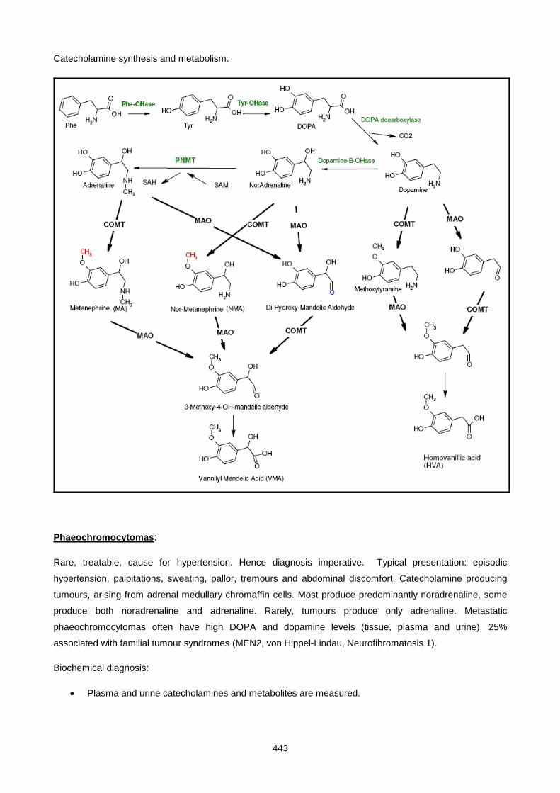
443
Catecholamine synthesis and metabolism:
Phaeochromocytomas:
Rare, treatable, cause for hypertension. Hence diagnosis imperative. Typical presentation: episodic
hypertension, palpitations, sweating, pallor, tremours and abdominal discomfort. Catecholamine producing
tumours, arising from adrenal medullary chromaffin cells. Most produce predominantly noradrenaline, some
produce both noradrenaline and adrenaline. Rarely, tumours produce only adrenaline. Metastatic
phaeochromocytomas often have high DOPA and dopamine levels (tissue, plasma and urine). 25%
associated with familial tumour syndromes (MEN2, von Hippel-Lindau, Neurofibromatosis 1).
Biochemical diagnosis:
• Plasma and urine catecholamines and metabolites are measured.

444
• Plasma free metanephrines most sensitive (97% hereditary; 100% sporadic) and highly specific (at
least 80%).
• If negative, phaeochromocytoma is unlikely.
• Urinary metanephrines (fractionated better than total) reasonably sensitive when plasma levels
cannot be measured, although less specific than plasma metanephrines. Entails 24 hour urine
collection in acid. Metanephrine:creatine ratio in random sample.
o Normal plasma free metanephrine or urinary metanephrines virtually excludes
phaemochromocytoma.
o >5 x upper limits of the reference interval implies phaeochromocytoma; warrants
further investigation for tumour localisation.
o Elevated levels < 5 x upper limit need further testing i.e. Clonidine suppression tests
to establish diagnosis (the lack of decrease in noradrenaline by clonidine
administration implies phaeochromocytoma).
False positive results may occur due to
• analytical interference (e.g. co-chromatography of paracetamol in HPLC; labetolol and caffeine)
• activation of sympathoadrenal system / intereference of catecholamine metabolism(e.g. Tricyclic
antidepressants, phenoxybenzamine, certain βblockers, hydralazine, MAO inhibitors, calcium
channel blockers, pseudoephedrine and levodopa as well as nicotine and theophylline)
Chromogranin A (CGA): see serotonin notes below. At least as sensitive and specific as plasma
catecholamines and urinary metanephrines.
B. SEROTONIN
Synthesised and stored at several sites in body. Large amounts found in intestinal mucosa (smaller
amounts in platelets and CNS). Synthesised by hydroxylation and decarboxylation of tryptophan to form 5-
OH tryptamine (=serotonin). See diagram. 5-OH tryptamine inactivated by MAO, forming 5-OH-
indolacetaldehyde, which is then mostly oxidised to 5-OH-indolacetic acid. A small fraction is converted to
5-OH tryptophol (increased in alcoholics).
Carcinoid tumours:
Low grade tumours arising from enterochromaffin cells. 74% found in GIT, respiratory tract (24%). [These
cells take up amine precursors and decarboxylate them (similar to phaeochromocytomas).]
May be associated with other endocrine tumours (producing gastrin, insulin, ACTH or catecholamines) and
MEN 1. Synthesise, store and release a variety of hormones and biogenic amines into the circulation, incl.
serotonin, histamine, prostaglandins, dopamine and adrenaline. Secretion of vasoactive amines may cause
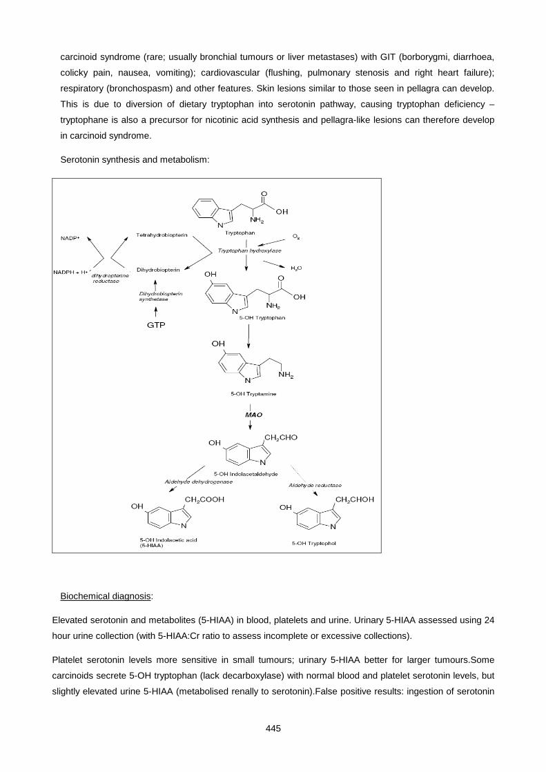
445
carcinoid syndrome (rare; usually bronchial tumours or liver metastases) with GIT (borborygmi, diarrhoea,
colicky pain, nausea, vomiting); cardiovascular (flushing, pulmonary stenosis and right heart failure);
respiratory (bronchospasm) and other features. Skin lesions similar to those seen in pellagra can develop.
This is due to diversion of dietary tryptophan into serotonin pathway, causing tryptophan deficiency –
tryptophane is also a precursor for nicotinic acid synthesis and pellagra-like lesions can therefore develop
in carcinoid syndrome.
Serotonin synthesis and metabolism:
Biochemical diagnosis:
Elevated serotonin and metabolites (5-HIAA) in blood, platelets and urine. Urinary 5-HIAA assessed using 24
hour urine collection (with 5-HIAA:Cr ratio to assess incomplete or excessive collections).
Platelet serotonin levels more sensitive in small tumours; urinary 5-HIAA better for larger tumours.Some
carcinoids secrete 5-OH tryptophan (lack decarboxylase) with normal blood and platelet serotonin levels, but
slightly elevated urine 5-HIAA (metabolised renally to serotonin).False positive results: ingestion of serotonin
446
rich foods e.g. bananas, kiwis, chocolate and guanfenesin (cough mixtures). False negative results: alcohol
and aspirin.
Chromogranin A :
More sensitive than urine 5-HIAA, but less specific: correlates well with tumour size. Major soluble protein of
the chromaffin granule (catecholamine storage vesicle). Widely expressed in neuroendocrine tissue, co-
secreted by neuroendocrine cells along with peptide hormones and neuropeptides. Useful marker of
neuroendocrine tumours including carcinoid, phaeochromocytoma and neuroblastoma. (Even in non-
functional carcinoid where serotonin is not secreted, CGA is still secreted. In phaeochromocytoma, at least as
sensitive and specific as plasma catecholamines or urinary metanephrines.) Neuron Specific Enolase,
substance P and neuropeptide K also useful in diagnosis and prognostication of carcinoid tumours.
NEWER “TUMOUR MARKERS”
a. Microarrays (DNA “chip”): This fairly new, powerful technology allows for the rapid identification of
sequence polymorphisms and mutations, and quantification of gene expression in thousands of genes
simultaneously (using mRNA). Expression patterns vary between healthy and diseased tissue. In cancer
tissue, this is useful in characterising tumours (e.g. breast tumours) into clinically relevant subgroups,
which in turn can act as prognostic markers and therapeutic guides.
b. Proteomics (protein “chip”): Proteomics is the study of the protein expression (vs mRNA expression in
DNA microarrays) in a given cell or tissue at a given time. Cells, healthy or diseased, have specific
protein signatures. These can be used as markers for cancer detection and diagnosis, and as treatment
guides. (Makes use of similar microarray technology developed for DNA analysis.)
c. Cell-free nucleic acids: Increased circulating levels of tumour-derived cell free nucleic acids (DNA or
RNA) have been detected in the serum / plasma of patients with malignancies. (Specific mutations, e.g.
N-ras mutation in acute myeloid leukaemia, have been demonstrated in plasma.) Levels correlate with
tumour burden and stage of disease. This is useful for non-invasive cancer diagnosis, predicting
prognosis and in monitoring progression of malignancies.
THE BODY'S RESPONSE TO MALIGNANCY
1. The "acute phase response" occurs in malignancy. The changes are cytokine-mediated and are
nonspecific: same as in any chronic inflammatory process.
◊ ↑ erythrocyte sedimentation rate (ESR)
◊ ↑ C-reactive protein (CRP), haptoglobin, α1-antitrypsin, fibrinogen, ferritin, ceruloplasmin (acute-
phase proteins)
2. Cachexia: Wasting and ↓ albumin:
◊ ↓ intake of food (anorexia, vomiting, depression)
◊ catabolic state: probably mediated by cytokines especially tumour necrosis factor (TNF)
447
3. Tumour lysis syndrome
ACUTE TUMOUR LYSIS SYNDROME
A group of metabolic abnormalities which arise during the course of treatment of cancer. Due to rapid
destruction of tumour cells & release of cellular breakdown products.
Features:
• hyperkalemia 12-24h after chemotherapy
• hyperuricemia 2 - 4 days after chemotherapy
• hyperphosphatemia 2 - 4 days
• hypocalcemia 2 - 4 days
Occurs with treatment of bulky, chemosensitive tumours. Most often seen with lymphomas (especially
Burkitt's lymphoma), and leukemias .
Complications and Management:
Hyperkalemia → cardiac arrhythmias. Treat with:
• K-losing (loop) diuretic + fluids
• Glucose + insulin
• dialysis
Hyperphosphatemia and hypocalcemia → metastatic calcification, nephrocalcinosis, renal failure:
• Fluids + loop diuretic
• Calcium gluconate I.V. infusion if tetany or convulsions.
Hyperuricemia → renal failure : acute urate nephropathy due to urate crystal deposition in renal tubules, and
acute gout.
• ↑ hydration
• alkalinize urine (sodium bicarbonate), if no hyperphosphatemia is present.
• allopurinol (xanthine oxidase inhibitor)

448
Lecture 29: Pregnancy And Parental Screening
PROF TS PILLAY 2008
LEARNING OBJECTIVES
1. Definition and objectives of prenatal screening
2. Properties of the various biochemical markers used for prenatal screening and their roles as adjuncts
to imaging techniques
3. HCG
4. PAPP-A
5. Inhibin A
6. Oestriol
7. Alpha fetoprotein
8. Risk assessment in prenatal screening; relative sensitivity and specificity of the different tests.
9. Differences between first and second trimester screening.
10. Differences in diagnostic marker levels in neutral tube defect, trisomy 21, trisomy 18 and the use of
these markers in assessment and diagnosis.
11. Understanding the nature of the Triple and Quadruple tests.
12. hCG as a tumour marker
PARENTAL SCREENING
What is prenatal screening?
It is the use of biochemical and other investigations to measure the likelihood that a certain disease is present
in an unborn child. This involves calculating a percentage “risk”. There are a number of biochemical markers
and emerging technologies aimed at increasing the sensitivity and specificity of screening tests. The risk is
quantified by comparison with the population mean or median. The results are often expressed as Multiples
of Median, MOMs and used as a basis for calculating risk. (Medians separate the lower half of the population
values from the upper half). An “average” result is 1.0 MOM. For each week of pregnancy, there are a set of
median values. Labs will either report individual results as MOM or as a total risk factor calculated by a
software program
What is the difference between a screening test and a diagnostic test?
• A screening tests estimates the risk
• However, there is a greater chance of being wrong, i.e either false positive or negative.
Screening tests Diagnostic tests
Healthy patients
Cheap Often expensive
Easy/noninvasive May be invasive/associated with risk
Define the population at risk Performed on the population at risk
Do not give a definitive answer Provide a definitive answer

449
Every pregnancy has a 2-3% risk of a baby with a congenital anomaly. Couples at risk for a genetic anomaly
may be identified prior to pregnancy, if there is pre-existing evidence of inherited disease.
OPTIONS FOR SCREENING TESTS
There are two different screening options.
First trimester: 10-13 weeks 6 days of pregnancy (10-14 weeks)
Second trimester: 14-18 weeks
Many women choose to have a first trimester screening test to find out early in their pregnancy if there is a
problem. Second trimester screening is valuable for women who are too late for the trimester screening test
or if the first trimester screening test is not available. Having a both first and second trimester screening test
is not recommended.
FIRST TRIMESTER SCREENING
This is offered at 10-14 weeks. It makes use of serum markers as PAPP-A and free HCG, as well as ultra
sound for nuchal translucency and nasal bone assessment. The nuchal translucency assess the amount of
fluid at the back of the neck and is often larger in Down’s syndrome.
IMPORTANCE OF SCREENING IN THE FIRST TRIMESTER
It allows earlier diagnosis ad earlier reassurance and is a safer option if termination is being considered. It is
superior to second trimester and is less traumatic.
(HUMAN) CHRONIC GONADOTROPHIN (hCG)
The detection of human Chorionic Gonadotrophin (hCG) is the sine qua non for the diagnosis of pregnancy
(with a few rare exceptions). The term “human” is superfluous but the terminology still persists amongst
clinicians. The hCG is a glycoprotein and is made by the synchtiotrophoblasts in the placenta. It has 2
subunits – alpha and beta. The a subunit is common to the following glycoprotein hormones and is encoded
by a single gene (chromosome 6) (TSH, FSH,LH,hCG). The B subunit is unique to hCG. hCG stimulates the
corpus luteum to continue producing progesterone in early pregnancy because the placenta does not make
enough progesterone in early pregnancy and is therefore important until the placenta is able to produce
enough progesterone. hCG is detectable in serum at 8-10 days after ovulation (approximately week 3 of
pregnancy-week 3 being calculated after the LPM- (the first day of the last menstrual period). This means that
pregnancy can be diagnosed before the missed period. The serum concentration starts increasing from 7-10
days after ovulation (LH surge) or 4-7 days after implantation. The serum levels are approximately >5IU/I at
this stage. The urine levels at week 5 are >25 IU/I and serum levels peak at 10-12 weeks (100 000 IU/I). In
early pregnancy the level increases exponentially, doubling every 2 days and reaching a maximum of
between 20 000 – 100 000 IU/I at 7-10 weeks of pregnancy. After this the levels decrease and level out at 13-
15 weeks, increase moderately at 30-33 weeks and decrease moderately thereafter until term. The following
serum levels delineate when the sample is regarded as being positive or negative for hCG.
• <5IU/I negative
• 5-25 IU/I indeterminate
• >25 IU/I positive

450
A single estimation of 25 IU/I is considered diagnostic of pregnancy or if is less than that, the levels double in
3 days. In situation where the hCG level is lower than expected or in the indeterminate range and pregnancy
is suspected on clinical grounds, then serial measurements are used. hCG curves can also be used to
delineate viable and non-viable pregnancies. A viable pregnancy will show at least a 50% increase in the hCG
over one day in the trimester. The mean value of hCG increase for normal pregnancies over 2 day are 124%.
However, borderline values can still exist in a symptomatic pregnancy (pain or bleeding) where an ectopic is
suspected. Therefore, ultrasound findings play an important role in overall assessment. Serial hCG
measurements can be assessed using a log-linear slope of –log hCG/?time. In ectopic pregnancy, the
median of this value tends to be 0.11. Normal pregnancies will have a value much greater than 0.11 (25%
increase in 2 days) with an average of 124% as described as above. Similarly, the decline in hCG can also be
used to distinguish between spontaneous complete abortion or an ectopic pregnancy. The average decline in
hCG is a 27% decrease in hCG over 2 days and this is lower than that observed in spontaneous abortion.
Failure to detect an intrauterine foetal sac when the hCG level is 1500 IU/I indicates that the pregnancy is
ectopic, but this does depend on the skill of the ultrasonographer. In addition, women at known risk for
ectopic pregnancy can be screened with ultrasound and serum hCG. This allows detection of pregnancy at a
gestational age of 37 days on average and this tends to be before symptoms occur. Early detection can
facilitate tube-sparing measures. Most hCG exists in dimeric form. 0.3% circulates as free BHCG. In
summary, hCG is used to detect normal and abnormal pregnancy (i.e. ectopic) and is used t screen for
Down’s syndrome and trisomy 18 and to monitor the course of cancer if the patient has an hCG producing
tumour.
PREGNANCY TESTS
The most common use for hCG determination is in the diagnosis of pregnancy. These commonly use urine
as a sample, but some are also able to use serum, plasma or whole blood. The pregnancy test is one of the
most reliable and useful laboratory tests available. Home pregnancy test kits are now widely available and
work well if used precisely according to the manufacturer’s instruction. hCG levels are detectable in urine
about 10 days after conception. Most tests work by placing a drop of urine on a tsrip and the test detects the
hCG in the urine. Testing before the 10 days have elapsed can give a false negative result because the levels
are too low. It is therefore advisable for the women to wait at least 5-10 days after the missed period to
endure that result is accurate. Most tests have a sensitivity of 25-50 IU/I. A detection limit of 25 IU/I is
considered optimal because non pregnant women can have levels of 10-15 IU/I. It is best to take the first
specimen in the morning since the hCG will be more concentrated in this sample. Otherwise the urine should
have been in the bladder at least 4 hours and the women should avoid drinking excessive fluids before the
test. The blood test for the hCG is rarely necessary in normal, uncomplicated pregnancy.
MATERNAL SERUM SCREENING FOR FOETAL ANOMALIES
The goal screening is the early identification of foetal anomalies. These include neural tube defects, trisomy
21 (Down syndrome), trisomy 18 (Edwards). There are a number of foeto-placental markers in maternal
serum that are used to improve the detection rate of foetal abnormalities. A combination of these markers
“triple test” increases the sensitivity and specificity of screening. The triple tests uses AFP, hCG and Oestriol.
The Quad test includes AFP, intact HCG or B subunit of Inhibin A (Quadruple test).
451
ALPHA-FOETOPROTEIN (AFP)
AFB has a similar function to albumin in the foetus. It is initially produced by the yolk sac and then later by the
liver as the yolk sac degenerates. AFP decreases to reach adult levels at 10 months of age. AFP is
measured in maternal serum and amniotic fluid. The AFP in maternal serum is raised in 85-95% of open
neural defects. It is decreased in 30% of Down’s syndrome foetuses.
Causes of Increased AFP
• Neutral tube defects
• Abdominal wall defects/exomphalos
• Foetal renal disease
• Oligohydramnios
• Foetal death
• Multiple pregnancy
• Tumour marker
• Incorrect dates
• Duodenal/oesophageal atresia
• Multiple pregnancy
CAUSES OF DECREASED AFP
Down’s syndrome-yolk sac and foetus are smaller than usual
• Trisomy 18
Raised maternal serum AFP can be confirmed by measuring AFP in the amniotic fluid following
amniocentesis. If the AFP is raised in the amniotic fluid then the Acetylcholinesterase can be used as a
confirmatory test. CSF contains high concentrations of the AChE and this leaks into the amniotic fluid if there
is an open neural tube defect.
UNCONGUGATED OESTRIOL (abbreviated as E3)
As the name suggests, this is an oestrogen with 3 hydroxyl groups. Maternal serum E3 increases with
gestational age and is produced in large amounts in the 3rd semester. It requires 3 organs to be produced.
The foetal adrenal, liver and placenta must be functional to create E3. It is decreased in the foetal death and
chromosome abnormalities.
CAUSES OF DECREASED E3
It is modestly decreased in Down’s syndrome-screening for this is the most common indication for doing the
test.
• Any disruption of the pathway will cause low maternal serum oestriol
• Anencephaly, placental sulphatase deficiency, foetal death, chromosomal abnormalities, molar
pregnancy, Smith-Lemli-opitz syndrome (deficiency in cholesterol synthesis). Facial features include
microcephaly, ptosis, broad nasal bridge, upturned nose and micrognathia. Cleft palate or bifid uvula
is also common. Limb anomalies include short proximally placed thumbs, single palmar creases,
clinodactyly, and postaxial polydactyly.
452
DIMERIC INHIBIN A (DIA)
Inhibin is a negative feedback regulator of FSH. Inhibins are members of the TGFB superfamily. There are 2
types of inhibins, A and B. The placenta produces large amounts of Inhibin A and this completely suppresses
FSH. It is a dimeric protein: (dimmers of a +B subunits). It is useful in screening for Down’s syndrome where
it is increased. It is commonly measured at 15-20 weeks of gestation as part of the quadruple test (Quad
test). Inhibin A can also be used in the assessement of ovarian cancer, ovulation disorders, invitro fertilization
and male infertility.
PREGNANCY ASSOCIATED PLASMA PROTEIN A (PAPP-A)
PPAP-A is associated with an increased risk of Down’s syndrome. It is produced the new fertilized egg. It can
used earlier in pregnancy for screening. PAPP-A is a glycoprotein produced by the trophoblast. It is found in
trace amounts in nonpregnant women. It increases the bioavailability of IGF-1. Low levels are seen in trisomy
21 and other aneuploidies,
NEUTRAL TUBE DEFECTS
The closure of the neural tube is complete 4 weeks after fertilization. Failure of neural tube fusion and closure
can lead to anencephaly, meningomyelocoele and encephalocoele. The causes are multifactorial but folate is
known to be important. Since there is no overlying skin amniotic fluid AFP increases and crosses into the
placenta into the maternal blood. Only open neural tube defects are detected by AFP testing. The screening
is optimally performed at 16-18 weeks. If the AFP is raised, then an amniocentesis to obtain amniotic fluid
may be necessary to test for AChE.
The ultrasound will also be used in conjunction with this.
DOWN SYNDROME (Trisomy 21)
The risk of down’s syndrome increases with maternal age. At 25 years of age the risk is 1:1000 at the age of
40 years the risk is 1:90. In women at risk (usually over 35 years of age) screening is achieved using a 2nd
trimester-triple test consisting of E3/AFP/hCG. If Dimeric inhibin A, there is a 8% increase in detection. If the
screen is positive, the additional confirmatory tests are required. Ultrasound is commonly added. One of the
most common causes for a positive screen is incorrect gestational age since the levels of these are based on
different cut-offs for different gestational age. If the gestational is overestimated then this may result in an
incorrect calculation of high risk. Since the ultrasound changes of nuchal translucency are not diagnostic, it
may be necessary to use amniocentesis and karyotyping. If any of the screening or diagnostic tests are
positive, then genetic counseling is offered.
Results of testing are as follows: If 1st Trimester testing (10-13 weeks) is undertaken using PAPP-A (↓ ) &
Free βhCG (↑ ), this gives a 60% detection rate . Ultrasound assessment of Nuchal Translucency yields a
60% detection rate. If these are combined then the detection rate is 85% with a 5% False positive rate. If first
and second trimester testing are combined i.e. 1st (PAPP-A & NT) & 2nd Trimester testing (AFP↓ ; E3↓;
hCG↑ & DIA↑) then there is an 85% detection rate and 1% false positive rate.

453
Why is the hCG high in Down’s syndrome? One possibility is that the extra copy of Chromosome 21 carries a
transcription factor that upregulates hCG synthesis. As a result the production of hCG is increased.
TRISOMY 18 (Edwards syndrome)
This occurs at an incidence of 1/8000 and results in a very high rate of foetal loss. 25% have neural tube
defects. 50% die within 5 days of birth and 90 % die within 3 months. If screening shows decreased AFP, E3
and hCG then amniocentesis and karyotyping may be undertaken. There are ultrasound abnormalities that
may be detected (heart, fist clenching etc.) but these are non-diagnostic.
Results of the Triple test at 16-18 Weeks
AFP bHCG E3
Down’s ↓ ↑ ↓ (25%)
Trisomy 18 ↓ ↓ ↓
Pseudopregnancy U U U
Neural tube defect ↑ N ↓/N
Twins ↑ ↑ ↑
New advances in prenatal screening
Foetal DNA circulates in maternal plasma. This has been used as a basis for prenatal screening. Sex
determination can be carried out by determining whether the maternal serum contains Y chromosome
markers that may have come from the foetus. In addition, it can also be used to provide a highly sensitive and
diagnostic marker for Trisomy 21 without the need for amniocentesis.
IMPORTANCE OF FOLIC ACID DURING PREGNANCY
The closure of the neural tube occurs around the 28th day of pregnancy. The incidence of neural tube defects
(spina bifida and anencephaly) is reduced by 400μg folic acid supplement/day before conception and during
the first month of pregnancy. The current recommendations are as follows:
• A folate-supplemented diet with additional daily supplements of multivitamins and 5 mg folic acid should
begin 3 months before conception and continue 10-12 weeks post.
• From 12 weeks post- multivitamins with folic acid (0.4-1.0 mg)
• 5 mg folate will not mask B12 deficiency
• Women planning conception should be advised about folate-rich diets -
fortified grains, spinach, lentils, chick peas, asparagus, broccoli, peas, Brussels
454
sprouts, corn and oranges.
• However, diet alone cannot provide the levels achieved using supplements.
• In general, all women of child-bearing age should be taking folate
supplements. Results of the Triple test at 16-18 Weeks
THE USE OF hCG FOR THE DIAGNOSIS AND MONITORING OF CANCER
hCG is increased in trophoblastic disease and some germ cell tumours of the testis and is therefore a
sensitive and specific tumour marker, possibly the most sensitive tumour marker known. With gestational
trophoblastic disease, the sensitivity and specificity are close to 100%. HCG can also be used to monitor
therapy and possible relapse of choriocarcinoma. About 50% of patients with germ cell tumours of the testis
have increased hCG concentrations.
FALSE POSITIVE RESULTS
As hCG is a very sensitive marker, in the absence of pregnancy an increased hCG level is a strong indicator
of trophoblastic disease and germ cell tumours. However, false positives do occur and patients have been
inappropriately treated with severe complications. There are a number of causes of false positives and this
needs to be taken into consideration when the circumstances arise. Indeed, this needs to be taken into
account whenever an immunoassay result is at odds with the clinical setting. The major cause of false
positives are heterophilic antibodies. These are antibodies to animal immunoglobulins. Many of us have
antibodies in our serum to animal antigens that have arisen after exposure to animal products including milk
or by exposure to animals or animal products. These can potentially interfere with immunoassays that use
mouse antibodies if the heterophilic antibody reacts with mouse immunoglobulins.
CAUSES OF FALSE POSITIVES
i. Interferents: Heterophilic antibodies, human anti-mouse antibodies (HAMAs), Rheumatoid factor
ii. Assay problems in the laboratory: sample mixup, contamination
iii. Increased pituitary hCG: (perimenopause; chemotherapy-induced suppression of gonadal function)
iv. Previous injection of hCG
Related Documents