Chemical Inhibition of RNA Viruses Reveals REDD1 as Host Defense Factor Miguel A. Mata 1,* , Neal Satterly 1,* , Gijs A. Versteeg 5,* , Doug Frantz 2 , Shuguang Wei 2 , Noelle Williams 2 , Mirco Schmolke 5 , Samuel Pena-Llopis 3 , James Brugarolas 3 , Christian Forst 4 , Michael A. White 1 , Adolfo Garcia-Sastre 5,6,7 , Michael G. Roth 2 , and Beatriz M. A. Fontoura 1,¶ 1 Department of Cell Biology, University of Texas Southwestern Medical Center, Dallas, TX 75390 2 Department of Biochemistry, University of Texas Southwestern Medical Center, Dallas, TX 75390 3 Department of Internal Medicine, Developmental Biology and Simmons Cancer Center, University of Texas Southwestern Medical Center, Dallas, TX 75390 4 Department of Clinical Sciences, University of Texas Southwestern Medical Center, Dallas, TX 75390 5 Department of Microbiology, Mount Sinai School of Medicine, New York, NY 10029 6 Department of Medicine, Division of Infectious Diseases, Mount Sinai School of Medicine, New York, NY 10029 7 Global Health and Emerging Pathogens Institute, Mount Sinai School of Medicine, New York, NY 10029 Abstract A chemical genetics approach was taken to identify inhibitors of NS1, a major influenza A virus virulence factor that inhibits host gene expression. A high-throughput screen of 200,000 synthetic compounds identified small molecules that reverted NS1-mediated inhibition of host gene expression. A counter-screen for suppression of influenza virus cytotoxicity identified naphthalimides that inhibited replication of influenza virus and vesicular stomatitis virus. The mechanism of action was through activation of REDD1 expression and concomitant inhibition of mTORC1 via TSC1/TSC2 complex. The antiviral activity of naphthalimides was abolished in REDD1−/− cells. Viruses inhibited REDD1 expression, resulting in activation of the mTORC1 pathway. REDD1−/− cells prematurely up-regulated viral proteins via mTORC1 activation and were permissive to virus replication. In contrast, cells conditionally expressing high levels of REDD1 down-regulated viral protein levels. Thus, REDD1 is a novel host defense factor and chemical activation of REDD1 expression represents a potent antiviral intervention strategy. ¶ Correspondence: Beatriz M. A. Fontoura, Department of Cell Biology, University of Texas Southwestern Medical Center, 5323 Harry Hines Blvd., mail code: 9039, room number K2.208, Dallas, TX 75390-9039, phone: (214) 648-9535, [email protected]. * These authors equally contributed to the manuscript. Author contributions: M.M., N.S., G.A.V., D.F., S.P.-L., J.B., C.F., M.A.W., A.G.-S., M.G.R., and B.M.A.F. designed research; M.M., N.S., G.A.V., S.W., N.W., M.S., S.P.-L., and C.F. performed research; D.F. contributed new reagents; M.M., N.S., G. A.V., D.F., N.W., M.S., S. P.-L., J.B., C.F., M.A.W., A.G.-S., M.G.R., and B.M.A.F analyzed data; M.G.R. and B.M.A.F. wrote the paper. Authors declare no competing financial interest. NIH Public Access Author Manuscript Nat Chem Biol. Author manuscript; available in PMC 2012 April 19. Published in final edited form as: Nat Chem Biol. ; 7(10): 712–719. doi:10.1038/nchembio.645. NIH-PA Author Manuscript NIH-PA Author Manuscript NIH-PA Author Manuscript

Welcome message from author
This document is posted to help you gain knowledge. Please leave a comment to let me know what you think about it! Share it to your friends and learn new things together.
Transcript
Chemical Inhibition of RNA Viruses Reveals REDD1 as HostDefense Factor
Miguel A. Mata1,*, Neal Satterly1,*, Gijs A. Versteeg5,*, Doug Frantz2, Shuguang Wei2, NoelleWilliams2, Mirco Schmolke5, Samuel Pena-Llopis3, James Brugarolas3, Christian Forst4,Michael A. White1, Adolfo Garcia-Sastre5,6,7, Michael G. Roth2, and Beatriz M. A.Fontoura1,
1Department of Cell Biology, University of Texas Southwestern Medical Center, Dallas, TX 753902Department of Biochemistry, University of Texas Southwestern Medical Center, Dallas, TX753903Department of Internal Medicine, Developmental Biology and Simmons Cancer Center,University of Texas Southwestern Medical Center, Dallas, TX 753904Department of Clinical Sciences, University of Texas Southwestern Medical Center, Dallas, TX753905Department of Microbiology, Mount Sinai School of Medicine, New York, NY 100296Department of Medicine, Division of Infectious Diseases, Mount Sinai School of Medicine, NewYork, NY 100297Global Health and Emerging Pathogens Institute, Mount Sinai School of Medicine, New York, NY10029
AbstractA chemical genetics approach was taken to identify inhibitors of NS1, a major influenza A virusvirulence factor that inhibits host gene expression. A high-throughput screen of 200,000 syntheticcompounds identified small molecules that reverted NS1-mediated inhibition of host geneexpression. A counter-screen for suppression of influenza virus cytotoxicity identifiednaphthalimides that inhibited replication of influenza virus and vesicular stomatitis virus. Themechanism of action was through activation of REDD1 expression and concomitant inhibition ofmTORC1 via TSC1/TSC2 complex. The antiviral activity of naphthalimides was abolished inREDD1−/− cells. Viruses inhibited REDD1 expression, resulting in activation of the mTORC1pathway. REDD1−/− cells prematurely up-regulated viral proteins via mTORC1 activation andwere permissive to virus replication. In contrast, cells conditionally expressing high levels ofREDD1 down-regulated viral protein levels. Thus, REDD1 is a novel host defense factor andchemical activation of REDD1 expression represents a potent antiviral intervention strategy.
¶Correspondence: Beatriz M. A. Fontoura, Department of Cell Biology, University of Texas Southwestern Medical Center, 5323Harry Hines Blvd., mail code: 9039, room number K2.208, Dallas, TX 75390-9039, phone: (214) 648-9535,[email protected].*These authors equally contributed to the manuscript.Author contributions: M.M., N.S., G.A.V., D.F., S.P.-L., J.B., C.F., M.A.W., A.G.-S., M.G.R., and B.M.A.F. designed research;M.M., N.S., G.A.V., S.W., N.W., M.S., S.P.-L., and C.F. performed research; D.F. contributed new reagents; M.M., N.S., G. A.V.,D.F., N.W., M.S., S. P.-L., J.B., C.F., M.A.W., A.G.-S., M.G.R., and B.M.A.F analyzed data; M.G.R. and B.M.A.F. wrote the paper.Authors declare no competing financial interest.
NIH Public AccessAuthor ManuscriptNat Chem Biol. Author manuscript; available in PMC 2012 April 19.
Published in final edited form as:Nat Chem Biol. ; 7(10): 712–719. doi:10.1038/nchembio.645.
NIH
-PA Author Manuscript
NIH
-PA Author Manuscript
NIH
-PA Author Manuscript
KeywordsmRNA export; NS1; influenza virus; S6K; mTORC1; REDD1; DDIT4; Rtp801; VSV
IntroductionInfections by influenza viruses are responsible for ~36,000 deaths annually in the UnitedStates 1 and ~500,000 deaths worldwide per year 2. Highly pathogenic strains have beenresponsible for many deaths worldwide, such as the 1918 pandemic which killed ~30 millionpeople. Currently, there are only two approaches available for preventing or treatingepidemic and pandemic influenza, vaccination and inhibitors of virus replication.Vaccination, although highly effective against homologous strains, looses efficacy in theelderly, and it is limited by the highly mutable nature of the virus and the large reservoir ofantigenically distinct virus strains. These factors require the annual re-formulation of thevaccine to match the antigenicity of the current influenza virus circulating strains. A numberof drugs have been approved for the treatment of influenza. These drugs inhibit virusuncoating (inhibitors of the viral protein M2) or virus spread (inhibitors of the viral proteinNA), but the use of these relatively small number of antiviral drugs is limited by theappearance of resistant virus strains. There is a clear need for additional therapeuticmodalities for the treatment of influenza virus disease as well as for a better understandingof mechanisms of viral-host interactions leading to the discovery of novel targets fortherapeutic intervention.
Many viruses target host mechanisms that are key steps within pathways that regulateantiviral responses. As an example, the NS1 protein of influenza virus is a multifunctionalvirulence factor that inhibits host gene expression and signal transduction required to induceantiviral responses. NS1 is found both in the nucleus and in the cytoplasm of influenza virusinfected cells3,4. The cytoplasmic pool of NS1 inhibits interferon (IFN) gene induction byantagonizing the cytoplasmic signal transduction pathway mediated by RIG-I 5–8. NS1 alsoprevents IFN action by sequestering double-stranded RNA and/or targeting the function ofdownstream antiviral effector proteins, such as PKR and the RNase L pathways 9,10. Inaddition, NS1 has been shown to activate phosphatidylinositol 3-kinase signaling (PI3K), afunction that supports virus replication 11.
The nuclear pool of NS1 inhibits host mRNA processing, including splicing12–14,polyadenylation15, and nuclear export16–18, thus preventing proper expression of hostantiviral genes but not nuclear export of viral RNAs 15,18. Disruption of NS1 functions bymutations yielded highly attenuated viruses that can only replicate in immunocompromisedhosts 19. These findings underscore the key role of NS1 as a pro-viral factor and emphasizethe need to identify both inhibitors of this virulence factor as well as novel host antiviralmechanisms that antagonize NS1 functions. Here, we performed a high-throughput screen toidentify small molecules that reverted NS1-mediated inhibition of host gene expression.Non-toxic small molecules from the naphthalimide family were identified and thesecompounds inhibited replication of evolutionarily diverse viruses, including influenza virusand VSV. We show that these small molecules served as probes to identify the mTORC1inhibitor REDD120,21 as a novel host defense factor. These findings underscore theimportance of regulating REDD1 expression as a novel strategy to trigger antiviral response.
Mata et al. Page 2
Nat Chem Biol. Author manuscript; available in PMC 2012 April 19.
NIH
-PA Author Manuscript
NIH
-PA Author Manuscript
NIH
-PA Author Manuscript
ResultsNaphthalimides Antagonize NS1 and Influenza Virus
We exploited the potent ability of NS1 to inhibit gene expression by blocking mRNAprocessing and export15,18, as the basis for a high-throughput assay that measured the effectof NS1 on luciferase expression. Luciferase activity was reduced ~95% in cells transfectedwith plasmids encoding NS1 when compared to cells expressing luciferase alone, as wepreviously reported18. We screened 200,000 compounds at 5 μM concentration with thisassay and then counter-screened for the ability of these small molecules to suppresscytotoxicity caused by influenza virus infection (Fig.1a,b). Among the most activecompounds was 4-[N-4-nitro-(1,8-naphthalimide)]-butanoic acid, compound 1, (Fig. 1c).
We obtained compounds structurally related to 1 and identified some that had no antiviralactivity (2) or more potent activity (3) (Fig. 1c) (Supplementary Results, Fig. S1).Compound 3 was much less cytotoxic than the original 1 (Fig. 2a) and had a much longerhalf-life than 1 (Fig. 2b). We have also shown that 3 did not alter bulk protein synthesis(Supplementary Results, Fig. S2). To investigate the effect of 3 on virus-mediatedcytotoxicity, we infected MDCK cells with influenza A/WSN/1933 virus at m.o.i. 0.001 for48 h in the presence or absence of 3. Widespread cytopathic effects were observed inMDCK cells in the absence of 3 after 48 h of infection, but 3 largely prevented this effect(Fig. 2c). Since this compound was derived from a screen in which a reversal of NS1-mediated inhibition of gene expression was observed, it would be expected that a significantreversal of the mRNA export block induced by influenza virus would occur in the presenceof active compound. Indeed, in cell populations infected with influenza virus in the presenceof 3, there was a decrease in the number of cells that retained poly(A) RNA in the nucleus,as compared to infected cells not treated with 3 (Fig. 2d,e). A sub-population of infectedcells still presented mRNA export block in the presence of 3; thus, it is possible that thesecells are at different phases of the cell cycle, a process known to regulate mRNA export 22.Thus, 3 partially antagonized the mRNA export block in virus-infected cells.
Naphthalimide Inhibits Virus ReplicationThe effect of 3 on virus replication was then assessed using various strains of influenzavirus, A/WSN/1933, A/Texas/1991, and the highly virulent A/Brevig/Mission/1/1918 strainthat killed ~ 30 million people 23 (Fig. 3a–c). Non-cytotoxic concentrations of 3 reducedviral titers by 103 to 106 between 24 to 36h post-infection, depending on the influenza virusstrain. The ratio of the concentration causing half maximum cytotoxicity (CC50) to halfvirus inhibition (IC50) for 3 was 31 (Fig. 2a, 3a). Similar results were also observed inhuman A549 cells (Supplementary Results, Fig. S3). As shown in Figure 3d, intracellularinfluenza virus proteins were also reduced in the presence of 3 (Fig. 3d). Thus, 3 decreasedviral protein levels, contributing to the reduction of virus replication.
The antiviral effect of 3 was not IFN-mediated. The mRNA levels of IFN-β and IFNeffectors were measured, by qPCR and microarray analysis, revealing that 3 did not induceIFN production or an IFN-mediated response (Fig. 3e; Supplementary Dataset 1).Furthermore, 3 protected cells impaired in interferon response, Vero Cells and Stat1 −/−cells, from influenza virus replication or cell death, respectively (Supplementary Results,Fig. S4). Compound 3 also antagonized expression of high levels of influenza virus proteinsin Vero cells (Supplementary Results, Fig. S5). Thus, 3 partially antagonized the block ofmRNA export in virus-infected cells, but this effect did not result in the production ofinterferon. However, the partial release of mRNA export by 3 likely occurred as aconsequence of low NS1 levels, resulting in the expression of a significant number of hostmRNAs that encode antiviral factors. To investigate whether 3 antagonized NS1 directly or
Mata et al. Page 3
Nat Chem Biol. Author manuscript; available in PMC 2012 April 19.
NIH
-PA Author Manuscript
NIH
-PA Author Manuscript
NIH
-PA Author Manuscript
promoted host antiviral functions regulated by NS1 that could also impact replication ofother viruses, we infected cells with VSV at 0.001 pfu/cell in the absence or presence ofcompounds 2 or 3. Compound 3 inhibited VSV replication (Fig. 3f). Thus, 3 targeted hostcell function(s) that conferred an antiviral state against diverse viruses.
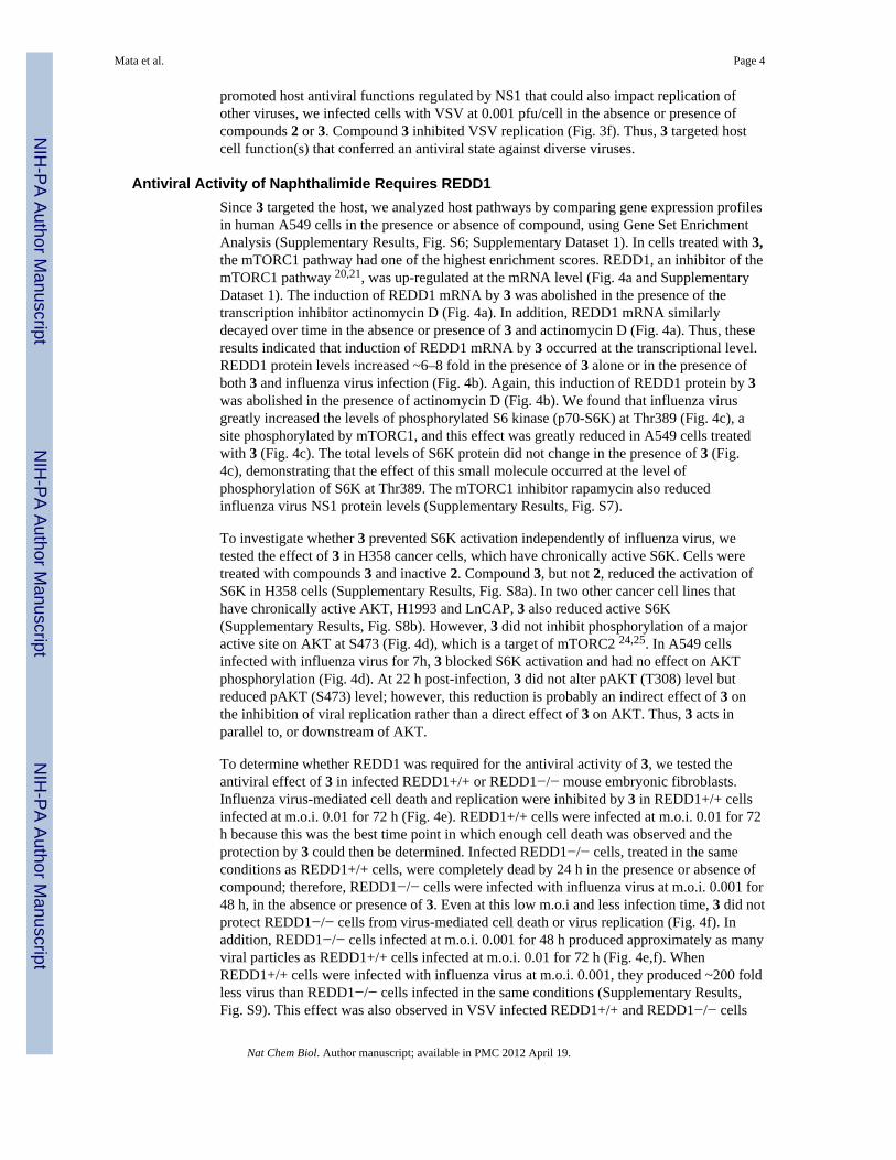
Antiviral Activity of Naphthalimide Requires REDD1Since 3 targeted the host, we analyzed host pathways by comparing gene expression profilesin human A549 cells in the presence or absence of compound, using Gene Set EnrichmentAnalysis (Supplementary Results, Fig. S6; Supplementary Dataset 1). In cells treated with 3,the mTORC1 pathway had one of the highest enrichment scores. REDD1, an inhibitor of themTORC1 pathway 20,21, was up-regulated at the mRNA level (Fig. 4a and SupplementaryDataset 1). The induction of REDD1 mRNA by 3 was abolished in the presence of thetranscription inhibitor actinomycin D (Fig. 4a). In addition, REDD1 mRNA similarlydecayed over time in the absence or presence of 3 and actinomycin D (Fig. 4a). Thus, theseresults indicated that induction of REDD1 mRNA by 3 occurred at the transcriptional level.REDD1 protein levels increased ~6–8 fold in the presence of 3 alone or in the presence ofboth 3 and influenza virus infection (Fig. 4b). Again, this induction of REDD1 protein by 3was abolished in the presence of actinomycin D (Fig. 4b). We found that influenza virusgreatly increased the levels of phosphorylated S6 kinase (p70-S6K) at Thr389 (Fig. 4c), asite phosphorylated by mTORC1, and this effect was greatly reduced in A549 cells treatedwith 3 (Fig. 4c). The total levels of S6K protein did not change in the presence of 3 (Fig.4c), demonstrating that the effect of this small molecule occurred at the level ofphosphorylation of S6K at Thr389. The mTORC1 inhibitor rapamycin also reducedinfluenza virus NS1 protein levels (Supplementary Results, Fig. S7).
To investigate whether 3 prevented S6K activation independently of influenza virus, wetested the effect of 3 in H358 cancer cells, which have chronically active S6K. Cells weretreated with compounds 3 and inactive 2. Compound 3, but not 2, reduced the activation ofS6K in H358 cells (Supplementary Results, Fig. S8a). In two other cancer cell lines thathave chronically active AKT, H1993 and LnCAP, 3 also reduced active S6K(Supplementary Results, Fig. S8b). However, 3 did not inhibit phosphorylation of a majoractive site on AKT at S473 (Fig. 4d), which is a target of mTORC2 24,25. In A549 cellsinfected with influenza virus for 7h, 3 blocked S6K activation and had no effect on AKTphosphorylation (Fig. 4d). At 22 h post-infection, 3 did not alter pAKT (T308) level butreduced pAKT (S473) level; however, this reduction is probably an indirect effect of 3 onthe inhibition of viral replication rather than a direct effect of 3 on AKT. Thus, 3 acts inparallel to, or downstream of AKT.
To determine whether REDD1 was required for the antiviral activity of 3, we tested theantiviral effect of 3 in infected REDD1+/+ or REDD1−/− mouse embryonic fibroblasts.Influenza virus-mediated cell death and replication were inhibited by 3 in REDD1+/+ cellsinfected at m.o.i. 0.01 for 72 h (Fig. 4e). REDD1+/+ cells were infected at m.o.i. 0.01 for 72h because this was the best time point in which enough cell death was observed and theprotection by 3 could then be determined. Infected REDD1−/− cells, treated in the sameconditions as REDD1+/+ cells, were completely dead by 24 h in the presence or absence ofcompound; therefore, REDD1−/− cells were infected with influenza virus at m.o.i. 0.001 for48 h, in the absence or presence of 3. Even at this low m.o.i and less infection time, 3 did notprotect REDD1−/− cells from virus-mediated cell death or virus replication (Fig. 4f). Inaddition, REDD1−/− cells infected at m.o.i. 0.001 for 48 h produced approximately as manyviral particles as REDD1+/+ cells infected at m.o.i. 0.01 for 72 h (Fig. 4e,f). WhenREDD1+/+ cells were infected with influenza virus at m.o.i. 0.001, they produced ~200 foldless virus than REDD1−/− cells infected in the same conditions (Supplementary Results,Fig. S9). This effect was also observed in VSV infected REDD1+/+ and REDD1−/− cells
Mata et al. Page 4
Nat Chem Biol. Author manuscript; available in PMC 2012 April 19.
NIH
-PA Author Manuscript
NIH
-PA Author Manuscript
NIH
-PA Author Manuscript
(Fig. 5c,d). Treatment of both REDD1+/+ and REDD1−/− cells with 3 alone did not causecytotoxicity (Supplementary Results, Fig. S10). Thus, REDD1 knockout cells were morepermissive to influenza virus replication than wild-type cells. As 3 did not inhibit virusreplication in the absence of REDD1 (Fig. 4f), REDD1 is required for the antiviral activityof 3.
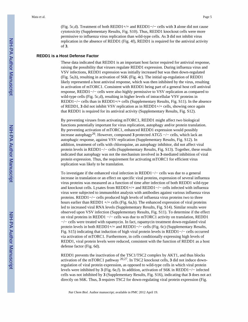
REDD1 is a Host Defense FactorThese data indicated that REDD1 is an important host factor required for antiviral response,raising the possibility that viruses regulate REDD1 expression. During influenza virus andVSV infections, REDD1 expression was initially increased but was then down-regulated(Fig. 5a,b), resulting in activation of S6K (Fig. 4c). The initial up-regulation of REDD1likely represented a host antiviral response, which was then inhibited by the virus, resultingin activation of mTORC1. Consistent with REDD1 being part of a general host cell antiviralresponse, REDD1−/− cells were also highly permissive to VSV replication as compared towild-type cells (Fig. 5c,d), resulting in higher levels of intracellular VSV proteins inREDD1−/− cells than in REDD1+/+ cells (Supplementary Results, Fig. S11). In the absenceof REDD1, 3 did not inhibit VSV replication as in REDD1+/+ cells, showing once againthat REDD1 is required for its antiviral activity (Supplementary Results, Fig. S12).
By preventing viruses from activating mTORC1, REDD1 might affect two biologicalfunctions potentially important for virus replication, autophagy and/or protein translation.By preventing activation of mTORC1, enhanced REDD1 expression would possiblyincrease autophagy26. However, compound 3 protected ATG5 −/− cells, which lack anautophagic response, against VSV replication (Supplementary Results, Fig. S12). Inaddition, treatment of cells with chloroquine, an autophagy inhibitor, did not affect viralprotein levels in REDD1−/− cells (Supplementary Results, Fig. S13). Together, these resultsindicated that autophagy was not the mechanism involved in 3-mediated inhibition of viralprotein expression. Thus, the requirement for activating mTORC1 for efficient virusreplication was likely to be translation.
To investigate if the enhanced viral infection in REDD1−/− cells was due to a generalincrease in translation or an effect on specific viral proteins, expression of several influenzavirus proteins was measured as a function of time after infection of both REDD1 wild-typeand knockout cells. Lysates from REDD1+/+ and REDD1−/− cells infected with influenzavirus were subjected to immunoblot analysis with antibodies against various influenza virusproteins. REDD1−/− cells produced high levels of influenza virus proteins two to threehours earlier than REDD1 +/+ cells (Fig. 6a,b). The enhanced expression of viral proteinsled to increased viral RNA levels (Supplementary Results, Fig. S14). Similar results wereobserved upon VSV infection (Supplementary Results, Fig. S11). To determine if the effecton viral proteins in REDD1 −/− cells was due to mTORC1 activity on translation, REDD1−/− cells were treated with rapamycin. In fact, rapamycin treatment down-regulated viralprotein levels in both REDD1+/+ and REDD1−/− cells (Fig. 6c) (Supplementary Results,Fig. S15) indicating that induction of high viral protein levels in REDD1−/− cells occurredvia activation of mTORC1. Furthermore, in cells conditionally expressing high levels ofREDD1, viral protein levels were reduced, consistent with the function of REDD1 as a hostdefense factor (Fig. 6d).
REDD1 prevents the inactivation of the TSC1/TSC2 complex by AKT1, and thus blocksactivation of the mTORC1 pathway 20,27. In TSC2 knockout cells, 3 did not induce down-regulation of viral protein expression, as opposed to wild-type cells in which viral proteinlevels were inhibited by 3 (Fig. 6e,f). In addition, activation of S6K in REDD1−/− infectedcells was not inhibited by 3 (Supplementary Results, Fig. S16), indicating that 3 does not actdirectly on S6K. Thus, 3 requires TSC2 for down-regulating viral protein expression (Fig.
Mata et al. Page 5
Nat Chem Biol. Author manuscript; available in PMC 2012 April 19.
NIH
-PA Author Manuscript
NIH
-PA Author Manuscript
NIH
-PA Author Manuscript
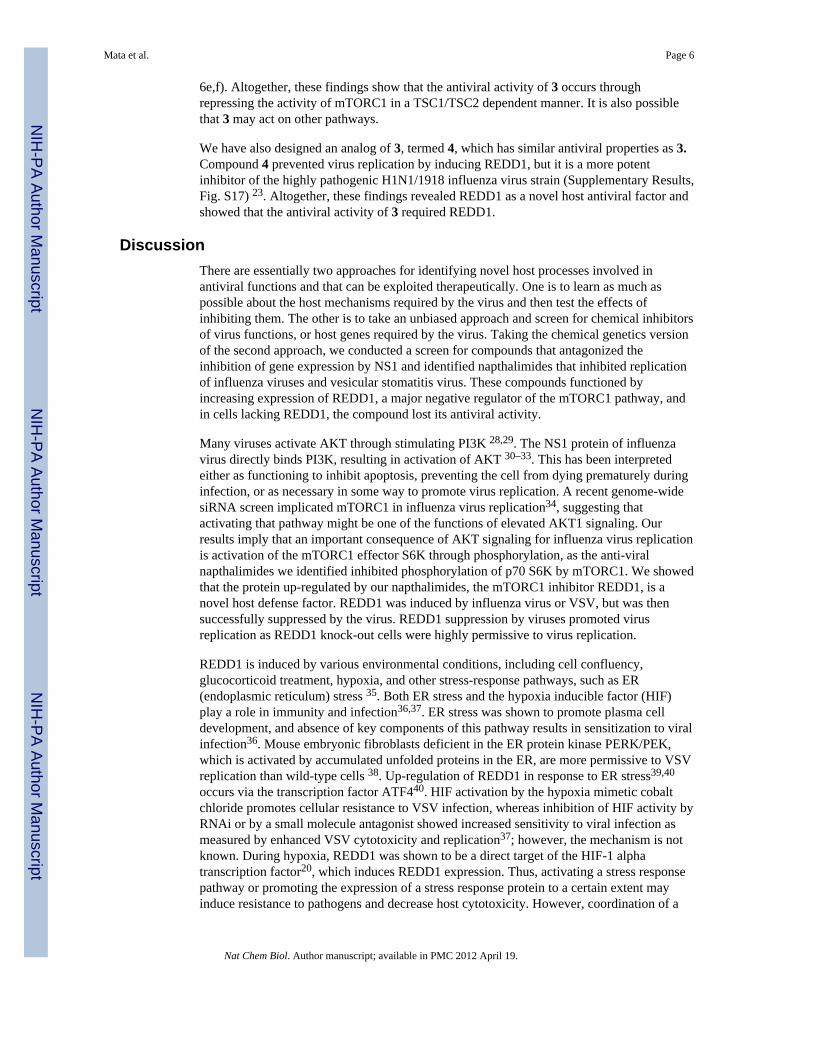
6e,f). Altogether, these findings show that the antiviral activity of 3 occurs throughrepressing the activity of mTORC1 in a TSC1/TSC2 dependent manner. It is also possiblethat 3 may act on other pathways.
We have also designed an analog of 3, termed 4, which has similar antiviral properties as 3.Compound 4 prevented virus replication by inducing REDD1, but it is a more potentinhibitor of the highly pathogenic H1N1/1918 influenza virus strain (Supplementary Results,Fig. S17) 23. Altogether, these findings revealed REDD1 as a novel host antiviral factor andshowed that the antiviral activity of 3 required REDD1.
DiscussionThere are essentially two approaches for identifying novel host processes involved inantiviral functions and that can be exploited therapeutically. One is to learn as much aspossible about the host mechanisms required by the virus and then test the effects ofinhibiting them. The other is to take an unbiased approach and screen for chemical inhibitorsof virus functions, or host genes required by the virus. Taking the chemical genetics versionof the second approach, we conducted a screen for compounds that antagonized theinhibition of gene expression by NS1 and identified napthalimides that inhibited replicationof influenza viruses and vesicular stomatitis virus. These compounds functioned byincreasing expression of REDD1, a major negative regulator of the mTORC1 pathway, andin cells lacking REDD1, the compound lost its antiviral activity.
Many viruses activate AKT through stimulating PI3K 28,29. The NS1 protein of influenzavirus directly binds PI3K, resulting in activation of AKT 30–33. This has been interpretedeither as functioning to inhibit apoptosis, preventing the cell from dying prematurely duringinfection, or as necessary in some way to promote virus replication. A recent genome-widesiRNA screen implicated mTORC1 in influenza virus replication34, suggesting thatactivating that pathway might be one of the functions of elevated AKT1 signaling. Ourresults imply that an important consequence of AKT signaling for influenza virus replicationis activation of the mTORC1 effector S6K through phosphorylation, as the anti-viralnapthalimides we identified inhibited phosphorylation of p70 S6K by mTORC1. We showedthat the protein up-regulated by our napthalimides, the mTORC1 inhibitor REDD1, is anovel host defense factor. REDD1 was induced by influenza virus or VSV, but was thensuccessfully suppressed by the virus. REDD1 suppression by viruses promoted virusreplication as REDD1 knock-out cells were highly permissive to virus replication.
REDD1 is induced by various environmental conditions, including cell confluency,glucocorticoid treatment, hypoxia, and other stress-response pathways, such as ER(endoplasmic reticulum) stress 35. Both ER stress and the hypoxia inducible factor (HIF)play a role in immunity and infection36,37. ER stress was shown to promote plasma celldevelopment, and absence of key components of this pathway results in sensitization to viralinfection36. Mouse embryonic fibroblasts deficient in the ER protein kinase PERK/PEK,which is activated by accumulated unfolded proteins in the ER, are more permissive to VSVreplication than wild-type cells 38. Up-regulation of REDD1 in response to ER stress39,40
occurs via the transcription factor ATF440. HIF activation by the hypoxia mimetic cobaltchloride promotes cellular resistance to VSV infection, whereas inhibition of HIF activity byRNAi or by a small molecule antagonist showed increased sensitivity to viral infection asmeasured by enhanced VSV cytotoxicity and replication37; however, the mechanism is notknown. During hypoxia, REDD1 was shown to be a direct target of the HIF-1 alphatranscription factor20, which induces REDD1 expression. Thus, activating a stress responsepathway or promoting the expression of a stress response protein to a certain extent mayinduce resistance to pathogens and decrease host cytotoxicity. However, coordination of a
Mata et al. Page 6
Nat Chem Biol. Author manuscript; available in PMC 2012 April 19.
NIH
-PA Author Manuscript
NIH
-PA Author Manuscript
NIH
-PA Author Manuscript
stress response to promote cellular resistance without significantly damaging the host uponpathogen invasion remains to be further investigated. We showed that induction of REDD1by small molecules is an efficient strategy for interfering with the functions of the mTORC1pathway that are required by viruses.
The effect of the napthalimide on influenza virus was a sharp attenuation of the productionof virus proteins early in infection. We found no effect of the napthalimide on global proteinsynthesis and no induction of an interferon response. In addition, in cells lacking REDD1, inwhich expression of influenza virus proteins is enhanced, rapamycin inhibited expression ofinfluenza virus proteins at a concentration in which bulk protein synthesis is known to beunaltered. Thus, this indicates selective translational regulation, which has been documentedin a number of conditions, including the general amino acid control response41 and othertypes of processes, such as survival and proliferation42. In addition, during nuclear mRNAprocessing and export, specific sequences either within the UTRs or in the coding region candictate differential binding by RNA-binding proteins (hnRNPs), which will regulateprocessing and export of specific subsets of mRNAs, resulting in differentialexpression43,44. This raises the possibility that the inhibition of the mTORC1 pathway mayalter translation in some way unfavorable to initiation of specific viral mRNAs relative tohost messages. In cells infected at a low multiplicity of infection, the first viral messagesmust compete with the far larger volume of host messages for access to ribosomes. In thisrespect, the early viral messages would have the same problem as a host cell message of lowabundance, such as mRNAs encoding certain transcription factors. However, at the earliestphases of infection, viruses are largely dependent upon normal host processes and it is theseprocesses that are likely to be the most useful therapeutic targets.
Although many viruses can be controlled by vaccination, there is still an important need forantiviral drugs. For viruses, such as influenza virus, that can infect other animals,vaccination will never eradicate the virus. Other viruses, such as small pox or measles canpotentially be eradicated by global immunization. However, once the incidence of disease isvery low, global vaccination is inevitably discontinued, leaving the human populationvulnerable to reemergence of the virus. The long lead times required to produce sufficientvaccine to protect the human population means that appearance of a new or re-occurringhighly infectious virus can lead to a pandemic of disease before the vaccine is available.However, antiviral drugs that target viral proteins have the disadvantage that resistance tothe drug will arise due to the high rates of mutation inherent in viruses and the largenumbers of progeny that they produce. A strategy targeting host processes that are essentialfor virus replication, such as the one discussed here, would avoid this problem, although itwould be limited by the possibility of toxic side effects. Thus, combinations of non-cytotoxic small molecules that target both viral and host proteins are desirable. Recently,influenza A nucleoprotein was identified as an antiviral target45. A small molecule thattriggered nucleoprotein aggregation and prevented its import into the nucleus protectedagainst influenza virus replication45. In addition, a chemical compound that inhibited hostpyrimidine biosynthesis has been recently shown to reduce influenza virus replication46.
In sum, the strategy of chemically inducing host antiviral activities targeting host pathwayswithout causing significant short-term toxic effects will likely have an important impact inantiviral therapy. One such strategy was identified here with the induction of the mTORC1inhibitor REDD1 by naphthalimides. Furthermore, small molecules that inhibit themTORC1 pathway in different ways have the potential for anti-cancer therapy as themTORC1 pathway is a major regulator of cell proliferation and cancer 47.
Mata et al. Page 7
Nat Chem Biol. Author manuscript; available in PMC 2012 April 19.
NIH
-PA Author Manuscript
NIH
-PA Author Manuscript
NIH
-PA Author Manuscript
MethodsCompound screen
The UT Southwestern Compound Library is composed of 200,000 synthetic drug-likecompounds arrayed in DMSO in 384 well plates. 293T cells were transfected with anapproximately 10:1 ratio of plasmid pCMV-Luc encoding luciferase and pCAGGS-NS1encoding NS1, using Lipofectamine2000 (Invitrogen). Cells were transfected with theluciferase plasmid alone as a positive control. After 16 hours, cells were dispensed at 5000cell/well in 384 well plates. After one hour, compounds from the library were added to afinal concentration of 5 μM in 1% DMSO in a one compound/one well format. Experimentalsamples were limited to columns 3 to 22, with controls treated with 1% DMSO in the firstand last two columns of wells. Wells in the first column of each plate contained cellstransfected with the luciferase plasmid alone; all other wells received cells transfected withboth plasmids. Plates were incubated 22 h at 37°C in 5% CO2, then cooled to roomtemperature and incubated with Bright-Glo luciferase substrate (Promega) for 4 min andluminescence was recorded. Plates with Z′-scores lower than 0.45 were repeated.Experimental values were normalized to the mean of the luciferase alone control on thesame plate. Compounds were ranked by z-score and 640 compounds having the mostpositive z-scores were selected and re-tested in the assay at 15, 5 and 1.7 μM concentrations.These compounds were also tested for the ability to prevent cell death of immortalizedhuman bronchial epithelial cells (HBEC) that had been infected with A/WS/33 influenzavirus by measuring cell ATP levels with ATP-lite (PerkinElmer). A supplementary tabledescribing this screen is added to the end of the Supplementary Information file.
Compound half-lifeCompound half-lives were measure in HBECs by LC/MS/MS. Metabolic stability half-lifewas determined by substrate depletion 48.
Cell survival/Cytotoxicity measurementsMEF, HBEC, or MDCK cells were seeded in white-walled 96 plates at a density of 3×10³
cells per well, 16 h prior to compound addition. Compounds dissolved in sterile DMSO(Sigma) at a concentration of 25 mM were diluted to 100 μM in OptiMEM I (Invitrogen) intriplicates. The 100 μM starting dilutions were serially diluted in two-fold steps to a finalconcentration of 0.2 μM. Control experiments, performed in the absence of compound, hadthe same final concentration of DMSO as in compound-treated samples. At the time pointsdepicted in the figures, cells were lysed and ATP levels measured by luminescence using theCell Titre-Glo kit (Promega) following the manufacturer’s instructions. In parallel, cellswere also counted at the beginning and at the end of each experiment and cell survival wasquantified by trypan blue exclusion assay.
Influenza virus replicationMDCK cells were infected with various strains of influenza virus depicted in the figures atan m.o.i. of 0.001 pfu/cell for 1 hr. Next, cells were washed with PBS and overlaid withOptiMEM containing two-fold compound dilutions ranging from 100 μM to 0.8 μM.Samples containing only the same volume of DMSO as the compounds were included. At 30h p.i., culture medium was collected, cell debris removed by centrifugation at 1000 ×g for 10minutes, and frozen at −80 °C. Viral titers were determined by plaque assay. Theexperiments conducted with the H1N1/1918 strain were performed in a high-containment(BSL3++) facility.
For experiments performed with A549 cells, REDD1+/+ and REDD1−/− cells, and TSC2cells, the methodology is described in the legends. For experiments performed with U20S
Mata et al. Page 8
Nat Chem Biol. Author manuscript; available in PMC 2012 April 19.
NIH
-PA Author Manuscript
NIH
-PA Author Manuscript
NIH
-PA Author Manuscript
cells, cells were plated in 12-well plates in DMEM containing 10% FBS and incubatedovernight. Cells were then incubated in media containing tetracycline (1 g/ml) for 2 h toinduce REDD1 overexpression. Cells were washed with PBS and infected with A/WSN/1933 or VSV at m.o.i. 2 for 1 h. Tetracycline was added back 1 h post-infection and celllysates were prepared at various time points post-infection, as indicated in the figure.
VSV Replication AssayVesicular stomatitis virus replication: MDCK cells seeded in 35-mm-diameter dishes wereinfected with VSV-GFP at m.o.i. 0.001 pfu/cell. At 24 h p.i., supernatants were clarified andused for titration on VERO cells. Four-fold serial dilutions of virus containing supernatantswere made in PBS containing serum and antibiotics. Fifty microliters of each dilution wasmixed with an equal volume of complete growth medium containing 8,000 VERO cells andincubated at 37 °C for 48 h in 96-well plates. Cells were fixed in 4% paraformaldehyde. Thenumber of wells with GFP expression were counted by fluorescence microscopy andsubsequently used to calculate relative virus titers. Infection of U2OS cells with VSV wasperformed in the same manner as influenza virus infection described above.
In situ hybridizationmRNA distribution in MDCK cells infected with influenza virus in the presence or absenceof compounds was performed as we previously described 18. Influenza proteins weredetected with mouse anti-influenza antibody (Biodesign International) and FITC labeledanti-mouse antibody.
Phospho-S6K analysisCells were starved for 18 h and then mock infected or infected as described in the legend offigure 5. Five percent serum was added to induce S6K phosphorylation in control lanes.H358 and H1993 cells were treated with 10 μM 3 and LnCap cells were treated with 30 μM.
All data presented here are representative of at least 3 independent experiments. In the linegraphs or histograms, data represent mean values +/− s.d.
Description of real-time RT-PCR, gene expression profiling and analysis, humanbiochemical network, compound synthesis, details of cells, plasmids and antibodies aredescribed in Supplementary Methods and Supplementary Information.
Supplementary MaterialRefer to Web version on PubMed Central for supplementary material.
AcknowledgmentsWe thank R. Sakthivel, L. Melito, and J. Naidoo for technical assistance. We thank S. de Celis, D.E. Levy and B.Levine for reagents. This work was supported by NIH R01 GM07159 to B.M.A.F.; R01 AI079110 andR01AI089539 to B.M.A.F. and M.G.R.; the Diane and Hal Brierley distinguished Chair in Biomedical research toM.G.R and C06-RR15437 from the NCRR; NIH grants R01AI046954, P01AI058113, U54AI057158,U01AI074539 and CRIP, an NIAID funded Center of Excellence for Influenza Research and Surveillance(HHSN266200700010C) to A.G.-S; R01 CA129387 to J.B.; M.M. was supported by the NIH Diversity SupplementR01GM06715908S1.
Abbreviations
MOI multiplicity of infection
Mata et al. Page 9
Nat Chem Biol. Author manuscript; available in PMC 2012 April 19.
NIH
-PA Author Manuscript
NIH
-PA Author Manuscript
NIH
-PA Author Manuscript
NS1 nonstructural 1
S6K S6 kinase
mTORC1 mammalian target of rapamycin
REDD1, DDIT4, or Rtp801 regulated in development and DNA damage response 1
VSV vesicular stomatitis virus
References1. Simonsen L, et al. The impact of influenza epidemics on mortality: introducing a severity index. Am
J Public Health. 1997; 87:1944–1950. [PubMed: 9431281]2. Smith DJ, et al. Mapping the antigenic and genetic evolution of influenza virus. Science. 2004;
305:371–376. [PubMed: 15218094]3. Krug RM, Etkind PR. Cytoplasmic and nuclear virus-specific proteins in influenza virus-infected
MDCK cells. Virology. 1973; 56:334–348. 0042-6822(73)90310-3 [pii]. [PubMed: 4795673]4. Li Y, Yamakita Y, Krug RM. Regulation of a nuclear export signal by an adjacent inhibitory
sequence: the effector domain of the influenza virus NS1 protein. Proceedings of the NationalAcademy of Sciences of the United States of America. 1998; 95:4864–4869. [PubMed: 9560194]
5. Guo Z, et al. NS1 protein of influenza A virus inhibits the function of intracytoplasmic pathogensensor, RIG-I. Am J Respir Cell Mol Biol. 2007; 36:263–269. 2006-0283RC [pii]. 10.1165/rcmb.2006-0283RC [PubMed: 17053203]
6. Mibayashi M, et al. Inhibition of Retinoic Acid-Inducible Gene-I-Mediated Induction of Interferon-{beta} by the NS1 protein of Influenza A Virus. J Virol. 2007; 81:514–524. [PubMed: 17079289]
7. Opitz B, et al. IFNbeta induction by influenza A virus is mediated by RIG-I which is regulated bythe viral NS1 protein. Cell Microbiol. 2007; 9:930–938. CMI841 [pii]. 10.1111/j.1462-5822.2006.00841.x [PubMed: 17140406]
8. Pichlmair A, et al. RIG-I-Mediated Antiviral Responses to Single-Stranded RNA Bearing 5′-Phosphates. Science. 2006; 314:997–1001. [PubMed: 17038589]
9. Li S, Min JY, Krug RM, Sen GC. Binding of the influenza A virus NS1 protein to PKR mediates theinhibition of its activation by either PACT or double-stranded RNA. Virology. 2006; 349:13–21.[PubMed: 16466763]
10. Min JY, Krug RM. The primary function of RNA binding by the influenza A virus NS1 protein ininfected cells: Inhibiting the 2′-5′ oligo (A) synthetase/RNase L pathway. Proceedings of theNational Academy of Sciences of the United States of America. 2006; 103:7100–7105. [PubMed:16627618]
11. Hale BG, Jackson D, Chen YH, Lamb RA, Randall RE. Influenza A virus NS1 protein bindsp85beta and activates phosphatidylinositol-3-kinase signaling. Proc Natl Acad Sci U S A. 2006;103:14194–14199. [PubMed: 16963558]
12. Fortes P, Beloso A, Ortin J. Influenza virus NS1 protein inhibits pre-mRNA splicing and blocksmRNA nucleocytoplasmic transport. EMBO J. 1994; 13:704–712. [PubMed: 8313914]
13. Qiu Y, Nemeroff M, Krug RM. The influenza virus NS1 protein binds to a specific region inhuman U6 snRNA and inhibits U6-U2 and U6-U4 snRNA interactions during splicing. Rna. 1995;1:304–316. [PubMed: 7489502]
14. Woff T, O’Neil RE, Palese P. NS1-Binding protein (NS1-BP): a novel human protein that interactswith the influenza A virus nonstructural NS1 protein is relocalized in the nuclei of infected cells. JVirol. 1998; 72:7170–7180. [PubMed: 9696811]
15. Nemeroff ME, Barabino SM, Li Y, Keller W, Krug RM. Influenza virus NS1 protein interacts withthe cellular 30 kDa subunit of CPSF and inhibits 3' end formation of cellular pre-mRNAs. MolCell. 1998; 1:991–1000. [PubMed: 9651582]
16. Qian XY, Alonso-Caplen F, Krug RM. Two functional domains of influenza virus NS1 protein arerequired for regulation of nuclear export of mRNa. J Virol. 1994; 68:2433–2441. [PubMed:8139028]
Mata et al. Page 10
Nat Chem Biol. Author manuscript; available in PMC 2012 April 19.
NIH
-PA Author Manuscript
NIH
-PA Author Manuscript
NIH
-PA Author Manuscript
17. Qiu Y, Krug RM. The influenza virus NS1 protein is a poly(A)-binding protein that inhibitsnuclear export of mRNAs containing poly(A). Journal of virology. 1994; 68:2425–2432.[PubMed: 7908060]
18. Satterly N, et al. Influenza virus targets the mRNA export machinery and the nuclear porecomplex. Proceedings of the National Academy of Sciences of the United States of America.2007; 104:1853–1858. [PubMed: 17267598]
19. Garcia-Sastre A, et al. Influenza A virus lacking the NS1 gene replicates in interferon-deficientsystems. Virology. 1998; 252:324–330. [PubMed: 9878611]
20. Brugarolas J, et al. Regulation of mTOR function in response to hypoxia by REDD1 and the TSC1/TSC2 tumor suppressor complex. Genes Dev. 2004; 18:2893–2904. gad.1256804 [pii]. 10.1101/gad.1256804 [PubMed: 15545625]
21. Ma XM, Blenis J. Molecular mechanisms of mTOR-mediated translational control. Nat Rev MolCell Biol. 2009; 10:307–318. nrm2672 [pii]. 10.1038/nrm2672 [PubMed: 19339977]
22. Chakraborty P, et al. Nucleoporin levels regulate cell cycle progression and phase-specific geneexpression. Developmental cell. 2008; 15:657–667. [PubMed: 19000832]
23. Tumpey TM, et al. Characterization of the reconstructed 1918 Spanish influenza pandemic virus.Science. 2005; 310:77–80. 310/5745/77 [pii]. 10.1126/science.1119392 [PubMed: 16210530]
24. Hresko RC, Mueckler M. mTOR.RICTOR is the Ser473 kinase for Akt/protein kinase B in 3T3-L1adipocytes. The Journal of biological chemistry. 2005; 280:40406–40416. M508361200 [pii].10.1074/jbc.M508361200 [PubMed: 16221682]
25. Sarbassov DD, Guertin DA, Ali SM, Sabatini DM. Phosphorylation and regulation of Akt/PKB bythe rictor-mTOR complex. Science. 2005; 307:1098–1101. 307/5712/1098 [pii]. 10.1126/science.1106148 [PubMed: 15718470]
26. Rubinsztein DC, Gestwicki JE, Murphy LO, Klionsky DJ. Potential therapeutic applications ofautophagy. Nat Rev Drug Discov. 2007; 6:304–312. nrd2272 [pii]. 10.1038/nrd2272 [PubMed:17396135]
27. Vega-Rubin-de-Celis S, et al. Structural analysis and functional implications of the negativemTORC1 regulator REDD1. Biochemistry. 2010; 49:2491–2501.10.1021/bi902135e [PubMed:20166753]
28. Buchkovich NJ, Yu Y, Zampieri CA, Alwine JC. The TORrid affairs of viruses: effects ofmammalian DNA viruses on the PI3K-Akt-mTOR signalling pathway. Nat Rev Microbiol. 2008;6:266–275. nrmicro1855 [pii]. 10.1038/nrmicro1855 [PubMed: 18311165]
29. Cooray S. The pivotal role of phosphatidylinositol 3-kinase-Akt signal transduction in virussurvival. The Journal of general virology. 2004; 85:1065–1076. [PubMed: 15105524]
30. Ehrhardt C, et al. Influenza A virus NS1 protein activates the PI3K/Akt pathway to mediateantiapoptotic signaling responses. Journal of virology. 2007; 81:3058–3067. JVI.02082-06 [pii].10.1128/JVI.02082-06 [PubMed: 17229704]
31. Hale BG, Jackson D, Chen YH, Lamb RA, Randall RE. Influenza A virus NS1 protein bindsp85beta and activates phosphatidylinositol-3-kinase signaling. Proceedings of the NationalAcademy of Sciences of the United States of America. 2006; 103:14194–14199. 0606109103 [pii].10.1073/pnas.0606109103 [PubMed: 16963558]
32. Shin YK, Liu Q, Tikoo SK, Babiuk LA, Zhou Y. Influenza A virus NS1 protein activates thephosphatidylinositol 3-kinase (PI3K)/Akt pathway by direct interaction with the p85 subunit ofPI3K. The Journal of general virology. 2007; 88:13–18. 88/1/13 [pii]. 10.1099/vir.0.82419-0[PubMed: 17170431]
33. Zhirnov OP, Klenk HD. Control of apoptosis in influenza virus-infected cells by up-regulation ofAkt and p53 signaling. Apoptosis. 2007; 12:1419–1432.10.1007/s10495-007-0071-y [PubMed:17468837]
34. Konig R, et al. Human host factors required for influenza virus replication. Nature. 2010; 463:813–817. nature08699 [pii]. 10.1038/nature08699 [PubMed: 20027183]
35. Brugarolas, J. mTORC1 signaling and hypoxia. In: Polunovsky, V.; Houghton, PJ., editors. mTORpathway and mTOR inhibitors in cancer therapy. Humana Press; 2010. in press
Mata et al. Page 11
Nat Chem Biol. Author manuscript; available in PMC 2012 April 19.
NIH
-PA Author Manuscript
NIH
-PA Author Manuscript
NIH
-PA Author Manuscript
36. Todd DJ, Lee AH, Glimcher LH. The endoplasmic reticulum stress response in immunity andautoimmunity. Nat Rev Immunol. 2008; 8:663–674. nri2359 [pii]. 10.1038/nri2359 [PubMed:18670423]
37. Zinkernagel AS, Johnson RS, Nizet V. Hypoxia inducible factor (HIF) function in innate immunityand infection. J Mol Med. 2007; 85:1339–1346.10.1007/s00109-007-0282-2 [PubMed: 18030436]
38. Krishnamoorthy J, Mounir Z, Raven JF, Koromilas AE. The eIF2alpha kinases inhibit vesicularstomatitis virus replication independently of eIF2alpha phosphorylation. Cell Cycle. 2008;7:2346–2351. 6323 [pii]. [PubMed: 18677106]
39. Jin H-O, et al. Activating transcription factor 4 and CCAAT/enhancer-binding protein-β negativelyregulate the mammalian target of rapamycin via Redd1 expression in response to oxidative andendoplasmic reticulum stress. Free Radical Biology & Medicine. 2009; 46:1158–1167. [PubMed:19439225]
40. Whitney ML, Jefferson LS, Kimball SR. ATF4 is necessary and sufficient for ER stress-inducedupregulation of REDD1 expression. Biochem Biophys Res Commun. 2009; 379:451–455.S0006-291X(08)02494-7 [pii]. 10.1016/j.bbrc.2008.12.079 [PubMed: 19114033]
41. Costa-Mattioli M, Sossin WS, Klann E, Sonenberg N. Translational control of long-lastingsynaptic plasticity and memory. Neuron. 2009; 61:10–26. S0896-6273(08)01089-1 [pii]. 10.1016/j.neuron.2008.10.055 [PubMed: 19146809]
42. Shahbazian D, et al. Control of cell survival and proliferation by mammalian eukaryotic initiationfactor 4B. Molecular and cellular biology. 2010; 30:1478–1485. MCB.01218-09 [pii]. 10.1128/MCB.01218-09 [PubMed: 20086100]
43. Farny NG, Hurt JA, Silver PA. Definition of global and transcript-specific mRNA export pathwaysin metazoans. Genes Dev. 2008; 22:66–78. gad.1616008 [pii]. 10.1101/gad.1616008 [PubMed:18086857]
44. Keene JD. RNA regulons: coordination of post-transcriptional events. Nat Rev Genet. 2007;8:533–543. nrg2111 [pii]. 10.1038/nrg2111 [PubMed: 17572691]
45. Kao RY, et al. Identification of influenza A nucleoprotein as an antiviral target. Nat Biotechnol.2010; 28:600–605.10.1038/nbt.1638 [PubMed: 20512121]
46. Hoffmann HH, Kunz A, Simon VA, Palese P, Shaw ML. Broad-spectrum antiviral that interfereswith de novo pyrimidine biosynthesis. Proceedings of the National Academy of Sciences of theUnited States of America. 2011; 108:5777–5782.10.1073/pnas.1101143108 [PubMed: 21436031]
47. Choo AY, Blenis J. Not all substrates are treated equally: implications for mTOR, rapamycin-resistance and cancer therapy. Cell Cycle. 2009; 8:567–572. 7659 [pii]. [PubMed: 19197153]
48. McNaney CA, et al. An automated liquid chromatography-mass spectrometry process to determinemetabolic stability half-life and intrinsic clearance of drug candidates by substrate depletion.Assay Drug Dev Technol. 2008; 6:121–129.10.1089/adt.2007.103 [PubMed: 18336089]
49. Yuen T, Wurmbach E, Pfeffer RL, Ebersole BJ, Sealfon SC. Accuracy and calibration ofcommercial oligonucleotide and custom cDNA microarrays. Nucleic Acids Res. 2002; 30:e48.[PubMed: 12000853]
50. Kanehisa M, et al. KEGG for linking genomes to life and the environment. Nucleic Acids Res.2008; 36:D480–484. gkm882 [pii]. 10.1093/nar/gkm882 [PubMed: 18077471]
Mata et al. Page 12
Nat Chem Biol. Author manuscript; available in PMC 2012 April 19.
NIH
-PA Author Manuscript
NIH
-PA Author Manuscript
NIH
-PA Author Manuscript

Figure 1. Identification of Small Molecules that Revert the Inhibition of Gene ExpressionMediated by the Influenza Virus NS1 Protein and that Protect Cells from Virus-Induced CellDeath(a) Luciferase expression in 293T cells transfected with NS1 and treated individually with200,000 synthetic compounds (5 μM) was normalized to on-plate controls treated with 0.3%DMSO. Values are expressed as z scores, using the mean value and standard deviation of theexperimental population screened on the same day. Red circle shows compound 1 studiedhere. (b) The most active 640 compounds were tested at three concentrations for the abilityto inhibit the cytopathic effect of A/WSN/1933 influenza virus infection in HBECs. z scoresfor compounds assayed at 1.7 μM are plotted according to activity. (c) The structure of themost active naphthalimide from the primary screen, compound 1, an inactive analog, 2, anda more potent related compound, 3, are shown.
Mata et al. Page 13
Nat Chem Biol. Author manuscript; available in PMC 2012 April 19.
NIH
-PA Author Manuscript
NIH
-PA Author Manuscript
NIH
-PA Author Manuscript
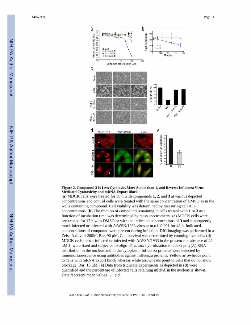
Figure 2. Compound 3 Is Less Cytotoxic, More Stable than 1, and Reverts Influenza Virus-Mediated Cytotoxicity and mRNA Export Block(a) MDCK cells were treated for 30 h with compounds 1, 2, and 3 at various depictedconcentrations and control cells were treated with the same concentration of DMSO as in thewells containing compound. Cell viability was determined by measuring cell ATPconcentrations. (b) The fraction of compound remaining in cells treated with 1 or 3 as afunction of incubation time was determined by mass spectrometry. (c) MDCK cells werepre-treated for 17 h with DMSO or with the indicated concentrations of 3 and subsequentlymock infected or infected with A/WSN/1933 virus at m.o.i. 0.001 for 48 h. Indicatedconcentrations of compound were present during infection. DIC imaging was performed in aZeiss Axiovert 200M. Bar, 90 μM. Cell survival was determined by counting live cells. (d)MDCK cells, mock-infected or infected with A/WSN/1933 in the presence or absence of 25μM 3, were fixed and subjected to oligo-dT in situ hybridization to detect poly(A) RNAdistribution in the nucleus and in the cytoplasm. Influenza proteins were detected byimmunofluorescence using antibodies against influenza proteins. Yellow arrowheads pointto cells with mRNA export block whereas white arrowheads point to cells that do not showblockage. Bar, 15 μM. (e) Data from triplicate experiments as depicted in (d) werequantified and the percentage of infected cells retaining mRNA in the nucleus is shown.Data represent mean values +/− s.d.
Mata et al. Page 14
Nat Chem Biol. Author manuscript; available in PMC 2012 April 19.
NIH
-PA Author Manuscript
NIH
-PA Author Manuscript
NIH
-PA Author Manuscript
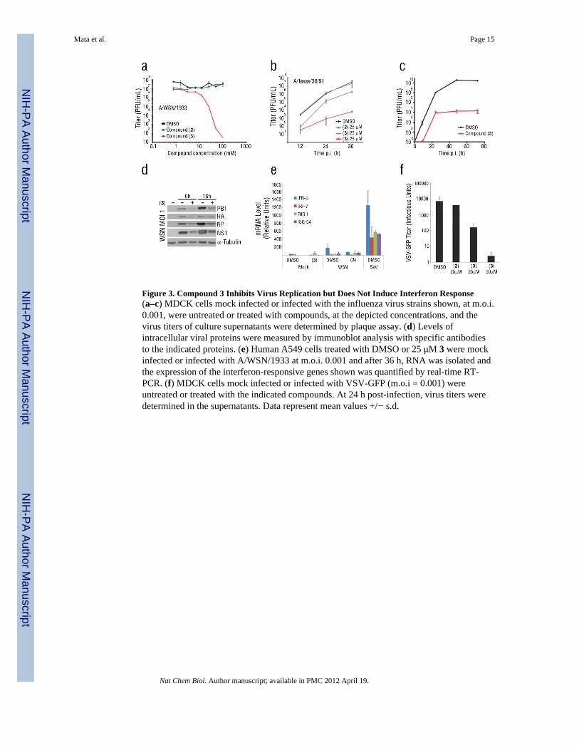
Figure 3. Compound 3 Inhibits Virus Replication but Does Not Induce Interferon Response(a–c) MDCK cells mock infected or infected with the influenza virus strains shown, at m.o.i.0.001, were untreated or treated with compounds, at the depicted concentrations, and thevirus titers of culture supernatants were determined by plaque assay. (d) Levels ofintracellular viral proteins were measured by immunoblot analysis with specific antibodiesto the indicated proteins. (e) Human A549 cells treated with DMSO or 25 μM 3 were mockinfected or infected with A/WSN/1933 at m.o.i. 0.001 and after 36 h, RNA was isolated andthe expression of the interferon-responsive genes shown was quantified by real-time RT-PCR. (f) MDCK cells mock infected or infected with VSV-GFP (m.o.i = 0.001) wereuntreated or treated with the indicated compounds. At 24 h post-infection, virus titers weredetermined in the supernatants. Data represent mean values +/− s.d.
Mata et al. Page 15
Nat Chem Biol. Author manuscript; available in PMC 2012 April 19.
NIH
-PA Author Manuscript
NIH
-PA Author Manuscript
NIH
-PA Author Manuscript
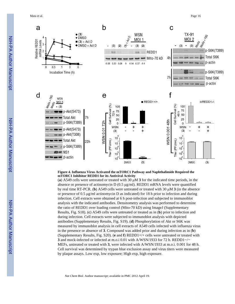
Figure 4. Influenza Virus Activated the mTORC1 Pathway and Naphthalimide Required themTORC1 Inhibitor REDD1 for its Antiviral Activity(a) A549 cells were untreated or treated with 30 μM 3 for the indicated time periods, in theabsence or presence of actinomycin D (0.5 μg/ml). REDD1 mRNA levels were quantifiedby real time RT-PCR. (b) A549 cells were untreated or treated with 30 μM 3 (in the absenceor presence of 0.5 μg/ml actinomycin D as indicated) for 18 h prior to infection and duringinfection. Cell extracts were obtained at 6 h post-infection and subjected to immunoblotanalysis with the indicated antibodies. Densitometry analysis was performed to determinethe ratio of REDD1 over loading control (Mito-70 kD) using ImageJ (SupplementaryResults, Fig. S18). (c) A549 cells were untreated or treated as in (b) prior to infection andduring infection. Cell extracts were subjected to immunoblot analysis with depictedantibodies (Supplementary Results, Fig. S19). (d) Phosphorylation of Akt or S6K wasmeasured by immunoblot analysis in cell extracts of A549 cells infected with influenza virusin the presence or absence of 3. Compound was added prior and during infection as in (b)(Supplementary Results, Fig. S20). (e and f) REDD1+/+ cells were untreated or treated with3 and mock-infected or infected at m.o.i 0.01 with A/WSN/1933 for 72 h. REDD1−/−MEFs, untreated or treated with 3, were infected with A/WSN/1933 at m.o.i. 0.001 for 48 h.Cell survival was determined by trypan blue exclusion assay and virus titers were measuredby plaque assays. Low exp, low exposure; High exp, high exposure.
Mata et al. Page 16
Nat Chem Biol. Author manuscript; available in PMC 2012 April 19.
NIH
-PA Author Manuscript
NIH
-PA Author Manuscript
NIH
-PA Author Manuscript
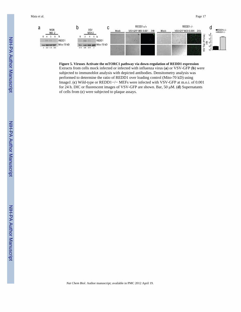
Figure 5. Viruses Activate the mTORC1 pathway via down-regulation of REDD1 expressionExtracts from cells mock infected or infected with influenza virus (a) or VSV-GFP (b) weresubjected to immunoblot analysis with depicted antibodies. Densitometry analysis wasperformed to determine the ratio of REDD1 over loading control (Mito-70 kD) usingImageJ. (c) Wild-type or REDD1−/− MEFs were infected with VSV-GFP at m.o.i. of 0.001for 24 h. DIC or fluorescent images of VSV-GFP are shown. Bar, 50 μM. (d) Supernatantsof cells from (c) were subjected to plaque assays.
Mata et al. Page 17
Nat Chem Biol. Author manuscript; available in PMC 2012 April 19.
NIH
-PA Author Manuscript
NIH
-PA Author Manuscript
NIH
-PA Author Manuscript
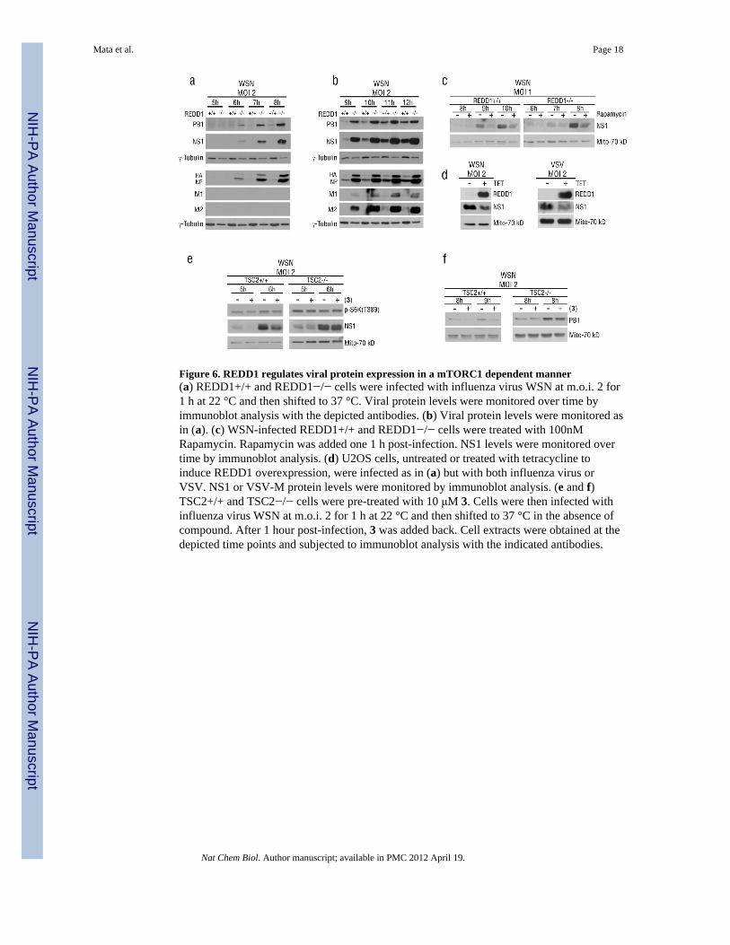
Figure 6. REDD1 regulates viral protein expression in a mTORC1 dependent manner(a) REDD1+/+ and REDD1−/− cells were infected with influenza virus WSN at m.o.i. 2 for1 h at 22 °C and then shifted to 37 °C. Viral protein levels were monitored over time byimmunoblot analysis with the depicted antibodies. (b) Viral protein levels were monitored asin (a). (c) WSN-infected REDD1+/+ and REDD1−/− cells were treated with 100nMRapamycin. Rapamycin was added one 1 h post-infection. NS1 levels were monitored overtime by immunoblot analysis. (d) U2OS cells, untreated or treated with tetracycline toinduce REDD1 overexpression, were infected as in (a) but with both influenza virus orVSV. NS1 or VSV-M protein levels were monitored by immunoblot analysis. (e and f)TSC2+/+ and TSC2−/− cells were pre-treated with 10 μM 3. Cells were then infected withinfluenza virus WSN at m.o.i. 2 for 1 h at 22 °C and then shifted to 37 °C in the absence ofcompound. After 1 hour post-infection, 3 was added back. Cell extracts were obtained at thedepicted time points and subjected to immunoblot analysis with the indicated antibodies.
Mata et al. Page 18
Nat Chem Biol. Author manuscript; available in PMC 2012 April 19.
NIH
-PA Author Manuscript
NIH
-PA Author Manuscript
NIH
-PA Author Manuscript
Related Documents