Chemistry & Biology, Vol. 10, 397–410, May, 2003, 2003 Elsevier Science Ltd. All rights reserved. DOI 10.1016/S1074-5521(03)00093-0 Chemical Genetic Modifier Screens: Small Molecule Trichostatin Suppressors as Probes of Intracellular Histone and Tubulin Acetylation potential of HDAC inhibitors in the treatment of T cell lymphoma [4] and neurodegenerative diseases [5] is also being explored in clinical trials of HDAC inhibitors like suberoylanilide hydroxamic acid (SAHA) [2]. Other small molecules known to inhibit HDACs (i.e., butyrate, Kathryn M. Koeller, 1,3 Stephen J. Haggarty, 1,2,3 Brian D. Perkins, 2 Igor Leykin, 5 Jason C. Wong, 1,3 Ming-Chih J. Kao, 5 and Stuart L. Schreiber 1,2,3,4,6 1 Department of Chemistry and Chemical Biology 2 Department of Molecular and Cellular Biology valproic acid) already enjoy widespread clinical accep- tance in the treatment of other disorders [6]. Based on 3 Harvard Institute of Chemistry and Chemical Biology these encouraging early clinical outcomes, numerous programs have been established for the discovery of 4 Howard Hughes Medical Institute Harvard University potent and potentially paralog-selective HDAC inhibi- tors [7]. 12 Oxford Street Cambridge, Massachusetts 02138 Histone acetylation is controlled by HDACs, histone acetyltransferases (HATs), and the proteins that position 5 Department of Biostatistics Harvard School of Public Health these enzymes to chromatin sites within the genome. HDAC inhibitors shift a reversible histone acetylation/ 655 Huntington Avenue Boston, Massachusetts 02115 deacetylation state (mediated by HATs and HDACs, re- spectively) toward a condition of histone hyperacetyla- tion (Figure 1). HDAC inhibitors trichostatin A (TSA) and SAHA promote acetylation of -tubulin as well as his- Summary tones, implying that tubulin acetylation is also controlled by competition between acetyltransferases and deacet- Histone deacetylase (HDAC) inhibitors are being de- ylases. Interestingly, not all HDAC inhibitors affect tu- veloped as new clinical agents in cancer therapy, in bulin acetylation, indicating that distinguishing features part because they interrupt cell cycle progression in exist for each class of these small molecules. Function- transformed cell lines. To examine cell cycle arrest ally, HDAC inhibitor-promoted histone acetylation corre- induced by HDAC inhibitor trichostatin A (TSA), a cy- lates with cell cycle arrest and transcriptional activation. toblot cell-based screen was used to identify small However, cellular effects of HDAC inhibitor-induced tu- molecule suppressors of this process. TSA suppres- bulin acetylation remain largely uncharacterized. As sors (ITSAs) counteract TSA-induced cell cycle arrest, such, tubulin acetylation represents a variable that has histone acetylation, and transcriptional activation. Hy- not been recognized in cellular and clinical analysis of droxamic acid-based HDAC inhibitors like TSA and HDAC inhibitors like SAHA. To probe further the cellular suberoylanilide hydroxamic acid (SAHA) promote acet- activities of TSA, a chemical genetic modifier screen ylation of cytoplasmic -tubulin as well as histones, was undertaken, specifically aimed at the discovery of a modification also suppressed by ITSAs. Although small molecule TSA suppressors. This strategy has iden- tubulin acetylation appears irrelevant to cell cycle pro- tified new probes of intracellular acetylation, the cell gression and transcription, it may play a role in other cycle, and acetylation-mediated transcriptional activa- cellular processes. Small molecule suppressors such tion in mammalian cell lines. as the ITSAs, available from chemical genetic suppres- sor screens, may prove to be valuable probes of many biological processes. Results and Discussion Introduction Cell-Based Screening Identifies Suppressors of TSA-Induced Cell Cycle Arrest Histone acetylation is tightly coordinated with transcrip- In classical genetics, a specific cellular phenotype is tional regulation, differentiation, and cell cycle progres- often characterized by identifying conditions or muta- sion in mammalian cell lines (Figure 1) [1]. Aberrant his- tions that modify (i.e., suppress or enhance) the pheno- tone acetylation patterns can alter differentiation type. By analogy to classical genetics, the cellular action pathways or disrupt cell cycle checkpoints relevant to of a chemical (here the HDAC inhibitor TSA) can be the onset or maintenance of cancerous states. Enzymes studied by identifying new small molecules that modify regulating histone modifications therefore represent at- its cellular effects. In this respect, a chemical genetic tractive chemotherapeutic targets. In particular, histone modifier screen was used to identify small molecules deacetylase (HDAC) inhibitors induce differentiation, cell that suppress TSA’s cell cycle effects. TSA [8] inhibits cycle arrest, or apoptosis in transformed cell lines, pro- all known class I and II HDACs in vitro and causes tran- cesses most likely involving transcriptional activation scriptional activation and cell cycle arrest in mammalian [2]. For example, HDAC inhibitors and retinoic acid act cell lines. Small molecule TSA suppressors (ITSAs, inhib- synergistically to reactivate silenced genes in “differenti- itors of TSA) may thus affect components up- or down- ation therapy” for acute promyelocytic leukemia [3]. The stream of HDAC inhibition. A cytoblot modifier screen in human A549 lung carci- noma cells was used to identify ITSAs (Figure 2A) [9]. 6 Correspondence: [email protected]

Welcome message from author
This document is posted to help you gain knowledge. Please leave a comment to let me know what you think about it! Share it to your friends and learn new things together.
Transcript
Chemistry & Biology, Vol. 10, 397–410, May, 2003, 2003 Elsevier Science Ltd. All rights reserved. DOI 10.1016/S1074-5521(03)00093-0
Chemical Genetic Modifier Screens:Small Molecule Trichostatin Suppressors as Probesof Intracellular Histone and Tubulin Acetylation
potential of HDAC inhibitors in the treatment of T celllymphoma [4] and neurodegenerative diseases [5] isalso being explored in clinical trials of HDAC inhibitorslike suberoylanilide hydroxamic acid (SAHA) [2]. Othersmall molecules known to inhibit HDACs (i.e., butyrate,
Kathryn M. Koeller,1,3 Stephen J. Haggarty,1,2,3
Brian D. Perkins,2 Igor Leykin,5
Jason C. Wong,1,3 Ming-Chih J. Kao,5
and Stuart L. Schreiber1,2,3,4,6
1Department of Chemistry and Chemical Biology2Department of Molecular and Cellular Biology valproic acid) already enjoy widespread clinical accep-
tance in the treatment of other disorders [6]. Based on3Harvard Institute of Chemistry andChemical Biology these encouraging early clinical outcomes, numerous
programs have been established for the discovery of4 Howard Hughes Medical InstituteHarvard University potent and potentially paralog-selective HDAC inhibi-
tors [7].12 Oxford StreetCambridge, Massachusetts 02138 Histone acetylation is controlled by HDACs, histone
acetyltransferases (HATs), and the proteins that position5 Department of BiostatisticsHarvard School of Public Health these enzymes to chromatin sites within the genome.
HDAC inhibitors shift a reversible histone acetylation/655 Huntington AvenueBoston, Massachusetts 02115 deacetylation state (mediated by HATs and HDACs, re-
spectively) toward a condition of histone hyperacetyla-tion (Figure 1). HDAC inhibitors trichostatin A (TSA) andSAHA promote acetylation of �-tubulin as well as his-Summarytones, implying that tubulin acetylation is also controlledby competition between acetyltransferases and deacet-Histone deacetylase (HDAC) inhibitors are being de-ylases. Interestingly, not all HDAC inhibitors affect tu-veloped as new clinical agents in cancer therapy, inbulin acetylation, indicating that distinguishing featurespart because they interrupt cell cycle progression inexist for each class of these small molecules. Function-transformed cell lines. To examine cell cycle arrestally, HDAC inhibitor-promoted histone acetylation corre-induced by HDAC inhibitor trichostatin A (TSA), a cy-lates with cell cycle arrest and transcriptional activation.toblot cell-based screen was used to identify smallHowever, cellular effects of HDAC inhibitor-induced tu-molecule suppressors of this process. TSA suppres-bulin acetylation remain largely uncharacterized. Assors (ITSAs) counteract TSA-induced cell cycle arrest,such, tubulin acetylation represents a variable that hashistone acetylation, and transcriptional activation. Hy-not been recognized in cellular and clinical analysis ofdroxamic acid-based HDAC inhibitors like TSA andHDAC inhibitors like SAHA. To probe further the cellularsuberoylanilide hydroxamic acid (SAHA) promote acet-activities of TSA, a chemical genetic modifier screenylation of cytoplasmic �-tubulin as well as histones,was undertaken, specifically aimed at the discovery ofa modification also suppressed by ITSAs. Althoughsmall molecule TSA suppressors. This strategy has iden-tubulin acetylation appears irrelevant to cell cycle pro-tified new probes of intracellular acetylation, the cellgression and transcription, it may play a role in othercycle, and acetylation-mediated transcriptional activa-cellular processes. Small molecule suppressors suchtion in mammalian cell lines.as the ITSAs, available from chemical genetic suppres-
sor screens, may prove to be valuable probes of manybiological processes.
Results and Discussion
Introduction Cell-Based Screening Identifies Suppressorsof TSA-Induced Cell Cycle Arrest
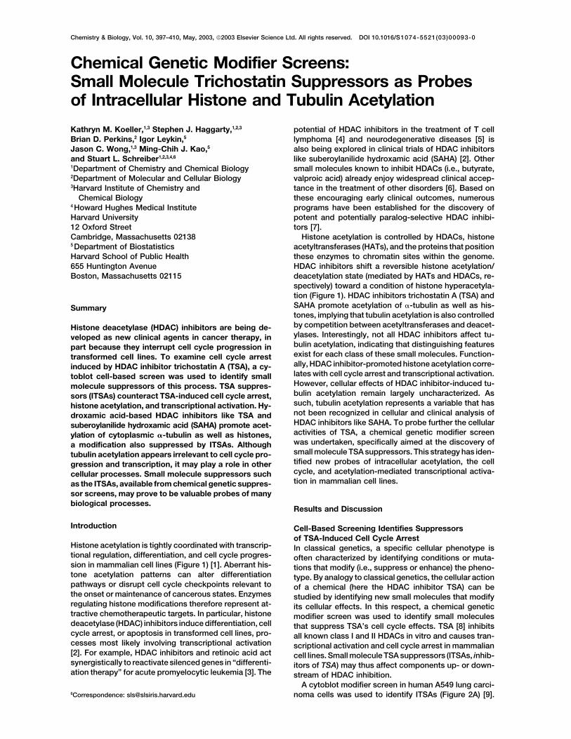
Histone acetylation is tightly coordinated with transcrip- In classical genetics, a specific cellular phenotype istional regulation, differentiation, and cell cycle progres- often characterized by identifying conditions or muta-sion in mammalian cell lines (Figure 1) [1]. Aberrant his- tions that modify (i.e., suppress or enhance) the pheno-tone acetylation patterns can alter differentiation type. By analogy to classical genetics, the cellular actionpathways or disrupt cell cycle checkpoints relevant to of a chemical (here the HDAC inhibitor TSA) can bethe onset or maintenance of cancerous states. Enzymes studied by identifying new small molecules that modifyregulating histone modifications therefore represent at- its cellular effects. In this respect, a chemical genetictractive chemotherapeutic targets. In particular, histone modifier screen was used to identify small moleculesdeacetylase (HDAC) inhibitors induce differentiation, cell that suppress TSA’s cell cycle effects. TSA [8] inhibitscycle arrest, or apoptosis in transformed cell lines, pro- all known class I and II HDACs in vitro and causes tran-cesses most likely involving transcriptional activation scriptional activation and cell cycle arrest in mammalian[2]. For example, HDAC inhibitors and retinoic acid act cell lines. Small molecule TSA suppressors (ITSAs, inhib-synergistically to reactivate silenced genes in “differenti- itors of TSA) may thus affect components up- or down-ation therapy” for acute promyelocytic leukemia [3]. The stream of HDAC inhibition.
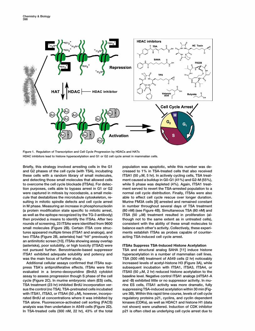
A cytoblot modifier screen in human A549 lung carci-noma cells was used to identify ITSAs (Figure 2A) [9].6Correspondence: [email protected]
Chemistry & Biology398
Figure 1. Regulation of Transcription and Cell Cycle Progression by HDACs and HATs
HDAC inhibitors lead to histone hyperacetylation and G1 or G2 cell cycle arrest in mammalian cells.
Briefly, this strategy involved arresting cells in the G1 population was apoptotic, while this number was de-creased to 1% in TSA-treated cells that also receivedand G2 phases of the cell cycle (with TSA), incubating
these cells with a random library of small molecules, ITSA1 (50 �M, 5 hr). In actively cycling cells, TSA treat-ment caused a buildup in G0-G1 (41%) and G2-M (55%),and detecting those small molecules that allowed cells
to overcome the cell cycle blockade (ITSAs). For detec- while S phase was depleted (4%). Again, ITSA1 treat-ment served to revert the TSA-arrested population to ation purposes, cells able to bypass arrest in G1 or G2
were captured in mitosis by nocodazole, a small mole- normal cell cycle distribution. Finally, ITSAs were alsoable to effect cell cycle rescue over longer duration.cule that destabilizes the microtubule cytoskeleton, re-
sulting in mitotic spindle defects and cell cycle arrest Murine FM3A cells [8] arrested and remained constantin number throughout several days of TSA treatmentin M phase. Measuring an increase in phosphonucleolin
(a protein modification state specific to mitotic arrest, (80 nM) (see Figure 4B). Simultaneous TSA (80 nM) andITSA (50 �M) treatment resulted in proliferation (al-as well as the epitope recognized by the TG-3 antibody)
then provided a means to identify the ITSAs. After two though not to the same extent as in untreated cells),consistent with the ability of these small molecules torounds of screening, 23 ITSAs were identified from 9600
small molecules (Figure 2B). Certain ITSA core struc- balance each other’s activity. Collectively, these experi-ments establish ITSAs as probes capable of counter-tures appeared multiple times (ITSA1 and analogs), and
two ITSAs (Figure 2B, asterisks) had “hit” previously in acting TSA-induced cell cycle arrest.an antimitotic screen [10]. ITSAs showing assay overlap(asterisks), poor solubility, or high toxicity (ITSA2) were ITSAs Suppress TSA-Induced Histone Acetylation
TSA and structural analog SAHA [11] induce histonenot pursued further. Benzotriazole-based suppressorITSA1 exhibited adequate solubility and potency and hyperacetylation in a number of mammalian cell lines.
TSA (300 nM) treatment of A549 cells (2 hr) noticeablywas the main focus of further study.Additional cellular assays confirmed that ITSAs sup- increased levels of acetyl-histone H3 (Figure 3A), while
subsequent incubation with ITSA1, ITSA3, ITSA4, orpress TSA’s antiproliferative effects. ITSAs were firstevaluated in a bromo-deoxyuridine (BrdU) cytoblot ITSA5 (50 �M, 2 hr) reduced histone acetylation to the
baseline level. Negative control ITSA1 analogs (nITSA1-Aassay to assess progression though S phase of the cellcycle (Figure 2C). In murine embryonic stem (ES) cells, and -B) exhibited little or no suppressor activity. In mu-
rine ES cells, ITSA1 activity was more dramatic, fullyTSA treatment (23 hr) inhibited BrdU incorporation ver-sus the control (no TSA). TSA-pretreated cells incubated suppressing TSA-induced acetylation within 30 min (Fig-
ure 3B). Within this rapid time course, levels of cell cyclewith ITSA1, ITSA3, or ITSA4 (50 �M), however, incorpo-rated BrdU at concentrations where it was inhibited by regulatory proteins p21, cyclins, and cyclin dependent
kinases (CDKs), as well as HDAC1 and histone H1 (dataTSA alone. Fluorescence-activated cell sorting (FACS)analysis was then undertaken in A549 cells (Figure 2D). not shown) were unaltered. Induction of CDK inhibitor
p21 is often cited as underlying cell cycle arrest due toIn TSA-treated cells (300 nM, 22 hr), 43% of the total
Small Molecule Trichostatin Suppressors399
HDAC inhibitors [12]. However, in murine ES cells the cleophilic functionality of this HDAC inhibitor [18]. Nota-bly, cell cycle arrest induced by HC Toxin was alsobasal p21 level was significant and insensitive to TSA.
Importantly, suppression of acetylation levels was insensitive to ITSA1 (Figure 2D). This intriguing abilityof ITSA1 to differentially suppress TSA-like versus TPX-only observable when ITSA1 was added concurrent with
or post TSA treatment. Cells pretreated with ITSA1 be- like molecules may reflect targeting of these HDAC in-hibitors to different multisubunit complexes with varyingfore addition of TSA exhibited elevated acetylation levels
characteristic of TSA treatment alone (data not shown). degrees of accessibility, rather than inhibition of specificHDAC orthologs. Indeed, the sets of genes upregulatedThis suggests that the target of ITSA1 is not present (or,
alternatively, not correctly localized) until induced by in TSA- versus TPX-treated cells (see Profiling sectionbelow) were observed to be nonoverlapping and associ-TSA. Possible targets include enzymes and complexes
recruited to or associated with acetylated histone tails. ated with different promoter elements, suggesting alter-native targeting.In this sense, chromatin remodeling complexes, bromo-
domain-containing proteins, and HATs all represent po-tential ITSA1 targets, as do components of signaling
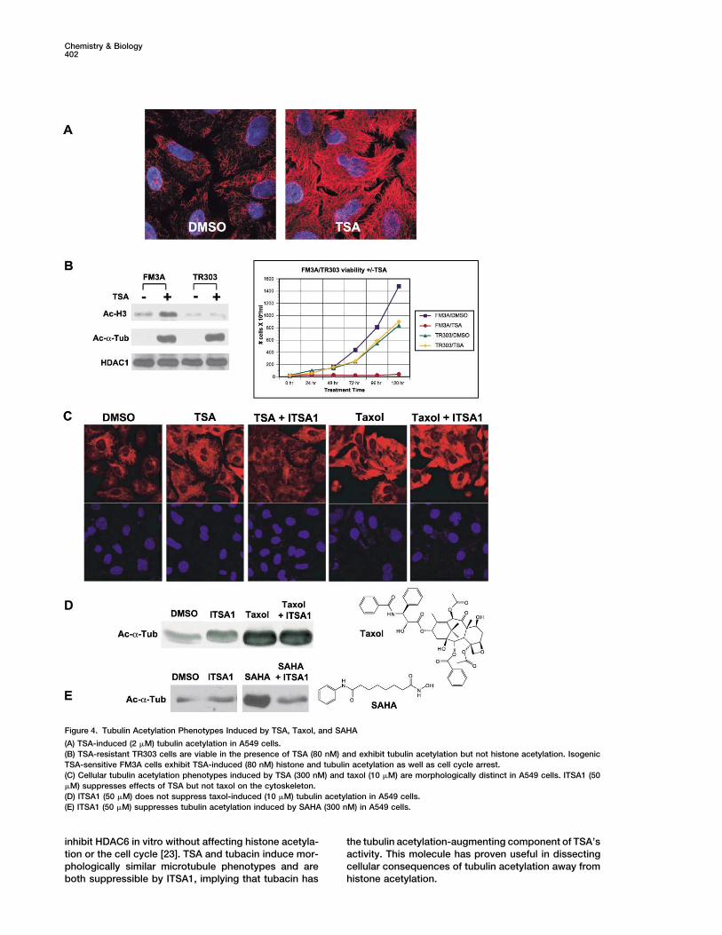
Several but Not All ITSAs Suppress TSA-Inducedcascades that lead to their recruitment [13]. BecauseTubulin AcetylationTSA pretreatment is necessary for suppression byTSA and SAHA induce acetylation of cytoplasmicITSA1, the possibility that ITSA1 inactivates TSA though�-tubulin at lysine-40. TSA treatment reversibly trans-chemical acylation was entertained. Acylation of TSA’sforms “short and curly” acetylated microtubules con-hydroxamic acid group would inactivate the functional-centrated around the centrosome and Golgi apparatusity required for HDAC inhibition, thus “suppressing”into an “extended and straight” acetylated microtubuleTSA’s activity. To test this hypothesis, total in vitro HDACpopulation throughout the entire cell body (Figure 4A).activity of a HeLa cell lysate was measured in the pres-This effect of TSA was observable in less than 2 hr inence of TSA, ITSA1, or TSA � ITSA1 (Figure 3C). ITSA1A549 or murine ES cells (Figures 3A and 3B). Subsequentalone (50 or 100 �M) had no effect on HDAC activity,ITSA1 or ITSA3 treatment (50 �M) suppressed tubulinmaking it unlikely that ITSA1 affects HDAC function di-acetylation (30 min–2 hr), while nITSA1-A and -B (50 �M)rectly. As expected, TSA (500 nM) partially inhibitedwere ineffective (Figure 3A). Interestingly, ITSA4 andHDAC activity of the total lysate. Notably, the level ofITSA5 (50 �M) also failed to suppress tubulin acetylation,inhibition for simultaneous treatment with TSA (500 nM)indicating that multiple categories of suppressors existand ITSA1 (50 or 100 �M) was nearly identical to thatwithin the ITSA population. Since only a few ITSAs sup-of TSA alone, suggesting that TSA and ITSA1 do notpress tubulin acetylation, this property is not requireddirectly interact, chemically or otherwise. To determineto bypass TSA-induced cell cycle arrest, the main condi-whether ITSA1 could possibly be acylating HATs or his-tion of the original screen. Furthermore, in TSA-resistanttone proteins, thereby destroying the acetylating en-cell line TR303 [8], proliferation was normal in the pres-zyme or substrate, cellular wash-out experiments wereence of TSA (80 nM). While TR303 cells were not subjectundertaken. A549 cells were treated with TSA (300 nM,to histone hyperacetylation by TSA, tubulin acetylation2 hr) followed by ITSA1 (50 �M, 2 hr). Cellular mediawas augmented in both FM3A (TSA-sensitive) andwas then removed and replaced with media containingTR303 cells (Figure 4B).TSA alone (300 nM) for various time periods. Even at
Tubulin acetylation is often associated with factorsthe shortest wash-out time point examined (30 min),that increase cytoskeletal stability, such as the microtu-TSA-induced acetylation of histones and tubulin wasbule-stabilizing agent taxol [19]. The possibility that TSAreadily observable (data not shown). As such, ITSA1causes an increase in �-tubulin acetylation by stabilizingdoes not likely covalently modify histones or HATs.the microtubule cytoskeleton was therefore investi-Trapoxin B (TPX) [14] and HC Toxin (both epoxy-gated. A549 cells were pretreated (2 hr) with either taxolketones) and FK228 [15] (a depsipeptide originally(10 �M) or TSA (300 nM) followed by the addition ofnamed FR901228) are potent HDAC inhibitors structur-ITSA1 (50 �M, 1 hr). Interestingly, ITSA1 decreased tu-ally distinct from hydroxamates TSA and SAHA. Collec-bulin acetylation caused by TSA (Figure 4C) but not taxoltively, these small molecules encompass various known(Figures 4C and 4D). Global acetylation phenotypes in-classes of HDAC inhibitors all thought to act by similarduced by TSA and taxol were also morphologically dis-mechanisms, with at least some functional redundancytinct. Whereas taxol caused visible clumping and(in vitro). While TSA and SAHA inhibit all class I and IIbundling of microtubules, TSA-treated cells lacked visi-HDACs in vitro, inhibitors like TPX inhibit all but HDAC6ble microtubule abnormalities (Figure 4C). A common[16] (and the recently identified, sequence-relatedmechanism of action for these agents is therefore un-HDAC10 [17]). Analysis of bulk histone acetylation levelslikely. TSA may instead augment tubulin acetylation byalone does not indicate differences in the activities ofdirectly inhibiting a tubulin deacetylase (TDAC), such asthese HDAC inhibitors in cells, as all induce histoneHDAC6 [20]. The ability of ITSA1 to differentially sup-acetylation (Figure 3D). However, ITSA1 possesses dif-press TSA-induced but not taxol-induced tubulin acet-ferential suppressor activity toward the different classesylation was exploited to screen and characterize a libraryand can be used to distinguish them. In this respect,of new acetylation-inducing small molecules (see ac-ITSA1 (50 �M) suppressed histone acetylation inducedcompanying manuscript [21]). In this screen, ITSA1 sup-by TSA (300 nM) and SAHA (3 �M) but not by TPX (50pressed �70% (338 of 475) of the newly discoverednM), HC Toxin (50 nM), or FK228 (10 nM). The inabilitytubulin acetylation-inducing hydroxamic acids (TSA-of ITSA1 to suppress FK228 further suggests that ITSA1
does not nonspecifically acylate thiol groups, the nu- like), while having no effect on the remaining �30%
Chemistry & Biology400
Small Molecule Trichostatin Suppressors401
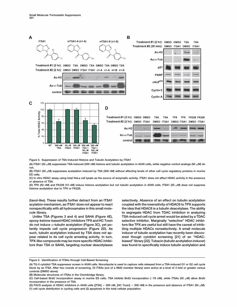
Figure 3. Suppression of TSA-Induced Histone and Tubulin Acetylation by ITSA1
(A) ITSA1 (50 �M) suppresses TSA-induced (300 nM) histone and tubulin acetylation in A549 cells, while negative control analogs (50 �M) donot.(B) ITSA1 (50 �M) suppresses acetylation induced by TSA (300 nM) without affecting levels of other cell cycle regulatory proteins in murineES cells.(C) In vitro HDAC assay using total HeLa cell lysate as the source of enzymatic activity. ITSA1 does not affect HDAC activity in the presenceor absence of TSA.(D) TPX (50 nM) and FK228 (10 nM) induce histone acetylation but not tubulin acetylation in A549 cells. ITSA1 (50 �M) does not suppresshistone acetylation due to TPX or FK228.
(taxol-like). These results further detract from an ITSA1 selectively. Absence of an effect on tubulin acetylationcoupled with the insensitivity of HDAC6 to TPX supportsacylation mechanism, as ITSA1 does not appear to react
nonspecifically with all hydroxamates in this small mole- the idea that HDAC6 is a tubulin deacetylase. The abilityto segregate HDAC from TDAC inhibition in analyzingcule library.
Unlike TSA (Figures 3 and 4) and SAHA (Figure 4E), TSA-induced cell cycle arrest would be aided by a TDACselective inhibitor. Marginally “selective” HDAC inhibi-epoxy-ketone-based HDAC inhibitors TPX and HC Toxin
do not induce �-tubulin acetylation (Figure 3C), yet po- tors like TPX are useful but still have the caveat of inhib-iting multiple HDACs nonselectively. A small moleculetently impede cell cycle progression (Figure 2D). As
such, tubulin acetylation induced by TSA does not ap- inducer of tubulin acetylation has recently been discov-ered though cytoblot screening [21] of an “HDAC-pear related to its cell cycle arresting activity. In turn,
TPX-like compounds may be more specific HDAC inhibi- biased” library [22]. Tubacin (tubulin acetylation inducer)was found to specifically induce tubulin acetylation andtors than TSA or SAHA, targeting nuclear deacetylases
Figure 2. Identification of ITSAs through Cell-Based Screening
(A) TG-3 cytoblot TSA suppressor screen in A549 cells. Nocodazole is used to capture cells released from a TSA-induced G1 or G2 cell cycleblock by an ITSA. After two rounds of screening, 23 ITSAs (out of a 9600 member library) were active at a level of 2-fold or greater versuscontrols (DMSO alone).(B) Molecular structures of ITSAs in the Chembridge library.(C) Cell-based BrdU incorporation assay in murine ES cells. TSA inhibits BrdU incorporation (�78 nM), while ITSAs (50 �M) allow BrdUincorporation in the presence of TSA.(D) FACS analysis of HDAC inhibitors in A549 cells ([TSA] � 500 nM, [HC Toxin] � 500 nM) in the presence and absence of ITSA1 (50 �M):(1) cell cycle distribution in cycling cells and (2) apoptosis in the total cellular population.
Chemistry & Biology402
Figure 4. Tubulin Acetylation Phenotypes Induced by TSA, Taxol, and SAHA
(A) TSA-induced (2 �M) tubulin acetylation in A549 cells.(B) TSA-resistant TR303 cells are viable in the presence of TSA (80 nM) and exhibit tubulin acetylation but not histone acetylation. IsogenicTSA-sensitive FM3A cells exhibit TSA-induced (80 nM) histone and tubulin acetylation as well as cell cycle arrest.(C) Cellular tubulin acetylation phenotypes induced by TSA (300 nM) and taxol (10 �M) are morphologically distinct in A549 cells. ITSA1 (50�M) suppresses effects of TSA but not taxol on the cytoskeleton.(D) ITSA1 (50 �M) does not suppress taxol-induced (10 �M) tubulin acetylation in A549 cells.(E) ITSA1 (50 �M) suppresses tubulin acetylation induced by SAHA (300 nM) in A549 cells.
inhibit HDAC6 in vitro without affecting histone acetyla- the tubulin acetylation-augmenting component of TSA’sactivity. This molecule has proven useful in dissectingtion or the cell cycle [23]. TSA and tubacin induce mor-
phologically similar microtubule phenotypes and are cellular consequences of tubulin acetylation away fromhistone acetylation.both suppressible by ITSA1, implying that tubacin has
Small Molecule Trichostatin Suppressors403
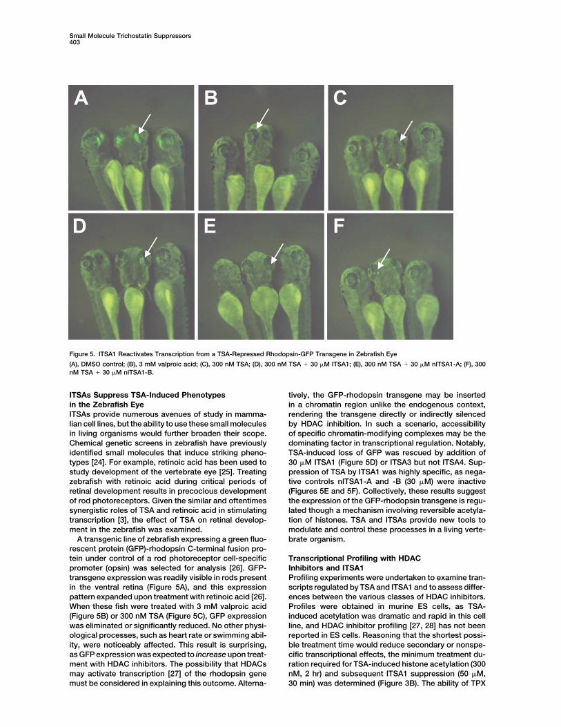
Figure 5. ITSA1 Reactivates Transcription from a TSA-Repressed Rhodopsin-GFP Transgene in Zebrafish Eye
(A), DMSO control; (B), 3 mM valproic acid; (C), 300 nM TSA; (D), 300 nM TSA � 30 �M ITSA1; (E), 300 nM TSA � 30 �M nITSA1-A; (F), 300nM TSA � 30 �M nITSA1-B.
ITSAs Suppress TSA-Induced Phenotypes tively, the GFP-rhodopsin transgene may be insertedin a chromatin region unlike the endogenous context,in the Zebrafish Eye
ITSAs provide numerous avenues of study in mamma- rendering the transgene directly or indirectly silencedby HDAC inhibition. In such a scenario, accessibilitylian cell lines, but the ability to use these small molecules
in living organisms would further broaden their scope. of specific chromatin-modifying complexes may be thedominating factor in transcriptional regulation. Notably,Chemical genetic screens in zebrafish have previously
identified small molecules that induce striking pheno- TSA-induced loss of GFP was rescued by addition of30 �M ITSA1 (Figure 5D) or ITSA3 but not ITSA4. Sup-types [24]. For example, retinoic acid has been used to
study development of the vertebrate eye [25]. Treating pression of TSA by ITSA1 was highly specific, as nega-tive controls nITSA1-A and -B (30 �M) were inactivezebrafish with retinoic acid during critical periods of
retinal development results in precocious development (Figures 5E and 5F). Collectively, these results suggestthe expression of the GFP-rhodopsin transgene is regu-of rod photoreceptors. Given the similar and oftentimes
synergistic roles of TSA and retinoic acid in stimulating lated though a mechanism involving reversible acetyla-tion of histones. TSA and ITSAs provide new tools totranscription [3], the effect of TSA on retinal develop-
ment in the zebrafish was examined. modulate and control these processes in a living verte-brate organism.A transgenic line of zebrafish expressing a green fluo-
rescent protein (GFP)-rhodopsin C-terminal fusion pro-tein under control of a rod photoreceptor cell-specific Transcriptional Profiling with HDAC
Inhibitors and ITSA1promoter (opsin) was selected for analysis [26]. GFP-transgene expression was readily visible in rods present Profiling experiments were undertaken to examine tran-
scripts regulated by TSA and ITSA1 and to assess differ-in the ventral retina (Figure 5A), and this expressionpattern expanded upon treatment with retinoic acid [26]. ences between the various classes of HDAC inhibitors.
Profiles were obtained in murine ES cells, as TSA-When these fish were treated with 3 mM valproic acid(Figure 5B) or 300 nM TSA (Figure 5C), GFP expression induced acetylation was dramatic and rapid in this cell
line, and HDAC inhibitor profiling [27, 28] has not beenwas eliminated or significantly reduced. No other physi-ological processes, such as heart rate or swimming abil- reported in ES cells. Reasoning that the shortest possi-
ble treatment time would reduce secondary or nonspe-ity, were noticeably affected. This result is surprising,as GFP expression was expected to increase upon treat- cific transcriptional effects, the minimum treatment du-
ration required for TSA-induced histone acetylation (300ment with HDAC inhibitors. The possibility that HDACsmay activate transcription [27] of the rhodopsin gene nM, 2 hr) and subsequent ITSA1 suppression (50 �M,
30 min) was determined (Figure 3B). The ability of TPXmust be considered in explaining this outcome. Alterna-
Chemistry & Biology404
to induce histone acetylation (50 nM, 2 hr), and tubacin ulated genes are related to histones and cell cycle pro-teins, such as granzyme A, a protease known to degradeto promote tubulin acetylation (2 �M, 2 hr) under these
conditions was also confirmed. Data sets were obtained histones and mediate cell death [32]. Interestingly, ex-pression of these TSA-induced genes was suppressedin duplicate by treating murine ES cells with the following
small molecule combinations (treatment 1 [2 hr]/treat- by ITSA1. When compared to profiles of murine stemcells in various stages of differentiation, TSA profile (2)ment 2 [30 min]): (1) DMSO/DMSO, (2) TSA/DMSO, (3)
TPX/DMSO, (4) tubacin/DMSO, (5) DMSO/ITSA1, and (6) includes genes not generally used by these cells at anystage of their developmental program [33]. In TSA profileTSA/ITSA1. Samples were processed following Affyme-
trix protocols, and data sets were analyzed with (2), the absence of general cell cycle regulatory proteinsoften cited in the context of HDAC inhibitor-induceddCHIP1.0 software [29] (see Supplemental Data at http://
www.chembiol.com/cgi/content/full/10/5/397/DC1 or arrest is noteworthy. This may reflect nonspecific activ-ity of TSA toward various deacetylases, metallopro-write to [email protected] for a PDF). When consider-
ing genes highly upregulated by TSA, TPX, or ITSA1, teases, and other proteins [34]. Surprisingly, TPX profile(3) was essentially nonoverlapping with TSA profile (2),reproducibility between the duplicates was high. Al-
though suppression was observed in both TSA/ITSA1 suggesting these HDAC inhibitors act though distinctmechanisms to regulate gene expression. This result(profile 6) data sets, one sample exhibited complete
suppression, while the other was only partially sup- may underlie the ability of ITSA1 to suppress cellularfunctions of TSA but not TPX. TPX-activated transcriptspressed. Expression values for all duplicate data sets
were pooled by a weighted averaging method. Fold- included those typically cited as responsive to HDACinhibitors, including cell cycle regulatory proteins likechange levels represent the lower statistical bound of
the actual measured fold-change versus control DMSO p21 and Jund1. Mapping TSA- and TPX-upregulatedgenes to their respective chromosomal locations re-profile (1).
TSA profile (2) exhibited robust transcriptional activa- vealed distinct patterns for each of these HDAC inhibi-tors (Figure 6F), possibly illustrating unique targetingtion (Figure 6A). Above a 2-fold cut-off, 133 of 139 genes
present in TSA profile (2) were upregulated. Notably, patterns of these molecules (see Supplemental Dataat http://www.chembiol.com/cgi/content/full/10/5/397/ITSA1 treatment abrogated expression of this highly ac-
tivated transcript population (Figure 6B), further corre- DC1 or write to [email protected] for a PDF).lating TSA-induced cell cycle arrest and histone acetyla-tion with transcriptional activation. ITSA1 profile (5)
Promoter Region Analysisshowed only low-level transcriptional activation and re-Analysis of promoter regions in TSA-, TPX-, and ITSA1-pression (Figure 6C). Compared to the suppression ac-responsive genes was next undertaken using recentlytivity of ITSA1 observed following TSA treatment, thisdeveloped HumanUpstream software (I.L., M.-C.J.K., O.result again suggests the target of ITSA1 is inducedLipan, X. Zhou, K.-F.S., A.M. Bowcock, and W.H. Wong,following TSA treatment. Genes downregulated in TSAunpublished data). Human orthologs of murine genesprofile (2) were only marginally decreased (�1.5-fold),on the Affymetrix U74Av2 array were obtained using theimplying that negative transcripts represent indirect re-Institute for Genomic Research (TIGR) TOGA database.sults of TSA treatment or a secondary cellular response.For TSA-activated transcripts, 28 human ortholog pro-TPX profile (3) contained a number of upregulatedmoter sequences were obtained (from a set of 111 totaltranscripts (Figure 6D) but at a level appreciably damp-genes). Likewise, 28 sequences were identified for TPX-ened versus TSA profile (2). Altered kinetics in the actionactivated genes (70 total genes), 28 for ITSA1-activatedof TSA and TPX may explain this result, as experimentsgenes (78 total genes), and 11 for ITSA1-repressedwere optimized for TSA-induced acetylation levels. Ingenes (24 total genes). Common frameworks of tran-TPX profile (3), several genes were highly upregulatedscription factor binding sites were defined for each clus-(�2-fold) and likely represent specific transcriptional tar-ter of promoter sequences, and significant modelsgets of TPX. Although the magnitude of ITSA1 profilewithin these subsets of the total data set identified (also(5) and TPX profile (3) appear similar (Figures 6C and 6D),see Supplemental Data at http://www.chembiol.com/ITSA1 profile (5) lacks a highly activated gene populationcgi/content/full/10/5/397/DC1 or write to chembiol@(�2-fold). Tubacin profile (4) was generally absent ofcell.com for a PDF).transcriptional activity, further segregating tubulin acet-
Promoter regions of TSA and TPX-upregulated genesylation from TSA’s effects on the cell cycle or transcrip-were enriched in Sp1 sites, alone or in combination withtion. Tubacin therefore has great utility in the analysisother transcription factor binding sites. Sp1 sites areof tubulin acetylation induced by HDAC inhibitors, in aresponsive to TSA [35] and TPX and are known to regu-fashion uncoupled from histone acetylation [23].late expression of p21. Sp1 binding sites thus appearto be general HDAC-related regulatory elements. Analy-sis of the most highly upregulated transcript populationsTranscripts Upregulated by HDAC Inhibitors
TSA and TPX Differ (TSA, �8-fold; TPX, �2-fold) provided evidence of spe-cificity between promoters targeted by TSA and TPX.Several transcripts activated in TSA profile (2) are con-
sistent with prior reports of HDAC-regulated genes (Fig- The E2F promoter was more generally associated withTSA-regulated transcripts, although it mediates the ac-ure 6E). For example, the prolactin promoter is dere-
pressed by TSA though inhibition of HDACs in the NCoR/ tivity of TPX to a lesser extent. E2F represses transcrip-tion though association with class I HDAC1 [36]. ThemSin3A complex [30], and TSA is known to induce ex-
pression of the lysozyme M gene [31]. Other TSA-upreg- cAMP-response element binding proteins (CREB) pro-
Small Molecule Trichostatin Suppressors405
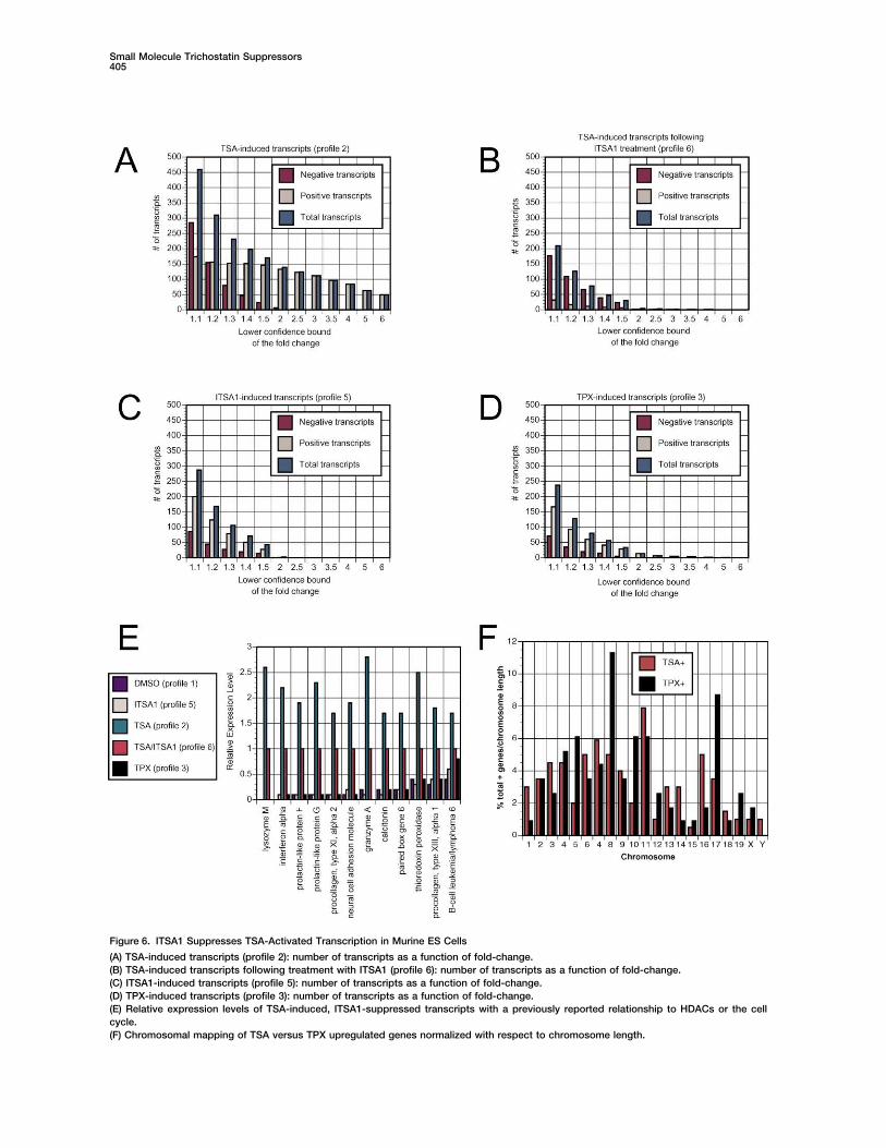
Figure 6. ITSA1 Suppresses TSA-Activated Transcription in Murine ES Cells
(A) TSA-induced transcripts (profile 2): number of transcripts as a function of fold-change.(B) TSA-induced transcripts following treatment with ITSA1 (profile 6): number of transcripts as a function of fold-change.(C) ITSA1-induced transcripts (profile 5): number of transcripts as a function of fold-change.(D) TPX-induced transcripts (profile 3): number of transcripts as a function of fold-change.(E) Relative expression levels of TSA-induced, ITSA1-suppressed transcripts with a previously reported relationship to HDACs or the cellcycle.(F) Chromosomal mapping of TSA versus TPX upregulated genes normalized with respect to chromosome length.
Chemistry & Biology406
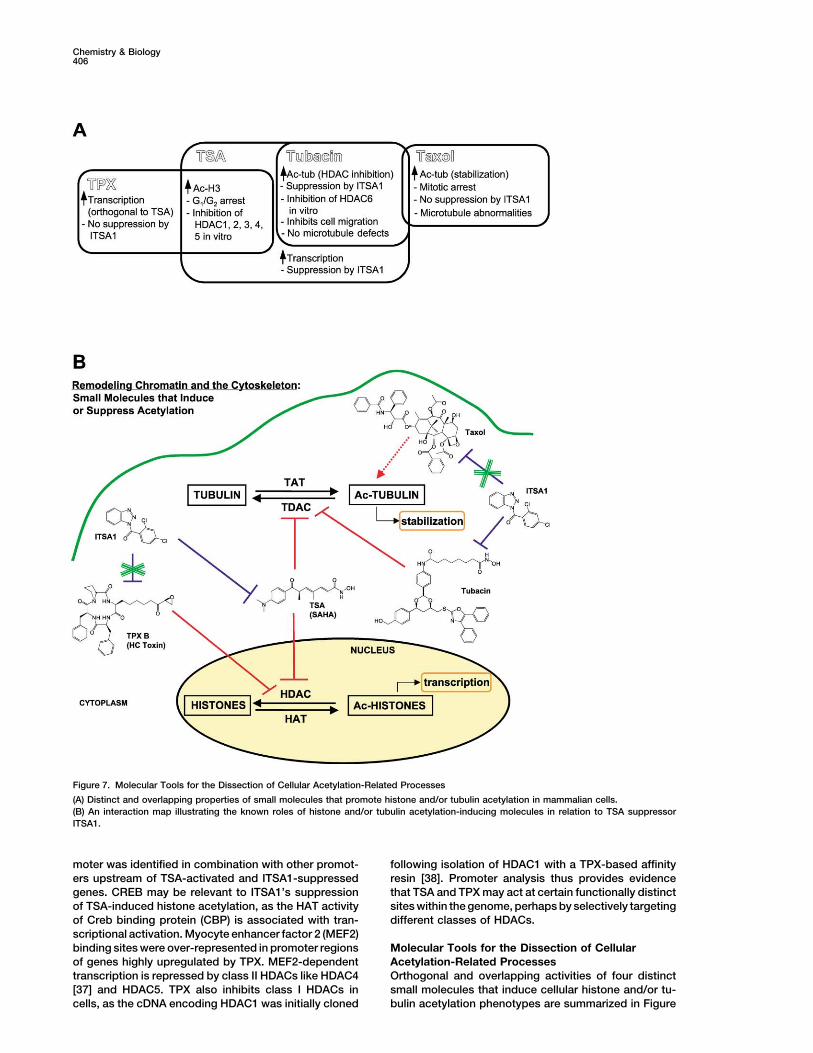
Figure 7. Molecular Tools for the Dissection of Cellular Acetylation-Related Processes
(A) Distinct and overlapping properties of small molecules that promote histone and/or tubulin acetylation in mammalian cells.(B) An interaction map illustrating the known roles of histone and/or tubulin acetylation-inducing molecules in relation to TSA suppressorITSA1.
moter was identified in combination with other promot- following isolation of HDAC1 with a TPX-based affinityresin [38]. Promoter analysis thus provides evidenceers upstream of TSA-activated and ITSA1-suppressed
genes. CREB may be relevant to ITSA1’s suppression that TSA and TPX may act at certain functionally distinctsites within the genome, perhaps by selectively targetingof TSA-induced histone acetylation, as the HAT activity
of Creb binding protein (CBP) is associated with tran- different classes of HDACs.scriptional activation. Myocyte enhancer factor 2 (MEF2)binding sites were over-represented in promoter regions Molecular Tools for the Dissection of Cellular
Acetylation-Related Processesof genes highly upregulated by TPX. MEF2-dependenttranscription is repressed by class II HDACs like HDAC4 Orthogonal and overlapping activities of four distinct
small molecules that induce cellular histone and/or tu-[37] and HDAC5. TPX also inhibits class I HDACs incells, as the cDNA encoding HDAC1 was initially cloned bulin acetylation phenotypes are summarized in Figure
Small Molecule Trichostatin Suppressors407
7A. Cellular activities associated with TSA, TPX, tubacin, cancer trials. Cell-based screening for chemical ge-netic modifiers of chromatin remodeling agents mayand taxol have been analyzed and distinguished though
ITSA1 suppression (Figure 7B). ITSAs represent new contribute to the current understanding of gene ex-pression and the cellular activities of small moleculesmolecular probes of protein acetylation in cell cycle pro-
gression, transcriptional activation, and cytoskeletal employed as therapeutics. Chemical genetic modifierscreens offer a powerful new approach to the discov-stability. Although the direct target(s) of ITSA1 remains
elusive, it likely is related to protein complexes associ- ery of small molecule probes.ated with an acetylated state of chromatin. Further stud-
Experimental Proceduresies aimed at elucidating the target are currently ongoing.ITSAs may serve as tools to indicate differences be-
Materialstween cell lines or tumors refractory to HDAC inhibition Trichostatin A, HC Toxin, nocodazole, 5�-bromo-2-deoxyuridine,or exhibiting altered responses [39]. Moreover, tumors paclitaxel (taxol), and camptothecin were from Sigma, Hoechsttargeted by HDAC inhibitor therapy will likely develop 33342 from Molecular Probes, and ITSA1, ITSA2, ITSA3, ITSA4,
ITSA5, nITSA1-A, and nITSA1-B from Chembridge. Suberoylanilideresistance to these agents. Cellular resistance mecha-hydroxamic acid was synthesized by Dr. Greg Copeland, and tra-nisms could presumably resemble ITSA suppression,poxin B was synthesized by Dr. Jack Taunton. FK228 was obtainedand identifying ITSA targets may shed light upon thefrom Dr. Minoru Yoshida (University of Tokyo), and anti-TG-3 anti-
adaptive resistance process. In addition, ITSAs may body was obtained from Dr. Peter Davies (Albert Einstein Collegeserve to sensitize cells to other chemotherapeutic drugs, of Medicine). Anti-BrdU and anti-PARP antibodies were from BDsince bypassing cell cycle checkpoints is known to con- Biosciences Pharmingen, anti-acetylated �-tubulin (6-11B-1), anti-
cyclin E, and anti-histone deacetylase 1 antibodies were fromtribute to genomic instability.Sigma, anti-diacetyl-histone H3 antibody was from Upstate Biotech-A substantial body of evidence has been amassednology, anti-cdc2p34 and anti-cyclin A antibodies were from Santaagainst a role for tubulin acetylation in TSA-induced cellCruz Biotechnology, and anti-p21 antibody (Ab-5) was from Onco-
cycle arrest. Consequently, tubulin acetylation repre- gene. Peroxidase-linked anti-rabbit and anti-mouse IgG antibodiessents an additional cellular consequence of treatment were from Amersham Pharmacia Biotech, and Texas methyl redwith hydroxamate-based HDAC inhibitors. In initial stud- (TMR)-conjugated anti-mouse IgG antibody was from Molecular
Probes.ies, the TDAC-selective inhibitor tubacin has been foundto inhibit cell migration [23], an activity of therapeutic
Cell Cultureinterest with respect to metastasis and angiogenesis.A549 cells (ATCC, human lung carcinoma) were maintained at 37�CThe overall clinical potential of molecules like SAHA andwith 5% CO2 in Dulbecco’s modified Eagle’s medium (DMEM) sup-
TSA may therefore be limited (or possibly enhanced) plemented with 10% fetal bovine serum (FBS), 100 units/ml penicillindue to effects on the cytoskeleton. Nonetheless, acet- G sodium, 100 �g/ml streptomycin sulfate (1% P/S), and 2 mM
L-glutamine (1% L-Gln) (all from Gibco BRL). Murine FM3A andylation of tubulin is a variable that should be consideredTR303 cells ([8]; from Dr. Minoru Yoshida, University of Tokyo) werein the ongoing clinical trials of hydroxamic acid-basedmaintained in RPMI 1640 medium at 37�C with 5% CO2 supple-HDAC inhibitors. Through cell-based screening, identifi-mented with 10% FBS, 1% P/S, 1% L-Gln. Murine embryonic stemcation of TSA-like HDAC inhibitors that lack TDAC activ-cells (Rosa 26.1; from Dr. Elizabeth Robertson, Harvard University)
ity is also possible [22]. Natural products like TPX may were maintained in DMEM supplemented with 15% defined fetalappear to function globally in this manner, but disparate bovine serum (Hyclone), 1% P/S, 1% L-Gln, 100 �M nonessential
amino acids (1%; Gibco BRL), 0.01 �l/ml -mercaptoethanolmechanisms of cell cycle arrest and transcriptional acti-(Sigma), 1 unit/ml leukemia inhibitory factor (LIF, Sigma).vation versus TSA have been observed.
TG-3 Cytoblot AssayA549 cells were seeded in 40 �l DMEM� (4000 cells/well) in whiteSignificance384-well plates (Nalge Nunc, tissue culture treated) using a liquiddispenser (Multidrop 384, Labsystems) and allowed to attach over-
Hydroxamate-based HDAC inhibitors related to tri- night at 37�C with 5% CO2. TSA (5 �l, 3 �M [(final) � 300 nM TSA/chostatin (TSA) are currently undergoing clinical trials well]) was added to each well and incubation continued (5 hr). Library
compounds (9600 molecules, Diverse E set, Chembridge) dissolvedfor the treatment of cancer. These small moleculesin DMSO (�5 mg/ml) were then pin transferred (50–200 nl to eachare generally believed to function by inducing cell cy-well, [final] � �20 �M compound/well) along with addition of noco-cle arrest or apoptosis in transformed cell lines. Dis-dazole (5 �l, 3.2 �M, [final] � 332 nM nocodaazole/well). Following
covery of new small molecules for use in analysis of 16 hr incubation, the percentage of cells in mitosis was assayedTSA-induced cell cycle arrest has been accomplished using the TG-3 mAb as described [9]. Data were collected on anthough a cell-based suppressor screen. Trichostatin Analyst plate reader (LJL Biosystems) with 0.1 s integration time.
After two rounds of screening, compounds that increased TG-3suppressors (ITSAs) represent valuable new tools forreactivity by 2-fold or greater than control cells were consideredthe study of acetylation-related phenotypes in mam-“hits” (ITSAs).malian cell lines. ITSAs suppress TSA-induced histone
and tubulin acetylation, transcriptional activation, andBrdU Cytoblot Assay
cell cycle arrest, providing numerous avenues of Murine ES cells were seeded in 40 �l ES medium (4000 cells/well)study. Furthermore, ITSA analysis has elucidated non- in white 384-well plates (Nalge Nunc, tissue culture treated) using
a liquid dispenser (Multidrop 384, Labsystems) and allowed to attachoverlapping mechanisms resulting in histone hyperacet-overnight at 37�C with 5% CO2. TSA (5 �l, [final] � 78 nM–5 �Mylation in the action of HDAC inhibitors TSA and TPX.TSA/well) was added to each well, and plates were incubated 5 hr.Our experiments also suggest that TSA-induced tu-ITSA1-4 (5 �l, [final] � 12.5–200 �M ITSA/well) or camptothecinbulin acetylation has no bearing on cell cycle or tran-(5 ml, [final] � 78 nM–5 �M camptothecin/well) were added and
scriptional events. However, the consequences of tu- incubation was continued 16 hr. BrdU (10 �l, [final] � 10 �M BrdU/bulin acetylation on other cellular pathways should be well) was added and incubation was continued 2 hr. Cells incorpo-
rating BrdU were detected using anti-BrdU Ab and ECL reagentsconsidered as HDAC inhibitors are assessed in clinical
Chemistry & Biology408
(Amersham Pharmacia) as described [9]. In a dark room, film Zebrafish ProtocolHomozygous transgenic zebrafish [26] were bred to wild-type zebra-(X-OMAT AR, Kodak Corporation) was placed on top of the plate
for 1–5 min, then developed in a Kodak M35A X-OMAT processor. fish in synchronized pairwise crosses as described [41] to generateembryos that all expressed GFP. Eggs were collected and kept inembryo buffer [41] supplemented with 40 U penicillin G and 40 �g
Immunoblotting Experimentsstreptomycin. At 24 hr post-fertilization (hpf), embryos were treated
Cells (A549 or murine ES) were seeded into 5 or 10 cm dishes,with 0.003% 1-phenyl-2-thiourea (PTU) to inhibit melanin synthesis
allowed to attach overnight, and small molecules were added as[41]. At 48 hpf, 10 fish were placed in a 6-well plate with 2 ml of PTU
DMSO stock solutions. Following treatment, cells were harvestedin embryo media. Stocks of valproic acid, TSA, ITSA1, nITSA1-A, and
using trypsin/EDTA (Gibco BRL), washed with phosphate bufferednITSA1-B were added directly to wells containing fish. Fish were
saline (PBS; pH 7.4; 2 5 ml), and lysed at 4�C (30 min) in ELB�
grown at 28.5�C for an additional 48 hr prior to examination. At 96buffer (50 mM HEPES [pH 7], 250 mM NaCl, 5mM EDTA [pH 8], 0.1%
hpf, treated fish were anesthetized with tricaine [41], viewed, andNP-40) with a protease inhibitor cocktail (PIC, Boehringer-Mannheim).
photographed under a Leica MZFLIII dissecting microscope (LeicaProteins were separated by SDS-PAGE gel electrophoresis, trans-
Microsystems, Bannockburn, IL) using a GFP filter set.ferred to PVDF membrane (Millipore), and incubated with antibodiesdiluted in PBST (PBS [pH 7.4], 0.1% Tween-20). Detection was ac-complished using ECL reagents. When not shown, a nonspecific Transcriptional Profilingprotein band was used to confirm protein normalization. Profiling samples were prepared using Affymetrix protocols. Dupli-
cate samples were independently prepared from different culturesof cells on different occasions. Murine ES cells (�10 107 cells, 10In Vitro Histone Deacetylase Assaycm dish, �80% confluent) were treated with DMSO, TSA (300 nM),[3H]acetate-incorporated histones were isolated from butyrate-TPX (50 nM), or tubacin (2 �M) for 2 hr, then with DMSO or ITSA1treated HeLa cells as described [40]. HeLa cell pellets (National Cell(50 �M) for 30 min. Cells were harvested and total RNA was isolatedCulture Facility) were lysed in JLB� (50 mM Tris [pH 8], 150 mMusing an RNeasy kit (Qiagen), yielding �150–250 �g/sample. Purifi-NaCl, 10% glycerol, 0.5% Triton X-100) supplemented with PIC.cation (Oligotex direct mRNA mini kit, Qiagen) then afforded �1–4Lysates were treated with TSA and ITSA1 and incubated with [3H]-�g poly A� mRNA/sample (1%–4% yield). T7(dT)24 primer (Gensetacetylated histones for 2 hr at 37�C. HDAC activity was determinedOligos) was used for cDNA synthesis with the Superscript Choiceby scintillation counting of the ethyl acetate soluble [3H]acetic acidSystem (Invitrogen) for first and second strand synthesis. Biotinyl-as described [38, 40]. Each assay point was run in triplicate.ated probes were prepared using the BioArray High-Yield RNA Tran-script Labeling Kit (Enzo Diagnostics), and fragmentation (35 min,
Immunofluorescence Microscopy 94�C) provided �30-60 �g cRNA/sample. Fragmented cRNA (15 �g)A549 cells were seeded onto glass coverslips at 80%–90% conflu- was then submitted to the MIT Biopolymers Facility for hybridizationency, and compounds were added diluted in cellular medium. Fol- to Affymetrix murine gene chip U74Av2. DChip1.0 software ([29],lowing incubation at 37�C for 18 hr, cells were fixed (0.2% glutaralde- http://www.biostat.harvard.edu/complab/dchip/) was used for datahyde), permeabilized (50 mM K-Pipes [pH 6.8], 5 mM EGTA, 0.5 mM analysis. Present calls ranged between 39%–49% for all 12 arraysMgCl2, 0.1% Triton X-100), and stained with an anti-acetylated- analyzed. Following chip normalization and model-based expres-�-tubulin antibody (6-11B-1) and Hoechst 33342 dye diluted in anti- sion processing, duplicate gene lists were pooled by a weightedbody dilution buffer (ADB, Tris buffered saline (TBS), 0.1% Triton averaging method. Samples were compared to DMSO control profileX-100, 2% bovine serum albumin, 0.1% sodium azide), followed by (1) at various levels of fold change, and the lower statistical bounda secondary TMR-conjugated anti-mouse IgG antibody (Molecular of the observed fold change was used for analysis (see Supplemen-Probes). Images were obtained using a Leitz microscope. tal Data at http://www.chembiol.com/cgi/content/full/10/5/397/DC1
or write to [email protected] for a PDF).
Cytotoxicity AssayExponentially growing cultures of FM3A and TR303 cells [8] were
Promoter Analysissuspended at an initial density of �2 105 cells/ml in RPMI�. After
To obtain the promoter sequence for each mouse gene in the clus-incubation with TSA (80 nM) or DMSO for variable times, viable cell
ters to be analyzed, the Institute for Genomic Research (TIGR) Or-numbers were determined in a hemacytometer using the trypan blue
thologous Gene Alignment (TOGA, now EGO, Eukaryotic Gene Or-exclusion method. The Western blot experiment in Figure 4B was
thologs) database was used to generate pairwise comparisonsobtained after a 48 hr treatment period.
between the tentative consensus sequences that comprise the TIGRHuman and Mouse Gene Indices (I.L., M.-C.J.K., O. Lipan, X. Zhou,K.-F.S., A.M. Bowcock, and W.H. Wong, unpublished data). Tenta-FACS Analysis
A549 cells (�75% confluent, 10 cm dishes) were treated with DMSO tive human consensus (THC) orthologs and their promoter se-quences were identified for many of the selected mouse genes. To(0.1% v/v), TSA (500 nM), or HC Toxin (500 nM) for 4.5 hr, then with
either DMSO (0.1% v/v) or ITSA1 (50 �M) for 17.5 hr. Samples were obtain a list of known transcription factor binding sites that occur inthe same relative order in most sequences, FrameWorker of Genomerinsed in PBS, trypsinized, fixed (2 hr, 70% ethanol, 30% PBS, 4�C),
washed, and stored overnight in PBS (4�C). Samples were blocked Exploring and Modeling Software (GEMS, Genomatix) was em-ployed to extract a common framework of binding sites from a set45 min in antibody dilution buffer (ADB � TBS, 0.1% Triton X-100,
2% bovine serum albumin, 0.1% sodium azide), aspirated, and incu- of DNA sequences. The framework model is sufficient to find thepromoter but is not a complete description of all functional elements.bated in ADB with mouse anti-acetylated tubulin IgG (6-11B-1,
Sigma, 1:500 v/v, overnight, 4�C). Samples were washed and incu- A model derived from such results can be used to scan databasesfor additional sequences that show a similar functional organizationbated in ADB with fluorescein isothiocyanate-conjugated goat anti-
mouse IgG (Santa Cruz, 1:200 v/v, 1 hr, room temperature). Cells even in the absence of direct sequence similarity. To evaluate thestatistical significance of models found with FrameWorker, 200 con-were washed twice in TBS, resuspended in ribonuclease A (100 �l,
Sigma, 100 �g/ml), and incubated 5 min (37�C). To measure the trol groups of sequences derived from the HumanUpstream data-base were randomly sampled, each containing the same number ofDNA content, propidium iodide (PI; 400 �l, Molecular Probes, 50
�g/ml) was added and incubated 1 hr at room temperature. (Samples sequences as the experimental group. These groups were analyzedwith ModelInspector professional (GEMS, Genomatix) utilizing thewere processed as above, but only information derived from DNA
content was necessary for this study.) Samples were analyzed using previously found models to scan sequences for regulatory unitsmatching the models. The program returns the numbers of matchesa FACScanII flow cytometer (Becton-Dickinson) at the Dana Farber
Cancer Institute, exciting at 488 nm and measuring PI fluorescence for each model in each of 200 control groups. The mean, standarddeviation, and Z score (observation minus mean divided by SD)though a 600 nm wavelength filter. Cell cycle distributions and apo-
ptosis analysis were calculated from 104 cells and modeled using were calculated for each model, and models with Z scores � 3 wereconsidered significant (see Supplemental Data at the URL above).ModFit LT (V2.0) software.

Small Molecule Trichostatin Suppressors409
Acknowledgments chi, S. (1998). FR901228, a potent antitumor antibiotic, is a novelhistone deacetylase inhibitor. Exp. Cell Res. 241, 126–133.
16. Furumai, R., Komatsu, Y., Nishino, N., Khochbin, S., Yoshida, M.,We thank Drs. Cheng Li and Wing H. Wong for providing dCHIPsoftware and assistance in its use, Dr. Greg Copeland for the synthe- and Horinouchi, S. (2001). Potent histone deacetylase inhibitors
built from trichostatin A and cyclic tetrapeptide antibiotics in-sis of SAHA, Dr. Chistina Grozinger for helpful discussions, and Dr.Carol Chang for assistance with database analysis. This research cluding trapoxin. Proc. Natl. Acad. Sci. USA 98, 87–92.
17. Guardiola, A.R., and Yao, T.-P. (2002). Molecular cloning andwas supported by the National Institute of General Medical Sciences(GM-38627, awarded to S.L.S.). K.M.K. was supported by Damon characterization of a novel histone deacetylase HDAC10. J. Biol.
Chem. 277, 3350–3356.Runyon Cancer Research Foundation Fellowship DRG-1650, I.L. byNSF grant DBI-0196176, J.C.W. by an NSF pre-doctoral fellowship, 18. Furumai, R., Matsuyama, A., Kobashi, N., Lee, K.H., Nishiyama,
M., Nakajima, H., Tanaka, A., Komatsu, Y., Nishino, N., Yoshida,and M.-C.J.K. by a Howard Hughes pre-doctoral fellowship. S.L.S.is an Investigator at the Howard Hughes Medical Institute in the M., et al. (2002). FK228 (depsipeptide) as a natural prodrug that
inhibits class I histone deacetylases. Cancer Res. 62, 4916–Department of Chemistry and Chemical Biology at Harvard Uni-versity. 4921.
19. Piperno, G., LeDizet, M., and Chang, X.-j. (1987). Microtubulescontaining acetylated �-tubulin in mammalian cells in culture.Received: January 24, 2003J. Cell Biol. 104, 289–302.Revised: March 27, 2003
20. Hubbert, C., Guardiola, A., Shao, R., Kawaguchi, Y., Ito, A.,Accepted: March 28, 2003Nixon, A., Yoshida, M., Wang, X.-F., and Yao, T.-P. (2002).Published: May 16, 2003HDAC6 is a microtubule-associated deacetylase. Nature 417,455–458.References
21. Haggarty, S.J., Koeller, K.M., Wong, J.C., Butcher, R.A., andSchreiber, S.L. (2003). Multidimensional chemical genetic analy-1. Cress, W.C., and Seto, E. (2000). Histone deacetylases, tran-sis of diversity-oriented synthesis-derived deacetylase inhibi-scriptional control, and cancer. J. Cell. Physiol. 184, 1–16.tors using cell-based assays. Chem. Biol., this issue, 383–396.2. Marks, P.A., Rifkind, R.A., Richon, V.M., Breslow, R., Miller, T.,
22. Sternson, S.M., Wong, J.C., Grozinger, C.M., and Schreiber, S.L.and Kelly, W.K. (2001). Histone deacetylases and cancer:
(2001). Synthesis of 7200 small molecules based on a substruc-causes and therapies. Nat. Rev. Cancer 1, 194–202.
tural analysis of the histone deacetylase inhibitors trichostatin3. He, L.-Z., Tolentino, T., Grayson, P., Zhong, S., Warrel, R.P.,
and trapoxin. Org. Lett. 3, 4239–4242.Jr., Rifkind, R.A., Marks, P.A., Richon, V.M., and Pandolfi, P.P.
23. Haggarty, S.J., Koeller, K.M., Wong, J.C., Grozinger, C.M., and(2001). Histone deacetylase inhibitors induce remission in trans-
Schreiber, S.L. (2003). Domain-selective small-molecule inhibi-genic models of therapy-resistant acute promyelocytic leuke-
tor of histone deacetylase 6 (HDAC6)-mediated tubulin deacety-mia. J. Clin. Invest. 108, 1321–1330. lation. Proc. Natl. Acad. Sci. USA 100, 4389–4394.
4. Piekarz, R.L., Robey, R., Sandor, V., Bakke, S., Wilson, W.H., 24. Peterson, R.T., Link, B.A., Dowling, J.E., and Schreiber, S.L.Dahmoush, L., Kingma, D.M., Turner, M.L., Altemus, R., and (2000). Small molecule developmental screens reveal the logicBates, S.E. (2001). Inhibitor of histone deacetylation, depsipep- and timing of vertebrate development. Proc. Natl. Acad. Sci.tide (FR901228), in the treatment of peripheral and cutaneous USA 97, 12965–12969.T-cell lymphoma: a case report. Blood 98, 2865–2868. 25. Hyatt, G.A., Schmitt, E.A., Fadool, J.M., and Dowling, J.E. (1996).
5. Hughes, R.E. (2002). Polyglutamine disease: acetyltransferase Retinoic acid alters photoreceptor development in vivo. Proc.awry. Curr. Biol. 12, R141–R143. Natl. Acad. Sci. USA 93, 13298–13303.
6. Williams, R.S.B., Cheng, L., Mudge, A.W., and Harwood, A.J. 26. Perkins, B.D., Kainz, P.M., O’Malley, D.M., and Dowling, J.E.(2002). A common mechanism of action for thee mood-stabiliz- (2002). Transgenic expression of a GFP-rhodopsin COOH-termi-ing drugs. Nature 417, 292–295. nal fusion protein in zebrafish rod photoreceptors. Vis. Neurosci.
7. Grozinger, C.M., and Schreiber, S.L. (2002). Deacetylase en- 19, 257–264.zymes: biological functions and the use of small-molecule inhib- 27. Bernstein, B.E., Tong, T.K., and Schreiber, S.L. (2000). Ge-itors. Chem. Biol. 9, 3–16. nomewide studies of histone deacetylase function in yeast.
8. Yoshida, M., Kijama, M., Akita, M., and Beppu, T. (1990). Potent Proc. Natl. Acad. Sci. USA 97, 13708–13713.and specific inhibition of mammalian histone deacetylase both 28. Mariadson, J.M., Corner, G.A., and Augenlicht, L.H. (2000). Ge-in vivo and in vitro by trichostatin A. J. Biol. Chem. 265, 17174– netic reprogramming in pathways of colonic cell maturation17179. induced by short chain fatty acids: comparison with trichostatin
9. Stockwell, B.R., Haggarty, S.J., and Schreiber, S.L. (1999). High- A, sulindac, and curcumin and implications for chemopreven-throughput screening of small molecules in miniaturized mam- tion of colon cancer. Cancer Res. 60, 4561–4572.malian cell-based assays involving post-translational modifica- 29. Li, C., and Wong, W.H. (2001). Model-based analysis of oligonu-tions. Chem. Biol. 6, 71–83. cleotide arrays: expression index computation and outlier de-
10. Haggarty, S.J., Mayer, T.U., Miyamoto, D.T., Fathi, R., King, tection. Proc. Natl. Acad. Sci. USA 98, 31–36.R.W., Mitchison, T.J., and Schreiber, S.L. (2000). Dissecting cel- 30. Diamond, S.E., and Gutierrez-Hartmann, A. (2000). The pit-1lular processes using small molecules: identification of colchi- domain dictates active repression and alteration of histone acet-cine-like, taxol-like, and other small molecules that perturb mito- ylation of the proximal prolactin promoter. J. Biol. Chem. 275,sis. Chem. Biol. 7, 275–286. 30977–30986.
11. Richon, V.M., Emiliani, S., Verdin, E., Webb, Y., Breslow, R., 31. Ammerpohl, O., Schmitz, A., Steinmiller, L., and Renkawitz, R.Rifkind, R.A., and Marks, P.A. (1998). A class of hybrid polar (1998). Repression of the mouse M-lysozyme gene involves bothinducers of transformed cell differentiation inhibits histone de- hindrance of enhancer factor binding to the methylated en-acetylases. Proc. Natl. Acad. Sci. USA 95, 3003–3007. hancer and histone deacetylation. Nucleic Acids Res. 26, 5256–
12. Kim, Y.B., Ki, S.W., Yoshida, M., and Horinouchi, S. (2000). 5260.Mechanism of cell cycle arrest caused by histone deacetylase 32. Fan, Z., Beresford, P.J., Zhang, D., Xu, Z., Novina, C.D., Yoshida,inhibitors in human carcinoma cells. J. Antibiot. (Tokyo) 53, A., Pommier, Y., and Lieberman, J. (2003). Cleaving the oxidative1191–1200. repair protein Ape1 enhances cell death mediated by granzyme
13. Schreiber, S.L., and Bernstein, B.E. (2002). Signaling network A. Nat. Immunol. 4, 145–153.model of chromatin. Cell 111, 771–778. 33. Ramalho-Santos, M., Yoon, S., Matsuzaki, Y., Mulligan, R.C.,
14. Kijima, M., Yoshida, M., Sugita, K., Horinouchi, S., and Beppu, and Melton, D.A. (2002). “Stemness”: transcriptional profiling ofT. (1993). Trapoxin, an antitumor cyclic tetrapeptide, is an irre- embryonic and adult stem cells. Science 298, 597–600.versible inhibitor of mammalian histone deacetylase. J. Biol. 34. Webb, Y., Zhou, X., Ngo, L., Cornish, V., Stahl, J., Erdjument-Chem. 268, 22429–22435. Bromage, H., Tempst, P., Rifkind, R.A., Marks, P.A., Breslow,
R., et al. (1999). Photoaffinity labeling and mass spectrometry15. Nakajima, H., Kim, Y.B., Terano, H., Yoshida, M., and Horinou-
Chemistry & Biology410
identify ribosomal protein S3 as a potential target for hybridpolar cytodifferentiation agents. J. Biol. Chem. 274, 14280–14287.
35. Sowa, Y., Orita, T., Minamikawa, S., Nakano, K., Mizuno, T.,Nomura, H., and Sakai, T. (1997). Histone deacetylase inhibitoractivates the WAF1/Cip1 gene promoter though the Sp1 sites.Biochem. Biophys. Res. Commun. 241, 142–150.
36. Brehm, A., Miska, E.A., McCance, D.J., Reid, J.L., Bannister,A.J., and Kouzarides, T. (1998). Retinoblastoma protein recruitshistone deacetylase to repress transcription. Nature 391,597–601.
37. Miska, E.A., Karlsson, C., Langley, E., Nielsen, S.J., Pines, J.,and Kouzarides, T. (1999). HDAC4 deacetylase associates withand represses the MEF2 transcription factor. EMBO J. 18, 5099–5107.
38. Taunton, J., Hassig, C.A., and Schreiber, S.L. (1996). A mamma-lian histone deacetylase related to the yeast transcriptional reg-ulator Rpd3. Science 272, 408–411.
39. Qiu, L., Burgess, A., Fairlie, D.P., Leonard, H., Parsons, P.G.,and Gabrielli, B.G. (2000). Histone deacetylase inhibitors triggera G2 checkpoint in normal cells that is defective in tumor cells.Mol. Biol. Cell 11, 2069–2083.
40. Hassig, C.A., Tong, J.K., Fleischer, T.C., Owa, T., Grable, P.G.,Ayer, D.E., and Schreiber, S.L. (1998). A role for histone deacety-lase activity in HDAC1-mediated transcriptional repression.Proc. Natl. Acad. Sci. USA 95, 3519–3524.
41. Westerfeld, M. (1995). The Zebrafish Book, Third Edition (Eu-gene, OR: University of Oregon Press).
Related Documents


