82 A. Lincoln MacKenzie [Ed]. Marine and Freshwater Harmful Algae 2014. Proceedings of the 16 th International Conference on Harmful. Algae. Cawthron Institute, Nelson, New Zealand and the International Society for the Study of Harmful Algae (ISSHA) Characterizing toxic activity from Heterosigma akashiwo: a tale of two assays Vera L. Trainer 1* , Leslie Moore 1 , Bich-Thuy L. Eberhart 1 , Brian D. Bill 1 , William P. Cochlan 2 , Christopher Ikeda 2 , Mark L. Wells 3 , John Incardona 1 , Tiffany Linbo 1 , Christopher O. Miles 4 and Charles G. Trick 5 1* Marine Biotoxins Program, Northwest Fisheries Science Center, National Marine Fisheries Service, National Oceanic and Atmospheric Administration, 2725 Montlake Blvd. East, Seattle, WA 98112 USA, [email protected]; 2 Romberg Tiburon Center for Environmental Studies, San Francisco State University, Tiburon, CA USA; 3 University of Maine, Orono, ME USA; 4 Norwegian Veterinary Institute, Oslo, Norway, 5 Western University, London, Ontario, Canada Abstract Blooms of the raphidophyte, Heterosigma akashiwo (Y. Hada) Y. Hada ex Y. Hara et M. Chihara, have caused severe economic damage to fish farms in the inland waterways of Washington State, USA, and British Columbia, Canada, and are believed to be increasing in frequency and severity. In our study, two laboratory tests were used to characterize H. akashiwo toxicity - a modified rainbow trout gill cell assay and embryonic and larval zebrafish exposures. The gill assay demonstrated that the H. akashiwo toxin is primarily intracellular, highly soluble in methanol and ethyl acetate, and pH stable, with no loss of activity upon storage at -20 o C. Stationary phase extracts from H. akashiwo culture were used to characterize the toxin‘s specific cellular targets on the development of zebr afish. At 48-hour postfertilization (hpf), intrinsic and specific effects to cardiomyocytes included reduced heart rate and atrial dilation, leading to pericardial edema. Zebrafish heart chambers formed normally, suggesting that the H. akashiwo toxin does not affect early cardiac development but is a physiological poison. In summary, the non-labile toxin from H. akashiwo is a largely intracellular, medium-to-low polarity organic compound that causes impairment of cardiac function in fish, possibly through impacts on cellular Ca 2+ homeostasis. Keywords: Heterosigma akashiwo, raphidophyte, toxicity, fish kill, zebrafish assay, gill cell assay Introduction Recurring threats from the raphidophyte, Heterosigma akashiwo have caused extensive devastation ($2-6 million USD per episode) to wild and net-penned fish of Puget Sound, Washington. The toxic activity of H. akashiwo has been attributed to the production of reactive oxygen species, brevetoxin-like compounds, excessive mucus, or hemolytic activity; however these mechanisms are not expressed consistently in all fish-killing events or cultured strains (e.g. Yang et al. 1995; Khan et al. 1997; Oda et al. 1997; Twiner and Trick, 2000). The difficulty of conducting research with active, toxin-producing field populations of H. akashiwo has resulted in conflicting findings from those obtained in lab culture studies, thereby limiting the ability of fish farmers to respond to these episodic blooms. However repeated studies have suggested that a neurotoxin is produced which causes hypoxic conditions of the blood and tissues as well as asphyxiation (e.g., Oda et al. 1997; Twiner and Trick 2000; Twiner 2002; Marshall et al. 2005; Khan et al. 1997). The goal of this study was to identify the primary neurotoxic element(s) associated with fish-killing H. akashiwo blooms, and thereby, to provide managers with the fundamental tools needed to monitor the toxicity associated with these harmful events. Material and Methods Heterosigma cell isolation and culture: Culture methods followed those of Guillard (1995). Single H. akashiwo cells were isolated from net tow samples. Cultures were grown in filter- sterilized natural seawater, enriched with nutrients according to Berges et al. (2001, 2004), with modifications following Cochlan et al. (2008). Nitrate, the sole nitrogen source, was reduced from 550 to 300 μM and silicate was not added. Cells were grown to mid-exponential phase through multiple transfers before testing for toxicity using the gill cell assay. H. akashiwo isolate NWFSC-513 was harvested during early stationary phase when H. akashiwo toxicity was highest (Cochlan et al., 2014) and used for solvent extraction experiments. EX5097-000001-TRB

Welcome message from author
This document is posted to help you gain knowledge. Please leave a comment to let me know what you think about it! Share it to your friends and learn new things together.
Transcript
82 A. Lincoln MacKenzie [Ed]. Marine and Freshwater Harmful Algae 2014. Proceedings of the 16th International Conference on
Harmful. Algae. Cawthron Institute, Nelson, New Zealand and the International Society for the Study of Harmful Algae (ISSHA)
Characterizing toxic activity from Heterosigma akashiwo: a tale of two assays
Vera L. Trainer 1*
, Leslie Moore
1, Bich-Thuy L. Eberhart
1, Brian D. Bill
1, William P. Cochlan
2, Christopher
Ikeda2, Mark L. Wells
3, John Incardona
1, Tiffany Linbo
1, Christopher O. Miles
4 and Charles G. Trick
5
1*Marine Biotoxins Program, Northwest Fisheries Science Center, National Marine Fisheries Service,
National Oceanic and Atmospheric Administration, 2725 Montlake Blvd. East, Seattle, WA 98112 USA,
[email protected]; 2Romberg Tiburon Center for Environmental Studies, San Francisco State
University, Tiburon, CA USA; 3University of Maine, Orono, ME USA;
4Norwegian Veterinary Institute,
Oslo, Norway,5Western University, London, Ontario, Canada
Abstract
Blooms of the raphidophyte, Heterosigma akashiwo (Y. Hada) Y. Hada ex Y. Hara et M. Chihara, have
caused severe economic damage to fish farms in the inland waterways of Washington State, USA, and
British Columbia, Canada, and are believed to be increasing in frequency and severity. In our study, two
laboratory tests were used to characterize H. akashiwo toxicity - a modified rainbow trout gill cell assay and
embryonic and larval zebrafish exposures. The gill assay demonstrated that the H. akashiwo toxin is
primarily intracellular, highly soluble in methanol and ethyl acetate, and pH stable, with no loss of activity
upon storage at -20oC. Stationary phase extracts from H. akashiwo culture were used to characterize the
toxin‘s specific cellular targets on the development of zebrafish. At 48-hour postfertilization (hpf), intrinsic
and specific effects to cardiomyocytes included reduced heart rate and atrial dilation, leading to pericardial
edema. Zebrafish heart chambers formed normally, suggesting that the H. akashiwo toxin does not affect
early cardiac development but is a physiological poison. In summary, the non-labile toxin from H. akashiwo
is a largely intracellular, medium-to-low polarity organic compound that causes impairment of cardiac
function in fish, possibly through impacts on cellular Ca2+
homeostasis.
Keywords: Heterosigma akashiwo, raphidophyte, toxicity, fish kill, zebrafish assay, gill cell assay
Introduction
Recurring threats from the raphidophyte,
Heterosigma akashiwo have caused extensive
devastation ($2-6 million USD per episode) to
wild and net-penned fish of Puget Sound,
Washington. The toxic activity of H. akashiwo
has been attributed to the production of reactive
oxygen species, brevetoxin-like compounds,
excessive mucus, or hemolytic activity; however
these mechanisms are not expressed consistently
in all fish-killing events or cultured strains (e.g.
Yang et al. 1995; Khan et al. 1997; Oda et al.
1997; Twiner and Trick, 2000). The difficulty of
conducting research with active, toxin-producing
field populations of H. akashiwo has resulted in
conflicting findings from those obtained in lab
culture studies, thereby limiting the ability of fish
farmers to respond to these episodic blooms.
However repeated studies have suggested that a
neurotoxin is produced which causes hypoxic
conditions of the blood and tissues as well as
asphyxiation (e.g., Oda et al. 1997; Twiner and
Trick 2000; Twiner 2002; Marshall et al. 2005;
Khan et al. 1997). The goal of this study was to
identify the primary neurotoxic element(s)
associated with fish-killing H. akashiwo blooms,
and thereby, to provide managers with the
fundamental tools needed to monitor the toxicity
associated with these harmful events.
Material and Methods
Heterosigma cell isolation and culture: Culture
methods followed those of Guillard (1995).
Single H. akashiwo cells were isolated from net
tow samples. Cultures were grown in filter-
sterilized natural seawater, enriched with nutrients
according to Berges et al. (2001, 2004), with
modifications following Cochlan et al.
(2008). Nitrate, the sole nitrogen source, was
reduced from 550 to 300 µM and silicate was not
added. Cells were grown to mid-exponential
phase through multiple transfers before testing for
toxicity using the gill cell assay. H. akashiwo
isolate NWFSC-513 was harvested during early
stationary phase when H. akashiwo toxicity was
highest (Cochlan et al., 2014) and used for solvent
extraction experiments.
EX5097-000001-TRB
83
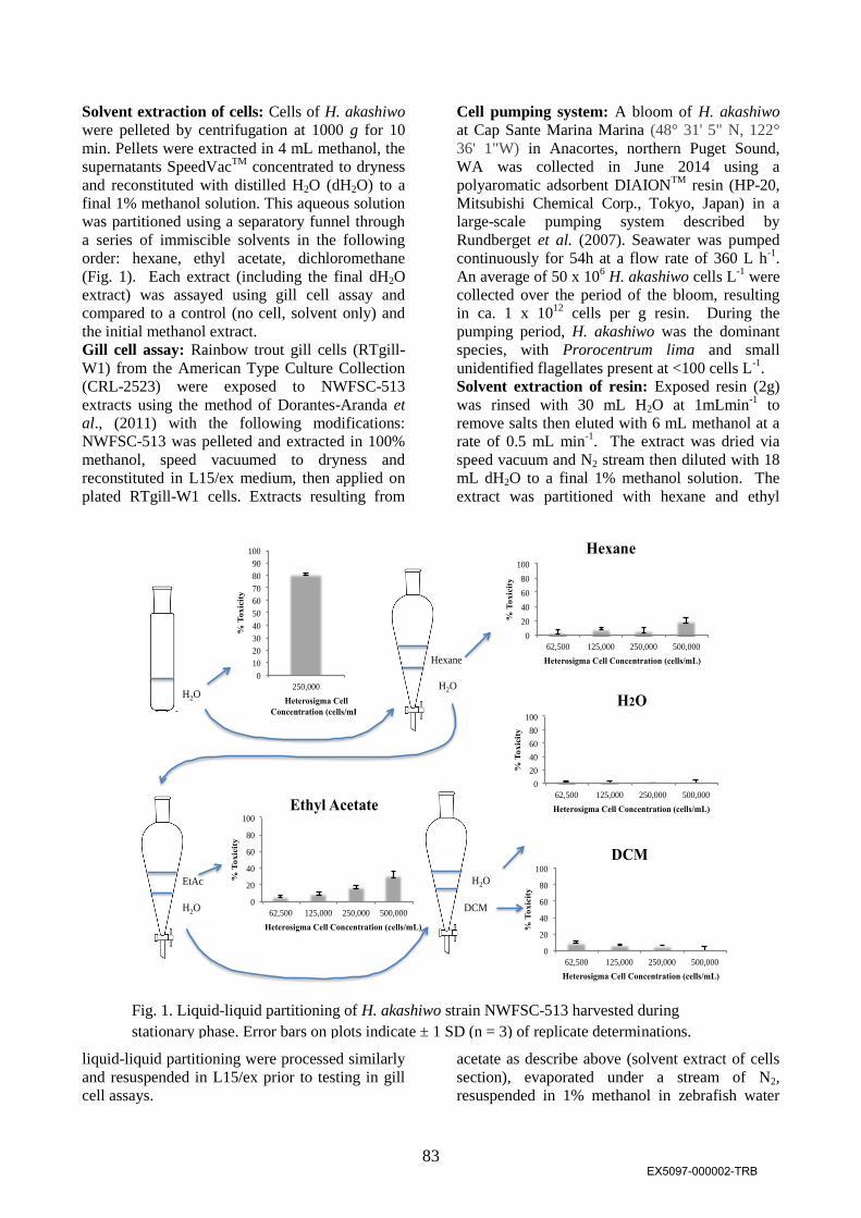
Solvent extraction of cells: Cells of H. akashiwo
were pelleted by centrifugation at 1000 g for 10
min. Pellets were extracted in 4 mL methanol, the
supernatants SpeedVacTM
concentrated to dryness
and reconstituted with distilled H2O (dH2O) to a
final 1% methanol solution. This aqueous solution
was partitioned using a separatory funnel through
a series of immiscible solvents in the following
order: hexane, ethyl acetate, dichloromethane
(Fig. 1). Each extract (including the final dH2O
extract) was assayed using gill cell assay and
compared to a control (no cell, solvent only) and
the initial methanol extract.
Gill cell assay: Rainbow trout gill cells (RTgill-
W1) from the American Type Culture Collection
(CRL-2523) were exposed to NWFSC-513
extracts using the method of Dorantes-Aranda et
al., (2011) with the following modifications:
NWFSC-513 was pelleted and extracted in 100%
methanol, speed vacuumed to dryness and
reconstituted in L15/ex medium, then applied on
plated RTgill-W1 cells. Extracts resulting from
liquid-liquid partitioning were processed similarly
and resuspended in L15/ex prior to testing in gill
cell assays.
Cell pumping system: A bloom of H. akashiwo
at Cap Sante Marina Marina (48° 31' 5" N, 122°
36' 1"W) in Anacortes, northern Puget Sound,
WA was collected in June 2014 using a
polyaromatic adsorbent DIAIONTM
resin (HP-20,
Mitsubishi Chemical Corp., Tokyo, Japan) in a
large-scale pumping system described by
Rundberget et al. (2007). Seawater was pumped
continuously for 54h at a flow rate of 360 L h-1
.
An average of 50 x 106 H. akashiwo cells L
-1 were
collected over the period of the bloom, resulting
in ca. 1 x 10¹²
cells per g resin. During the
pumping period, H. akashiwo was the dominant
species, with Prorocentrum lima and small
unidentified flagellates present at <100 cells L-1
.
Solvent extraction of resin: Exposed resin (2g)
was rinsed with 30 mL H2O at 1mLmin-1
to
remove salts then eluted with 6 mL methanol at a
rate of 0.5 mL min-1
. The extract was dried via
speed vacuum and N2 stream then diluted with 18
mL dH2O to a final 1% methanol solution. The
extract was partitioned with hexane and ethyl
acetate as describe above (solvent extract of cells
section), evaporated under a stream of N2,
resuspended in 1% methanol in zebrafish water
H2O H2O
H2O
H2O
Hexane
EtAc
DCM
0
20
40
60
80
100
62,500 125,000 250,000 500,000
% T
oxic
ity
Heterosigma Cell Concentration (cells/mL)
Hexane
0
20
40
60
80
100
62,500 125,000 250,000 500,000
% T
ox
icit
y
Heterosigma Cell Concentration (cells/mL)
H2O
0
20
40
60
80
100
62,500 125,000 250,000 500,000
% T
oxic
ity
Heterosigma Cell Concentration (cells/mL)
DCM
0
20
40
60
80
100
62,500 125,000 250,000 500,000
% T
oxic
ity
Heterosigma Cell Concentration (cells/mL)
Ethyl Acetate
0
10
20
30
40
50
60
70
80
90
100
250,000
% T
ox
icit
y
Heterosigma Cell
Concentration (cells/mL)
Fig. 1. Liquid-liquid partitioning of H. akashiwo strain NWFSC-513 harvested during
stationary phase. Error bars on plots indicate ± 1 SD (n = 3) of replicate determinations.
EX5097-000002-TRB

84
(deionized and reverse osmosis filtered water
amended with Instant Ocean Salt to a conductivity
of 1500 μS cm-1
) for zebrafish exposure.
Concentrations used for exposures (in triplicate)
ranged from 4-15 x 106 H. akashiwo cell-
equivalents per treatment.
Zebrafish exposures: Zebrafish embryos (n=15)
at 256-cell stage (2.5 hpf) were placed in 5 ml
zebrafish water in 6-well glass plates (Corning,
Costar 3516) and kept in the dark at 28.5°C with
water replacement every 24 h until 72 hpf.
Controls were zebrafish water containing 1%
methanol and zebrafish water alone. Digital video
clips of embryos were collected from each
treatment using a Nikon SMZ800
stereomicroscope or a Nikon Eclipse E600 and a
Unibrain Fire-I800 camera with BTV Carbon Pro
software as described in Incardona et al. (2014).
For each treatment, both heart rate and percentage
of zebrafish with edema were recorded (n=15).
Results and Discussion
The ethyl acetate extract of NWFSC-513 culture
resulting from liquid-liquid partitioning showed
the greatest dose-dependent toxicity in the gill cell
assay compared to other fractions. About 25% of
the activity observed in the original methanol
extract was retained in the ethyl acetate fraction
(Fig. 1). Typically, when extracted sequentially in
this manner: the hexane fraction contains very
lipophilic compounds (e.g. fatty acids); the ethyl
acetate fraction contains the less polar uncharged
molecules (e.g. pectenotoxins); the
dichloromethane fraction contains the more polar
organic molecules (e.g. okadaic acid); and, salts
and highly polar or ionic organic compounds (e.g.
yessotoxins) remain in the water fraction. Resin
extracts from the high density Heterosigma bloom
(June 2014) also showed the highest activity in the
ethyl acetate fraction and, in general, showed
more reliable toxicity than cultures, in particular
because the latter appeared to lose their toxicity
after ~1 year. For this reason, resin extracts were
used for zebrafish exposure experiments.
Zebrafish embryos exposed to the HP20 resin
ethyl acetate extract showed severe pericardial
edema (Figs. 2, 3), atrial dilation, and
hydrocephalus shown by a clear space in the 4th
ventricle of the brain (Fig. 2). The incidence of
edema and decrease in heart rate was dose-
dependent with exposure to HP-20 resin extract
(Fig. 3). Together, these data suggest that H.
akashiwo produces a stable toxin that is
Fig. 3. Dose dependent cardiac effects of H.
akashiwo ethyl acetate extracts on zebrafish
pericardial edema (A), atrial dilation (B), and
heart rate (C). Control heart rate is 211 beats per
minute (bpm) and highest exposed is 132 bpm at
48 hpf. Average std. dev. in (A) = 2 bpm (n=15).
A
B
C
A
V
Treated Control
*
Figure. 2. Zebrafish exposed to H. akashiwo extract
2-48 hpf shows severe edema, hydrocephalus (*),
and atrial (A) dilation. V=ventricle
0
20
40
60
80
100
120
control 4 million 8 million 15 million
% w
ith
ed
ema
Pericardial edema
48h
60h
72h
0
50
100
150
200
250
control 4 million 8 million 15 million
Hea
rt r
ate
(b
eats
/min
)
Heterosigma extract (cell equivalents)
Heart rate
48h
60h
0
20
40
60
80
100
120
control 4 million 8 million 15 million
Atr
ium
wid
th (
mm
)
Atrial dilation
48 hr
60 hr
EX5097-000003-TRB
85
moderately lipophilic. It is proven to be a
chemical entity because toxic activity is recovered
after drying down in methanol and resuspended in
assay buffer. Solubility in ethyl acetate suggests
that it may be a relatively non-polar, neutral
polyether-like compound. The toxin causes
bradycardia, edema and atrial dilation has a
negative chronotropic effect in zebrafish.
Because the cardiac-specific effects occurred by
48h exposure and the heart chambers were formed
normally, the effect is intrinsic to heart
development and not a secondary effect, such as
at the acetylcholine receptor which appears at a
later stage in zebrafish development. This
evidence suggests that the H. akashiwo toxin is a
physiological poison.
Twiner (2002) proposed that H. akashiwo extracts
had an effect on Ca2+
release by muscarinic-1
transfected sf9 insect cells pretreated with
lanthanum, a Ca2+
channel blocker (Twiner 2002).
The effects of H. akashiwo extracts on cardiac
function that we observed also point to the
possibility that H. akashiwo impairs Ca2+
homeostasis. The negative chronotropic effects
shown by H. akashiwo extracts on zebrafish are
consistent with Ca2+
blockage during the plateau
phase of the action potential. However, this study
is not a confirmation of H. akashiwo impacts
solely on Ca2+
homeostasis as the zebrafish
exposures resulted in mixed phenotypes,
suggesting other toxic mechanisms. Head-first
hatching which is characteristic of brevetoxin
effects on zebrafish embryos (Kimm-Brinson &
Ramsdell 2001), was not observed, suggesting
that H. akashiwo toxins are not brevetoxins, as has
been suggested (Khan et al. 1997).
Acknowledgements
We thank Kevin Bright, American Gold Seafood,
for alerting us to H. akashiwo blooms. We
appreciate the assistance of the Cap Sante marina
personnel. This work is a result of ECOHAB
research funded by the U.S. National Oceanic and
Atmospheric Administration Center for Sponsored
Coastal Ocean Research Project No.
NA10NOS4780160 awarded to the University of
Maine (MLW) and San Francisco State University
(WPC), an internal grant to the NWFSC (VLT),
with a subcontract to Western University and a
Canadian NSERC Discovery Grant awarded to
CGT. This is ECOHAB publication No. 813.
References
Berges, J.A., Franklin, D.J., & Harrison, P.J.
(2001). J. Phycol. 37: 1138-1145.
Berges, J.A., Franklin, D.J.,& Harrison, P.J.
(2004). J. Phycol. 40: 619.
Cochlan, W.P. Herndon, J., & Kudela, R.M.
(2008). Harmful Algae 8: 111-118.
Cochlan W.P., Trainer, V.L., Trick, C.G. et al.
(2014). In: Kim, H.G., B. Reguera, G.M.
Hallegraeff et al. (eds). Harmful Algae 2012,
Proceedings of the 15th International Conference
on Harmful Algae, 2014, pp. 203-206.
Dorantes-Aranda, J.J., Waite, T.D., Godrant, A.,
et al. (2011). Harmful Algae 10(4):366-373.
Guillard, R. R. L. (1995). Culture methods. In:
Manual on Harmful Marine Microalgae,
Hallegraeff G. M., Anderson D. M., Cembella A.
D. (eds), UNESCO IOC Manuals and Guides No.
33. 551pp.
Khan, S., Arakawa, O., & Onoue, Y. (1997).
Aquac. Res. 28: 9-14.
Kimm-Brison, K.L. & Ramsdell, J.S. (2001).
Environ. Health Persp. 109: 377-381.
Marshall, J.A., de Salas, M., Oda, T., &
Hallegraeff, G. (2005). Mar.Biol. 147: 533-540.
Oda, T., Nakamura, M., Shikayama, M. et al.
(1997). Biosci. Biotech. Biochem. 61: 1658-1662.
Rundberget, T., Sandvik, M., Larsen, K. et al.
(2007). Toxicon 50: 960-970.
Twiner, M.J. & Trick, C.G. (2000). J. Plankton
Res. 22: 1961-1975.
Twiner, M.J. (2002). Bioactive extracellular
metabolites from Heterosigma akashiwo. Ph.D.
Thesis, The University of Western Ontario,
London, Ontario, 127 pp.
Yang, C.Z., Albright, L.J., & Yousif, A.N. (1995).
Dis. Aquat. Org. 23: 101-108.
EX5097-000004-TRB
Related Documents