Characterization of the Targeting Signal of the Arabidopsis 22-kD Integral Peroxisomal Membrane Protein 1[w] Mary A. Murphy 2 , Belinda A. Phillipson 3 , Alison Baker, and Robert T. Mullen* Department of Botany, University of Guelph, Guelph, Ontario, Canada N1G 2W1 (M.A.M., R.T.M.); and Centre for Plant Sciences, University of Leeds, Leeds, United Kingdom LS2 9JT (B.A.P., A.B.). Using a combination of in vivo and in vitro assays, we characterized the sorting pathway and molecular targeting signal for the Arabidopsis 22-kD peroxisome membrane protein (PMP22), an integral component of the membrane of all peroxisomes in the mature plant. We show that nascent PMP22 is sorted directly from the cytosol to peroxisomes and that it is inserted into the peroxisomal boundary membrane with its N- and C-termini facing the cytosol. This direct sorting of PMP22 to peroxisomes contrasts with the indirect sorting reported previously for cottonseed (Gossypium hirsutum) ascorbate peroxi- dase, an integral PMP that sorts to peroxisomes via a subdomain of the endoplasmic reticulum. Thus, at least two different sorting pathways for PMPs exist in plant cells. At least four distinct regions within the N-terminal one-half of PMP22, including a positively charged domain present in most peroxisomal integral membrane-destined proteins, functions in a cooperative manner in efficient peroxisomal targeting and insertion. In addition, targeting with high fidelity to peroxisomes requires all four membrane-spanning domains in PMP22. Together, these results illustrate that the PMP22 membrane peroxisomal targeting signal is complex and that different elements within the signal may be responsible for mediating unique aspects of PMP22 biogenesis, including maintaining the solubility before membrane insertion, targeting to peroxi- somes, and ensuring proper assembly in the peroxisomal boundary membrane. Peroxisomes are multifunctional organelles that are generally defined as containing at least one hydrogen peroxide-generating oxidase and catalase. Some per- oxisomal functions are virtually universal among evolutionarily diverse organisms such as the -oxidation of fatty acids and defense against oxida- tive stresses. Other peroxisomal functions are more specialized and depend upon the organism in which the organelle resides, e.g. key steps in the synthesis of ether-linked phospholipids and bile salts in mamma- lian peroxisomes, and portions of the glycolate and glycerate pathways of photorespiration in plant leaf and leaf-type peroxisomes. Recent studies have shown that peroxisomes also are involved in the biosynthesis of important signaling molecules, in- cluding indole acetic acid (Zolman et al., 2000) and jasmonate (Stintz and Browse, 2001), nitric oxide, and various reactive oxygen species (Corpas et al., 2001) that modulate many aspects of the plant’s life cycle. In Arabidopsis, the isolation of mutants with defects in a peroxisomal ATP-binding cassette transporter (Zolman et al., 2001; Footitt et al., 2002; Hayashi et al., 2002) or the peroxisome biogenesis protein factor (a peroxin) Pex2p (Hu et al., 2002) have suggested un- expected links between the organelle and fundamen- tal processes, including the breaking of seed dor- mancy (Footitt et al., 2002) and light-regulated gene expression (Hu et al., 2002). Although the molecular mechanisms of these and other plant peroxisomal functions remain to be clarified, it is apparent that the peroxisome is a source and sensor of molecules that can affect plant growth and development in pro- found ways. The biogenesis of peroxisomes is proposed to take place in three distinct steps: the formation of a “nas- cent” or “preperoxisomal” vesicle that, depending upon the organism, is thought to be derived from the endoplasmic reticulum (ER) or another endomem- brane source, or from a pre-existing peroxisome; and the targeting and insertion/assembly of peroxisome membrane proteins (PMP), which include many of the components required for the targeting and im- port of matrix proteins (Sacksteder and Gould, 2000; Purdue and Lazarow, 2001; Sparkes and Baker, 2002). Overall, the mechanisms governing matrix protein targeting and import are best understood, although specific differences exist between evolutionary dis- 1 This work was supported by the Natural Sciences and Engi- neering Research Council of Canada (grant no. 217291), by the Ontario Premier’s Research in Excellence Award (to R.T.M.), and by the Biotechnology and Biology Research Council (grant no. 24/C12039 to A.B.). 2 Present address: Laboratory Services Division, 95 Stone Road West, University of Guelph, Guelph, Ontario, Canada N1H 8J7. 3 Present address: Central Science Laboratory, Sand Hutton, York, UK. [w] The online version of this article contains Web-only information. * Corresponding author; e-mail [email protected]; fax 519 –767–1991. Article, publication date, and citation information can be found at www.plantphysiol.org/cgi/doi/10.1104/pp.103.027870. Plant Physiology, October 2003, Vol. 133, pp. 813–828, www.plantphysiol.org © 2003 American Society of Plant Biologists 813 www.plantphysiol.org on March 26, 2018 - Published by Downloaded from Copyright © 2003 American Society of Plant Biologists. All rights reserved.

Welcome message from author
This document is posted to help you gain knowledge. Please leave a comment to let me know what you think about it! Share it to your friends and learn new things together.
Transcript
Characterization of the Targeting Signal of theArabidopsis 22-kD Integral PeroxisomalMembrane Protein1[w]
Mary A. Murphy2, Belinda A. Phillipson3, Alison Baker, and Robert T. Mullen*
Department of Botany, University of Guelph, Guelph, Ontario, Canada N1G 2W1 (M.A.M., R.T.M.); andCentre for Plant Sciences, University of Leeds, Leeds, United Kingdom LS2 9JT (B.A.P., A.B.).
Using a combination of in vivo and in vitro assays, we characterized the sorting pathway and molecular targeting signal forthe Arabidopsis 22-kD peroxisome membrane protein (PMP22), an integral component of the membrane of all peroxisomesin the mature plant. We show that nascent PMP22 is sorted directly from the cytosol to peroxisomes and that it is insertedinto the peroxisomal boundary membrane with its N- and C-termini facing the cytosol. This direct sorting of PMP22 toperoxisomes contrasts with the indirect sorting reported previously for cottonseed (Gossypium hirsutum) ascorbate peroxi-dase, an integral PMP that sorts to peroxisomes via a subdomain of the endoplasmic reticulum. Thus, at least two differentsorting pathways for PMPs exist in plant cells. At least four distinct regions within the N-terminal one-half of PMP22,including a positively charged domain present in most peroxisomal integral membrane-destined proteins, functions in acooperative manner in efficient peroxisomal targeting and insertion. In addition, targeting with high fidelity to peroxisomesrequires all four membrane-spanning domains in PMP22. Together, these results illustrate that the PMP22 membraneperoxisomal targeting signal is complex and that different elements within the signal may be responsible for mediatingunique aspects of PMP22 biogenesis, including maintaining the solubility before membrane insertion, targeting to peroxi-somes, and ensuring proper assembly in the peroxisomal boundary membrane.
Peroxisomes are multifunctional organelles that aregenerally defined as containing at least one hydrogenperoxide-generating oxidase and catalase. Some per-oxisomal functions are virtually universal amongevolutionarily diverse organisms such as the�-oxidation of fatty acids and defense against oxida-tive stresses. Other peroxisomal functions are morespecialized and depend upon the organism in whichthe organelle resides, e.g. key steps in the synthesis ofether-linked phospholipids and bile salts in mamma-lian peroxisomes, and portions of the glycolate andglycerate pathways of photorespiration in plant leafand leaf-type peroxisomes. Recent studies haveshown that peroxisomes also are involved in thebiosynthesis of important signaling molecules, in-cluding indole acetic acid (Zolman et al., 2000) and
jasmonate (Stintz and Browse, 2001), nitric oxide, andvarious reactive oxygen species (Corpas et al., 2001)that modulate many aspects of the plant’s life cycle.In Arabidopsis, the isolation of mutants with defectsin a peroxisomal ATP-binding cassette transporter(Zolman et al., 2001; Footitt et al., 2002; Hayashi et al.,2002) or the peroxisome biogenesis protein factor (aperoxin) Pex2p (Hu et al., 2002) have suggested un-expected links between the organelle and fundamen-tal processes, including the breaking of seed dor-mancy (Footitt et al., 2002) and light-regulated geneexpression (Hu et al., 2002). Although the molecularmechanisms of these and other plant peroxisomalfunctions remain to be clarified, it is apparent that theperoxisome is a source and sensor of molecules thatcan affect plant growth and development in pro-found ways.
The biogenesis of peroxisomes is proposed to takeplace in three distinct steps: the formation of a “nas-cent” or “preperoxisomal” vesicle that, dependingupon the organism, is thought to be derived from theendoplasmic reticulum (ER) or another endomem-brane source, or from a pre-existing peroxisome; andthe targeting and insertion/assembly of peroxisomemembrane proteins (PMP), which include many ofthe components required for the targeting and im-port of matrix proteins (Sacksteder and Gould, 2000;Purdue and Lazarow, 2001; Sparkes and Baker, 2002).Overall, the mechanisms governing matrix proteintargeting and import are best understood, althoughspecific differences exist between evolutionary dis-
1 This work was supported by the Natural Sciences and Engi-neering Research Council of Canada (grant no. 217291), by theOntario Premier’s Research in Excellence Award (to R.T.M.), andby the Biotechnology and Biology Research Council (grant no.24/C12039 to A.B.).
2 Present address: Laboratory Services Division, 95 Stone RoadWest, University of Guelph, Guelph, Ontario, Canada N1H 8J7.
3 Present address: Central Science Laboratory, Sand Hutton,York, UK.
[w] The online version of this article contains Web-onlyinformation.
* Corresponding author; e-mail [email protected]; fax519 –767–1991.
Article, publication date, and citation information can be foundat www.plantphysiol.org/cgi/doi/10.1104/pp.103.027870.
Plant Physiology, October 2003, Vol. 133, pp. 813–828, www.plantphysiol.org © 2003 American Society of Plant Biologists 813 www.plantphysiol.orgon March 26, 2018 - Published by Downloaded from Copyright © 2003 American Society of Plant Biologists. All rights reserved.

tant organisms. Peroxisomal matrix proteins are syn-thesized on free polyribosomes in the cytosol andcontain one of two different peroxisomal targetingsignals (PTS), i.e. the type I PTS (PTS1) consisting ofa carboxy-terminal tripeptide motif (-SKL) and thetype 2 PTS (PTS2) an amino terminal nonpeptidemotif (-R-X6-H/Q-A/L/F-; Mullen, 2002). The PTS1and PTS2 on nascent matrix proteins are recognizedin the cytosol by their cognate receptors, Pex5p andPex7p, respectively, and the resulting receptor-cargo-protein complexes are targeted to the surface of theperoxisomal boundary membrane. Subsequent dock-ing and translocation of the PTS-bearing cargo, aswell as possibly the bound receptor, and then therecycling of the receptor back to the cytosol for ad-ditional rounds of targeting and import requires theparticipation of several other soluble and membrane-bound peroxins.
In comparison with our understanding of the tar-geting and import of matrix proteins, much less isknown about the targeting and insertion/assemblyof PMPs. Although it is generally accepted that manyPMPs are synthesized in the cytosol and insertedposttranslationally into the peroxisome singleboundary membrane, a consensus has not beenreached on the functional role(s) for some of theprotein components (peroxins) involved in PMP tar-geting/insertion or what constitutes a prototypicmembrane peroxisomal targeting signal (mPTS).PMPs do not possess a PTS1 or PTS2 and, dependingupon the protein and/or organism, the mPTS canvary from a short stretch of three to six positivelycharged amino acids residues to large nonoverlap-ping segments that do not contain obvious consensusmotifs (Subramani et al., 2000; Sparkes and Baker,2002; Trelease, 2002). In addition, mPTSs character-ized seem to be inconsistent in terms of whether theyare orientated topologically on the matrix or cytosolicside of the peroxisomal boundary membrane andwhether one or more transmembrane domains(TMDs) are required for their proper functioning.Also a matter of question is whether the ER serves asthe initial sorting site for at least a subset of PMPs.For example, in tobacco (Nicotiana tabacum) BrightYellow (BY)-2 suspension culture cells, transientlyexpressed cottonseed (Gossypium hirsutum) ascorbateperoxidase (APX) targets to a subdomain of the ERbefore its sorting to pre-existing peroxisomes(Mullen et al., 1999). In Yarrowia lipolyitica, Pex2p andPex16p are sorted to the ER while en route to peroxi-somes (Titorenko and Rachubinski, 1998), whereasno evidence for the delivery of these proteins toperoxisomes via ER could be found in mammaliancells (Voorn-Brouwer et al., 2001). Saccharomyces cer-evisiae Pex15p has been reported also to sort to per-oxisomes via the ER (Elgersma et al., 1997) but thishas been disputed (Hettema et al., 2000). The conflict-ing data published for different PMPs in differentorganisms have prevented the formation of a consis-
tent working model on the nature of the mPTS, thesorting pathways used by PMPs, or the manner inwhich peroxins mediate PMP insertion and assemblyin the peroxisomal boundary membrane. This is fur-ther exemplified in plants, where a paucity of infor-mation on PMPs exists because only a few authenticPMPs have been described, and only one of these,APX, has been characterized in terms of its mPTS andsorting pathways (Mullen et al., 1999, 2000; Nito etal., 2001; Lisenbee and Trelease, 2003).
Arabidopsis PMP22 is an integral membrane pro-tein that is prominent in all organs of the matureplant (Tugal et al., 1999). Related proteins includePMP22 in rat, PMP22, Mpv17 and M-LP in mouse,and PMP22 and Mpv17 in human (Tugal et al., 1999;Iida et al., 2003 and refs. therein). Although the pre-cise molecular function of PMP22 and PMP22-likeproteins remains to be elucidated, recent studies withmouse Mpv17 (Wagner et al., 2001) and M-LP (Iida etal., 2003) suggest that they are involved in enzymaticantioxidant defense systems. Studies on the in vitroinsertion of PMP22 revealed that the rat and Arabi-dopsis proteins are inserted into isolated peroxisomemembranes (Diestelkotter and Just, 1993; Tugal et al.,1999). Studies of the targeting information in mam-malian PMP22 and PMP22-like proteins (Pause et al.,2000; Brosius et al., 2002; Iida et al., 2003) haveyielded radically different conclusions on the natureof the mPTS(s), most likely because large deletionswere used to determine the targeting signals, a strat-egy that is unreliable if multiple signals act cooper-atively and are distributed throughout the protein.
Here, we describe the results of a comprehensivestudy of molecular signals involved in the targetingand insertion of Arabidopsis PMP22 in vivo and invitro. We show that, unlike the sorting of cottonseedAPX to peroxisomes via the ER, newly synthesizedPMP22 is sorted directly from the cytosol to peroxi-somes, and the protein is inserted into the peroxi-some boundary membrane with N- and C-terminalparts facing the cytosol. We also demonstrate, usinga combination of fusion proteins and modified ver-sions of PMP22 (e.g. site-specific substitutions, inter-nal deletions, and truncations), that at least four dis-tinct regions within PMP22 are required for efficientperoxisomal targeting and integration with high fi-delity. Efficient targeting of PMP22 to peroxisomesalso requires all four of the protein’s TMDs. Theimplications of these results and nature of the mPTSin Arabidopsis PMP22 are discussed.
RESULTS
Intracellular Sorting and Membrane Insertion ofEpitope-Tagged Arabidopsis PMP22
When nontransformed tobacco BY-2 suspension-cultured cells stained with anti-Arabidopsis PMP22immunoglobulin (Ig) Gs were examined by immuno-fluorescence microscopy, a punctate fluorescence
Murphy et al.
814 Plant Physiol. Vol. 133, 2003 www.plantphysiol.orgon March 26, 2018 - Published by Downloaded from Copyright © 2003 American Society of Plant Biologists. All rights reserved.

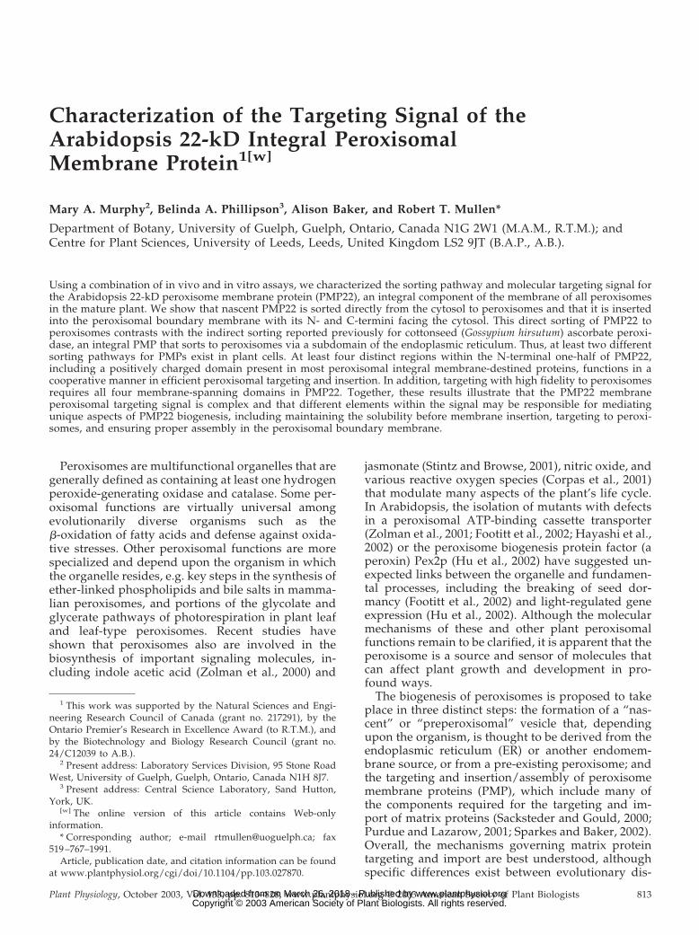
pattern was observed, characteristic of an antigenicprotein, presumably a PMP22, localized to individualperoxisomes (Fig. 1A, a). To distinguish between thisendogenous BY-2 PMP22 and ectopically expressedArabidopsis PMP22, a single copy of the myc epitopetag was fused to the N terminus of ArabidopsisPMP22. Figure 1A (b and c) illustrates that myc-PMP22 was localized exclusively to peroxisomes, asevidenced by its colocalization with the endogenousperoxisomal matrix enzyme catalase. Several otherepitope-tagged versions of Arabidopsis PMP22 alsolocalized to BY-2 peroxisomes, including anN-terminal hemagglutinin (HA)-tagged PMP22 (HA-PMP22), C-terminal myc-tagged PMP22 (PMP22-myc), and a double-epitope-tagged version of PMP22whereby HA and myc epitopes were fused to the Nand C terminus of PMP22, respectively (HA-PMP22-myc; Fig. 1A, d–f).
Different epitope-tagged versions of PMP22 werealso imported into peroxisomes in vitro. Figure 1Bshows the results of representative in vitro importreactions in which radiolabeled wild-type PMP22,myc-PMP22, PMP22-myc, and HA-PMP22-myc wereincubated independently with or without isolatedsunflower peroxisomes, and in the presence or ab-sence of ATP and an ATP-regeneration system, theprotease thermolysin, and/or the detergent TritonX-100. After all import reactions, peroxisomes werereisolated through a 0.7 m Suc cushion and polypep-tides were analyzed by SDS-PAGE and phosphorim-aging. Consistent with previously published results(Tugal et al., 1999), wild-type PMP22 bound to peroxi-somes in a largely ATP-independent manner. That is,the majority of the radiolabeled PMP22 reisolated withperoxisomes in the pellet fraction after incubations inthe presence or absence of ATP (Fig. 1C, a; comparelanes 2 and 4). However, a greater degree of proteaseprotection for PMP22 was observed in the presence ofATP (Fig. 1C, a; lane 3) than in the absence of ATP(Fig. 1C, a; lane 5), indicating that the energy mayfacilitate the protein, acquiring its final protease-resistant state within the peroxisomal boundary mem-brane (Tugal et al., 1999). Similar data were obtainedfor the import of myc-PMP22 (Fig. 1C, panel b), HA-PMP22-myc (Fig. 1C, panel c), and PMP22-myc (Fig.1C, panel d); all three epitope-tagged PMP22 proteinsacquired a greater degree of protection to appliedthermolysin in the presence of ATP. When importreactions with each of the PMP22 proteins weretreated with thermolysin in the presence of TritonX-100 (Fig. 1C, a-d, lane 6) or when peroxisomes wereomitted (Fig. 1C, a-d, lane 7), no protected wild-typeor epitope-tagged PMP22 proteins were observed inthe pellet fractions. This is consistent with proteaseprotection being due to integration of the proteins intothe lipid bilayer and not due to protein misfoldingand/or aggregation. Separate immunoprecipitationexperiments with the soluble fraction from each trans-lation reaction (Fig. 1C, lane 1, b–d) and anti-myc IgGs
confirmed the identity of each epitope-tagged PMP22protein (data not shown). Taken together, the resultspresented in Figure 1 (A and B) indicate that epitopetags fused to the N and/or C terminus of PMP22 didnot disturb peroxisomal targeting or insertion. Be-cause all epitope-tagged proteins behaved in a similarmanner to native PMP22, except PMP22-myc, whichimported poorly, subsequent experiments to deter-mine the location of the peroxisomal targeting infor-mation in PMP22 made use of myc-PMP22.
Figure 1C illustrates the localization myc-PMP22 inrepresentative transformed BY-2 cells at 5, 12, 20, and45 h postbombardment. At the earlier stages of ex-pression and sorting (i.e. 5 h postbombardment), themajority of nascent myc-PMP22 resided in the non-organelle cytosol (Fig. 1C, a), with only a small pro-portion localized to catalase-containing peroxisomes(Fig. 1E, compare a and b). However, after 12 and20 h of transient expression, myc-PMP22 localizedalmost exclusively to individual peroxisomes distrib-uted throughout the cell (Fig. 1C, e and f). The lack offluorescence staining attributable to myc-PMP22 lo-calized in other subcellular compartment(s) besideperoxisomes indicates that nascent PMP22 is sorteddirectly from its site of synthesis in the cytosol toperoxisomes, and not indirectly to peroxisomes viathe ER.
At 45 h postbombardment, myc-PMP22 remainedlocalized to peroxisomes (Fig. 1C, g and h), but themorphology and distribution of myc-PMP22-containing peroxisomes was altered by this timepoint. Most of the peroxisomes in a myc-PMP22-transformed cell 45 h after bombardment were redis-tributed into large globular structures up to 6 �m indiameter and that usually numbered 10 to 20 per cell.These globular-like peroxisomes were not apparentin surrounding nontransformed cells (Fig. 1C, h).Large globular peroxisomes also accumulated by45 h postbombardment in PMP22-myc and HA-PMP22-myc-transformed cells (data not shown).
The N and C Termini of PMP22 AreExposed to the Cytosol
The topology of PMP22 within the peroxisomemembrane was determined by the differential per-meabilization/immunofluorescence method (Lee etal., 1997; Mullen et al., 2001b). Myc-PMP22-transformed cells at 20 h postbombardment weretreated with digitonin, which selectively permeabi-lizes plasma membranes, leaving intraorganellar an-tigenic sites inaccessible to antibodies. Figure 2A (aand b) shows that transiently expressed myc-PMP22and endogenous �-tubulin in cytosolic microtubuleswere immunostained in the same cell, indicating thatthe N terminus of myc-PMP22 is accessible to anti-myc antibodies. A simultaneous control experimentverified that only the plasma membrane was per-meabilized in the same batch of myc-PMP22-
Targeting Signal for Arabidopsis Peroxisomal Membrane Protein 22
Plant Physiol. Vol. 133, 2003 815 www.plantphysiol.orgon March 26, 2018 - Published by Downloaded from Copyright © 2003 American Society of Plant Biologists. All rights reserved.
Figure 1. Peroxisomal targeting and membrane insertion of eptiope-tagged-PMP22s. A, Subcellular localization of endogenousPMP22 and different versions of epitope-tagged Arabidopsis PMP22 in BY-2 cells. Nontransformed (a) or transiently trans-formed (b-f) BY-2 cells were fixed in formaldehyde, permeabilized with pectolyase and Triton X-100, and incubated inappropriate antibodies. a, Punctate immunofluorescence pattern in nontransformed BY-2 cells incubated with anti-ArabidopsisPMP22 IgGs. b, Transient-expressed myc-PMP22 and endogenous catalase (c) in the same transformed cell; solid arrowsindicated obvious colocalizations. Punctate immunofluorescence patterns attributable to expressed HA-PMP22 (d), PMP22-myc (e), and HA-PMP22-myc (f) in transformed cells; colocalizations with endogenous catalase in peroxisomes are not shown.No fluorescence was detected in control experiments including omission of anti-Arabidopsis PMP22, anti-myc, or anti-HA IgGsor mock transformations with pRTL2 vector alone (data not shown). Bar in f � 10 �m. B, Insertion of wild-type andepitope-tagged versions of Arabidopsis PMP22 into isolated peroxisomes in vitro. PMP22 (a), myc-PMP22 (b), HA-PMP22-myc(c), and PMP22-myc (d) were translated in vitro in the presence 35S-Met with the wheat (Triticum aestivum) germ extract systemand soluble radiolabeled translation products used in an in vitro import assay with isolated sunflower (Helianthus annuus)peroxisomes. Solid arrows to the left of each panel indicate the location of wild-type or epitope-tagged PMP22 species; thelatter was confirmed by immunoprecipitation reactions with anti-myc IgGs (data not shown). Lane 1, Translation productsequivalent to 40% of the amount shown in the other lanes. Lane 2, Reisolated radiolabeled protein from an import reactioncontaining ATP and peroxisomes. Lane 3, The same as lane 2 except that import reactions were treated with the proteasethermolysin before reisolation of peroxisomes. Lane 4, Radiolabeled protein reisolated with peroxisomes after an importreaction in the absence of ATP. Lane 5, The same as lane 4 except that import reactions were treated with thermolysin beforereisolation of peroxisomes. Lane 6, After an import reaction was carried out in the presence of ATP peroxisomes werereisolated, lysed with the detergent Triton X-100, and treated with thermolysin. Lane 7, The same as lane 5 except that, as acontrol, peroxisomes were omitted from the mock import reaction. M, Molecular mass markers; upper band (where shown) is30.1 kD, and the lower band is 20 kD. C, Intracellular sorting of nascent myc-PMP22 from the cytosol to peroxisomes in BY-2cells. Transiently expressed myc-PMP22 (a, c, e, and g) and endogenous catalase (b, d, f, and h) in transformed cells 5 h (a andb), 12 h (c and d), 20 h (e and f), or 45 h (g and h) after biolistic bombardment. Obvious colocalizations of myc-PMP22 withcatalase in individual or globular peroxisomes are indicated with black and white arrows, respectively. Bar in a � 10 �m.
Murphy et al.
816 Plant Physiol. Vol. 133, 2003 www.plantphysiol.orgon March 26, 2018 - Published by Downloaded from Copyright © 2003 American Society of Plant Biologists. All rights reserved.

transformed cells, i.e. the peroxisomal matrix enzymecatalase was not immunostained, whereas cytosolicmicrotubules were readily visualized in the same cell(Fig. 2A, c and d). However, permeabilization of cellswith Triton X-100 resulted in peroxisomal catalase andcytosolic microtubules being immunodetected (Fig.2A, e and f). Identical results were obtained withdigitonin-permeabilized PMP22-myc-transformedcells (data not shown), indicating that the C-terminalpart of PMP22-myc was also accessible to antibodies.
When HA-PMP22-myc-transformed cells were per-meabilized with digitonin, identical (superimpos-sible) staining patterns attributable to myc and HAwere observed in the same cell (Fig. 2A, g and h). Incontrast, endogenous peroxisomal catalase was notdetected in HA-PMP22-myc transformed cells thatwere permeabilized with digitonin and double-stained with anticatalase and anti-HA or anti-mycantibodies (Fig. 2A, I–l). Control experiments withtransiently expressed HA-tagged catalase, a well-established matrix enzyme (Mullen et al., 1997), re-vealed that the HA epitope was not exposed to thecytosolic face of the peroxisomal boundary mem-brane (Fig. 2A, m–o).
Collectively, results obtained from differential per-meabilization experiments with various epitope-tagged PMP22 constructs indicated that the N- andC-terminal portions of PMP22 are exposed to thecytosol. These data are consistent with the model forthe topology of Arabidopsis PMP22 based on pri-mary sequence and hydrophobicity. In this model(Fig. 2B), PMP22 is predicted to consist of four TMDs(TMD1-4; residues 55–73, 99–117, 133–150, and 159–177), two matrix-exposed hydrophilic sequences or“loops” (residues 74–98 and 151–158), and at leastthree cytosolic-exposed hydrophilic sequences, in-cluding one loop region (residues 118–133), and theN- and C-terminal portions (residues 1–54 and 178–190) of the protein.
ized with Triton X-100. Immunostaining of HA (g) and myc (h)epitopes in a HA-PMP22-myc-transformed cell permeabilized withdigitonin. HA-PMP22-myc-bombarded, digitonin-permeabilizedcells used for (g and h) immunostained for endogenous catalase (iand k) and the HA (j) or myc epitope (l). Immunostaining of expressedHA-catalase (m) in cells permeabilized with Triton X-100. HA-catalase-bombarded, digitonin-permeabilized cells used for (m) im-munostained for the HA epitope (n) and endogenous tubulin (p). Barin a � 10 �m. B, Predicted topological map of PMP22. Regions ofPMP22 proposed to be hydrophobic membrane-spanning domains orhydrophilic domains facing the cytosol or peroxisomal matrix wereidentified using the TMHMM program (version 2.0) (http://www.cbs.dtu.dk/services/TMHMM-2.0/). Shaded rectangles denote thefour membrane-spanning domains (TMD 1–4) and the numbers oftheir first and last amino acid residues of each TMD are alsoindicated.
Figure 2. Topological orientation of PMP22. A, Immunostaining at-tributable to transiently expressed myc-PMP22, HA-PMP22-myc,HA-catalase, or to endogenous �-tubulin or catalase in differentialpermeabilized BY-2 cells. BY-2 cells were formaldehyde fixed, per-meabilized with pectolyase and with digitonin (a–d, g–l, n, and o) orTriton X-100 (e, f, and m), and then incubated with appropriateantibodies. Transiently expressed myc-PMP22 (a) and endogenous�-tubulin (b) in the same digitonin-permeabilized transformed cell.Myc-PMP22-bombarded, digitonin-permeabilized cells used for (aand b) immunostained for endogenous peroxisomal matrix catalase(c) and cytosolic �-tubulin (d). Immunostaining of endogenous cata-lase (e) and �-tubulin (f) in myc-PMP22-bombarded cells permeabil-
Targeting Signal for Arabidopsis Peroxisomal Membrane Protein 22
Plant Physiol. Vol. 133, 2003 817 www.plantphysiol.orgon March 26, 2018 - Published by Downloaded from Copyright © 2003 American Society of Plant Biologists. All rights reserved.

The Peroxisomal Targeting Information IsLocated in the N-Terminal Region of PMP22
To define the peroxisomal targeting information inPMP22, a series of fusion proteins in which differentportions of PMP22 were appended to the N or Cterminus of the bacterial passenger protein chloram-phenicol acetyltransferase (CAT) were generated. Allchimeric proteins as well as mutant versions ofepitope-tagged PMP22 described below are listed inFigure 3. The efficiency with which proteins sorted toperoxisomes in BY-2 cells was compared with wild-
type myc-PMP22 and was assessed or scored by co-localization with endogenous peroxisomal catalase.That is, PMP22 constructs that colocalized exclu-sively with catalase in the same cell but no otherorganelles were designated “�.” Those that targetedpartially to peroxisomes and to other organellesand/or cytosol in the same cell were designated “�,”and those that showed no apparent colocalizationwith catalase but instead localized to other organellesand/or cytosol were designated “–“ (Fig. 3).
Figure 4A (a–d) shows that CAT alone, as well asfusion proteins PMP22 1-27-CAT and PMP22 1-54-CAT accumulated in the cytosol of individual trans-formed BY-2 cells. In contrast, PMP22 1-78-CAT,which includes the N-terminal hydrophilic domainand the first putative membrane-spanning domain(TMD1) of PMP22, was sufficient, albeit in an ineffi-cient manner, in redirecting the passenger protein toperoxisomes. Figure 4A (e and f) shows that at leasta portion of transiently expressed PMP22 1-78-CATcolocalized with endogenous catalase, and that theperoxisomes in the transformed cell possessed a dra-matically altered distribution and morphology simi-lar to myc-PMP22-transformed cells at later stages ofexpression (Fig. 1C, g). These globular peroxisomalstructures in PMP22 1-78-CAT-transfomed cells werestrikingly similar to the aggregated peroxisomes incells expressing a CAT-APX fusion protein. Figure4A (g and h) shows, for comparison, replicate imagesof those shown previously (Mullen et al., 1999, 2001b)of the localization of CAT-APX (CAT plus the 36C-terminal residues of cottonseed peroxisomal APX)to various subcellular structures, including catalase-containing aggregated peroxisomes.
Transiently expressed PMP22 1-78-CAT did notcolocalize with endogenous calreticulin, an ERmarker (Fig. 4A, i and j). The reticular/circular fluo-rescence pattern attributable to the fusion proteinthat was not localized to catalase-containing globularperoxisomes is similar to reticular/circular structuresobserved in CAT-APX-transformed cells (Fig. 4A,compare e, g, and i). CAT-APX and other APX fusionproteins have been shown previously localized topER, before their sorting to (globular) peroxisomes(Mullen et al., 1999, 2001b), as well as to plastids andmitochondria due to overexpression and/or mislo-calization (Lisenbee et al., 2003).
Similar to the results presented above for PMP221-78-CAT, other PMP22-CAT fusion proteins thatconsisted of larger N-terminal portions of PMP22,e.g. PMP22 1-99-CAT, PMP22 1-120-CAT, and PMP221-155-CAT showed only partial colocalization withendogenous catalase in globular peroxisomes (Fig.4A, k–n). However, full-length PMP22 (residues1–190) fused to the N terminus of CAT (PMP22 1-190-CAT) was efficiently targeted to peroxisomes be-cause the fusion protein colocalized entirely withendogenous catalase (Fig. 4A, o and p). Individualperoxisomes, and not globular or aggregated perox-
Figure 3. Peroxisomal targeting of PMP22 mutant and fusion pro-teins. Black boxes represent the four predicted TMDs (1–4) inPMP22. Other regions of PMP22 containing putative mPTSs (i.e.amino acids 7 and 8, 14–26, 49–54, and 82–85) are marked withasterisks. Epitope tags and CAT fused to the N or C terminus ofPMP22 are indicated. Schematic representations also show deletionsor truncations in PMP22 proteins as spaces and site-specific Glysubstitutions are marked with vertical bars. The numbers in the nameof each fusion construct or myc-PMP22 mutant denotes the specificamino acid residues from PMP22 (1–190 residues) that were fused tothe N or C terminus of CAT or delete/replaced with Gly residues.Targeting of PMP22 mutant and fusion proteins to peroxisomes inBY-2 cells was scored as follows: �, exclusively localized to peroxi-somes; �, partially localized to peroxisomes; -, not localized toperoxisomes. Results shown for each construct are a representative ofall the transformants (�50) observed from at least two independentbiolistic bombardment experiments.
Murphy et al.
818 Plant Physiol. Vol. 133, 2003 www.plantphysiol.orgon March 26, 2018 - Published by Downloaded from Copyright © 2003 American Society of Plant Biologists. All rights reserved.
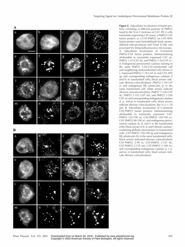
Figure 4. Subcellular localization of fusion pro-teins consisting of different portions of PMP22fused to the N or C terminus of CAT. BY-2 cellstransiently expressing CAT alone, a PMP22-CATfusion protein, or a CAT-PMP22 (or CAT-APX)fusion protein were formaldehyde fixed, perme-abilized with pectolyase and Triton X-100, andprocessed for immunofluorescence microscopy.A, Subcellular localization of N-terminalPMP22-CAT fusion proteins. Immunostainingattributable to transiently expressed CAT (a),PMP22 1-27-CAT (b), and PMP22 1-54-CAT (c).d, Endogenous peroxisomal catalase staining inthe same PMP22 1-54-CAT-transformed celland neighboring nontransformed cells shown inc. Expressed PMP22 1-78-CAT (e) and CAT-APX(g) and corresponding endogenous catalase (fand h) in transformed cells; black arrows indi-cate obvious colocalizations. PMP22 1-78-CAT(i) and endogenous ER calreticulin (j) in thesame transformed cell; white arrows indicateobvious noncolocalizations. PMP22 1-120 CAT(k), PMP22 1-155 CAT (m), and PMP22 1-190CAT (o) and corresponding endogenous catalase(l, n, and p) in transformed cells; black arrowsindicate obvious colocalizations. Bar in a � 10�m. B, Subcellular localization of C-terminalCAT-PMP22 fusion proteins. Immunostainingattributable to transiently expressed CAT-PMP22 155-190 (a), CAT-PMP22 120-190 (c),CAT-PMP22 88-190 (e), and endogenous perox-isomal catalase (b, d, and f) in the transformedcells; black arrows in b, d, and f denote catalase-containing globular peroxisomes in transformedcells. CAT-PMP22 120-190 (g) and endogenousER calreticulin (h) in the same transformed cells;black arrows indicated obvious colocalizationsCAT-PMP22 2-99 (i), CAT-PMP22 2-121 (k),CAT-PMP22 2-155 (m), CAT-PMP22 2-190 (o),and corresponding endogenous catalase (j, l, n,and p) in transformed cells; black arrows indi-cate obvious colocalizations.
Targeting Signal for Arabidopsis Peroxisomal Membrane Protein 22
Plant Physiol. Vol. 133, 2003 819 www.plantphysiol.orgon March 26, 2018 - Published by Downloaded from Copyright © 2003 American Society of Plant Biologists. All rights reserved.

isomal structures, were observed in the majority ofPMP22 1-190-CAT-transformed cells.
Next, the sufficiency of various C-terminal regionsof PMP22 for redirecting CAT from the cytosol toperoxisomes was tested. No apparent colocalizationswere observed between endogenous peroxisomalcatalase and several transiently expressed CAT-PMP22 fusion proteins (Fig. 4B), including CAT-PMP22 155-190 (Fig. 4B, a and b), CAT-PMP22 121-190 (Fig. 4B, c and d), and CAT-PMP22 88-190 (Fig.4B, e and f). Each of these fusion proteins insteadlocalized to a reticular network that consisted of ER,as evidenced by colocalizations with endogenous cal-reticulin (data shown only for CAT-PMP22 121-190;Fig. 4B, g and h). Although CAT-PMP22 155-190,CAT-PMP22 121-190, and CAT-PMP22 88-190 didnot appear to contain peroxisomal targeting informa-tion, globular peroxisomes were observed in cellstransiently expressing each one of these three fusionproteins. We noted also that the size and/or numberof the peroxisomal structures that were formedseemed to vary depending on proportion of the Cterminus of PMP22 that was appended to CAT; glob-ular peroxisomes were more prevalent in cells ex-pressing CAT-PMP22 88-190 or CAT-PMP22 121-190than in cells expressing CAT-PMP22 155-190. At leastpartial localization to catalase-containing globularperoxisomes was observed for several other CAT-PMP22 fusion proteins, including CAT-PMP22 55-190, CAT-PMP22 2-99, CAT-PMP22 2-121, and CAT-PMP22 2-155 (Figs. 3 and 4B, i–n). Each of thesefusion proteins, like the N-terminal PMP22-CAT fu-sions shown in Figure 4A also localized to othersubcellular compartment(s) with a reticular/circularappearance that did not colocalize with calreticulinin the ER (data not shown). Figure 4B (o and p)shows that CAT-PMP22 2-190, consisting of full-length PMP22 fused to the C terminus of CAT local-ized exclusively to catalase-containing globular per-oxisomes. Overall, the results presented in Figure 4indicate that although the N-terminal one-half ofPMP22 contains the peroxisomal targeting informa-tion, efficient sorting of the passenger protein CATfrom the cytosol to peroxisomes required that it wasappended to nearly the entire PMP22 sequence, in-cluding all four TMDs.
PMP22 Contains Several Distinct Regions That Are AllNecessary for Efficient Peroxisomal Targeting
To define more precisely the region(s) withinPMP22 responsible for targeting to peroxisomes,amino acid sequences were sought within the proteinthat resemble the so-called prototypic mPTS found inmost other PMPs. These consist of a cluster of three tofive positively charged amino acid residues adjacentto at least one TMD (for review, see Subramani et al.,2000; Purdue and Lazarow, 2001; Trelease et al.,2002).
Figure 5 shows an alignment of deduced aminoacid sequences for PMP22s from Arabidopsis, mouse,rat, and human. Overall, the Arabidopsis sequencewas similar (45%–57%) to mammalian PMP22s, withthe most notable difference being that the relativepositions of the first three (of four) predicted TMDsvaried between Arabidopsis PMP22 and the mamma-lian proteins. At least three amino acid sequenceswithin Arabidopsis PMP22 closely resembled theprototypic mPTS (bold and underlined in Fig. 5).These included the basic cluster -KIQLRR- (residues49–54) that is immediately adjacent to amino-terminal end of TMD1 and conserved among otherPMP22s, and the basic clusters -KGKK- (residues82–85) and -RERIKK- (residues 126–131) that appearto be unique to the Arabidopsis protein and areadjacent to TMD1 and TMD3, respectively.
To determine whether any of these three clusters ofbasic amino acid residues in PMP22 confer necessaryperoxisomal targeting information, they were inde-pendently deleted or altered in myc-PMP22, and thelocalization of resulting mutant proteins was exam-ined in transformed BY-2 cells. As shown in Figure6A (a and b), myc-PMP22�126-131, which lacks theamino acids -RERIKK- immediately upstream ofTMD3, colocalized entirely with endogenous perox-isomal catalase. In contrast, myc-PMP22�49-54 (Fig.6A, c and d) and myc-PMP22�82-85 (Fig. 6A, e and f)only partially colocalized with endogenous catalaseand a proportion of each expressed protein mislocal-ized to the cytosol and ER. Partial localization ofmyc-PMP22�49-54 and myc-PMP22�82-85, as wellseveral other myc-PMP22 mutant proteins describedbelow, to the ER was evident by their colocalizationwith endogenous calreticulin; data are shown only
Figure 5. Sequence comparison of PMP22s from rat, mouse, human,and Arabidopsis. Deduced amino acid sequences were obtainedfrom GenBank (accession nos.: rat Q07066; mouse P42925; humanAY044439; and Arabidopsis AJ006053) and were aligned usingClustalW and visual inspection. Identical amino acid residues ineach of the aligned PMP22s are indicated by asterisks, and similarresidues are indicated by dots. TMDs were identified using theTMHMM program (version 2.0). The four predicted TMDs in each ofthe proteins are shaded and regions tested in Arabidopsis PMP22 inthis study to function as mPTSs are in bold and underlined.
Murphy et al.
820 Plant Physiol. Vol. 133, 2003 www.plantphysiol.orgon March 26, 2018 - Published by Downloaded from Copyright © 2003 American Society of Plant Biologists. All rights reserved.

Figure 6. Subcellular localization of modified ver-sions of myc-PMP22. Transiently transformed BY-2cells were formaldehyde fixed, permeabilized withpectolyase and Triton X-100, and processed for im-munofluorescence microscopy. A, Subcellular local-ization of myc-PMP22 mutants with alterations inamino acids sequences that resemble positivelycharged mPTSs in other PMPs. Transiently expressedmyc-PMP�126-131 (a), myc-PMP22�49-54 (c),myc-PMP22�82-85 (e), and corresponding endoge-nous catalase (b, d, and f) in transformed cells; blackarrows indicate obvious colocalizations. Myc-PMP22�49-54 (g) and endogenous ER calreticulin(h) in the same transformed cell; black arrows indi-cate obvious colocalizations. Coexpressed myc-PMP22�49-54 (i) and CAT-APX (j) in the same trans-formed cell; white arrows indicate obviousnoncolocalizations. Myc-PMP22K49R53R54�G (k),myc-PMP22K82K84K85�G (m), PMP22K49R53R54K82-K84K85�G (o), and corresponding endogenous cata-lase (l, n, and p) in transformed cells; black arrowsindicate obvious colocalizations. Bar in a � 10 �m.B, Subcellular localization of myc-PMP22 mutantswith Gly substitutions of amino acids sequencesthat have been proposed to function as mPTSs inmammalian PMP22s. Transiently expressed myc-PMP22K7K8�G (a), myc-PMP22K92K93�G (c), myc-PMP22Y14L18P22K26�G (e), myc-PMP22K7K8Y14L18-P22K26�G (g), myc-PMP22�1-33 (i), myc-PMP22K7-K8Y14L18P22K26K49K53K54�G (k), and correspondingendogenous catalase (b, d, f, h, j, and l) in trans-formed cells; black arrows indicate obvious colocal-izations. Expressed myc-PMP22K7K8Y14L18P22-K26K49K53K54K82K84K85�G (m and o) and endoge-nous catalase (n) or endogenous calreticulin (p) intransformed cells; white arrows in m and n indicateobvious noncolocalization and black arrows in oand p indicate obvious colocalizations.
Targeting Signal for Arabidopsis Peroxisomal Membrane Protein 22
Plant Physiol. Vol. 133, 2003 821 www.plantphysiol.orgon March 26, 2018 - Published by Downloaded from Copyright © 2003 American Society of Plant Biologists. All rights reserved.
for myc-PMP22�49-54 (Fig. 6A, g and h). Interest-ingly, no myc-PMP22 mutant protein tested was mis-localized readily to pER; no apparent colocalizationwas observed, for example, when myc-PMP22�49-54and CAT-APX, as marker for pER, were coexpressedin the same BY-2 cell (Fig. 6A, i and j).
We next tested whether peroxisomal targeting ofPMP22 was affected when all the basic residues atposition 49 through 54 (underlined, KIQLRR) or po-sition 82 through 85 (KGKK) or at both positionswere replaced with noncharged glycines. Figure 6A(k–p) shows that the resulting proteins (myc-PMP22K49R53R54�G, myc-PMP22K82K84K85�G, andmyc-PMP22 K49R53R54K82K84K85�G) were only par-tially localized to peroxisomes (Fig. 6A, k–p), and allthree mutants mislocalized to a similar extent to theER. Taken together, the results presented in Figure6A indicate that although at least two regions inPMP22 that resemble a prototypic mPTS, namelyregions 49 through 54 and 82 through 85, are neces-sary for efficient sorting to peroxisomes, additionaltargeting information exists within the protein.
Because the C-terminal one-half of PMP22 was in-sufficient in sorting CAT to peroxisomes (Fig. 5), itwas reassured that any peroxisomal targeting infor-mation in PMP22, in addition to regions 49 through54 and 82 through 85, was contained in theN-terminal region of the protein. From the results ofprevious studies of mammalian PMP22s (Pause et al.,2000; Brosius et al., 2002), at least three separateregions within the N-terminal one-half of the pro-teins were speculated, but not experimentallyproven, to function as mPTSs, and each of theseregions are divergent from the prototypic “basic clus-ter” mPTS described above. Inspection of the Arabi-dopsis PMP22 sequence revealed that all three ofthese putative mPTSs were conserved in the plantprotein, -KK- at positions 7 and 8, -KK- at positions92 and 93, and the motif -Y-x3-l-x3-P-x3-K- at posi-tions 14 through 26 (bold and underlined in Fig. 5).
Figure 6B (a and b) shows that when the two Lysresidues at positions 7 and 8 in PMP22 were eachreplaced with a Gly residue, the resulting mutantprotein (myc-PMP22K7K8�G) was not completelylocalized to peroxisomes. Instead, a portion ofmyc-PMP22K7K8�G was mislocalized to the ER, asevidenced by colocalization with endogenouscalreticulin (data not shown). In contrast, expressedmyc-PMP22K92K93�G colocalized entirely with en-dogenous catalase (Fig. 6B, c and d). These datasuggest that the di-Lys residues at positions 7 and 8,but not those at positions 92 and 93, are necessary forPMP22 to be targeted efficiently to peroxisomes. Thetargeting function of the proposed Y-x3-l-x3-P-x3-Kmotif identified in rat PMP22 was tested by replacingthe conserved Y, L, P, and K residues at positions 14through 26 in Arabidopsis PMP22 with Gly. Figure6B (Fig. 6B, e and f) shows that the mutant myc-PMP22Y14L18P22K26�G only partially localized to
peroxisomes. Targeting to peroxisomes was also di-minished when residues at position 14 through 26were deleted (myc-PMP22�14-26) or when the Y andL (myc-PMP22Y14L18�G) or the P and K (myc-PMP22P22K26�G) alone were replaced with Gly res-idues (data not shown and Fig. 3), suggesting that allfour conserved residues in this motif were equallyimportant for efficient peroxisomal localization ofPMP22.
The effect of disrupting several regions at once inPMP22 on the peroxisomal targeting of the proteinwas investigated. Figure 6B (g and h) shows thatwhen Lys 7 and 8 as well as the conserved residues inthe -Y-x3-l-x3-P-x3-K- motif at positions 14 through26 were replaced with Gly, the resulting mutant pro-tein (myc-PMP22K7K8Y14L18P22K26�G) was still par-tially localized to peroxisomes. Similarly, deletion ofthe first 33 residues of PMP22 (myc-PMP22�1-33) didnot abolish peroxisomal targeting completely (Fig.6B, i and j). However, these results for the partialtargeting of myc-PMP22K7K8Y14L18P22K26�G andmyc-PMP22�1–33 to peroxisomes were not entirelyunexpected because at least two other putative tar-geting regions within PMP22 (i.e. -KIQLRR- and-KGKK- at positions 49–54 and 82–85, respectively)remained intact in each of these mutants. Partiallocalization to peroxisomes was still observed whenall three of the putative targeting regions inN-terminal hydrophilic domain of PMP22 were mu-tated (myc-PMP22K7K8Y14L18P22K26K49K53K54�G;Fig. 6B, k and l). Only when all four of the regionsconsidered to be important for targeting PMP22were altered by substitutions of specific residueswith Gly was the resulting mutant protein (myc-PMP22K7K8Y14L18P22K26K49K53K54K82K84K85�G) notlocalized to peroxisomes, but instead localized to ER(Fig. 6A, m–p).
Import of myc-PMP22 Mutants in Vitro
The ability of selected myc-PMP22 mutants to in-sert into isolated peroxisomes in vitro was also tested(Fig. 7). In vitro-translated proteins were incubatedin duplicate with isolated sunflower peroxisomes inthe presence of ATP and an ATP-regeneration systemfor 5, 15, 30, or 60 min. Reactions were stopped bytransferring the samples to ice and one aliquot wasthen treated with the protease thermolysin. A directcomparison of samples treated or not treated withprotease allowed discrimination between myc-PMP22 mutants that have achieved the protease-resistance characteristic similar to that of wild-typemyc-PMP22 (and native PMP22; Tugal et al., 1999)and mutant myc-PMP22s that had simply reisolatedwith peroxisomes. Moreover, an analysis of selectedPMP22 mutants ability to acquire protease resistancein time-course experiments allowed us to determinewhether they inserted in vitro into peroxisomes withslower kinetics than wild-type myc-PMP22.
Murphy et al.
822 Plant Physiol. Vol. 133, 2003 www.plantphysiol.orgon March 26, 2018 - Published by Downloaded from Copyright © 2003 American Society of Plant Biologists. All rights reserved.
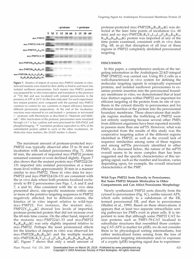
The maximum amount of protease-protected myc-PMP22 was typically observed after 15 to 30 min ofincubation with isolated peroxisomes (Fig. 7). After60 min, the amount of imported myc-PMP22 proteinremained constant or even declined slightly. Figure 7also shows that the mutant protein myc-PMP22�126-131 imported into isolated peroxisomes at a maxi-mum level within approximately 30 min in a mannersimilar to myc-PMP22. These in vitro data for myc-PMP22 and myc-PMP22�126-131 are consistent withthe in vivo data where both proteins localized exclu-sively to BY-2 peroxisomes (see Figs. 1, A and E and7, a and b). Also consistent with the in vivo datapresented above, site-specific mutations within oneor more of the putative targeting sequences in PMP22negatively affected, but to different extents, thekinetics of in vitro import relative to wild-typemyc-PMP22. For instance, the mutant myc-PMP22Y14L18�G showed low levels of imported,protease-protected protein that did not increase overthe 60-min time course. On the other hand, import ofthe mutants myc-PMP22�1-33 and mycPMP22K82K84K85�G was only slightly impaired relative tomyc-PMP22. Perhaps the most pronounced effectsfor the kinetics of import in vitro was observed formyc-PMP22K49R53R54�G and the multiple mutantmyc-PMP22K7K8Y14L18L18P22K26K49K53K54K82K84K85�G. Figure 7 shows that only a small amount of
protease-protected myc-PMP22K49R53R54�G was de-tected at the later time points of incubation (i.e. 60min) and no myc-PMP22K7K8Y14L18L18P22K26K49K53K54K82K84K85�G protein was imported at any of thetime points examined, consistent with in vivo data(Fig. 6B, m–p) that disruption of all four of theseregions in PMP22 completely abolished peroxisomaltargeting.
DISCUSSION
In this paper, a comprehensive analysis of the tar-geting information in the Arabidopsis 22-kD integralPMP (PMP22) was carried out. Using BY-2 cells as awell-characterized in vivo system for defining themolecular targeting signals in ectopically expressedproteins, and isolated sunflower peroxisomes to ex-amine protein insertion into the peroxisomal bound-ary membrane in vitro, we demonstrated that at leastfour distinct regions within PMP22 are important forefficient targeting of the protein from its site of syn-thesis in the cytosol directly to peroxisomes and forefficient insertion of the protein into the peroxisomalboundary membrane. These observations that multi-ple regions mediate the trafficking of PMP22 werenot entirely surprising because several other PMPsfrom different organisms have been reported recentlyto possess more than one mPTS. However, what wasunexpected from the results of this study was thecooperative targeting action of the different regionsidentified in PMP22, as well as the overall lack ofsequence similarities among some of these regionsand among mPTSs previously identified in otherPMPs. As discussed below, the nature of the mPTSoverall seems to be more complex than has beensuggested and the specific characteristics of this tar-geting signal, such as the number and location, variesdepending upon, for example, the overall structuralcharacteristics of the PMP.
Wild-Type PMP22 Sorts Directly to Peroxisomes,But Some PMP22 Mutants Mislocalize to OtherCompartments and Can Affect Peroxisome Morphology
Newly synthesized PMP22 sorts directly from thecytosol to peroxisomes (Fig. 1C), unlike nascent APX,which sorts initially to a subdomain of the ER,termed peroxisomal ER, and then to peroxisomes(Mullen et al., 1999). Based on these observations, itappears that at least two separate intracellular sort-ing pathways for PMPs exist in plant cells. It is im-portant to note that although some PMP22 CAT fu-sion proteins such as PMP1-78-CAT localized tosubcellular structures that resembled those contain-ing CAT-APX (a marker for pER), we do not considerthem to be physiological sorting intermediates, butrather mislocalized forms arising from the loss ofperoxisomal targeting information and/or exposureof a cryptic (pER) targeting signal due to experimen-
Figure 7. Kinetics of import of various myc-PMP22 mutants in vitro.Selected mutants were tested for their ability to bind to and insert intoisolated sunflower peroxisomes. Each mutant myc-PMP22 proteinwas prepared by in vitro transcription and translation in the presenceof 35[S] Met and was incubated with isolated peroxisomes in thepresence of ATP at 26°C for the time indicated. For each experiment,two mutant proteins were compared with the parental myc-PMP22construct to control for any variations in import efficiency betweendifferent peroxisome preparations. At the end of the incubation,reactions were returned to ice and treated (� protease) or not treated(� protease) with thermolysin as described in “Materials and Meth-ods.” After inactivation of the protease, peroxisomes were reisolatedthrough a 0.7 M Suc cushion and were processed for SDS-PAGE andphosphorimaging. “T” translation products equivalent to 40% of theradiolabeled protein added to each of the other incubations. M,Molecular mass markers; the 20-kD marker is shown.
Targeting Signal for Arabidopsis Peroxisomal Membrane Protein 22
Plant Physiol. Vol. 133, 2003 823 www.plantphysiol.orgon March 26, 2018 - Published by Downloaded from Copyright © 2003 American Society of Plant Biologists. All rights reserved.
tal manipulation. Similarly, several myc-PMP22 mu-tants mislocalized to the more general ER rather thanpER; experiments to explain these differences in themislocalization of the various PMP22 mutants to sep-arate regions of the ER are currently under way.
Expression of PMP22 at later time points after bi-olistic bombardment (e.g. 45 h; Fig. 1C, g and h) andof several PMP22-CAT (or CAT-PMP22) fusion pro-teins (Fig. 4) resulted in a striking alteration of per-oxisome morphology. Typically, a large number ofglobular peroxisomes were observed that were usu-ally concentrated around the nucleus. The formationof such globular peroxisomal structures can resultfrom the oligomerization or “zippering” of cytosoli-cally exposed CAT moieties after insertion of APXfusion proteins into the peroxisomal boundary mem-brane (Mullen et al., 2001b), and this is the likelyexplanation for the formation of the globular peroxi-somes seen with the PMP22-1-78 CAT and someother CAT constructs. However, not all fusion pro-teins in which CAT was located on the cytosolic sideof the membrane resulted in this effect. Expression ofPMP22-1-190-CAT did not result in formation ofglobular peroxisomes (Fig. 4A, o and p) even thoughthe CAT moiety was shown by digitonin permeabi-lization to be cytosolic (data not shown). Even morebizarre was that some CAT-PMP22 fusion proteinsthat are not themselves targeted to peroxisomescaused this alteration in peroxisome morphology(e.g. CAT-PMP22-155-190, CAT-PMP22-121-190, andCAT-PMP22-88-190; Fig. 4B, a–f), which excludes thezippering mechanism (described in Mullen et al.,2001b) in these cases. We speculate that thesetargeting-defective PMP22 constructs acted in adominant-negative manner, sequestering the ma-chinery required for the proper localization of PMPs,including protein components required for normalperoxisome division and segregation.
Features of the Arabidopsis PMP22 mPTS
Two of the four regions we identified as part of themPTS(s) within Arabidopsis PMP22 corresponded toa pair of Lys residues at positions 7 and 8 (-KK-) anda sequence of 13 amino acids at positions 14 through26 (-YLSQLQQHPLRTK-) that were previously pro-posed to be core components of one or two distinctmPTSs in rat and human PMP22s. For instance,Pause et al. (2000) proposed that the core componentof a single mPTS in rat PMP22 consisted of the motif-Y-x3-l-x3-P-x3-K- located in the N-terminal hydro-philic region of the protein. In contrast, Brosius andcoworkers (2002) speculated that two N- andC-terminal mPTSs, each consisting of a pair of Lysand/or Arg residues, as well as adjacent sequencesthat included at least two TMDs, functioned in ratand human PMP22. However, as with the -Y-x3-l-x3-P-x3-K- motif identified by Pause et al. (2000), neitherpair of basic residues in rat and human PMP22 was
shown experimentally to be necessary for peroxiso-mal targeting in these studies. Therefore, it is possi-ble that different key residues in the N- andC-terminal region of rat and human PMP22 functionas the core components of the mPTS(s). Using gain-of-function and loss-of-function experiments, weshowed that the di-Lys pair at position 7 and 8 aswell as the -Y-x3-l-x3-P-x3-K- motif in the N-terminalcytosolic tail are important for peroxisomal targetingof Arabidopsis PMP22. In contrast, the di-Lys pair atpositions 92 and 93 are not necessary. In fact, wefound no evidence that any portion of the C terminusof Arabidopsis PMP22 (residues 88–190) was suffi-cient for peroxisomal sorting. These data suggest thatthe uncharacterized mPTS reported to exist in theC-terminal one-half of rat and human PMP22 (Bro-sius et al., 2002) does not exist in Arabidopsis PMP22,although all three proteins probably use anN-terminal basic cluster and -Y-x3-l-x3-P-x3-K- motifas important components of their mPTS.
Two other regions located at positions 49 through54 (-KIQLRR-) and 82 through 85 (-KGKK-) in theN-terminal one-half of Arabidopsis PMP22 wereidentified as possible elements of one or more mPTSsdue to their resemblance to the basic clusters of fourto six amino acid residues located in the mPTSs ofother PMPs. These include Candida boidinii PMP47(-KIKKR-; Dyer et al., 1996), Pex3p (-RHKKK-;Soukupova et al., 1999; Baerends et al., 2000), S.cerevisiae Pex15p (-RKKK-; Elgersma et al., 1997), andcottonseed APX (-RKRMK; Mullen and Trelease,2000). Consistent with this premise, peroxisomal tar-geting and membrane insertion were impaired whenpositively charged residues in either of these basiccluster were replaced with Gly. In addition, certainportions of PMP22 containing one or both of thesebasic clusters were sufficient, albeit in an inefficientmanner, for sorting CAT to peroxisomes. However,another basic cluster located at positions 126 through131 (-RERIKK-), between TMD2 and TMD3, was notsufficient (CAT-PMP22 88-190; Fig. 4A) or necessary(myc-PMP22�126-131; Figs. 6A and 7) for peroxiso-mal targeting. This indicates that in ArabidopsisPMP22, not all basic clusters are required for efficientperoxisomal targeting. The results with myc-PMP22�126-131 and certain other modified versionsof PMP22 (e.g. myc-PMP22 K92K93�G) also indicatethat mutations of the protein did not always affectthe fidelity of peroxisomal targeting. This latter pointis an important one because a substantive caveat ofthis study (and all other analyses of PMP targeting) isthat aberrant protein folding, rather than disruptionsin key components of one or more peroxisomal tar-geting signals, can result in mislocalization and/orpoor membrane insertion. Although the possibilitythat such mutations caused deleterious affects in theprotein’s overall structure cannot be excluded, thestrength of using a combination of loss-of-function,gain-of-function, and in vitro membrane insertion
Murphy et al.
824 Plant Physiol. Vol. 133, 2003 www.plantphysiol.orgon March 26, 2018 - Published by Downloaded from Copyright © 2003 American Society of Plant Biologists. All rights reserved.

experiments allowed the definition of the regions inPMP22 that contain bona fide targeting/insertioninformation.
Although the basic clusters located at positions 49through 54 and 82 through 85 appear to participate insome way in the peroxisomal targeting of Arabidop-sis PMP22, neither sequence was involved in theperoxisomal targeting of mammalian PMP22s. Forinstance, mammalian PMP22s do not possess se-quences similar to the basic cluster located at position82 through 85 in Arabidopsis PMP22 (refer to Fig. 5).Also, in mammalian PMP22s, the basic cluster equiv-alent to that cluster at positions 49 through 54 inArabidopsis PMP22 was not necessary or sufficientfor peroxisomal targeting (Pause et al., 2000; Brosiuset al., 2002). On the other hand, mouse M-LP, a PMPclosely related to mammalian PMP22s, contains amPTS that includes a basic cluster equivalent to thebasic cluster at positions 49 through 54 in Arabidop-sis PMP22 (Iida et al., 2003). It is also worth pointingout that other notable differences exist between thebasic clusters that form part of the mPTS(s) in Ara-bidopsis PMP22 and the basic clusters that are a keycomponent of the mPTS identified in most otherPMPs. For instance, in PMP47, Pex3p, Pex15p, andAPX, the basic cluster is located on the matrix side ofthe peroxisomal boundary membrane (McCammonet al., 1994; Elgersma et al., 1997; Soukupova et al.,1999; Baerends et al., 2000; Mullen and Trelease,2000), whereas in PMP22, differential permeabiliza-tion studies (Fig. 2A) and computer-based predic-tions (Fig. 2B) indicated that the basic cluster at po-sition 49 through 54 was located on cytosolic side ofthe membrane and the basic cluster at position 82through 85 was matrix orientated. Due to the differ-ence in the position of TMD1 between the plant andmammalian PMP22-like proteins (Fig. 5), the se-quence in M-LP equivalent to the basic cluster atposition 49 through 54 in Arabidopsis PMP22 is pre-dicted to be matrix oriented. Another difference be-tween the basic clusters in mPTS(s) of ArabidopsisPMP22 and other PMPs is the amount of sequenceadjacent to the cluster that together is minimallysufficient for proper (efficient) localization to peroxi-somes. We showed that only when full-length PMP22(190 amino acids), including both basic clusters, theproposed targeting information located at residues 7and 8 and 14 through 26, and all four predictedTMDs, was fused to CAT (to the N or C terminus ofCAT) was the resulting fusion protein(s) sorted effi-ciently to peroxisomes; all other fusion proteins con-taining shorter fragments of PMP22 and includingthose containing one or both of the basic clusterswere sorted to peroxisomes in an inefficient manner,i.e. partially mislocalized to other nonperoxisomalstructures (Fig. 4). In contrast to the results forPMP22, the mPTSs reported as sufficient for peroxi-somal targeting of other PMPs are relatively short,ranging, for example, from 29 amino acids long for
APX (approximately 10% of the protein; Mullen andTrelease, 2000) to 83 amino acids long for C. boidiniiPMP47 (20% of the protein; Dyer et al., 1996). Severalother PMPs such as human PMP34, PMP70, andPex13p (Sacksteder et al., 2000; Jones et al., 2001)have been shown to contain at least two distinctregions that are minimally sufficient for peroxisomaltargeting. However, none of these regions corre-sponds in position or sequence to the single mPTSs ofAPX, Pex3p, and PMP47.
Taken together, how can all of the conflicting datadescribed above for the nature of the mPTS in Ara-bidopsis PMP22, mammalian PMP22s, and otherPMPs be easily reconciled? Part of the problem maybe defining what is meant by a PMP targeting signal,and that most targeting assays, ours included, mea-sure the end point of a complex multistep processthat begins during synthesis on free polyribosomes inthe cytosol and ends with assembly in the peroxiso-mal boundary membrane. For single membrane-spanning PMPs such as Pex3p, APX, and Pex15p,there is general agreement that the targeting infor-mation is contained within one distinct region thatincludes a matrix-orientated basic cluster and adja-cent TMD. However, as suggested in models pro-posed by Wang et al. (2001) and Jones et al. (2001),multispanning PMPs such as PMP22, PMP47, andPMP34 have more complex requirements. These in-clude preventing aggregation of the PMP by havingto shield multiple hydrophobic TMDs from the cy-tosol and ensuring proper assembly of the PMP in theperoxisomal boundary membrane by promoting thecorrect insertion of multiple TMDs into the lipidbilayer. Each of these functions is likely performedby one or more chaperone/receptor proteins thatwould interact with multiple sites on the PMP. Inter-fering with any of these auxiliary sites would ad-versely affect targeting to peroxisomes whether ornot they play a role in determining the specificity ofthe final sorting destination. It would also seemlikely that chaperones/receptors would need to in-teract with the nascent multispanning PMP duringtranslation to prevent aggregation and misfolding,even though the protein itself is inserted into peroxi-somes posttranslationally. Our observations that se-quences near the amino terminus of PMP22 includingthe di-Lys pair (residues 7 and 8) and the -Y-x3-l-x3-P-x3-K- motif (residues 14–26) are important for effi-cient targeting to peroxisomes are consistent withthis idea.
Wang et al. (2001) proposed that a matrix-orientated basic cluster was the key component of themPTS in all PMPs, although other sequences withless basic characteristics overall can suffice for thebasic cluster and thus act as redundant targetingsignals. Indirect evidence in support of this premisecame from their data showing that a basic cluster ina minimally sufficient PMP47-green fluorescent pro-tein fusion need not contain more than two basic
Targeting Signal for Arabidopsis Peroxisomal Membrane Protein 22
Plant Physiol. Vol. 133, 2003 825 www.plantphysiol.orgon March 26, 2018 - Published by Downloaded from Copyright © 2003 American Society of Plant Biologists. All rights reserved.

charges, or may function with even fewer in theproper context. Thus, it is possible that for thoseother PMPs reported to contain multiple mPTSs, butthat lack conspicuous basic clusters, there must existat least some positively charged residues in eachregion of the protein that is minimally sufficient forperoxisomal targeting. In the case of ArabidopsisPMP22, the requirement for the matrix-orientatedbasic sequence -KGKK- at positions 82 through 85would conform to this model.
Model for the PMP22 mPTS
Based on the hypothetical model presented above,we propose that Arabidopsis PMP22 contains a sin-gle mPTS consisting of several critical elements in-cluding a matrix-orientated basic cluster at position82 through 85 that serves as a key targeting compo-nent, an N-terminal cytosolic-orientated hydrophilicdomain that is required to mediate the proper contextfor the basic cluster to function, and all four TMDsfor correct insertion and assembly in the peroxisomalboundary membrane. Only when all of these ele-ments in PMP22 are intact is targeting with highfidelity to peroxisomes achieved.
This working model for the PMP22 mPTS providesa reasonable explanation for the partial colocalizationof PMP22 1-78-CAT to peroxisomes, a fusion proteinlacking the basic cluster at position 82 through 85.We suspect that this portion of PMP22 is sufficientfor sorting CAT to peroxisomes (although in an in-efficient manner) because it retains the sequence-KIQLRR- at positions 49 through 54 that suffices asa matrix-localized targeting element in this fusionprotein. Digitonin permeabilization experimentswith PMP22 1-78-CAT-transformed cells revealed theCAT moiety was exposed to the cytosol (data notshown), suggesting that the N-terminal domain ofthis fusion protein (residues 1–54, including-KIQLRR-) was topologically orientated in the perox-isomal matrix. Therefore, this fusion has created anartificial single spanning membrane PMP, invertingthe topological orientation that the N-terminal seg-ment possesses in native PMP22. This result impliesan important role for the basic cluster at position 82through 85, possibly in conjunction with TMD1 and2, in determining the topology of the native PMP22.In vitro import experiments (Fig. 7) indicate also thatthe basic cluster -KIQLRR- can function as a targetingelement because the mutant in which -KGKK- atpositions 82 through 85 was altered to -GGGG- wasstill imported, although less effectively than the pa-rental myc-PMP22. Although we cannot rule out thepossibility that the second matrix-orientated se-quence in PMP22 (-NYKVPLHRF-, residues 151–158)may function also as a redundant mPTS, this possi-bility seems unlikely because minimal fusion pro-teins, including the loop sequence at positions 151through 158, were not sufficient in any way for sort-
ing to peroxisomes. However, the minimally suffi-cient fragment reported to exist in the C-terminalone-half of mammalian PMP22s (Brosius et al., 2002)suggests the sequence -KMR- in the second matrixloop of these proteins can suffice as a basic clusterwith redundant targeting function, and it is notewor-thy to point out also that we observed that myc-PMP22 K49R53R54K82K84K85�G at least partially local-ized to peroxisomes (Fig. 6A, o and p). Hence, it maybe possible that other yet undetected targeting sig-nals exist in PMP22.
Probably the most difficult component of thePMP22 mPTS to assess a specific function for is theprotein’s TMDs. It is well known that TMDs arecrucial for the proper localization of PMPs. However,because TMDs do not appear to contain specific tar-geting information (Mullen and Trelease, 2000), theTMDs in PMP22 (and those in other PMPs) are mostlikely required to form an overall three-dimensionalconformation that enables the nascent protein in thecytosol to be efficiently recognized by a receptor(s)and then integrated/assembled into the peroxisomalboundary membrane. Thus, although we are nowcloser to understanding the nature of the mPTS, fur-ther investigation is needed to characterize the mech-anisms involved in early steps in PMP folding in thecytosol and subsequent targeting to peroxisomes, in-cluding the protein components (e.g. molecular chap-erones and receptors) that mediate these events.
MATERIALS AND METHODS
Recombinant DNA Procedures and Reagents
Restriction enzymes and other DNA-modifying enzymes were purchasedfrom Promega (Madison, WI) or New England Biolabs (Beverly, MA).Custom synthetic oligonucleotides were purchased from Invitrogen Canada(Burlington, Ontario, Canada). Site-directed mutagenesis of plasmid DNAwas performed using PCR and the QuikChange site-directed mutagenesiskit (Stratagene, La Jolla, CA). PCRs were performed with a GeneAmp PCRsystem 2400 programmable thermal controller from Perkin Elmer (Welles-ley, MA). Isolation of DNA fragments and plasmids was carried out usingQiagen kit reagents (Mississauga, Ontario, Canada). Automated fluorescentdye-terminator cycle sequencing to confirm all PMP22-derived constructswas performed at the University of Guelph Molecular Supercenter (Guelph,Ontario, Canada) using an ABI Prism 377 Automated Sequencer (AppliedBiosystems, Foster City, CA). Standard recombinant DNA procedures werecarried out as described by Sambrook et al. (1989).
Construction of Plasmids
All PMP22 constructs used in this study are shown schematically in Figure3. A complete description of the construction of these constructs along with alist of the sequences of oligonucleotide primers used is available as supple-mental information that can be viewed at http://www.plantphysiol.org.
Tobacco (Nicotiana tabacum) BY-2 Cell Cultures,Microprojectile Bombardment, andImmunofluorescence Microscopy
Tobacco suspension cultures were maintained and prepared for biolisticbombardment as described previously (Banjoko and Trelease, 1995). Forexperiments designed to determine the topological orientation of PMP22within the peroxisomal boundary membrane, BY-2 cells were prepared for
Murphy et al.
826 Plant Physiol. Vol. 133, 2003 www.plantphysiol.orgon March 26, 2018 - Published by Downloaded from Copyright © 2003 American Society of Plant Biologists. All rights reserved.

bombardment as described by Mullen et al. (2001b). Transient transforma-tions were carried out using 10 �g of plasmid DNA with a Biolistic ParticleDelivery System (Bio-Rad Laboratories, Mississauga, Ontario, Canada; Leeet al., 1997). For cotransient expression experiments, cells were bombardedwith 5 �g of each plasmid DNA. After biolistic bombardment, cells were leftfor 4 to 45 h to allow transient expression of the introduced gene(s). BY-2cells were then fixed in formaldehyde, incubated with pectolyase Y-23(Kyowa Chemical Products, Osaka), and permeabilized in Triton X-100 or,for topology experiments, in digitonin (Lee et al., 1997).
Fixed, permeabilized cells were then processed for immunofluorescencemicroscopy as described previously (Trelease et al., 1996). Antibodies andsources were as follows (IgGs were affinity purified using a proteinA-Sepharose column): mouse anti-myc IgGs (clone 9E10) and rabbit anti-myc IgGs (Berkeley Antibody Company, Richmond, CA); mouse anti-HAIgGs (clone 12CA5; Boehringer Mannheim, Mississauga, Ontario, Canada);mouse anti-�-tubulin IgGs (clone DM 1A) and rabbit anti-CAT IgGs (Sigma-Aldrich, St. Louis); mouse anti-CAT hybridoma medium; rabbit anti-cottonseed (Gossypium hirsutum) catalase IgGs (Kunce et al., 1988); rabbitanti-Arabidopsis PMP22 IgGs (Tugal et al., 1999); rabbit anti-castor bean(Ricinus communis) calreticulin (Coughlan et al., 1997); goat anti-mouse andgoat anti-rabbit Alexa Fluor 488 IgGs (Cedar Lane Laboratories, Hornby,Ontario, Canada); and goat anti-mouse cyanine 3 (Cy3) IgGs and goatanti-rabbit rhodamine red-X IgGs (Jackson ImmunoResearch Laboratories,West Grove, PA). Controls included omitting primary antibodies and mocktransformations with vector (pRTL2) alone.
Labeled cells were mounted on glass slides and were viewed using anepifluorescence microscope (Axioskop 2 MOT; Carl Zeiss, Thornwood, NY).Epifluorescence images were captured using a CCD camera (Retiga 1300;Qimaging, British Columbia, Burnaby, Canada). Images were deconvolved(a computational technique to reduce fluorescence from sources not in theplane of focus) and then adjusted for brightness and contrast using northernEclipse 6.0 software (Empix Imaging, Mississauga, Ontario, Canada). Im-ages were composed into figures using Adobe Photoshop 6.0 (Adobe Sys-tems, Klamath Falls, OR).
In Vitro Membrane Association Experiments
Plasmids encoding various epitope-tagged and mutant versions ofPMP22 were transcribed and translated in vitro using a TNT-coupled wheat(Triticum aestivum) germ extract system (Promega). Translation-gradel-35[S]-Met (specific activity � 1,000 Ci mmol�1) was from ICN Pharmaceu-ticals (Basingstoke, Hants, UK).
In vitro membrane association assays were performed using radiolabeledproteins and peroxisomes isolated from 3-d postimbibition sunflower (He-lianthus annuus) cotyledons as described previously (Horng et al., 1995;Tugal et al., 1999). For each import assay, 150 to 200 �g of peroxisomalprotein and 15 �L of translation product were used. After the importreaction, samples were reisolated by centrifugation through a 0.7 m Succushion and the supernatant plus Suc cushion was carefully removed.Pellets were solubilized in 1� SDS-PAGE sample buffer (Laemmli, 1970) at37°C for 60 min. Twenty-five percent of each pellet fraction was separatedon 14% (w/v) SDS-polyacrylamide gels alongside 4C-labeled marker pro-teins (Amersham Biosciences, Little Chalfont, Buckinghamshire, UK), andthe radioactivity was detected by using a phosphorimager (Fuji1000; Fuji-film Electronic Imaging, Hempstead, Herts, UK).
Distribution of Materials
Upon request, all novel materials described in this publication will bemade available in a timely manner for noncommercial research purposes,subject to the requisite permission from any third-party owners of all orparts of the material. Obtaining permission will be the responsibility of therequestor.
ACKNOWLEDGMENTS
We thank Imogen Sparkes for some preliminary results and helpfuldiscussions with in vitro insertion experiments, Barbara Johnson and YeenTing Huang for technical assistance, and Derek Bewley for critical readingof the manuscript. We thank also Richard Trelease and Sean Coughlan forproviding antibodies. Erin Anderson and Andrew McCartney maintained
tobacco BY-2 suspension cell cultures. We are grateful also to John Dyer forinsightful discussion during the course of this work.
Received June 2, 2003; returned for revision June 19, 2003; accepted June 26,2003.
LITERATURE CITED
Baerends RJ, Faber KN, Kram AM, Kiel JAKW, van der Klei IJ, VeenhuisM (2000) A stretch of positively charged amino acids at the N terminusof Hansenula polymorpha Pex3p is involved in incorporation of the proteininto the peroxisomal membrane. J Biol Chem 275: 9985–9995
Banjoko A, Trelease RN (1995) Development and application of an in vivoplant peroxisome import system. Plant Physiol 107: 1201–1208
Brosius U, Dehmel T, Gartner J (2002) Two different targeting signals directhuman peroxisomal membrane protein 22 to peroxisomes. J Biol Chem277: 774–784
Corpas FJ, Barroso JB, del Rio LA (2001) Peroxisomes as a source of reactiveoxygen species and nitric oxide signal molecules in plant cells. TrendsPlant Sci 6: 145–150
Coughlan SJ, Hastings C, Winfrey R (1997) Congin and characterization ofthe calreticulin gene from Ricinus communis L. Plant Mol Biol 34: 897–911
Diestelkotter P, Just WW (1993) In vitro insertion of the 22-kD peroxisomalmembrane protein into isolated rat liver peroxisomes. J Cell Biol 123:1717–1725, 1993
Dyer JM, McNew JA, Goodman JM (1996) The sorting sequence of theperoxisomal integral membrane protein PMP47 is contained within ashort hydrophilic loop. J Cell Biol 133: 269–280
Elgersma Y, Kwast L, van den Berg M, Snyder WB, Distel B, Subramani S,Tabak HF (1997) Overexpression of Pex15p, a phosphorylated peroxiso-mal integral membrane protein required for peroxisome assembly in S.cerevisiae, causes proliferation of the endoplasmic reticulum membrane.EMBO J 16: 7326–7341
Footitt S, Slocombe SP, Larner V, Kurup S, Wu Y, Larson T, Graham I,Baker A, Holdsworth M (2002) Control of germination and lipid mobi-lization by COMATOSE, the Arabidopsis homologue of human ALDP.EMBO J 21: 2912–2922
Hayashi M, Nito K, Takei-Hoshi R, Yagi M, Kondo M, Suenaga A, YamayaT, Nishimura M (2002) Ped3p is a peroxisomal ATP-binding cassettetransporter that might supply substrates for fatty acid �-oxidation. PlantCell Physiol 43: 1–11
Hettema EH, Girzalsky W, Van den Berg M, Erdmann R, Distel B (2000)Saccharomyces cerevisiae Pex3p and Pex19p are required for proper local-ization and stability of peroxisomal membrane proteins. EMBO J 19:223–233, 2000
Horng JT, Behari R, Burke CA, Baker A (1995) Investigation of the energyrequirement and targeting signal for the import of glycolate oxidase intoglyoxysomes. Eur J Biochem 230: 157–163
Hu J, Aguirre M, Peto C, Alonso J, Ecker J, Chory J (2002) A role forperoxisomes in photomorphogenesis and development of Arabidopsis.Science 297: 405–409
Iida R, Yasuda T, Tsubota E, Takatsuka H, Masuyama M, Matsuki T, KishiK (2003) M-LP, Mpv17-like protein, has a peroxisomal membrane target-ing signal comprising a transmembrane domain and a positively-chargedloop and up-regulates expression of the manganese superoxide dis-mutase gene. J Biol Chem 278: 6301–6306
Jones JM, Morrell JC, Gould SJ (2001) Multiple distinct targeting signals inintegral peroxisomal membrane proteins. J Cell Biol 153: 1141–1149
Kunce CM, Trelease RN, Turley RB (1988) Purification and biosynthesis ofcottonseed (Gossypium hirsutum L.) catalase. Biochem J 251: 147–155
Laemmli UK (1970) Cleavage of structural proteins during the assembly ofthe head of bacteriophage T4. Nature 227: 680–685
Lee MS, Mullen RT, Trelease RN (1997) Oilseed isocitrate lyases lackingtheir essential type 1 peroxisomal targeting signal are piggybacked toglyoxysomes. Plant Cell 9: 186–197
Lisenbee CS, Karnik SK, Trelease RN (2003) Overexpression of a tail-anchored GFP fusion protein leads to “zippered” aggregation of plastids,mitochondria, and peroxisomes in plant cells. Traffic 4: 491–501
Lisenbee CS, Trelease RN (2003) Peroxisomal ascorbate peroxidase resideswithin a domain of rough endoplasmic reticulum in wild-type Arabidopsiscells. Plant Physiol 132: 870–882
Targeting Signal for Arabidopsis Peroxisomal Membrane Protein 22
Plant Physiol. Vol. 133, 2003 827 www.plantphysiol.orgon March 26, 2018 - Published by Downloaded from Copyright © 2003 American Society of Plant Biologists. All rights reserved.
McCammon MT, McNew JA, Willy PJ, Goodman JM (1994) An internalregion of the peroxisomal membrane protein PMP47 is essential forsorting to peroxisomes. J Cell Biol 124: 915–925
Mullen RT (2002) Targeting and import of matrix proteins into peroxi-somes. In A Baker, I Graham, eds, Plant Peroxisomes: Biochemistry, CellBiology and Biotechnological Applications. Kluwer Academic Publishers,Dordrecht, The Netherlands, pp 339–385
Mullen RT, Flynn CR, Trelease RN (2001a) How are peroxisomes formed?The role of the endoplasmic reticulum and peroxins. Trends Plant Sci 6:256–261
Mullen RT, Lee MS, Trelease RN (1997) Identification of the peroxisomaltargeting signal for peroxisomal catalase. Plant J 12: 313–322
Mullen RT, Lisenbee CS, Flynn CR, Trelease RN (2001b) Stable andtransient expression of chimeric peroxisomal membrane proteins inducesan independent “zippering” of peroxisomes and an endoplasmic reticu-lum subdomain. Planta 213: 849–863
Mullen RT, Lisenbee CS, Miernyk JA, Trelease RN (1999) Peroxisomalmembrane ascorbate peroxidase is sorted to a membranous network thatresembles a subdomain of the endoplasmic reticulum. Plant Cell 11:2167–2185
Mullen RT, Trelease RN (2000) The sorting signals for peroxisomalmembrane-bound ascorbate peroxidase are within its C-terminal tail.J Biol Chem 275: 16337–16344
Nito K, Yamaguchi K, Kondo M, Hayashi M, Nishimura M (2001) Pump-kin peroxisomal ascorbate peroxidase is localized on peroxisomal mem-branes and unknown membranous structures. Plant Cell Physiol 42:20–27
Pause B, Saffrich R, Hunziker A, Ansorge W, Just WW (2000) Targeting ofthe 22 kDa integral peroxisomal membrane protein. FEBS Lett 471: 23–28
Purdue PE, Lazarow PB (2001) Peroxisome biogenesis. Annu Rev Cell DevBiol 17: 701–752
Sacksteder KA, Gould SJ (2000) The genetics of peroxisome biogenesis.Annu Rev Genet 34: 623–652
Sacksteder KA, Jones JM, South ST, Li X, Liu Y, Gould SJ (2000) PEX19binds multiple peroxisomal membrane proteins, is predominantly cyto-plasmic, and is required for peroxisome membrane synthesis. J Cell Biol148: 931–944
Sambrook J, Fritsch EF, Maniatis T (1989) Molecular Cloning: A LaboratoryManual, Ed 2. Cold Spring Harbor Laboratory Press, Cold Spring Harbor,NY
Soukupova M, Sprenger C, Gorgas K, Kunau W-H, Dodt G (1999) Identi-fication and characterization of the human peroxin PEX3. Eur J Cell Biol78: 357–374
Sparkes IA, Baker A (2002) Peroxisome biogenesis and protein import inplants animals and yeasts: enigma and variations. Mol Membrane Biol 19:171–185
Stintzi A, Browse J (2000) The Arabidopsis male-sterile mutant, opr3, lacksthe 12-oxophytodienoic acid reductase required for jasmonate synthesis.Proc Natl Acad Sci USA 97: 10625–10630
Subramani S, Koller A, Snyder WB (2000) Import of peroxisomal matrixand membrane proteins. Annu Rev Biochem 69: 399–418
Titorenko VI, Rachubinski RA (1998) Mutants of the yeast Yarrowia lipoly-tica defective in protein exit from the endoplasmic reticulum are alsodefective in peroxisome biogenesis. Mol Cell Biol 18: 2789–2803
Trelease RN (2002) Peroxisomal biogenesis and acquisition of membraneproteins. In A Baker, I Graham, eds. Plant Peroxisomes: Biochemistry,Cell Biology and Biotechnological Applications. Kluwer Academic Pub-lishers, Dordrecht, The Netherlands, pp 305–337
Trelease RN, Lee MS, Banjoko A, Bunkelmann J (1996) C-terminalpolypeptides are necessary and sufficient for in vivo targeting of tran-siently expressed proteins to peroxisomes in suspension-cultured plantcells. Protoplasma 195: 156–167
Tugal HB, Pool M, Baker A (1999) Arabidopsis 22-kilodalton peroxisomalmembrane protein: nucleotide sequence and biochemical characteriza-tion. Plant Physiol 120: 309–320
Voorn-Brouwer T, Kragt A, Tabak HF, Distel B (2001) Peroxisomal mem-brane proteins are properly targeted to peroxisomes in the absence ofCOPI- and COPII-mediated vesicular transport. J Cell Sci 114: 2199–2204
Wagner G, Stettmaier K, Bors W, Sies H, Wagner EM, Reuter A, Weiher H(2001) Enhanced �-glutamyl transpeptidase expression and superoxideproduction in Mpv17�/� glomerulosclerosis mice. Biol Chem 382:1019–1025
Wang X, Unruh MJ, Goodman JM (2001) Discrete targeting signals directPmp47 to oleate-induced peroxisomes in Saccharomyces cerevisiae. J BiolChem 276: 10897–10905
Zolman BK, Silva ID, Bartel B (2001) The Arabidopsis pxa1 mutant isdefective in an ATP-binding cassette transporter-like protein required forperoxisomal �-oxidation. Plant Physiol 127: 1266–1278
Zolman BK, Yoder A, Bartel B (2000) Genetic analysis of indole-3-butyricacid responses in Arabidopsis thaliana reveals four mutant classes. Genet-ics 156: 1323–1337
Murphy et al.
828 Plant Physiol. Vol. 133, 2003 www.plantphysiol.orgon March 26, 2018 - Published by Downloaded from Copyright © 2003 American Society of Plant Biologists. All rights reserved.
Related Documents