Characterization of the Neurospora crassa Cell Fusion Proteins, HAM-6, HAM-7, HAM-8, HAM-9, HAM-10, AMPH- 1 and WHI-2 Ci Fu 1 , Jie Ao 1 , Anne Dettmann 2 , Stephan Seiler 2,3 , Stephen J. Free 1 * 1 Department of Biological Sciences, SUNY University at Buffalo, Buffalo, New York, United States of America, 2 Institute for Biology II, Albert-Ludwigs University Freiburg, Freiburg, Germany, 3 Freiburg Institute for Advanced Studies (FRIAS), Albert-Ludwigs University Freiburg, Freiburg, Germany Abstract Intercellular communication of vegetative cells and their subsequent cell fusion is vital for different aspects of growth, fitness, and differentiation of filamentous fungi. Cell fusion between germinating spores is important for early colony establishment, while hyphal fusion in the mature colony facilitates the movement of resources and organelles throughout an established colony. Approximately 50 proteins have been shown to be important for somatic cell-cell communication and fusion in the model filamentous fungus Neurospora crassa. Genetic, biochemical, and microscopic techniques were used to characterize the functions of seven previously poorly characterized cell fusion proteins. HAM-6, HAM-7 and HAM-8 share functional characteristics and are proposed to function in the same signaling network. Our data suggest that these proteins may form a sensor complex at the cell wall/plasma membrane for the MAK-1 cell wall integrity mitogen-activated protein kinase (MAPK) pathway. We also demonstrate that HAM-9, HAM-10, AMPH-1 and WHI-2 have more general functions and are required for normal growth and development. The activation status of the MAK-1 and MAK-2 MAPK pathways are altered in mutants lacking these proteins. We propose that these proteins may function to coordinate the activities of the two MAPK modules with other signaling pathways during cell fusion. Citation: Fu C, Ao J, Dettmann A, Seiler S, Free SJ (2014) Characterization of the Neurospora crassa Cell Fusion Proteins, HAM-6, HAM-7, HAM-8, HAM-9, HAM-10, AMPH-1 and WHI-2. PLoS ONE 9(10): e107773. doi:10.1371/journal.pone.0107773 Editor: Michael Freitag, Oregon State University, United States of America Received May 22, 2014; Accepted August 14, 2014; Published October 3, 2014 Copyright: ß 2014 Fu et al. This is an open-access article distributed under the terms of the Creative Commons Attribution License, which permits unrestricted use, distribution, and reproduction in any medium, provided the original author and source are credited. Data Availability: The authors confirm that all data underlying the findings are fully available without restriction. All relevant data are within the paper and its Supporting Information files. Funding: Funding for this study was provided by grants R01GM078589 and 3R01GM078589-04S1 from National Institutes of Health to SF, by grants SE1054/4-2 and SE1054/6-1 from the Deutsche Forschungsgemeinschaft to SS, funds from UB Foundation, and grant SU-12-08 from UB Graduate Student Association Mark Dimond Research Fund to CF. Funding for the confocal microscope was by grant DBI0923133 from National Science Foundation to SUNY University at Buffalo. Funding for the creation of the single gene deletion library was provided by the grant PO1 GM068087. The funders had no role in study design, data collection and analysis, decision to publish, or preparation of the manuscript. Competing Interests: The authors have declared that no competing interests exist. * Email: [email protected] Introduction Cell-to-cell fusion between vegetative cells plays a critical role in the life cycles of the filamentous fungi. The fusion between germinating conidia allows the cells to share resources and helps them to establish a colony [1–4]. As the fungal colony matures, cell fusion is important for the movement of resources throughout the colony, a prerequisite for asexual and sexual development. In the model filamentous fungus, Neurospora crassa, cell-to-cell fusion plays an important role during colony establishment, as well as during conidiation (asexual development) and protoperithecium formation (sexual development) [5–7]. During colony establish- ment, fusion between germinating conidia occurs between specialized cells called conidial anastomosis tubes (CATs), which are morphologically and physiologically distinct from germ tubes [7,8]. Germ tubes are wider and exhibit negative chemotrophic interactions, while CATs are thinner and exhibit chemotrophic attraction towards each other [8]. Mutants that are defective in cell fusion can’t form an interconnected hyphal network to support nutrient transport within the colony [1,9]. During the N. crassa asexual life cycle, wild type colonies transport nutrients from a vegetative hyphal network into the growing aerial hyphae, which generate conidia (asexual spores). Cell fusion mutants are defective in producing the long aerial hyphae typical of wild type cells. They produce short aerial hyphae, which give a ‘‘flat’’ carpet-like conidiation phenotype. [10]. Cell fusion is also important for the N. crassa sexual life cycle. Cell fusion mutants are female sterile, and this may be because the efficient transport of amino acids and other nutrients from a vegetative hyphal network into the developing protoperithecia is needed to support sexual develop- ment. Various groups have defined approximately 50 genes required for cell-cell communication and fusion in N. crassa [9–17]. Many of these cell fusion genes encode components of the MAK-1 and MAK-2 mitogen-activated protein kinase (MAPK) signal trans- duction pathways [1,18–24], which are homologous to the yeast cell wall integrity (CWI) and pheromone response cascades, respectively [25–27]. MAK-2 and HAM-1/SO, a protein of unknown molecular function, display oscillatory recruitment to opposing cell tips during CAT communication, suggesting that the chemotrophic interactions between two CATs are coordinated by the MAK-2/SO Ping-Pong signaling behavior [23,28]. NRC-1 PLOS ONE | www.plosone.org 1 October 2014 | Volume 9 | Issue 10 | e107773

Welcome message from author
This document is posted to help you gain knowledge. Please leave a comment to let me know what you think about it! Share it to your friends and learn new things together.
Transcript

Characterization of the Neurospora crassa Cell FusionProteins, HAM-6, HAM-7, HAM-8, HAM-9, HAM-10, AMPH-1 and WHI-2Ci Fu1, Jie Ao1, Anne Dettmann2, Stephan Seiler2,3, Stephen J. Free1*
1Department of Biological Sciences, SUNY University at Buffalo, Buffalo, New York, United States of America, 2 Institute for Biology II, Albert-Ludwigs University Freiburg,
Freiburg, Germany, 3 Freiburg Institute for Advanced Studies (FRIAS), Albert-Ludwigs University Freiburg, Freiburg, Germany
Abstract
Intercellular communication of vegetative cells and their subsequent cell fusion is vital for different aspects of growth,fitness, and differentiation of filamentous fungi. Cell fusion between germinating spores is important for early colonyestablishment, while hyphal fusion in the mature colony facilitates the movement of resources and organelles throughoutan established colony. Approximately 50 proteins have been shown to be important for somatic cell-cell communicationand fusion in the model filamentous fungus Neurospora crassa. Genetic, biochemical, and microscopic techniques wereused to characterize the functions of seven previously poorly characterized cell fusion proteins. HAM-6, HAM-7 and HAM-8share functional characteristics and are proposed to function in the same signaling network. Our data suggest that theseproteins may form a sensor complex at the cell wall/plasma membrane for the MAK-1 cell wall integrity mitogen-activatedprotein kinase (MAPK) pathway. We also demonstrate that HAM-9, HAM-10, AMPH-1 and WHI-2 have more generalfunctions and are required for normal growth and development. The activation status of the MAK-1 and MAK-2 MAPKpathways are altered in mutants lacking these proteins. We propose that these proteins may function to coordinate theactivities of the two MAPK modules with other signaling pathways during cell fusion.
Citation: Fu C, Ao J, Dettmann A, Seiler S, Free SJ (2014) Characterization of the Neurospora crassa Cell Fusion Proteins, HAM-6, HAM-7, HAM-8, HAM-9, HAM-10,AMPH-1 and WHI-2. PLoS ONE 9(10): e107773. doi:10.1371/journal.pone.0107773
Editor: Michael Freitag, Oregon State University, United States of America
Received May 22, 2014; Accepted August 14, 2014; Published October 3, 2014
Copyright: � 2014 Fu et al. This is an open-access article distributed under the terms of the Creative Commons Attribution License, which permits unrestricteduse, distribution, and reproduction in any medium, provided the original author and source are credited.
Data Availability: The authors confirm that all data underlying the findings are fully available without restriction. All relevant data are within the paper and itsSupporting Information files.
Funding: Funding for this study was provided by grants R01GM078589 and 3R01GM078589-04S1 from National Institutes of Health to SF, by grants SE1054/4-2and SE1054/6-1 from the Deutsche Forschungsgemeinschaft to SS, funds from UB Foundation, and grant SU-12-08 from UB Graduate Student Association MarkDimond Research Fund to CF. Funding for the confocal microscope was by grant DBI0923133 from National Science Foundation to SUNY University at Buffalo.Funding for the creation of the single gene deletion library was provided by the grant PO1 GM068087. The funders had no role in study design, data collectionand analysis, decision to publish, or preparation of the manuscript.
Competing Interests: The authors have declared that no competing interests exist.
* Email: [email protected]
Introduction
Cell-to-cell fusion between vegetative cells plays a critical role in
the life cycles of the filamentous fungi. The fusion between
germinating conidia allows the cells to share resources and helps
them to establish a colony [1–4]. As the fungal colony matures, cell
fusion is important for the movement of resources throughout the
colony, a prerequisite for asexual and sexual development. In the
model filamentous fungus, Neurospora crassa, cell-to-cell fusionplays an important role during colony establishment, as well as
during conidiation (asexual development) and protoperithecium
formation (sexual development) [5–7]. During colony establish-
ment, fusion between germinating conidia occurs between
specialized cells called conidial anastomosis tubes (CATs), which
are morphologically and physiologically distinct from germ tubes
[7,8]. Germ tubes are wider and exhibit negative chemotrophic
interactions, while CATs are thinner and exhibit chemotrophic
attraction towards each other [8]. Mutants that are defective in
cell fusion can’t form an interconnected hyphal network to support
nutrient transport within the colony [1,9]. During the N. crassaasexual life cycle, wild type colonies transport nutrients from a
vegetative hyphal network into the growing aerial hyphae, which
generate conidia (asexual spores). Cell fusion mutants are defective
in producing the long aerial hyphae typical of wild type cells. They
produce short aerial hyphae, which give a ‘‘flat’’ carpet-like
conidiation phenotype. [10]. Cell fusion is also important for the
N. crassa sexual life cycle. Cell fusion mutants are female sterile,
and this may be because the efficient transport of amino acids and
other nutrients from a vegetative hyphal network into the
developing protoperithecia is needed to support sexual develop-
ment.
Various groups have defined approximately 50 genes required
for cell-cell communication and fusion in N. crassa [9–17]. Many
of these cell fusion genes encode components of the MAK-1 and
MAK-2 mitogen-activated protein kinase (MAPK) signal trans-
duction pathways [1,18–24], which are homologous to the yeast
cell wall integrity (CWI) and pheromone response cascades,
respectively [25–27]. MAK-2 and HAM-1/SO, a protein of
unknown molecular function, display oscillatory recruitment to
opposing cell tips during CAT communication, suggesting that the
chemotrophic interactions between two CATs are coordinated by
the MAK-2/SO Ping-Pong signaling behavior [23,28]. NRC-1
PLOS ONE | www.plosone.org 1 October 2014 | Volume 9 | Issue 10 | e107773
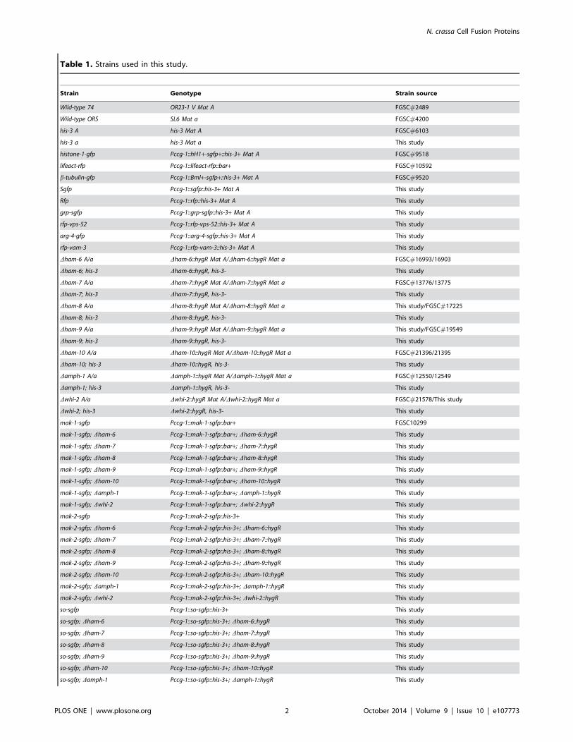
Table 1. Strains used in this study.
Strain Genotype Strain source
Wild-type 74 OR23-1 V Mat A FGSC#2489
Wild-type ORS SL6 Mat a FGSC#4200
his-3 A his-3 Mat A FGSC#6103
his-3 a his-3 Mat a This study
histone-1-gfp Pccg-1::hH1+-sgfp+::his-3+ Mat A FGSC#9518
lifeact-rfp Pccg-1::lifeact-rfp::bar+ FGSC#10592
b-tubulin-gfp Pccg-1::Bml+-sgfp+::his-3+ Mat A FGSC#9520
Sgfp Pccg-1::sgfp::his-3+ Mat A This study
Rfp Pccg-1::rfp::his-3+ Mat A This study
grp-sgfp Pccg-1::grp-sgfp::his-3+ Mat A This study
rfp-vps-52 Pccg-1::rfp-vps-52::his-3+ Mat A This study
arg-4-gfp Pccg-1::arg-4-sgfp::his-3+ Mat A This study
rfp-vam-3 Pccg-1::rfp-vam-3::his-3+ Mat A This study
Dham-6 A/a Dham-6::hygR Mat A/Dham-6::hygR Mat a FGSC#16993/16903
Dham-6; his-3 Dham-6::hygR, his-3- This study
Dham-7 A/a Dham-7::hygR Mat A/Dham-7::hygR Mat a FGSC#13776/13775
Dham-7; his-3 Dham-7::hygR, his-3- This study
Dham-8 A/a Dham-8::hygR Mat A/Dham-8::hygR Mat a This study/FGSC#17225
Dham-8; his-3 Dham-8::hygR, his-3- This study
Dham-9 A/a Dham-9::hygR Mat A/Dham-9::hygR Mat a This study/FGSC#19549
Dham-9; his-3 Dham-9::hygR, his-3- This study
Dham-10 A/a Dham-10::hygR Mat A/Dham-10::hygR Mat a FGSC#21396/21395
Dham-10; his-3 Dham-10::hygR, his-3- This study
Damph-1 A/a Damph-1::hygR Mat A/Damph-1::hygR Mat a FGSC#12550/12549
Damph-1; his-3 Damph-1::hygR, his-3- This study
Dwhi-2 A/a Dwhi-2::hygR Mat A/Dwhi-2::hygR Mat a FGSC#21578/This study
Dwhi-2; his-3 Dwhi-2::hygR, his-3- This study
mak-1-sgfp Pccg-1::mak-1-sgfp::bar+ FGSC10299
mak-1-sgfp; Dham-6 Pccg-1::mak-1-sgfp::bar+; Dham-6::hygR This study
mak-1-sgfp; Dham-7 Pccg-1::mak-1-sgfp::bar+; Dham-7::hygR This study
mak-1-sgfp; Dham-8 Pccg-1::mak-1-sgfp::bar+; Dham-8::hygR This study
mak-1-sgfp; Dham-9 Pccg-1::mak-1-sgfp::bar+; Dham-9::hygR This study
mak-1-sgfp; Dham-10 Pccg-1::mak-1-sgfp::bar+; Dham-10::hygR This study
mak-1-sgfp; Damph-1 Pccg-1::mak-1-sgfp::bar+; Damph-1::hygR This study
mak-1-sgfp; Dwhi-2 Pccg-1::mak-1-sgfp::bar+; Dwhi-2::hygR This study
mak-2-sgfp Pccg-1::mak-2-sgfp::his-3+ This study
mak-2-sgfp; Dham-6 Pccg-1::mak-2-sgfp::his-3+; Dham-6::hygR This study
mak-2-sgfp; Dham-7 Pccg-1::mak-2-sgfp::his-3+; Dham-7::hygR This study
mak-2-sgfp; Dham-8 Pccg-1::mak-2-sgfp::his-3+; Dham-8::hygR This study
mak-2-sgfp; Dham-9 Pccg-1::mak-2-sgfp::his-3+; Dham-9::hygR This study
mak-2-sgfp; Dham-10 Pccg-1::mak-2-sgfp::his-3+; Dham-10::hygR This study
mak-2-sgfp; Damph-1 Pccg-1::mak-2-sgfp::his-3+; Damph-1::hygR This study
mak-2-sgfp; Dwhi-2 Pccg-1::mak-2-sgfp::his-3+; Dwhi-2::hygR This study
so-sgfp Pccg-1::so-sgfp::his-3+ This study
so-sgfp; Dham-6 Pccg-1::so-sgfp::his-3+; Dham-6::hygR This study
so-sgfp; Dham-7 Pccg-1::so-sgfp::his-3+; Dham-7::hygR This study
so-sgfp; Dham-8 Pccg-1::so-sgfp::his-3+; Dham-8::hygR This study
so-sgfp; Dham-9 Pccg-1::so-sgfp::his-3+; Dham-9::hygR This study
so-sgfp; Dham-10 Pccg-1::so-sgfp::his-3+; Dham-10::hygR This study
so-sgfp; Damph-1 Pccg-1::so-sgfp::his-3+; Damph-1::hygR This study
N. crassa Cell Fusion Proteins
PLOS ONE | www.plosone.org 2 October 2014 | Volume 9 | Issue 10 | e107773
and MEK-2, the upstream MAPKKK and MAPKK in MAK-2
pathway, were also found to have the oscillatory signaling behavior
during cell fusion [24]. The N. crassa MAK-1 CWI pathway
initiates through a set of transmembrane sensors. Signals are
integrated by the small GTPase RHO1, which activates a
conserved mitogen-activated protein kinase (MAPK) cascade
through its interaction with protein kinase C [29–32]. In addition
to its function in cell wall stress integration, the CWI pathway is
also a central component of the cell-cell communication machin-
ery. The functional relationship between the two signaling
pathways during intercellular communication is poorly under-
stood, but evidence for cross-talk between MAK-1 and MAK-2 is
provided by work on the striatin interacting phosphatase and
kinase (STRIPAK) complex [33,34]. Its subunits, HAM-2, HAM-
3, HAM-4, MOB-3, PP2A and PPG-1, are all required for cell-cell
communication [9,13,35,36]. Phosphorylation of MOB-3 by
MAK-2 is required for nuclear localization of MAK-1 in
vegetative hyphae, suggesting that MAK-1-dependent expression
of cell fusion genes may be required for establishing cell-cell
communication competence.
Among the identified cell fusion genes, there were six genes
whose functions were largely uncharacterized. To better under-
stand how cell-to-cell fusion is regulated, these six genes, ham-6,ham-8, ham-9, ham-10, amph-1, and whi-2 have been further
characterized. The expression patterns, intracellular locations, and
how the loss of these genes affects the activation status of the
MAK-1 and MAK-2 pathway were examined. A seventh gene,
ham-7, which has been shown to function as a sensor for the
MAK-1 pathway [31], was included in the analysis to examine the
cell type expression pattern and cellular location of the HAM-7
sensor. In further characterizing these genes, we demonstrate here
that HAM-6, HAM-7 and HAM-8 function as upstream elements
in the pathway regulating MAK-1 kinase activity during cell
fusion. We further demonstrate that HAM-9, HAM-10, AMPH-1
and WHI-2 are proteins with general functions in regulating N.crassa growth, and we suggest that HAM-9, HAM-10, and WHI-2
may provide cross-talk between the two MAP kinase pathways and
other signaling cascades during cell fusion.
Materials and Methods
Strains, media and growth conditionsThe strains used in this study are listed in Table 1. Wild type A
(FGSC#2489), wild type a (FGSC#4200), his-3 A (FGSC#6103),
histone-1-gfp (FGSC#9518) [37], lifeact-rfp (FGSC#10592) [38],
b-tubulin-gfp (FGSC#9520) [37], and mak-1-gfp (FGSC#10299)
[39] were obtained from Fungal Genetics Stock Center (Kansas
City, MO). The Dham-6 (NCU02767), Dham-7 (NCU00881),
Dham-8 (NCU02811), Dham-9 (NCU07389), Dham-10(NCU02833), Damph-1 (NCU01069) and Dwhi-2(NCU10518)
strains were obtained from the FGSC N. crassa single gene
deletion mutant library [40]. All other strains used in this study
were either obtained through transformation experiments or
mating. The presence of the gene deletion in all of the deletion
mutant strains was verified by PCR. The growth media and
growth condition for regular strain maintenance, mating, and for
conidial anastomosis tube (CAT) formation are available through
the FGSC website (www.fgsc.net). The screening procedure to
identify cell fusion mutants was performed as previously described
[9]. Deletion mutant isolates were further characterized by co-
segregation experiments to assess whether the gene deletion
(marked by the presence of the hygromycin resistance gene
cassette) was responsible for the mutant phenotype [9,40].
Deletion mutants showing co-segregation of hygromycin resistance
with the cell fusion mutant phenotypes were verified by
complementation experiments.
Plasmid construction and expression of tagged proteinsPlasmids used in this study are listed in Table S1. The primers
used to construct GFP-tagged, RFP-tagged and HA-tagged
protein constructs are listed in Table S2. GFP and dsRed RFP
fusion proteins were generated using the pMF272 and pMF334
vectors [37,41] and were expressed under the control of ccg-1promoter at the his-3 locus [42].
HA-tagged proteins (HA-HAM-6, HA-HAM-8, HA-HAM-9,
HA-AMPH-1 and HA-WHI-2) were cloned using the vectors
pBM60 and pBM61 [43–45] and were expressed under the
control of their own promoters at the his-3 locus. To identify sites
for HA tagging, the protein sequences for HAM-6, HAM-8,
HAM-9, AMPH-1 and WHI-2 were used to generate multiple
sequence alignments with homologous proteins from other fungi
[46]. Protein sequences were also analyzed by the online program
Globplot to predict protein structural information [47]. Non-
conserved protein sequence regions, which were predicted to be
exposed, were chosen for HA tagging. For ham-6, ham-8 and ham-9, the HA tag coding sequence was inserted immediately before
the stop codon. For amph-1, the HA tag coding sequence was
Table 1. Cont.
Strain Genotype Strain source
so-sgfp; Dwhi-2 Pccg-1::so-sgfp::his-3+; Dwhi-2::hygR This study
HA-ham-6; Dham-6 Pham-6::HA-ham-6::his-3+; Dham-6::hygR This study
HA-ham-7; Dham-7 Pham-7::HA-ham-7::his-3+; Dham-7::hygR This study
HA-ham-8; Dham-8 Pham-8::HA-ham-8::his-3+; Dham-8::hygR This study
HA-ham-9; Dham-9 Pham-9::HA-ham-9::his-3+; Dham-9::hygR This study
HA-amph-1; Damph-1 Pamph-1::HA-amph-1::his-3+; Damph-1::hygR This study
HA-whi-2; Dwhi-2 Pwhi-2::HA-whi-2::his-3+; Dwhi-2::hygR This study
ham-8-sgfp; Dham-8 Pccg-1::ham-8-sgfp::his-3+; Dham-8::hygR This study
rfp-ham-8; Dham-8 Pccg-1::rfp-ham-8::his-3+; Dham-8::hygR This study
rfp-ham-10; Dham-10 Pccg-1::rfp-ham-10::his-3+; Dham-10::hygR This study
rfp-amph-1; Damph-1 Pccg-1::rfp-amph-1::his-3+; Damph-1::hygR This study
doi:10.1371/journal.pone.0107773.t001
N. crassa Cell Fusion Proteins
PLOS ONE | www.plosone.org 3 October 2014 | Volume 9 | Issue 10 | e107773

inserted between the third and the fourth amino acid codons. For
whi-2, the peptide sequence VPVDPASGA (amino acid 118–126)
was replaced with the HA tag. The cloning of these HA-tagged
protein constructs involved two PCR amplification steps. PCR
primers were used to amplify approximately 1,500 bp of 59 UTR
sequence, the coding sequence upstream of the HA tag insertion
site, and the HA tag coding sequence. A second set of primers
were used to amplify the HA tag coding sequence, the rest of the
coding sequence of the gene, and the approximately 500 bp 39
UTR sequence. The two PCR products were then mixed together
and used as templates to amplify the entire HA-tagged gene. The
primers designed for the two ends of the genes contained an added
restriction enzyme site to allow the insertion of the amplified DNA
into the multicloning sites of pBM60 or pBM61. An HA-tagged
version of HAM-7 with its endogenous promoter was generated by
Retrogen Inc. (San Diego, CA). The DNA sequence encoding
amino acids 190–198 (YTINILESG), which are located immedi-
ately in front of the GPI anchor addition site, were replaced by the
HA tag sequence (YPYDVPDYA) in the HA-tagged HAM-7.
Plasmids pgrp-GFP, pRFP-vps-52, parg-4-GFP and pRFP-vam-
3 were obtained from the FGSC as fluorescent protein markers for
ER, Golgi, mitochondria, and vacuoles respectively [48], and used
in co-localization experiments. Plasmids pso-GFP and pmak-2-
GFP were kind gifts from Dr. Louise Glass’s lab, and were used to
study the MAK-2 signal transduction pathway [23].
Analysis of HA-tagged protein expressionTo examine the expression of HA-tagged proteins in vegetative
hyphae, cultures were grown in 100 ml liquid Vogel’s sucrose
medium in shaking Erlenmeyer flasks at room temperature for 36
to 48 hours. To examine the expression of HA-tagged proteins in
germ tubes and CATs, conidia were used to inoculate 16 ml of
Vogel’s sucrose medium at a titer of 106 per ml and grown in
100615 mm Petri dishes at 34uC without agitation for 4 hours to
allow the formation of germ tubes and CATs [8]. Cells were
harvested by filtration using a Buchner funnel and ground in liquid
nitrogen. Protein extraction buffer [100 mM Tris/HCL pH 7.4,
1% (w/v) SDS; supplemented with 1X protease cocktail (P-
8340 Sigma-Aldrich, St. Louis, MO)] was added, and the cell
extracts were collected after centrifugation.
For Western blot analysis of HA-tagged proteins, the protein
concentration of the cell extracts was determined by using the DC
protein assay kit (BioRad, Hercules, CA). Samples containing
60 mg of protein were subjected to SDS-PAGE and transferred to
nitrocellulose membrane. The nitrocellulose membranes were
then subjected to Ponceau S red (Sigma-Aldrich, St. Louis, MO)
staining to verify equal loading of different protein samples.
Western blot experiments with mouse monoclonal anti-HA
(Covance, Princeton, NJ) and rabbit anti-mouse IgG-HRP
(Sigma-Aldrich, St. Louis, MO) were used to assess the level of
protein expression. Chemiluminescent signal was detected by
using a ChemiDoc XRS+ System and images were analyzed with
Image Lab Software (BioRad, Hercules, CA).
Evaluation of MAK-1 and MAK-2 phosphorylation statusPolyclonal antibody directed against the phosphorylated acti-
vation site in common on MAK-1 and MAK-2 was used to
evaluate their activation status. To examine the status of the
pathways in vegetative hyphae, liquid N. crassa cultures were
grown at room temperature and harvested by filtration using a
Buchner funnel [31]. In some experiments, vegetative cultures
were subjected to 8 mM H2O2 for 10 minutes just prior to being
harvested to determine if the pathways could be activated by
oxidative stress. To study the MAK-1 and MAK-2 phosphoryla-
tion status in germ tubes and CATs, conidia were grown for four
hours under the conditions described above to allow germ tube
and CAT formation. Germlings were harvested by gently scraping
them off the culture dishes and collected on a Buchner funnel. The
harvested vegetative hyphae samples and germ tubes/CATs
samples were ground to a fine powder in a mortar and pestle in
liquid nitrogen. Protein extraction for the analysis of the MAK-1
and MAK-2 phosphorylation status was performed as described by
Maddi et al. [31].
Preparation of cells expressing HA-tagged proteins forimmunolocalizationFor each HA-tagged strain, six Petri dishes containing 16 ml of
conidial suspension (106 conidia/ml) were grown at 34uC for 4
hours to allow germ tube and CAT formation. Cells were
harvested by gently scraping them off Petri dishes, and were
collected by centrifugation at 60006g for 5 minutes. Cells were
transferred into a microcentrifuge tube and fixed in PBS
(phosphate buffered saline) with 3.7% formaldehyde for 15 min-
utes. After washing with PBS, the samples were incubated for
30 minutes in PBS containing 1 mg/ml of Novozyme 234
(InterSpex Products Inc., Foster City, CA) and 1% bovine serum
albumin to digest the cell wall [49,50]. Following the cell wall
digestion step, the cells were collected by centrifugation and
washed with PBS. Cell samples were then incubated in
permeabilization buffer (PBS with 1% BSA and 0.5% Triton-X
100) for 5 minutes to permeabilize the membrane. After the
membrane permeabilization step, the cells were collected and
washed with PBS. Cell samples were then incubated overnight at
4uC in mouse monoclonal anti-HA primary antibody (Covance,
Princeton, NJ) used at a 1:200 dilution in PBS with 1% BSA. After
the primary antibody incubation, samples were washed in PBS
three times and then incubated for 2 hours in Alexa Fluor 488-
conjugated goat anti-mouse secondary antibody (Life Technolo-
gies, Carlsbad, CA) used at a 1:100 dilution in PBS with 1% BSA.
After three washes, cell samples were used for microscopic
observation. All of the incubation buffers and washing buffers
used in this assay were supplemented with 1X protease inhibitor
cocktail and 1 mM PMSF (Sigma Aldrich, St. Louis, MO).
Live cell imaging for the quantification of CATsLive-cell imaging was performed using an inverted microscope
Diaphot-TMD inverted microscope (Nikon, Japan) to quantify
CAT fusion activity for wild type and mutant strains as previously
described [8,51]. In brief, 1 ml of fresh conidia at a density of 106
per ml were grown in Petri dishes (35610 mm) at 34uC. Benomyl
was added to distinguish CATs from germ tubes. This is because
benomyl inhibits the formation of long germ tubes but not CAT
formation or fusion [52,53]. After 4 hours of incubation, the cells
were examined under the microscope for the presence of CATs
and CAT fusion. For each sample, five random images were
captured for later quantification of CAT fusion activity. The
frequency of cell fusion observed in the mutant samples was
compared to the wild type cell fusion frequency to obtain a relative
CAT fusion activity for each of the mutants.
Sample preparation and confocal microscopyFor live-cell imaging of CAT formation, an inverted agar block
method was adapted for this study [54]. A 1 ml aliquot of conidia
expressing GFP-tagged protein or RFP-tagged protein (106
conidia per ml) was placed in a fluorodish (World Precision
Instruments, FL). A thick agar block was then placed in the middle
of the fluorodish to facilitate attachment of the conidia to the
N. crassa Cell Fusion Proteins
PLOS ONE | www.plosone.org 4 October 2014 | Volume 9 | Issue 10 | e107773
surface of the cover glass bottom. Benomyl was added in both the
liquid medium and the agar block to inhibit germ tube formation.
The cells were observed between 3 and 6 hours to follow the
formation of CATs.
For imaging of germ tubes and hyphal cells expressing
fluorescent protein constructs, conidia (105 conidia per ml) were
grown in Vogel’s sucrose liquid medium for 6 to 8 hours. Cell
samples were placed on slides for immediate microscopic
observation.
To examine the nuclear localization of MAK-1-GFP and
MAK-2-GFP in germ tubes/CATs, conidia (106 per ml) were
grown in Vogel’s sucrose liquid medium at 34uC for 4 hours to
allow conidia germination and CAT formation. Cell samples were
then collected, fixed, digested with Novozyme, and permeabilized
following the procedure described above. After membrane
permeabilization, cells were treated with 1 mg/ml RNAase for
30 minutes at room temperature to digest RNA. Cells were then
washed once with PBS and stained with propidium iodide (5 mg/ml propidium iodide in PBS) for 20 minutes [55]. After three
washing steps, cells were placed on slides for microscopic
observation.
Confocal laser scanning microscopy was performed using a
Zeiss LSM 710 Confocal Microscope (Carl Zeiss, Inc., Thorn-
wood, NY). Plan-Apochromat 636/1.40 Oil DIC M27 objective
or Plan-Apochromat 406/1.3 Oil DIC M27 objective lenses were
used for imaging. Excitation wavelength and detection wavelength
were set up either according to references [37,41,56] or by using
the smart setup program of the ZEN2012 image capture software.
GFP images were collected at 493 to 598 nm with excitation at
488 nm. RFP images were collected at .570 nm with excitation
at 558 nm. When imaging samples with both GFP and RFP
signals, the images were collected at 493 to 539 nm and at 554 to
703 nm with excitation at 490 nm and 514 nm. When examining
GFP protein samples that have been stained with propidium
iodide, the images were collected at 499 to 560 nm and at 572 to
719 nm with excitation at 488 nm and 535 nm. The images were
collected sequentially using either a line sequential scanning mode
or plane scanning mode. Bright-field images were captured with a
transmitted light detector. Time-lapse imaging was performed to
evaluate Ping-Pong signaling at time intervals of 10 s to 60 s for
periods up to 10 minutes. Images were analyzed with image
processing software ZEN lite and Image J.
For the detection of HA-tagged proteins, anti-HA primary
antibody was used in conjunction with Alexa Fluor 488 conjugated
secondary antibody. Immunofluorescent images were collected at
497 to 622 nm with excitation at 488 nm.
Results
Characterization of cell fusion mutants reveals twofunctional mutant groupsA screening of plates 110 to 120 from the single gene deletion
library identified a new cell fusion gene, whi-2 (NCU10518),
which is a predicted homolog of yeast stress response factor protein
Whi2p [57,58]. We confirmed that the cell-fusion defect and
hygromycin resistance co-segregated. Moreover, ectopic expres-
sion of HA-WHI-2 at the his-3 locus complemented the Dwhi-2mutant phenotypes (Figure 1 and 2), demonstrating that the loss of
whi-2 was responsible for the mutant defects. An examination of
the conidial morphology of Dwhi-2 revealed a conidial phenotype
similar to that of the Dham-10 and Damph-1 mutants (Figure S1).
Instead of making mature macroconidia, Dham-10, Damph-1 and
Dwhi-2 produced chains of macroconidia that stopped develop-
ment at the major constriction stage (Figure S1) [59]. Dham-10
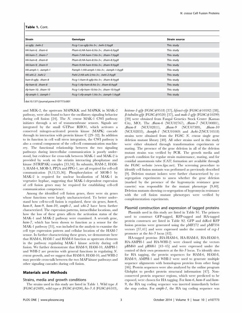
Figure 1. Strains used in this study. Slants containing Vogel’s sucrose medium were inoculated with different strain isolates and grown for 4days. Strains shown in the top panel from left to right include wild type (WT), Dham-6, Dham-6 transformed with HA-ham-6, Dham-7, Dham-7transformed with HA-ham-7, Dham-8, Dham-8 transformed with HA-ham-8, Dham-8 transformed with ham-8-GFP, and Dham-8 transformed with RFP-ham-8. The bottom panel shows Dham-9, Dham-9 transformed with HA-ham-9, Dham-10, Dham-10 transformed with RFP-ham-10, Damph-1, Damph-1 transformed with HA-amph-1, Damph-1 transformed with RFP-amph-1, Dwhi-2, and Dwhi-2 transformed with HA-whi-2.doi:10.1371/journal.pone.0107773.g001
N. crassa Cell Fusion Proteins
PLOS ONE | www.plosone.org 5 October 2014 | Volume 9 | Issue 10 | e107773

produced fewer conidia than Damph-1 and Dwhi-2. In contrast,
Dham-6, Dham-7, Dham-8 and Dham-9 generated normal
macroconidia (Figure S1).
All of the cell fusion mutants had a flat conidiation phenotype,
which is due to a defect in the generation of long aerial hyphae
(Figure 1). The phenotypic differences between the Dham-6,Dham-7, Dham-8 and Dham-9 group of mutants, and the Dham-10, Damph-1 and Dwhi-2 mutants suggest that there are
functional differences between the proteins encoded by these two
groups of cell fusion genes.
HAM-6, HAM-7 and HAM-8 are specifically expressed ingerm tubes/CATsTo examine the cell type expression pattern for these cell fusion
proteins, we generated HA-tagged versions that were expressed at
the his-3 locus under the control of their own promoters. The HA-
tagged proteins fully rescued the mutant developmental and CAT
fusion defects of Dham-6, Dham-7, Dham-8, Dham-9 and Dwhi-2(Figures 1 and 2) [9]. The HA-tagged version of AMPH-1
provided only a partial rescue of Damph-1 (32.3% of the wild
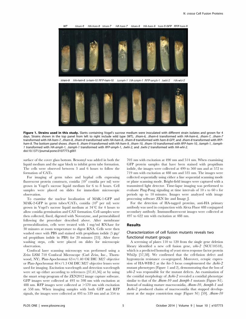
type cell fusion level) (Figures 1 and 2). Western blot analysis was
performed to examine the size and expression patterns of the HA-
tagged proteins (Figure 3). The predicted MW (molecular weight)
for HAM-6, HAM-7, HAM-8, HAM-9, AMPH-1 and WHI-2 are
15.9 KD, 24.4 KD, 57.7 KD, 96.7 KD, 29.9 KD and 32.2 KD
respectively. HAM-9, AMPH-1 and WHI-2 are predicted to be
cytosolic proteins, and their HA-tagged proteins gave MWs very
close to the predicted MWs. HAM-6 and HAM-8 contain three
and four predicted TM (transmembrane) domains respectively,
and the measured MWs of their HA-tagged proteins were also
very close to their predicted MWs. HAM-7 has been shown to be a
GPI-anchored cell wall protein [31]. The measured MW for HA-
HAM-7 was 42 KD, 18 KD larger than the predicted MW, which
suggests the GPI-anchored cell wall protein is heavily glycosylated.
HA-HAM-6 and HA-HAM-8 displayed a germ tubes/CATs-
specific expression pattern, with only a trace amount of expression
in vegetative hyphae (Figure 3). HA-HAM-7 was expressed at a
very high level in germ tubes and CATs, and at a 5-fold reduced
level in vegetative hyphae. In contrast, we determined that HA-
HAM-9 and HA-WHI-2 were expressed at about equal level in the
germ tubes/CATs and hyphae samples, while HA-AMPH-1 was
expressed at higher level in hyphae than in germ tubes/CATs
(Figure 3). In summary, these expression experiments support our
phenotypic classification of the mutants, and suggest that HAM-6,
HAM-7, and HAM-8 form a group of proteins that primarily
functions in germlings and during CAT fusion, while HAM-9,
WHI-2 and AMPH-1 have general functions during growth and
differentiation.
HAM-7 and HAM-8 are found in a punctate pattern nearthe tips of germ tubes and CATsIn order to determine the location of the cell fusion proteins, we
expressed them as GFP- and dsRed RFP-tagged constructs under
the control of the ccg-1 promoter in their respective mutant
backgrounds (Figure 1). The HAM-9-GFP, RFP-HAM-9, HAM-
10-GFP and AMPH-1-GFP fusion proteins failed to rescue the
mutant phenotypes. We were unable to detect the GFP and RFP
signals from these tagged proteins, suggesting that the tagged
proteins were rapidly degraded. HAM-8-GFP and RFP-HAM-8
rescued the Dham-8 conidiation defects, but failed to restore CAT
Figure 2. Complementation of CAT fusion activities by different HA, GFP and RFP tagged proteins. A) The levels of CAT fusion activityfor the gene deletion mutants and for transformants expressing a tagged version of the deleted gene are shown as a percentile of the cell fusionactivity for wild type CATs. B) Photograph of CAT fusion activities in wild type (WT), Dham-8, and Dham-8 transformed with HA-ham-8. Arrows pointto examples of CAT fusion in the wild-type and Dham-8 transformed with HA-ham-8 panels.doi:10.1371/journal.pone.0107773.g002
N. crassa Cell Fusion Proteins
PLOS ONE | www.plosone.org 6 October 2014 | Volume 9 | Issue 10 | e107773
fusion activity, indicating that the tagged HAM-8 proteins were
only partially functional (Figures 1 and 2). Moreover, some of the
GFP and RFP signals were detected at large vacuolar-like
structures, suggesting that the tagged HAM-8 proteins might have
been targeted to the vacuole for degradation, and may not reflect
the normal localization for HAM-8 (Figure S2A). Co-localization
experiments with marker proteins showed that HAM-8-GFP and
the vacuolar marker RFP-VAM-3 showed co-localization (Figure
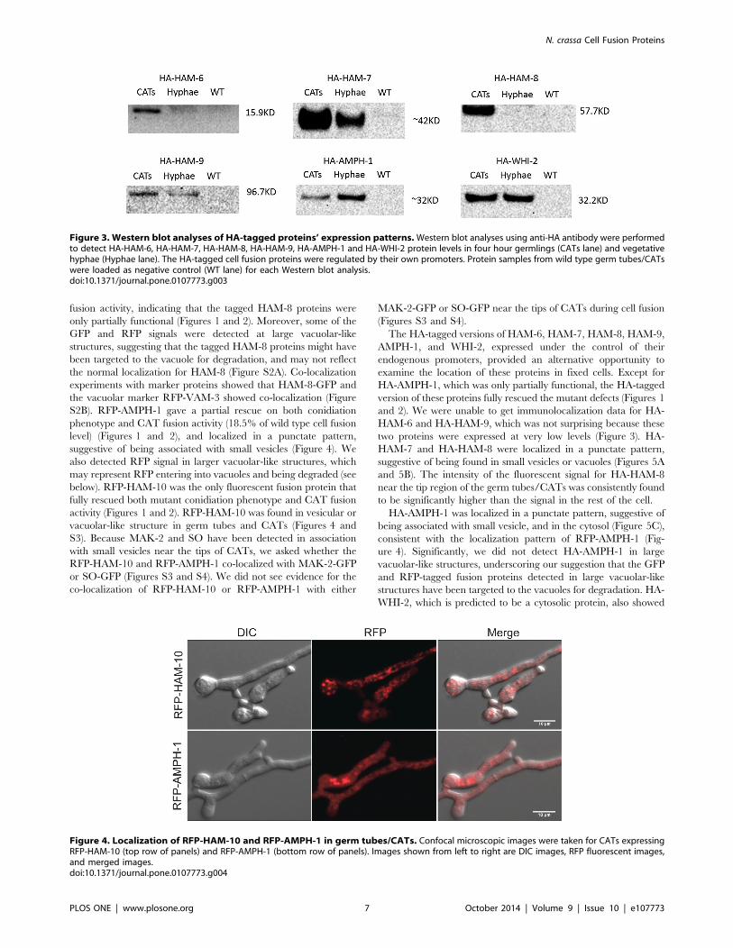
S2B). RFP-AMPH-1 gave a partial rescue on both conidiation
phenotype and CAT fusion activity (18.5% of wild type cell fusion
level) (Figures 1 and 2), and localized in a punctate pattern,
suggestive of being associated with small vesicles (Figure 4). We
also detected RFP signal in larger vacuolar-like structures, which
may represent RFP entering into vacuoles and being degraded (see
below). RFP-HAM-10 was the only fluorescent fusion protein that
fully rescued both mutant conidiation phenotype and CAT fusion
activity (Figures 1 and 2). RFP-HAM-10 was found in vesicular or
vacuolar-like structure in germ tubes and CATs (Figures 4 and
S3). Because MAK-2 and SO have been detected in association
with small vesicles near the tips of CATs, we asked whether the
RFP-HAM-10 and RFP-AMPH-1 co-localized with MAK-2-GFP
or SO-GFP (Figures S3 and S4). We did not see evidence for the
co-localization of RFP-HAM-10 or RFP-AMPH-1 with either
MAK-2-GFP or SO-GFP near the tips of CATs during cell fusion
(Figures S3 and S4).
The HA-tagged versions of HAM-6, HAM-7, HAM-8, HAM-9,
AMPH-1, and WHI-2, expressed under the control of their
endogenous promoters, provided an alternative opportunity to
examine the location of these proteins in fixed cells. Except for
HA-AMPH-1, which was only partially functional, the HA-tagged
version of these proteins fully rescued the mutant defects (Figures 1
and 2). We were unable to get immunolocalization data for HA-
HAM-6 and HA-HAM-9, which was not surprising because these
two proteins were expressed at very low levels (Figure 3). HA-
HAM-7 and HA-HAM-8 were localized in a punctate pattern,
suggestive of being found in small vesicles or vacuoles (Figures 5A
and 5B). The intensity of the fluorescent signal for HA-HAM-8
near the tip region of the germ tubes/CATs was consistently found
to be significantly higher than the signal in the rest of the cell.
HA-AMPH-1 was localized in a punctate pattern, suggestive of
being associated with small vesicle, and in the cytosol (Figure 5C),
consistent with the localization pattern of RFP-AMPH-1 (Fig-
ure 4). Significantly, we did not detect HA-AMPH-1 in large
vacuolar-like structures, underscoring our suggestion that the GFP
and RFP-tagged fusion proteins detected in large vacuolar-like
structures have been targeted to the vacuoles for degradation. HA-
WHI-2, which is predicted to be a cytosolic protein, also showed
Figure 3. Western blot analyses of HA-tagged proteins’ expression patterns.Western blot analyses using anti-HA antibody were performedto detect HA-HAM-6, HA-HAM-7, HA-HAM-8, HA-HAM-9, HA-AMPH-1 and HA-WHI-2 protein levels in four hour germlings (CATs lane) and vegetativehyphae (Hyphae lane). The HA-tagged cell fusion proteins were regulated by their own promoters. Protein samples from wild type germ tubes/CATswere loaded as negative control (WT lane) for each Western blot analysis.doi:10.1371/journal.pone.0107773.g003
Figure 4. Localization of RFP-HAM-10 and RFP-AMPH-1 in germ tubes/CATs. Confocal microscopic images were taken for CATs expressingRFP-HAM-10 (top row of panels) and RFP-AMPH-1 (bottom row of panels). Images shown from left to right are DIC images, RFP fluorescent images,and merged images.doi:10.1371/journal.pone.0107773.g004
N. crassa Cell Fusion Proteins
PLOS ONE | www.plosone.org 7 October 2014 | Volume 9 | Issue 10 | e107773
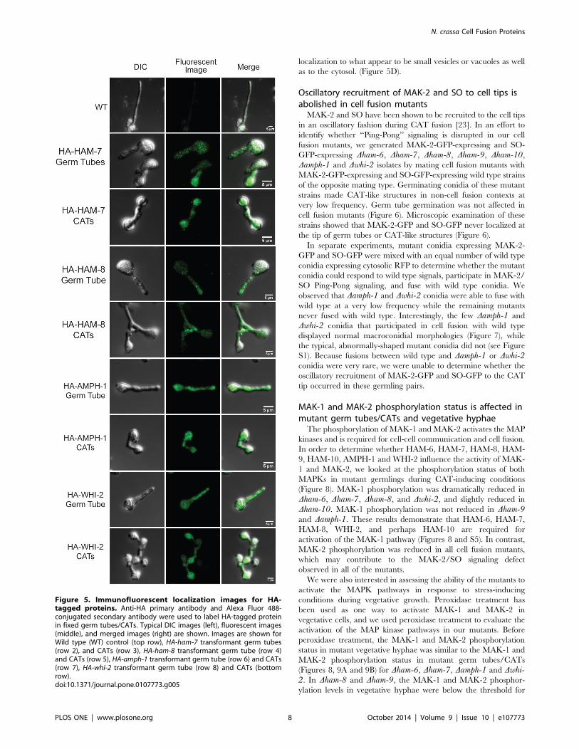
localization to what appear to be small vesicles or vacuoles as well
as to the cytosol. (Figure 5D).
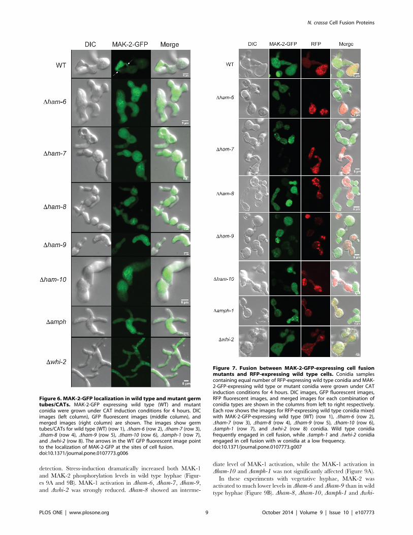
Oscillatory recruitment of MAK-2 and SO to cell tips isabolished in cell fusion mutantsMAK-2 and SO have been shown to be recruited to the cell tips
in an oscillatory fashion during CAT fusion [23]. In an effort to
identify whether ‘‘Ping-Pong’’ signaling is disrupted in our cell
fusion mutants, we generated MAK-2-GFP-expressing and SO-
GFP-expressing Dham-6, Dham-7, Dham-8, Dham-9, Dham-10,Damph-1 and Dwhi-2 isolates by mating cell fusion mutants with
MAK-2-GFP-expressing and SO-GFP-expressing wild type strains
of the opposite mating type. Germinating conidia of these mutant
strains made CAT-like structures in non-cell fusion contexts at
very low frequency. Germ tube germination was not affected in
cell fusion mutants (Figure 6). Microscopic examination of these
strains showed that MAK-2-GFP and SO-GFP never localized at
the tip of germ tubes or CAT-like structures (Figure 6).
In separate experiments, mutant conidia expressing MAK-2-
GFP and SO-GFP were mixed with an equal number of wild type
conidia expressing cytosolic RFP to determine whether the mutant
conidia could respond to wild type signals, participate in MAK-2/
SO Ping-Pong signaling, and fuse with wild type conidia. We
observed that Damph-1 and Dwhi-2 conidia were able to fuse with
wild type at a very low frequency while the remaining mutants
never fused with wild type. Interestingly, the few Damph-1 and
Dwhi-2 conidia that participated in cell fusion with wild type
displayed normal macroconidial morphologies (Figure 7), while
the typical, abnormally-shaped mutant conidia did not (see Figure
S1). Because fusions between wild type and Damph-1 or Dwhi-2conidia were very rare, we were unable to determine whether the
oscillatory recruitment of MAK-2-GFP and SO-GFP to the CAT
tip occurred in these germling pairs.
MAK-1 and MAK-2 phosphorylation status is affected inmutant germ tubes/CATs and vegetative hyphaeThe phosphorylation of MAK-1 and MAK-2 activates the MAP
kinases and is required for cell-cell communication and cell fusion.
In order to determine whether HAM-6, HAM-7, HAM-8, HAM-
9, HAM-10, AMPH-1 and WHI-2 influence the activity of MAK-
1 and MAK-2, we looked at the phosphorylation status of both
MAPKs in mutant germlings during CAT-inducing conditions
(Figure 8). MAK-1 phosphorylation was dramatically reduced in
Dham-6, Dham-7, Dham-8, and Dwhi-2, and slightly reduced in
Dham-10. MAK-1 phosphorylation was not reduced in Dham-9and Damph-1. These results demonstrate that HAM-6, HAM-7,
HAM-8, WHI-2, and perhaps HAM-10 are required for
activation of the MAK-1 pathway (Figures 8 and S5). In contrast,
MAK-2 phosphorylation was reduced in all cell fusion mutants,
which may contribute to the MAK-2/SO signaling defect
observed in all of the mutants.
We were also interested in assessing the ability of the mutants to
activate the MAPK pathways in response to stress-inducing
conditions during vegetative growth. Peroxidase treatment has
been used as one way to activate MAK-1 and MAK-2 in
vegetative cells, and we used peroxidase treatment to evaluate the
activation of the MAP kinase pathways in our mutants. Before
peroxidase treatment, the MAK-1 and MAK-2 phosphorylation
status in mutant vegetative hyphae was similar to the MAK-1 and
MAK-2 phosphorylation status in mutant germ tubes/CATs
(Figures 8, 9A and 9B) for Dham-6, Dham-7, Damph-1 and Dwhi-2. In Dham-8 and Dham-9, the MAK-1 and MAK-2 phosphor-
ylation levels in vegetative hyphae were below the threshold for
Figure 5. Immunofluorescent localization images for HA-tagged proteins. Anti-HA primary antibody and Alexa Fluor 488-conjugated secondary antibody were used to label HA-tagged proteinin fixed germ tubes/CATs. Typical DIC images (left), fluorescent images(middle), and merged images (right) are shown. Images are shown forWild type (WT) control (top row), HA-ham-7 transformant germ tubes(row 2), and CATs (row 3), HA-ham-8 transformant germ tube (row 4)and CATs (row 5), HA-amph-1 transformant germ tube (row 6) and CATs(row 7), HA-whi-2 transformant germ tube (row 8) and CATs (bottomrow).doi:10.1371/journal.pone.0107773.g005
N. crassa Cell Fusion Proteins
PLOS ONE | www.plosone.org 8 October 2014 | Volume 9 | Issue 10 | e107773
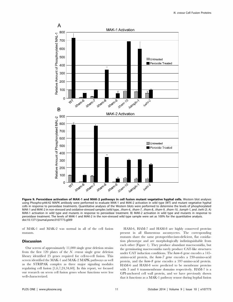
detection. Stress-induction dramatically increased both MAK-1
and MAK-2 phosphorylation levels in wild type hyphae (Figur-
es 9A and 9B). MAK-1 activation in Dham-6, Dham-7, Dham-9,and Dwhi-2 was strongly reduced. Dham-8 showed an interme-
diate level of MAK-1 activation, while the MAK-1 activation in
Dham-10 and Damph-1 was not significantly affected (Figure 9A).
In these experiments with vegetative hyphae, MAK-2 was
activated to much lower levels in Dham-6 and Dham-9 than in wild
type hyphae (Figure 9B). Dham-8, Dham-10, Damph-1 and Dwhi-
Figure 6. MAK-2-GFP localization in wild type andmutant germtubes/CATs. MAK-2-GFP expressing wild type (WT) and mutantconidia were grown under CAT induction conditions for 4 hours. DICimages (left column), GFP fluorescent images (middle column), andmerged images (right column) are shown. The images show germtubes/CATs for wild type (WT) (row 1), Dham-6 (row 2), Dham-7 (row 3),Dham-8 (row 4), Dham-9 (row 5), Dham-10 (row 6), Damph-1 (row 7),and Dwhi-2 (row 8). The arrows in the WT GFP fluorescent image pointto the localization of MAK-2-GFP at the sites of cell fusion.doi:10.1371/journal.pone.0107773.g006
Figure 7. Fusion between MAK-2-GFP-expressing cell fusionmutants and RFP-expressing wild type cells. Conidia samplescontaining equal number of RFP-expressing wild type conidia and MAK-2-GFP-expressing wild type or mutant conidia were grown under CATinduction conditions for 4 hours. DIC images, GFP fluorescent images,RFP fluorescent images, and merged images for each combination ofconidia types are shown in the columns from left to right respectively.Each row shows the images for RFP-expressing wild type conidia mixedwith MAK-2-GFP-expressing wild type (WT) (row 1), Dham-6 (row 2),Dham-7 (row 3), Dham-8 (row 4), Dham-9 (row 5), Dham-10 (row 6),Damph-1 (row 7), and Dwhi-2 (row 8) conidia. Wild type conidiafrequently engaged in cell fusion, while Damph-1 and Dwhi-2 conidiaengaged in cell fusion with w conidia at a low frequency.doi:10.1371/journal.pone.0107773.g007
N. crassa Cell Fusion Proteins
PLOS ONE | www.plosone.org 9 October 2014 | Volume 9 | Issue 10 | e107773

2 showed an intermediate level of MAK-2 activation, while MAK-
2 activation was normal in Dham-7 (Figure 9B). The differences in
MAK-1 and MAK-2 phosphorylation status between germ tubes/
CATs and vegetative hyphae for some of the mutants suggest that
there may be differences in how the two MAP kinase pathways are
being regulated during the various stages of the N. crassa life cycle.
MAK-1 and MAK-2 nuclear accumulation is normal inmutant germ tubes/CATsMutants of the STRIPAK complex have been shown to have a
defect in MAK-1 nuclear accumulation, which is regulated by
MAK-2 phosphorylation of MOB-3 [34]. MAK-2 nuclear
localization is also required during cell fusion [23]. In order to
examine if MAK-1 or MAK-2 nuclear accumulation is compro-
mised in the cell fusion mutants, propidium iodide was used to
label nuclei in MAK-1-GFP and MAK-2-GFP-expressing mutant
strains (Figures S6 and S7). We found that nuclear accumulation
Figure 8. MAK-1 and MAK-2 phosphorylation status in germ tubes/CATs. Western blot analysis using Phospho-p44/42 MAPK antibody wasperformed to determine MAK-1 and MAK-2 phosphorylation status in wild type (WT) and mutant (Dham-6, Dham-7, Dham-8, Dham-9, Dham-10,Damph-1, Dwhi-2, Dmik-1, and Dnrc-1) germ tubes/CATS. The positions of the phosphorylated MAK-1 (p-MAK-1) and phosphorylated MAK-2 (p-MAK-2) in the Western blot are noted in the left margin of the figure. B) The relative MAK-1 phosphorylation status in mutant germ tubes/CATs relative tothe MAK-1 phosphorylation status in wild type germ tubes/CATs (WT value is set at 100%). C) The relative MAK-2 phosphorylation status in mutantgerm tubes/CATs compared to the MAK-2 phosphorylation status in wild type germ tubes/CATs (WT value is set at 100%).doi:10.1371/journal.pone.0107773.g008
N. crassa Cell Fusion Proteins
PLOS ONE | www.plosone.org 10 October 2014 | Volume 9 | Issue 10 | e107773
of MAK-1 and MAK-2 was normal in all of the cell fusion
mutants.
Discussion
Our screen of approximately 11,000 single gene deletion strains
from the first 120 plates of the N. crassa single gene deletion
library identified 25 genes required for cell-to-cell fusion. This
screen identified the MAK-1 and MAK-2 MAPK pathways as well
as the STRIPAK complex as three major signaling modules
regulating cell fusion [1,6,7,24,34,60]. In this report, we focused
our research on seven cell fusion genes whose functions were less
well-characterized.
HAM-6, HAM-7 and HAM-8 are highly conserved proteins
present in all filamentous ascomycetes. The corresponding
mutants share the same protoperithecium-deficient, flat conidia-
tion phenotype and are morphologically indistinguishable from
each other (Figure 1). They produce abundant macroconidia, but
the germinating macroconidia rarely produce CAT-like structures
under CAT induction conditions. The ham-6 gene encodes a 145-
amino-acid protein, the ham-7 gene encodes a 230-amino-acid
protein, and the ham-8 gene encodes a 597-amino-acid protein.
HAM-6 and HAM-8 were predicted to be membrane proteins
with 3 and 4 transmembrane domains respectively. HAM-7 is a
GPI-anchored cell wall protein, and we have previously shown
that it functions as a MAK-1 pathway sensor during hyphal fusion
Figure 9. Peroxidase activation of MAK-1 and MAK-2 pathways in cell fusion mutant vegetative hyphal cells. Western blot analysesusing Phospho-p44/42 MAPK antibody were performed to evaluate MAK-1 and MAK-2 activation in wild type (WT) and mutant vegetative hyphalcells in response to peroxidase treatments. Quantitative analyses of the Western blots were performed to determine the levels of phosphorylatedMAK-1 and MAK-2 in non-stressed and oxidative-stressed samples (wild type, Dham-6, Dham-7, Dham-8, Dham-9, Dham-10, Damph-1, and Dwhi-2). A)MAK-1 activation in wild type and mutants in response to peroxidase treatment. B) MAK-2 activation in wild type and mutants in response toperoxidase treatment. The levels of MAK-1 and MAK-2 in the non-stressed wild type sample were set as 100% for the quantitative analysis.doi:10.1371/journal.pone.0107773.g009
N. crassa Cell Fusion Proteins
PLOS ONE | www.plosone.org 11 October 2014 | Volume 9 | Issue 10 | e107773
[31]. We found that the three proteins were expressed at much
higher levels in germ tubes/CATs than in vegetative hyphae
(Figure 3). HAM-7 and HAM-8 were found to be localized in
punctate pattern, suggestive of small vesicular or vacuolar
structures (Figures 5A and 5B). The HAM-8 containing structures
were found to be concentrated near the tip of the germlings and
CATs (Figure 5B). Although HAM-7 has been shown to be a GPI-
anchored cell wall protein, we did not see immunolocalization of
HAM-7 at the plasma membrane/cell wall boundary. We
attribute this to the heavily glycosylated status of HAM-7, which
could block to interaction between the glycosylated HAM-7 and
the antibody used for immunolocalization. The HAM-7 observed
in our localization studies (Figure 5A) may well represent newly
synthesized HAM-7 in transit through the secretory pathway that
hasn’t been fully glycosylated. Tip localization of MAK-2-GFP
and SO-GFP was missing in the few CAT-like structures formed
by these mutants and the mutants failed to fuse with wild type
conidia (Figures 6 and 7). We found that HAM-6, HAM-7 and
HAM-8 are required for MAK-1 kinase activation during conidial
germination and CAT formation (Figure 8). Despite the lower
levels of expression for HAM-6, HAM-7 and HAM-8 in vegetative
cells, MAK-1 phosphorylation was dramatically reduced in Dham-6, Dham-7, and Dham-8 during vegetative hyphal growth. Leeder
et al. [1] determined that the expression of the three genes is co-
regulated and controlled by the MAK-2 pathway-dependent
transcription factor PP-1. In summary, we propose that the GPI-
anchored cell wall HAM-7 and the two transmembrane proteins,
HAM-6 and HAM-8, function together to regulate the MAK-1
pathway. Given the cell wall/plasma membrane location for the
GPI-anchored protein HAM-7, we suggest that the three proteins
might participate in a signaling complex at the cell wall/plasma
membrane boundary, but our data would also be consistent with a
signaling complex localized to intracellular membranes.
The ham-9 gene encodes an 869-amino-acid protein containing
a SAM domain and two PH domains. The SAM domain has been
identified in yeast Ste11p (S. cerevisae homolog of N. crassa NRC-
1) [61], and the PH domains have been suggested to play a role in
targeting signal transduction proteins to intracellular membrane in
signaling events [62]. The C-terminal GFP-tagged and N-terminal
RFP-tagged HAM-9 fusion proteins were not functional, preclud-
ing any live-imaging analysis. HA-HAM-9 was expressed in both
vegetative hyphae and germ tubes/CATs (Figure 3), but its
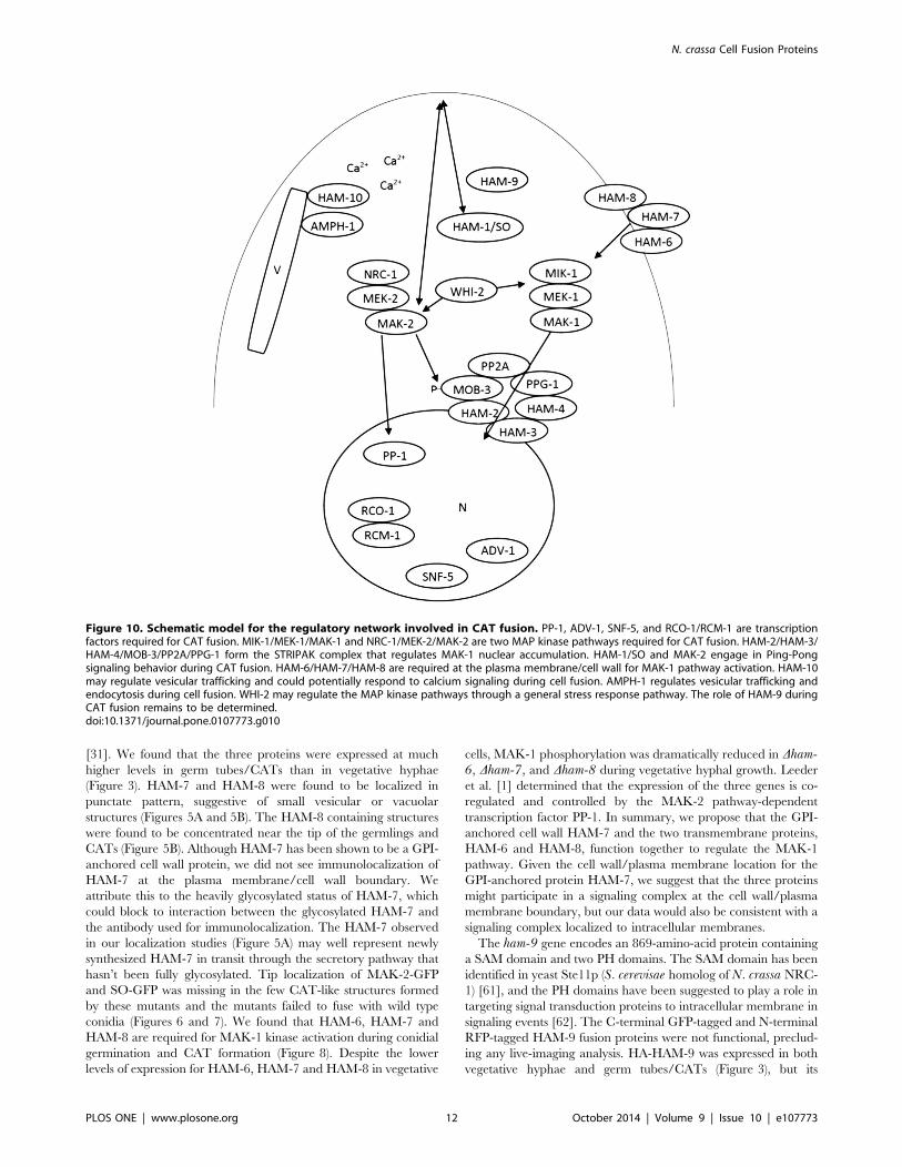
Figure 10. Schematic model for the regulatory network involved in CAT fusion. PP-1, ADV-1, SNF-5, and RCO-1/RCM-1 are transcriptionfactors required for CAT fusion. MIK-1/MEK-1/MAK-1 and NRC-1/MEK-2/MAK-2 are two MAP kinase pathways required for CAT fusion. HAM-2/HAM-3/HAM-4/MOB-3/PP2A/PPG-1 form the STRIPAK complex that regulates MAK-1 nuclear accumulation. HAM-1/SO and MAK-2 engage in Ping-Pongsignaling behavior during CAT fusion. HAM-6/HAM-7/HAM-8 are required at the plasma membrane/cell wall for MAK-1 pathway activation. HAM-10may regulate vesicular trafficking and could potentially respond to calcium signaling during cell fusion. AMPH-1 regulates vesicular trafficking andendocytosis during cell fusion. WHI-2 may regulate the MAP kinase pathways through a general stress response pathway. The role of HAM-9 duringCAT fusion remains to be determined.doi:10.1371/journal.pone.0107773.g010
N. crassa Cell Fusion Proteins
PLOS ONE | www.plosone.org 12 October 2014 | Volume 9 | Issue 10 | e107773
expression level was too low for immunolocalization. The
requirement of HAM-9 for both MAK-1 and MAK-2 activation
in vegetative hyphae (Figures 9A and 9B), may suggest that HAM-
9 regulates cross-communication of the two MAPK pathways
during vegetative growth.
The amph-1 gene encodes a 262-amino-acid protein containing
a bar domain, a domain frequently involved in protein-protein
interaction and regulation of membrane curvature [63]. N. crassaAMPH-1 is a homolog of the yeast Rvs161p and Rvs167p
proteins. Rvs161p and Rvs167p are required for endocytosis and
cell fusion during yeast mating [63,64]. HA-AMPH-1 and RFP-
AMPH-1 localized to small vesicles in the germ tubes/CATs, and
some of the vesicles appeared to be associated with the plasma
membrane (Figures 4 and 5C), suggesting that N. crassa AMPH-1
plays a role in vesicular trafficking and endocytosis. MAK-1 and
MAK-2 activity in Damph-1 germlings were similar to the wild
type control, indicating that AMPH-1 does not affect cell fusion by
regulating these pathways. This is consistent with our observation
that a few Damph-1 conidia having wild type morphology were
able to participate in cell fusion with wild type conidia (Figure 7).
HA-AMPH-1 was expressed in both vegetative hyphae and
germlings, suggesting it is a general factor required for all stages of
N. crassa life cycle. In summary, we suggest that AMPH-1
functions during vesicular trafficking and endocytosis.
The ham-10 gene encodes a 1,422-amino-acid protein contain-
ing a C2 domain near the C terminus. C2 domains function as
calcium-dependent lipid-binding domains and are thought to be
involved in vesicular trafficking, exocytosis, and signal transduc-
tion [65]. HAM-10 tagged with RFP at its N terminus fully
rescued Dham-10, but HAM-10 tagged with GFP at the C
terminus did not (Figures 1 and 2), suggesting that modification
near the C terminal C2 domain may affect the function and
stability of HAM-10. RFP-HAM-10 localized in the cytosol and in
a punctate pattern, suggestive of a vesicular or vacuolar network
location (Figure 4). However, our proposed localization of HAM-
10 should be considered as a tentative assignment. We did not
demonstrate that the RFP-tag remained attached to HAM-10, nor
have we carried out extensive co-localization studies with known
vesicle and vacuolar marker proteins to definitively demonstrate
co-localization of the RFP-HAM-10 with organelle-specific
markers. Our results demonstrate that HAM-10 was not enriched
at CAT tips during cell fusion (Figures 4 and S3). The requirement
of HAM-10 in both MAPK pathways during different develop-
ment stages suggests HAM-10 could be a general factor in
regulating cell growth.
The whi-2 gene encodes a 298-amino-acid protein with
homology to the yeast general stress response protein Whi2p,
which has been shown to activate autophagy and mitophagy under
nutrient starvation conditions [66,67]. N. crassa Dwhi-2 displayed
a conidial development defect (Figure S1). HA-WHI-2 was
expressed in both vegetative hyphae and germ tubes/CATs and
was localized in cytosol and in a punctate pattern suggestive of
small vesicles or vacuoles (Figures 3 and 5D). MAK-2/SO
signaling was abolished in Dwhi-2 (Figure 6), but we found that
a few mutant macroconidia with wild type morphology were able
to participate in cell fusion with wild type conidia (Figure 7).
Interestingly, the phosphorylation levels of both MAK-1 and
MAK-2 were reduced in germlings and during vegetative hyphal
growth (Figures 8 and 9), suggesting WHI-2 functions as a general
stress response factor regulating both MAPK pathways.
In summary, our studies on the seven cell fusion genes
confirmed that the MAK-1 and the MAK-2 pathways play critical
roles during conidia germination and CAT fusion. Figure 10
shows a diagrammatic representation of a CAT tip with many of
the proteins we have discussed. The phenotypic characteristics,
cell type-specific expression patterns, cellular locations, and MAP
kinase activity status of HAM-6, HAM-7 and HAM-8 suggest that
the three proteins may form a multimeric sensor complex at the
cell wall/plasma membrane or on intracellular vesicles and
regulate MAK-1 activation during CAT fusion. Our studies on
HAM-9, HAM-10, AMPH-1 and WHI-2 suggest that cell fusion is
also affected in mutants lacking proteins with general functions in
growth and development. HAM-10, AMPH-1 and WHI-2 clearly
play a role in conidial development as well as during CAT fusion.
The importance of HAM-9, HAM-10 and WHI-2 for both MAK-
1 and MAK-2 signaling may provide opportunities to study cross-
talk regulation between MAP kinase pathways and other signaling
modules.
Supporting Information
Figure S1 CAT fusion in wild type and mutants. Wild
type (WT) and mutant conidia cells were grown under CAT
induction conditions for 4 hours. Images for Dham-6, Dham-7,Dham-8, Dham-9, Dham-10, Damph-1, Dwhi-2, and wild type are
shown. The images show that conidia from the mutant isolates are
unable to generate CATs. The wild type conidia participate in
CAT formation and fusion. The arrows in the Dham-10, Damph-1, and Dwhi-2 panels point to chains of abnormal conidia. The
arrows in the wild type panel point to a site of CAT fusion.
(TIF)
Figure S2 Localization of HAM-8-GFP, RFP-HAM-8, andRFP-VAM-3. Confocal microscopic images were taken of cells
expression GFP- and RFP-tagged proteins. A) Images for germ
tubes/CATs expressing HAM-8-GFP (top row of panels) and
germ tubes/CATs expressing RFP-HAM-8 (bottom row of
panels). Fluorescent images (left column), DIC images (middle
column), and merged images (right column) are shown. B)
Confocal microscopic images were taken for cells expressing both
HAM-8-GFP and RFP-VAM-3. GFP fluorescent image (HAM-8-
GFP localization in top left panel), DIC image (top right panel),
RFP fluorescent image (RFP-VAM-3 localization in bottom left
panel), and a merged image (bottom right panel) are shown.
Yellow fluorescent signal in the merged image shows co-
localization of HAM-8-GFP and RFP-VAM-3.
(TIF)
Figure S3 Localization of RFP-HAM-10 with SO-GFP.Heterokaryotic conidia expressing RFP-HAM-10 and SO-GFP
were grown under CAT induction conditions for 4 hours.
Confocal microscopic images were taken for CATs engaging in
cell fusion. GFP fluorescent image (SO-GFP localization in top left
panel), DIC image (top right panel), RFP fluorescent image (RFP-
HAM-10 localization in bottom left panel), and a merged image
(bottom right panel) are shown. The arrows in the fluorescent
images point to a site of cell fusion. Note the presence of SO-GFP
and the absence of RFP-HAM-10 at the fusion site.
(TIF)
Figure S4 Localization of RFP-AMPH-1 with MAK-2-GFP. Heterokaryotic conidia expressing RFP-AMPH-1 and
MAK-2-GFP were grown under CAT induction conditions for 4
hours. Confocal microscopic images were taken for CATs
engaging in cell fusion. GFP fluorescent image (MAK-2-GFP
localization in top left panel), DIC image (top right panel), RFP
fluorescent image (RFP-AMPH-1 localization in bottom left
panel), and a merged image (bottom right panel) are shown.
The arrows in the fluorescent images point to a site where cell
N. crassa Cell Fusion Proteins
PLOS ONE | www.plosone.org 13 October 2014 | Volume 9 | Issue 10 | e107773
fusion will occur. Note the presence of MAK-2-GFP and the
absence of RFP-AMPH-1 at the tip of CATs.
(TIF)
Figure S5 Ponceau stain for MAK-1 and MAK-2 phos-phorylation status in mutant germ tubes/CATs. Ponceaustain image is shown below the Western blot image for the MAK-1
and MAK-2 phosphorylation status in wild type (WT) and mutant
germ tubes/CATs. The Western blot image is found as Figure 8 in
the manuscript and the Ponceau stain image is given here to
demonstrate equal loading of the samples used in the Western blot.
(TIF)
Figure S6 Nuclear localization of MAK-1-GFP in mutantgerm tubes. Propidium iodide was used to stain nuclei in MAK-
1-GFP-expressing wild type (WT) and mutant germ tubes. The
figure shows DIC images, GFP fluorescent images, propidium
iodide red fluorescent images, and merged images (from left to
right respectively). Images are shown for wild type (WT) (row 1),
Dham-6 (row 2), Dham-7 (row 3), Dham-8 (row 4), Dham-9 (row 5),
Dham-10 (row 6), Damph-1 (row 7), and Dwhi-2 (row 8) germ
tubes.
(TIF)
Figure S7 Nuclear localization of MAK-2-GFP in mutantgerm tubes. Propidium iodide was used to stain nuclei in MAK-
2-GFP-expressing wild type (WT) and mutant germ tubes. The
figure shows DIC images, GFP fluorescent images, propidium
iodide red fluorescent images, and merged images (from left to
right respectively). Images are shown for wild type (WT) (row 1),
Dham-6 (row 2), Dham-7 (row 3), Dham-8 (row 4), Dham-9 (row 5),
Dham-10 (row 6), Damph-1 (row 7), and Dwhi-2 (row 8) germ
tubes.
(TIF)
Table S1 Plasmids used in this study.
(DOCX)
Table S2 Primers used in this study.
(DOCX)
Acknowledgments
We appreciate the kind gift of pmak-2-GFP and pso-GFP from Dr. Louise
Glass. We thank Alan Siegel and James Stamos for the help with
microscopy and photography.
Author Contributions
Conceived and designed the experiments: CF AD SS SF. Performed the
experiments: CF JA AD SS SF. Analyzed the data: CF JA AD SS SF.
Contributed reagents/materials/analysis tools: CF AD SS SF. Contributed
to the writing of the manuscript: CF SS SF.
References
1. Leeder AC, Jonkers W, Li J, Glass NL (2013) Germination and Early Colony
Establishment in Neurospora crassa Requires a MAP Kinase Regulatory
Network. Genetics 195: 883–898.
2. Roca MG, Read ND, Wheals AE (2005) Conidial anastomosis tubes in
filamentous fungi. FEMS Microbiol Lett 249: 191–198.
3. Richard F, Glass NL, Pringle A (2012) Cooperation among germinating spores
facilitates the growth of the fungus, Neurospora crassa. Biology Letters 8: 419–
422.
4. Simonin A, Palma-Guerrero J, Fricker M, Glass NL (2012) Physiological
significance of network organization in fungi. Eukaryot Cell 11: 1345–1352.
5. Glass NL, Rasmussen C, Roca MG, Read ND (2004) Hyphal homing, fusion
and mycelial interconnectedness. Trends in microbiology 12: 135–141.
6. Fleissner A, Simonin AR, Glass NL (2008) Cell fusion in the filamentous fungus,
Neurospora crassa. Methods Mol Biol 475: 21–38.
7. Read ND, Fleissner A, Roca MG, Glass NL (2010) Hyphal fusion. In: Borkovich
KA, Ebbole DJ, editors. Cellular and molecular biology of filamentous fungi.
Washington, DC.: ASM Press. 260–273.
8. Roca MG, Arlt J, Jeffree CE, Read ND (2005) Cell biology of conidial
anastomosis tubes in Neurospora crassa. Eukaryot Cell 4: 911–919.
9. Fu C, Iyer P, Herkal A, Abdullah J, Stout A, et al. (2011) Identification and
characterization of genes required for cell-to-cell fusion in Neurospora crassa.
Eukaryot Cell 10: 1100–1109.
10. Aldabbous MS, Roca MG, Stout A, Huang IC, Read ND, et al. (2010) The
ham-5, rcm-1 and rco-1 genes regulate hyphal fusion in Neurospora crassa.
Microbiology 156: 2621–2629.
11. Fleissner A, Sarkar S, Jacobson DJ, Roca MG, Read ND, et al. (2005) The so
locus is required for vegetative cell fusion and postfertilization events in
Neurospora crassa. Eukaryot Cell 4: 920–930.
12. Cano-Dominguez N, Alvarez-Delfin K, Hansberg W, Aguirre J (2008) NADPH
oxidases NOX-1 and NOX-2 require the regulatory subunit NOR-1 to control
cell differentiation and growth in Neurospora crassa. Eukaryot Cell 7: 1352–
1361.
13. Maerz S, Dettmann A, Ziv C, Liu Y, Valerius O, et al. (2009) Two NDR kinase-
MOB complexes function as distinct modules during septum formation and tip
extension in Neurospora crassa. Mol Microbiol 74: 707–723.
14. Mahs A, Ischebeck T, Heilig Y, Stenzel I, Hempel F, et al. (2012) The essential
phosphoinositide kinase MSS-4 is required for polar hyphal morphogenesis,
localizing to sites of growth and cell fusion in Neurospora crassa. PLoS One 7:
e51454.
15. Schurg T, Brandt U, Adis C, Fleissner A (2012) The Saccharomyces cerevisiae
BEM1 homologue in Neurospora crassa promotes co-ordinated cell behaviour
resulting in cell fusion. Mol Microbiol 86: 349–366.
16. Palma-Guerrero J, Glass NL (2013) LFD-1 is a component of the membrane
merger machinery during cell-cell fusion in Neurospora crassa. 27th Fungal
Genetics Conference. Asilomar, CA.
17. Read ND, Goryachev AB, Lichius A (2012) The mechanistic basis of self-fusionbetween conidial anastomosis tubes during fungal colony initiation. Fungal
Biology Reviews 26: 1–11.
18. Kothe GO, Free SJ (1998) The isolation and characterization of nrc-1 and nrc-2,two genes encoding protein kinases that control growth and development in
Neurospora crassa. Genetics 149: 117–130.
19. Pandey A, Roca MG, Read ND, Glass NL (2004) Role of a mitogen-activatedprotein kinase pathway during conidial germination and hyphal fusion in
Neurospora crassa. Eukaryot Cell 3: 348–358.
20. Li D, Bobrowicz P, Wilkinson HH, Ebbole DJ (2005) A mitogen-activatedprotein kinase pathway essential for mating and contributing to vegetative
growth in Neurospora crassa. Genetics 170: 1091–1104.
21. Park G, Pan S, Borkovich KA (2008) Mitogen-activated protein kinase cascaderequired for regulation of development and secondary metabolism in
Neurospora crassa. Eukaryot Cell 7: 2113–2122.
22. Maerz S, Ziv C, Vogt N, Helmstaedt K, Cohen N, et al. (2008) The nuclearDbf2-related kinase COT1 and the mitogen-activated protein kinases MAK1
and MAK2 genetically interact to regulate filamentous growth, hyphal fusion
and sexual development in Neurospora crassa. Genetics 179: 1313–1325.
23. Fleissner A, Leeder AC, Roca MG, Read ND, Glass NL (2009) Oscillatory
recruitment of signaling proteins to cell tips promotes coordinated behavior
during cell fusion. Proc Natl Acad Sci U S A 106: 19387–19392.
24. Dettmann A, Illgen J, Marz S, Schurg T, Fleissner A, et al. (2012) The NDR
kinase scaffold HYM1/MO25 is essential for MAK2 map kinase signaling in
Neurospora crassa. PLoS Genet 8: e1002950.
25. Borkovich KA, Alex LA, Yarden O, Freitag M, Turner GE, et al. (2004) Lessons
from the genome sequence of Neurospora crassa: tracing the path from genomic
blueprint to multicellular organism. Microbiol Mol Biol Rev 68: 1–108.
26. Rispail N, Soanes DM, Ant C, Czajkowski R, Grunler A, et al. (2009)
Comparative genomics of MAP kinase and calcium-calcineurin signalling
components in plant and human pathogenic fungi. Fungal Genet Biol 46: 287–
298.
27. Saito H (2010) Regulation of cross-talk in yeast MAPK signaling pathways. Curr
Opin Microbiol 13: 677–683.
28. Goryachev AB, Lichius A, Wright GD, Read ND (2012) Excitable behavior can
explain the ‘‘ping-pong’’ mode of communication between cells using the same
chemoattractant. Bioessays 34: 259–266.
29. Vogt N, Seiler S (2008) The RHO1-specific GTPase-activating protein LRG1
regulates polar tip growth in parallel to Ndr kinase signaling in Neurospora. Mol
Biol Cell 19: 4554–4569.
30. Khatun R, Lakin-Thomas P (2010) Activation and localization of protein kinase
C in Neurospora crassa. Fungal Genet Biol 48: 465–473.
31. Maddi A, Dettman A, Fu C, Seiler S, Free SJ (2012) WSC-1 and HAM-7 are
MAK-1 MAP kinase pathway sensors required for cell wall integrity and hyphalfusion in Neurospora crassa. PLoS One 7: e42374.
32. Richthammer C, Enseleit M, Sanchez-Leon E, Marz S, Heilig Y, et al. (2012)
RHO1 and RHO2 share partially overlapping functions in the regulation of cell
N. crassa Cell Fusion Proteins
PLOS ONE | www.plosone.org 14 October 2014 | Volume 9 | Issue 10 | e107773
wall integrity and hyphal polarity in Neurospora crassa. Mol Microbiol 85: 716–
733.
33. Bloemendal S, Bernhards Y, Bartho K, Dettmann A, Voigt O, et al. (2012) A
homologue of the human STRIPAK complex controls sexual development in
fungi. Mol Microbiol 84: 310–323.
34. Dettmann A, Heilig Y, Ludwig S, Schmitt K, Illgen J, et al. (2013) HAM-2 and
HAM-3 are central for the assembly of the Neurospora STRIPAK complex at
the nuclear envelope and regulate nuclear accumulation of the MAP kinase
MAK-1 in a MAK-2-dependent manner. Mol Microbiol. 90: 796–812.
35. Xiang Q, Rasmussen C, Glass NL (2002) The ham-2 locus, encoding a putative
transmembrane protein, is required for hyphal fusion in Neurospora crassa.
Genetics 160: 169–180.
36. Simonin AR, Rasmussen CG, Yang M, Glass NL (2010) Genes encoding a
striatin-like protein (ham-3) and a forkhead associated protein (ham-4) are
required for hyphal fusion in Neurospora crassa. Fungal Genet Biol 47: 855–
868.
37. Freitag M, Hickey PC, Raju NB, Selker EU, Read ND (2004) GFP as a tool to
analyze the organization, dynamics and function of nuclei and microtubules in
Neurospora crassa. Fungal Genet Biol 41: 897–910.
38. Berepiki A, Lichius A, Shoji JY, Tilsner J, Read ND (2010) F-actin dynamics in
Neurospora crassa. Eukaryot Cell 9: 547–557.
39. Lichius A, Lord KM, Jeffree CE, Oborny R, Boonyarungsrit P, et al. (2012)
Importance of MAP kinases during protoperithecial morphogenesis in
Neurospora crassa. PLoS One 7: e42565.
40. Colot HV, Park G, Turner GE, Ringelberg C, Crew CM, et al. (2006) A high-
throughput gene knockout procedure for Neurospora reveals functions for
multiple transcription factors. Proc Natl Acad Sci USA 103: 10352–10357.
41. Freitag M, Selker EU (2005) Expression and visualization of red fluorescent
protein (RFP) in Neurospora crassa. Fungal Genet Newsl 52: 14–17.
42. McNally MT, Free SJ (1988) Isolation and characterization of a Neurospora
glucose-repressible gene. Curr Genet 14: 545–551.
43. Margolin BS, Freitag M, Selker EU (1997) Improved plasmids for gene targeting
at the his-3 locus of Neurospora crassa by electroporation. Fungal Genet Newsl
44: 34–36.
44. Kawabata T, Inoue H (2007) Detection of physical interactions by immuno-
precipitation of FLAG- and HA tagged proteins expressed at the his-3 locus in
Neurospora crassa. Fungal Genet Newsl 54: 5–8.
45. Honda S, Selker EU (2009) Tools for fungal proteomics: multifunctional
neurospora vectors for gene replacement, protein expression and protein
purification. Genetics 182: 11–23.
46. Chenna R, Sugawara H, Koike T, Lopez R, Gibson TJ, et al. (2003) Multiple
sequence alignment with the Clustal series of programs. Nucleic Acids Res 31:
3497–3500.
47. Linding R, Russell RB, Neduva V, Gibson TJ (2003) GlobPlot: Exploring
protein sequences for globularity and disorder. Nucleic Acids Res 31: 3701–
3708.
48. Bowman BJ, Draskovic M, Freitag M, Bowman EJ (2009) Structure and
distribution of organelles and cellular location of calcium transporters in
Neurospora crassa. Eukaryot Cell 8: 1845–1855.
49. Tinsley JH, Minke PF, Bruno KS, Plamann M (1996) p150Glued, the largest
subunit of the dynactin complex, is nonessential in Neurospora but required for
nuclear distribution. Mol Biol Cell 7: 731–742.
50. Seiler S, Kirchner J, Horn C, Kallipolitou A, Woehlke G, et al. (2000) Cargo
binding and regulatory sites in the tail of fungal conventional kinesin. Nat CellBiol 2: 333–338.
51. Roca MG, Lichius A, Read ND (2010) How to analyze and quantify conidial
anastomosis tube (CAT)-mediated cell fusion. The Neurospora protocol guide.52. Lichius A, Roca MG, Read ND (2010) How to distinguish conidial anastomosis
tubes (CATs) from germ tubes, and to discriminate between cell fusion mutantsblocked in CAT formation and CAT homing. The Neurospora protocol guide
1–6.
53. Roca MG, Kuo HC, Lichius A, Freitag M, Read ND (2010) Nuclear dynamics,mitosis, and the cytoskeleton during the early stages of colony initiation in
Neurospora crassa. Eukaryot Cell 9: 1171–1183.54. Hickey PC, Jacobson D, Read ND, Glass NL (2002) Live-cell imaging of
vegetative hyphal fusion in Neurospora crassa. Fungal Genet Biol 37: 109–119.55. Maniatis T, Fritsch EF, Sambrook J (1982) Molecular cloning: a laboratory
manual. Cold Spring Harbor Laboratory, Cold Spring Harbor, NY.
56. Hickey PC, Swift SR, Roca MG, Read ND (2004) Live-cell Imaging ofFilamentous Fungi Using Vital Fluorescent Dyes and Confocal Microscopy. In:
Savidge T, Charalabos P, editors. Methods in Microbiology: Academic Press.63–87.
57. Radcliffe PA, Binley KM, Trevethick J, Hall M, Sudbery PE (1997) Filamentous
growth of the budding yeast Saccharomyces cerevisiae induced by overexpressionof the WHi2 gene. Microbiology 143 (Pt 6): 1867–1876.
58. Kaida D, Yashiroda H, Toh-e A, Kikuchi Y (2002) Yeast Whi2 and Psr1-phosphatase form a complex and regulate STRE-mediated gene expression.
Genes Cells 7: 543–552.59. Greenwald CJ, Kasuga T, Glass NL, Shaw BD, Ebbole DJ, et al. (2010)
Temporal and spatial regulation of gene expression during asexual development
of Neurospora crassa. Genetics 186: 1217–1230.60. Glass NL, Jacobson DJ, Shiu PK (2000) The genetics of hyphal fusion and
vegetative incompatibility in filamentous ascomycete fungi. Annu Rev Genet 34:165–186.
61. Grimshaw SJ, Mott HR, Stott KM, Nielsen PR, Evetts KA, et al. (2004)
Structure of the sterile alpha motif (SAM) domain of the Saccharomycescerevisiae mitogen-activated protein kinase pathway-modulating protein STE50
and analysis of its interaction with the STE11 SAM. J Biol Chem 279: 2192–2201.
62. Rebecchi MJ, Scarlata S (1998) Pleckstrin homology domains: a common foldwith diverse functions. Annu Rev Biophys Biomol Struct 27: 503–528.
63. Ren G, Vajjhala P, Lee JS, Winsor B, Munn AL (2006) The BAR domain
proteins: molding membranes in fission, fusion, and phagy. Microbiol Mol BiolRev 70: 37–120.
64. Youn JY, Friesen H, Kishimoto T, Henne WM, Kurat CF, et al. (2010)Dissecting BAR domain function in the yeast Amphiphysins Rvs161 and Rvs167
during endocytosis. Mol Biol Cell 21: 3054–3069.
65. Sutton RB, Davletov BA, Berghuis AM, Sudhof TC, Sprang SR (1995)Structure of the first C2 domain of synaptotagmin I: a novel Ca2+/phospholipid-binding fold. Cell 80: 929–938.
66. Leadsham JE, Miller K, Ayscough KR, Colombo S, Martegani E, et al. (2009)
Whi2p links nutritional sensing to actin-dependent Ras-cAMP-PKA regulationand apoptosis in yeast. J Cell Sci 122: 706–715.
67. Mendl N, Occhipinti A, Muller M, Wild P, Dikic I, et al. (2011) Mitophagy in
yeast is independent of mitochondrial fission and requires the stress responsegene WHI2. J Cell Sci 124: 1339–1350.
N. crassa Cell Fusion Proteins
PLOS ONE | www.plosone.org 15 October 2014 | Volume 9 | Issue 10 | e107773
Related Documents