Copyright © 2003 by Lipid Research, Inc. 994 Journal of Lipid Research Volume 44, 2003 This article is available online at http://www.jlr.org Characterization of the long pentraxin PTX3 as a TNF-induced secreted protein of adipose cells Anissa Abderrahim-Ferkoune, 1, * Olivier Bezy, 1, * Chiara Chiellini, § Margherita Maffei, † Paul Grimaldi, † Frédéric Bonino,* Naïma Moustaid-Moussa,** Fabio Pasqualini, †† Alberto Mantovani, †† Gérard Ailhaud,* and Ez-Zoubir Amri 2, * Institute of Signaling, Developmental Biology and Cancer Research, Centre de Biochimie, UMR 6543 CNRS* and INSERM U470, † Faculté des Sciences, Parc Valrose, 06108 Nice cedex 2, France; Department of Endocrinology and Metabolism, § University of Pisa, Pisa, Italy; Department of Nutrition,** University of Tennessee, Knoxville, TN 37939; Department of Immunology and Cell Biology, †† Mario Negri Institute for Pharmacology Research, Via Eritrea 62, Milano, Italy Abstract Exposure of preadipocytes to long-chain fatty ac- ids induces the expression of several markers of adipocyte differentiation. In an attempt to identify novel genes and proteins that are regulated by fatty acids in preadipocytes, we performed a substractive hybridization screening and identified PTX3, a protein of the pentraxin family. PTX3 mRNA expression is transient during adipocyte differentia- tion of clonal cell lines and is absent in fully differentiated cells. Stable overexpression of PTX3 in preadipocytes has no effect on adipocyte differentiation. In line with this, PTX3 mRNA is expressed in the stromal-vascular fraction of adipose tissue, but not in the adipocyte fraction; how- ever, in 3T3-F442A adipocytes, the PTX3 gene can be rein- duced by tumor necrosis factor (TNF) in a dose-depen- dent manner. This effect is accompanied by PTX3 protein secretion from both 3T3-F442A adipocytes and explants of mouse adipose tissue. PTX3 mRNA levels are found to be higher in adipose tissue of genetically obese mice versus control mice, consistent with their increased TNF lev- els. In conclusion, PTX3 appears as a TNF-induced pro- tein that provides a new link between chronic low-level in- flammatory state and obesity.—Abderrahim-Ferkoune, A., O. Bezy, C. Chiellini, M. Maffei, P. Grimaldi, F. Bonino, N. Moustaid-Moussa, F. Pasqualini, A. Mantovani, G. Ailhaud, and E-Z. Amri. Characterization of the long pentraxin PTX3 as a TNF-induced secreted protein of adipose cells. J. Lipid Res. 44: 994–1000. Supplementary key words tumor necrosis factor • cytokines • adipo- cytes An increasing body of evidence correlates the obese phenotype with chronically elevated systemic levels of acute-phase reactants and inflammatory cytokines (1–3). These elevated levels may in turn contribute directly or in- directly to the increased incidence of cardiovascular dis- eases (4). A significant correlation has been found be- tween body mass index and circulating levels of C-reactive protein (CRP), interleukin-6 (IL-6), and tumor necrosis factor (TNF) (5–7). Recent evidence shows that adipo- cytes secrete these cytokines as well as inflammatory pro- teins, i.e., serum amyloid A3 and haptoglobin (8–10). Al- though the role of these reactants has remained largely unknown, TNF and IL-6 have been shown to inhibit in- sulin signaling and to induce both hypertriglyceridemia and endothelial activation (11–13). Altogether, these ob- servations suggest strongly that adipose tissue through adipocytes is an important determinant of a chronic low- level inflammatory state (14–16). Clearly, secretion of cy- tokines in adipose tissue is not confined to adipocytes, as we have previously reported that preadipocytes secrete leukemia inhibitory factor (LIF) transiently at a time when their cognate cell surface receptor (LIF-R) is also expressed (17). The LIF/LIF-R system then promotes adi- pocyte differentiation via the activation of extracellular signal-regulated kinases that mediate the expression of CCAAT/enhancer binding proteins and (18), and LIF production ceases with the ongoing differentiation pro- cess. During the course of our investigations in searching early-expressed, fatty acid-responsive genes, we have iden- tified a novel mRNA that encodes a long form of pen- traxin, i.e., PTX3. Up to now, PTX3, also known as TSG14, has been known as a member of the pentraxin gene fam- ily that is expressed at extrahepatic sites, predominantly in vascular endothelial cells of heart and skeletal muscle Abbreviations: LIF, leukemia inhibitory factor; PPAR, peroxisome proliferator-activated receptor ; TNF, tumor necrosis factor . 1 A. Abderrahim-Ferkoune and O. Bezy contributed equally to this work. 2 To whom correspondence should be addressed. e-mail: [email protected] Manuscript received 26 September 2002 and in revised form 13 February 2003. Published, JLR Papers in Press, March 1, 2003. DOI 10.1194/jlr.M200382-JLR200 by guest, on December 16, 2015 www.jlr.org Downloaded from

Welcome message from author
This document is posted to help you gain knowledge. Please leave a comment to let me know what you think about it! Share it to your friends and learn new things together.
Transcript

Copyright © 2003 by Lipid Research, Inc.
994 Journal of Lipid Research
Volume 44, 2003
This article is available online at http://www.jlr.org
Characterization of the long pentraxin PTX3 as a TNF
�
-induced secreted protein of adipose cells
Anissa Abderrahim-Ferkoune,
1,
* Olivier Bezy,
1,
* Chiara Chiellini,
§
Margherita Maffei,
†
Paul Grimaldi,
†
Frédéric Bonino,* Naïma Moustaid-Moussa,** Fabio Pasqualini,
††
Alberto Mantovani,
††
Gérard Ailhaud,* and Ez-Zoubir Amri
2,
*
Institute of Signaling, Developmental Biology and Cancer Research, Centre de Biochimie, UMR 6543 CNRS* and INSERM U470,
†
Faculté des Sciences, Parc Valrose, 06108 Nice cedex 2, France; Department of Endocrinology and Metabolism,
§
University of Pisa, Pisa, Italy; Department of Nutrition,** University of Tennessee, Knoxville, TN 37939; Department of Immunology and Cell Biology,
††
Mario Negri Institute for Pharmacology Research, Via Eritrea 62, Milano, Italy
Abstract Exposure of preadipocytes to long-chain fatty ac-ids induces the expression of several markers of adipocytedifferentiation. In an attempt to identify novel genes andproteins that are regulated by fatty acids in preadipocytes,we performed a substractive hybridization screening andidentified PTX3, a protein of the pentraxin family. PTX3mRNA expression is transient during adipocyte differentia-tion of clonal cell lines and is absent in fully differentiatedcells. Stable overexpression of PTX3 in preadipocytes hasno effect on adipocyte differentiation. In line with this,PTX3 mRNA is expressed in the stromal-vascular fractionof adipose tissue, but not in the adipocyte fraction; how-ever, in 3T3-F442A adipocytes, the PTX3 gene can be rein-duced by tumor necrosis factor
�
(TNF
�
) in a dose-depen-dent manner. This effect is accompanied by PTX3 proteinsecretion from both 3T3-F442A adipocytes and explants ofmouse adipose tissue. PTX3 mRNA levels are found to behigher in adipose tissue of genetically obese mice versuscontrol mice, consistent with their increased TNF
�
lev-els. In conclusion, PTX3 appears as a TNF
�
-induced pro-tein that provides a new link between chronic low-level in-flammatory state and obesity.
—Abderrahim-Ferkoune, A., O.Bezy, C. Chiellini, M. Maffei, P. Grimaldi, F. Bonino, N.Moustaid-Moussa, F. Pasqualini, A. Mantovani, G. Ailhaud,and E-Z. Amri.
Characterization of the long pentraxin PTX3as a TNF
�
-induced secreted protein of adipose cells.
J.Lipid Res.
44:
994–1000.
Supplementary key words
tumor necrosis factor
�
•
cytokines
•
adipo-cytes
An increasing body of evidence correlates the obesephenotype with chronically elevated systemic levels ofacute-phase reactants and inflammatory cytokines (1–3).These elevated levels may in turn contribute directly or in-
directly to the increased incidence of cardiovascular dis-eases (4). A significant correlation has been found be-tween body mass index and circulating levels of C-reactiveprotein (CRP), interleukin-6 (IL-6), and tumor necrosisfactor
�
(TNF
�
) (5–7). Recent evidence shows that adipo-cytes secrete these cytokines as well as inflammatory pro-teins, i.e., serum amyloid A3 and haptoglobin (8–10). Al-though the role of these reactants has remained largelyunknown, TNF
�
and IL-6 have been shown to inhibit in-sulin signaling and to induce both hypertriglyceridemiaand endothelial activation (11–13). Altogether, these ob-servations suggest strongly that adipose tissue throughadipocytes is an important determinant of a chronic low-level inflammatory state (14–16). Clearly, secretion of cy-tokines in adipose tissue is not confined to adipocytes, aswe have previously reported that preadipocytes secreteleukemia inhibitory factor (LIF) transiently at a timewhen their cognate cell surface receptor (LIF-R) is alsoexpressed (17). The LIF/LIF-R system then promotes adi-pocyte differentiation via the activation of extracellularsignal-regulated kinases that mediate the expression ofCCAAT/enhancer binding proteins
�
and
�
(18), and LIFproduction ceases with the ongoing differentiation pro-cess. During the course of our investigations in searchingearly-expressed, fatty acid-responsive genes, we have iden-tified a novel mRNA that encodes a long form of pen-traxin, i.e., PTX3. Up to now, PTX3, also known as TSG14,has been known as a member of the pentraxin gene fam-ily that is expressed at extrahepatic sites, predominantly invascular endothelial cells of heart and skeletal muscle
Abbreviations: LIF, leukemia inhibitory factor; PPAR
�
, peroxisomeproliferator-activated receptor
�
; TNF
�
, tumor necrosis factor
�
.
1
A. Abderrahim-Ferkoune and O. Bezy contributed equally to thiswork.
2
To whom correspondence should be addressed.e-mail: [email protected]
Manuscript received 26 September 2002 and in revised form 13 February 2003.
Published, JLR Papers in Press, March 1, 2003.DOI 10.1194/jlr.M200382-JLR200
by guest, on Decem
ber 16, 2015w
ww
.jlr.orgD
ownloaded from
Abderrahim-Ferkoune et al.
Expression of PTX3 in adipocytes 995
(19–21). We show herein that, despite the fact that PTX3expression becomes low, if any, in adipocytes, its expres-sion and secretion are induced dramatically during TNF
�
exposure. We show also that expression of the PTX3 geneis elevated in mouse models of monogenic obesity, stronglysuggesting that PTX3 provides a new link between thechronic low-level inflammatory state and obesity.
MATERIALS AND METHODS
Cell culture
Cells were grown in Dulbecco’s modified Eagle’s medium(DMEM) supplemented with 8% fetal bovine serum, 200 U/mlpenicillin, and 50
�
g/ml streptomycin (standard medium) toconfluence (hereafter referred to as Day 0). For differentiation,confluent 3T3-F442A cells were shifted to standard medium sup-plemented with 5 nM insulin and 2 nM triiodothyronine (differ-entiation medium). For experiments in serum-free medium,cells were first inoculated in standard medium at a density of 10
3
cells/cm
2
and washed 24 h later with DMEM-Ham’s F12 (50:50,v/v). Cells were grown to confluence in 4-F medium (22) consist-ing of DMEM-Ham’s F12 supplemented with insulin (5
�
g/ml),transferrin (10
�
g/ml), submaxillary gland extract (SMGE) (2
�
g/ml), and fibroblast growth factor (25 ng/ml). At confluence,cells were shifted to the same medium in the absence of fibro-blast growth factor and SMGE, supplemented with fetuin (100
�
g/ml) growth hormone (2 nM), triiodothyronine (0.2 nM),BRL 49653 (rosiglitazone, 10 nM), ascorbate (100
�
M), and se-lenite (20 nM). Confluent 3T3-L1 cells were stimulated to differ-entiate by addition of hormonal cocktail [0.5 mM 1-methyl-3-isobutylmethyl-xanthine, 0.5
�
M dexamethasone, and 170 nMinsulin (MDI)] to the standard medium for 2 days and weremaintained thereafter in standard medium with only 170 nM in-sulin. Typically, by Day 6, more than 95% of the cells fully differ-entiate into adipocytes. Media were changed every other day.
Substractive hybridization
Substractive hybridization of RNA from untreated and fattyacid-treated Ob1771 preadipocyte cells was performed by the useof a substraction kit (Stratagene, The Netherlands) according tothe manufacturer’s instructions.
Retrovirus production and transduction
BOSC23 cells were transfected with viral DNA at 50% conflu-ence by the use of FuGENE 6 reagent (Roche Molecular Bio-chemicals, France) as described by the manufacturer. Forty-eighthours after transfection, viral supernatant was harvested, centri-fuged, and filtered. Fifty percent confluent 3T3-F442A cells weretransduced with viral supernatant diluted with one volume offresh standard medium in the presence of 6
�
g/ml Polybrene.On the following day, cells were split and subjected to neomycin(Sigma) selection (200
�
g/ml). Stable cell populations were ob-tained after 7–10 days of selection. After confluence, cells weremaintained in differentiation medium.
Culture of adipose tissue explants
Adipose tissue from epididymal fat pads were dissected understerile conditions, freed as much as possible from blood capillar-ies, and bathed in DMEM medium containing antibiotics asabove. Adipose tissue was then minced into
�
100 mm
3
piecesand incubated in differentiation medium for 2 h to 15 h.
Stromal-vascular and adipocyte fractions were obtained as al-ready described in (23).
Secretion media
Adipose tissue explants or differentiated 3T3-F442A cells werewashed twice in PBS (pH 7.4), once with ITT medium (DMEM-Ham’s F12 supplemented with 5 nM insulin, 2 nM triiodothyro-nine, and 10
�
g/ml transferring), and then incubated in ITTmedium for 2 h. Cytokines were added for 3 h and media werecollected. Secretion media were centrifuged for 5 min at 2,000rpm at 4
�
C, then proteins were concentrated either by acetoneprecipitation or by centrifugation with Amicon filters as de-scribed by the manufacturer (Millipore, France).
Isolation and analysis of RNA
Total RNA from cells and mouse adipose tissue was extractedusing Tri-Reagent™ kit (Euromedex, France) according to man-ufacturer’s instructions and analyzed by Northern blot as de-scribed previously (24). Blots were subjected to digital imaging(FujixBAS 1000).
An amount of 1
�
g of total RNA, digested with DNaseI (RocheMolecular Biochemicals), was subjected to RT-PCR analysis. TheRT reactions were carried out in the presence of pd(N)6 randomhexamer (Roche Molecular Biochemicals), 200 units of Moloneymurine leukemia virus reverse transcriptase (Promega, France)and supplied buffer, dNTPs (0.5 mM) and RNase inhibitor (4units) at 37
�
C for 1 h in a 25
�
l reaction volume. Real-timePCR reactions were performed on an ABI Prism 7700 (Perkin-Elmer Applied Biosystem). For each PCR run, a master mixturewas prepared on ice with 1
�
TaqMan Universal PCR Master Mix(Perkin-Elmer Applied Biosystem), 300 nM of each primer ex-cept for PTX3 reverse (900 nM), 200 nM probe, and 1
�
l of di-luted (1:2, v/v) reverse-transcribed cDNA. PCR conditions com-prised 2 min at 50
�
C, an initial denaturation step at 95
�
for 10min, and 40 cycles of 15 sec at 95
�
C, 1 min at 60
�
C. Gene expressionwas quantified using the comparative-Ct method. Experiments wereperformed with triplicate for each data point. TATA-binding pro-tein (TBP) was used as an internal standard. The oligonucle-otides of each target of interest, designed using Primer Expresssoftware (Perkin-Elmer), were (forward and reverse): PTX3, 5
-GACAACGAAATAGACAATGGACTTCA-3
, 5
-GCGAGTTCTCC-AGCATGATGA-3
; and TBP, 5
-ACCCTTCACCAATGACTCCT-ATG-3
, 5
-ATGATGACTGCAGCAAATCGC-3
. The TaqMan probeswere Fam/Tamra-labeled: PTX3, 5
-CACCGAGGACCCCAC-3
and TBP, 5
-AGCTCTGGAATTGTACCGCAGCTTCAAAATA-3
.
Plasmids
The retroviral construct containing full-length PTX3 cDNA wasderived from pSG5-PTX3 and cloned into the
Not
I site of pAkvi-Bipe2 (gift of Dr. K. Kristiansen, University of Odense, Denmark).
Oil red O staining
Culture dishes were washed in PBS (pH 7.4) and cells werefixed in 3.7% formaldehyde for 1 h, followed by staining with OilRed O for 1 h. Oil Red O was prepared by diluting a stock solu-tion (0.5 g of Oil Red O) (Sigma) in 100 ml of isopropanol withwater (60:40, v/v) followed by filtration. After staining, plateswere washed twice in water and photographed.
Protein analysis
Whole-cell extracts.
Plates were washed twice in 10 mM Tris-HClbuffer (pH 7.5) containing 150 mM NaCl. Cells were directlylysed with a solution containing 2.5% SDS, 10% glycerol, 50 mMTris-HCl (pH 6.8), 10 mM dithiothreitol, 10 mM
�
-glycerophos-phate, 10 mM NaF, 0.1 mM sodium orthovanadate, 1 mM phenyl-methylsulfonyl fluoride, and the complete protease inhibitormixture (1:50 tablet per ml) (Roche Molecular Biochemicals).Lysis of cells was immediately followed by boiling for 3 min. Ly-sates were subsequently treated with benzon nuclease (Merck,
by guest, on Decem
ber 16, 2015w
ww
.jlr.orgD
ownloaded from
996 Journal of Lipid Research
Volume 44, 2003
France). Whole-cell extracts were stored at
80
�
C. Protein con-centrations were determined by the Bradford method (Bio-Rad,France).
Western blotting.
Fifty to one hundred micrograms of proteinwere loaded in each lane. After SDS-polyacrylamide gel electro-phoresis, proteins were blotted onto polyvinylidene difluoridemembranes (Amersham Biosciences, France) using a Bio-Radsemidry blotter. Equal loading/transfer was confirmed by Pon-ceau S staining of membranes. Membranes were blocked over-night in TBS [10 mM Tris-Cl (pH 7.5), 150 mM NaCl] containing0.1% Tween 20 (TBS-T) and 5% nonfat dry milk. Incubationwith primary and secondary antibodies was performed in TBS-Tcontaining 5% nonfat dry milk for 2 h at room temperature.After incubation with antibodies, membranes were washed inTBS-T. Rat anti-human PTX3 was already described (25), andcross-reacted well with the mouse PTX3. Anti-TBP (Santa-Cruz)was used to monitor the equal loading. Secondary antibodieswere horseradish peroxidase-conjugated anti-rat (Jackson Labo-ratories) or anti-rabbit antibodies (Promega). Enhanced chemi-luminescence (Amersham Biosciences) was used for detection.Stripping of membranes was done by boiling for 5 min in water.
Materials
Culture media, fetal calf serum, cytokines, and geneticin werefrom Life Technologies, Inc. (France). Other chemicals werepurchased from Sigma and Aldrich (France). Radioactive materi-als, random priming kit, and nylon membranes were from Amer-sham Biosciences. BRL 49653 was a kind gift (S. Michel and U.Reicher, Galderma RandD, Sophia-Antipolis, France). Mice(C57BL/6J, ob/ob, db/db) were purchased from Janvier (LeGenest Saint Isle, France) and used at 6–8 weeks of age. Animalswere killed by cervical dislocation. All experimental protocolswere performed in accordance with the recommendations of theFrench Accreditation of Laboratory Animal Care.
RESULTS
Identification of PTX3 cDNA and deducedamino acid sequence
To search for new genes that are expressed early inpreadipocytes and are responsive to fatty acids, we em-ployed a substractive hybridization technique. Two cDNAlibraries were obtained from RNA prepared either fromDay 2 postconfluent Ob1771 cells (26) or from RNA ofDay 1 postconfluent Ob1771 cells exposed to 300
�
M
�
-linolenate for 24 h. After substractive hybridizationscreening among various clones, we identified a clone thatcontained a cDNA fragment of about 1.7 kb. Using thisDNA fragment as a probe for Northern blot analysis, a sin-gle transcript around 2 kb was observed at higher levels inuntreated and at very low levels in fatty acid-treated pread-ipocytes (
Fig. 1A
). As shown previously (24), aP2 mRNAexpression was, in contrast, strongly induced by fatty acidtreatment (Fig. 1A). A nearly full-length cDNA clone wassubsequently sequenced, and Blastn searching of nucleic aciddatabases at the National Center for Biotechnology Infor-mation to look for sequence homologies (27) identified thecDNA sequence as identical to mouse PTX3 (GenBank™accession number X83601). The PTX3 gene encodes a 42kDa glycoprotein with a carboxy-terminal half that shares
high homology with the entire sequence of CRP and se-rum amyloid protein, which are acute-phase proteins ofthe pentraxin family (28–30), whereas the NH2-terminalpart does not show any significant homology with otherknown proteins.
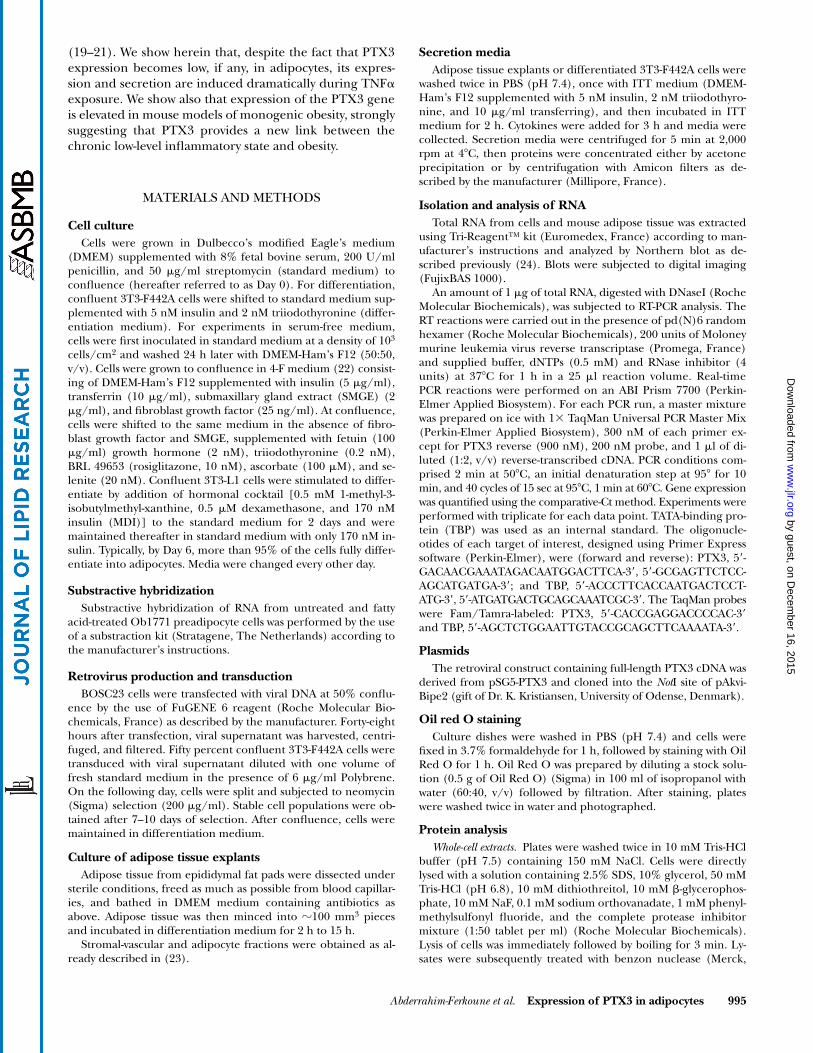
Expression of PTX3 mRNA during adipocyte differentiation
White and brown adipose tissues were shown to expresshigh and low levels of PTX3 mRNA in the stromal-vascularfraction and the adipocyte fraction, respectively. In con-trast to PTX3, aP2 and peroxisome proliferator-activatedreceptor
�
(PPAR
�
) were predominantly expressed in theadipocyte fraction (Fig. 1B, and not shown). These obser-vations led us to examine PTX3 expression as a functionof differentiation of 3T3-F442A (31), 3T3-L1 (32), andOb1771 cells. As shown in
Fig. 2A
, PTX3 mRNA was al-ready expressed in growing cells but became expressed athigher levels in confluent 3T3-F442A cells, whereas its ex-pression dramatically decreased thereafter. Its kinetics ofexpression appeared opposite to those of aP2 and PPAR
�
,which characterize the terminal differentiation process(33, 34). Identical results were obtained with 3T3-L1 (Fig.2A) and Ob1771 cells (data not shown). PTX3 mRNA ex-pression appeared as a differentiation-related event as themessage was maintained in the absence of induction ofdifferentiation of 3T3-L1 cells (Fig. 2A), and a weak andcontinuous expression was observed in 3T3-C2 cells (datanot shown), a fibroblast nonadipocyte clonal line (35).Consistent with this interpretation, in Ob1771 cells ex-posed to growth hormone or to fatty acids, in order to ac-celerate the differentiation process, the disappearance ofPTX3 mRNA is faster than that observed in untreatedcells (data not shown). The same pattern of expressionwas obtained in 3T3-F442A cells differentiated in a serum-free medium (Fig. 2B). This result excluded nonspecific
Fig. 1. Effect of fatty acid on PTX3 mRNA expression in preadi-pocytes and its distribution in adipose tissue. A: One day post-con-fluent Ob1771 cells were maintained for 24 h in standard mediumin the absence () or presence (�) of 300 �M �-linolenate. Twentymicrograms of RNA were analyzed as described in Materials andMethods. The results are representative of four independent exper-iments. B: RNA from stromal-vascular and adipocyte fractions ofwhite adipose tissue (WAT) and brown adipose tissue from six micewas blotted and hybridized with PTX3 probe. The results are repre-sentative of two independent experiments.
by guest, on Decem
ber 16, 2015w
ww
.jlr.orgD
ownloaded from
Abderrahim-Ferkoune et al.
Expression of PTX3 in adipocytes 997
effects of serum components, including cytokines, and fa-vored the existence of a differentiation-specific event.Thus, the finding that PTX3, a member of the long pen-traxin family, was transiently expressed in preadipocytesprompted us to examine its role, if any, in adipocyte dif-ferentiation.
Differentiation of stable 3T3-F442A cells overexpressing PTX3
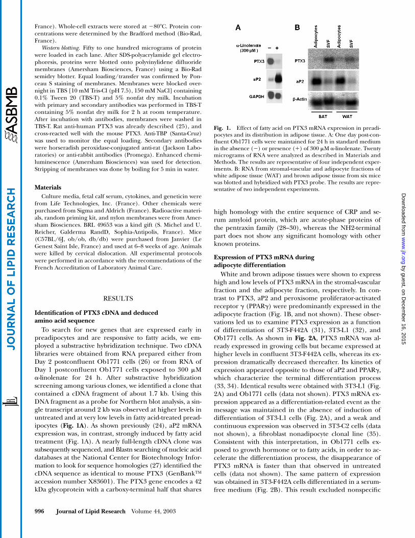
Overexpression of PTX3 in 3T3-F442A cells was accom-plished by retroviral infection as shown in
Fig. 3
controlcells, i.e., cells infected with the empty vector expressedthe endogenous PTX3 mRNA at the expected size of 2 kb.Cells infected with the retroviral vector containing thePTX3 coding sequence expressed higher levels of PTX3protein, and both the 2 kb signal and a 4 kb signal corre-sponding to the viral transcript were observed (Fig. 3A,
B). The expression of the 4 kb mRNA remained un-changed throughout the postconfluent period, consistentwith similar levels of PTX3 protein during differentiation(Fig. 3A). As shown in Fig. 3B and C, adipocyte differenti-ation of PTX3-overexpressing cells was unimpaired com-pared with that of control cells, as assessed by microscopeexamination and expression of differentiation-specificmarkers, excluding a direct role of PTX3 in the differenti-ation process. However, owing to the involvement ofPTX3 in the inflammatory process and its postulated rolein the defense against bacterial infections (36–38), studieswere performed with 3T3-F442A adipocytes in response toinflammatory cytokines, i.e., TNF
�
, as PTX3 expression,which is also termed TSG-14, is highly induced by TNF
�
,and as both human and mouse PTX3 promoters areknown to be responsive to TNF
�
(39, 40).
Modulation by TNF
�
of PTX3 mRNA andprotein expression
Upon exposure of differentiated 3T3-F442A cells toTNF
�
present in the nanomolar range of concentrations,the induction of the PTX3 gene occurred within 1 h. Peaklevels were reached within 3 h and decreased somewhatthereafter (
Fig. 4
). Interestingly, IL-6 and LIF were inac-tive at concentrations up to 1 nM, in contrast to li-popolysaccharide (LPS) which is active at 100 ng/ml. 3T3-F442A cells were able to respond to TNF
�
at concentra-tions as low as 0.01 nM (Fig. 4). Likewise, PTX3 proteinexpression was enhanced following TNF
�
treatment.PTX3 protein was clearly detectable by Western blot analy-sis of incubation media from differentiated 3T3-F442A ad-ipocytes as well as from adipose tissue explants (
Fig. 5
).Collectively, these data emphasize that PTX3 is a TNF
�
-induced protein secreted from adipocytes. As TNF
�
levelswere reported to be elevated in adipose tissue of geneti-cally obese mice, PTX3 levels were examined in these ani-mals.
Fig. 2. PTX3 mRNA expression in 3T3-F442A and 3T3-L1 cells during adipocyte differentiation in serum-containing and in serum-free medium. A: RNA was extracted from 3T3-F442A or 3T3-L1 cells that had beenmaintained in serum-supplemented medium at different intervals relative to confluence. 3T3-L1 cells havebeen induced to differentiate (�MDI) or not (MDI) in the presence of an hormonal cocktail (0.5 mM1-methyl-3-isobutylmethyl-xanthine, 0.5 �M dexamethasone, and 170 nM insulin) for 2 days. B: Total RNAwas extracted from 3T3-F442A cells that had been maintained in serum-free medium at different intervalsrelative to confluence. Twenty micrograms of RNA of each sample were loaded and analyzed by Northernblot as described in Materials and Methods. The results are representative of two independent experiments.
Fig. 3. Ectopic expression of PTX3 and differentiation of 3T3-F442A preadipocytes. Stable 3T3-F442A-vector cells and 3T3-F442A-PTX3 transfectants were maintained after confluence in differenti-ation medium. A: Total protein was extracted at Days 0 and 13, and50 �g was analyzed by Western blot. TATA-binding protein (TBP)was used as the internal standard. B: RNA was extracted at Days 2and 10 after confluence, and 20 �g were analyzed by Northernblot. C: Cells were fixed and stained with Oil red O at Day 10 afterconfluence and photographed. The results are representative oftwo independent experiments.
by guest, on Decem
ber 16, 2015w
ww
.jlr.orgD
ownloaded from
998 Journal of Lipid Research Volume 44, 2003
Enhanced expression of PTX3 gene in adipose tissue of genetically obese mice
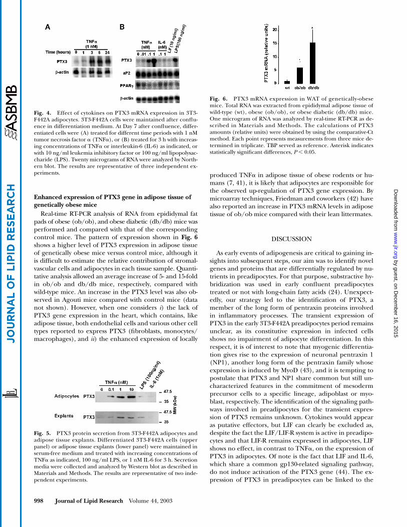
Real-time RT-PCR analysis of RNA from epididymal fatpads of obese (ob/ob), and obese diabetic (db/db) mice wasperformed and compared with that of the correspondingcontrol mice. The pattern of expression shown in Fig. 6shows a higher level of PTX3 expression in adipose tissueof genetically obese mice versus control mice, although itis difficult to estimate the relative contribution of stromal-vascular cells and adipocytes in each tissue sample. Quanti-tative analysis allowed an average increase of 5- and 15-foldin ob/ob and db/db mice, respectively, compared withwild-type mice. An increase in the PTX3 level was also ob-served in Agouti mice compared with control mice (datanot shown). However, when one considers i) the lack ofPTX3 gene expression in the heart, which contains, likeadipose tissue, both endothelial cells and various other celltypes reported to express PTX3 (fibroblasts, monocytes/macrophages), and ii) the enhanced expression of locally
produced TNF� in adipose tissue of obese rodents or hu-mans (7, 41), it is likely that adipocytes are responsible forthe observed up-regulation of PTX3 gene expression. Bymicroarray techniques, Friedman and coworkers (42) havealso reported an increase in PTX3 mRNA levels in adiposetissue of ob/ob mice compared with their lean littermates.
DISCUSSION
As early events of adipogenesis are critical to gaining in-sights into subsequent steps, our aim was to identify novelgenes and proteins that are differentially regulated by nu-trients in preadipocytes. For that purpose, substractive hy-bridization was used in early confluent preadipocytestreated or not with long-chain fatty acids (24). Unexpect-edly, our strategy led to the identification of PTX3, amember of the long form of pentraxin proteins involvedin inflammatory processes. The transient expression ofPTX3 in the early 3T3-F442A preadipocytes period remainsunclear, as its constitutive expression in infected cellsshows no impairment of adipocyte differentiation. In thisrespect, it is of interest to note that myogenic differentia-tion gives rise to the expression of neuronal pentraxin 1(NP1), another long form of the pentraxin family whoseexpression is induced by MyoD (43), and it is tempting topostulate that PTX3 and NP1 share common but still un-characterized features in the commitment of mesodermprecursor cells to a specific lineage, adipoblast or myo-blast, respectively. The identification of the signaling path-ways involved in preadipocytes for the transient expres-sion of PTX3 remains unknown. Cytokines would appearas putative effectors, but LIF can clearly be excluded as,despite the fact the LIF/LIF-R system is active in preadipo-cytes and that LIF-R remains expressed in adipocytes, LIFshows no effect, in contrast to TNF�, on the expression ofPTX3 in adipocytes. Of note is the fact that LIF and IL-6,which share a common gp130-related signaling pathway,do not induce activation of the PTX3 gene (44). The ex-pression of PTX3 in preadipocytes can be linked to the
Fig. 4. Effect of cytokines on PTX3 mRNA expression in 3T3-F442A adipocytes. 3T3-F442A cells were maintained after conflu-ence in differentiation medium. At Day 7 after confluence, differ-entiated cells were (A) treated for different time periods with 1 nMtumor necrosis factor � (TNF�), or (B) treated for 3 h with increas-ing concentrations of TNF� or interleukin-6 (IL-6) as indicated, orwith 10 ng/ml leukemia inhibitory factor or 100 ng/ml lipopolysac-charide (LPS). Twenty micrograms of RNA were analyzed by North-ern blot. The results are representative of three independent ex-periments.
Fig. 5. PTX3 protein secretion from 3T3-F442A adipocytes andadipose tissue explants. Differentiated 3T3-F442A cells (upperpanel) or adipose tissue explants (lower panel) were maintained inserum-free medium and treated with increasing concentrations ofTNF� as indicated, 100 ng/ml LPS, or 1 nM IL-6 for 3 h. Secretionmedia were collected and analyzed by Western blot as described inMaterials and Methods. The results are representative of two inde-pendent experiments.
Fig. 6. PTX3 mRNA expression in WAT of genetically-obesemice. Total RNA was extracted from epididymal adipose tissue ofwild-type (wt), obese (ob/ob), or obese diabetic (db/db) mice.One microgram of RNA was analyzed by real-time RT-PCR as de-scribed in Materials and Methods. The calculations of PTX3amounts (relative units) were obtained by using the comparative-Ctmethod. Each point represents measurements from three mice de-termined in triplicate. TBP served as reference. Asterisk indicatesstatistically significant differences, P � 0.05.
by guest, on Decem
ber 16, 2015w
ww
.jlr.orgD
ownloaded from

Abderrahim-Ferkoune et al. Expression of PTX3 in adipocytes 999
macrophage-like functions of preadipocytes, consistentwith the involvement of adipose tissue in immune re-sponse during inflammation (45).
PTX3 is a long-form member of the pentraxin family,and we have identified the PTX3 gene as a TNF�-respon-sive gene leading to the protein secretion. TNF� and IL-1,two major mediators of inflammation, have been reportedto induce the transcriptional activation of the PTX3 gene(19, 21, 46). LPS, which raises PTX3 plasma levels after in-jection in mice, shows a direct effect by activating thePTX3 gene. It is assumed that, as in 3T3-L1 adipocytes,LPS binding to constitutively-expressed Toll-like recep-tor-4 (TLR4) results in the fast induction of TLR-2 and thesynthesis of a set of secretory proteins that would includePTX3 (47). Although cultured adipocytes and adipose tis-sue explants respond to very low concentrations of TNF�(0.01–1 nM), it remains unclear whether Type 1 or Type 2TNF� receptors are involved in this response. Furtherstudies using adipocytes lacking each or both of these re-ceptors should clarify this issue (48). Preliminary evidenceshows that significant levels of PTX3 are present in mouseplasma, but a possible increase in plasma of geneticallyobese mice remains to be shown. If it is so, consideringthe fact that PTX3 binds to C1q (25), which is known tobe homologous to adiponectin (49, 50), it is tempting topostulate that higher circulating levels of PTX3 in achronic low-level inflammatory state may lower the con-centration of unbound adiponectin and may participatein the aggravation of the metabolic syndrome observed inobese animals and individuals. Studies in transgenic miceand gene-targeted mice suggest an important role forPTX3 in the regulation of inflammatory reactions and in-nate immunity (51–54). The study of the development ofadipose tissue in these knock-out animals as well as the useof obese TNF�-deficient mice should shed some light onthe physiological role of PTX3.
This work was supported by the Centre National de la Recher-che Scientifique (CNRS), by a special grant from the BristolMyers Squibb Foundation (to G.A.), and by the AssociationFrançaise contre les Myopathies (fellowship to A.A-F.). M.M. isan assistant Telethon Scientist and C.C. is supported by a Tele-thon fellowship. The authors are grateful to M. T. Ravier for ex-pert technical assistance, G. Oillaux for skillful secretarial assis-tance, and W. Cousin for help with real-time PCR experiments.
REFERENCES
1. Fantuzzi, G., and R. Faggioni. 2000. Leptin in the regulation of im-munity, inflammation, and hematopoiesis. J. Leukoc. Biol. 68: 437–446.
2. Ahima, R. S., and J. S. Flier. 2000. Adipose tissue as an endocrineorgan. Trends Endocrinol. Metab. 11: 327–332.
3. Sethi, J. K., and G. S. Hotamisligil. 1999. The role of TNF alpha inadipocyte metabolism. Semin. Cell Dev. Biol. 10: 19–29.
4. Sorisky, A. 2002. Molecular links between obesity and cardiovascu-lar disease. Am. J. Ther. 9: 516–521.
5. Hotamisligil, G. S., N. S. Shargill, and B. M. Spiegelman. 1993. Ad-ipose expression of tumor necrosis factor-alpha: direct role in obe-sity-linked insulin resistance. Science. 259: 87–91.
6. Yudkin, J. S., C. D. Stehouwer, J. J. Emeis, and S. W. Coppack.1999. C-reactive protein in healthy subjects: associations with obe-sity, insulin resistance, and endothelial dysfunction: a potentialrole for cytokines originating from adipose tissue? Arterioscler.Thromb. Vasc. Biol. 19: 972–978.
7. Kern, P. A., S. Ranganathan, C. Li, L. Wood, and G. Ranganathan.2001. Adipose tissue tumor necrosis factor and interleukin-6 ex-pression in human obesity and insulin resistance. Am. J. Physiol. En-docrinol. Metab. 280: E745–E751.
8. Hotamisligil, G. S., P. Arner, J. F. Caro, R. L. Atkinson, and B. M.Spiegelman. 1995. Increased adipose tissue expression of tumornecrosis factor-alpha in human obesity and insulin resistance. J.Clin. Invest. 95: 2409–2415.
9. Lin, Y., M. W. Rajala, J. P. Berger, D. E. Moller, N. Barzilai, and P. E.Scherer. 2001. Hyperglycemia-induced production of acute phasereactants in adipose tissue. J. Biol. Chem. 276: 42077–42083.
10. Chiellini, C., A. Bertacca, S. E. Novelli, C. Z. Gorgun, A. Ciccarone,A. Giordano, H. Xu, A. Soukas, M. Costa, D. Gandini, R. Dimitri, P.Bottone, P. Cecchetti, E. Pardini, L. Perego, R. Navalesi, F. Folli, L.Benzi, S. Cinti, J. M. Friedman, G. S. Hotamisligil, and M. Maffei.2002. Obesity modulates the expression of haptoglobin in thewhite adipose tissue via TNFalpha. J. Cell. Physiol. 190: 251–258.
11. van der Poll, T., S. J. van Deventer, G. Pasterkamp, J. A. vanMourik, H. R. Buller, and J. W. ten Cate. 1992. Tumor necrosis fac-tor induces von Willebrand factor release in healthy humans.Thromb. Haemost. 67: 623–626.
12. Hardardottir, I., C. Grunfeld, and K. R. Feingold. 1994. Effects ofendotoxin and cytokines on lipid metabolism. Curr. Opin. Lipidol.5: 207–215.
13. Hotamisligil, G. S., A. Budavari, D. Murray, and B. M. Spiegelman.1994. Reduced tyrosine kinase activity of the insulin receptor inobesity-diabetes. Central role of tumor necrosis factor-alpha. J.Clin. Invest. 94: 1543–1549.
14. Grimble, R. F. 2002. Inflammatory status and insulin resistance.Curr. Opin. Clin. Nutr. Metab. Care. 5: 551–559.
15. Visser, M., L. M. Bouter, G. M. McQuillan, M. H. Wener, and T. B.Harris. 1999. Elevated C-reactive protein levels in overweight andobese adults. JAMA. 282: 2131–2135.
16. Visser, M., L. M. Bouter, G. M. McQuillan, M. H. Wener, and T. B.Harris. 2001. Low-grade systemic inflammation in overweight chil-dren. Pediatrics. 107: E13–E18.
17. Aubert, J., S. Dessolin, N. Belmonte, M. Li, F. R. McKenzie, L. Stac-cini, P. Villageois, B. Barhanin, A. Vernallis, A. G. Smith, G. Ail-haud, and C. Dani. 1999. Leukemia inhibitory factor and its recep-tor promote adipocyte differentiation via the mitogen-activatedprotein kinase cascade. J. Biol. Chem. 274: 24965–24972.
18. Belmonte, N., B. W. Phillips, F. Massiera, P. Villageois, B. Wdzie-konski, P. Saint-Marc, J. Nichols, J. Aubert, K. Saeki, A. Yuo, S.Narumiya, G. Ailhaud, and C. Dani. 2001. Activation of extracellu-lar signal-regulated kinases and CREB/ATF-1 mediate the expres-sion of CCAAT/enhancer binding proteins beta and delta inpreadipocytes. Mol. Endocrinol. 15: 2037–2049.
19. Breviario, F., E. M. d’Aniello, J. Golay, G. Peri, B. Bottazzi, A. Bai-roch, S. Saccone, R. Marzella, V. Predazzi, M. Rocchi, G. DellaValle, E. Dejana, A. Mantovani, and M. Introna. 1992. Interleukin-1-inducible genes in endothelial cells. Cloning of a new gene re-lated to C-reactive protein and serum amyloid P component. J.Biol. Chem. 267: 22190–22197.
20. Introna, M., V. V. Alles, M. Castellano, G. Picardi, L. De Gioia, B. Bot-tazzai, G. Peri, F. Breviario, M. Salmona, L. De Gregorio, T. A.Dragani, N. Srinivasan, T. L. Blundell, T. A. Hamilton, and A. Manto-vani. 1996. Cloning of mouse PTX3, a new member of the pentraxingene family expressed at extrahepatic sites. Blood. 87: 1862–1872.
21. Lee, G. W., T. H. Lee, and J. Vilcek. 1993. TSG-14, a tumor necrosisfactor- and IL-1-inducible protein, is a novel member of the pen-taxin family of acute phase proteins. J. Immunol. 150: 1804–1812.
22. Gaillard, D., G. Ailhaud, and R. Negrel. 1985. Fetuin modulatesgrowth and differentiation of Ob17 preadipose cells in serum-freehormone-supplemented medium. Biochim. Biophys. Acta. 846: 185–191.
23. Cannon, B., and J. Nedergaard. 2001. Cultures of adipose precursorcells from brown adipose tissue and of clonal brown-adipocyte-likecell lines. Methods Mol. Biol. 155: 213–224.
24. Amri, E. Z., B. Bertrand, G. Ailhaud, and P. Grimaldi. 1991. Regu-lation of adipose cell differentiation. I. Fatty acids are inducers ofthe aP2 gene expression. J. Lipid Res. 32: 1449–1456.
25. Bottazzi, B., V. Vouret-Craviari, A. Bastone, L. De Gioia, C. Mat-
by guest, on Decem
ber 16, 2015w
ww
.jlr.orgD
ownloaded from
1000 Journal of Lipid Research Volume 44, 2003
teucci, G. Peri, F. Spreafico, M. Pausa, C. D’Ettorre, E. Gianazza, A.Tagliabue, M. Salmona, F. Tedesco, M. Introna, and A. Mantovani.1997. Multimer formation and ligand recognition by the long pen-traxin PTX3. Similarities and differences with the short pentraxinsC-reactive protein and serum amyloid P component. J. Biol. Chem.272: 32817–32823.
26. Negrel, R., P. Grimaldi, and G. Ailhaud. 1978. Establishment ofpreadipocyte clonal line from epididymal fat pad of ob/ob mousethat responds to insulin and to lipolytic hormones. Proc. Natl.Acad. Sci. USA. 75: 6054–6058.
27. Altschul, S. F., T. L. Madden, A. A. Schaffer, J. Zhang, Z. Zhang, W.Miller, and D. J. Lipman. 1997. Gapped BLAST and PSI-BLAST: anew generation of protein database search programs. Nucleic AcidsRes. 25: 3389–3402.
28. Pepys, M. B., and M. L. Baltz. 1983. Acute phase proteins with spe-cial reference to C-reactive protein and related proteins (pentax-ins) and serum amyloid A protein. Adv. Immunol. 34: 141–212.
29. Gewurz, H., X. H. Zhang, and T. F. Lint. 1995. Structure and func-tion of the pentraxins. Curr. Opin. Immunol. 7: 54–64.
30. Goodman, A. R., T. Cardozo, R. Abagyan, A. Altmeyer, H. G. Wis-niewski, and J. Vilcek. 1996. Long pentraxins: an emerging groupof proteins with diverse functions. Cytokine Growth Factor Rev. 7:191–202.
31. Green, H., and O. Kehinde. 1976. Spontaneous heritable changesleading to increased adipose conversion in 3T3 cells. Cell. 7: 105–113.
32. Green, H., and M. Meuth. 1974. An established pre-adipose cellline and its differentiation in culture. Cell. 3: 127–133.
33. Cook, K. S., C. R. Hunt, and B. M. Spiegelman. 1985. Developmen-tally regulated mRNAs in 3T3-adipocytes: analysis of transcrip-tional control. J. Cell Biol. 100: 514–520.
34. Rosen, E. D., and B. M. Spiegelman. 2000. Molecular regulation ofadipogenesis. Annu. Rev. Cell Dev. Biol. 16: 145–171.
35. Green, H., and O. Kehinde. 1975. An established preadipose cellline and its differentiation in culture. II. Factors affecting the adi-pose conversion. Cell. 5: 19–27.
36. Muller, B., G. Peri, A. Doni, V. Torri, R. Landmann, B. Bottazzi,and A. Mantovani. 2001. Circulating levels of the long pentraxinPTX3 correlate with severity of infection in critically ill patients.Crit. Care Med. 29: 1404–1407.
37. Luchetti, M. M., G. Piccinini, A. Mantovani, G. Peri, C. Matteucci,G. Pomponio, M. Fratini, P. Fraticelli, P. Sambo, C. Di Loreto, A.Doni, M. Introna, and A. Gabrielli. 2000. Expression and produc-tion of the long pentraxin PTX3 in rheumatoid arthritis (RA).Clin. Exp. Immunol. 119: 196–202.
38. Peri, G., M. Introna, D. Corradi, G. Iacuitti, S. Signorini, F. Avan-zini, F. Pizzetti, A. P. Maggioni, T. Moccetti, M. Metra, L. D. Cas, P.Ghezzi, J. D. Sipe, G. Re, G. Olivetti, A. Mantovani, and R. Latini.2000. PTX3, A prototypical long pentraxin, is an early indicator ofacute myocardial infarction in humans. Circulation. 102: 636–641.
39. Basile, A., A. Sica, E. d’Aniello, F. Breviario, G. Garrido, M. Castellano,A. Mantovani, and M. Introna. 1997. Characterization of the pro-moter for the human long pentraxin PTX3. Role of NF-kappaB intumor necrosis factor-alpha and interleukin-1beta regulation. J.Biol. Chem. 272: 8172–8178.
40. Altmeyer, A., L. Klampfer, A. R. Goodman, and J. Vilcek. 1995.Promoter structure and transcriptional activation of the murineTSG-14 gene encoding a tumor necrosis factor/interleukin-1-induc-ible pentraxin protein. J. Biol. Chem. 270: 25584–25590.
41. Yamakawa, T., S. Tanaka, Y. Yamakawa, Y. Kiuchi, F. Isoda, S. Kawa-moto, K. Okuda, and H. Sekihara. 1995. Augmented productionof tumor necrosis factor-alpha in obese mice. Clin. Immunol. Immu-nopathol. 75: 51–56.
42. Soukas, A., P. Cohen, N. D. Socci, and J. M. Friedman. 2000. Lep-tin-specific patterns of gene expression in white adipose tissue.Genes Dev. 14: 963–980.
43. Wyzykowski, J. C., T. I. Winata, N. Mitin, E. J. Taparowsky, and S. F.Konieczny. 2002. Identification of novel MyoD gene targets in pro-liferating myogenic stem cells. Mol. Cell. Biol. 22: 6199–6208.
44. Heinrich, P. C., I. Behrmann, G. Muller-Newen, F. Schaper, and L.Graeve. 1998. Interleukin-6-type cytokine signalling through thegp130/Jak/STAT pathway. Biochem. J. 334: 297–314.
45. Cousin, B., O. Munoz, M. Andre, A. M. Fontanilles, C. Dani, J. L.Cousin, P. Laharrague, L. Casteilla, and L. Penicaud. 1999. A rolefor preadipocytes as macrophage-like cells. FASEB J. 13: 305–312.
46. Goodman, A. R., D. E. Levy, L. F. Reis, and J. Vilcek. 2000. Differ-ential regulation of TSG-14 expression in murine fibroblasts andperitoneal macrophages. J. Leukoc. Biol. 67: 387–395.
47. Lin, Y., H. Lee, A. H. Berg, M. P. Lisanti, L. Shapiro, and P. E.Scherer. 2000. The lipopolysaccharide-activated toll-like receptor(TLR)-4 induces synthesis of the closely related receptor TLR-2 inadipocytes. J. Biol. Chem. 275: 24255–24263.
48. Sethi, J. K., H. Xu, K. T. Uysal, S. M. Wiesbrock, L. Scheja, and G. S.Hotamisligil. 2000. Characterisation of receptor-specific TNFalphafunctions in adipocyte cell lines lacking type 1 and 2 TNF recep-tors. FEBS Lett. 469: 77–82.
49. Berg, A. H., T. P. Combs, and P. E. Scherer. 2002. ACRP30/adi-ponectin: an adipokine regulating glucose and lipid metabolism.Trends Endocrinol. Metab. 13: 84–89.
50. Tsao, T. S., H. F. Lodish, and J. Fruebis. 2002. ACRP30, a new hor-mone controlling fat and glucose metabolism. Eur. J. Pharmacol.440: 213–221.
51. Dias, A. A., A. R. Goodman, J. L. Dos Santos, R. N. Gomes, A. Alt-meyer, P. T. Bozza, M. F. Horta, J. Vilcek, and L. F. Reis. 2001. TSG-14 transgenic mice have improved survival to endotoxemia and toCLP-induced sepsis. J. Leukoc. Biol. 69: 928–936.
52. Souza, D. G., A. C. Soares, V. Pinho, H. Torloni, L. F. Reis, M. T.Martins, and A. A. Dias. 2002. Increased mortality and inflamma-tion in tumor necrosis factor-stimulated gene-14 transgenic mice af-ter ischemia and reperfusion injury. Am. J. Pathol. 160: 1755–1765.
53. Varani, S., J. A. Elvin, C. Yan, J. DeMayo, F. J. DeMayo, H. F. Hor-ton, M. C. Byrne, and M. M. Matzuk. 2002. Knockout of pentraxin 3,a downstream target of growth differentiation factor-9, causes fe-male subfertility. Mol. Endocrinol. 16: 1154–1167.
54. Garlanda, C., E. Hirsch, S. Bozza, A. Salustri, M. De Acetis, R.Nota, A. Maccagno, F. Riva, B. Bottazzi, G. Peri, A. Doni, L. Vago,M. Botto, R. De Santis, P. Carminati, G. Siracusa, F. Altruda, A. Vecchi,L. Romani, and A. Mantovani. 2002. Non-redundant role of thelong pentraxin PTX3 in anti-fungal innate immune response. Na-ture. 420: 182–186.
by guest, on Decem
ber 16, 2015w
ww
.jlr.orgD
ownloaded from
Related Documents