7698–7713 Nucleic Acids Research, 2007, Vol. 35, No. 22 Published online 5 November 2007 doi:10.1093/nar/gkm538 Characterization of the G-quadruplexes in the duplex nuclease hypersensitive element of the PDGF-A promoter and modulation of PDGF-A promoter activity by TMPyP4 Yong Qin 1 , Evonne M. Rezler 1 , Vijay Gokhale 1 , Daekyu Sun 1 and Laurence H. Hurley 1,2,3,4, * 1 College of Pharmacy, 1703 E. Mabel, University of Arizona, Tucson, Arizona 85721, 2 Arizona Cancer Center, 1515 N. Campbell Avenue, Tucson, Arizona 85724, 3 Department of Chemistry, University of Arizona, Tucson, Arizona, 85721 and 4 BIO5 Collaborative Research Institute, 1657 E. Helen Street, Tucson, Arizona 85721, USA Received April 13, 2007; Revised June 27, 2007; Accepted June 28, 2007 ABSTRACT The proximal 5’-flanking region of the human platelet-derived growth factor A (PDGF-A) promoter contains one nuclease hypersensitive element (NHE) that is critical for PDGF-A gene transcription. On the basis of circular dichroism (CD) and electrophoretic mobility shift assay (EMSA), we have shown that the guanine-rich (G-rich) strand of the DNA in this region can form stable intramolecular parallel G-quadru- plexes under physiological conditions. A Taq poly- merase stop assay has shown that the G-rich strand of the NHE can form two major G-quadruplex structures, which are in dynamic equilibrium and differentially stabilized by three G-quadruplex-inter- active drugs. One major parallel G-quadruplex structure of the G-rich strand DNA of NHE was identified by CD and dimethyl sulfate (DMS) foot- printing. Surprisingly, CD spectroscopy shows a stable parallel G-quadruplex structure formed within the duplex DNA of the NHE at temperatures up to 100uC. This structure has been characterized by DMS footprinting in the double-stranded DNA of the NHE. In transfection experiments, 10 kM TMPyP4 reduced the activity of the basal promoter of PDGF- A »40%, relative to the control. On the basis of these results, we have established that ligand-mediated stabilization of G-quadruplex structures within the PDGF-A NHE can silence PDGF-A expression. INTRODUCTION Platelet-derived growth factor (PDGF), which was originally isolated from human platelets, is composed of four polypeptide chains designated A, B, C and D. These polypeptide chains make up five biologically active dimeric PDGF isoforms: PDGF-AA, -AB, -BB, -CC and -DD, which exert their effects through two tyrosine kinase receptors, PDGFR-a and PDGFR-b (1–3). PDGF-A and PDGF-B are considered to be major mitogens for a variety of mesenchymal cell types. They play critical roles in normal embryonic development, cellular differentiation and wound healing (1–4). The overactivity of the PDGFs has been implicated in the pathogenesis of a number of diseases characterized by excessive cell growth, including cancer, atherosclerosis and various fibrotic conditions (3,5,6). Significantly augmented expression of the PDGF-A gene has been observed in a number of human tumor cell lines and tissues, particularly in gliomas, sarcomas and ostoastrocytomas (6–10). Abnormal PDGF-A expression is linked to accelerated malignant cell growth through autocrine and paracrine pathways (1,6,11). For these reasons, PDGF signaling pathways are important targets for anticancer therapeutics; specifically, inhibitors of PDGF-A gene expression could be valuable in the treatment of some cancers. Transcription of the PDGF-A gene is controlled by several positive and negative regulatory elements located in the promoter region and the 5 0 -distal upstream region of the gene (Figure 1) (12–14). An important, highly guanine- and cytosine-rich region located at nucleotides –120 to –33 within the PDGF-A promoter accounts for 80% of basal promoter activity. This GC-rich region was originally identified by its sensitivity to the single-strand- specific endonuclease S1 nuclease and has been termed the nuclease hypersensitive element (NHE) (12–14). PDGF-A is also subject to transcriptional repression by two discrete S1 nuclease hypersensitive (SHS) elements. One is located within the 5 0 -distal region (–1418 to –1338) of the promoter (5 0 SHS), while the other is located within the first intron (+1605 to +1630, intron SHS) (14–17). Multiple transcription factors bind to and regulate *To whom correspondence should be addressed. Tel: +1 520 626 5622; Fax: +1 520 626 5623; Email: [email protected] ß 2007 The Author(s) This is an Open Access article distributed under the terms of the Creative Commons Attribution Non-Commercial License (http://creativecommons.org/licenses/ by-nc/2.0/uk/) which permits unrestricted non-commercial use, distribution, and reproduction in any medium, provided the original work is properly cited. by guest on February 25, 2014 http://nar.oxfordjournals.org/ Downloaded from

Welcome message from author
This document is posted to help you gain knowledge. Please leave a comment to let me know what you think about it! Share it to your friends and learn new things together.
Transcript

7698–7713 Nucleic Acids Research, 2007, Vol. 35, No. 22 Published online 5 November 2007doi:10.1093/nar/gkm538
Characterization of the G-quadruplexes in the duplexnuclease hypersensitive element of the PDGF-Apromoter and modulation of PDGF-A promoteractivity by TMPyP4Yong Qin1, Evonne M. Rezler1, Vijay Gokhale1, Daekyu Sun1 and Laurence H. Hurley1,2,3,4,*
1College of Pharmacy, 1703 E. Mabel, University of Arizona, Tucson, Arizona 85721, 2Arizona Cancer Center,1515N. Campbell Avenue, Tucson, Arizona 85724, 3Department of Chemistry, University of Arizona, Tucson,Arizona, 85721 and 4BIO5 Collaborative Research Institute, 1657 E. Helen Street, Tucson, Arizona 85721, USA
Received April 13, 2007; Revised June 27, 2007; Accepted June 28, 2007
ABSTRACT
The proximal 5’-flanking region of the humanplatelet-derived growth factor A (PDGF-A) promotercontains one nuclease hypersensitive element (NHE)that is critical for PDGF-A gene transcription. On thebasis of circular dichroism (CD) and electrophoreticmobility shift assay (EMSA), we have shown that theguanine-rich (G-rich) strand of the DNA in this regioncan form stable intramolecular parallel G-quadru-plexes under physiological conditions. A Taq poly-merase stop assay has shown that the G-rich strandof the NHE can form two major G-quadruplexstructures, which are in dynamic equilibrium anddifferentially stabilized by three G-quadruplex-inter-active drugs. One major parallel G-quadruplexstructure of the G-rich strand DNA of NHE wasidentified by CD and dimethyl sulfate (DMS) foot-printing. Surprisingly, CD spectroscopy shows astable parallel G-quadruplex structure formed withinthe duplex DNA of the NHE at temperatures up to100uC. This structure has been characterized byDMS footprinting in the double-stranded DNA of theNHE. In transfection experiments, 10kM TMPyP4reduced the activity of the basal promoter of PDGF-A »40%, relative to the control. On the basis of theseresults, we have established that ligand-mediatedstabilization of G-quadruplex structures within thePDGF-A NHE can silence PDGF-A expression.
INTRODUCTION
Platelet-derived growth factor (PDGF), which wasoriginally isolated from human platelets, is composedof four polypeptide chains designated A, B, C and D.
These polypeptide chains make up five biologically activedimeric PDGF isoforms: PDGF-AA, -AB, -BB, -CC and-DD, which exert their effects through two tyrosine kinasereceptors, PDGFR-a and PDGFR-b (1–3). PDGF-A andPDGF-B are considered to be major mitogens for avariety of mesenchymal cell types. They play critical rolesin normal embryonic development, cellular differentiationand wound healing (1–4). The overactivity of the PDGFshas been implicated in the pathogenesis of a number ofdiseases characterized by excessive cell growth, includingcancer, atherosclerosis and various fibrotic conditions(3,5,6). Significantly augmented expression of thePDGF-A gene has been observed in a number of humantumor cell lines and tissues, particularly in gliomas,sarcomas and ostoastrocytomas (6–10). AbnormalPDGF-A expression is linked to accelerated malignantcell growth through autocrine and paracrine pathways(1,6,11). For these reasons, PDGF signaling pathways areimportant targets for anticancer therapeutics; specifically,inhibitors of PDGF-A gene expression could be valuablein the treatment of some cancers.
Transcription of the PDGF-A gene is controlled byseveral positive and negative regulatory elements locatedin the promoter region and the 50-distal upstream regionof the gene (Figure 1) (12–14). An important, highlyguanine- and cytosine-rich region located at nucleotides–120 to –33 within the PDGF-A promoter accounts for80% of basal promoter activity. This GC-rich region wasoriginally identified by its sensitivity to the single-strand-specific endonuclease S1 nuclease and has been termed thenuclease hypersensitive element (NHE) (12–14). PDGF-Ais also subject to transcriptional repression by two discreteS1 nuclease hypersensitive (SHS) elements. One is locatedwithin the 50-distal region (–1418 to –1338) of thepromoter (50SHS), while the other is located within thefirst intron (+1605 to +1630, intron SHS) (14–17).Multiple transcription factors bind to and regulate
*To whom correspondence should be addressed. Tel: +1 520 626 5622; Fax: +1 520 626 5623; Email: [email protected]
� 2007 The Author(s)
This is an Open Access article distributed under the terms of the Creative Commons Attribution Non-Commercial License (http://creativecommons.org/licenses/
by-nc/2.0/uk/) which permits unrestricted non-commercial use, distribution, and reproduction in any medium, provided the original work is properly cited.
by guest on February 25, 2014http://nar.oxfordjournals.org/
Dow
nloaded from
PDGF-A genes through the NHE region, such as Sp1,Egr-1, Wilms’ tumor protein (WT1), GC factor 2 (GCF2),nuclear factor 1-X (NF1-X) and the metastasis suppressorprotein NM23 (17–23). This region is structurally dynamicand capable of adopting non-B-DNA conformations (24).The importance of the unwound paranemic structure ofNHE in PDGF-A promoter activity was confirmed bydemonstrating that a single-stranded DNA oligomer,derived from the guanine-rich strand (–74 to –51), inhibitsthe promoter activity of PDGF-A (24). It is believed thatthe equilibrium between the B-form DNA, the single-stranded DNA, and intramolecular secondary DNAstructures is instrumental in the determination of thebinding of transcription factors to the NHE region ofPDGF-A (NHEPDGF-A). However, the molecular detailsof the unwinding of secondary structures in theNHEPDGF-A are poorly understood.
Tandem repeats of the guanine-tract (G-tract) sequenceadopt specific intramolecular G-quadruplex structures inthe telomeres and promoters of several important genes,such as c-MYC, KRAS, Bcl-2, c-Kit and VEGF (25–29).The basic building block, the G-tetrad, and examples offolding patterns are shown in Figure 1B. The involvementof G-quadruplex structures in cellular processes, suchas transcriptional control of c-MYC expression, hasstimulated the development of anticancer drugs thatare selective for G-quadruplexes. The G-rich strand ofNHEPDGF-A contains five contiguous guanine runs,each separated by one base. The G-tract motif inNHEPDGF-A has some similarity to the G-tract motifin the NHE region of c-MYC (NHEc-MYC). However,in NHEc-MYC the minimum number of guanines in anyrun is three rather than four, and NHEPDGF-A contains avery long run of 13 contiguous guanines at the 50-end(see sequences in Figure 2A) (25,30). Since the G-richstrand of NHEc-MYC is known to form G-quadruplex
structures, by analogy the G-rich strand of NHEPDGF-A
has the potential to form G-quadruplex structuresunder physiological conditions. Consequently, theseG-quadruplex structures may play an important role inmodulating the expression of PDGF-A. For these reasons,this work examines the in vitro formation and propertiesof G-quadruplex structures within the G-rich strand ofNHEPDGF-A, including its transcriptional modulation byTMPyP4.
MATERIALS AND METHODS
Oligodeoxynucleotides, enzymes, materials and drugs
DNA oligomers (sequences shown in Table 1) wereobtained from Sigma Genosys and Biosearch Techno-logies, Inc. All DNA oligomers were PAGE-purified anddissolved in double-distilled water before use. Theextinction coefficients that the manufacturers providedfor a given DNA sequence were calculated using thenearest-neighbor method (31). When the DNA oligomersfold into a G-quadruplex, the base stacking results inhyperchromicity. Thus, the extinction coefficients of DNAoligomers that can form DNA secondary structures needto be corrected for the hyperchromicity. To address thisissue, we used UV/vis thermal denaturation techniques todetermine the high-order structural hyperchromicity ofthe G-quadruplex-forming DNA oligomers (32). The UVexperiments were carried out on a Cary 300 BioUV-visible spectrophotometer. Cells with optical pathlengths of 10mm were used, and the temperature of thecell holder was controlled by an external circulating waterbath (Varian). The UV melting experiments were done bycollecting spectra every five degrees as the sample heatedup from 20 to 1008C, and monitoring the absorptionchange at �max. The plateau at the top of the melting curve
Figure 1. (A) Promoter structure of PDGF-A and locations of the important NHEs. The sequences of the 50SHS, the G-rich strand of the NHE andthe intron SHS are shown. TSS= transcriptional start site. (B) Structure of a G-tetrad and examples of the folding patterns of known intramolecularG-quadruplexes.
Nucleic Acids Research, 2007, Vol. 35, No. 22 7699
by guest on February 25, 2014http://nar.oxfordjournals.org/
Dow
nloaded from
(usually 208C above the respective Tm) was extrapolatedback to 208C to obtain the hyperchromicity. Theextinction coefficient for the G-quadruplex-formingDNA oligomer was corrected by using the followingequation: “260(Q)=“260(N)/(1+ (%hyperchromicity/100)). “260(Q) represents the extinction coefficient for theG-quadruplex-forming DNA oligomer, and “260(N)represents the extinction coefficient for the non-structuredsingle-stranded DNA oligomers. The corrected “260(Q)values for select DNA oligomers in our study are asfollows: PDGF-A Pu48, 533680M–1 cm–1; PDGF-A Pu60,672353M–1 cm–1; PDGF-A Pu90, 1000303M–1 cm–1;PA-5W, 379701M–1 cm–1; PA-Mut1, 360524M–1 cm–1;
PA-Mut2, 369612M–1 cm–1; c-MYC Pu27,354938M–1 cm–1; TBA, 151770M–1 cm–1 and Bcl-2Pu38WT, 453766M–1 cm–1. Acrylamide:bisacrylamide(29:1) solution and ammonium persulfate were purchasedfrom Bio-Rad, and N,N,N0,N0-tetramethylethylenedia-mine was purchased from Fisher. T4 polynucleotidekinase and Taq DNA polymerase were purchased fromPromega. [g-32P]ATP was purchased from NEN Dupont.TMPyP2, TMPyP4 and Se2SAP were synthesized in ourlaboratory. Telomestatin was kindly provided byDr Kazuo Shin-ya (University of Tokyo, Japan). Stocksolutions of telomestatin and Se2SAP (10mM) weremade using DMSO (10%) and double-distilled water,
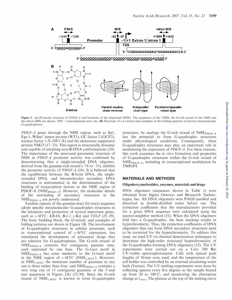
Figure 2. (A) Comparative CD spectra of three known G-quadruplex-forming sequences with the PDGF-A Pu48-mer. Blue line=PDGF-A Pu48-mer (25mM KCl), red line=c-Myc Pu27-mer (parallel G-quadruplex in 25mM KCl), green line=Bcl-2 Pu39WT (mixed parallel/antiparallelG-quadruplex in 100mM KCl), black line=TBA (antiparallel G-quadruplex in 100mM KCl). All CD data were obtained with a 5mM strandconcentration at 258C. Comparison of sequences of these two DNA oligomers is shown under the CD spectra. (B) Native gel electrophoresisindicating the formation of intramolecular G-quadruplex structures by Pu48-mer. The fast mobility major band represents the intramolecularG-quadruplex and the slow mobility bands represent the higher-order intermolecular G-quadruplexes. (C) Effect of alkali metals on the Pu48-merCD spectra. Effect of KCl (red line) and NaCl (blue line) on the ellipticity signal compared to the signal in the absence of salt (black line). All CDdata were obtained with a 5 mM strand concentration at 258C.
7700 Nucleic Acids Research, 2007, Vol. 35, No. 22
by guest on February 25, 2014http://nar.oxfordjournals.org/
Dow
nloaded from

respectively. Further dilutions to working concentrationswere made with double-distilled water immediately priorto use. Plasmid pGL3-basic, pRL-TK and dual luciferaseassay kits were purchased from Promega.
Circular dichroism spectroscopy
CD spectra were recorded on a Jasco-810 spectropolari-meter (Easton, MD, USA) using a quartz cell of 1mmoptical path length and an instrument scanning speed of100 nm/min, with a response time of 1 s, over a wavelengthrange of 200–330 nm. All DNA samples were dissolvedand diluted in Tris–HCl buffer (50mM, pH 7.6), andwhere appropriate, the samples also contained differentconcentrations of KCl and/or NaCl. The DNA strandconcentrations were 5 mM, and the CD data representfour averaged scans taken at an experimental temperature(25–1108C). All CD spectra are baseline-corrected forsignal contributions due to the buffer.
Labeling and purification of oligodeoxyribonucleotides
The DNA oligomers were 50-end-labeled with [g-32P]ATPusing T4 polynucleotide kinase for 1 h at 378C. Thelabeling reaction was inactivated by heating the samples at908C for 8min after the addition of 1.5 ml of 0.5M EDTA.The 50-end labeled DNA was then purified using a Bio-Spin 6 chromatography column (Bio-Rad). The labeledDNA was further purified by running a 12% denaturedpolyacrylamide gel that contained 8.0M urea.
Preparation of intramolecular G-quadruplex structures byEMSA
The 50-end 32P-labeled single-stranded DNA oligomer wasincubated in Tris–HCl buffer (50mM, pH 7.6) containingdifferent concentrations of KCl and/or NaCl for 1 h atroom temperature. The denatured marker DNA oligomerswere prepared separately before the EMSA. The controlDNA oligomers were heated at 958C for 15min. Theywere then immediately put on ice and cooled to 48C beforeloading the gel. The secondary structures of the DNAoligomers were obtained by heating the stock solutions at908C for 10min and then slowly cooling to roomtemperature over 4 h. The monomolecular G-quadruplexstructure of the G-rich strand of the NHE was isolated bynon-denatured gel electrophoresis in the presence of10mM NaCl and KCl in Tris-Borate-EDTA buffer.Electrophoresis of the DNA was conducted at 150V.The different mobility shift DNA bands were cut from thenon-denatured gel and soaked in double-distilled water.The double-stranded 60-mer NHE was obtained first byannealing equal amounts of 60-mer G-rich strand (Pu60-mer, 50-end 32P-labeled) and C-rich strand (Py60-mer) inannealing buffer (10mM Tris–HCl, pH 7.5, 10mMNaCl),heating the mixtures at 758C for 15min, and then slowlycooling the DNA sample to room temperature. The majordouble-stranded DNA band was isolated from a 12%native polyacrylamide gel. For inducing the formation ofG-quadruplexes in the duplex DNA, the double-stranded60-mer NHE was incubated in different concentrations ofKCl buffers at room temperature for at least 1 h, thenheated at 658C and allowed to slowly cool to roomtemperature over 4 h.
Taq polymerase stop assay
The DNA primer 50-d(TCGACTCTAAGCAAATGCGTCGAG)-30 was 50-end-labeled with 32P as described above.The labeled primer was annealed to the DNA template(sequence shown in Table 1) by using the aforementionedprotocol. The DNA complex formed by annealing theprimer to the template was purified using gel electrophore-sis on a 12% native polyacrylamide gel. The purified DNAwas then diluted to a concentration of 0.2 nM and mixedwith the reaction buffer (10mM MgCl2, 0.5mM DTT,0.1mM EDTA, 1.5 mg/ml BSA) and 0.1mM dNTPs.Where appropriate, KCl, NaCl and/or drugs were alsoadded. The reaction mixtures were incubated for 1 h atroom temperature, allowing the alkali metal ions or drugsto stabilize the G-quadruplexes. Taq DNA polymerasewas then added, and samples were incubated for 30minat 608C for polymerase extension. The polymeraseextension reaction was stopped by adding 2� stop buffer(10mM EDTA, 10mM NaOH, 0.1% xylene cyanole,0.1% bromophenol blue in formamide solution), andthe samples were analyzed on a 16% denatured poly-acrylamide gel (33).
DMS footprinting
Each band of interest was excised from the EMSA andsoaked in 1� Tris-Borate-EDTA buffer containing 10mM
Table 1. Oligodeoxynucleotides used in this study
DNA oligomer Sequence (50 ! 30)
PDGF-A Pu48 GGAGGC GGGGG GGGGG GGGCG GGGGCGGGGG CGGGG GAGGG GCGCG GC
PDGF-A Pu60 CCGGG GAGGC GGGGG GGGGG GGGCGGGGGC GGGGG CGGGG GAGGG GCGCGGCGGC GGCGG
PDGF-A Py60 CCGCC GCCGC CGCGC CCCTC CCCCGCCCCC GCCCC CGCCC CCCCC CCCCCGCCTC CCCGG
PDGF-A Pu90 TGCGG GTCCC AGGCC CGGAA TCCGGGGAGG CGGGG GGGGG GGGGC GGGGG
CGGGG GCGGG GGAGG GGCGC GGCGGCGGCG GCTAT AACCC
Templatea CGGG GAGGC GGGGG GGGGG GGGCGGGGGC GGGGG CGGGG GAGGG GCGCGGTTAG TCAGA CCTCG ACGCA TTTGCTTAGA GTCGA
Primerb TCGAC TCTAA GCAAA TGCGT CGAGc-MYC Pu27 TGGGG AGGGT GGGGA GGGT GGGG AAGGTBA GGTT GGTG TGGT TGG
Bcl-2 Pu39WT AGGGG CGGGC GCGGG AGGAA GGGGG
CGGGA GCGGGG CTGPA-5W AGGC GGGGG GGGGG GGGCG GGGGC
GGGGG CGGPA-Mut1 AGGC GGGGG GGGGG GGGCT GGGGC
TGGGG CGGPA-Mut2 AGGC GGGGG GGGGG GGGCA GGGGC
AGGGG CGG
aTemplate used in Taq DNA polymerase stop assay.bPrimer used in Taq DNA polymerase stop assay.
Nucleic Acids Research, 2007, Vol. 35, No. 22 7701
by guest on February 25, 2014http://nar.oxfordjournals.org/
Dow
nloaded from

NaCl and KCl. The samples were then vigorously agitatedat room temperature for 3 h in water. The solutionswere filtered and 50 000 c.p.m. per reaction of DNAsolution was further diluted with 0.1� TE (10mM Tris,1mM EDTA, pH 7.5) to a total volume of 90 ml perreaction. Following the addition of 1 ml calf thymus DNA(0.1 mg/ml), the reaction mixture was incubated with 1 ml of25% dimethyl sulfate solution (DMS:ethanol; 1:4, vol/vol)for 5 or 10min. Each reaction was stopped by adding 18 mlof stop buffer (3M b-mercaptoethanol:water:NaOAc;1:6:7, vol/vol). After ethanol precipitation and washing,the piperidine cleavage reaction was performed byheating the samples to 908C for 30min in 30 ml piperidine.The samples were separated on a 20% denaturedpolyacrylamide gel and visualized on a phosphorimager(Storm 820). DMS analysis of random unstructuredcontrol DNA oligomer was performed in the sameway by using heat-denatured (quick cooling on ice)50-end-labeled oligomer (25).
Imaging and quantitation
The dried gel was exposed on a phosphor screen. Imagingand quantitation were performed using a phosphorimager(Storm 820) and ImageQuant 5.1 software fromAmersham Biosciences.
Plasmid construction
The DNA sequence of the –222 to +119 region of thePDGF-A promoter, relative to the transcriptional start site(12,13), was obtained by PCR-amplifying human genomeDNA extracted from HPDE-6 cells with the primer pair(50-GGGGCTTTGATGGATTTAGC-30 and 50-CCTGAGGGCGGCGCAAGGCCG-30). This fragment was sub-cloned into the multiple cloning sites region, betweenHindIII and ScaI, of the pGL3-basic vector (Promega),which is a promoter-less plasmid containing the fireflyluciferase reporter gene. The resultant clone pA361 wascharacterized by DNA sequencing, and the correctorientation of the insert was verified by restrictionenzyme analysis.
Cell culture, transient transfection and dual luciferase assay
Human pancreatic cancer MIA PaCa-2 cells were culturedin Dulbecco’s Modified Eagle’s Medium, containing100 U/ml penicillin, 100 mg/ml streptomycin, 200mML-glutamine and 10% FBS (Mediated Inc.). Cells weremaintained in a logarithmic phase of growth. Fortransfection experiments, cells were seeded in 6-welldishes with a concentration of 2� 105 cells per well andtreated with varying concentrations of TMPyP4 orTMPyP2 (up to 50 mM). Each well was co-transfectedwith 2 mg of pA361 and 25 ng of pRL-TK withTransFectin (BioRad). pRL-TK (Promega), containing arenilla luciferase gene driven by the herpes simplex virusthymidine kinase promoter, was used as a control fortransfection efficiency. The expression of firefly luciferase,with respect to that of renilla luciferase, was determinedby dual luciferase assay (Promega), 24 h after transfection(as described by the manufacturer). Cell lysate (20 ml) wasmixed with 100 ml of reconstituted luciferase assay reagent,
and light output was measured for 12 s with a FB12Luminometer (Berthold).
RESULTS
The G-rich strand of thePDGF-ANHE formsintramolecular G-quadruplex structures
Since signature CD spectra have been determined forseveral well-defined G-quadruplex structures in solution,comparative CD analysis can provide primary evidencefor the existence of G-quadruplex structures in anunknown DNA sequence and also provide insight intothe topology of G-quadruplexes (34–44). To determinethe potential presence of G-quadruplex structureswithin the G-rich strand of NHEPDGF-A, we used CD toanalyze the purine strand Pu48-mer, which represents thewhole core sequence of the G-rich strand of NHEPDGF-A
(–40 to –87 in the PDGF-A promoter). Figure 2A (blueline) shows that Pu48-mer exhibited a CD spectrumcharacterized by a maximum positive ellipticity at 266 nm,a negative band at 240 nm and a minor positive band at212 nm. This spectrum is similar to that of the guanine-rich DNA oligomer Pu27-mer of NHEc-MYC (red linein Figure 2A), which forms intramolecular parallelG-quadruplex structures that have been well characterizedby NMR, CD and DMS footprinting studies (29,38,45).To further identify the topology of the G-quadruplexstructures formed in Pu48-mer, we compared the CDspectrum of Pu48-mer to two other DNA sequences, thethrombin binding aptamer (TBA) (black line inFigure 2A) and the Bcl-2 Pu39WT (green line inFigure 2A). The chair-type antiparallel G-quadruplexstructure of TBA has been proposed on the basis of NMRand X-ray crystallography (46), and it has a signatureCD spectrum characterized by a positive maximumband around 290 nm, a negative band at 265 nm anda smaller positive band at 246 nm. The hybrid parallel/antiparallel G-quadruplex structure of Bcl-2 Pu39WT wassolved by NMR (47). The CD spectrum of this hybridG-quadruplex structure shows a major positive band at� 265 nm with a negative band at 240 nm, which is similarto the parallel G-quadruplex. However, it has a uniquepronounced shoulder absorption between 280 and300 nm, which is not observed in the CD spectrum ofthe parallel G-quadruplex (27). The CD spectrum of thePu48-mer is quite different from those of Bcl-2 Pu39WTand TBA, and it resembles most closely that of the c-MYCPu27-mer. Collectively, these data suggest that the majorG-quadruplex structure in the Pu48-mer is a parallelstructure.
DNA oligomers folding into intramolecularG-quadruplex structures migrate faster than non-structured single-stranded DNA and the correspondingintermolecular dimers in a native gel (48,49). Thus, it ispossible to isolate the intramolecular G-quadruplexstructure from the other DNA structures by nativegel electrophoresis. To determine whether theG-quadruplexes formed by Pu48-mer in our CD studiesare intra- or intermolecular, we analyzed the electro-phoretic mobilities of different concentrations of
7702 Nucleic Acids Research, 2007, Vol. 35, No. 22
by guest on February 25, 2014http://nar.oxfordjournals.org/
Dow
nloaded from
Pu48-mer in the presence of 50mM KCl. When the DNAconcentration was 15 mM or lower (Figure 2B, lanes 2–4),the major DNA species of Pu48-mer migrated in an 8%native polyacrylamide gel much faster than the corre-sponding denatured Pu48-mer that represents the randomlinear DNA structure (Figure 2B, lane 1 as control).These results suggest that the Pu48-mer folds into acompact intramolecular G-quadruplex at low DNAconcentrations. However, when the DNA concentrationwas 20 mM, more than 40% of the Pu48-mer was found asa slowly migrating species, which is presumed to representhigher-order intermolecular DNA complexes (Figure 2B,lane 5). Since the concentration of the DNA oligomerused in our CD studies is 5 mM, it is reasonable to assumethat the G-quadruplexes formed in these conditions areintramolecular.
K+ facilitates the formation of intramolecularG-quadruplexes in the G-rich strand of NHEPDGF-A
To examine the effects of the alkali metal ions (K+ andNa+) on the formation of G-quadruplexes in the G-richstrand of NHEPDGF-A, we monitored the CD spectra ofPu48-mer in the presence of either 100mM KCl or100mM NaCl. These CD spectra showed similar parallelG-quadruplex characteristics. However, a dramaticincrease in the elliptic intensity was observed with thesample that was pre-incubated with 100mM KCl
(Figure 3A, red line), while 100mM NaCl only slightlyincreased the magnitude of the CD signal around280–300 nm (Figure 2C, blue line) as compared to thespectrum of DNA in the Tris–HCl buffer alone(Figure 2C, black line). This indicates that K+ or Na+
can increase the formation of the PDGF-A Pu48-merG-quadruplex, but a more significant effect occurs withK+. K+ strongly increases the formation of the parallelPu48-mer G-quadruplex structure, while Na+ appears toinduce the formation of a hybrid parallel/antiparallelPu48-mer G-quadruplex structure. Further evidence thatK+ stabilizes the Pu48-mer G-quadruplex structure wasobtained by comparing the melting curve of Pu48-mer inthe absence or presence of KCl. We monitored the CDspectra of Pu48-mer as a function of temperature and thenplotted the ellipticity at 266 nm versus temperature togenerate a melting curve. The full melting curve of Pu48-mer in Tris–HCl (50mM, pH 7.2) showed a Tm of 758C(data not shown). This high Tm suggests that intramole-cular Pu48-mer G-quadruplex structures are very stable.Surprisingly, in the presence of 25mM KCl, the Pu48-merstill showed a detectable parallel G-quadruplex CD signalat 1108C. Because of the high-temperature limit of our CDinstrument, we could not obtain a full melting curve forthe Pu48-mer in 25mM KCl. This result implies that K+
can significantly stabilize the Pu48-mer G-quadruplex andincrease its Tm.
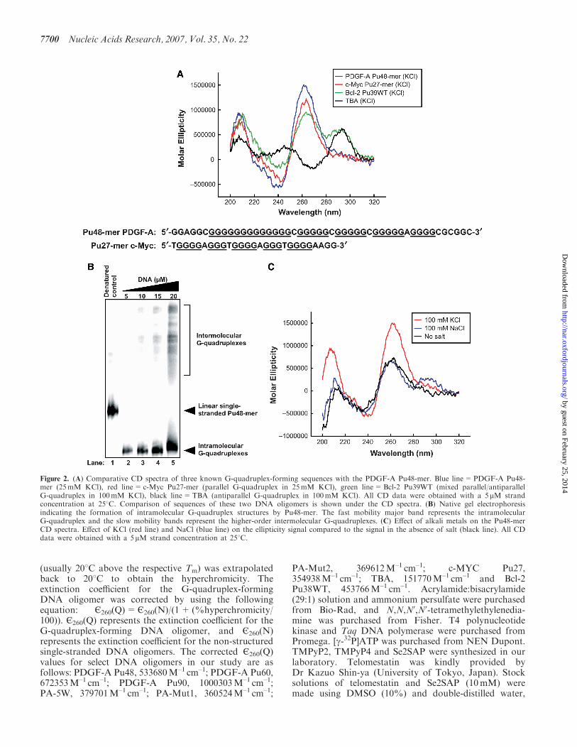
Figure 3. Effect of K+ and Na+ on the formation of the NHEPDGF-A G-quadruplex in a Taq polymerase stop assay. Two stop products aredesignated as the 50-end product and the 30-end stop product. The corresponding arrest sites are indicated on the core G-tract sequence.
Nucleic Acids Research, 2007, Vol. 35, No. 22 7703
by guest on February 25, 2014http://nar.oxfordjournals.org/
Dow
nloaded from
The specific stabilizing effect of K+ on the formation ofG-quadruplexes in the NHEPDGF-A was confirmed by Taqpolymerase stop assay. This assay is a useful tool toinvestigate the capability of G-rich DNA sequences toform stable G-quadruplex structures in vitro, based on theassumption that a stable DNA secondary structure inthe DNA template is able to stop primer extension by theDNA polymerase (33). A cassette template DNA contain-ing the Pu48-mer of NHEPDGF-A was annealed with alabeled primer, which subsequently was extended with Taqpolymerase in the presence of increasing concentrations ofKCl or NaCl. A full-length product was synthesized in thepresence of NaCl (Figure 3, lanes 3–5); however, uponreplacement of NaCl by KCl to the reaction, two majorpremature primer extension products were produced anddesignated as the 50-end stop product and the 30-end stopproduct (Figure 3, lanes 7–9). Both of these arrestproducts increased in a K+-dependent manner but werenot affected by Na+. Thus, the K+-specific block to DNAsynthesis is not due to general effects of alkali ions.Collectively, these results suggest that the G-rich strand ofNHEPDGF-A has the capacity to form two separateintramolecular parallel G-quadruplex structures that arepreferentially stabilized by K+.
Formation of multiple G-quadruplex structures inthe G-rich strand of NHEPDGF-A
The G-rich strand of NHEPDGF-A is composed of fiveG-tracts and has the potential to form multipleG-quadruplexes structures, based on the assumption thateach G-tract has an equal possibility of participating inthe formation of G-quadruplexes. The Taq polymerasestop assay in Figure 3 showed that two significant stopproducts are formed within the NHEPDGF-A. Thisindicates that at least two major, stable intramolecularG-quadruplex structures are formed by different G-tractsin the G-rich strand of NHEPDGF-A. The Taq polymerasearrest sites for these two major stop products weredetermined by sequencing the template DNA. Thearrested site for the 30-end stop product corresponded toG41 in the PDGF-A Pu48-mer (core sequence ofNHEPDGF-A, as shown under the gel in Figure 3). Thearrest product at G41 suggest that bases G37–G41 mustform a stable cap structure on the 30-end of theG-quadruplex. The arrested site for the 50-end stopproduct corresponded to G25 in the Pu48-mer(Figure 3). This indicates that the three consecutive runsof guanines at the 50-end of Pu48-mer (G1–G25) formthe G-quadruplex structure of the 50-end stop product.
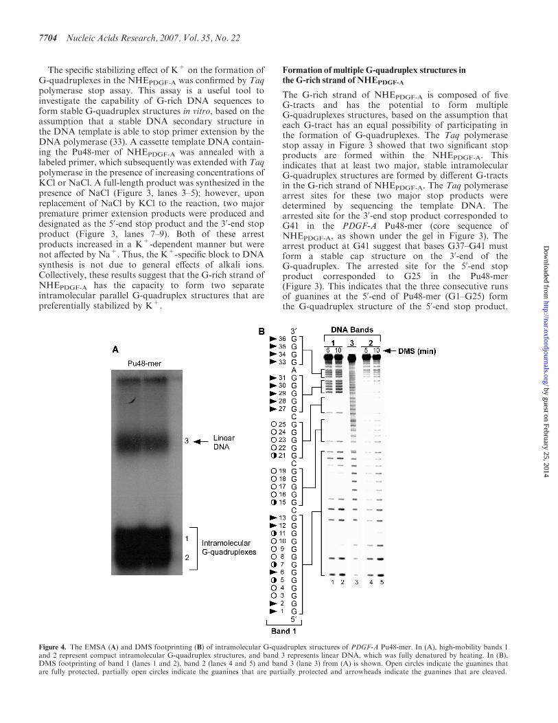
Figure 4. The EMSA (A) and DMS footprinting (B) of intramolecular G-quadruplex structures of PDGF-A Pu48-mer. In (A), high-mobility bands 1and 2 represent compact intramolecular G-quadruplex structures, and band 3 represents linear DNA, which was fully denatured by heating. In (B),DMS footprinting of band 1 (lanes 1 and 2), band 2 (lanes 4 and 5) and band 3 (lane 3) from (A) is shown. Open circles indicate the guanines thatare fully protected, partially open circles indicate the guanines that are partially protected and arrowheads indicate the guanines that are cleaved.
7704 Nucleic Acids Research, 2007, Vol. 35, No. 22
by guest on February 25, 2014http://nar.oxfordjournals.org/
Dow
nloaded from
One structural characteristic of particular interest isthat the 13-mer G-tract must provide the two runsof presumably four guanines at the 50-end of theG-quadruplex.
Native polyacrylamide gel electrophoresis of Pu48-mer,which was pre-incubated with 100mM KCl, showstwo fast migrating bands (Figure 4A, bands 1 and 2)that move faster than the denatured unstructured linearDNA species (band 3). These high-mobility speciesare presumed to represent compact intramolecularG-quadruplex structures. The close mobility andbroadness of two intramolecular G-quadruplexes(bands 1 and 2) indicate that the Pu48-mer mayform multiple similarly structured intramolecularG-quadruplexes.
To further probe the structures of the two intramolec-ular G-quadruplexes, a DMS footprinting experimentwas carried out. Since N7 of guanine in a G-tetrad isinvolved in Hoogsteen bonding and is inaccessible toalkylation by DMS, we can identify those guanine basesengaged in the formation of a G-quadruplex structure.The DMS methylation pattern produced by band 3 wasconsistent with a linear single-stranded DNA (Figure 4B,lane 3). For bands 1 and 2 (Figure 4B, lanes 1 and 2 andlanes 3 and 4), the DMS protection patterns suggested theformation of intramolecular G-quadruplexes. As shown inFigure 4B (lanes 1 and 2), full DMS protection occurredwithin the four runs of guanines (G3–G4, G8–G10, G16–G19 and G22–G25) of band 1. For band 1, the DMSprotection of the two four-guanine G-tracts (G16–G19,
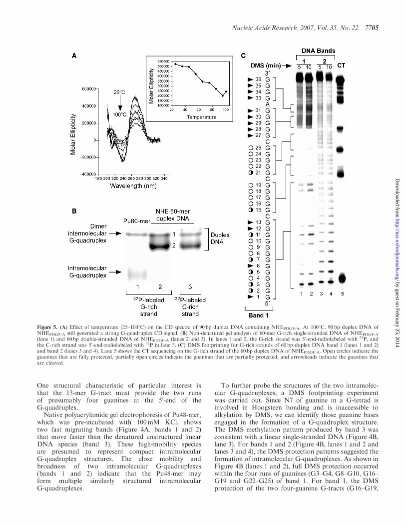
Figure 5. (A) Effect of temperature (25–1008C) on the CD spectra of 90 bp duplex DNA containing NHEPDGF-A. At 1008C, 90 bp duplex DNA ofNHEPDGF-A still generated a strong G-quadruplex CD signal. (B) Non-denatured gel analysis of 60-mer G-rich single-stranded DNA of NHEPDGF-A
(lane 1) and 60 bp double-stranded DNA of NHEPDGF-A (lanes 2 and 3). In lanes 1 and 2, the G-rich strand was 50-end-radiolabeled with 32P, andthe C-rich strand was 50-end-radiolabeled with 32P in lane 3. (C) DMS footprinting for G-rich strands of 60 bp duplex DNA band 1 (lanes 1 and 2)and band 2 (lanes 3 and 4). Lane 5 shows the CT sequencing on the G-rich strand of the 60 bp duplex DNA of NHEPDGF-A. Open circles indicate theguanines that are fully protected, partially open circles indicate the guanines that are partially protected, and arrowheads indicate the guanines thatare cleaved.
Nucleic Acids Research, 2007, Vol. 35, No. 22 7705
by guest on February 25, 2014http://nar.oxfordjournals.org/
Dow
nloaded from

G22–G25) at the 30-end of the NHEPDGF-A is clear, whilethe remaining two G-tracts within the run of 13 guaninesat the 50-end of the NHEPDGF-A are less well defined.Within the long run of 13 guanines there are two runs ofguanines that are clearly protected: a run of two guanines(G3–G4) and three guanines (G8–G10). Partial protectionof the intervening run of guanines (G5 and G7) occursbetween these two fully protected regions, and likewiseG11 is partially protected. The first guanine involved inthe formation of the G-tetrad at the 30-end of band 1 isG25. This corresponds to the arrest site for the 50-end stopproduct in the Taq polymerase stop assay (Figure 3). Forband 2 (lanes 4 and 5), most guanines within the two50-runs of guanine (G1–G13 and G15–G19), with theexception of G1 and G2, were partially protected fromDMS methylation, with only two runs of guanines (G22–G25 and G27–G30) showed full protection (Figure 4B,lanes 4 and 5). These somewhat ill-defined N7 methylationprotection patterns do not provide straightforward insightinto the specific G-quadruplex structures for the DNAspecies in bands 1 and 2. Most probably these ill-definedbands result from mixtures of multiple intramolecularG-quadruplexes existing in dynamic equilibrium (seeDiscussion Section).
Formation of a G-quadruplex within the double-strandedDNA of NHEPDGF-A. To date, all well-characterizedG-quadruplex structures have been determined by study-ing single-stranded DNA in vitro (25–30,35,36,41,42). Toinvestigate the potential for G-quadruplex formationwithin the duplex DNA of NHEPDGF-A, we used CD tostudy a 90 base pair duplex DNA, derived from the –24 to–113 region of the PDGF-A promoter, that containedabout 20 base pairs flanking each side of the NHE coresequence. Significantly, the CD spectrum of the NHE90 bp double-stranded DNA exhibited an almost identicalCD spectrum to the parallel G-quadruplex formed in thesingle-stranded DNA (Figure 2A, blue line), which ischaracterized by a positive ellipticity maximum at 266 nmand a negative minimum at 240 nm (Figure 5A). This CDspectrum is substantially different from the CD signalof B-form duplex DNA, which typically has an ellipticitymaximum absorption around 275–280 nm and zeroellipticity at 258 nm. In addition, the CD bands ofB-form DNA are much weaker (34,50,51). This CDprofile of the NHE 90 bp double-stranded DNA indicatesthat a dominant intramolecular parallel G-quadruplex isformed within the duplex DNA of NHEPDGF-A. Todetermine the stability of this G-quadruplex structure,we monitored the CD spectra of the NHE 90 bp double-stranded DNA over a 25–1008C temperature range.Notably, the DNA sample still generated very strongparallel G-quadruplex CD signals, even above 1008C(Figure 5A). This indicates that the G-quadruplexstructure formed in the NHE duplex DNA is very stable.To identify the G-quadruplex structure formed in the
NHE duplex DNA, a 60 bp double-stranded DNA ofNHE (–33 to –92 in the PDGF-A promoter), which wasshown to have better resolution for DMS footprintingthan the 90 bp double-stranded DNA, was used. The CDspectrum of purified NHE 60-mer double-stranded DNA
was consistent with that of the NHE 90 bp double-stranded DNA (data not shown). EMSA was performedto isolate the duplex DNA species containing theG-quadruplex structure, which was 50-end 32P-labeled inthe G-rich and C-rich strands. Two bands (Figure 5B,bands 1 and 2) were observed in both duplex DNAsamples (lanes 2 and 3) pre-incubated with 100mM KCl.These bands were duplex DNA species, since they bothmigrated differently to the single-stranded intramolecularG-quadruplex species. The duplex DNA species contain-ing the partially unwound region was less compact thanthe linear duplex DNA and as expected showed slowermigration in EMSA. Thus band 1 corresponds to theduplex DNA species containing the G-quadruplex struc-ture, while band 2 corresponds to the fully linear duplexDNA. By coincidence, this band migrates at the samepoint as the presumed dimer intermolecular species fromthe Pu60-mer. However, it cannot correspond to thisspecies since the NHE 60 bp duplex DNA labeled on theC-rich strand also gives this species. Bands 1 and 2 wereboth extracted and subjected to DMS footprinting. Thefull DMS cleavage pattern of band 2 confirmed that it wasa linear duplex DNA (Figure 5C, lanes 3 and 4). A distinctDMS protection pattern among three runs of guanines(G3–G11, G16–G19 and G22–G25) was observed for theG-rich strand of band 1 (Figure 5C, lanes 1 and 2).Although it is not quite as well defined as the DMSfootprinting patterns of the G-quadruplex speciesobtained from single-stranded DNA (see Figure 4B,
Figure 6. Sequences (A) and comparative CD spectra (B) of the wild-type sequence of the three 50-end runs of guanines and the G15/G21double mutation sequences in the presence of 100mM KCl. Blackline=wild-type sequence PA-5W, red line=double G-to-T mutationsequence PA-Mut1 and blue line=double G-to-A mutation sequencePA-Mut2. All CD data were obtained with a 5mM strand concentra-tion at 258C.
7706 Nucleic Acids Research, 2007, Vol. 35, No. 22
by guest on February 25, 2014http://nar.oxfordjournals.org/
Dow
nloaded from

lanes 1 and 2), the DMS footprinting pattern of band 1corresponds very closely to that of the single-strandedspecies associated with the 50-end product seen in the Taqpolymerase stop assay.
Mutation of guanines in the loops leads to dramaticdestabilization of the PDGF-A G-quadruplexes
A full mutational analysis of the three consecutive 50-runsof guanines provided further insight into the sequencerequirements for stability of the NHEPDGF-A
G-quadruplexes. In accordance with the DMS footprint-ing studies on the predicted mixture of loop isomers (seeDiscussion Section), 12 sets of mutants, in which theindividual loop isomers were selected for, were comparedby the relative intensity of the CD at 285 nM (data notshown). In all cases, the mutant sequences that werepredicted to form defined loop isomers showed lowerstability. Surprisingly, all these guanine mutant isomersshowed a significant decrease in parallel G-quadruplexsignals and an increase in antiparallel signals. This showsthat these mutants predominantly form antiparallel orhybrid parallel/antiparallel G-quadruplex structures,which is different from the biologically relevantNHEPDGF-A G-quadruplex. Among these guaninemutant isomers, the most surprising results originatedfrom two dual mutants, PA-Mut1 and PA-Mut2(Figure 6A). These two mutant oligomers were designedto provide dual G-to-T or G-to-A base mutations at G15and G21 in the core sequence of NHEPDGF-A. PA-Mut1and PA-Mut2 were chosen because of the identifiedcleavage sites shown in the DMS footprinting of thePDGF-A Pu48 and the 60 bp duplex of NHEPDGF-A
(Figures 4B and 5C). Therefore, G15 and G21 are notrequired for G-tetrad formation, and it was predicted thata mutation at these positions would not affect the stabilityor folding of the G-quadruplex in the NHEPDGF-A.Unexpectedly, PA-Mut1 and PA-Mut2 both showed asignificant decrease in ellipticity at 212 and 266 nm and anincrease in the antiparallel shoulder absorption between280 and 300 nm, compared with the wild-type sequence ofthe three consecutive 50-runs of guanines of NHEPDGF-A
(PA-5W) (Figure 6B). Substitution of other loop-positionguanines with other bases also resulted in the samechanges in CD.
Stabilization of the G-quadruplexes in the NHEPDGF-A withsmall molecules
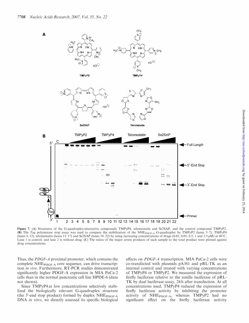
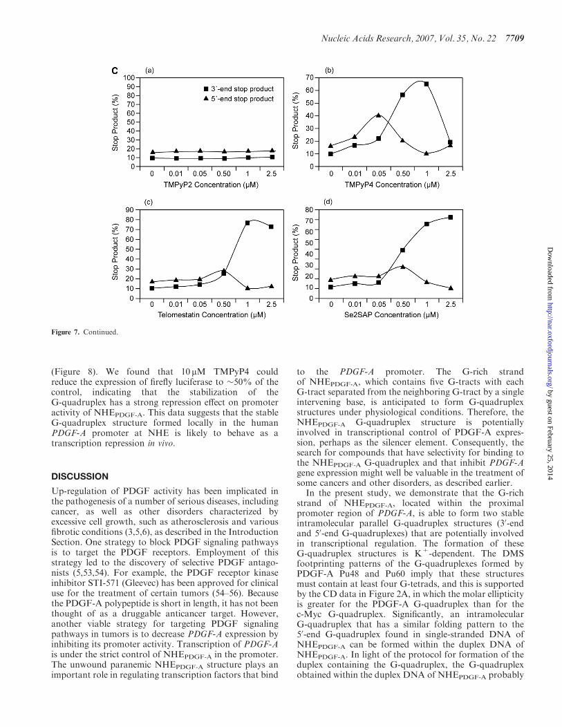
One important goal of our studies is to identifysmall molecules with the ability to specifically modulatePDGF-A gene expression through stabilization or mod-ification of the biologically relevant G-quadruplexstructures in the NHEPDGF-A. To gain further insightinto the selective action of G-quadruplex-interactiveagents with the NHEPDGF-A sequence, three well-established G-quadruplex-interactive agents, TMPyP4,telomestatin and Se2SAP (Figure 7A), were evaluated bya Taq polymerase stop assay with a DNA templatecontaining the whole core sequence of the G-rich strandof NHEPDGF-A. TMPyP2 (Figure 7A), a positional isomerof TMPyP4 that lacks the ability to insert or stack into
the G-tetrad structures (52), served as the negative controlcompound in our studies. Since there was a considerableamount of 50-end stop products formed in the absence ofdrugs and KCl (Figure 7B, lane 2), we used an elevatedprimer extension temperature (608C) to partially destabi-lize the arresting G-quadruplex structure and therebypermit a larger window for drug stabilization of theG-quadruplex structures. The pausing at the 50-end stopproduct in the absence of G-quadruplex-interactiveligands and KCl at a high temperature of 608C is anotherindication that the G-quadruplex structure in the 50-endproduct is more stable than other possible G-quadruplexstructures formed by the G-rich strand of NHEPDGF-A
(Figure 7B, lane 2). This suggests that the 50-endG-quadruplex is the primary G-quadruplex structureformed in NHEPDGF-A, corresponding to the one formedin the NHE duplex DNA.The results in Figure 7B (lanes 3–7) and Figure 7C (a)
show that TMPyP2 had a negligible effect on theformation of polymerase stop arrest products. When theconcentration of drugs was 0.05 mM and lower (Figure 7B,lanes 8 and 9), TMPyP4 significantly increased stabiliza-tion of the 50-end stop products in a concentration-dependent manner, but telomestatin and Se2SAP onlymarginally increased the formation of 50-end stopproducts (lanes 13, 14, 19 and 20 in Figure 7B). Incontrast, when the concentration of all three drugs was0.5mM and higher, decreased formation of the 50-end stopproducts was observed for all three G-quadruplex-interactive drugs, because of the accumulation of the30-end stop product. TMPyP4 achieved the maximumstabilizing effect on the 50-end stop products at aconcentration of 0.05 mM [Figure 7B, lane 9, and C (b)].The Taq polymerase stop assay also showed that thesethree G-quadruplex-interactive drugs induced formationof the 30-end stop products at high drug concentrations(�0.5 mM). A concentration-dependent increase in stabi-lization of the 30-end stop products was also observed forall three drugs. However, telomestatin and Se2SAP weremore selective at stabilizing the 30-end stop product;both achieved the best stabilizing effect on the 30-endG-quadruplex at a concentration of 1 mM [Figure 7B, lane21, and C (c and d)]. In addition, at drug concentrations of1 and 2.5 mM, TMPyP4 exhibited preferential bindingfor duplex DNA over the G-quadruplex structures(lanes 11 and 12 in Figure 7B).
TMPyP4 selectively inhibits the promoter activity of theNHEPDGF-A
In light of our results demonstrating that the duplex DNAof NHEPDGF-A can form a G-quadruplex structure, wesought to determine if stabilization of this G-quadruplexstructure affects PDGF-A transcription in vivo. Therefore,we constructed pGL3-Basic derivative (pA361) in whichthe human genomic DNA fragment containing theproximal promoter region of PDGF-A (–218 to +142,relative to the transcriptional start site) was insertedupstream of the firefly luciferase reporter gene. Whentransfected into MIA PaCa-2 human pancreatic cancercells, pA361 drove luciferase expression into these cells.
Nucleic Acids Research, 2007, Vol. 35, No. 22 7707
by guest on February 25, 2014http://nar.oxfordjournals.org/
Dow
nloaded from
Thus, the PDGF-A proximal promoter, which contains thecomplete NHEPDGF-A core sequence, can drive transcrip-tion in vivo. Furthermore, RT-PCR studies demonstratedsignificantly higher PDGF-A expression in MIA PaCa-2cells than in the normal pancreatic cell line HPDE-6 (datanot shown).Since TMPyP4 at low concentrations selectively stabi-
lized the biologically relevant G-quadruplex structure(the 50-end stop product) formed by duplex NHEPDGF-A
DNA in vitro, we directly assessed its specific biological
effects on PDGF-A transcription. MIA PaCa-2 cells wereco-transfected with plasmids pA361 and pRL-TK as aninternal control and treated with varying concentrationsof TMPyP4 or TMPyP2. We measured the expression offirefly luciferase relative to the renilla luciferase of pRL-TK by dual luciferase assay, 24 h after transfection. At allconcentrations used, TMPyP4 reduced the expression offirefly luciferase activity by inhibiting the promoteractivity of NHEPDGF-A, whereas TMPyP2 had nosignificant effect on the firefly luciferase activity
Figure 7. (A) Structures of the G-quadruplex-interactive compounds TMPyP4, telomestatin and Se2SAP, and the control compound TMPyP2.(B) The Taq polymerase stop assay was used to compare the stabilization of the NHEPDGF-A G-quadruplex by TMPyP2 (lanes 3–7), TMPyP4(lanes 8–12), telomestatin (lanes 13–17) and Se2SAP (lanes 18–22) by using increasing concentrations of drugs (0.01, 0.05, 0.5, 1 and 2.5 mM) at 608C.Lane 1 is control, and lane 2 is without drug. (C) The ratios of the major arrest products of each sample to the total product were plotted againstdrug concentrations.
7708 Nucleic Acids Research, 2007, Vol. 35, No. 22
by guest on February 25, 2014http://nar.oxfordjournals.org/
Dow
nloaded from
(Figure 8). We found that 10 mM TMPyP4 couldreduce the expression of firefly luciferase to �50% of thecontrol, indicating that the stabilization of theG-quadruplex has a strong repression effect on promoteractivity of NHEPDGF-A. This data suggests that the stableG-quadruplex structure formed locally in the humanPDGF-A promoter at NHE is likely to behave as atranscription repression in vivo.
DISCUSSION
Up-regulation of PDGF activity has been implicated inthe pathogenesis of a number of serious diseases, includingcancer, as well as other disorders characterized byexcessive cell growth, such as atherosclerosis and variousfibrotic conditions (3,5,6), as described in the IntroductionSection. One strategy to block PDGF signaling pathwaysis to target the PDGF receptors. Employment of thisstrategy led to the discovery of selective PDGF antago-nists (5,53,54). For example, the PDGF receptor kinaseinhibitor STI-571 (Gleevec) has been approved for clinicaluse for the treatment of certain tumors (54–56). Becausethe PDGF-A polypeptide is short in length, it has not beenthought of as a druggable anticancer target. However,another viable strategy for targeting PDGF signalingpathways in tumors is to decrease PDGF-A expression byinhibiting its promoter activity. Transcription of PDGF-Ais under the strict control of NHEPDGF-A in the promoter.The unwound paranemic NHEPDGF-A structure plays animportant role in regulating transcription factors that bind
to the PDGF-A promoter. The G-rich strandof NHEPDGF-A, which contains five G-tracts with eachG-tract separated from the neighboring G-tract by a singleintervening base, is anticipated to form G-quadruplexstructures under physiological conditions. Therefore, theNHEPDGF-A G-quadruplex structure is potentiallyinvolved in transcriptional control of PDGF-A expres-sion, perhaps as the silencer element. Consequently, thesearch for compounds that have selectivity for binding tothe NHEPDGF-A G-quadruplex and that inhibit PDGF-Agene expression might well be valuable in the treatment ofsome cancers and other disorders, as described earlier.In the present study, we demonstrate that the G-rich
strand of NHEPDGF-A, located within the proximalpromoter region of PDGF-A, is able to form two stableintramolecular parallel G-quadruplex structures (30-endand 50-end G-quadruplexes) that are potentially involvedin transcriptional regulation. The formation of theseG-quadruplex structures is K+-dependent. The DMSfootprinting patterns of the G-quadruplexes formed byPDGF-A Pu48 and Pu60 imply that these structuresmust contain at least four G-tetrads, and this is supportedby the CD data in Figure 2A, in which the molar ellipticityis greater for the PDGF-A G-quadruplex than for thec-Myc G-quadruplex. Significantly, an intramolecularG-quadruplex that has a similar folding pattern to the50-end G-quadruplex found in single-stranded DNA ofNHEPDGF-A can be formed within the duplex DNA ofNHEPDGF-A. In light of the protocol for formation of theduplex containing the G-quadruplex, the G-quadruplexobtained within the duplex DNA of NHEPDGF-A probably
Figure 7. Continued.
Nucleic Acids Research, 2007, Vol. 35, No. 22 7709
by guest on February 25, 2014http://nar.oxfordjournals.org/
Dow
nloaded from

forms prior to the formation of the duplex species.Thus, the species formed during the annealing is a resultof hybridization of the single-stranded G-rich strandalready containing the G-quadruplex with the single-stranded C-rich strand. This is unlikely to be the samemechanism for the formation of the G-quadruplexstructure within the duplex DNA in in vivo situationswhere superhelicity is involved (see subsequently).Nevertheless, this is the first time that we have beenable to isolate the duplex DNA containing aG-quadruplex structure in vitro. This unique structure islikely to represent the dominant biologically relevantG-quadruplex structure formed in the PDGF-A promoter.It has been proposed that superhelicity of genomic DNAmay play an important role in determining the presence ofsuch an interconversion (57,58). The functional analysis ofthe PDGF-A promoter revealed that the NHEPDGF-A
adopted a non-B-DNA conformation and was hypersen-sitive to the S1 nuclease in the supercoiled plasmids(13–16). This provides further evidence that the formationof a G-quadruplex in the NHEPDGF-A can competewith the superhelical duplex conformation in vivo. Theimplication of the formation of a stable G-quadruplexin duplex DNA is that the C-rich strand forms a single-stranded DNA loop, which may be in equilibrium with ani-motif structure.The folding pattern of the biologically relevant
G-quadruplex structure formed by the three consecutiveG-tracts (G1–G25) at the 50-end of NHEPDGF-A duplexDNA most likely represents a mixture of different loopisomers. Of particular interest, the 13-mer G-tract(G1–G13) must provide the two runs of guanines at the50-end. The flanking regions of NHEPDGF-A are also madeup of polypurine and polypyrimidine elements, whichmay have a significant impact on the propensity to formthe G-quadruplex in the G-rich strand of NHEPDGF-A
double-stranded DNA. The overall folding patterns ofthe parallel G-quadruplex structure in NHEPDGF-A
duplex DNA has been defined by its characteristic CDspectra, which demonstrates that it has a unique
double-chain reversal structure. Previously, examples ofG-quadruplexes consisting of double-chain reversals inpromoter regions have been confined to three tetrads witha single base in the internal loop. This is the first exampleof a G-quadruplex with four tetrads and a two-baseinternal loop. The DMS footprinting pattern of theG-quadruplex in NHEPDGF-A duplex DNA reveals thatthe 30-face of this G-quadruplex structure has two runs offour guanines, which are separated by a two-base (CG)internal loop. A molecular model of the four G-tetrads inthe NHEPDGF-A G-quadruplex structure shows that atwo-base internal loop is the minimum required to bridgetwo runs of four guanines (Figure 9A). The opposite50-face of this G-quadruplex structure is constructed fromthe run of 13 contiguous guanines, in which the DMSfootprinting pattern predicts a mixture of at least fourdouble-chain reversal loop isomers (Figure 9B). Loopisomers ‘a’, ‘b’, and ‘c’ in Figure 9B have a two-baseinternal loop on the 50-face dictated by the demonstratedDMS cleavages of G11, G12 and G13 (‘a’ isomer),G12 and G13 (‘b’ isomer) and G1 and G2 (‘c’ isomer) inthe core sequence of NHEPDGF-A (see Figure 4B). Finally,the cleavage of G5, G6 and G7 predicts loop isomer ‘d’.By analogy with the c-Myc parallel G-quadruplexstructure, in which the double-chain reversal loops onthe opposite face are sufficient to stabilize a thirdintervening loop size of six bases, the double-chainreversals in the NHEPDGF-A G-quadruplex can stabilizea third internal loop of up to five bases (as predicted inloop isomer ‘a’ in Figure 9B) (38). Two views of amolecular model of loop isomer ‘a’ having the predictedfolding pattern are shown in Figure 9A.
A surprising finding illustrated by the CD results shownin Figure 6B for two dual mutant oligomers is that basechanges, even in potential loop positions, result indestabilization and apparent dramatic changes in thefolding patterns. This is in contrast to the c-MycG-quadruplex, where elimination of the redundancy inloop isomers by judicial G-to-T mutations does notdramatically change the stability or folding patternsof the parallel-stranded G-quadruplex (38). The effectsof G-to-N mutations in the NHEPDGF-A G-quadruplexesare not limited to the run of 13 contiguous guanines, butalso occur when the 30 terminal guanines in the two runs of5 guanines (G15 and G21) are mutated to thymines oradenines. On the basis of DMS cleavage results, theseguanines are predicted to be predominantly in the loops,implying that the dynamic equilibrium between differentloop isomers is an important feature of both stability andformation of the parallel-stranded species. This phenom-enon may be uniquely linked to the 13 contiguousguanines in this promoter sequence.
An important aim of our research program is to identifysmall molecules that can selectively modulate PDGF-Atranscription by stabilizing or modifying the uniqueG-quadruplex structures formed by NHEPDGF-A.Therefore, we examined the affinities of three well-known G-quadruplex-interactive drugs (TMPyP4,Se2SAP and telomestatin) for the NHEPDGF-A
G-quadruplex. The results of our Taq polymerase stopassays revealed that these three drugs preferentially
Figure 8. Dual luciferase assay to determine the effect of TMPyP4 andTMPyP2 on the transcriptional activity of PDGF-A basal promotercontaining the NHE. The comparative firefly luciferase expressions(firefly/renilla) of TMPyP2 and TMPyP4 are shown in the histograms.The values are the average of three independent experiments. Errorbars are � SE.
7710 Nucleic Acids Research, 2007, Vol. 35, No. 22
by guest on February 25, 2014http://nar.oxfordjournals.org/
Dow
nloaded from

interact with and stabilize the 50-end NHEPDGF-A
G-quadruplex, which has a similar folding pattern to thebiological G-quadruplexes formed in the NHEPDGF-A
duplex DNA at low concentrations. TMPyP4 exhibitedstronger selectivity than either Se2SAP or telomestatinfor stabilization of the 50-end stop product. Thus,TMPyP4 appeared to be an ideal candidate for modulat-ing PDGF-A transcription. Our data confirms that, incells, TMPyP4 can specifically downregulate PDGF-Apromoter activity by interacting with the NHEPDGF-A
G-quadruplex. Because TMPyP4, and not TMPyP2,inhibits PDGF-A proximal promoter activity, this isstrong evidence that G-quadruplexes exist in vivo andthat NHEPDGF-A G-quadruplex structures act as tran-scriptional silencers for PDGF-A.
Finally, the relationship of the 50SHS and intron SHSelements to the dynamic equilibrium of the G-quadruplexand associated C-strand structures remains to be deter-mined. The complementarity of the 50SHS and the 50-endof the intron SHS is suggestive of the interaction of theseelements. Experiments addressing this question are inprogress.
ACKNOWLEDGEMENTS
This research has been supported by grants from theNational Institutes of Health (CA94166) and the ArizonaBiomedical Research Commission (9006). We thank JohnFitch, Michael Cusanovich and members of the
Figure 9. (A) Model of the biologically relevant NHEPDGF-A G-quadruplex [loop isomer 50-(2,5,2)-30], which contains two 2-base double-chainreversal loops and one 5-base intervening loop (guanines= red, cytosines=yellow and K+ ions=white). For clarity, hydrogen atoms have beenremoved. In the left panel, the two 2-base double-chain reversal loops are shown on each side of model, and in the right panel, the model has beenrotated to show the 5-base intervening loop on the right side of model. (B) Proposed folding patterns of the four different loop isomers formed in thecore sequence of NHEPDGF-A. Guanines= red, cytosines=yellow.
Nucleic Acids Research, 2007, Vol. 35, No. 22 7711
by guest on February 25, 2014http://nar.oxfordjournals.org/
Dow
nloaded from
Cusanovich lab (University of Arizona) for providingequipment and technical support for oligonucleotideextinction coefficient calculation. We are grateful toDavid Bishop for preparing, proofreading and editingthe final version of the manuscript and figures. Funding topay the Open Access publication charges for this articlewas provided by NIH CA94166.
Conflict of interest statement. None declared.
REFERENCES
1. Heldin,C.H. and Westermark,B. (1999) Mechanism of action andin vivo role of platelet-derived growth factor. Physiol. Rev., 79,1283–1316.
2. Heldin,C.H., Eriksson,U. and Ostman,A. (2002) New membersof the platelet-derived growth factor family of mitogens.Arch. Biochem. Biophys., 398, 284–290.
3. Alvarez,R.H., Kantarjian,H.M. and Cortes,J.E. (2006) Biology ofplatelet-derived growth factor and its involvement in disease. MayoClin. Proc., 81, 1241–1257.
4. Betsholtz,C. (2003) Biology of platelet-derived growth factors indevelopment. Birth Defects Res. C Embryo. Today, 69, 272–285.
5. Ostman,A. and Heldin,C.H. (2001) Involvement of platelet-derivedgrowth factor in disease: development of specific antagonists.Adv. Cancer Res., 80, 1–38.
6. Yu,J., Ustach,C. and Kim,H.R. (2003) Platelet-derived growthfactor signaling and human cancer. J. Biochem. Mol. Biol., 36,49–59.
7. Westermark,B., Heldin,C.H. and Nister,M. (1995) Platelet-derivedgrowth factor in human glioma. Glia, 15, 257–263.
8. Sulzbacher,I., Birner,P., Trieb,K., Traxler,M., Lang,S. and Chott,A.(2003) Expression of platelet-derived growth factor-AA is associatedwith tumor progression in osteosarcoma. Mod. Pathol., 16, 66–71.
9. Afrakhte,M., Nister,M., Ostman,A., Westermark,B. andPaulsson,Y. (1996) Production of cell-associated PDGF-AA by ahuman sarcoma cell line: evidence for a latent autocrine effect.Int. J. Cancer, 68, 802–809.
10. Guha,A., Dashner,K., Black,PM., Wagner,J.A. and Stiles,C.D.(1995) Expression of PDGF and PDGF receptors in humanastrocytoma operation specimens supports the existence of anautocrine loop. Int. J. Cancer, 60, 168–173.
11. Sulzbacher,I., Traxler,M., Mosberger,I., Lang,S. and Chott,A.(2000) Platelet-derived growth factor-AA and -a receptor expressionsuggests an autocrine and/or paracrine loop in osteosarcoma.Mod. Pathol., 13, 632–637.
12. Takimoto,Y., Wang,Z.Y., Kobler,K. and Deuel,T.F. (1991)Promoter region of the human platelet-derived growth factorA-chain gene. Proc. Natl Acad. Sci. USA, 88, 1686–1690.
13. Lin,X., Wang,Z., Gu,L. and Deuel,T.F. (1992) Functional analysisof the human platelet-derived growth factor A-chain promoterregion. J. Biol. Chem., 267, 25614–25619.
14. Kaetzel,D.M.Jr, Maul,R.S., Liu,B., Bonthron,D.,Fenstermaker,R.A. and Coyne,D.W. (1994) Platelet-derived growthfactor A-chain gene transcription is mediated by positive andnegative regulatory regions in the promoter. Biochem. J., 301,321–327.
15. Liu,B., Maul,R.S. and Kaetzel,D.M.Jr. (1996) Repression ofplatelet-derived growth factor A-chain gene transcription by anupstream silencer element. J. Biol. Chem., 271, 26281–26290.
16. Wang,Z.Y. and Deuel,T.F. (1996) S1-nuclease-sensitive structurescontribute to transcriptional regulation of the human PDGFA-chain gene. Prog. Nucleic Acid Res. Mol. Biol., 55, 227–244.
17. Kaetzel,D.M. (2003) Transcription of the platelet-derived growthfactor A-chain gene. Cytokine Growth Factor Rev., 14, 427–446.
18. Silverman,E.S., Khachigian,L.M., Lindner,V., Williams,A.J. andCollins,T. (1997) Inducible PDGF A-chain transcription in smoothmuscle cells is mediated by Egr-1 displacement of Sp1 and Sp3.Am. J. Physiol., 273, H1415–H1426.
19. Gashler,A.L., Bonthron,D.T., Madden,S.L., Rauscher,F.J.III,Collins,T. and Sukhatme,V.P. (1992) Human platelet-derivedgrowth factor A chain is transcriptionally repressed by the Wilms
tumor suppressor WT1. Proc. Natl Acad. Sci. USA, 89,10984–10988.
20. Wang,Z.Y., Madden,S.L., Deuel,T.F. and Rauscher,F.J.III (1992)The Wilms’ tumor gene product, WT1, represses transcription ofthe platelet-derived growth factor A-chain gene. J. Biol. Chem., 267,21999–22002.
21. Khachigian,L.M., Santiago,F.S., Rafty,L.A., Chan,O.L.-W.,Delbridge,G.J., Bobik,A., Collins,T. and Johnson,A.C. (1999) GCfactor 2 represses platelet-derived growth factor A-chain genetranscription and is itself induced by arterial injury. Circ. Res., 84,1258–1267.
22. Rafty,L.A., Santiago,F.S. and Khachigian,L.M. (2002) NF1/Xrepresses PDGF A-chain transcription by interacting with Sp1 andantagonizing Sp1 occupancy of the promoter. EMBO J., 21,334–343.
23. Ma,D., Xing,Z., Liu,B., Pedigo,N.G., Zimmer,S.G., Bai,Z.,Postel,E.H. and Kaetzel,D.M. (2002) NM23-H1 and NM23-H2repress transcriptional activities of nuclease-hypersensitive elementsin the platelet-derived growth factor-A promoter. J. Biol. Chem.,277, 1560–1567.
24. Wang,Z.Y., Lin,X.H., Nobuyoshi,M., Qiu,Q.Q. and Deuel,T.F.(1992) Binding of single-stranded oligonucleotides to a non-B-formDNA structure results in loss of promoter activity of the platelet-derived growth factor-A chain gene. J. Biol. Chem., 267,13669–13674.
25. Siddiqui-Jain,A., Grand,C.L., Bearss,D.J. and Hurley,L.H. (2002)Direct evidence for a G-quadruplex in a promoter region and itstargeting with a small molecule to repress c-MYC transcription.Proc. Natl Acad. Sci. USA, 99, 11593–11598.
26. Cogoi,S. and Xodo,L.E. (2006) G-quadruplex formation withinthe promoter of the KRAS proto-oncogene and its effect ontranscription. Nucleic Acids Res., 34, 2536–2549.
27. Dexheimer,T.S., Sun,D. and Hurley,L.H. (2006)Deconvoluting the structural and drug-recognition complexity ofthe G-quadruplex-forming region upstream of the bcl-2 P1promoter. J. Am. Chem. Soc., 128, 5404–5415.
28. Fernando,H., Reszka,A.P., Huppert,J., Ladame,S., Rankin,S.,Venkitaraman,A.R., Neidle,S. and Balasubramanian,S. (2006) Aconserved quadruplex motif located in a transcription activation siteof the human c-kit oncogene. Biochemistry, 45, 7854–7860.
29. Sun,D., Guo,K., Rusche,J.J. and Hurley,L.H. (2005) Facilitation ofa structural transition in the polypurine/polypyrimidine tract withinthe proximal promoter region of the human VEGF gene by thepresence of potassium and G-quadruplex-interactive agents.Nucleic Acids Res., 33, 6070–6080.
30. Yang,D. and Hurley,L.H. (2006) Structure of the biologicallyrelevant G-quadruplex in the c-MYC promoter. Nucleosides,Nucleotides Nucleic Acids, 25, 951–968.
31. Cantor,C.R., Warshaw,M.M. and Shapiro,H. (1970)Oligonucleotide interactions. III. Circular dichroism studies of theconformation of deoxyoligonucleotides. Biopolymers, 9, 1059–1077.
32. Sun,X.G., Cao,E.H., He,Y.J. and Qin,J.F. (1999) Spectroscopiccomparison of different DNA structures formed by oligonucleo-tides. J. Biomol. Struct. Dyn., 16, 863–872.
33. Han,H., Hurley,L.H. and Salazar,M. (1999) A DNA polymerasestop assay for G-quadruplex-interactive compounds.Nucleic Acids Res., 27, 537–542.
34. Maurizot,J.C. (2000) Circular dichroism of nucleic acids:nonclassical conformations and modified oligonucleotides.In: Berova,N., Nakanishi,K. and Woody,R.W. (eds), CircularDichroism: Principles and Applications, 2nd ed. Wiley-VCH,New York, pp. 719–736.
35. Dapic,V., Abdomerovic,V., Marrington,R., Peberdy,J., Rodger,A.,Trent,J.O. and Bates,P.J. (2003) Biophysical and biologicalproperties of quadruplex oligodeoxyribonucleotides. Nucleic AcidsRes., 31, 2097–2107.
36. Petraccone,L., Erra,E., Esposito,V., Randazzo,A., Mayol,L.,Nasti,L., Barone,G. and Giancola,C. (2004) Stability and structureof telomeric DNA sequences forming quadruplexes containing fourG-tetrads with different topological arrangements. Biochemistry, 43,4877–4884.
37. Rezler,E.M., Seenisamy,J., Bashyam,S., Kim,M.Y., White,E.,Wilson,W.D. and Hurley,L.H. (2005) Telomestatin and diselenosapphyrin bind selectively to two different forms of the human
7712 Nucleic Acids Research, 2007, Vol. 35, No. 22
by guest on February 25, 2014http://nar.oxfordjournals.org/
Dow
nloaded from
telomeric G-quadruplex structure. J. Am. Chem. Soc., 127,9439–9447.
38. Seenisamy,J., Rezler,E.M., Powell,T.J., Tye,D., Gokhale,V.,Joshi,C.S., Siddiqui-Jain,A. and Hurley,L.H. (2004)The dynamic character of the G-quadruplex element in thec-MYC promoter and modification by TMPyP4. J. Am. Chem. Soc.,126, 8702–8709.
39. Rujan,I.N., Meleney,J.C. and Bolton,P.H. (2005) Vertebrate telo-mere repeat DNAs favor external loop propeller quadruplexstructures in the presence of high concentrations of potassium.Nucleic Acids Res., 33, 2022–2031.
40. Miyoshi,D., Nakao,A. and Sugimoto,N. (2003) Structural transitionfrom antiparallel to parallel G-quadruplex of d(G4T4G4) induced byCa2+. Nucleic Acids Res., 31, 1156–1163.
41. Ambrus,A., Chen,D., Dai,J., Bialis,T., Jones,R.A. and Yang,D.(2006) Human telomeric sequence forms a hybrid-type intramole-cular G-quadruplex structure with mixed parallel/antiparallelstrands in potassium solution. Nucleic Acids Res., 34, 2723–2735.
42. Miyoshi,D., Matsumura,S., Nakano,S. and Sugimoto,N. (2004)Duplex dissociation of telomere DNAs induced by molecularcrowding. J. Am. Chem. Soc., 126, 165–169.
43. Balagurumoorthy,P. and Brahmachari,S.K. (1994) Structure andstability of human telomeric sequence. J. Biol. Chem., 269,21858–21869.
44. Risitano,A. and Fox,K.R. (2004) Influence of loop size on thestability of intramolecular DNA quadruplexes. Nucleic Acids Res.,32, 2598–2606.
45. Phan,A.T., Modi,Y.S. and Patel,D.J. (2004) Propeller-typeparallel-stranded G-quadruplexes in the human c-myc promoter.J. Am. Chem. Soc., 126, 8710–8716.
46. Kelly,J.A., Feigon,J. and Yeates,T.O. (1996) Reconciliationof the X-ray and NMR structures of the thrombin-bindingaptamer d(GGTTGGTGTGGTTGG). J. Mol. Biol., 256, 417–422.
47. Dai,J., Dexheimer,T.S., Chen,D., Carver,M., Ambrus,A.,Jones,R.A. and Yang,D. (2006) An intramolecular G-quadruplexstructure with mixed parallel/antiparallel G-strands formed in thehuman BCL-2 promoter region in solution. J. Am. Chem. Soc., 128,1096–1098.
48. Henderson,E., Hardin,C.C., Walk,S.K., Tinoco,I.Jr andBlackburn,E.H. (1987) Telomeric DNA oligonucleotides form novelintramolecular structures containing guanine–guanine base pairs.Cell, 51, 899–908.
49. Williamson,J.R., Raghuraman,M.K. and Cech,T.R. (1989)Monovalent cation-induced structure of telomeric DNA: theG-quartet model. Cell, 59, 871–880.
50. Vorlickova,M. and Kypr,J. (1985) Conformational variability ofpoly(dA-dT)�poly(dA-dT) and some other deoxyribonucleic acidsincludes a novel type of double helix. J. Biomol. Struct. Dyn., 3,67–83.
51. Kypr,J., Fialova,M., Chladkova,J., Tumova,M. and Vorlickova,M.(2001) Conserved guanine–guanine stacking in tetraplex and duplexDNA. Eur. Biophys. J., 30, 555–558.
52. Han,F.X., Wheelhouse,R.T. and Hurley,L.H. (1999) Interactions ofTMPyP4 and TMPyP2 with quadruplex DNA. Structural basis forthe differential effects on telomerase inhibition. J. Am. Chem. Soc.,121, 3561–3570.
53. Pietras,K., Sjoblom,T., Rubin,K., Heldin,C.H. and Ostman,A.(2003) PDGF receptors as cancer drug targets. Cancer Cell, 3,439–443.
54. George,D. (2001) Platelet-derived growth factor receptors: atherapeutic target in solid tumors. Semin. Oncol., 28, 27–33.
55. Madhusudan,S. and Ganesan,T.S. (2004) Tyrosine kinase inhibitorsin cancer therapy. Clin. Biochem., 37, 618–635.
56. Hofer,M.D., Fecko,A., Shen,R., Setlur,S.R., Pienta,K.G.,Tomlins,S.A., Chinnaiyan,A.M. and Rubin,M.A. (2004) Expressionof the platelet-derived growth factor receptor in prostate cancer andtreatment implications with tyrosine kinase inhibitors. Neoplasia, 6,503–512.
57. Havas,K., Flaus,A., Phelan,M., Kingston,R., Wade,P.A.,Lilley,D.M. and Owen-Hughes,T. (2000) Generation of superhelicaltorsion by ATP-dependent chromatin remodeling activities. Cell,103, 1133–1142.
58. Kumar,N. and Maiti,S. (2004) Quadruplex to Watson-Crick duplextransition of the thrombin binding aptamer: a fluorescenceresonance energy transfer study. Biochem. Biophys. Res. Commun.,319, 759–767.
Nucleic Acids Research, 2007, Vol. 35, No. 22 7713
by guest on February 25, 2014http://nar.oxfordjournals.org/
Dow
nloaded from
Related Documents