Characterization of the Cofactor-Induced Folding Mechanism of a Zinc-Binding Peptide Using Computationally Designed Mutants Jia Tang†, Seung-Gu Kang†, Jeffery G. Saven⁎ and Feng Gai⁎ Department of Chemistry, University of Pennsylvania, Philadelphia, PA 19104, USA Received 7 January 2009; received in revised form 26 March 2009; accepted 31 March 2009 Available online 8 April 2009 Metals are the most commonly encountered protein cofactors, and they play important structural and functional roles in biology. In many cases, metal binding provides a major driving force for a polypeptide chain to fold. While there are many studies on the structure, stability, and function of metal-binding proteins, there are few studies focusing on understanding the kinetic mechanism of metal-induced folding. Herein, the Zn 2+ -induced folding kinetics of a small zinc-binding protein are studied; the CH1 1 pep- tide is derived from the first cysteine/histidine-rich region (CH1 domain) of the protein interaction domains of the transcriptional coregulator CREB- binding protein. Computational design is used to introduce tryptophan and histidine mutations that are structurally consistent with CH1 1 ; these mutants are studied using stopped-flow tryptophan fluorescence experi- ments. The Zn 2+ -induced CH1 1 folding kinetics are consistent with two parallel pathways, where the initial binding of Zn 2+ occurs at two sites. However, the initially formed Zn 2+ -bound complexes can proceed either directly to the folded state where zinc adopts a tetrahedral coordination or to an off-pathway misligated intermediate. While elimination of those ligands responsible for misligation simplifies the folding kinetics, it also leads to a decrease in the zinc binding constant. Therefore, these results suggest why these nonnative zinc ligands in the CH1 1 motif are conserved in several distantly related organisms and why the requirement for function can lead to kinetic frustration in folding. In addition, the loop closure rate of the CH1 1 peptide is determined based on the proposed model and temperature-dependent kinetic measurements. © 2009 Elsevier Ltd. All rights reserved. Edited by C. R. Matthews Keywords: protein folding; computational design; stopped-flow fluorescence; zinc-binding peptides; folding intermediate Introduction The study of the folding mechanism of single- domain proteins that lack cofactors has reached an advanced stage wherein the sequence of events along the course of refolding after denaturation begins to be understood. 1–10 In several cases, it has been even possible to describe in detail the energetics, posi- tion, and structure of the folding transition-state en- sembles and/or intermediate states involved. 2,3,7–9 In contrast, there are far fewer studies on cofactor- induced folding kinetics, even though a large number of proteins require cofactors for proper folding and/or function. 11,12 While the effect of cofactor binding on structure and stability may differ for different proteins, many polypeptides can only attain a well-folded structure when associated with their respective cofactors. For example, cytochrome c, an important electron transfer protein, cannot fold without incorporation of its heme cofactor. 12 Simi- larly, many small metal-binding peptides, such as the zinc-finger motif, 13,14 also require their respec- tive cofactors to maintain the integrity of their native fold. For such systems, cofactor binding thus pro- vides not only stabilization but also a major driving force directing the folding of an otherwise unstruc- *Corresponding authors. E-mail addresses: [email protected]; [email protected]. † J.T. and S.-G.K. contributed equally to this work. Abbreviation used: SCADS, statistical computationally assisted design strategy. doi:10.1016/j.jmb.2009.03.074 J. Mol. Biol. (2009) 389, 90–102 Available online at www.sciencedirect.com 0022-2836/$ - see front matter © 2009 Elsevier Ltd. All rights reserved.

Welcome message from author
This document is posted to help you gain knowledge. Please leave a comment to let me know what you think about it! Share it to your friends and learn new things together.
Transcript
doi:10.1016/j.jmb.2009.03.074 J. Mol. Biol. (2009) 389, 90–102
Available online at www.sciencedirect.com
Characterization of the Cofactor-Induced FoldingMechanism of a Zinc-Binding Peptide UsingComputationally Designed Mutants
Jia Tang†, Seung-Gu Kang†, Jeffery G. Saven⁎ and Feng Gai⁎
Department of Chemistry,University of Pennsylvania,Philadelphia, PA 19104, USA
Received 7 January 2009;received in revised form26 March 2009;accepted 31 March 2009Available online8 April 2009
*Corresponding authors. E-mail [email protected]; [email protected]† J.T. and S.-G.K. contributed equAbbreviation used: SCADS, statis
assisted design strategy.
0022-2836/$ - see front matter © 2009 E
Metals are the most commonly encountered protein cofactors, and they playimportant structural and functional roles in biology. In many cases, metalbinding provides a major driving force for a polypeptide chain to fold.While there are many studies on the structure, stability, and function ofmetal-binding proteins, there are few studies focusing on understanding thekinetic mechanism of metal-induced folding. Herein, the Zn2+-inducedfolding kinetics of a small zinc-binding protein are studied; the CH11 pep-tide is derived from the first cysteine/histidine-rich region (CH1 domain) ofthe protein interaction domains of the transcriptional coregulator CREB-binding protein. Computational design is used to introduce tryptophan andhistidine mutations that are structurally consistent with CH11; thesemutants are studied using stopped-flow tryptophan fluorescence experi-ments. The Zn2+-induced CH11 folding kinetics are consistent with twoparallel pathways, where the initial binding of Zn2+ occurs at two sites.However, the initially formed Zn2+-bound complexes can proceed eitherdirectly to the folded state where zinc adopts a tetrahedral coordination orto an off-pathway misligated intermediate. While elimination of thoseligands responsible for misligation simplifies the folding kinetics, it alsoleads to a decrease in the zinc binding constant. Therefore, these resultssuggest why these nonnative zinc ligands in the CH11 motif are conservedin several distantly related organisms and why the requirement for functioncan lead to kinetic frustration in folding. In addition, the loop closure rate ofthe CH11 peptide is determined based on the proposed model andtemperature-dependent kinetic measurements.
© 2009 Elsevier Ltd. All rights reserved.
Keywords: protein folding; computational design; stopped-flow fluorescence;zinc-binding peptides; folding intermediate
Edited by C. R. MatthewsIntroduction
The study of the folding mechanism of single-domain proteins that lack cofactors has reached anadvanced stagewherein the sequence of events alongthe course of refolding after denaturation begins tobe understood.1–10 In several cases, it has been evenpossible to describe in detail the energetics, posi-tion, and structure of the folding transition-state en-
resses:n.edu.ally to this work.tical computationally
lsevier Ltd. All rights reserve
sembles and/or intermediate states involved.2,3,7–9
In contrast, there are far fewer studies on cofactor-induced folding kinetics, even though a largenumber of proteins require cofactors for properfolding and/or function.11,12 While the effect ofcofactor binding on structure and stabilitymay differfor different proteins, many polypeptides can onlyattain a well-folded structure when associated withtheir respective cofactors. For example, cytochromec, an important electron transfer protein, cannot foldwithout incorporation of its heme cofactor.12 Simi-larly, many small metal-binding peptides, such asthe zinc-finger motif,13,14 also require their respec-tive cofactors to maintain the integrity of their nativefold. For such systems, cofactor binding thus pro-vides not only stabilization but also a major drivingforce directing the folding of an otherwise unstruc-
d.

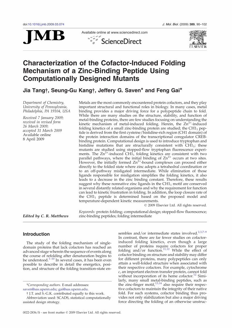
Fig. 1. The NMR structures of the CH11 Zn-bindingdomain (Protein Data Bank code 1liq).40 Zn-coordinatingresidues (gray) and residues of the triple mutant L7W/H9V/H22I (magenta) are shown.
91Zn2+-Induced Folding
tured protein to a well-defined structure. Nonethe-less, the prerequisite of a cofactor for folding maycomplicate the folding free-energy landscape andthus the kinetic mechanism15 because cofactorbinding often requires the formation of a preciselydefined cofactor-binding environment in the nativestate. In addition, a protein may also transientlyassociate with a cofactor in a nonnative fashion,leading to kinetic frustration in folding. For ex-ample, cytochrome c exhibits rather complex foldingkinetics arising from nonnative iron ligation duringor prior to folding.16–20 Herein, the folding of thedesigned variants of a small Zn2+-binding protein isexamined in an effort to probe the roles of native andnonnative interactions in the folding kinetics ofmetal-binding proteins.Metal ions such as Zn2+, Mg2+, and Ca2+ are
among the most commonly encountered cofactors.Metal binding not only plays many important struc-tural and functional roles in cells21 but also is inc-reasingly used in de novo protein designs to generateor stabilize a specific fold.22–32 For example, whencoordinated with a metal ion, polypeptides com-posed of several heptad repeats are observed toundergo transition from a disordered conformationto an α-helical coiled-coil structure.24,32 Despite theincrease in the number of metal-binding peptides (ordomains) designed and discovered in recent years,studies on metal-induced folding kinetics arescarce.25,33–35 Moreover, small miniproteins haveled to advances in our understanding of elementaryevents in protein folding and detailed comparisonsbetween theory and experiment.36–39 Similarly, it isuseful to identify and develop small cofactor-con-taining miniproteins that lend themselves to kineticfolding studies. To provide further insight intounderstanding the mechanism of metal-mediatedprotein folding processes, herein we have carriedout detailed stopped-flow studies of the Zn2+-in-duced folding kinetics of computationally designedvariants of a zinc-binding motif, the CH11 peptide.The CH11 peptide (sequence 1EVRACSLPHC–
11RTMKNVLNHM–21THCQAGK) is a highly con-served motif found in the first cysteine/histidine-rich (Cys/His-rich) region (CH1 domain) of theprotein interaction domains of the transcriptionalcoregulator CREB-binding protein.40 NMR studieshave revealed that the CH11 peptide binds a singleZn2+ through coordination with the side chains ofC5, C10, H19, and C23 residues, and that the foldedstructure is composed of a 310-helix and a short α-helical stretch packing around the zinc (Fig. 1). TheCH11 peptide has also been termed CHANCE finger(or a Cys/His-rich peptide exhibiting an unexpectedconformational ensemble) as it constitutes a newprotein fold.40 Previous studies40,41 have shown thatthis CHANCE fold is not only thermally stable (nosubstantial secondary structural changes occur up to85 °C, as judged by CD) but also tolerant to bothmultiple alanine (Ala) mutations and changes in thesequence spacing of zinc-ligating ligands. In addi-tion, it is found that the two nonnative ligands,especiallyHis22, are conserved in the CH11 sequence
in several distantly related organisms,40 suggestingthat they may be functionally important, for ex-ample, by facilitating ligand binding via nonnativeinteractions. However, such interactions are ex-pected to occur at the expense of introducing frust-ration into the folding kinetics. Thus, the advantagesof studying the folding kinetics of the CH11 peptideare multifold: (a) the robustness of the peptide se-quence renders multiple mutations feasible, a neces-sity for elucidating the folding mechanism; (b) thesmall size of the peptide also makes it feasible toachieve a mechanistic description of the foldingprocess; (c) it allows one to examine the kinetic role ofthose nonnative ligands, thus providing a rationali-zation of their possible functional role; and (d)because of its small size, thisminiprotein is amenableand should provide a rich model system for furthercomputer simulation studies.To study the zinc-induced folding process of the
CH11 peptide, it is useful to introduce a probe that issensitive to local structure and to mutate potentialnonnative ligands H9 and H22 while retaining theparent structure. For the CH11 variants studiedherein, a single tryptophan (Trp) residue was intro-duced to serve as a fluorescent probe of structure,since both cysteine (Cys) and histidine (His) sidechains are efficient quenchers of Trp fluorescence.42
Although the peptide is tolerant of multiple alaninemutations, mutations involving larger residuesmay not be compatible with folding to the nativestructure. Computational design methods facilitatethe identification of viable mutations. Four struc-turally consistent mutants (L7W, T21W, L7W/H9V,and L7W/H9V/H22I) were designed using a

92 Zn2+-Induced Folding
statistical computationally assisted design strategy(SCADS).43,44 The folding process was initiated bymixing a Zn2+ solution with a CH11 variant solutionusing a stopped-flow technique, and the foldingkinetics were monitored by following changes inTrp fluorescence intensity. The Zn2+-induced fold-ing kinetics of the CH11 peptide are complex andinvolve at least three kinetic phases. Based on ex-tensive concentration and temperature-dependentstudies, a model that is capable of describing theobserved stopped-flow kinetics of these mutants isproposed. This model suggests that the binding ofZn2+ to the unfolded CH11 peptide involves parallelpathways, and that the initially formed Zn2+-boundpeptide can proceed either directly to the nativestate or to an off-pathway misligated state.
Results
Identification of Trp-tag sites
Native fluorescence, especially that arising fromTrp, is often used in protein folding and bindingstudies. However, the native sequence of the CH11peptide does not contain a suitable fluorophore formonitoring folding or cofactor binding. Therefore,we have employed a statistical computational de-sign method (i.e., SCADS) to identify Trp-containingmutants consistent with the known structure ofCH11.
40 While the calculations suggest that severalsites may be tolerant of Trp mutation, two single
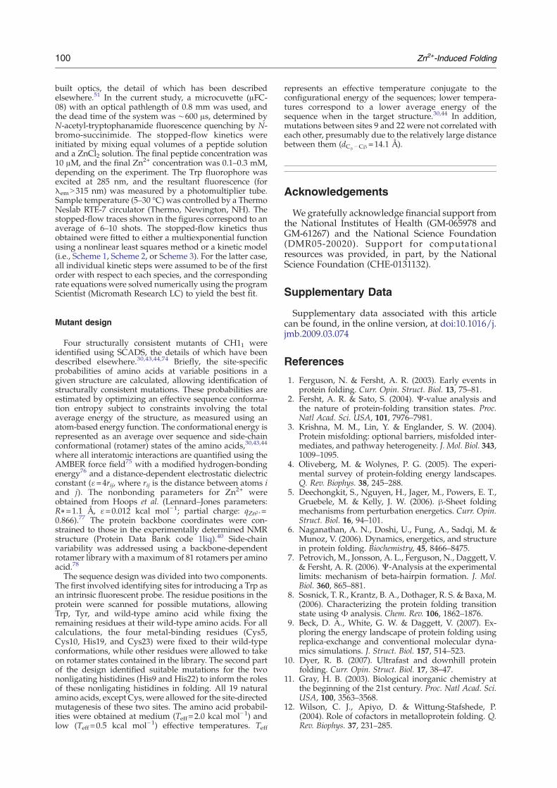
Fig. 2. Site-specific amino acid probability profiles for thereplacement for H9 and H22 (right). Teff=0.5 kcal mol−1. For H2.0 and 0.5 kcal mol−1 are presented with gray squares and ryellow. Zn-coordinating residues (gray) and residues of the tr
mutants L7W and T21W were targeted because, inboth cases, the Trp residue is in proximity to twozinc ligands: C5/C10 for L7W and H19/C23 forT21W. The probabilities of these two Trp residues attheir respective positions are shown in Fig. 2. Asshown in the predicted model structures (Fig. 2),Trp7 in L7W is located close to the metal-bindingpocket; thus, it is expected to be sensitive to Zn2+–ligand coordination. On the other hand, while theside chain of Trp21 in T21W is highly exposed tosolvent (Fig. 2), it is near the zinc-binding site, andthe location (i.e., sandwiched between H19 and C23)makes it a good candidate for the observation oflocal environmental changes associated with transi-tion from a heterogeneous ensemble of conforma-tions in the unfolded state to a well-organizedstructure upon folding.
Identification of mutations for His9 and His22
To elucidate the roles of the two nonbinding Hisresidues (i.e., H9 and H22) in CH11 folding, the site-specific amino acid probability profiles for sites 9and 22 were calculated. At each position, 19 aminoacids were permitted (i.e., Cys was excluded), andthe modeled structure of the mutant L7W was used.As shown (Fig. 2), glutamic acid (Glu) shows aprobability for site 9 that is slightly higher than thosefor several other amino acids at Teff=2 kcal mol−1.However, at lower energies (Teff=0.5 kcal mol−1),the aliphatic nonpolar amino acid valine (Val) is themost probable amino acid for this site due tostabilizing short-ranged van der Waals interactions.
introduction of Trp mutation on L7 and T21 (left) and His9, probabilities at two different sampling temperatures ofed solid bars, respectively. A mutated residue appears iniple mutant L7W/H9V/H22I (magenta) are shown.
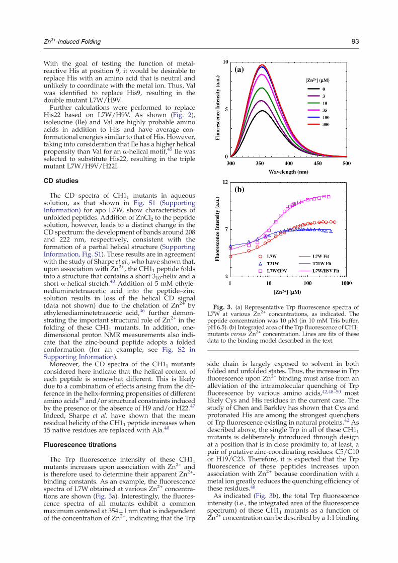
Fig. 3. (a) Representative Trp fluorescence spectra ofL7W at various Zn2+ concentrations, as indicated. Thepeptide concentration was 10 μM (in 10 mM Tris buffer,pH 6.5). (b) Integrated area of the Trp fluorescence of CH11mutants versus Zn2+ concentration. Lines are fits of thesedata to the binding model described in the text.
93Zn2+-Induced Folding
With the goal of testing the function of metal-reactive His at position 9, it would be desirable toreplace His with an amino acid that is neutral andunlikely to coordinate with the metal ion. Thus, Valwas identified to replace His9, resulting in thedouble mutant L7W/H9V.Further calculations were performed to replace
His22 based on L7W/H9V. As shown (Fig. 2),isoleucine (Ile) and Val are highly probable aminoacids in addition to His and have average con-formational energies similar to that of His. However,taking into consideration that Ile has a higher helicalpropensity than Val for an α-helical motif,45 Ile wasselected to substitute His22, resulting in the triplemutant L7W/H9V/H22I.
CD studies
The CD spectra of CH11 mutants in aqueoussolution, as that shown in Fig. S1 (SupportingInformation) for apo L7W, show characteristics ofunfolded peptides. Addition of ZnCl2 to the peptidesolution, however, leads to a distinct change in theCD spectrum: the development of bands around 208and 222 nm, respectively, consistent with theformation of a partial helical structure (SupportingInformation, Fig. S1). These results are in agreementwith the study of Sharpe et al., who have shown that,upon association with Zn2+, the CH11 peptide foldsinto a structure that contains a short 310-helix and ashort α-helical stretch.40 Addition of 5 mM ethyle-nediaminetetraacetic acid into the peptide–zincsolution results in loss of the helical CD signal(data not shown) due to the chelation of Zn2+ byethylenediaminetetraacetic acid,46 further demon-strating the important structural role of Zn2+ in thefolding of these CH11 mutants. In addition, one-dimensional proton NMR measurements also indi-cate that the zinc-bound peptide adopts a foldedconformation (for an example, see Fig. S2 inSupporting Information).Moreover, the CD spectra of the CH11 mutants
considered here indicate that the helical content ofeach peptide is somewhat different. This is likelydue to a combination of effects arising from the dif-ference in the helix-forming propensities of differentamino acids45 and/or structural constraints inducedby the presence or the absence of H9 and/or H22.47
Indeed, Sharpe et al. have shown that the meanresidual helicity of the CH11 peptide increases when15 native residues are replaced with Ala.40
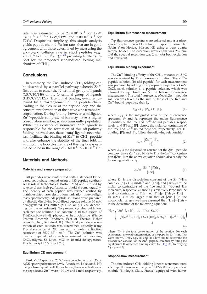
Fluorescence titrations
The Trp fluorescence intensity of these CH11mutants increases upon association with Zn2+ andis therefore used to determine their apparent Zn2+-binding constants. As an example, the fluorescencespectra of L7W obtained at various Zn2+ concentra-tions are shown (Fig. 3a). Interestingly, the fluores-cence spectra of all mutants exhibit a commonmaximum centered at 354±1 nm that is independentof the concentration of Zn2+, indicating that the Trp
side chain is largely exposed to solvent in bothfolded and unfolded states. Thus, the increase in Trpfluorescence upon Zn2+ binding must arise from analleviation of the intramolecular quenching of Trpfluorescence by various amino acids,42,48–50 mostlikely Cys and His residues in the current case. Thestudy of Chen and Barkley has shown that Cys andprotonated His are among the strongest quenchersof Trp fluorescence existing in natural proteins.42 Asdescribed above, the single Trp in all of these CH11mutants is deliberately introduced through designat a position that is in close proximity to, at least, apair of putative zinc-coordinating residues: C5/C10or H19/C23. Therefore, it is expected that the Trpfluorescence of these peptides increases uponassociation with Zn2+ because coordination with ametal ion greatly reduces the quenching efficiency ofthese residues.48As indicated (Fig. 3b), the total Trp fluorescence
intensity (i.e., the integrated area of the fluorescencespectrum) of these CH11 mutants as a function ofZn2+ concentration can be described by a 1:1 binding
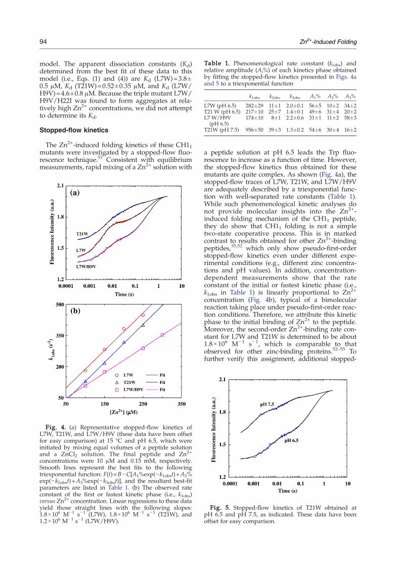
Table 1. Phenomenological rate constant (kiobs) andrelative amplitude (Ai%) of each kinetics phase obtainedby fitting the stopped-flow kinetics presented in Figs. 4aand 5 to a triexponential function
k1obs k2obs k3obs A1% A2% A3%
L7W (pH 6.5) 282±29 11±1 2.0±0.1 56±5 10±2 34±2T21 W (pH 6.5) 217±10 25±7 1.4±0.1 49±6 31±4 20±2L7 W/H9V
(pH 6.5)174±10 8±1 2.2±0.6 31±1 11±2 58±3
T21W (pH 7.5) 956±50 39±3 1.3±0.2 54±6 30±4 16±2
94 Zn2+-Induced Folding
model. The apparent dissociation constants (Kd)determined from the best fit of these data to thismodel (i.e., Eqs. (1) and (4)) are Kd (L7W)=3.8±0.5 μM, Kd (T21W)=0.52±0.35 μM, and Kd (L7W/H9V)=4.6±0.8 μM. Because the triple mutant L7W/H9V/H22I was found to form aggregates at rela-tively high Zn2+ concentrations, we did not attemptto determine its Kd.
Stopped-flow kinetics
The Zn2+-induced folding kinetics of these CH11mutants were investigated by a stopped-flow fluo-rescence technique.51 Consistent with equilibriummeasurements, rapid mixing of a Zn2+ solution with
Fig. 4. (a) Representative stopped-flow kinetics ofL7W, T21W, and L7W/H9V (these data have been offsetfor easy comparison) at 15 °C and pH 6.5, which wereinitiated by mixing equal volumes of a peptide solutionand a ZnCl2 solution. The final peptide and Zn2+
concentrations were 10 μM and 0.15 mM, respectively.Smooth lines represent the best fits to the followingtriexponential function: F(t)=B−C[A1%exp(−k1obst)+A2%exp(−k2obst)+A3%exp(−k3obst)], and the resultant best-fitparameters are listed in Table 1. (b) The observed rateconstant of the first or fastest kinetic phase (i.e., k1obs)versus Zn2+ concentration. Linear regressions to these datayield those straight lines with the following slopes:1.8×10⁶ M−1 s−1 (L7W), 1.8×10⁶ M−1 s−1 (T21W), and1.2×106 M−1 s−1 (L7W/H9V).
a peptide solution at pH 6.5 leads the Trp fluo-rescence to increase as a function of time. However,the stopped-flow kinetics thus obtained for thesemutants are quite complex. As shown (Fig. 4a), thestopped-flow traces of L7W, T21W, and L7W/H9Vare adequately described by a triexponential func-tion with well-separated rate constants (Table 1).While such phenomenological kinetic analyses donot provide molecular insights into the Zn2+-induced folding mechanism of the CH11 peptide,they do show that CH11 folding is not a simpletwo-state cooperative process. This is in markedcontrast to results obtained for other Zn2+-bindingpeptides,35,52 which only show pseudo-first-orderstopped-flow kinetics even under different expe-rimental conditions (e.g., different zinc concentra-tions and pH values). In addition, concentration-dependent measurements show that the rateconstant of the initial or fastest kinetic phase (i.e.,k1obs in Table 1) is linearly proportional to Zn2+
concentration (Fig. 4b), typical of a bimolecularreaction taking place under pseudo-first-order reac-tion conditions. Therefore, we attribute this kineticphase to the initial binding of Zn2+ to the peptide.Moreover, the second-order Zn2+-binding rate con-stant for L7W and T21W is determined to be about1.8×106 M−1 s−1, which is comparable to thatobserved for other zinc-binding proteins.52–55 Tofurther verify this assignment, additional stopped-
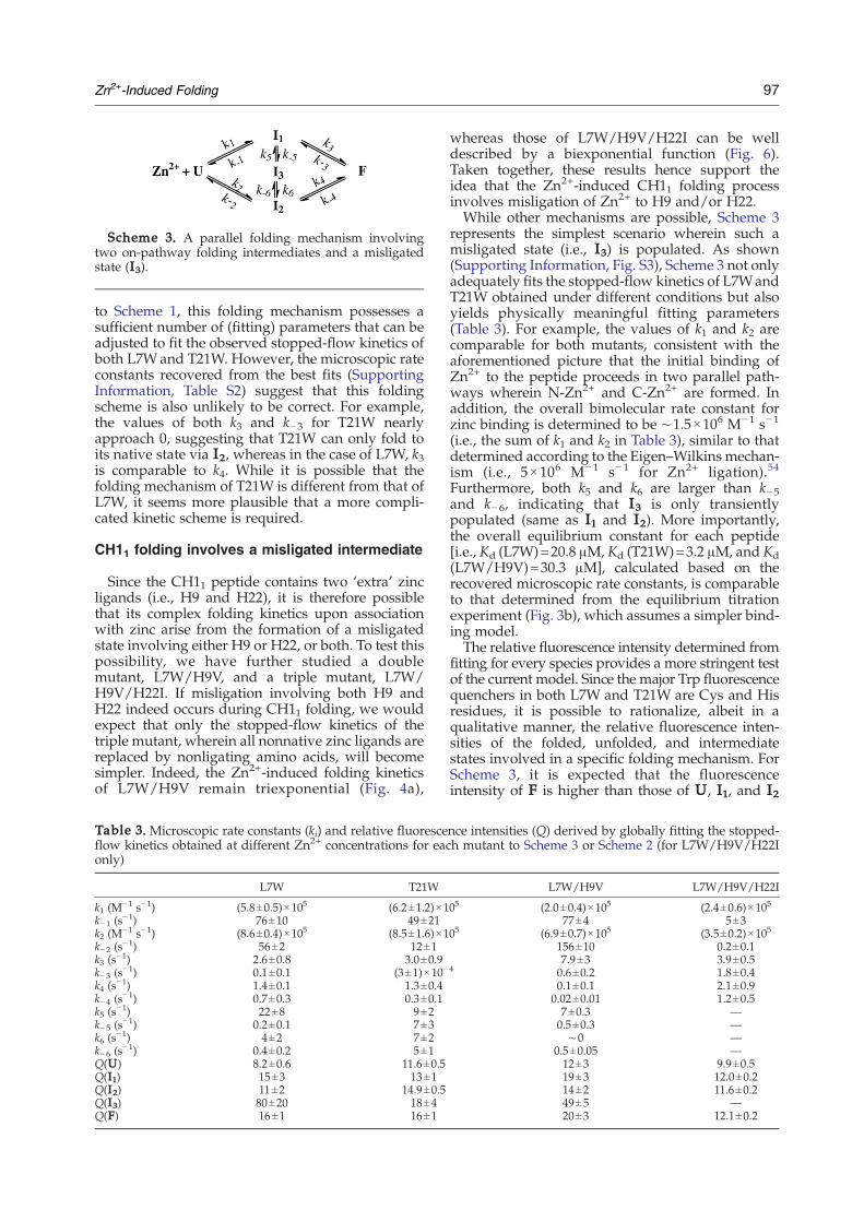
Fig. 5. Stopped-flow kinetics of T21W obtained atpH 6.5 and pH 7.5, as indicated. These data have beenoffset for easy comparison.

Fig. 6. Stopped-flow kinetics obtained by mixing equalvolumes of a L7W/H9V/H22I peptide solution and aZnCl2 solution at 15 °C. The final peptide and Zn2+
concentrations were 10 μMand 0.15 mM, respectively. Thesmooth line is the best fit of these data to a biexponentialfunction, and the resultant rate constants are k1obs=92 s−1
and k2obs=6.8 s−1.
Table 2.Apparent activation energy of each stopped-flowkinetic phase
L7W T21W L7W/H9V
Ea1 (kcal mol−1) 6.2±0.5 7.1±1.7 8.4±1.5
Ea2 (kcal mol−1) 14.1±1.2 10.2±0.5 12.8±0.3
Ea3 (kcal mol−1) 9.3±0.6 12.9±0.3 12.0±1.5
95Zn2+-Induced Folding
flowmixing experiments were carried out at differentpH conditions. As expected (Fig. 5 and Table 1),taking T21W as an example, the rate of the firststopped-flow kinetic phase increases approximatelyfour times when the pH of the final mixed solution isincreased from 6.5 to 7.5, due to an increase in thepopulation of deprotonated ligands (especially de-protonated His). Interestingly, the Zn2+-inducedfolding kinetics of L7W/H9V/H22I can be welldescribed by a biexponential function (Fig. 6), sug-gesting that replacement of H9 and H22 with non-ligating amino acids simplifies the folding process.Finally, temperature-dependent measurements
were carried out in the temperature range of5–30 °C to further characterize the apparent activa-tion energy (Ea) encountered by each kinetic phase.As shown (Fig. 7 and Table 2), all the rate constants
Fig. 7. Arrhenius plots of the rate constants obtained by(as indicated) to the triexponential function discussed in Filn(A)−Ea
i/RT, where Eai is the apparent activation energy fo
Table 2.
show Arrhenius temperature dependence, with Earanging from 6 to 14 kcal mol−1.
Discussion
The CH11 peptide is a novel zinc-binding motiffound in the first Cys/His-rich region of CREB-binding protein.40 It is known that this peptidebinds to zinc through a consensus CCHC motif (i.e.,Cys-X4-Cys-X8-His-X3-Cys, wherein Cys and Hisresidues are the ligands for Zn2+) and forms athermostable well-defined tertiary structure that isextremely tolerant to sequence mutations at non-ligating positions.40,41 In other words, Zn2+ bindingprovides a major driving force for CH11 folding.Therefore, this peptide provides an ideal modelsystem for understanding the mechanism of metal-induced protein folding processes.Since the native sequence of CH11 peptide does
not contain a suitable fluorophore (e.g., Trp) that canbe used to follow the kinetic process of binding orfolding using fluorescence-based techniques, wehave employed a statistical computational designmethod43,44 to identify structurally consistent Trp-containing mutants of CH11. First, two singlemutants, L7W and T21W, were identified as thebest targets because, in both cases, the Trp residue issandwiched between two zinc ligands (i.e., C5/C10for L7W and H19/C23 for T21W). Thus, theirfluorescence is likely sensitive to Zn2+ binding.Second, a double mutant, L7W/H9V, and a triplemutant, L7W/H9V/H22I, were designed to inves-tigate the role of nonligating His residues (i.e., His9
fitting the stopped-flow kinetics of the CH11 mutantsg. 4. Lines are fits to the Arrhenius equation: ln(kiobs)=r kinetic phase i (i=1, 2, or 3), and its value is listed in

Fig. 8. Two putative Zn2+–peptide complexes formedafter the initial binding of zinc, where H9 and H22 are thetwo nonnative ligands that may also participate inbinding.
96 Zn2+-Induced Folding
and His22) in the kinetics of CH11 folding. Indeed,both equilibrium and stopped-flow fluorescencemeasurements show that, upon coordination withZn2+, the Trp fluorescence of these mutantsincreases significantly, indicative of the sensitivityof this fluorescent probe to metal-induced foldingprocesses. Furthermore, the structural integrity ofthese mutants was verified by low-resolutionmethods (i.e., CD and one-dimensional NMR).
A simple sequential model is inadequate todescribe the folding mechanism of the CH11peptide
Compared to other metal-binding motifs,34,35,54–56
these CH11 mutants exhibit rather complex andslow cofactor-induced folding kinetics. As shown(Fig. 4a), the stopped-flow kinetics of L7W consist of(at least) three exponential phases, indicative of anon-two-state folding scenario. While this result isnot surprising, it is nevertheless interesting as manyproteins of similar size that lack cofactors have beenshown to fold in a two-state manner37,57–59 and on amuch faster timescale.37,60–62 Therefore, the slowerand more complex folding kinetics of L7W mustarise from the fact that the folding of CH11necessitates Zn2+ ligation in a tetrahedral geometry.The fastest phase in the stopped-flow kinetics of
L7W is characteristic of a bimolecular reaction, asits rate constant (i.e., k1obs in Table 1) dependslinearly on Zn2+ concentration (Fig. 4b). Therefore,we attribute this kinetic phase to the initialassociation of the peptide with zinc, wherein aZn2+–peptide complex (I1) is formed. Given the factthat the four native ligands in CH11 are segregatedinto two pairs—one (C5 and C10) close to the N-terminus and the other (C23 and H19) close to theC-terminus—it is likely that only two ligands in I1are ligated. Indeed, Bombarda et al. have recentlyshown that the first step in the Zn2+-inducedfolding kinetics of the distal CCHC finger motif ofthe human immunodeficiency virus-1 nucleocapsidprotein corresponds to the formation of a bidentatestate wherein only C36 and H44 are coordinatedwith the cofactor.35 Therefore, the simplest inter-pretation of the triexponential stopped-flow fluor-escence kinetics of L7W is that this initially formedZn2+–peptide complex I1 further evolves to pro-duce the final folded state (F) via a sequentialmechanism involving an on-pathway folding inter-mediate (I2), as indicated in Scheme 1. In fact, zinccoordination has been proposed to proceed in anintrinsically multistep and sequential manner forseveral peptides.35,54,63 While Scheme 1 can indeedfit the stopped-flow kinetics of L7W obtained atdifferent Zn2+ concentrations, the microscopic rate
Scheme 1. A sequential folding mechanism involvingan on-pathway folding intermediate.
constants recovered from the best fit (SupportingInformation, Table S1), however, give rise to anequilibrium constant that is not overwhelminglyfavorable for the folded state. Instead, this modelindicates that the folding intermediate states aresignificantly populated at equilibrium (e.g., [U]:[I1]:[I2]:[F]=1:9:9:14 at 300 μM Zn2+), inconsistent withprevious40 and current equilibrium results (Fig. 3b).This inconsistency suggests that a simple sequentialmodel, such as Scheme 1, is inadequate to describethe Zn2+-induced folding mechanism of the CH11peptide.
CH11 folding involves parallel pathways
The inadequacy of Scheme 1 in describing thefolding mechanism of the CH11 peptide promptedus to design and study a second mutant (i.e., T21W)wherein the Trp is placed near the second group ofthe Zn2+ ligands H19 and C23. The rationale is toexplore whether Zn2+-induced CH11 folding in-volves parallel pathways. In principle, the initialbinding of Zn2+ to the peptide could occur at bothN-terminal and C-terminal regions, leading to theformation of N-Zn2+ and C-Zn2+ (Fig. 8). Since Trpfluorescence quenching by various amino acids isstrongly distance dependent, we expect both L7Wand T21W to exhibit similar fluorescence intensitychanges upon the initial binding of Zn2+ if both N-Zn2+ and C-Zn2+ are formed. Indeed, the relativeamplitude of the first stopped-flow kinetic phase ofT21W (Table 1), arising from the initial Zn2+–peptide association, is 49%, similar to that (i.e.,56%) of L7W. Taken together, these results thus sug-gest that the Zn2+-induced CH11 folding involves atleast two parallel pathways, as shown in Scheme 2,which represents the simplest parallel foldingmechanism wherein N-Zn2+ (I1) and C-Zn2+ (I2)are directly converted into the folded state. Similar
Scheme 2. A parallel folding mechanism involvingtwo on-pathway folding intermediates.
Scheme 3. A parallel folding mechanism involvingtwo on-pathway folding intermediates and a misligatedstate (I3).
97Zn2+-Induced Folding
to Scheme 1, this folding mechanism possesses asufficient number of (fitting) parameters that can beadjusted to fit the observed stopped-flow kinetics ofboth L7Wand T21W. However, the microscopic rateconstants recovered from the best fits (SupportingInformation, Table S2) suggest that this foldingscheme is also unlikely to be correct. For example,the values of both k3 and k−3 for T21W nearlyapproach 0, suggesting that T21W can only fold toits native state via I2, whereas in the case of L7W, k3is comparable to k4. While it is possible that thefolding mechanism of T21W is different from that ofL7W, it seems more plausible that a more compli-cated kinetic scheme is required.
CH11 folding involves a misligated intermediate
Since the CH11 peptide contains two ‘extra’ zincligands (i.e., H9 and H22), it is therefore possiblethat its complex folding kinetics upon associationwith zinc arise from the formation of a misligatedstate involving either H9 or H22, or both. To test thispossibility, we have further studied a doublemutant, L7W/H9V, and a triple mutant, L7W/H9V/H22I. If misligation involving both H9 andH22 indeed occurs during CH11 folding, we wouldexpect that only the stopped-flow kinetics of thetriple mutant, wherein all nonnative zinc ligands arereplaced by nonligating amino acids, will becomesimpler. Indeed, the Zn2+-induced folding kineticsof L7W/H9V remain triexponential (Fig. 4a),
Table 3. Microscopic rate constants (ki) and relative fluoresceflow kinetics obtained at different Zn2+ concentrations for eaconly)
L7W T21W
k1 (M−1 s−1) (5.8±0.5)×105 (6.2±1.2)×1
k−1 (s−1) 76±10 49±21
k2 (M−1 s−1) (8.6±0.4)×105 (8.5±1.6)×1
k−2 (s−1) 56±2 12±1
k3 (s−1) 2.6±0.8 3.0±0.9
k−3 (s−1) 0.1±0.1 (3±1)×10−
k4 (s−1) 1.4±0.1 1.3±0.4
k−4 (s−1) 0.7±0.3 0.3±0.1
k5 (s−1) 22±8 9±2
k−5 (s−1) 0.2±0.1 7±3
k6 (s−1) 4±2 7±2
k−6 (s−1) 0.4±0.2 5±1
Q(U) 8.2±0.6 11.6±0.5Q(I1) 15±3 13±1Q(I2) 11±2 14.9±0.5Q(I3) 80±20 18±4Q(F) 16±1 16±1
whereas those of L7W/H9V/H22I can be welldescribed by a biexponential function (Fig. 6).Taken together, these results hence support theidea that the Zn2+-induced CH11 folding processinvolves misligation of Zn2+ to H9 and/or H22.While other mechanisms are possible, Scheme 3
represents the simplest scenario wherein such amisligated state (i.e., I3) is populated. As shown(Supporting Information, Fig. S3), Scheme 3 not onlyadequately fits the stopped-flow kinetics of L7WandT21W obtained under different conditions but alsoyields physically meaningful fitting parameters(Table 3). For example, the values of k1 and k2 arecomparable for both mutants, consistent with theaforementioned picture that the initial binding ofZn2+ to the peptide proceeds in two parallel path-ways wherein N-Zn2+ and C-Zn2+ are formed. Inaddition, the overall bimolecular rate constant forzinc binding is determined to be ∼1.5×10⁶ M−1 s−1
(i.e., the sum of k1 and k2 in Table 3), similar to thatdetermined according to the Eigen–Wilkins mechan-ism (i.e., 5×10⁶ M− 1 s− 1 for Zn2+ ligation).54
Furthermore, both k5 and k6 are larger than k−5and k− 6, indicating that I3 is only transientlypopulated (same as I1 and I2). More importantly,the overall equilibrium constant for each peptide[i.e., Kd (L7W)=20.8 μM, Kd (T21W)=3.2 μM, and Kd(L7W/H9V)=30.3 μM], calculated based on therecovered microscopic rate constants, is comparableto that determined from the equilibrium titrationexperiment (Fig. 3b), which assumes a simpler bind-ing model.The relative fluorescence intensity determined from
fitting for every species provides a more stringent testof the current model. Since the major Trp fluorescencequenchers in both L7W and T21W are Cys and Hisresidues, it is possible to rationalize, albeit in aqualitative manner, the relative fluorescence inten-sities of the folded, unfolded, and intermediatestates involved in a specific folding mechanism. ForScheme 3, it is expected that the fluorescenceintensity of F is higher than those of U, I1, and I2
nce intensities (Q) derived by globally fitting the stopped-h mutant to Scheme 3 or Scheme 2 (for L7W/H9V/H22I
L7W/H9V L7W/H9V/H22I
05 (2.0±0.4)×105 (2.4±0.6)×105
77±4 5±305 (6.9±0.7)×105 (3.5±0.2)×105
156±10 0.2±0.17.9±3 3.9±0.5
4 0.6±0.2 1.8±0.40.1±0.1 2.1±0.90.02±0.01 1.2±0.57±0.3 —0.5±0.3 —∼0 —
0.5±0.05 —12±3 9.9±0.519±3 12.0±0.214±2 11.6±0.249±5 —20±3 12.1±0.2
98 Zn2+-Induced Folding
because more Trp fluorescence quenchers are boundto Zn2+ in the folded state (F). Similarly, if I1 and I2represent N-Zn2+ and C-Zn2+ in Scheme 3, respec-tively, one would expect that, for L7W (T21W), thefluorescence intensity of I1 (I2) is larger than that ofI2 (I1) because, in this case, the Trp fluorophore islocated near the N-terminal (C-terminal) ligands. Asindicated (Table 3), the relative fluorescence inten-sities of F, I1, and I2 recovered from the best fits ofthe stopped-flow data of L7W and T21W to Scheme3 indeed meet these expectations. Moreover, forboth mutants, the off-pathway intermediate I3exhibits the largest relative fluorescence intensity,suggesting that it adopts a conformation whereinTrp fluorescence quenching by all potential quench-ers (i.e., all Cys and His residues) is maximallyreduced. Thus, this result corroborates the idea thatthe misligated state involves H9 and/or H22.While the structure of the misligated state cannot
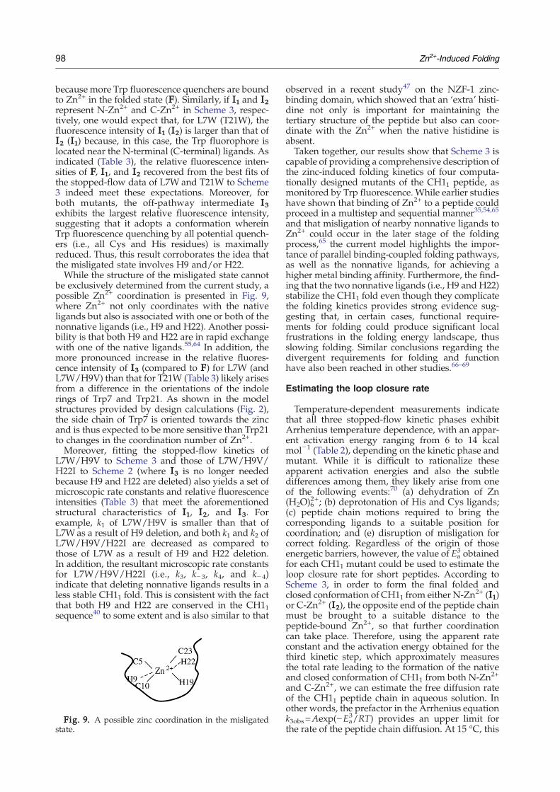
be exclusively determined from the current study, apossible Zn2+ coordination is presented in Fig. 9,where Zn2+ not only coordinates with the nativeligands but also is associated with one or both of thenonnative ligands (i.e., H9 and H22). Another possi-bility is that both H9 and H22 are in rapid exchangewith one of the native ligands.55,64 In addition, themore pronounced increase in the relative fluores-cence intensity of I3 (compared to F) for L7W (andL7W/H9V) than that for T21W (Table 3) likely arisesfrom a difference in the orientations of the indolerings of Trp7 and Trp21. As shown in the modelstructures provided by design calculations (Fig. 2),the side chain of Trp7 is oriented towards the zincand is thus expected to be more sensitive than Trp21to changes in the coordination number of Zn2+.Moreover, fitting the stopped-flow kinetics of
L7W/H9V to Scheme 3 and those of L7W/H9V/H22I to Scheme 2 (where I3 is no longer neededbecause H9 and H22 are deleted) also yields a set ofmicroscopic rate constants and relative fluorescenceintensities (Table 3) that meet the aforementionedstructural characteristics of I1, I2, and I3. Forexample, k1 of L7W/H9V is smaller than that ofL7Was a result of H9 deletion, and both k1 and k2 ofL7W/H9V/H22I are decreased as compared tothose of L7W as a result of H9 and H22 deletion.In addition, the resultant microscopic rate constantsfor L7W/H9V/H22I (i.e., k3, k− 3, k4, and k−4)indicate that deleting nonnative ligands results in aless stable CH11 fold. This is consistent with the factthat both H9 and H22 are conserved in the CH11sequence40 to some extent and is also similar to that
Fig. 9. A possible zinc coordination in the misligatedstate.
observed in a recent study47 on the NZF-1 zinc-binding domain, which showed that an ‘extra’ histi-dine not only is important for maintaining thetertiary structure of the peptide but also can coor-dinate with the Zn2+ when the native histidine isabsent.Taken together, our results show that Scheme 3 is
capable of providing a comprehensive description ofthe zinc-induced folding kinetics of four computa-tionally designed mutants of the CH11 peptide, asmonitored by Trp fluorescence. While earlier studieshave shown that binding of Zn2+ to a peptide couldproceed in a multistep and sequential manner35,54,65
and that misligation of nearby nonnative ligands toZn2+ could occur in the later stage of the foldingprocess,65 the current model highlights the impor-tance of parallel binding-coupled folding pathways,as well as the nonnative ligands, for achieving ahigher metal binding affinity. Furthermore, the find-ing that the two nonnative ligands (i.e., H9 and H22)stabilize the CH11 fold even though they complicatethe folding kinetics provides strong evidence sug-gesting that, in certain cases, functional require-ments for folding could produce significant localfrustrations in the folding energy landscape, thusslowing folding. Similar conclusions regarding thedivergent requirements for folding and functionhave also been reached in other studies.66–69
Estimating the loop closure rate
Temperature-dependent measurements indicatethat all three stopped-flow kinetic phases exhibitArrhenius temperature dependence, with an appar-ent activation energy ranging from 6 to 14 kcalmol−1 (Table 2), depending on the kinetic phase andmutant. While it is difficult to rationalize theseapparent activation energies and also the subtledifferences among them, they likely arise from oneof the following events:70 (a) dehydration of Zn(H2O)6
2+; (b) deprotonation of His and Cys ligands;(c) peptide chain motions required to bring thecorresponding ligands to a suitable position forcoordination; and (e) disruption of misligation forcorrect folding. Regardless of the origin of thoseenergetic barriers, however, the value of Ea
3 obtainedfor each CH11 mutant could be used to estimate theloop closure rate for short peptides. According toScheme 3, in order to form the final folded andclosed conformation of CH11 from either N-Zn2+ (I1)or C-Zn2+ (I2), the opposite end of the peptide chainmust be brought to a suitable distance to thepeptide-bound Zn2+, so that further coordinationcan take place. Therefore, using the apparent rateconstant and the activation energy obtained for thethird kinetic step, which approximately measuresthe total rate leading to the formation of the nativeand closed conformation of CH11 from both N-Zn2+
and C-Zn2+, we can estimate the free diffusion rateof the CH11 peptide chain in aqueous solution. Inother words, the prefactor in the Arrhenius equationk3obs=Aexp(−Ea
3/RT) provides an upper limit forthe rate of the peptide chain diffusion. At 15 °C, this
99Zn2+-Induced Folding
rate was estimated to be 2.1×10⁷ s−1 for L7W,6.6×106 s−1 for L7W/H9V, and 7.0×10⁹ s−1 forT21W. Despite its simplicity, this simple analysisyields peptide chain diffusion rates that are in goodagreement with those determined by measuring theend-to-end collision rate in short peptides (e.g.,7.1×106 to 1.5×10⁸ s−1),71,72 providing further sup-port for the proposed zinc-induced folding me-chanism of CH11.
Conclusions
In summary, the Zn2+-induced CH11 folding canbe described by a parallel pathway wherein Zn2+
first binds to either the N-terminal group of ligands(C5/C10/H9) or the C-terminal group of ligands(H19/C23/H22). This initial binding event is fol-lowed by a rearrangement of the peptide chain,leading to the closure of the peptide loop and theconcomitant formation of the native zinc tetrahedralcoordination. During folding, however, a misligatedZn2+–peptide complex, which may have a highercoordination number, is also transiently populated.While the existence of nonnative Zn2+ ligands isresponsible for the formation of this off-pathwayfolding intermediate, these ‘extra’ ligands neverthe-less facilitate the binding of Zn2+ to CH11 peptideand also enhance the stability of the final fold. Inaddition, the loop closure rate of this peptide is esti-mated to be in the range of 6.6×10⁶ to 7.0×10⁹ s−1.
Materials and Methods
Materials and sample preparation
All peptides were synthesized with a standard Fmoc-based solid-phase method using a PS3 peptide synthesi-zer (Protein Technologies, Boston, MA) and purified byreverse-phase high-performance liquid chromatography.The identity of each peptide was further verified bymatrix-assisted laser desorption/ionization time-of-flightmass spectrometry. All peptide solutions were preparedby directly dissolving lyophilized peptide solid in 10 mMdeoxygenated Tris buffer (pH 6.5 or pH 7.5, depend-ing on the experiment). To prevent cysteine oxidation,each peptide solution also contains a 10-fold excess ofTris(2-carboxyethyl) phosphine hydrochloride (PierceProtein Research Products, Part of Thermo FisherScientific, Inc., Rockford, IL). The final peptide concen-tration of each solution was determined optically usingTrp absorbance at 280 nm and a molar extinctioncoefficient of 5600 M−1 cm−1. The Zn2+ solution wasfreshly prepared before each experiment by dissolvingZnCl2 (Sigma, St. Louis, MO) in 10 mM deoxygenatedTris buffer (pH 6.5 or pH 7.5).
Equilibrium CD measurement
Far-UV CD spectra at 25 °C were collected with an AVIV62DS spectropolarimeter (Aviv Associates, Lakewood, NJ)usinga 1-mmquartz cell. For each case, the concentrationsofthe peptide and Zn2+ were∼30 μMand 1mM, respectively.
Equilibrium fluorescence measurement
Trp fluorescence spectra were collected under a nitro-gen atmosphere on a Fluorolog 3.10 spectrofluorometer(Jobin Yvon Horiba, Edison, NJ) using a 1-cm quartzsample holder. The excitation wavelength was 285 nm,and the spectral resolution was 2 nm (for both excitationand emission).
Equilibrium binding experiment
The Zn2+-binding affinity of the CH11 mutants at 15 °Cwas determined by Trp fluorescence titration. The Zn2+–peptide solution (10 μM peptide) for each measurementwas prepared by adding an appropriate aliquot of a 4 mMZnCl2 stock solution to a peptide solution, which wasallowed to equilibrate for 5 min before fluorescencemeasurement. The total fluorescence of each Zn2+–peptidesolution was taken as the sum of those of the free andZn2+-bound peptides, that is,
Fint = Fbd P½ �b + Ffd P½ �f ð1Þwhere Fint is the integrated area of the fluorescencespectrum; Ff and Fb represent the molar fluorescenceintensities of the free and Zn2+-bound peptides, respec-tively; and [P]f and [P]b denote the molar concentrations ofthe free and Zn2+-bound peptides, respectively. For 1:1binding, [P]f and [P]b follow the following relationship:
Kd =Zn2 +� �
P½ �fP½ �b
ð2Þ
where Kd is the dissociation constant of the Zn2+–peptidecomplex. Since Zn2+ also binds to Tris, the Zn2+ concentra-tion ([Zn2+]) in the above equation should also satisfy thefollowing relationship:
Kd V=Zn2 +� �
Tris½ �fTris½ �b
ð3Þ
where Kd′ is the dissociation constant of the Zn2+–Triscomplex (Kd′=11.5 mM),73 and [Tris]f and [Tris]b are themolar concentrations of the free and Zn2+-bound Trismolecules, respectively. Since Kd′ is relatively large and thetotal concentration of Tris (i.e., [Tris]T=[Tris]f+ [Tris]b=10 mM) is much larger than that of [Zn2+] (in themicromolar range), we have assumed that [Tris]f= [Tris]Tin the derivation of the following equation:
½P�b =ðð½Zn2 + �T + ½P�T +Kd + ½Tris�TKd=K′dÞ
�ffiffiffiffiffiffiffiffiffiffiffiffiffiffiffiffiffiffiffiffiffiffiffiffiffiffiffiffiffiffiffiffiffiffiffiffiffiffiffiffiffiffiffiffiffiffiffiffiffiffiffiffiffiffiffiffiffiffiffiffiffiffiffiffiffiffiffiffiffiffiffiffiffiffiffiffiffiffiffiffiffiffiffiffiffiffiffiffiffiffiffiffiffiffiffiffiffiffiffiffiffiffiffiffiffiffiffiffiffiffið½Zn2 + �T + ½P�T +Kd + ½Tris�TKd=K′
dÞ2 � 4½Zn2 + �T½P�Tq Þ
2ð4Þ
where [P]T is the total concentration of the peptide. For eachexperiment, the total concentrations of the peptide, Zn2+, and Triswere known. Thus, Eqs. (1) and (4) allow one to determine thedissociation constant of the Zn2+–peptide complex by fitting theequilibrium fluorescence binding curve (i.e., Fig. 3b) by varyingKd, Fb, and Ff.
Stopped-flow measurement
The zinc-induced CH11 folding kinetics were monitoredvia Trp fluorescence using an SFM-300 stopped-flowmodule (Bio-logic, Claix, France) equipped with home-
100 Zn2+-Induced Folding
built optics, the detail of which has been describedelsewhere.51 In the current study, a microcuvette (μFC-08) with an optical pathlength of 0.8 mm was used, andthe dead time of the system was ∼600 μs, determined byN-acetyl-tryptophanamide fluorescence quenching by N-bromo-succinimide. The stopped-flow kinetics wereinitiated by mixing equal volumes of a peptide solutionand a ZnCl2 solution. The final peptide concentration was10 μM, and the final Zn2+ concentration was 0.1–0.3 mM,depending on the experiment. The Trp fluorophore wasexcited at 285 nm, and the resultant fluorescence (forλemN315 nm) was measured by a photomultiplier tube.Sample temperature (5–30 °C) was controlled by a ThermoNeslab RTE-7 circulator (Thermo, Newington, NH). Thestopped-flow traces shown in the figures correspond to anaverage of 6–10 shots. The stopped-flow kinetics thusobtained were fitted to either a multiexponential functionusing a nonlinear least squares method or a kinetic model(i.e., Scheme 1, Scheme 2, or Scheme 3). For the latter case,all individual kinetic steps were assumed to be of the firstorder with respect to each species, and the correspondingrate equations were solved numerically using the programScientist (Micromath Research LC) to yield the best fit.
Mutant design
Four structurally consistent mutants of CH11 wereidentified using SCADS, the details of which have beendescribed elsewhere.30,43,44,74 Briefly, the site-specificprobabilities of amino acids at variable positions in agiven structure are calculated, allowing identification ofstructurally consistent mutations. These probabilities areestimated by optimizing an effective sequence conforma-tion entropy subject to constraints involving the totalaverage energy of the structure, as measured using anatom-based energy function. The conformational energy isrepresented as an average over sequence and side-chainconformational (rotamer) states of the amino acids,30,43,44
where all interatomic interactions are quantified using theAMBER force field75 with a modified hydrogen-bondingenergy76 and a distance-dependent electrostatic dielectricconstant (ɛ=4rij, where rij is the distance between atoms iand j). The nonbonding parameters for Zn2+ wereobtained from Hoops et al. (Lennard–Jones parameters:R⁎=1.1 Å, ɛ=0.012 kcal mol−1; partial charge: qZn2+=0.866).77 The protein backbone coordinates were con-strained to those in the experimentally determined NMRstructure (Protein Data Bank code 1liq).40 Side-chainvariability was addressed using a backbone-dependentrotamer library with a maximum of 81 rotamers per aminoacid.78
The sequence design was divided into two components.The first involved identifying sites for introducing a Trp asan intrinsic fluorescent probe. The residue positions in theprotein were scanned for possible mutations, allowingTrp, Tyr, and wild-type amino acid while fixing theremaining residues at their wild-type amino acids. For allcalculations, the four metal-binding residues (Cys5,Cys10, His19, and Cys23) were fixed to their wild-typeconformations, while other residues were allowed to takeon rotamer states contained in the library. The second partof the design identified suitable mutations for the twononligating histidines (His9 and His22) to inform the rolesof these nonligating histidines in folding. All 19 naturalamino acids, except Cys, were allowed for the site-directedmutagenesis of these two sites. The amino acid probabil-ities were obtained at medium (Teff=2.0 kcal mol−1) andlow (Teff=0.5 kcal mol−1) effective temperatures. Teff
represents an effective temperature conjugate to theconfigurational energy of the sequences; lower tempera-tures correspond to a lower average energy of thesequence when in the target structure.30,44 In addition,mutations between sites 9 and 22 were not correlated witheach other, presumably due to the relatively large distancebetween them (dCβ−Cβ=14.1 Å).
Acknowledgements
We gratefully acknowledge financial support fromthe National Institutes of Health (GM-065978 andGM-61267) and the National Science Foundation(DMR05-20020). Support for computationalresources was provided, in part, by the NationalScience Foundation (CHE-0131132).
Supplementary Data
Supplementary data associated with this articlecan be found, in the online version, at doi:10.1016/j.jmb.2009.03.074
References
1. Ferguson, N. & Fersht, A. R. (2003). Early events inprotein folding. Curr. Opin. Struct. Biol. 13, 75–81.
2. Fersht, A. R. & Sato, S. (2004). Ψ-value analysis andthe nature of protein-folding transition states. Proc.Natl Acad. Sci. USA, 101, 7976–7981.
3. Krishna, M. M., Lin, Y. & Englander, S. W. (2004).Protein misfolding: optional barriers, misfolded inter-mediates, and pathway heterogeneity. J. Mol. Biol. 343,1009–1095.
4. Oliveberg, M. & Wolynes, P. G. (2005). The experi-mental survey of protein-folding energy landscapes.Q. Rev. Biophys. 38, 245–288.
5. Deechongkit, S., Nguyen, H., Jager, M., Powers, E. T.,Gruebele, M. & Kelly, J. W. (2006). β-Sheet foldingmechanisms from perturbation energetics. Curr. Opin.Struct. Biol. 16, 94–101.
6. Naganathan, A. N., Doshi, U., Fung, A., Sadqi, M. &Munoz, V. (2006). Dynamics, energetics, and structurein protein folding. Biochemistry, 45, 8466–8475.
7. Petrovich, M., Jonsson, A. L., Ferguson, N., Daggett, V.& Fersht, A. R. (2006). Ψ-Analysis at the experimentallimits: mechanism of beta-hairpin formation. J. Mol.Biol. 360, 865–881.
8. Sosnick, T. R., Krantz, B. A., Dothager, R. S. & Baxa, M.(2006). Characterizing the protein folding transitionstate using Φ analysis. Chem. Rev. 106, 1862–1876.
9. Beck, D. A., White, G. W. & Daggett, V. (2007). Ex-ploring the energy landscape of protein folding usingreplica-exchange and conventional molecular dyna-mics simulations. J. Struct. Biol. 157, 514–523.
10. Dyer, R. B. (2007). Ultrafast and downhill proteinfolding. Curr. Opin. Struct. Biol. 17, 38–47.
11. Gray, H. B. (2003). Biological inorganic chemistry atthe beginning of the 21st century. Proc. Natl Acad. Sci.USA, 100, 3563–3568.
12. Wilson, C. J., Apiyo, D. & Wittung-Stafshede, P.(2004). Role of cofactors in metalloprotein folding. Q.Rev. Biophys. 37, 231–285.
101Zn2+-Induced Folding
13. Frankel, A. D., Berg, J. M. & Pabo, C. O. (1987). Metal-dependent folding of a single zinc finger from trans-cription factor IIIA. Proc. Natl Acad. Sci. USA, 84,4841–4845.
14. Krizek, B. A., Amann, B. T., Kilfoil, V. J., Merkle, D. L.& Berg, J. M. (1991). A consensus zinc finger peptide:design, high-affinity metal-binding, a pH-dependentstructure, and a His to Cys sequence variant. J. Am.Chem. Soc. 113, 4518–4523.
15. Zong, C. H., Wilson, C. J., Shen, T. Y., Wittung-Stafshede, P., Mayo, S. L. & Wolynes, P. G. (2007).Establishing the entatic state in folding metallatedPseudomonas aeruginosa azurin. Proc. Natl Acad. Sci.USA, 104, 3159–3164.
16. Jones, C. M., Henry, E. R., Hu, Y., Chan, C. K., Luck,S. D., Bhuyan, A. et al. (1993). Fast events in protein-folding initiated by nanosecond laser photolysis.Proc. Natl Acad. Sci. USA, 90, 11860–11864.
17. Shastry, M. C. R., Sauder, J. M. & Roder, H. (1998).Kinetic and structural analysis of submillisecondfolding events in cytochrome c. Acc. Chem. Res. 31,717–725.
18. Englander, S. W., Sosnick, T. R., Mayne, L. C.,Shtilerman, M., Qi, P. X. & Bai, Y. W. (1998). Fastand slow folding in cytochrome c. Acc. Chem. Res. 31,737–744.
19. Guidry, J. &Wittung-Stafshede, P. (2000). Cytochromec(553), a small heme protein that lacks misligation inits unfolded state, folds with rapid two-state kinetics.J. Mol. Biol. 301, 769–773.
20. Chen, E. F., Goldbeck, R. A. & Kliger, D. S. (2004). Theearliest events in protein folding: a structural require-ment for ultrafast folding in cytochrome c. J. Am.Chem. Soc. 126, 11175–11181.
21. Bertini, I., Gray, H. B., Lippard, S. J. & Valentine, J. S.(1994). Bioinorganic Chemistry, 2nd edit UniversityScience Books, Mill Valley, CA.
22. Cerasoli, E., Sharpe, B. K. & Woolfson, D. N. (2005).ZiCo: a peptide designed to switch folded state uponbinding zinc. J. Am. Chem. Soc. 127, 15008–15009.
23. DeGrado, W. F., Summa, C. M., Pavone, V., Nastri, F.& Lombardi, A. (1999). De novo design and structuralcharacterization of proteins and metalloproteins.Annu. Rev. Biochem. 68, 779–819.
24. Kohn, W. D., Kay, C. M., Sykes, B. D. & Hodges, R. S.(1998). Metal ion induced folding of a de novo designedcoiled-coil peptide. J. Am. Chem. Soc. 120, 1124–1132.
25. Kharenko, O. A. & Ogawa, M. Y. (2004). Metal-induced folding of a designed metalloprotein. J. Inorg.Biochem. 98, 1971–1974.
26. Maglio, O., Nastri, F., Calhoun, J. R., Lahr, S., Wade,H., Pavone, V. et al. (2005). Artificial di-iron proteins:solution characterization of four helix bundles con-taining two distinct types of inter-helical loops. J. Biol.Inorg. Chem. 10, 539–549.
27. Shiraishi, Y., Imanishi, M., Morisaki, T. & Sugiura, Y.(2005). Swapping of the β-hairpin region between Sp1and GLI zinc fingers: significant role of the β-hairpinregion in DNA binding properties of C2H2-type zincfinger peptides. Biochemistry, 44, 2523–2538.
28. Iranzo, O., Ghosh, D. & Pecoraro, V. L. (2006).Assessing the integrity of designed homomericparallel three-stranded coiled coils in the presence ofmetal ion. Inorg. Chem. 45, 9959–9973.
29. Papworth, M., Kolasinska, P. & Minczuk, M. (2006).Designer zinc-finger proteins and their applications.Gene, 17, 27–38.
30. Calhoun, J. R., Kono, H., Lahr, S., Wang, W., DeGrado,W. F. & Saven, J. G. (2003). Computational design and
characterization of a monomeric helical dinuclearmetalloprotein. J. Mol. Biol. 334, 1101–1115.
31. Nanda, V., Rosenblatt, M. M., Osyczka, A., Kono, H.,Getahun, Z., Dutton, P. L. et al. (2005). De novo designof a redox active minimal rubredoxin mimic. J. Am.Chem. Soc. 127, 5804–5805.
32. Suzuki, K., Hiroaki, H., Kohda, D., Nakamura, H. &Tanaka, T. (1998). Metal ion induced self-assembly of adesigned peptide into a triple-stranded α-helicalbundle: a novel metal binding site in the hydrophobiccore. J. Am. Chem. Soc. 120, 13008–13015.
33. Farrer, B. T. & Pecoraro, V. L. (2003). Hg(II) binding toa weakly associated coiled coil nucleates an encodedmetalloprotein fold: a kinetic analysis. Proc. Natl Acad.Sci. USA, 100, 3760–3765.
34. Pozdnyakova, I. & Wittung-Stafshede, P. (2001).Biological relevance of metal binding before proteinfolding. J. Am. Chem. Soc. 123, 10135–10136.
35. Bombarda, E., Grell, E., Roques, B. & Mely, Y. (2007).Molecular mechanism of the Zn2+-induced folding ofthe distal CCHC finger motif of the HIV-1 nucleocap-sid protein. Biophys. J. 93, 208–217.
36. Du, D. G., Zhu, Y. J., Huang, C. Y. & Gai, F. (2004).Understanding the key factors that control the rate ofbeta-hairpin folding. Proc. Natl Acad. Sci. USA, 101,15915–15920.
37. Bunagan, M. R., Yang, X., Saven, J. G. & Gai, F. (2006).Ultrafast folding of a computationally designedTrp-cage mutant: Trp2-cage. J. Phys. Chem. B, 110,3759–3763.
38. Xu, Y., Purkayastha, P. & Gai, F. (2006). Nanosecondfolding dynamics of a three-stranded β-sheet. J. Am.Chem. Soc. 128, 15836–15842.
39. Sadqi, M., de Alba, E., Pérez-Jiménez, R., Sanchez-Ruiz, J. M. & Muñoz, V. (2009). A designed protein asexperimental model of primordial folding. Proc. NatlAcad. Sci. USA, 106, 4127–4132.
40. Sharpe, B. K., Matthews, J. M., Kwan, A. H., Newton,A., Gell, D. A., Crossley, M. & Mackay, J. P. (2002). Anew zinc binding fold underlines the versatility of zincbinding modules in protein evolution. Structure, 10,639–648.
41. Sharpe, B. K., Liew, C. K., Kwan, A. H., Wilce, J. A.,Crossley, M., Matthews, J. M. & Mackay, J. P. (2005).Assessment of the robustness of a serendipitous zincbinding fold: mutagenesis and protein grafting.Structure, 13, 257–266.
42. Chen, Y. & Barkley, M. D. (1998). Toward under-standing tryptophan fluorescence in proteins.Biochemistry, 37, 9976–9982.
43. Zou, J. & Saven, J. G. (2000). Statistical theory ofcombinatorial libraries of folding proteins: energeticdiscrimination of a target structure. J. Mol. Biol. 296,281–294.
44. Kono, H. & Saven, J. G. (2001). Statistical theory forprotein combinatorial libraries. Packing interactions,backbone flexibility, and the sequence variability of amain-chain structure. J. Mol. Biol. 306, 607–628.
45. Oneil, K. T. & DeGrado, W. F. (1990). A thermo-dynamic scale for the helix-forming tendencies ofthe commonly occurring amino-acids. Science, 250,646–651.
46. Nyborg, J. K. & Peersen, O. B. (2004). That zincing feel-ing: the effects of EDTA on the behaviour of zinc-bind-ing transcriptional regulators. Biochem. J. 381, e3–e4.
47. Berkovits-Cymet, H. J., Amann, B. T. & Berg, J. M.(2004). Solution structure of a CCHHC domain ofneural zinc finger factor-1 and its implications forDNA binding. Biochemistry, 43, 898–903.
102 Zn2+-Induced Folding
48. Mély, Y., De Rocquigny, H., Piemont, E., Demene, H.,Jullian, N., Fournie-Zaluski, M. C. et al. (1993).Influence of the N-terminal and C-terminal chainson the zinc-binding and conformational propertiesof the central zinc-finger structure of Moloneymurine leukemia-virus nucleocapsid protein: asteady-state and time-resolved fluorescence study.Biochim. Biophys. Acta, 1161, 6–18.
49. Mély, Y., De Rocquigny, H., Morellet, N., Roques, B. P.& Gerad, D. (1996). Zinc binding to the HIV-1nucleocapsid protein: a thermodynamic investigationby fluorescence spectroscopy. Biochemistry, 35,5175–5182.
50. Chen, Y., Liu, B., Yu, H. T. & Barkley, M. D. (1996). Thepeptide bond quenches indole fluorescence. J. Am.Chem. Soc. 118, 9271–9278.
51. Tucker, M. J., Tang, J. & Gai, F. (2006). Probing thekinetics of membrane-mediated helix folding. J. Phys.Chem. B, 110, 8105–8109.
52. Buchsbaum, J. C. & Berg, J. M. (2000). Kinetics of metalbinding by a zinc finger peptide. Inorg. Chim. Acta,297, 217–219.
53. Keeble, A. H., Hemmings, A. M., James, R., Moore,G. R. & Kleanthous, C. (2002). Multistep binding oftransition metals to the H-N-H endonuclease toxincolicin E9. Biochemistry, 41, 10234–10244.
54. Bombarda, E., Roques, B. P., Mély, Y. & Grell, E. (2005).Mechanism of zinc coordination by point-mutatedstructures of the distal CCHC binding motif of theHIV-1NCp7 proteins. Biochemistry, 44, 7315–7325.
55. Heinz, U., Kiefer, M., Tholey, A. & Adolph, H. W.(2005). On the competition for available zinc. J. Biol.Chem. 280, 3197–3207.
56. Dutta, S. J., Liu, J. & Mitra, B. (2005). Kinetic analysisof metal binding to the amino-terminal domain ofZntA by monitoring metal–thiolate charge-transfercomplexes. Biochemistry, 44, 14268–14274.
57. Bunagan, M. R., Cristian, L., DeGrado, W. F. & Gai, F.(2006). Truncation of a cross-linkedGCN4-p1 coiled-coilleads to ultrafast folding. Biochemistry, 45, 10981–10986.
58. Du, D. G. & Gai, F. (2006). Understanding the foldingmechanism of an α-helical hairpin. Biochemistry, 45,13131–13139.
59. Qiu, L. L., Pabit, S. A., Roitberg, A. E. & Hagen,S. J. (2002). Smaller and faster: the 20-residue Trp-cage protein folds in 4 μs. J. Am. Chem. Soc. 124,12952–12953.
60. Williams, S., Causgrove, T. P., Gilmanshin, R.,Fang, K. S., Callender, R. H., Woodruff, W. H. &Dyer, R. B. (1996). Fast events in protein folding:helix melting and formation in a small peptide.Biochemistry, 35, 691–697.
61. Du, D. G., Tucker, M. J. & Gai, F. (2006).Understanding the mechanism of β-hairpin foldingvia ϕ-value analysis. Biochemistry, 45, 2668–2678.
62. Wang, T., Zhu, Y. J., Getahun, Z., Du, D. G., Huang,C. -Y., DeGrado, W. F. & Gai, F. (2004). Lengthdependent helix-coil transition kinetics of ninealanine-based peptides. J. Phys. Chem. B, 108,15301–15310.
63. Bombarda, E., Morellet, N., Cherradi, H., Spiess, B.,Bouaziz, S., Grell, E. et al. (2001). Determination of the
pKa of the four Zn2+-coordinating residues of thedistal finger motif of the HIV-1 nucleocapsid protein:consequences on the binding of Zn2+. J. Mol. Biol. 310,659–672.
64. Siemann, S., Badiei, H. R., Karanassios, V.,Viswanatha, T. &Dmitrienko, G. I. (2006). 68Zn isotopeexchange experiments reveal an unusual kineticlability of the metal ions in the di-zinc form of IMP-1metallo-β-lactamase. Chem. Commun., 532–534.
65. Li, W. F., Zhang, J., Wang, J. & Wang, W. (2007).Metal-coupled folding of Cys2His2 zinc-finger. J. Am.Chem. Soc. 130, 892–900.
66. Jager, M., Zhang, Y., Bieschke, J., Nguyen, H., Dendle,M., Bowman, M. E. et al. (2006). Structure–function–folding relationship in a WW domain. Proc. Natl Acad.Sci. USA, 103, 10648–10653.
67. Ferreiro, D. U., Hegler, J. A., Komives, E. A. &Wolynes, P. G. (2007). Localizing frustration in nativeproteins and protein assemblies. Proc. Natl Acad. Sci.USA, 104, 19819–19824.
68. Hills, R. D. & Brooks, C. L. (2008). Coevolution offunction and the folding landscape: correlation withdensity of native contact. Biophys. J. 95, L57–L59.
69. Ivarsson, Y., Travaglini-Allocatelli, C., Brunori, M. &Gianni, S. (2008). Folding and misfolding in anaturally occurring circularly permuted PDZ domain.J. Biol. Chem. 283, 8954–8960.
70. Blasie, C. A. & Berg, J. M. (2002). Structure-basedthermodynamic analysis of a coupled metal bind-ing–protein folding reaction involving a zinc fingerpeptide. Biochemistry, 41, 15068–15073.
71. Lapidus, L. J., Eaton, W. A. & Hofrichter, J. (2000).Measuring the rate of intramolecular contact forma-tion in polypeptides. Proc. Natl Acad. Sci. USA, 97,7220–7225.
72. Roccatano, D., Sahoo, H., Zacharias, M. & Nau, W. M.(2007). Temperature dependence of looping rates in ashort peptide. J. Phys. Chem. B, 111, 2639–2646.
73. Magyara, J. S. & Godwin, H. A. (2003).Spectropotentiometric analysis of metal bindingto structural zinc-binding sites: accounting quan-titatively for pH and metal ion buffering effect.Anal. Biochem. 320, 39–54.
74. Zhu, Y. J., Fu, X. R., Wang, T., Tamura, A., Takada, S.,Saven, J. G. & Gai, F. (2004). Guiding the search for aprotein's maximum rate of folding. Chem. Phys. 307,99–109.
75. Weiner, S. J., Kollman, P. A., Case, D. A., Singh,U. C., Ghio, C., Alagona, G. et al. (1984). A newforce-field for molecular mechanical simulation ofnucleic-acids and proteins. J. Am. Chem. Soc. 106,765–784.
76. Kono, H. & Doi, J. (1996). A newmethod for sidechainconformation prediction using a Hopfield networkand reproduced rotamers. J. Comput. Chem. 17,1667–1683.
77. Hoops, S. C., Anderson, K. W. & Merz, K. M. (1991).Force-field design for metalloproteins. J. Am. Chem.Soc. 113, 8262–8270.
78. Dunbrack, R. L. & Cohen, F. E. (1997). Bayesianstatistical analysis of protein side-chain rotamerpreferences. Protein Sci. 6, 1661–1681.
Related Documents


