Chemistry & Biology Article Characterization of SyrC, an Aminoacyltransferase Shuttling Threonyl and Chlorothreonyl Residues in the Syringomycin Biosynthetic Assembly Line Gitanjali M. Singh, 1 Fre ´ de ´ ric H. Vaillancourt, 1,2 Jun Yin, 1,3 and Christopher T. Walsh 1, * 1 Department of Biological Chemistry & Molecular Pharmacology, Harvard Medical School, Boston, MA 02115, USA 2 Present address: Department of Biological Sciences, Research and Development, Boehringer Ingelheim (Canada) Ltd., Laval, Quebec, H7S 2G5, Canada. 3 Present address: Department of Chemistry, The University of Chicago, Chicago, IL 60637, USA. *Correspondence: [email protected] DOI 10.1016/j.chembiol.2006.11.005 SUMMARY Syringomycin, a lipopeptidolactone assembled from nine amino acid monomers by four enzymes, SyrB1, SyrB2, SyrC, and SyrE, is a cyclic nonribosomal peptide made by plant- associated Pseudomonas spp. This assembly is unusual because the terminal residue, 4- chlorothreonine, has been proposed to be added in trans since the ninth module of the megasynthetase SyrE lacks an adenylation do- main required for Thr/Cl-Thr activation. SyrC is now identified as a Thr/Cl-Thr aminoacyltrans- ferase, shuttling the Thr/Cl-Thr moiety between the pantetheinyl arms of the thiolation domain of SyrB1 and the thiolation domain in module nine of SyrE. SyrC uses Cys224 as a catalytic nucleophile to generate a Thr/Cl-Thr-S-enzyme intermediate during transfer. SyrC joins a grow- ing family of such aminoacyl-shuttling enzymes that also use covalent catalysis to move amino- acyl groups from carrier proteins during cou- mermycin and coronamic acid biosynthesis. INTRODUCTION The phytotoxic natural product syringomycin E is a cyclic lipodepsipeptide produced by Pseudomonas syringae pv. syringae [1, 2]. The nonapeptidolactone comprising the syringomycin scaffold contains unusual amino acids at 5 of the 9 residues, including D-diaminobutyric acid, L-diaminobutyric acid, dehydro-Thr, L-threo-OH-Asp, and 4-Cl-L-Thr (Figure 1A). The syringomycin gene cluster in P. syringae pv. syringae B301D contains four genes rele- vant to syringomycin biosynthesis, namely, syrB1, syrB2, syrC, and syrE [1]. The roles of SyrB1 and SyrB2 in the generation of the 4-Cl-L-Thr residue have been well char- acterized. SyrB1 is an adenylation-thiolation didomain enzyme that is responsible for activating and loading L-Thr. The non-heme Fe II halogenase SyrB2 chlorinates L-Thr when it is tethered to the thiolation domain of SyrB1, thereby generating the tethered 4-Cl-L-Thr moiety that is later incorporated into the syringomycin nonapep- tide [3]. SyrE is an NRPS megasynthetase composed of nine modules (Figure 1B), the first eight of which contain a condensation, adenylation, and thiolation domain. In contrast, the ninth module of SyrE lacks an adenylation domain, suggesting that the final amino acid, 4-Cl-Thr 9 , must be loaded onto the ninth T domain in trans. SyrC belongs to the a/b-hydrolase superfamily and shows homology to a small group of acyltransferases that have recently been shown to be involved in other NRPS and PKS systems [4, 5]. Prior efforts to identify the role of SyrC in syringomycin biosynthesis have sug- gested that the enzyme may be capable of hydrolyzing the CoA moiety from long-chain fatty acids, including 3-hydroxydodecanoyl-CoA, the lipid attached to the N- terminal serine of syringomycin [6]. However, no evidence for that role has been forthcoming. Instead, we report here that SyrC is an aminoacyltransferase, shuttling the threonyl moiety in trans between the thiolation domain of SyrB1 and SyrE, setting up the final elongation to the full-length nonapeptidyl chain. RESULTS Cloning and Expression of SyrB1, SyrC, SyrC C224A, and SyrE 8,9 Constructs The genes encoding the 66 kDa SyrB1 and 44 kDa SyrC were amplified from the Pseudomonas syringae pv. syrin- gae B301D gene cluster and were cloned into N-terminally His 6 -tagged expression vectors. The SyrE-A 8 T 8 C 9 T 9 TE 160 kDa five-domain fragment (hereafter termed SyrE 8,9 ) of the 28 kbp syrE gene was amplified and cloned in the same manner. The point mutant SyrC C224A was gen- erated by SOE mutagenesis of the SyrC construct de- scribed above and was cloned into an N-terminally His 6 - tagged expression vector. The expression vectors for all of the constructs mentioned above were transformed into E. coli, and the proteins were expressed at 15 C after induction of the cultures with 0.1 mM IPTG. All proteins were purified by nickel-affinity chromatography (Fig- ure 1C). Yields of 10–15 mg protein/l culture were obtained. Chemistry & Biology 14, 31–40, January 2007 ª2007 Elsevier Ltd All rights reserved 31

Welcome message from author
This document is posted to help you gain knowledge. Please leave a comment to let me know what you think about it! Share it to your friends and learn new things together.
Transcript
Chemistry & Biology
Article
Characterization of SyrC, an AminoacyltransferaseShuttling Threonyl and Chlorothreonyl Residuesin the Syringomycin Biosynthetic Assembly LineGitanjali M. Singh,1 Frederic H. Vaillancourt,1,2 Jun Yin,1,3 and Christopher T. Walsh1,*1 Department of Biological Chemistry & Molecular Pharmacology, Harvard Medical School, Boston, MA 02115, USA2 Present address: Department of Biological Sciences, Research and Development, Boehringer Ingelheim (Canada) Ltd., Laval,
Quebec, H7S 2G5, Canada.3 Present address: Department of Chemistry, The University of Chicago, Chicago, IL 60637, USA.
*Correspondence: [email protected]
DOI 10.1016/j.chembiol.2006.11.005
SUMMARY
Syringomycin, a lipopeptidolactone assembledfrom nine amino acid monomers by fourenzymes, SyrB1, SyrB2, SyrC, and SyrE, is acyclic nonribosomal peptide made by plant-associated Pseudomonas spp. This assemblyis unusual because the terminal residue, 4-chlorothreonine, has been proposed to beadded in trans since the ninth module of themegasynthetase SyrE lacks an adenylation do-main required for Thr/Cl-Thr activation. SyrC isnow identified as a Thr/Cl-Thr aminoacyltrans-ferase, shuttling the Thr/Cl-Thr moiety betweenthe pantetheinyl arms of the thiolation domainof SyrB1 and the thiolation domain in modulenine of SyrE. SyrC uses Cys224 as a catalyticnucleophile to generate a Thr/Cl-Thr-S-enzymeintermediate during transfer. SyrC joins a grow-ing family of such aminoacyl-shuttling enzymesthat also use covalent catalysis to move amino-acyl groups from carrier proteins during cou-mermycin and coronamic acid biosynthesis.
INTRODUCTION
The phytotoxic natural product syringomycin E is a cyclic
lipodepsipeptide produced by Pseudomonas syringae pv.
syringae [1, 2]. The nonapeptidolactone comprising the
syringomycin scaffold contains unusual amino acids at
5 of the 9 residues, including D-diaminobutyric acid,
L-diaminobutyric acid, dehydro-Thr, L-threo-OH-Asp, and
4-Cl-L-Thr (Figure 1A). The syringomycin gene cluster in
P. syringae pv. syringae B301D contains four genes rele-
vant to syringomycin biosynthesis, namely, syrB1, syrB2,
syrC, and syrE [1]. The roles of SyrB1 and SyrB2 in the
generation of the 4-Cl-L-Thr residue have been well char-
acterized. SyrB1 is an adenylation-thiolation didomain
enzyme that is responsible for activating and loading
L-Thr. The non-heme FeII halogenase SyrB2 chlorinates
L-Thr when it is tethered to the thiolation domain of
Chemistry & Biology 14, 3
SyrB1, thereby generating the tethered 4-Cl-L-Thr moiety
that is later incorporated into the syringomycin nonapep-
tide [3]. SyrE is an NRPS megasynthetase composed of
nine modules (Figure 1B), the first eight of which contain
a condensation, adenylation, and thiolation domain. In
contrast, the ninth module of SyrE lacks an adenylation
domain, suggesting that the final amino acid, 4-Cl-Thr9,
must be loaded onto the ninth T domain in trans.
SyrC belongs to the a/b-hydrolase superfamily and
shows homology to a small group of acyltransferases
that have recently been shown to be involved in other
NRPS and PKS systems [4, 5]. Prior efforts to identify
the role of SyrC in syringomycin biosynthesis have sug-
gested that the enzyme may be capable of hydrolyzing
the CoA moiety from long-chain fatty acids, including
3-hydroxydodecanoyl-CoA, the lipid attached to the N-
terminal serine of syringomycin [6]. However, no evidence
for that role has been forthcoming. Instead, we report
here that SyrC is an aminoacyltransferase, shuttling the
threonyl moiety in trans between the thiolation domain of
SyrB1 and SyrE, setting up the final elongation to the
full-length nonapeptidyl chain.
RESULTS
Cloning and Expression of SyrB1, SyrC, SyrC C224A,
and SyrE8,9 Constructs
The genes encoding the 66 kDa SyrB1 and 44 kDa SyrC
were amplified from the Pseudomonas syringae pv. syrin-
gae B301D gene cluster and were cloned into N-terminally
His6-tagged expression vectors. The SyrE-A8T8C9T9TE
160 kDa five-domain fragment (hereafter termed SyrE8,9)
of the 28 kbp syrE gene was amplified and cloned in
the same manner. The point mutant SyrC C224A was gen-
erated by SOE mutagenesis of the SyrC construct de-
scribed above and was cloned into an N-terminally His6-
tagged expression vector. The expression vectors for all
of the constructs mentioned above were transformed
into E. coli, and the proteins were expressed at 15�C after
induction of the cultures with 0.1 mM IPTG. All proteins
were purified by nickel-affinity chromatography (Fig-
ure 1C). Yields of 10–15 mg protein/l culture were
obtained.
1–40, January 2007 ª2007 Elsevier Ltd All rights reserved 31
Chemistry & Biology
Acyl Transfer in Syringomycin Biosynthesis
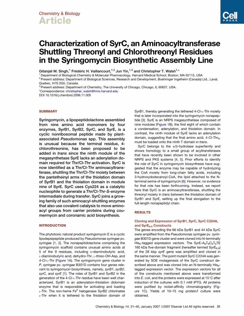
Figure 1. Syringomycin E Structure and
the Proteins Involved in Its Biosynthesis
(A) Syringomycin E structure.
(B) SyrE module organization.
(C) SDS polyacrylamide gel of purified Syr
proteins.
Several errors were found when comparing the cloned
sequences of SyrE8,9 and SyrC to the published se-
quences [1]. Multiple clones from different PCR reactions
were sequenced to confirm that the errors did not result
from errors incurred during PCR amplification. The correct
sequences are shown in Figure S1 (see the Supplemental
Data available with this article online). Similar types of
errors were found in SyrB1 and SyrB2 by the original group
that published the sequences, but these errors have since
been corrected. The initial errors were simply due to the
poor quality of the initial sequencing results, not to a differ-
ence in strains.
Demonstration of L-[14C]Thr Transfer from SyrB1 to
SyrC; Formation of a SyrC Acyl-Enzyme Intermediate
Prior work has shown that SyrB1 is the adenylation-thiola-
tion (A-T) didomain protein that activates and then cova-
lently tethers L-Thr in thioester linkage to the phosphopan-
tetheinyl arm in the thiolation domain, where it then
undergoes halogenation to produce 4-Cl-Thr-S-SyrB1
[3]. However, the multimodular syringomycin synthetase
SyrE lacks a ninth adenylation domain to load the final
4-Cl-Thr residue onto its ninth thiolation domain (T9)
(Figure 1B). Therefore, we propose that SyrC transfers
4-Cl-Thr from SyrB1 to SyrE. To test the hypothesis that
SyrC may engage in two-step aminoacyl transfer via a
Thr/Cl-Thr-enzyme intermediate, the transfer of L-[14C]Thr
from [14C]Thr-S-SyrB1 to SyrC was evaluated (Figure 4B,
first half reaction). Although 4-Cl-Thr is the form of Thr
incorporated into the syringomycin scaffold, we used
L-[14C]Thr to test for SyrC acyltransferase activity since it
is readily available and can be detected by autoradiogra-
phy, and since Thr9 variants of syringomycin are known [7].
32 Chemistry & Biology 14, 31–40, January 2007 ª2007 Elsevie
Recombinant SyrC was incubated with phosphopante-
theinylated SyrB1 (holo-SyrB1) in the presence of
L-[14C]Thr and ATP in HEPES buffer. Aliquots of the reac-
tion were quenched in SDS-PAGE loading buffer at
various time points, and the proteins were subjected to
SDS-PAGE. The autoradiogram of the gel revealed a de-
tectable amount of radiolabel accumulating on SyrC in
a time-dependent fashion (Figure 2A), suggesting the for-
mation of an acyl-enzyme intermediate. This intermediate
is only apparent in the absence of reducing agents, such
as b-mercaptoethanol and dithiothreitol, indicating that it
is quite labile to thiol nucleophiles.
Demonstration of SyrC-Mediated Transfer
of L-[14C]Thr from SyrB1 to SyrE
To determine whether SyrC can mediate L-[14C]Thr trans-
fer from SyrB1 to SyrE, we used an equivalent gel-based
assay as described above, but we now monitored ac-
cumulation of the radiolabeled SyrE8,9 fragment. Both
SyrB1 and SyrE8,9 were preincubated with the phospho-
pantetheinyltransferase Sfp [8, 9] to convert inactive apo
forms of the thiolation domains to the HS-pantetheinyl-
containing holo forms. SyrE8,9 has two T domains that
must be posttranslationally primed. SyrC was incubated
with holo-SyrB1 and holo-SyrE8,9 in the presence of
L-[14C]Thr and ATP in HEPES buffer. A build-up of radiola-
bel on SyrE was detectable over a 1 hr time period, show-
ing that L-[14C]Thr is indeed transferred from SyrB1 to
SyrE. Control experiments verified that the transfer was
both time and SyrC dependent (Figures 3A and 3B). In
the absence of reducing agents, the SyrC acyl-enzyme
intermediate is visible (Figure 3C).
r Ltd All rights reserved
Chemistry & Biology
Acyl Transfer in Syringomycin Biosynthesis
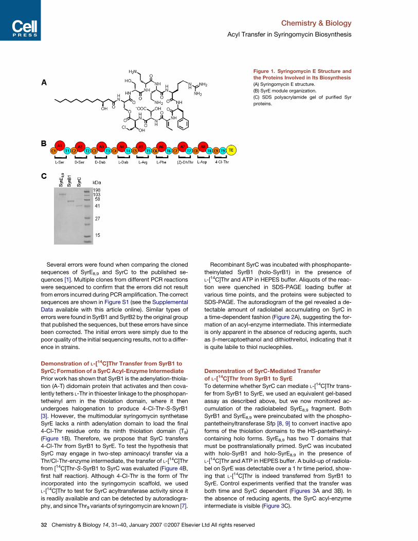
Figure 2. Assays for Transfer of
L-[14C]Thr from SyrB1 to SyrC and Acyl-
Enzyme Intermediate Formation
(A) Autoradiogram and SDS polyacrylamide
gel of the transfer assay with SyrC under
nonreducing conditions during gel sample
preparation.
(B) Autoradiogram and SDS polyacrylamide
gel of the transfer assay with SyrC C224A
under nonreducing conditions during gel
sample preparation.
Identification of the Catalytic Residue Necessary
for Acyltransferase Activity
Sequence comparison to other members of the a/b-hy-
drolase protein superfamily, notably CmaE [4], CouN7,
and CloN7 [5], suggested that the thiolate side chain of
Cys224 could be the catalytic nucleophile; thus, the cor-
responding SyrC C224A mutant was constructed, ex-
pressed, and purified (Figure S2). The SyrC C224A mutant
protein did not become detectably radiolabeled, suggest-
ing that the point mutation prevents formation of the acyl-
enzyme intermediate (Figures 2B and 4A). Therefore,
given the first half reaction (Thr-S-SyrB1 to SyrC) and
the overall reaction (Thr-S-SyrB1 to SyrE8,9), we conclude
that SyrC is competent to catalyze the second half reac-
tion of the scheme depicted in Figure 4B (Thr-S-SyrC to
SyrE8,9). The migrating aminoacyl moiety is shuttling be-
tween thiol nucleophiles, Cys224 in SyrC, and the HS-
pantetheinyl arms of the T domains in SyrB1 and SyrE.
Chemistry & Biology 14
Characterization of SyrC Amino Acid and Acceptor
T Domain Specificity
Testing the selectivity of SyrC for transfer of amino acids
other than L-Thr onto SyrE8,9 required the use of alternate
aminoacyl-S-SyrB1 proteins as donor substrates. From
our prior studies, SyrB1 has very high specificity for load-
ing L-Thr and cannot load alternate amino acids to any sig-
nificant extent [3]. Therefore, instead of using SyrB1 as the
donor T domain, we turned to a comparable free-standing
A-T didomain enzyme, CmaA from the coronamic acid
biosynthetic pathway [4]. Although CmaA normally acti-
vates and tethers the allo diastereomer of L-isoleucine
on its T domain, CmaA is also capable of loading L-valine,
which is available in radioactive form.
When SyrC was incubated with holo-CmaA and SyrE8,9
in the presence of L-[14C]Val and ATP in HEPES buffer and
the transfer reaction was monitored by autoradiography
(Figure 5), the build-up of radioactive label on SyrE in
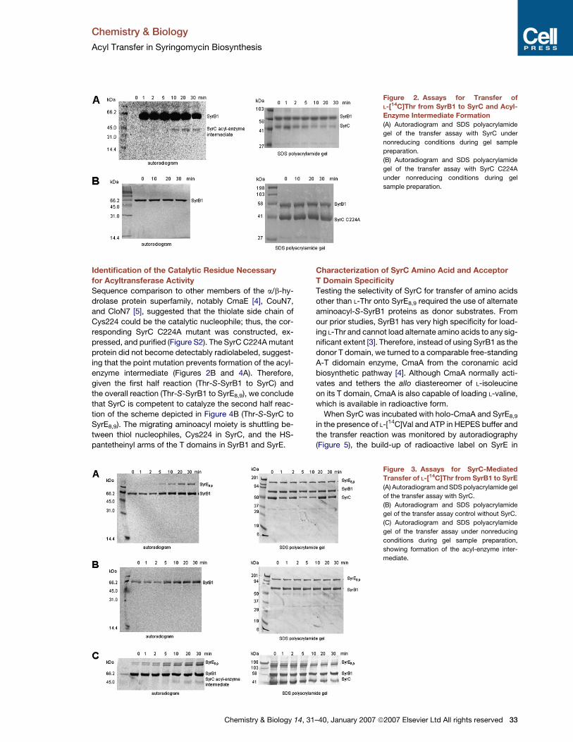
Figure 3. Assays for SyrC-Mediated
Transfer of L-[14C]Thr from SyrB1 to SyrE
(A) Autoradiogram and SDS polyacrylamide gel
of the transfer assay with SyrC.
(B) Autoradiogram and SDS polyacrylamide
gel of the transfer assay control without SyrC.
(C) Autoradiogram and SDS polyacrylamide
gel of the transfer assay under nonreducing
conditions during gel sample preparation,
showing formation of the acyl-enzyme inter-
mediate.
, 31–40, January 2007 ª2007 Elsevier Ltd All rights reserved 33

Chemistry & Biology
Acyl Transfer in Syringomycin Biosynthesis
Figure 4. Assay of the SyrC C224A Mutant and Proposed Acyltransfer Mechanism
(A) Assay for L-[14C]Thr transfer from SyrB1 to SyrE with the SyrC C224A mutant.
(B) Scheme depicting the two proposed half reactions involved in L-Thr transfer from SyrB1 to SyrE8,9 via SyrC.
a time- and SyrC-dependent fashion was detected. This
result shows that SyrC can both use an alternate amino-
acyl moiety and recognize an alternate T domain donor
scaffold when it mediates the transfer of L-[14C]Val from
CmaA to SyrE8,9.
Analogous logic was used to evaluate the ability of SyrC
to transfer a leucyl group from an L-[14C]Leu-S-T domain.
In this case, we had to turn to the barbamide cluster [10,
11] using the free-standing adenylation domain BarD
and its cognate 10 kDa T domain BarA, in posttranslation-
ally primed holo form. Purified BarD was used to adenylate
and load L-[14C]Leu onto holo-BarA. Next, SyrC was
incubated with L-[14C]Leu-S-BarA and SyrE8,9, and the
reaction was monitored as described above. Again,
a SyrC- and time-dependent build-up of radioactive label
on SyrE was observed, adding Leu and Val to Thr as
shuttling aminoacyl groups for SyrC action (data not
shown).
34 Chemistry & Biology 14, 31–40, January 2007 ª2007 Elsevie
Using an autoradiographic assay, we found that SyrC
is capable of transferring L-[14C]-Thr from SyrB1 to the
stand-alone T9 domain. When similar experiments were
done to determine whether SyrC can mediate the transfer
of L-[14C]Thr from SyrB1 to the alternate acceptor T do-
main, CmaD from the coronamic acid pathway [4], no
transfer was observed, perhaps suggesting more strin-
gent recognition of the T domain scaffold in its acceptor
mode. In future experiments, we will carry out an accurate
kinetic comparison between various T8 and T9 constructs,
embedded and excised, along with comparison of other
excised domains from the nine-module SyrE protein.
Examination of SyrC Acyltransferase Specificity
for the T8 versus T9 Domain of SyrE
Our hypothesis that SyrC acts in trans to load L-Thr onto T9
of SyrE raises the question of whether SyrC is specific
enough to distinguish T9 from all of the other T domains
r Ltd All rights reserved
Chemistry & Biology
Acyl Transfer in Syringomycin Biosynthesis
Figure 5. SyrC-Mediated Transfer of
Valine to SyrE
Autoradiogram and SDS polyacrylamide gel
showing SyrC-mediated transfer of L-[14C]Val
from CmaA to SyrE.
in the megadalton, nine-module SyrE. Numerous attempts
at site-directed mutagenesis in T8 and T9 of the 160 kDa
SyrE8,9 fragment were attempted, but due to the large
size and high GC content of the SyrE construct, we have
not been able to successfully generate the mutant
constructs.
Therefore, an alternate limited proteolysis approach
was taken to probe SyrC specificity for T8 versus T9.
SyrE was loaded with L-[14C]Thr via SyrB1 and SyrC as
described above and then subjected to cleavage at the
unique thrombin site directly preceding the T9 domain of
SyrE (Figure 6A). The resulting fragments were analyzed
by SDS-PAGE and autoradiography. Cleavage of the
SyrE8,9 construct with thrombin results in the formation
of two fragments: A8T8C9 (118 kDa) and T9TE (42 kDa). If
SyrC were to specifically transfer L-[14C]Thr only to the
T9 domain, and not to the T8 domain, only the 42 kDa
band should be radiolabeled in the autoradiogram
(Figure 6B). However, both the 118 kDa and 42 kDa bands
are radiolabeled, suggesting that the SyrC aminoacyl-
transferase does not distinguish between the two do-
mains in this transfer experiment. Future kinetic studies
may reveal if there are rate differences for Thr transfer to
the pantetheinyl arms of T8 versus T9.
Since aminoacylation of T8 via SyrC would jam the syrin-
gomycin assembly line, it is possible that T8 is aminoacyl-
ated in cis via A8 much faster than it is aminoacylated in
trans via SyrC. This would prevent SyrC from misloading
T8 with threonine. To test this hypothesis, we repeated
the thrombin proteolysis assay described above, but this
time added nonradiolabeled L-Asp to the reaction mixture.
In this case, if L-Asp were activated and loaded onto T8 at
a greater rate than SyrC could load L-[14C]Thr in trans, we
Chemistry & Biology 14,
would expect to see radiolabel only on the T9TE fragment
since the loading of nonradiolabeled L-Asp would pre-
clude misloading of L-[14C]Thr on T8. However, we again
observed radiolabel on both the A8T8C9 and the T9TE frag-
ments, suggesting that there is some other mechanism of
regulation that we cannot replicate in vitro.
Determination of SyrE-A8 Substrate Specificity
by ATP-PPi Exchange Assay
The eighth amino acid in the mature syringomycin lipo-
peptidolactone is L-threo-3-OH-Asp. The five-domain
SyrE8,9 has the A8 domain intact and thus could be tested
to determine whether L-threo-3-OH-Asp is activated and
loaded onto T8, or whether L-Asp is loaded and hydroxyl-
ation occurs later. The aminoacyl-AMP half reaction of the
A8 domain was assayed by amino acid-dependent ATP-
PPi exchange assay [12]. At a substrate concentration
of 10 mM, A8 in the SyrE8,9 160 kDa fragment was able
to activate L-Asp with a kobs of 18.6 min�1, whereas it
was able to activate L-threo-3-OH-Asp with a kobs of
0.98 min�1, suggesting that hydroxylation of Asp occurs
on or after the assembly line. N-acetyl-L-Asp and the
syringomycin octapeptide comprising residues 1–8 did
not support the ATP-PPi exchange, consistent with
a lack of recognition by the A8 domain.
Condensation of L-[14C]Thr with the Syringomycin
Octapeptide Catalyzed by C9
Given that SyrE8,9 could load Asp onto T8 and that SyrC,
in the presence of SyrB1, could transfer Thr to T9, the
question of whether the C domain in module nine could
catalyze peptide bond formation arose. It did not yield
detectable amounts of Asp-Thr dipeptide, so we postulate
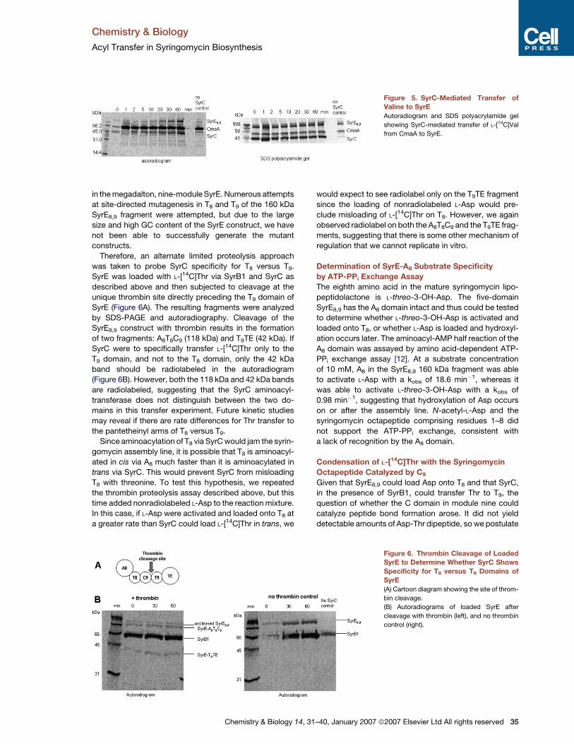
Figure 6. Thrombin Cleavage of Loaded
SyrE to Determine Whether SyrC Shows
Specificity for T8 versus T9 Domains of
SyrE
(A) Cartoon diagram showing the site of throm-
bin cleavage.
(B) Autoradiograms of loaded SyrE after
cleavage with thrombin (left), and no thrombin
control (right).
31–40, January 2007 ª2007 Elsevier Ltd All rights reserved 35
Chemistry & Biology
Acyl Transfer in Syringomycin Biosynthesis
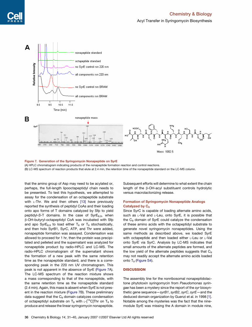
Figure 7. Generation of the Syringomycin Nonapeptide on SyrE
(A) HPLC chromatogram indicating products of the nonapeptide formation reaction and control reactions.
(B) LC-MS spectrum of reaction products that elute at 2.4 min, the retention time of the nonapeptide standard on the LC-MS column.
that the amino group of Asp may need to be acylated or,
perhaps, the full-length lipooctapeptidyl chain needs to
be presented. To test this hypothesis, we attempted to
assay for the condensation of an octapeptide substrate
with L-Thr. We and then others [13] have previously
reported the synthesis of peptidyl CoAs and their loading
onto apo forms of T domains catalyzed by Sfp to yield
peptidyl-S-T domains. In the case of SyrE8,9, when
b-OH-butyryl-octapeptidyl CoA was incubated with Sfp
and apo SyrE8,9 to load either T8 or T9 stochastically,
and then holo SyrB1, SyrC, ATP, and Thr were added,
nonapeptide formation was assayed. Condensation was
allowed to proceed for 1 hr, then the protein was precipi-
tated and pelleted and the supernatant was analyzed for
nonapeptide product by radio-HPLC and LC-MS. The
radio-HPLC chromatogram of the supernatant shows
the formation of a new peak with the same retention
time as the nonapeptide standard, and there is a corre-
sponding peak in the 220 nm UV chromatogram. This
peak is not apparent in the absence of SyrE (Figure 7A).
The LC-MS spectrum of the reaction mixture shows
a mass corresponding to that of the nonapeptide, with
the same retention time as the nonapeptide standard
(2.4 min). Again, this mass is absent when SyrE is not pres-
ent in the reaction mixture (Figure 7B). These preliminary
data suggest that the C9 domain catalyzes condensation
of octapeptidyl substrate on T8 with L-[14C]Thr on T9 to
produce and release the linear syringomycin nonapeptide.
36 Chemistry & Biology 14, 31–40, January 2007 ª2007 Elsevie
Subsequent efforts will determine to what extent the chain
length of the b-OH-acyl substituent controls hydrolytic
versus macrolactonizing release.
Formation of Syringomycin Nonapeptide Analogs
Catalyzed by C9
Since SyrC is capable of loading alternate amino acids,
such as L-Val and L-Leu, onto SyrE, it is possible that
the C9 domain of SyrE could catalyze the condensation
of these amino acids with the octapeptidyl substrate to
generate novel syringomycin nonapeptides. Using the
same methods as described above, we loaded SyrE
with octapeptide and then loaded either L-Leu or L-Val
onto SyrE via SyrC. Analysis by LC-MS indicates that
small amounts of the alternate peptides are formed, and
the low yield of the alternate peptides suggests that C9
may not readily accept the alternate amino acids loaded
onto T9 (Figure S4).
DISCUSSION
The assembly line for the nonribosomal nonapeptidolac-
tone phytotoxin syringomycin from Pseudomonas syrin-
gae has been a mystery since the report of the syr biosyn-
thetic gene sequence—syrB1, syrB2, syrC, syrE—and the
deduced domain organization by Guenzi et al. in 1998 [1].
Notable among the mysteries was the fact that the nine-
module SyrE was missing the A domain in module nine,
r Ltd All rights reserved
Chemistry & Biology
Acyl Transfer in Syringomycin Biosynthesis
suggesting that the MDa protein SyrE would be incompe-
tent to activate and load the terminal residue, Thr9 or 4-Cl-
Thr9, to finish chain elongation. Further, the A domain in
the free-standing A-T didomain enzyme SyrB1 was able
to activate Thr. This suggested that SyrB1 was in effect
the ‘‘missing’’ A9, and that the Thr9 must come in trans
from SyrB1 to module nine of SyrE each time a syringomy-
cin chain is completed on the SyrE assembly line. We
clarified part of the puzzle by recently demonstrating the
function of SyrB2 as a novel mononuclear nonheme FeII
halogenase that acts on Thr-S-SyrB1 to produce 4-Cl-
Thr-S-SyrB1. This is the catalytic step generating the
Cl-Thr9 donor on the pantetheinyl arm of SyrB1 [3].
In the present work, we demonstrate the function of
the remaining Syr protein, the 44 kDa SyrC, and validate
the in trans shuttling of Thr/Cl-Thr from SyrB1 to SyrE.
When the sequence of SyrC was reported, it was clearly
a member of the a/b-hydrolase superfamily. Postulated
functions included thioesterase, haloperoxidase, or per-
haps an acyltransferase for the N-terminal b-OH-fatty
acyl moiety [1, 6]. We have previously assayed SyrC for
such N-acylation activity of Ser1-S-SyrE without any suc-
cess (R.G. Kruger, F.H.V., C.T.W., data not shown).
Two parallel efforts on other NRPS systems in which
aminoacyl/acyl moieties are shuttled gave us some inkling
of the function of SyrC demonstrated in this work. One
was in the coronamic acid biosynthetic pathway in which
the free-standing A-T didomain CmaA corresponds to
SyrB1 and the nonheme iron halogenase CmaB corre-
sponds to SyrB2 [4]. CmaE, a homolog of SyrC, shuttles
an L-allo-Ile moiety between the pantetheinyl arm of the
T domain of CmaA and the free-standing T domain of
CmaD. Only when the aminoacyl group is presented as
L-allo-Ile-S-CmaD will the CmaB halogenase recognize it
[4]. CmaE is thus an aminoacyltransferase that shuttles
the allo-Ile moiety between holo T domain scaffolds.
The second example is the recent demonstration that
the last step in coumermycin and clorobiocin biosynthesis
involves transfer of the acyl group, pyrrole-2-carbonyl,
from the pantetheinyl arm of a T domain to the 30-OH of
the noviosyl ring of the antibiotic [5]. CmaE uses an active
site Cys as a nucleophile, while CouN7 uses an active site
Ser as a nucleophile, to make acyl-S- and acyl-O-enzyme
intermediates, respectively, to shuttle the substrate acyl
fragments between acceptors. In this context, SyrC joins
this family of acyl/aminoacyl-S-pantetheinyl-T domain
shuttle enzymes and solves the need for an in trans deliv-
ery of an activated Thr/Cl-Thr moiety to module nine of
SyrE.
The assay of SyrC activity was not entirely straightfor-
ward. The protein scaffold for the aminoacyl donor is the
66 kDa holo form of the SyrB1 protein, which can be
made in multimilligram quantities. The 4-Cl-Thr amino
acyl moiety is not available in radioactive form, cannot
be loaded by the A domain of SyrB1 [3], and is not quan-
titatively available by action of SyrB2 on Thr-S-SyrB1.
Therefore, we used L-[14C]Thr as a surrogate for Cl-Thr.
The acceptor substrate is even harder to come by:
P. syringae SyrE has a molecular weight of 1,038,663 Da
Chemistry & Biology 14,
and is not readily expressable in E. coli. Of various frag-
ments of SyrE, we chose the 160 kDa A8-T8-C9-T9-TE
(deemed SyrE8,9) to express and purify in soluble form to
evaluate various aspects of the last step of syringomycin
assembly. In principle, SyrE8,9 could allow assay of in trans
import of L-[14C]Thr from SyrB1, loading of Asp/b-OH-Asp
by A8 on T8, condensation activity of C9, and release activ-
ity of the TE domain. In this study, we have focused mostly
on demonstrating SyrC’s aminoacyl shuttle activity.
The SyrC activity assay devolved to monitoring
L-[14C]Thr transfer from L-[14C]Thr-S-SyrB1 to holo
SyrE8,9. Our hypothesis that SyrC, like CmaE and CouN7
[4, 5], may engage in covalent catalysis is confirmed by
autoradiography of SyrC in SDS-PAGE studies, with the
thiolate side chain of Cys224 as the likely catalytic nucle-
ophile. Thus, the shuttle enzymes CmaE and SyrC carry
out energetically neutral aminoacyl transfers between
thiolates of pantetheinyl prosthetic groups by way of a
covalent thioester linkage in the shuttling enzyme. In
contrast, CouN7, which uses a Ser-OH nucleophile, cata-
lyzes the energetically favorable transfer of the pyrrole-
2-carboxy acyl group from a pantetheinyl thioester in
CouN1 to an oxoester in the pyrrolyl-coumarin antibiotic
scaffold [5].
The SyrE8,9 fragment is not ideal for determination of the
T domain selectivity of SyrC, but it appears that the itiner-
ant L-[14C]Thr can end up on the pantetheinyl forms of both
T8 and T9 of this enzyme fragment. Whether occupancy of
T8 by the waiting octapeptidyl chain is a default director
of the incoming Thr to T9, or there are higher-order con-
former constraints in the full-length, million molecular
weight SyrE is yet unknown. Presumably, transfer of the
L-[14C]Thr/Cl-Thr to an empty holo-T8 would jam the
assembly line and, thus, would have to be avoided or
hydrolytically released. In subsequent efforts, we shall
begin to address the action of C9 and the TE domain, per-
haps in a smaller C9-T9-TE construct, to begin to study the
affinity of SyrC for SyrE T domains, the kinetics and spec-
ificity of transfers of the aminoacyl moiety that will become
residue 9, and the permissivity of the C9 condensation
step and macrolactonization.
Given our recent detection of the three examples noted,
the question of how frequently aminoacyl shuttle enzymes
will turn up in NRPS assembly lines arises. They should
allow in trans aminoacyl group insertions to create
diversity at particular sites. If different shuttling enzymes
recognize distinct sets of T domains, then it may become
possible to increase positional diversity in nonribosomal
peptides in their presence.
SIGNIFICANCE
The final step of the syringomycin biosynthetic
assembly line, which involves the incorporation of
chlorothreonine into the syringomycin scaffold, has
long been a mystery due to the fact that SyrE, the meg-
adalton syringomycin synthetase, lacks an adenyla-
tion domain in its ninth module. Prior work has shown
that chlorothreonine is generated by the action of the
31–40, January 2007 ª2007 Elsevier Ltd All rights reserved 37
Chemistry & Biology
Acyl Transfer in Syringomycin Biosynthesis
FeII/a-ketoglutarate-dependent halogenase SyrB2 on
a threonine residue that is tethered via a phosphopan-
tetheinyl linkage to the A-T didomain protein SyrB1.
Here, we demonstrate that SyrC is an acyltransferase
that is capable of shuttling threonine from SyrB1 to
SyrE. Furthermore, we present preliminary data
suggesting that the ninth condensation domain of
SyrE is capable of catalyzing the condensation of
threonyl-S-SyrE-T9 with an octapeptidyl substrate to
generate a linear nonapeptide, which is then hydro-
lyzed by the thioesterase domain. We also show that
SyrC is capable of transferring alternate amino acids,
such as leucine and valine, onto SyrE, and that these
amino acids can then be incorporated into a linear
nonapeptide. These results suggest that SyrC may
be useful in the combinatorial enzymatic synthesis of
syringomycin analogs, and they set the stage for
further studies into the final steps of syringomycin
biosynthesis. In addition, our identification of SyrC
as an aminoacyltransferase adds another member to
the small but growing family of NRPS/PKS acyltrans-
ferases that use covalent catalysis to shuttle amino-
acyl groups between carrier proteins. This family of
aminoacyltransferases may be useful in creating posi-
tional diversity in nonribosomal peptides.
EXPERIMENTAL PROCEDURES
Materials and General Methods
All radiolabeled chemicals were obtained from American Radiolabeled
Chemicals, Inc. (ARC). L-threo-3-OH-Asp was obtained from Tocris
Pharmaceuticals. All other chemicals used were from Sigma, unless
otherwise specified. The TOP10- and BL21(DE3)-competent E. coli
strains were purchased from Stratagene. The PfuTurbo DNA Polymer-
ase used in PCR was purchased from Stratagene. During protein
purification, cells were lysed with an Avestin Emulsiflex-C5 cell disrup-
tor. Thrombin was obtained from Novagen. SDS polyacrylamide gels
were obtained from BioRad, and autoradiography was performed on a
Typhoon 9400 scanner (GE Healthcare). HPLC was carried out on
a Beckman Coulter System Gold by using a Vydac C18 column.
Radio-HPLC was performed on an identical system equipped with
a b-Ram radioisotope detector (IN/US). LC-MS analysis was per-
formed on an LCMS-QP8000a spectrometer (Shimadzu) with a Vydac
C18 LC-MS column. Recombinant BarA and BarD were provided by
Danica Galonic, and recombinant CmaA was provided by Eric Strieter.
Cloning of Syr Genes
SyrB1 was cloned as described [3]. The SyrC and SyrE8,9 constructs
were obtained by PCR amplification of genomic DNA from Pseudomo-
nas syringae pv. syringae B301D, which was a gift from Dennis C.
Gross (Texas A&M University, College Station, Texas) [14]. P. syringae
pv. syringae was grown in nutrient broth-yeast extract medium at
30�C, and genomic DNA was isolated by using the Bactozol kit for bac-
terial DNA extraction (Molecular Research). Amplification was carried
out by using PfuTurbo DNA Polymerase in accordance with the man-
ufacturer’s instructions. The following oligonucleotide primers were
used in PCR amplification: SyrC: 50-GGAATTCCATATGCGCGTTTG
CGGCATT-30 and 50-CCCAAGCTTCATCATGGGAAGCTGGGACA-30;
SyrE8,9: 50-CGGAATTCACTACTCACTGGCGCGGT-30 and 50-GGAAT
TCCATATGCTTGAGCAGGATCCGGCA-30. The SyrC PCR product
was digested with NdeI and HindIII, and the SyrE8,9 construct was
digested with NdeI and EcoRI. All digested PCR products were ligated
into similarly digested pET28b plasmids to create N-terminally His6-
tagged constructs.
38 Chemistry & Biology 14, 31–40, January 2007 ª2007 Elsevie
Overexpression and Purification of Syr Proteins
The pET-28a expression vectors containing the Syr proteins were
transformed into BL21 (DE3)-competent cells. Cultures were grown
in Luria-Bertani medium supplemented with 30 mg/ml kanamycin
at 37�C until the OD600 reached �0.3, at which time the cultures
were cooled to 25�C and grown until the OD600 reached �0.6. The
cultures were induced with 0.1 mM IPTG and were grown at 15�C
overnight. Cells were harvested by centrifugation at 6,000 rpm for
30 min, flash frozen in liquid N2, and stored at �80�C until further
purification.
Cells were thawed, resuspended in Buffer A (300 mM NaCl, 5 mM
imidazole, 20 mM Hepps [pH 8.0]), and lysed by cell disruption. Cell
debris was removed from the lysate by centrifugation at 15,000 rpm
for 30 min, and the supernatant was removed and bound to Ni-NTA
resin by rocking at 4�C for 2 hr. The resin was added to a Bio-Rad
Econo-Sphere column and washed with Buffer A. Protein was eluted
with Buffer B (300 mM NaCl, 30 mM imidazole, 20 mM Hepps
[pH 8.0]) and Buffer C (100 mM NaCl, 200 mM imidazole, 20 mM
Hepps [pH 8.0]). Protein-containing fractions were identified by
SDS-PAGE, combined, and dialyzed overnight in 100 mM NaCl,
1 mM EDTA, 50 mM Hepps (pH 8.0) with 10% glycerol. The dialyzed
protein was concentrated, flash frozen in liquid N2, and stored
at �80�C.
Site-Directed Mutagenesis of SyrC
The pET28a plasmid containing the syrC gene was used as the tem-
plate for splicing by overlap extension (SOE) to generate the C224A
mutant protein. The standard two-step SOE PCR method was used
to create a mutation at the desired site [15]. In the first round of
PCR, the 50 end of the SyrC protein was amplified with the following
primers: 50-CGGGGATCCCATATGACTATTTCCTCCGAT-30 (forward)
and 50-GCCGGCGATGCCCATCAGATGTGCGGTCGA-30 (reverse
internal). Also in the first round of PCR, the 30 end of the SyrC protein
was amplified with the following primers: 50-CGGATAGAGCTCT
CAGGCGACAGCGGGCTG-30 (reverse) and 50-CATCTGATGGGCAT
CGCCGGCGGCGCGGTCATC-30 (forward internal). Underlined bases
indicate restriction sites, and bold base pairs indicate the sites of
mutation. The two resulting fragments were purified by using the Qia-
gen PCR Purification kit and were then mixed and used in the second
round of PCR with the forward and reverse primers from the first step.
The resulting PCR product was digested with XhoI and NdeI and
ligated into the pET28a plasmid. The presence of the mutation in the
resulting vector was verified by sequencing. The SyrC C224A mutant
protein was expressed and purified as described above.
Generation of Phosphopantetheinlyated Enzymes
Recombinant enzymes containing thiolation domains, such as SyrB1,
SyrE, CmaA, and BarA, must be phosphopantetheinlyated prior to
their use in assays. In a typical reaction, 1 nmol enzyme is incubated
with MgCl2 (0.5 mmol), CoA (50 nmol), Sfp (10 nmol) in 50 mM HEPES
buffer (pH 7.5), in a total reaction volume of 25 ml for 30 min at room
temperature to produce the holo enzyme.
Solid-Phase Synthesis of Octapeptide, COOH-L-Asp-L-Abu-L-
Phe-L-Arg-L-Dab-D-Dab-D-Ser-L-Ser-N-Ac, and Nonapeptide,
COOH-L-Thr-L-Asp-L-Abu-L-Phe-L-Arg-L-Dab-D-Dab-D-Ser-L-
Ser-N-Ac
Peptide synthesis was performed with a PerSeptive Biosystems
9050 synthesizer (0.3 mM scale) by using diisopropylcarbodiimide
(DIPCDI)/hydroxybenzotriazole (HOBt) chemistry. The peptide was
cleaved from the resin, deprotected in a single treatment with 95% tri-
fluoroacetic acid (TFA), 2.5% water, and 2.5% triisopropylsilane (TIS)
at room temperature for 3 hr. The solution was then added to cold ether
dropwise, and the precipitated peptide was collected by centrifuga-
tion. The purified peptides (Figure S3) were lyophilized to yield a white
powder, and the identity and purity were established by analysis
on LCMS.
r Ltd All rights reserved
Chemistry & Biology
Acyl Transfer in Syringomycin Biosynthesis
Synthesis of Coenzyme A Derivatives of Octapeptide
and Nonapeptide
The peptidyl-CoA derivatives (Figure S3) were prepared based on
a previously reported protocol [16]. The peptide was cleaved from
the resin with 1:1:3 acetic acid:trifluoroethanol (TFE):dichloromethane
(DCM) and was incubated at room temperature for 3 hr. The resin was
removed by filtration, and n-hexane was added to precipitate the fully
protected peptide. After rotary evaporation to remove the solvent, the
peptide was redissolved in DCM and precipitated with n-hexane; this
was repeated twice. The CoA coupling to the protected peptide was
accomplished by the addition of 1 equivalent of CoA (Li+ salt; Sigma),
4 equivalents of potassium carbonate, and 1.5 equivalents of PyBOP
in 1:1 THF:water. The reaction was mixed by tipping for 2 hr at room
temperature, and then the solvent was removed by rotary evaporation
followed by lyophilization. Removal of the N-terminal Boc-protecting
groups was achieved by treatment with 95% TFA, 2.5% water, and
2.5% TIS at room temperature for 3 hr. The solution was then added
to cold ether dropwise, and the precipitate was removed by centrifuga-
tion after overnight incubation at �20�C. The peptidyl-CoA was then
dissolved in acetonitrile/water and purified by preparative HPLC on
a reverse-phase C18 column with a gradient of 0%–100% acetonitrile
in 0.1% TFA/water over 35 min. The pure peptidyl-CoA had a retention
time of 16 min. The purified compounds were lyophilized, and the iden-
tity and purity were established by analysis in negative ion mode
on LCMS and MALDI-TOF mass spectrometry: octapeptidyl-CoA
1711.5 [(M � H)�] calculated, 1711.1 observed; nonapeptidyl-CoA
1811.6 [(M � H)�] calculated, 1811.7 observed.
Assay for Transfer of L-[14C]Thr from SyrB1 to SyrC
Holo-SyrB1 (0.4 nmol) was incubated with L-[14C]Thr (9 nmol) and ATP
(200 nmol) in 50 mM HEPES buffer (pH 7.5), in a total reaction volume
of 62 ml for 10 min at room temperature to generate L-[14C]Thr-
S-SyrB1. To this reaction mixture, SyrC (0.6 nmol) was added, and
8 ml aliquots of the reaction were quenched in 23 SDS-PAGE loading
buffer (without reducing agent) at various time points. The quenched
aliquots were heated to 70�C for 10 min and then run on a 12% SDS
polyacrylamide gel. The gel was stained, destained, dried, and
exposed to a phosphorimager screen for 3 days, after which the
screen was scanned with a Typhoon imager.
Assay for SyrC-Mediated Transfer of [14C]-Labeled Amino Acids
from SyrB1 to SyrE
In a typical reaction (66 ml), holo-SyrB1 (0.4 nmol) and holo-SyrE8,9
(0.2 nmol) were incubated with L-[14C]Thr (9 nmol) and ATP
(200 nmol) in 50 mM HEPES (pH 7.5). Recombinant SyrC (0.6 nmol)
was added to initiate the reaction, and time points were quenched
and run on a gel as described above. In tandem, a reaction lacking
SyrC was performed as a negative control. The gels were processed
as described above. Similar reactions to examine the ability of SyrC
to transfer alternate amino acids were carried out with L-[14C]Leu
and L-[14C]Val, with donor T domains BarA and CmaA, respectively.
Assay for Formation of the Syringomycin Nonapeptide
on SyrE8,9
Holo-SyrE8,9 (1.4 nmol) was incubated with ATP (100 nmol) and
octapeptidyl-S-CoA (20 nmol) in 50 mM HEPES (pH 7.5), in a total
reaction volume of 45 ml for 1 hr at room temperature. The octapep-
tidyl-S-SyrE was then incubated with holo-SyrB1 (2.8 nmol),
L-[14C]Thr (9 nmol), and ATP (450 nmol) in HEPES. Recombinant
SyrC (4.2 nmol) was added to initiate the reaction (140 ml total volume),
which was incubated at room temperature for 1 hr to allow nonapep-
tide formation to proceed. In tandem, reactions lacking peptide,
L-[14C]Thr, SyrB1, SyrC, or SyrE were carried out as controls.
The reactions were quenched in 100 ml 10% (v/v) trichloroacetic acid
(TCA) to precipitate the proteins. The proteins were pelleted by centri-
fugation at 13,000 rpm for 20 min, and the supernatant was removed
and saved (supernatant A). The pellet was washed twice with 100 ml
10% TCA and then redissolved in 100 ml 0.1M LiOH and heated to
Chemistry & Biology 14,
85�C to release any peptide still bound to the SyrE8,9 scaffold. The
protein was then reprecipitated by the addition of 20 ml 50% TCA
and pelleted by centrifugation, and the supernatant was removed
and saved (supernatant C).
The two supernatants from each reaction were analyzed by HPLC
and LC-MS to determine if they contained the radiolabeled nonapep-
tide product. The HPLC analysis was carried out by using a Vydac C18
small-pore column with a water/acetonitrile gradient going from
0%–100% acetonitrile over 30 min. HPLC was monitored both for
absorbance at 220 nm and for 14C radioactive counts. By LC-MS,
the nonapeptide was observed in supernatant A of the reaction con-
taining all components: 1083.1 [(M + H+)] calculated, 1082.0 observed.
ATP-PPi Exchange Assays to Determine SyrE-A8 Substrate
Specificity
Each reaction contained 10 mM amino acid, 10 mM MgCl2, 1 mM ATP,
1 mM DTT, 2 mM SyrE8,9, and 5 mM sodium [32P]pyrophosphate in
a total volume of 500 ml with 50 mM HEPES (pH 7.5). In tandem, reac-
tions containing no enzyme were carried out as negative controls.
Reactions were initiated by the addition of enzyme, and aliquots
were quenched at 0, 1, 2, 5, 10, 20, 30, and 60 min by the addition
of 750 ml of a solution containing 1.6% (w/v) activated charcoal,
200 mM sodium pyrophosphate, and 3.5% (v/v) perchloric acid. For
each time point, the charcoal was pelleted by centrifugation and
washed twice with 750 ml of a solution containing 200 mM sodium
pyrophosphate with 3.5% perchloric acid (wash buffer). The charcoal
pellet was then resuspended in 750 ml wash buffer and mixed with
liquid scintillation fluid. Radioactivity bound to the charcoal was
measured by liquid scintillation counting.
Assay for SyrC Specificity for SyrE8,9 T Domains by Thrombin
Cleavage
To determine whether SyrC can specifically transfer L-[14C]Thr from
SyrB1 to the T9 domain of SyrE, or whether it transfers the amino
acid to both T8 and T9, holo-SyrE8,9 (0.2 nmol) was loaded with
L-[14C]Thr via SyrC and SyrB1 as described above in a total reaction
volume of 78 ml. Time points were collected at 0, 30, and 60 min, by
flash freezing 20 ml aliquots of the reaction in liquid nitrogen. Then,
the aliquots were thawed, and 10 ml of each aliquot was incubated
with 0.1 units of thrombin in thrombin-cleavage buffer for 3 hr at
22�C. The remaining 10 ml of each aliquot was incubated under the
same conditions in thrombin-cleavage buffer without the addition of
thrombin as a negative control. Samples were run on a 12% SDS
polyacrylamide gel, and the gel was processed as described above.
Supplemental Data
Supplemental Data include four figures and are available at http://
www.chembiol.com/cgi/content/full/14/1/31/DC1/.
ACKNOWLEDGMENTS
We thank Danica Galonic and Eric Strieter for providing BarA/BarD and
CmaA, respectively, as well as for many helpful discussions. This work
was supported in part by National Institutes of Health grant GM20011
(C.T.W.), a National Science Foundation Predoctoral fellowship
(G.M.S), a Merck-sponsored fellowship of the Helen Hay Whitney
Foundation (F.H.V.), and a Natural Sciences and Engineering Research
Council of Canada Postdoctoral fellowship (F.H.V).
Received: August 14, 2006
Revised: October 27, 2006
Accepted: November 3, 2006
Published: January 26, 2007
REFERENCES
1. Guenzi, E., Galli, G., Grgurina, I., Gross, D.C., and Grandi, G.
(1998). Characterization of the syinrgomycin synthetase gene
31–40, January 2007 ª2007 Elsevier Ltd All rights reserved 39
Chemistry & Biology
Acyl Transfer in Syringomycin Biosynthesis
cluster. A link between prokaryotic and eukaryotic peptide synthe-
tases. J. Biol. Chem. 273, 32857–32863.
2. Raaijmakers, J.M., de Bruijn, I., and de Kock, M.J. (2006). Cyclic
lipopeptide production by plant-associated Pseudomonas spp.:
diversity, activity, biosynthesis, and regulation. Mol. Plant Microbe
Interact. 19, 699–710.
3. Vaillancourt, F.H., Yin, J., and Walsh, C.T. (2005). SyrB2 in syringo-
mycin E biosynthesis is a nonheme, FeII, a-ketoglutarate, and O2
dependent halogenase. Proc. Natl. Acad. Sci. USA 102, 10111–
10116.
4. Vaillancourt, F.H., Yeh, E., Vosburg, D.A., O’Connor, S.E., and
Walsh, C.T. (2005). Cryptic chlorination by a non-haem iron
enzyme during cyclopropyl amino acid biosynthesis. Nature 436,
1191–1194.
5. Garneau-Tsodikova, S., Stapon, A., Kahne, D., and Walsh, C.T.
(2006). Installation of the pyrrolyl-2-carboxyl pharmacophore by
CouN1 and CouN7 in the late biosynthetic steps of the aminocou-
marin antiobiotics clorobiocin and coumermycin A(1). Biochemis-
try 45, 8568–8578.
6. Grgurina, I., Gross, D.C., Deligiovas, I., and Zhang, J.H. (1997).
SyrC, an enzyme involved in syringomycin biosynthesis, shows
thioesterasic activity. In Pseudomonas syringae Pathovars and
Related Pathogens, T.J.B.K. Rudolf, J.W. Mansfield, D. Stead,
A. Vivian, and J. Von Kietzell, eds. (Dordrecht, The Netherlands:
Kluwer Academic Publishers), pp. 192–197.
7. Grgurina, I., Barca, A., Cervigni, S., Gallo, M., Scaloni, A., and
Pucci, P. (1994). Relevance of chlorine-substituent for the antifun-
gal activity of syringomycin and syringotoxin, metabolites of the
phytopathogenic bacterium Pseudomonas syringae pv. syringae.
Experientia 50, 130–133.
8. Lambalot, R.H., and Walsh, C.T. (1995). Cloning, overproduction,
and characterization of the Escherichia coli holo-acyl carrier
protein synthase. J. Biol. Chem. 270, 24658–24661.
40 Chemistry & Biology 14, 31–40, January 2007 ª2007 Elsevi
9. Quadri, L.E., Weinreb, P.H., Lei, M., Nakano, M.M., Zuber, P.,
and Walsh, C.T. (1998). Characterization of Sfp, a Bacillus
subtilis phosphopantetheinyl transferase for peptidyl carrier pro-
tein domains in peptide synthetases. Biochemistry 37, 1585–
1595.
10. Chang, Z., Flatt, P., Gerwick, W.H., Nguyen, V.A., Willis, C.L., and
Sherman, D.H. (2002). The barbamide biosynthetic gene cluster:
a novel marine cyanobacterial system of mixed PKS-NRPS origin
involving an unusual trichloroleucyl starter unit. Gene 296,
235–247.
11. Galonic, D.P., Vaillancourt, F.H., and Walsh, C.T. (2006). Haloge-
nation of unactivated carbon centers in natural product biosyn-
thesis: trichlorination of leucine during barbamide biosynthesis.
J. Am. Chem. Soc. 128, 3900–3901.
12. Chen, H., Hubbard, B.K., O’Connor, S.E., and Walsh, C.T. (2002).
Formation of b-hydroxy histidine in the biosynthesis of nikkomycin
antibiotics. Chem. Biol. 9, 103–112.
13. Belshaw, P.J., Walsh, C.T., and Stachelhaus, T. (1999). Amino-
acyl-CoAs as probes of condensation domain selectivity in non-
ribosomal peptide synthesis. Science 284, 486–489.
14. Zhang, J.H., Quigley, N.B., and Gross, D.C. (1997). Analysis of the
SyrP gene, which regulates syringomycin production by Pseudo-
monas syringae pv. syringae. Appl. Environ. Microbiol. 63,
2771–2778.
15. Ho, S.N., Hunt, H.D., Horton, R.M., Pullen, J.K., and Pease, L.R.
(1989). Site-directed mutagenesis by overlap extension using the
polymerase chain reaction. Gene 77, 51–59.
16. Sieber, S.A., Walsh, C.T., and Marahiel, M.A. (2003). Loading pep-
tidyl-coenzyme A onto peptidyl carrier proteins: a novel approach
in characterizing macrocyclization by thioesterase domains.
J. Am. Chem. Soc. 125, 10862–10866.
er Ltd All rights reserved
Related Documents