Chemical Physics ELSEVIER Chemical Physics 221 (1997) 121-133 Characterization of reduced porphyrinatozinc(II) complexes by EPR/ENDOR/TRIPLE and optical absorption spectroscopy 1 Jiirgen Pawlik, Lileta Gherghel, Stoyan Karabunarliev 2, Martin Baumgarten * Max-Planck-lnstitut fiir Polymerforschung, Ackermannweg 10, D-55128 Mainz, Germany Received 11 November 1996; in final form 18 April 1997 Abstract The reduction stages of ZnTPP, ZnOEP and different meso-tetraaryl substituted zinc metalated porphyrins have been studied in depth by EPR/ENDOR/TRIPLE and optical absorption spectroscopy. The striking differences between the characteristic g values and peak to peak linewidths (A H) for monoanions of free base or Zn metalated porphyrins ranging from g = 1.980 to g = 2.003 and AH = 0.5-2.5 mT reported in the literature could be resolved by demonstrating that the ease of reduction leads to easily accessible trianions, sometimes even in admixtures with diamagnetic dianions and paramagnetic monoanions, also evidenced by their typical Vis-NIR absorptions. Thus the monoanions exhibit g values below 2.0000 indicating strong spin orbit coupling as in the fullerenes. Their linewidths ranges from 1.8 to 2.5 mT. The trianions show larger g values around g = 2.0023 and much smaller linewidths than the monoanions with AH = 0.5-0.7 mT. From the relative signs of the hyperfine coupling constants and their multiplicity derived from ENDOR/TRIPLE spectroscopy of the trianions an assignment to the molecular positions of the protons could be achieved. No considerable spin density was found on the nitrogen centres. And in accord with theoretical considerations the trifold charging leads to a lowering of symmetry from D4h to D2h (B2g) such that only two (the opposite ones) instead of four meso-positions are equivalent and Gouterman's four orbital model no longer holds for the description of the spin density distribution. © 1997 Elsevier Science B.V. 1. Introduction Due to their important role in a variety of biologi- cal processes and applied material research the elec- tronic properties of metalloporphyrins and related tetrapyrrolic macrocyclic molecules have been sub- ject of intense investigation over the past decades [1-6]. The high redox activity of metalloporphyrins, * Corresponding author. LThis work is dedicated to Professor Harry Kurreck on the occasion of his 65th birthday. 2 Permanent address: Technical University, Dept. of Physics, 8010 Bourgas, Bulgaria. indicated by reversible formation of each state from the dication [7] up to the hexaanion [8-11], makes porphyrinic complexes attractive candidates for ele- ments in photovoltaic devices or rechargeable batter- ies. Coordination polymers containing metallopor- phyrins assembled in a stacked structure have been developed in search for conducting organic materials [12-15]. Prominent examples of biological redox systems which use metalloporphyrins as reaction centres are iron porphyrins in oxidases, cytochromes and catalases, and hydrogenically reduced porphyrins such as magnesium chlorins serve as the prosthetic group in photosynthetic pigments in the initial events of light-energy-conversion in photosynthesis. Most 0301-0104/97/$17.00 © 1997 Elsevier Science B.V. All rights reserved. PII S0301-01 04(97)001 21-3

Welcome message from author
This document is posted to help you gain knowledge. Please leave a comment to let me know what you think about it! Share it to your friends and learn new things together.
Transcript

Chemical Physics
ELSEVIER Chemical Physics 221 (1997) 121-133
Characterization of reduced porphyrinatozinc(II) complexes by EPR/ENDOR/TRIPLE and optical absorption spectroscopy 1
Jiirgen Pawlik, Lileta Gherghel, Stoyan Karabunarliev 2, Martin Baumgarten * Max-Planck-lnstitut fiir Polymerforschung, Ackermannweg 10, D-55128 Mainz, Germany
Received 11 November 1996; in final form 18 April 1997
Abstract
The reduction stages of ZnTPP, ZnOEP and different meso-tetraaryl substituted zinc metalated porphyrins have been studied in depth by EPR/ENDOR/TRIPLE and optical absorption spectroscopy. The striking differences between the characteristic g values and peak to peak linewidths (A H) for monoanions of free base or Zn metalated porphyrins ranging from g = 1.980 to g = 2.003 and AH = 0.5-2.5 mT reported in the literature could be resolved by demonstrating that the ease of reduction leads to easily accessible trianions, sometimes even in admixtures with diamagnetic dianions and paramagnetic monoanions, also evidenced by their typical Vis-NIR absorptions. Thus the monoanions exhibit g values below 2.0000 indicating strong spin orbit coupling as in the fullerenes. Their linewidths ranges from 1.8 to 2.5 mT. The trianions show larger g values around g = 2.0023 and much smaller linewidths than the monoanions with AH = 0.5-0.7 mT. From the relative signs of the hyperfine coupling constants and their multiplicity derived from ENDOR/TRIPLE spectroscopy of the trianions an assignment to the molecular positions of the protons could be achieved. No considerable spin density was found on the nitrogen centres. And in accord with theoretical considerations the trifold charging leads to a lowering of symmetry from D4h to D2h (B2g) such that only two (the opposite ones) instead of four meso-positions are equivalent and Gouterman's four orbital model no longer holds for the description of the spin density distribution. © 1997 Elsevier Science B.V.
1. Introduction
Due to their important role in a variety of biologi- cal processes and applied material research the elec- tronic properties of metalloporphyrins and related tetrapyrrolic macrocyclic molecules have been sub- ject of intense investigation over the past decades [1-6]. The high redox activity of metalloporphyrins,
* Corresponding author. L This work is dedicated to Professor Harry Kurreck on the
occasion of his 65th birthday. 2 Permanent address: Technical University, Dept. of Physics,
8010 Bourgas, Bulgaria.
indicated by reversible formation of each state from the dication [7] up to the hexaanion [8-11], makes porphyrinic complexes attractive candidates for ele- ments in photovoltaic devices or rechargeable batter- ies. Coordination polymers containing metallopor- phyrins assembled in a stacked structure have been developed in search for conducting organic materials [12-15]. Prominent examples of biological redox systems which use metalloporphyrins as reaction centres are iron porphyrins in oxidases, cytochromes and catalases, and hydrogenically reduced porphyrins such as magnesium chlorins serve as the prosthetic group in photosynthetic pigments in the initial events of light-energy-conversion in photosynthesis. Most
0301-0104/97/$17.00 © 1997 Elsevier Science B.V. All rights reserved. PII S 0 3 0 1 - 0 1 0 4 ( 9 7 ) 0 0 1 21-3
122 J. Pawlik et al. / Chemical Physics 221 (1997) 121-133
of the studies up to the present focused on the electronic donor properties of metalloporphyrins but surprisingly there are only a few and besides contro- versial informations concerning the paramagnetic an- ionic redox states. The investigations among the latter primarily focused, up to date, on the characteri- sation of the first redox states of porphyrins and metalloporphyrins [16-28].
An examination of the EPR-results for the monoanion of ZnTPP (TPP = 5,10,15,20- tetraphenylporphyrin) reveal the common finding of just one unresolved signal, but large variation in linewidths (0.5 to 2.6 mT) and its g value ranging from 1.9986 to 2.0030. Recent EPR-studies [29] seem to clarify the first reduction state of ZnTPP. These results and the Resonance-Raman investiga- tions [30] indicate that the macrocycle of the mononegative complex is subject to strain and dy- namic Jahn-Teller-distortion, leading independent from the choice of solvent, isotopic substitution or the corresponding counterion, to a broad unresolved structure of the EPR-signal with a linewidth of A H = 2.6 mT and an isotropic g value of giso =
1.998(6), which is substantially below the g value of the free electron (g~ = 2.0023).
In order to clarify the contradictions concerning the significant variation in the g values and linewidths of the published data and to provide a better understanding of the properties of porphyrin anions, there is considerable interest in a reliable characterisation of the various anionic reduction states. This prompted us to accomplish a detailed investigation of the paramagnetic states of porphyri- natozinc(II) complexes by EPR-spectroscopy and multiple electron-nuclear resonance techniques, such as ENDOR and TRIPLE, with simultaneous charac- terisation of all anionic states by optical-absorption- spectroscopy. The inherently higher resolution poten- tial of electron-nuclear double resonance (ENDOR) and electron-nuclear-nuclear triple resonance (TRI- PLE), makes these methods most appropriate for studying the electronic structure of complex molecu- lar systems [31-35].
In the present work, we report the optical-absorp- tion spectra from the reduction sequence of ZnTPP, special substituted ZnTPP-derivatives and ZnOEP (OEP = octaethylporphyrin) (Scheme 1) from the mono- up to the hexaanion in solution and a detailed
R ~ R1
ZnTPP : R1 = .... ~\ /~ R2=H
ZnTHPP : RI = ..... R2=H
ZnTDPP : R1 = ........ ( ~ R2 = I-I
ZnOEP : RI = H R2 = "CH2"CH3
Scheme 1.
investigation of the paramagnetic redox states by EPR-, ENDOR- and TRIPLE-resonance. In order to increase the poor solubility of the ZnTPP-anions [17] alkylsubstituents were used on the phenyl rings, either two tert.-butyl groups at each meta-position leading to ZnTDPP or one hexylsubstituent in each para-position giving ZnTHPP (Scheme 1).
The different substitution pattern of the porphyrin complexes should allow the unambiguous identifica- tion of the electron spin density distribution in the anionic porphyrinic radicals and facilitate the investi- gation how structural changes imposed by different positions of the peripheral substituents infuence the reduction behaviour.
2. Experimental section
The alkylsubstituted meso-tetraarylporphyrins were synthesized by the method of Adler-Rothe- mund [15,36,37], involving the condensation of 4- hexylbenzaldehyd, its 3,5-di-tert.-butyl analogue and pyrrol in propionic acid. The purification was per- formed on silica columns with chloroform/hexane (3 /1) as eluting solvent. Zinc insertion was carried out according to literature procedures [38]. H2TPP and H2OEP (chlorine free) were obtained from Aldrich and metalated as described. The correspond- ing Zn(II)complexes (ZnTHPP and ZnTDPP) were purified on silica columns by using CHC13 as eluting solvent [ 15]. The reduction of each metalloporphyrin, solved in tetrahydrofuran (THF), was performed on a potassium mirror under high vacuum in a sealed glass tube carrying two side chambers and an adapted quartz tube. THF was dried by refluxing over sodium, purified by distillation and stored over

J. Pawlik et al. / Chemical Physics 221 (1997) 121-133 123
sodium/potassium alloy. Prior to use, the solvent was degassed by the freeze, pump and thaw tech- nique. The EPR sample tube can be used for parallel EPR- and optical-measurements. Therefore, the as- signment of the redox states is based on the appear- ance and disappearance of the EPR-signal-intensity upon reduction, allowing the clean separation of para- and diamagnetic states and parallel detection of their optical-absorption spectra (UV/Vis ib le /NIR) in the same quartz tube without changing the condi- tions.
The optical absorption spectra are received at ambient temperature on a Perkin-Elmer Lambda 9, and a Bruker ESP-300 system (X-band) served for the EPR/ENDOR and TRIPLE investigations. The control of the temperature was achieved by using a Bruker continuous flow N2-cryostat, g values were determined with a Bruker ER035M gaussmeter and a Bruker 3120 XL microwave frequency counter, cali- brated to the known value of DPPH.
3. Results
The various reduction states (mono to hexaanion) characterised by EPR and optical absorption spectra in THF solution are summarised in Figs. 1 and 2, and Tables 1 and 2, respectively. The EPR and optical absorption spectra of ZnTPP, ZnTDPP and ZnTHPP anions were found to be similar to one another, therefore only the spectra of ZnTHPP and ZnOEP anions are illustrated. For sake of clarity the different anionic states will be described separately.
A. ZnTHPP
t,,,2 re'l" I
.
J
ZnOEP
.
Fig. 1. EPR spectra of the paramagnetic reduction stages of Zn(lI)porphyrins in THF taken at room temperature, obtained by reduction with potassium: (A) ZnTHPP, (B) ZnOEP.
3.1. Monoanions
The EPR signals of ZnTPP' - and Z n T H P P - exhibit no hyperfine structure and their peak to peak linewidths of A H = 2.1 mT, is not affected by the different substitution pattern of these complexes (Fig. 1). Their g values are similar to one another (g = 1.9989-1.9994) and are substantially below the value of ge (see Table 1), which indicates that the orbital angular momentum is not completely quenched in the 2Eg ground states of these anions. The EPR parameters determined from our spectra for the first redox state are generally almost consistent with those
for Z n T P P - reported by Seth and Bocian [29]. The general characteristics of the Z n O E P - spectrum at ambient temperature were found to be very similar to those of the meso-substituted analogues, whereas only the linewidth deviates slightly (A Hpp = 1.8 roT) from the aforementioned parameters.
A slightly different behaviour was observed for ZnTDPP. While the EPR intensity of the broad signal for the m o n o a n i o n (AHpp = 2.1 mT) was still increasing upon reduction of the neutral compound, a second overlaying much narrower signal, with g = 2.0023 and a width of A H = 0.67 mT, was detected. A microwave power-saturation experiment revealed
124 J. Pawlik et al. / Chemical Physics 221 (1997) 121-133
that in contrast to the broad signal, the narrow signal is easily to saturate (even at room temperature) and thus belongs to a second paramagnetic species. Its
A 14
12
10 ,..2,.
, . ~ 8 °:l o e, iI £ o
e~ <
2
0
ZnTHPP -6
~ 7 7 5 ~ -
~ ~ ~ - 31- 4
660 ' 860 '10'00'12'00'1400
Wavelength [nm]
B
O e,,
.Q t_ O
e~ <
10 -592
5 -
\ 629
7 8 O ~ ZnOEP
-3
- 2
o
i
660 ' 860 '10'00'12'00'1400 Wavelength [rim]
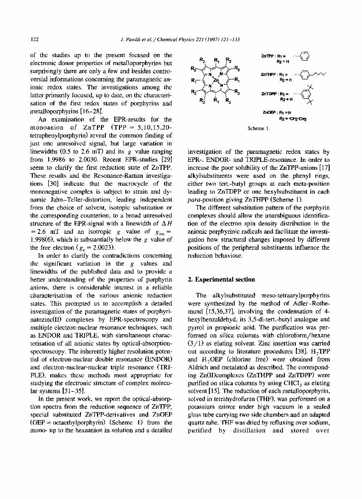
Fig. 2. Vis /NIR optical absorption spectra of the six anionic redox stages of Zn(II)porphyrins in THF taken at room tempera- ture, obtained by reduction with potassium: (A) ZnTHPP, (B) ZnOEP. The spectra are shown for the visible to NIR region from 480 to 1400 nm since the Soret region is far to intensive. Acquisition time was 230 s per spectrum.
identification as trianion will be given by the results described later.
The anisotropy of the monoanion low-temperature EPR spectra at 135 K is significant. The dominant narrow feature, with A H < 1.0 mT, centred near ge and the much less intensive broad feature (AHpp = 2.0-3.0 mT), centred at g = 1.9990, are indicative for an axially symmetric spin system which is con- sistent with the observations reported for Z n T P P - in earlier studies [29]. The fact, that the signals of all monoanions described herein could not be saturated in liquid solution, made it impossible to apply the ENDOR technique for an investigation of the elec- tron spin density distribution in these radicals.
The optical absorption spectra of Z n T P P - , ZnTHPP and Z n T D P P - are generally similar to one another. The spectral features include a domi- nant Soret absorption at A = 456 nm, red shifted compared to the neutral compounds, and weaker, but very characteristic absorptions, in the visible region around A = 705, 720 and 890 nm, while the colour of the solutions at maximum monoanion concentra- tion has turned from pink to brownish. An isosbestic point (IP) for the transition between neutral to monocharged forms was found at 606-610 nm.
The optical spectrum of ZnOEP' - is almost com- parable in its shape to the ones already described, but the characteristic absorption bands at longest wave- length (Q-bands) are shifted hypsochromically to A = 629 and 835 nm with an IP at 582 nm. In this regard it must be noted that, according to our studies, the optical bands at A = 535 and 571 nm do not belong to Z n O E P - as described earlier [30], but are due to the presence of residual, yet unreduced ZnOEP.
3.2. Di- and trianions
Upon further contact of the solutions with the potassium mirror, the colour changes from brownish to green, with a simultaneous loss of EPR signal intensity, which is consistent with the reported dia- magnetic character of the dianion. While the bands for the monoanions disappear in the optical spectra, new absorptions gain intensity in the Q-band region at A = 556 and 610 nm for ZnTPP 2-, ZnTHPP 2-, ZnTDPP 2- (IP:646,655,648 nm) and at A = 545 and
J. Pawlik et al. / Chemical Physics 221 (1997) 121-133
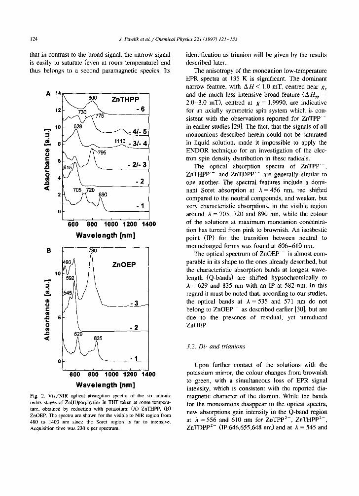
Table 1
EPR parameters for the Zn(II)porphyrin radical anions in T H F / K taken at room temperature ~
125
Reduction state ZnTPP ZnTHPP ZnTDPP ZnOEP
1 - g = 1.9989 g = 1.9989 g = 1.9989 A H = 2.1 mT A H = 2.1 mT A H = 2.1 mT
3 - g = 2.0023 g = 2.0023 g = 2.0023
A H = 0.65 mT A H = 0.66 mT A H = 0.67 mT
5 - g = 2.0043 g = 2.0043 g = 2.0043
A H = 1 0 G A H = 10G A H = 1 0 G
g = 1.9994 A H = 1.8 mT
g = 2.0023 A H = ~
The g values were measured at the zero crossing point, A H is the peak-to-peak width, experimental errors: A g = +0.0002,
AH(1 - ) = _+0.1 mT, AH(3 - ) = +0 .001 mT, AH(5 - ) = +0.1 mT.
b The averaged splitting of the dominant triplet is 6.85 G.
592 nm for ZnOEP 2- (IP: 602 nm). A slight hyp- sochromic shift but no clean separation is found in their Soret-absorptions. Upon further reduction (charging very shortly), a new EPR signal is mea- sured for the next paramagnetic redox state: the trianion. The most characteristic feature in the opti- cal spectra of the trianions is a new band at around A = 795-800 nm for ZnTPP 3 - , ZnTHPP 3 - and
ZnTDPP 3- and at A = 493, 780 nm for ZnOEP 3-, while the Q-bands observed already for the dianions remain and the Soret absorptions are shifted slightly to higher energy like in the monoanions (Table 2, Fig. 2). At this point it must be noted, that the band at 800 nm which we detected were not due to reaction products via protonation of the dianions, leading to phlorin and chlorine derivatives which has
Table 2
Visible and infrared spectroscopic data in [nm] of the reduced Zn(II)porphyrins taken at room temperature in T H F / K ~
ZnTPP ZnTHPP ZnTDPP ZnOEP
neura l 421(Soret) 423(Soret) 422(Soret)
555,595 556,595 555,595
IP 606 610 608
monoanion 456(Sorer), 456(Soret), 456(Sorer),
705,720, 705,720, 705,720,
890 890 888
IP 646 658 644
dianion 436(Sore0, 440(Sorer), 440(Soret),
558,615 556, 615 556, 610 1P 700 675 x
~ianion 459(Sore0, 456(Sorer), 456(Soret),
800 795 795
tetraanion 459(Sore0, 459(Soret), 459(Sorer), 1110 628 ,1110 1100
pentaanion 645,730, 645,730, 645,730, 775 775 775
hexaanion 420, 840 420, 800 420, 840
399(Soret)
535 ,570
582
629 ,835
519 ,602
545,592 531 ,627
493,780
a Maxima of characteristic bands in each reduction state, experimental error: + 5 nm. 1P = isosbestic points, x = not determined.

126 J. Pawlik et al. // Chemical Physics 221 (1997) 121-133
been described by several authors [18,20], since the reduction sequences were completely reversible and no second paramagnetic signal belonging to reaction or decomposition products could be detected in their EPR spectra. This is a quite usual progress since the same alkali metal reduction technique with THF solution contact on a potassium mirror has been used to obtain and characterize the diamagnetic di-, tetra-, and hexaanions by high resolution NMR techniques [8-10]. Also cyclic voltammetry has been used to measure the redox potentials of the reversible forma- tion of the mono- to hexaanion [8] with half wave potentials at - 144, (le), - 1.56(le), - 2.78(2e), - 3.20(2e).
The EPR spectra of the trianions are distinctly different from those of the monoanions. They show no resolved hyperfine structure at ambient tempera- ture, but they exhibit a remarkable narrow and al- most identical linewidth of A H = 0.65-0.66 mT. A similar spectrum with A Hg = 0.67 mT was obtained for ZnTDPP' 3-, where it was nearly impossible to trap solely the mono or the dianion upon reduction of the neutral compound. A rotational steric hinder- ance of the aryl substituents which stabilise a part of the excess charge, could be responsible for the devi- ating reduction behaviour. A striking hyperfine split- ting in three dominant lines with an average distance of 0.685 mT is observed for ZnOEP 3 - , which presumably has to be attributed to the essential dif- ferent substitution pattern of this porphyrin, com- pared to the meso-substituted derivatives. Whereas the EPR lines of the monoanions are resistant to saturation, all radical trianions described herein ex- hibit g values quite similar to ge and can readily be saturated. The last and most important feature en- abled us for the first time to study ZnTPP 3 - , Z n O E P 3-, Z n T H P P 3- and Z n T D P P 3- by EN- DOR and General TRIPLE-resonance spectroscopy in order to gain concrete information about the elec- tron spin density distribution in porphyrin anions.
3.3. ENDOR and TRIPLE spectra of trianions
3.3.1. ZnOEP "3- Fig. 3A shows the ENDOR spectrum of
Z n O E P 3- in solution, recorded at 290 K. Analysis of the hyperfine pattern yields four pairs of signals, due to four proton hyperfine coupling constants a H
v H
~ EP
~ 1 . 1 . 1 . 1 . 1 . 1 . 1 , 1 , | . 1 ,
6 8 10 12 14 16 18 20 22 24
v H
13 ¢ PP
' l I 0 ' 1 ' 1 ' 112 " 1'3 ' 14 " 1'5 ' 1'6 ' 1'7 ' tt8 ' 1~9 '
D vH
13 14 15 16
frequency / MHz
Fig. 3. ENDOR spectra of the radical trianions ZnOEP 3- (A), ZnTPP 3- (B), and ZnTHPP 3- (C), together with an extended view of the ENDOR specU'a of three tetraarylporphyrins ZnTPP" 3 -, ZnTHP" 3-, ZnTDPP' 3 - (D) in THF/K; temperature T = 290 K.
(hfc's), which are centred at the proton Lamor fre- quency v H, and denoted as a~ = 0.572 mT, a 2 = 0.147 mT, a 3 = 0.110 mT and a 4 - - - - -0 .030 mT. Not
14 detected are signals from the N nuclei (above 2
J. Pawlik et al. / Chemical Physics 221 (1997) 121-133 127
MHz and at different temperatures between T = 200-300 K, even though they could be expected by following the numerous predicted charge density dis- tributions described up to the present [10,16-29].
Special-TRIPLE experiments for resolving the multiplicity of equivalent nuclei are difficult to ad- just since our experimental setup allows measure- ments only from ___ 1 MHz, where informations about the smaller couplings are completely lost.
General TRIPLE resonance experiments per- formed on the signals belonging to a 3 indicate that the largest (a 1) and the smallest hfc 's ( a 4 ) possess the same sign while a 2 and a~ possess opposite
Tab le 3
Pro ton hyper f ine coupl ing cons tants [mT] o f the porphyr in ic radi-
cal t r ianions ( T H F / K , 2 7 0 - 2 9 0 K)
Coup l ing cons tan ts N u m b e r o f A s s i g n m e n t
protons
Z n O E P 3 -
a~ - 0 .572 2 m e s o ]
a 2 + 0 .147 2 m e s o 2
a 3 +0.110 8 pyra(13-H) a 4 - - 0.030 8 pyre(13_H) a
ZnTPP 3 a t - 0.223 4 pyr l a 2 -0.063 6 orthoj, para]
b a 3 + 0 .038 4 p y r 2
a 4 + 0 . 0 3 2 4 m e t a t b a s - 0 . 0 1 2 d c
Z n T H P P 3 -
a I - 0 . 2 2 3 4 py r I
a 2 - 0 .062 4 or tho I
a 3 + 0 . 0 4 0 4 ( + 4 ) pyr2 (+pa ra t (13_H) ) b
a 4 + 0 .036 4 m e t a 1 u a 5 + 0 . 0 1 1 d c
Z n T D P P " 3 -
a I 0 .223 4 py r I a_~ 0 .062 6 or tho t b
b a 3 0 .045 4 pyre , pa ra I a 4 0.011 a c
a This coup l ing could also be due to 2t-protons o f pyr I.
b Not c o m p l e t e l y separated.
T h e smal l coup l ings canno t u n a m b i g u o u s l y be ass igned. T h e y
should s t em f r o m the pro tons o f the two pheny l units a t tached at
m e s o posi t ions o f smal l spin densi ty.
d N u m b e r o f pro tons undef ined , not r e so lvab le f r o m c o m p u t e r
s imula t ion .
3 - General-TRIPLE Vpump Vpump ZnTPP
l ,
i i I i i i i i i i
10 11 12 13 14 15 16 17 18 19
frequency / MHz
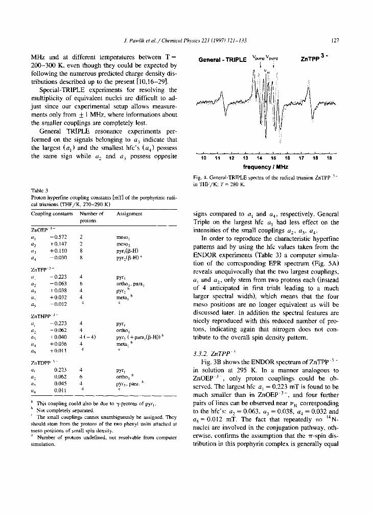
Fig. 4. General-TRIPLE spectra of the radical trianion ZnTPP' 3 - in THF/K; T = 280 K.
signs compared to a] and a4, respectively. General Triple on the largest hfc a] had less effect on the intensities of the small couplings a 2, a3, a 4.
In order to reproduce the characteristic hyperfine patterns and by using the hfc values taken from the ENDOR experiments (Table 3) a computer simula- tion of the corresponding EPR spectrum (Fig. 5A) reveals unequivocally that the two largest couplings, a~ and a z, only stem from two protons each (instead of 4 anticipated in first trials leading to a much larger spectral width), which means that the four meso positions are no longer equivalent as will be discussed later. In addition the spectral features are nicely reproduced with this reduced number of pro- tons, indicating again that nitrogen does not con- tribute to the overall spin density pattern.
3.3.2. Z n T P P 3
Fig. 3B shows the ENDOR spectrum of Z n T P P 3- in solution at 295 K. In a manner analogous to ZnOEP '3- , only proton couplings could be ob- served. The largest hfc a 1 = 0.223 mT is found to be much smaller than in ZnOEP 3 - , and four further pairs of lines can be observed near vn corresponding to the hfc's: a 2 = 0.063, a 3 = 0 . 0 3 8 , a 4 = 0.032 and a 5 = 0.012 mT. The fact that repeatedly no 14N- nuclei are involved in the conjugation pathway, oth- erwise, confirms the assumption that the ,rr-spin dis- tribution in this porphyrin complex is generally equal

128 J. Pawlik et al. / Chemical Physics 221 (1997) 121-133
ZnOEP
A ~ experiment ................ / "" i ~ simulation
', / ", /
, , , i ,
B I Gauss
to the one in ZnOEP 3 - , although the substitution pattern deviates significantly. A further differentia- tion by determining the relative signs of the cou- plings via General TRIPLE resonance is inalienable.
A General TRIPLE experiment performed on the a 3 / a 4 signals of ZnTPP 3- (Fig. 4) reveals that only the a 3 / a 4 signals possess different signs than the other hfc's. This is a most relevant information for further interpretation and assignment of the coupling constants to the proton positions (see discussion). The computer simulation of the EPR spectrum of ZnTPP 3 based on this assignment agrees well with the experimental one (Fig. 5B).
ZnTPP
experiment B simulation
B I Gauss
ZnTHPP
C /
, i i 34OO
experiment simulation
i i , , i ,
3410 342O 343O
B I Gauss
Fig. 5. (A) EPR spectrum of the radical trianion ZnOEP 3- in THF/K; T = 295 K (Top) and simulation using the proton cou- pling constants a I - a4 ,and their multiplicities given Table 3; line shape Lorentzian. (B). EPR spectrum of the radical trianion ZnTPP - 3 in THF/K; T = 295 K (Top). Simulation with the use of the proton coupling constants a i -a4 , and their multiplicities given in Table 3 (Bottom); line shape Lorentzian. (C) EPR spectrum of the radical trianion ZnTHPP - 3 in THF/K; T = 295 K (Top). Simulation with the use of the proton coupling constants a l - a 5, and their multiplicities given in Table 3 (Bottom); line shape Lorentzian.
3.3.3. Z n T H P P ~ and Z n T D P P " ~-
The ENDOR spectrum of ZnTHPP 3- in solution at 295 K is shown in Fig. 3C. In analogy to ZnTPP '3- five pairs of lines are detected, which correspond to similar proton hyperfine coupling val- ues: a 1 -- 0.223, a z = 0.062, a 3 = 0.040, a 4 = 0.033 and a 5 = 0.011 mT. Due to the fact, that the alkyl substituents do not affect the linewidth of the EPR signal, relative to ZnTPP 3 - , the alkyl-protons in para-position contribute only to negligible extent to the EPR spectrum. General TRIPLE experiments performed on the a 3 / a 4 signals of ZnTHPP '3- indicate the same signs for the large hfc's as for ZnTPP 3- with an exception for the smallest hfc a 5. The EPR spectrum of ZnTHPP 3- can be simulated properly (Fig. 5C) by taking the experimental values for the proton hfc's and their multiplicities as de- scribed above. The hfc' s of ZnTHPP 3 - are listed in Table 3.
The hyperfine coupling constants for the meta-al- kyl substituted derivate ZnTDPP 3-, listed in Table 3, yield minor differences compared to the other two arylporphyrins. While the two largest couplings a~ and az are nearly identical a 3 is slightly larger than before and the former a 4 coupling is missing as can be seen in the comparison of the extended proton ENDOR spectra in Fig. 3Dc. This should help in the assignment of the phenyl protons as discussed in Section 4.
Thus despite varying the substitution pattern of the macrocycle, the w-spin carrying molecular or- bitals of all investigated metalloporphyrin trianions show similar electron spin density distributions.

J. Pawlik et al. / Chemical Physics 221 (1997) 121-133 129
3.4. Tetra-, penta- and hexaanions
Upon reducing the trianions the EPR signal inten- sity is lost, consistent with the reported diamagnetic character of the tetraanionic redox state which has shown to be accessible to standard high resolution NMR spectroscopy [8-10], while the trianion bands in the optical spectra decrease and a new absorption in the NIR-region at around A--1110 nm gains intensity. Beginning with formation of ZnTPP 4- and ZnOEP 4- we observed a flaky black precipitate, due to the increasing insolubility. For ZnOEP this is already the highest reduction stage accessible [8].
The next reduction state ( 5 - ) of ZnTHPP and ZnTDPP is characterised by two new overlapping absorption in the visible region at around A = 645, 730, and 775 nm, while bands of residual amounts of the lower redox state still can be detected. The colour of the solutions has changed thereby from green to blue and a new EPR signal can be observed with a highly anisotropic line shape measured at ambient temperature. Due to the alkylsubstituents of ZnTHPP and ZnTDPP, no precipitate occurred for these compounds on this redox level and the EPR signal intensity (Fig. 1) is higher, compared to the one for ZnTPP 5-. Similar to the monoanions, the signals exhibit no hyperfine structure and the mi- crowave power-saturation experiments performed on this state revealed that their EPR signals can not be saturated, which made it impossible to apply multi- ple electron-nuclear resonance techniques for a thor- ough analysis.
After prolonged reduction time the colour changes to yellow-brownish and two absorptions are mea- sured in the optical spectra, one with high intensity at A = 420 nm and one broad absorption with low intensity at around 3. = 800-840 nm. The solutions are diamagnetic again and therefore assigned to the hexaanion.
4. Discussion
The monoanions of the zinc metalated porphyrins considered here exhibit very characteristic broad EPR signals with exceptionally small g values indicating strong spin orbit coupling contributions. Since earlier studies had shown that this signal in liquid solution
is nearly unaffected by isotopic substitution as XSN, 13C, 8D [29] and in accord with our study cannot be saturated by microwave power, thus not allowing application of continuous wave ENDOR spec- troscopy. The reason for these fast relaxation pro- cesses may most reasonable be dynamic Jahn-Teller distortions but further experimental proofs were be- yond the purpose of this classification and should more general include other Zn(II) complexes.
The major focus here is the characterisation of the trianions and the spin density distribution in them. We therefore start with the discussion of the assign- ment of well resolved proton hfc's to the molecular positions. The surprising finding that only two pro- tons of the four meso protons in the monoradical trianion ZnOEP 3- are responsible for the three line EPR spectrum, let us to begin with a closer look at the ZnOEP.
For large hfc's of protons directly attached to • r-centres, the sign is normally assumed to be nega- tive, as required by published theoretical studies and experimental results [39]. Therefore, considering all aspects, it is reasonable to assess each of the two largest couplings of relative low intensity (aj and a 2) to the two sets of non equivalent protons in meso-positions (also established by their multiplicity in agreement with the computer simulation), with a negative sign for the value of al and a positive sign for the value of a~. It is further reasonable to assign a 3 to the [3-protons of one set of four equivalent ethylsubstituents (Table 3). With regard to the fact that ~-protons at positions of small spin density have rarely been observed the smallest coupling a 4 should be assigned to the second group of B-protons from the other ethylsubstituents, The reversed sign of a 4 does not help for the differentiation between [~-pyr- rol 2 and ~/-pyrroll since they should possess the same sign and reversed sign compared to a 3 as also established from the calculations of the spin density discussed later on and shown in Fig. 7.
Following the results of EPR/ENDOR investiga- tions and molecular orbital calculations (RHF- INDO/SP) of the cation radical of ZnTPP [40-42] it is to be expected that the proton hfc's of the phenyl- substituents, should not differ more than by the factor 2 from each other. Therefore, the largest cou- pling, a 1, which differs more then by the factor 3.5 from the second largest coupling in ZnTPP 3 - ,
130 J. Pawlik et al. / Chemical Physics 221 (1997) 121-133
should be assigned to protons of the pyrrol subunits (apyr). With respect to the results for ZnOEP '3-, a significantly reduced value is to be expected for the coupling of the remaining second half of nuclei in [3-pyrrol position (apyr2).
Since the phenyl rings of ZnTPP are twisted out of the plane spread by the macrocycle (ca. 60°), spin density into the phenylsubstituents is transferred mainly via hyperconjugation. The best results in explaining the hyperfine values of such a twisted w-system were obtained by the 'phenyl-hyperconju- gation-model' [40-42]. According to the hyperconju- gation-model only couplings from meta-protons ( a m)
exhibit an opposite sign compared to the signs for the couplings of ortho- and para-protons (a o and ap). The latter are suggested to have negative signs [40-42] while they may possess the same size. This enables us to suggest the assignment of coupling constants given in Table 3.
Since the nuclei belonging to apyr, and the apyr2
are c~-protons, the signs of the hfc's invert relative to those of the [3-protons from the ethylsubstituents attached to the same position in ZnOEP 3-. Conse- quently apyr, got to have a negative and apyr2 a
positive sign, respectively. Therefore, a 2 is assigned to the ortho- and para-protons of two phenylsub- stituents in ZnTPP 3 - The ENDOR lines of a 3 and a 4 correspond to hfc's with positive signs and should therefore stem from the four protons in [3-pyrrol- position with reduced spin density (pyr2-H) and the four meta-protons (meta 1) of two phenyl rings, re- spectively. Due to the reduced electron spin density at two of the four meso-carbon centres (see ZnOEP 3 - ) additional small couplings are to be expected for the protons of the last two phenyl rings which can no longer be separated and assigned clearly.
In analogy to ZnTPP 3 - , the two largest cou- plings a I and a 2 in ZnTHPP 3-, possess the same signs and their signs have to be negative. These couplings can therefore be assessed to four protons in [3-position of the pyrrol subunits (pyrl-H) and to the ortho-protons of two phenylsubstituents attached at meso positions of large spin density. Then the protons in pyr 2- and metal-positions to a 3 and a 4, respectively, as given before. Since the protons in the hexyl groups are separated by two bonds ([3-pro- tons) or more bonds (~/-, ~-protons, etc.) from the
~r-system spin density is transferred via hyperconju- gation [39-42] and the signs of the hfc's from [3-protons invert relative to those of the a-protons in para-phenyl position of ZnTPP 3-. Therefore, the increase in signal intensity of the a 3 lines is at- tributed to the additional contributions to this cou- pling by four [3-protons of the hexyl substituents (Fig. 3D).
The other difference obtained compared to ZnTPP, namely the reversed sign of a 5 could then in princi- ple be explained by the [3-protons in para-position of the second group of hexylphenyl substituents. But since we are not able to resolve this region better and since the second group of phenyl rings again should lead to different ortho, meta, and para proton cou- plings this is somewhat speculative and we hesitate to make a clear assignment to this small coupling in general.
When looking at the ENDOR spectra of the ZnTDP "3- (Fig. 3Dc) it is obvious that the two largest proton couplings are comparable to the ones in ZnTPP. The next coupling a 3 seems to be slightly larger than before while the former a 4 signals are completely missing. This supports our earlier sugges- tions to assign a n to the protons in meta-phenyl position, which are now substituted by tert.-butyl groups.
According to our results, the nitrogen nuclei in the trianions of the porphyrin macrocycle do not contribute to the width of the EPR signal and should therefore possess negligible spin density. Therefore, we come to the conclusion that the unpaired electron in all trianions considered here is delocalized over the 'outer' perimeter of 20 carbon centres with alter- nating spin density. The results show further, that significantly reduced spin density ( ~ 1/4) is found at two of the four meso-positions and at four of the eight [3-positions in the four pyrrol subunits. Accord-
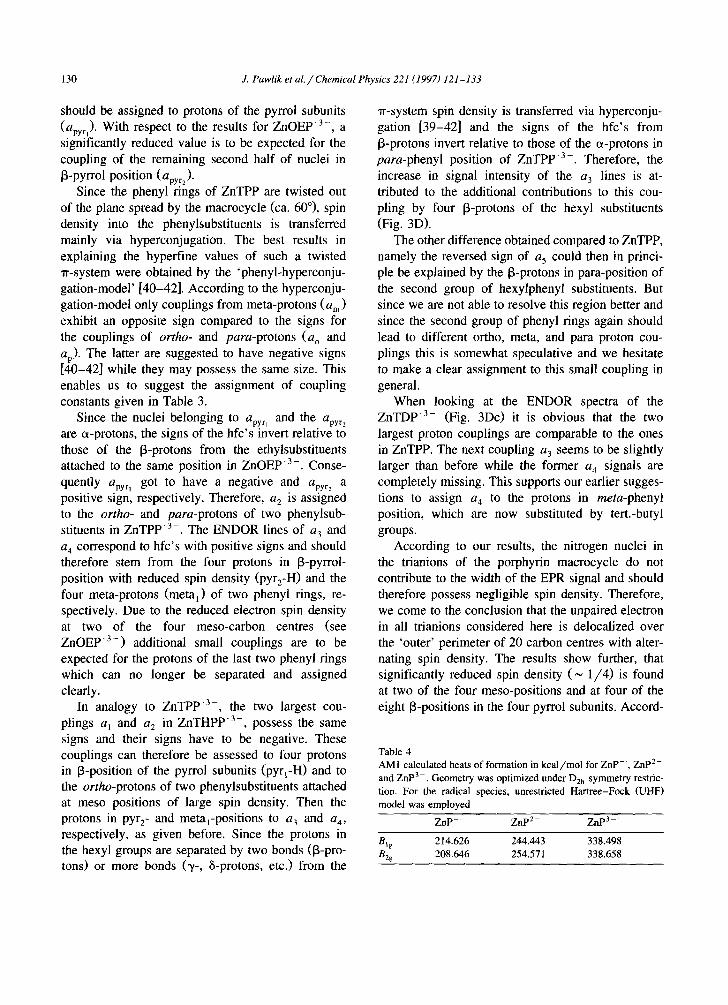
Table 4 AM1 calculated heats of formation in kcal/mol for Z n P - , ZnP 2- and ZnP 3- . Geometry was optimized under Dzh symmetry restric- tion. For the radical species, unrestricted Hartree-Fock (UHF) model was employed
Z n P - ZnP 2- ZnP 3-
Big 214.626 244.443 338.498 Bzg 208.646 254.571 338.658
A
J. Pawlik et al. / Chemical Physics 221 (1997) 121 - 133 131
B
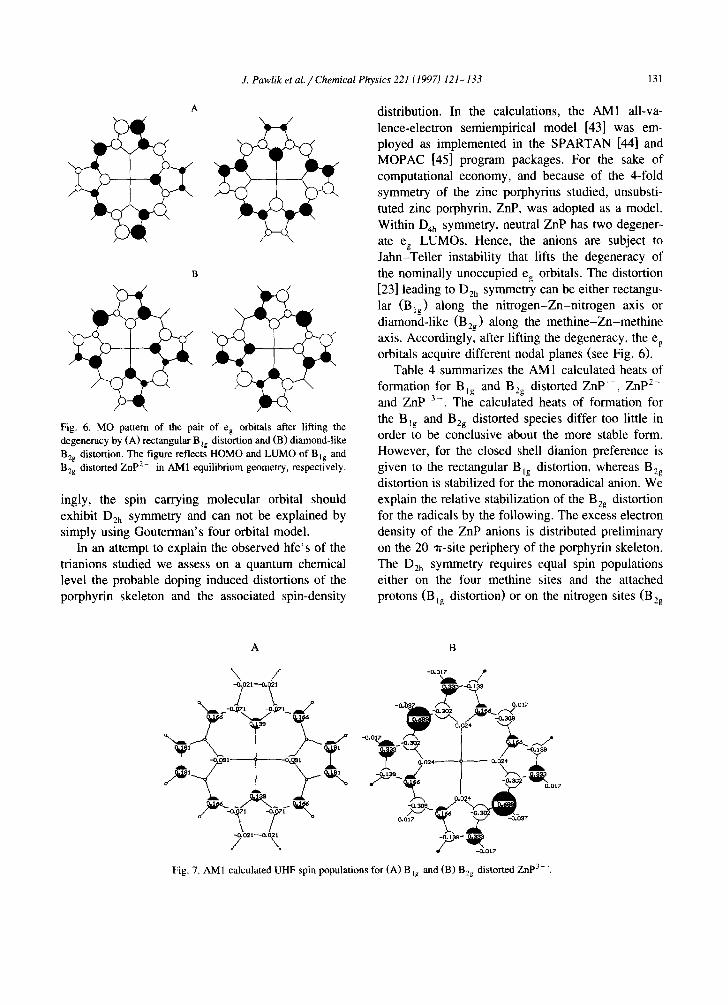
Fig. 6. MO pattern of the pair of eg orbitals after lifting the degeneracy by (A) rectangular B~g distortion and (B) diamond-like B2g distortion. The figure reflects HOMO and LUMO of Big and B2s distorted ZnP 2- in AM1 equilibrium geometry, respectively.
ingly, the spin carrying molecular orbital should exhibit D2h symmetry and can not be explained by simply using Gouterman's four orbital model.
In an attempt to explain the observed hfc's of the trianions studied we assess on a quantum chemical level the probable doping induced distortions of the porphyrin skeleton and the associated spin-density
distribution. In the calculations, the AM I all-va- lence-electron semiempirical model [43] was em- ployed as implemented in the SPARTAN [44] and MOPAC [45] program packages. For the sake of computational economy, and because of the 4-fold symmetry of the zinc porphyrins studied, unsubsti- tuted zinc porphyrin, ZnP, was adopted as a model. Within D4h symmetry, neutral ZnP has two degener- ate eg LUMOs. Hence, the anions are subject to Jahn-Teller instability that lifts the degeneracy of the nominally unoccupied eg orbitals. The distortion [23] leading to D2h symmetry can be either rectangu- lar (Big) along the nitrogen-Zn-nitrogen axis or diamond-like (B2g) along the methine-Zn-methine axis. Accordingly, after lifting the degeneracy, the eg orbitals acquire different nodal planes (see Fig. 6).
Table 4 summarizes the AM1 calculated heats of formation for Big and Bzg distorted Z n P - , ZnP 2- and ZnP 3-. The calculated heats of formation for the Bzg and B2g distorted species differ too little in order to be conclusive about the more stable form. However, for the closed shell dianion preference is given to the rectangular Big distortion, whereas B2g distortion is stabilized for the monoradical anion. We explain the relative stabilization of the Beg distortion for the radicals by the following. The excess electron density of the ZnP anions is distributed preliminary on the 20 at-site periphery of the porphyrin skeleton. The Dzh symmetry requires equal spin populations either on the four methine sites and the attached protons (BIg distortion) or on the nitrogen sites (B2g
A B
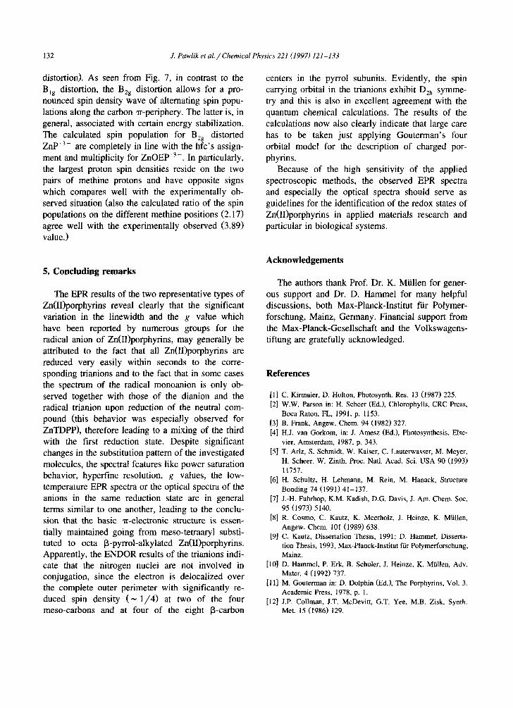
\ / Z
.k ' .k 0-
. - _°.°,7
Fig. 7. AM1 calculated UHF spin populations for (A) Big and (B) B2g distorted ZnP 3- .
132 J. Pawlik et al. / Chemical Physics 221 (1997) 121-133
distortion). As seen from Fig. 7, in contrast to the Big distortion, the B2g distortion allows for a pro- nounced spin density wave of alternating spin popu- lations along the carbon at-periphery. The latter is, in general, associated with certain energy stabilization. The calculated spin population for B2g distorted ZnP 3 - are completely in line with the hfc's assign- ment and multiplicity for ZnOEP' 3-. In particularly, the largest proton spin densities reside on the two pairs of methine protons and have opposite signs which compares well with the experimentally ob- served situation (also the calculated ratio of the spin populations on the different methine positions (2.17) agree well with the experimentally observed (3.89) value.)
centers in the pyrrol subunits. Evidently, the spin carrying orbital in the trianions exhibit D2h symme- try and this is also in excellent agreement with the quantum chemical calculations. The results of the calculations now also clearly indicate that large care has to be taken just applying Gouterman's four orbital model for the description of charged por- phyrins.
Because of the high sensitivity of the applied spectroscopic methods, the observed EPR spectra and especially the optical spectra should serve as guidelines for the identification of the redox states of Zn(II)porphyrins in applied materials research and particular in biological systems.
5. Concluding remarks
The EPR results of the two representative types of Zn(II)porphyrins reveal clearly that the significant variation in the linewidth and the g value which have been reported by numerous groups for the radical anion of Zn(II)porphyrins, may generally be attributed to the fact that all Zn(II)porphyrins are reduced very easily within seconds to the corre- sponding trianions and to the fact that in some cases the spectrum of the radical monoanion is only ob- served together with those of the dianion and the radical trianion upon reduction of the neutral com- pound (this behavior was especially observed for ZnTDPP), therefore leading to a mixing of the third with the first reduction state. Despite significant changes in the substitution pattern of the investigated molecules, the spectral features like power saturation behavior, hyperfine resolution, g values, the low- temperature EPR spectra or the optical spectra of the anions in the same reduction state are in general terms similar to one another, leading to the conclu- sion that the basic at-electronic structure is essen- tially maintained going from meso-tetraaryl substi- tuted to octa [3-pyrrol-alkylated Zn(II)porphyrins. Apparently, the ENDOR results of the trianions indi- cate that the nitrogen nuclei are not involved in conjugation, since the electron is delocalized over the complete outer perimeter with significantly re- duced spin density ( ~ 1/4) at two of the four meso-carbons and at four of the eight B-carbon
Acknowledgements
The authors thank Prof. Dr. K. Miillen for gener- ous support and Dr. D. Hammel for many helpful discussions, both Max-Planck-Institut for Polymer- forschung, Mainz, Germany. Financial support from the Max-Planck-Gesellschaft and the Volkswagens- tiftung are gratefully acknowledged.
References
[1] C. Kirmaier, D. Holton, Photosynth. Res. 13 (1987) 225. [2] W.W. Parson in: H. Scheer (Ed.), Chlorophylls, CRC Press,
Boca Raton, FL, 1991, p. 1153. [3] B. Frank, Angew. Chem. 94 (1982) 327. [4] H.J. van Gorkom, in: J. Amesz (Ed.), Photosynthesis, Else-
vier, Amsterdam, 1987, p. 343. [5] T. Arlz, S. Schmidt, W. Kaiser, C. Lauterwasser, M. Meyer,
H. Scheer, W. Zinth, Proc. Natl. Acad. Sci. USA 90 (1993) 11757.
[6] H. Schultz, H. Lehmann, M. Rein, M. Hanack, Structure Bonding 74 (1991) 41-137.
[7] J.-H. Fuhrhop, K.M. Kadish, D.G. Davis, J. Am. Chem. Soc. 95 (1973) 5140.
[8] R. Cosmo, C. Kautz, K. Meerholz, J. Heinze, K. Miillen, Angew. Chem. 101 (1989) 638.
[9] C. Kautz, Dissertation Thesis, 1991; D. Hammel, Disserta- tion Thesis, 1993, Max-Planck-Institut f'tir Polymerforschung, Mainz.
[10] D. Hammel, P. Erk, B. Schuler, J. Heinze, K. Miillen, Adv. Mater. 4 (1992) 737.
[11] M. Gouterman in: D. Dolphin (Ed.), The Porphyrins, Vol. 3, Academic Press, 1978, p. 1.
[12] J.P. Collman, J.T. McDevitt, G.T. Yee, M.B. Zisk, Synth. Met. 15 (1986) 129.

J. Pawlik et al. / Chemical Physics 221 (1997) 121-133 133
[13] J,P. Collman, J.T. McDevitt, C.R. Leidner, G.T. Yee, J.B. Torrance, W.A. Little, J. Am. Chem. Soc. 109 (1987) 4606.
[14] M. Mezger, M. Hanack, A. Hirsch, J. Kleinw~ichter, K.-M. Mangold, L. Ramaswami Subramanian, Chem. Ber. 124 (1991) 841.
[15] J. Pawlik, C. Kautz, M. Baumgarten, J. Inorg. Organomet. Polym. 4 (1994) 237.
[16] J. Fajer, M.S. Davis, in: D. Dolphin (Ed.), The Porphyrins, Vol. 4, Academic Press, 1978, p. 230.
[17] G.L. Closs, L.E. Closs, J. Am. Chem. Soc. 85 (1963) 818. [18] G. Peychal-Heiling, G.S. Wilson, J. Electrochem. Soc. 119
(1972) 1039. [19] M.J. Gouterman, J. Mol. Spectrosc. 6 (1961) 138. [20] N.S. Hush, J.R. Rowlands, J. Am. Chem. Soc. 89 (1967)
2976. [21] D.F. Bocian, J.-H. Perng, J. Phys. Chem. 96 (1992) 4804. [22] M. Atamian, R.J. Donohoe, J.S. Lindsey, D.F. Bocian, J.
Phys. Chem. 93 (1989) 2236. [23] K. Prendergast, T.G. Spiro, J. Phys. Chem. 95 (1991) 9728. [24] R.A. Reeds, R. Purrello, K. Prendergast, T.G. Spiro, J. Phys.
Chem. 95 (1991) 9720. [25] V.A. Waiters, J.C. dePaula, G.T. Babcock, G.E. Leroi, J.
Am. Chem. Soc. 111 (1989) 8300. [26] R.S. Czernuszewicz, K.A. Macor, X.Y. Li, J.R. Kincaid,
T.G. Spiro, J. Am. Chem. Soc. 111 (1989) 3860. [27] V.E. Kholmogorov, V.G. Maslow, Opt. Spectrosc. 195
(1971). [28] M. Gouterman, Ann. N.Y. Acad. Sci. 206 (1973) 70. [29] J. Seth, D.F. Bocian, J. Am. Chem. Soc. 116 (1994) 143. [30] J.H. Perng, D.F. Bocian, J. Phys. Chem. 96 (1992) 4804. [31] J.H. Freeds, in: M.M, Dorio, J.H. Freeds (Eds.), Multiple
Electron Resonance Spectroscopy, Plenum Press, New York, 1979, p. 73.
[32] K. M~Sbius, R. Biehl, in: M.M. Dorio, J.H. Freeds (Eds.), Multiple Electron Resonance Spectroscopy, Plenum Press, New York, 1979, p. 475.
[33] H. Kurreck, B. Kirste, W. Lubitz, Angew. Chem. 96 (1984) 171.
[34] K. M6bius, M. Plato, W. Lubitz, Physics Rept. 87 (1982) 171.
[35] H. Kurreck, B. Kirste, W. Lubitz, Electron Nuclear Double Resonance Spectroscopy of Radicals in Solution, Methods Stereochem. Anal., VHC, Weinheim, 1988.
[36] A.D. Adler, F.R. Longo, W. Shergalis, J. Am. Chem. Soc. 86 (1964) 3145.
[37] A.D. Adler, F.R. Longo, J.D. Finarelli, J. Goldmacher, J. Assur, L. Korsakoff, J. Org. Chem. 32 (1967) 476.
[38] J.H. Fuhrhop, in: K.M. Smith (Ed.), Porphyrins and Metallo- porphyrins, Elsevier, Amsterdam, 1975, p. 798.
[39] See, for example: F. Gerson, High-Resolution ESR Spec- troscopy, Wiley, New York, 1970, (a) Chapter 1.5; (b) Appendix A.2.2.
[40] M. Huber, Dissertation Thesis, FU Berlin, 1989. [41] M. Huber, T. Galili, K. MiSbius, H. Levanon, Isr. J. Chem.
29 (1989) 65. [42] M. Huber, H. Kurreck, B. von Mahlzan, M. Plato, K. MiSbius,
J. Chem. Soc. Faraday Trans. 86 (1990) 1087. [43] M.J. S Dewar, E.G. Zoebisch, E.F. Healy, J.J.P. Stewart, J.
Am. Chem. Soc. 107 (1985) 3902. [44] SPARTAN (Version 4.0.3), Wavefunction Inc., 1995. [45] MOPAC: a general molecular orbital package (Version 7.2),
QCPE, 1995.
Related Documents