This article was downloaded by: [Mohammad Morshed] On: 20 October 2014, At: 16:04 Publisher: Taylor & Francis Informa Ltd Registered in England and Wales Registered Number: 1072954 Registered office: Mortimer House, 37-41 Mortimer Street, London W1T 3JH, UK International Journal of Polymeric Materials and Polymeric Biomaterials Publication details, including instructions for authors and subscription information: http://www.tandfonline.com/loi/gpom20 Characterization of PLGA/Chitosan Electrospun Nano- Biocomposite Fabricated by Two Different Methods Sedigheh Vaezifar a b , Shahnaz Razavi a , Mohammad Ali Golozar b , Hamid Zarkesh Esfahani c , Mohammad Morshed d & Saeed Karbasi e a Department of Anatomical Sciences and Molecular Biology, School of Medicine , Isfahan University of Medical Sciences , Isfahan , Iran b Department of Materials Engineering , Isfahan University of Technology , Isfahan , Iran c Department of Biology , Isfahan University of Medical Sciences , Isfahan , Iran d Department of Textile Engineering , Isfahan University of Technology , Isfahan , Iran e Department of Medical Physics and Biomedical Engineering, School of Medicine , Isfahan University of Medical Sciences , Isfahan , Iran Published online: 16 Oct 2015. To cite this article: Sedigheh Vaezifar , Shahnaz Razavi , Mohammad Ali Golozar , Hamid Zarkesh Esfahani , Mohammad Morshed & Saeed Karbasi (2015) Characterization of PLGA/Chitosan Electrospun Nano-Biocomposite Fabricated by Two Different Methods, International Journal of Polymeric Materials and Polymeric Biomaterials, 64:2, 64-75, DOI: 10.1080/00914037.2014.886244 To link to this article: http://dx.doi.org/10.1080/00914037.2014.886244 PLEASE SCROLL DOWN FOR ARTICLE Taylor & Francis makes every effort to ensure the accuracy of all the information (the “Content”) contained in the publications on our platform. However, Taylor & Francis, our agents, and our licensors make no representations or warranties whatsoever as to the accuracy, completeness, or suitability for any purpose of the Content. Any opinions and views expressed in this publication are the opinions and views of the authors, and are not the views of or endorsed by Taylor & Francis. The accuracy of the Content should not be relied upon and should be independently verified with primary sources of information. Taylor and Francis shall not be liable for any losses, actions, claims, proceedings, demands, costs, expenses, damages, and other liabilities whatsoever or howsoever caused arising directly or indirectly in connection with, in relation to or arising out of the use of the Content. This article may be used for research, teaching, and private study purposes. Any substantial or systematic reproduction, redistribution, reselling, loan, sub-licensing, systematic supply, or distribution in any form to anyone is expressly forbidden. Terms & Conditions of access and use can be found at http:// www.tandfonline.com/page/terms-and-conditions

Welcome message from author
This document is posted to help you gain knowledge. Please leave a comment to let me know what you think about it! Share it to your friends and learn new things together.
Transcript
This article was downloaded by: [Mohammad Morshed]On: 20 October 2014, At: 16:04Publisher: Taylor & FrancisInforma Ltd Registered in England and Wales Registered Number: 1072954 Registered office: Mortimer House,37-41 Mortimer Street, London W1T 3JH, UK
International Journal of Polymeric Materials andPolymeric BiomaterialsPublication details, including instructions for authors and subscription information:http://www.tandfonline.com/loi/gpom20
Characterization of PLGA/Chitosan Electrospun Nano-Biocomposite Fabricated by Two Different MethodsSedigheh Vaezifar a b , Shahnaz Razavi a , Mohammad Ali Golozar b , Hamid Zarkesh Esfahanic , Mohammad Morshed d & Saeed Karbasi ea Department of Anatomical Sciences and Molecular Biology, School of Medicine , IsfahanUniversity of Medical Sciences , Isfahan , Iranb Department of Materials Engineering , Isfahan University of Technology , Isfahan , Iranc Department of Biology , Isfahan University of Medical Sciences , Isfahan , Irand Department of Textile Engineering , Isfahan University of Technology , Isfahan , Irane Department of Medical Physics and Biomedical Engineering, School of Medicine , IsfahanUniversity of Medical Sciences , Isfahan , IranPublished online: 16 Oct 2015.
To cite this article: Sedigheh Vaezifar , Shahnaz Razavi , Mohammad Ali Golozar , Hamid Zarkesh Esfahani , MohammadMorshed & Saeed Karbasi (2015) Characterization of PLGA/Chitosan Electrospun Nano-Biocomposite Fabricated byTwo Different Methods, International Journal of Polymeric Materials and Polymeric Biomaterials, 64:2, 64-75, DOI:10.1080/00914037.2014.886244
To link to this article: http://dx.doi.org/10.1080/00914037.2014.886244
PLEASE SCROLL DOWN FOR ARTICLE
Taylor & Francis makes every effort to ensure the accuracy of all the information (the “Content”) containedin the publications on our platform. However, Taylor & Francis, our agents, and our licensors make norepresentations or warranties whatsoever as to the accuracy, completeness, or suitability for any purpose of theContent. Any opinions and views expressed in this publication are the opinions and views of the authors, andare not the views of or endorsed by Taylor & Francis. The accuracy of the Content should not be relied upon andshould be independently verified with primary sources of information. Taylor and Francis shall not be liable forany losses, actions, claims, proceedings, demands, costs, expenses, damages, and other liabilities whatsoeveror howsoever caused arising directly or indirectly in connection with, in relation to or arising out of the use ofthe Content.
This article may be used for research, teaching, and private study purposes. Any substantial or systematicreproduction, redistribution, reselling, loan, sub-licensing, systematic supply, or distribution in anyform to anyone is expressly forbidden. Terms & Conditions of access and use can be found at http://www.tandfonline.com/page/terms-and-conditions
Characterization of PLGA/Chitosan ElectrospunNano-Biocomposite Fabricated by Two Different Methods
SEDIGHEH VAEZIFAR1,2, SHAHNAZ RAZAVI1, MOHAMMAD ALI GOLOZAR2, HAMID ZARKESH ESFAHANI3,MOHAMMAD MORSHED4, and SAEED KARBASI5
1Department of Anatomical Sciences andMolecular Biology, School of Medicine, Isfahan University of Medical Sciences, Isfahan, Iran2Department of Materials Engineering, Isfahan University of Technology, Isfahan, Iran3Department of Biology, Isfahan University of Medical Sciences, Isfahan, Iran4Department of Textile Engineering, Isfahan University of Technology, Isfahan, Iran5Department of Medical Physics and Biomedical Engineering, School of Medicine, Isfahan University of Medical Sciences,Isfahan, Iran
Received 2 November 2013, Accepted 19 January 2014
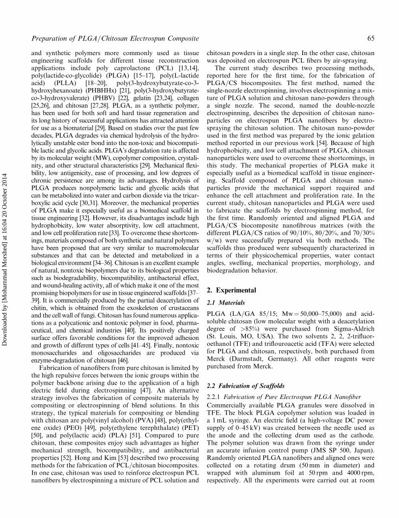
Nano-biocomposites composed of poly (lactide-co-glycolide) (PLGA) and chitosan (CS) were electrospun using two fabricationmethods. In the single nozzle method, the CS nano-powders dispersed in PLGA solutions were electrospun through a single nozzlebut in the double-nozzle method, PLGA and CS were simultaneously electrospun from two syringes and the electrospun PLGAnanofiber and electrosprayed CS nanoparticles were mixed and collected on the rotating drum (randomly oriented [A] and aligned[B]) to prepare the nano-biocomposite membrane. The PLGA=CS scaffolds were prepared at the different ratios. The single-nozzlemethod was associated with decreasing fiber diameter when the CS content was increased and exhibited improve mechanical andhydrophilic properties.
Keywords: Chitosan nanoparticles, electrospinning, nano-biocomposite, PLGA
1. Introduction
Many researchers have employed the electrospinningtechnique to fabricate biodegradable and biocompatiblescaffolds [1–5]. Biodegradable and biocompatible syntheticor natural polymers have been used to develop scaffoldsespecially designed to mimic the structure and biological
function of the native extracellular matrix (ECM) proteins.Such scaffolds provide the mechanical support requiredand enhance the cell attachment and proliferation rate [6].Among the current methods used for fabricating tissueengineering scaffolds, electrospinning has many advantages.It is a simple and convenient technique for producing finefibers with diameters ranging from nanometer to micronscales. The polymeric scaffolds fabricated by electrospinningare promising materials or substrates [7–9] with high specificsurface area, high porosity, and interconnected pores similarto the natural extracellular matrix (ECM), which can regu-late cell activities [10]. Both synthetic polymers and naturalmaterials, though different in nature, have been used for fab-ricating biomedical scaffolds [11,12]. Some of the natural
Address correspondence to: Shahnaz Razavi, Department ofAnatomical Sciences and Molecular Biology, School ofMedicine, Isfahan University of Medical Sciences, Isfahan,81744–176, Iran. E-mail: [email protected]
Color versions of one or more of the figures in the articlecan be found online at www.tandfonline.com/gpom.
International Journal of Polymeric Materials and Polymeric Biomaterials, 64: 64–75
Copyright # 2015 Taylor & Francis Group, LLC
ISSN: 0091-4037 print/1563-535X online
DOI: 10.1080/00914037.2014.886244
Dow
nloa
ded
by [
Moh
amm
ad M
orsh
ed]
at 1
6:04
20
Oct
ober
201
4
and synthetic polymers more commonly used as tissueengineering scaffolds for different tissue reconstructionapplications include poly caprolactone (PCL) [13,14],poly(lactide-co-glycolide) (PLGA) [15–17], poly(L-lactideacid) (PLLA) [18–20], poly(3-hydroxybutyrate-co-3-hydroxyhexanoate) (PHBHHx) [21], poly(3-hydroxybutyrate-co-3-hydroxyvalerate) (PHBV) [22], gelatin [23,24], collagen[25,26], and chitosan [27,28]. PLGA, as a synthetic polymer,has been used for both soft and hard tissue regeneration andits long history of successful applications has attracted attentionfor use as a biomaterial [29]. Based on studies over the past fewdecades, PLGA degrades via chemical hydrolysis of the hydro-lytically unstable ester bond into the non-toxic and biocompati-ble lactic and glycolic acids. PLGA’s degradation rate is affectedby its molecular weight (MW), copolymer composition, crystali-nity, and other structural characteristics [29]. Mechanical flexi-bility, low antigenicity, ease of processing, and low degrees ofchronic persistence are among its advantages. Hydrolysis ofPLGA produces nonpolymeric lactic and glycolic acids thatcan be metabolized into water and carbon dioxide via the tricar-boxylic acid cycle [30,31]. Moreover, the mechanical propertiesof PLGA make it especially useful as a biomedical scaffold intissue engineering [32]. However, its disadvantages include highhydrophobicity, low water absorptivity, low cell attachment,and low cell proliferation rate [33]. To overcome these shortcom-ings, materials composed of both synthetic and natural polymershave been proposed that are very similar to macromolecularsubstances and that can be detected and metabolized in abiological environment [34–36]. Chitosan is an excellent exampleof natural, nontoxic biopolymers due to its biological propertiessuch as biodegradability, biocompatibility, antibacterial effect,and wound-healing activity, all of which make it one of the mostpromising biopolymers for use in tissue engineered scaffolds [37–39]. It is commercially produced by the partial deacetylation ofchitin, which is obtained from the exoskeleton of crustaceansand the cell wall of fungi. Chitosan has found numerous applica-tions as a polycationic and nontoxic polymer in food, pharma-ceutical, and chemical industries [40]. Its positively chargedsurface offers favorable conditions for the improved adhesionand growth of different types of cells [41–45]. Finally, nontoxicmonosaccharides and oligosaccharides are produced viaenzyme-degradation of chitosan [46].
Fabrication of nanofibers from pure chitosan is limited bythe high repulsive forces between the ionic groups within thepolymer backbone arising due to the application of a highelectric field during electrospinning [47]. An alternativestrategy involves the fabrication of composite materials bycompositing or electrospinning of blend solutions. In thisstrategy, the typical materials for compositing or blendingwith chitosan are poly(vinyl alcohol) (PVA) [48], poly(ethyl-ene oxide) (PEO) [49], poly(ethylene terephthalate) (PET)[50], and poly(lactic acid) (PLA) [51]. Compared to purechitosan, these composites enjoy such advantages as highermechanical strength, biocompatibility, and antibacterialproperties [52]. Hong and Kim [53] described two processingmethods for the fabrication of PCL=chitosan biocomposites.In one case, chitosan was used to reinforce electrospun PCLnanofibers by electrospinning a mixture of PCL solution and
chitosan powders in a single step. In the other case, chitosanwas deposited on electrospun PCL fibers by air-spraying.
The current study describes two processing methods,reported here for the first time, for the fabrication ofPLGA=CS biocomposites. The first method, named thesingle-nozzle electrospinning, involves electrospinning a mix-ture of PLGA solution and chitosan nano-powders througha single nozzle. The second, named the double-nozzleelectrospinning, describes the deposition of chitosan nano-particles on electrospun PLGA nanofibers by electro-spraying the chitosan solution. The chitosan nano-powderused in the first method was prepared by the ionic gelationmethod reported in our previous work [54]. Because of highhydrophobicity, and low cell attachment of PLGA, chitosannanoparticles were used to overcome these shortcomings, inthis study. The mechanical properties of PLGA make itespecially useful as a biomedical scaffold in tissue engineer-ing. Scaffold composed of PLGA and chitosan nano-particles provide the mechanical support required andenhance the cell attachment and proliferation rate. In thecurrent study, chitosan nanoparticles and PLGA were usedto fabricate the scaffolds by electrospinning method, forthe first time. Randomly oriented and aligned PLGA andPLGA=CS biocomposite nanofibrous matrices (with thedifferent PLGA=CS ratios of 90=10%, 80=20%, and 70=30%w=w) were successfully prepared via both methods. Thescaffolds thus produced were subsequently characterized interms of their physicochemical properties, water contactangles, swelling, mechanical properties, morphology, andbiodegradation behavior.
2. Experimental
2.1 Materials
PLGA (LA=GA 85=15; Mw¼ 50,000–75,000) and acid-soluble chitosan (low molecular weight with a deacetylationdegree of >85%) were purchased from Sigma-Aldrich(St. Louis, MO, USA). The two solvents 2, 2, 2-trifluor-oethanol (TFE) and triflouroacetic acid (TFA) were selectedfor PLGA and chitosan, respectively, both purchased fromMerck (Darmstadt, Germany). All other reagents werepurchased from Merck.
2.2 Fabrication of Scaffolds
2.2.1 Fabrication of Pure Electrospun PLGA Nanofiber
Commercially available PLGA granules were dissolved inTFE. The block PLGA copolymer solution was loaded ina 1mL syringe. An electric field (a high-voltage DC powersupply of 0–45 kV) was created between the needle used asthe anode and the collecting drum used as the cathode.The polymer solution was drawn from the syringe underan accurate infusion control pump (JMS SP 500, Japan).Randomly oriented PLGA nanofibers and aligned ones werecollected on a rotating drum (50mm in diameter) andwrapped with aluminum foil at 50 rpm and 4000 rpm,respectively. All the experiments were carried out at room
Preparation of PLGA/Chitosan Electrospun Composite 65
Dow
nloa
ded
by [
Moh
amm
ad M
orsh
ed]
at 1
6:04
20
Oct
ober
201
4
temperature. The scaffolds were dried overnight undervacuum at room temperature. To obtain the optimumconditions for the fabrication of electrospun nanofibers,the effects of such parameters as polymer solution concen-tration, feed rate, voltage, and distance of collector fromthe needle tip were investigated on the morphology anddiameter distribution of electrospun nanofibers.
The optimum conditions were obtained with a polymersolution concentration of 24% w=v at a feeding rate of0.25mL=h and a high voltage of 13.5 kV. The distance ofcollector from the needle tip was 10 cm.
2.2.2 Nano-Biocomposite Preparation by Electrospinning ofPLGA=Chitosan
The PLGA=CS nano-biocomposites were electrospun onto arotating collector from a PLGA solution (24% w=v in TFE)containing different weight ratios of 10, 20, and 30wt%chitosan nano-powders prepared by the ionic gelationmethod as described in our previous work [54]. The hom-ogenous solutions were obtained by slowly adding chitosannano-powders and gentle stirring for 12 h at room tempera-ture. The same optimum electrospinning conditions wereobtained as mentioned previously.
For the double-nozzle method, the deposition of chitosannanoparticles on the electrospun PLGA fibers was initiallyoptimized by electro-spraying the chitosan solution intonano-sized particles at a lower concentration with negligiblechain entanglement. This is because continuous fibers can-not be produced by electrospinning at high concentrations.Chitosan nanoparticles were distributed uniformly on thePLGA nanofibrous structure by simultaneous electrospin-ning, and the nanoparticles appeared to adhere strongly tothe PLGA nanofibers. In this method, chitosan was dis-solved in TFA at a concentration of 2% w=v before beingsprayed onto the target by electrostatic charge and at a feed-ing rate of 0.33mL=h and an applied voltage of 13–14 kV.The distance between the spraying nozzle and the mat was10 cm. In the double-nozzle method, the PLGA and chito-san solutions were simultaneously electrospun from two dif-ferent syringes and mixed on the rotating drum to preparethe nanofibrous biocomposite membrane. A 90=10 weightratio of PLGA=CS was obtained by simultaneous electro-spinning of the two solutions. To obtain the 80=20 and70=30 weight ratios of PLGA=CS, the duration of electro-spraying the chitosan solution was increased without elec-trospinning the PLGA solution in multi-steps. Randomlyoriented PLGA=CS nano-biocomposites and aligned nano-fibers were formed using a rotating drum at 50 and4000 rpm, respectively. The fabricated scaffolds were driedovernight under vacuum at room temperature.
2.3 Characterization of Scaffolds
2.3.1 Morphology
Electrospun nanofibrous membranes were sputtered withgold, and their morphology was observed using a scanningelectron microscope (SEM; Seron Technology AIS 2500,India). The diameters of the resulting nanofibers weredetermined using the Image J software from the SEM
micrographs. The sample thickness was evaluated usingcross sections prepared by cryocut (cryocut 1800, reichert,JUNG, Germany) and measured by the Image J softwareat three points. The measured values were averaged.A Philips CM12 transmission electron microscope, operatedat 120 kV was used to investigate the distribution of nano-particles. TEM micrographs showed a good distribution ofchitosan nanoparticles in the PLGA fiber prepared by thesingle-nozzle method.
2.3.2 Water Contact Angle (WCA)
To evaluate the effect of chitosan on the hydrophilicity ofelectrospun PLGA fiber webs, WCAs were measured usinga contact angle analyzer (Dataphysics, Model: OCA 15 plus,Canada). Measurements were acquired at five independentpoints and presented as the average value � standarddeviation (SD).
2.3.3 PBS Absorption (Swelling)
Electrospun membranes of PLGA and PLGA=CS werecut into 10mm� 10mm square shapes for PBS absorp-tion. Pre-weighed specimens were placed in closed bottlescontaining 10mL of PBS (pH¼ 7.40) and incubatedin vitro at 37.0� 0.1�C for 24 h. The wet weight of themembranes was determined by weighing them immedi-ately on an electronic balance after removing the mem-branes from PBS and dehydrating them with filter paperto absorb water on the membrane surface. The wateruptake of the electrospun membranes in PBS were thencalculated using Eq. 1:
Swellingð%Þ ¼ ðw1 � w0=w0Þ � 100% ð1Þ
where, W0 and W1 are the weights of the membranesbefore and after immersion in the PBS, respectively.
2.3.4 Mechanical Properties
The specimens were carefully cut into 10mm� 50mm rec-tangular strips, and their tensile properties were evaluated usinga tensile testingmachine (Zwick Tensile Tester, 1446, Germany)equipped with a 20-N load-cell. The cross-head speed was10mm=min. The reported tensile moduli, tensile strengths,and elongations were presented as averages of three tests.
2.3.5 Biodegradation Behavior
The loss of scaffold weights was evaluated after cultivation.The biodegradation behavior of the scaffolds was evaluatedby measuring the weight change in PBS as follows. Theinitial mass of the scaffolds was measured. They were thenallowed to degrade by placing them in phosphate buffer sol-ution pH 7.4 and incubated at 37�C. At selected time inter-vals (1, 2, 7, 14, 21, and 30 days), the scaffolds were removedfrom the solution, blotted with an absorbent tissue, dried ina vacuum oven, and weighed. The biodegradation percent-age, D (%), was defined as in Eq. 2:
Dð%Þ ¼ ðwi � wf =wiÞ � 100% ð2Þwhere Wi is the initial weight of the scaffold and Wf is thefinal weight of scaffold at the selected time intervals.
66 S. Vaezifar et al.
Dow
nloa
ded
by [
Moh
amm
ad M
orsh
ed]
at 1
6:04
20
Oct
ober
201
4
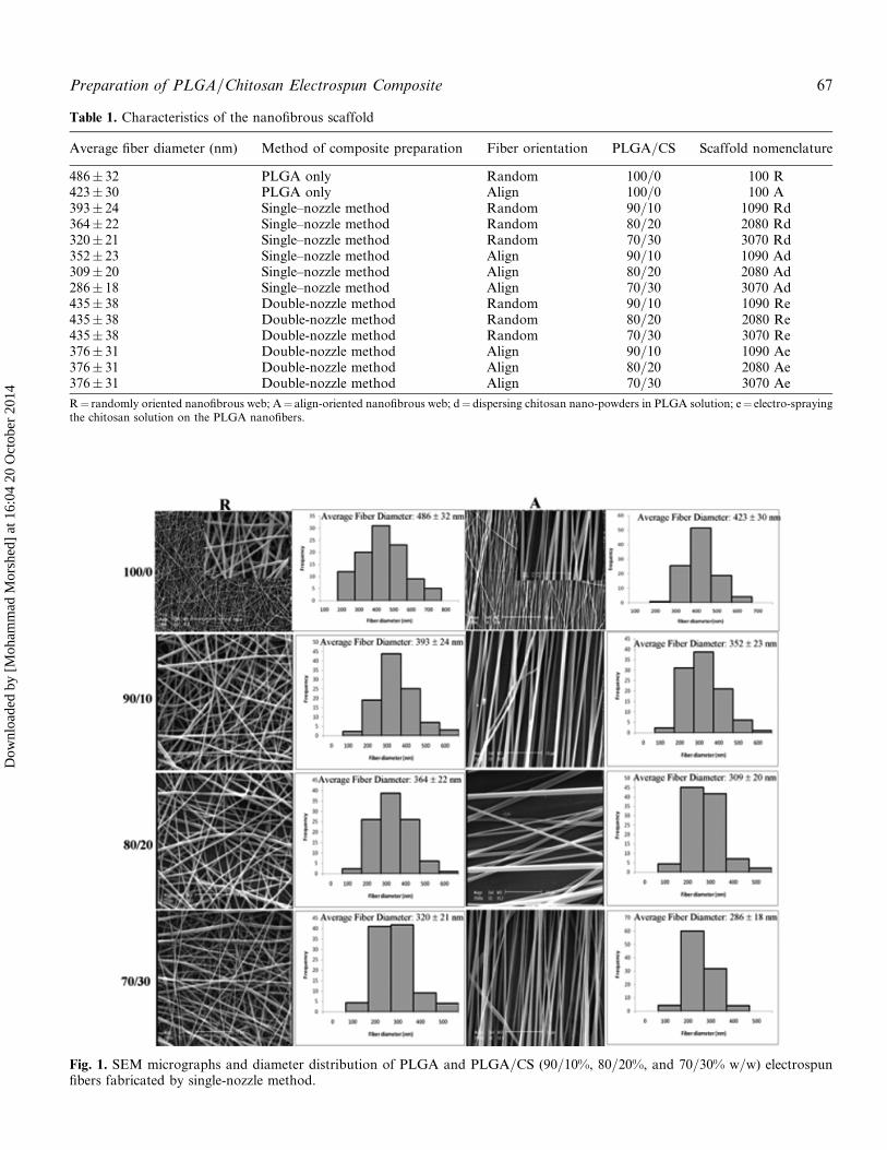
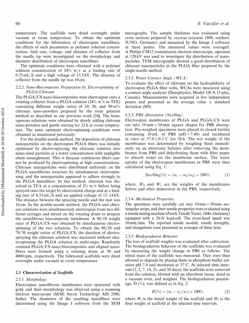
Table 1. Characteristics of the nanofibrous scaffold
Average fiber diameter (nm) Method of composite preparation Fiber orientation PLGA=CS Scaffold nomenclature
486� 32 PLGA only Random 100=0 100 R423� 30 PLGA only Align 100=0 100 A393� 24 Single–nozzle method Random 90=10 1090 Rd364� 22 Single–nozzle method Random 80=20 2080 Rd320� 21 Single–nozzle method Random 70=30 3070 Rd352� 23 Single–nozzle method Align 90=10 1090 Ad309� 20 Single–nozzle method Align 80=20 2080 Ad286� 18 Single–nozzle method Align 70=30 3070 Ad435� 38 Double-nozzle method Random 90=10 1090 Re435� 38 Double-nozzle method Random 80=20 2080 Re435� 38 Double-nozzle method Random 70=30 3070 Re376� 31 Double-nozzle method Align 90=10 1090 Ae376� 31 Double-nozzle method Align 80=20 2080 Ae376� 31 Double-nozzle method Align 70=30 3070 Ae
R¼ randomly oriented nanofibrous web; A¼ align-oriented nanofibrous web; d¼ dispersing chitosan nano-powders in PLGA solution; e¼ electro-sprayingthe chitosan solution on the PLGA nanofibers.
Fig. 1. SEM micrographs and diameter distribution of PLGA and PLGA=CS (90=10%, 80=20%, and 70=30% w=w) electrospunfibers fabricated by single-nozzle method.
Preparation of PLGA/Chitosan Electrospun Composite 67
Dow
nloa
ded
by [
Moh
amm
ad M
orsh
ed]
at 1
6:04
20
Oct
ober
201
4
2.3.6 Attenuated Total Reflection Fourier Transform Infrared(ATR-FT-IR)
Chemical characteristics of the electrospun PLGA andPLGA=CS nanofibrous scaffolds and pure powders of chit-osan were evaluated by an attenuated total reflection Fouriertransform infrared (ATR-FT-IR) spectrophotometer(JASCO FT=IR-6300, Japan). All spectra represent theaverage of 30 scans between 400 and 4000 cm�1.
2.3.7 Thermal Property
The thermal property of the scaffolds was studied using thethermogravimeter (TG, Rheometric Scientific, Inc 1998,USA) at a constant heating rate of 10�C=min over a tem-perature range of 25–600�C.
2.4 Statistical Analysis
All the data in this paper were presented as means �standard deviation and analyzed using single-factor analysesof variance (ANOVAs). The significance level was set atp< 0.05.
3. Results
3.1 Morphology of Electrospun Nanofibers
The morphology of the electrospun nanofibers is influencedby various parameters such as applied voltage, solution feed
rate, distance between the capillary and the collector, andespecially the properties of the polymer solutions includingconcentration, surface tension, and the nature of the solvent[6,55]. The scaffold nomenclature, fiber orientation, methodof composite preparation, PLGA=CS ratio and the averagefiber diameter (nm) are presented in Table 1. No significantdifferences (p< 0.05) in the diameter were observed for ran-dom compared to aligned nanofibers for the respectivePLGA and PLGA=CS nanofibers.
Figure 1 shows the morphology of the randomly orientedand aligned electrospun PLGA from a 24% PLGA solutionin TFE as the solvent. The insets display the correspondingdiameter distribution. As can be seen, highly uniform andsmooth nanofibers were formed without the occurrence ofbead defects in all the randomly oriented and aligned nano-fibrous scaffolds. Using the Image J software of the SEMmicrographs, the average fiber diameters of the randomlyoriented and aligned PLGA fibers were determined to be486� 32 nm and 423� 30, respectively. Figure 1 also showsthe SEM micrograph and the insets display the correspond-ing diameter distribution of the PLGA=CS electrospunfibers with three different ratios (90=10%, 80=20%, and70=30% w=w), fabricated by the single-nozzle method. Inthis method, the chitosan nano-powders dispersed in thePLGA solutions were electrospun through a single nozzle.As shown, the average diameter of the PLGA=CS membrane
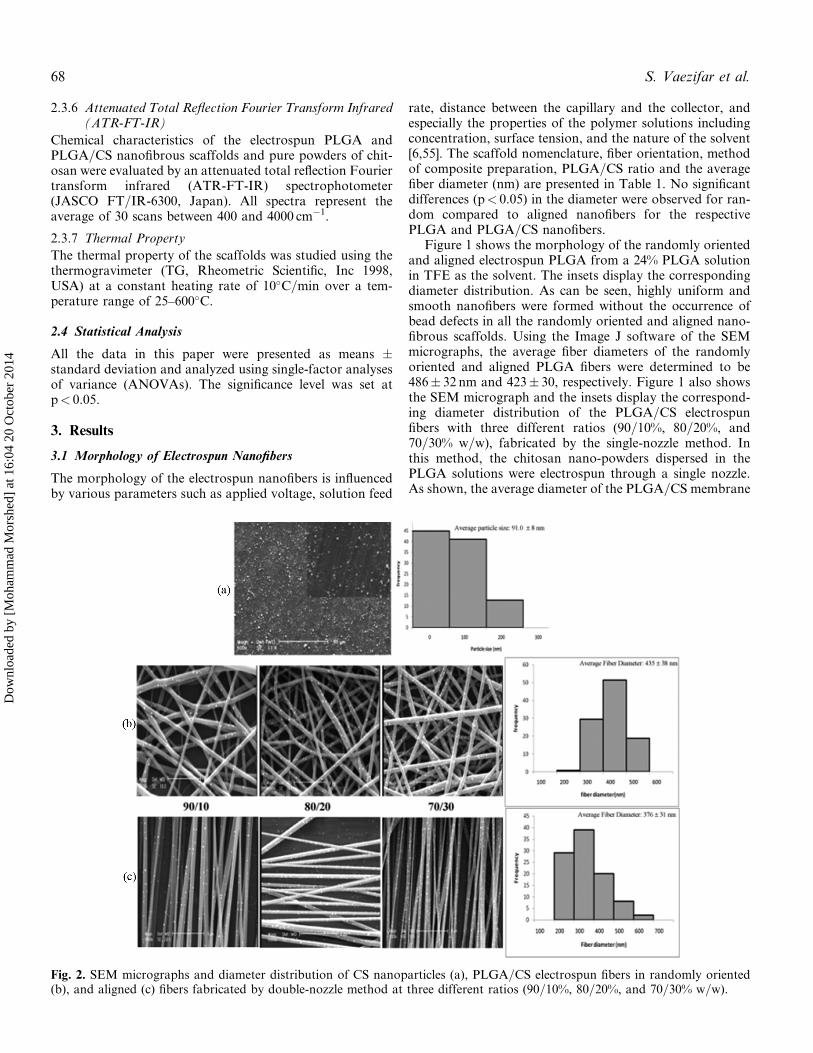
Fig. 2. SEM micrographs and diameter distribution of CS nanoparticles (a), PLGA=CS electrospun fibers in randomly oriented(b), and aligned (c) fibers fabricated by double-nozzle method at three different ratios (90=10%, 80=20%, and 70=30% w=w).
68 S. Vaezifar et al.
Dow
nloa
ded
by [
Moh
amm
ad M
orsh
ed]
at 1
6:04
20
Oct
ober
201
4
prepared by dispersing the chitosan nano-powders in PLGAsolution is lower than the others. The nanofiber diameterdecreased and the diameter distribution broadened withincreasing chitosan content. There were significant differ-ences (p< 0.05) in the diameter by increasing chitosan con-tent. The presence of chitosan in the PLGA solutionincreased conductivity and surface charge densities, whichenhanced the whipping instability.
SEM micrograph of chitosan nanoparticles electro-sprayed from a 2% w=v solution in TFA at a feeding rateof 0.33mL=h and an applied voltage of 13–14 kV is shownin Figure 2a. The distance between the spraying nozzleand the mat was 10 cm. Chitosan nanoparticles obtainedunder this condition were highly uniform. However, theaverage diameter of chitosan particles was 91� 8 nm andits distribution was in the range of 26–250 nm. The SEMmicrographs and the insets in Figures 2b and 2c displaythe corresponding diameter distribution of the PLGA=CSelectrospun fibers=electro-sprayed nanoparticles with threedifferent ratios (90=10%, 80=20%, and 70=30% w=w) fabri-cated by the second method. In this method, PLGA andchitosan solutions were simultaneously electrospun from
two different syringes and the electrospun PLGA nanofiberand electro-sprayed CS nanoparticles were mixed and col-lected on the rotating drum to prepare the nanofibrouscomposite membrane. As shown, the average fiber diameterof the PLGA=CS electrospun fibers prepared by electro-spraying the chitosan solution on the PLGA nanofibers islower than the pure PLGA nanofibers but higher than theones fabricated by the first method. This could be due tothe simultaneous effect of two electrical fields. In order toprepare the aligned scaffold, a high speed rotating drumwas used as the collector at a speed of 4000 rpm. Comparedwith the randomly oriented nanofibers, the aligned oneswere smaller in diameter but no significant differences(p< 0.05) in the diameter were observed.
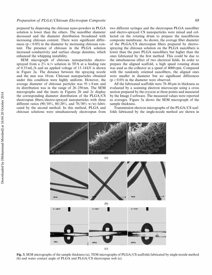
All the fabricated scaffolds were 70–80 mm in thickness asevaluated by a scanning electron microscope using a crosssection prepared by the cryocut at three points and measuredby the Image J software. The measured values were reportedas averages. Figure 3a shows the SEM micrograph of thesample thickness.
Transmission electron micrographs of the PLGA=CS scaf-folds fabricated by the single-nozzle method are shown in
Fig. 3. SEMmicrographs of the sample thickness (a), TEM micrographs of PLGA=CS scaffolds fabricated by single-nozzle method(b) and water contact angle of PLGA and PLGA=CS electrospun web (c).
Preparation of PLGA/Chitosan Electrospun Composite 69
Dow
nloa
ded
by [
Moh
amm
ad M
orsh
ed]
at 1
6:04
20
Oct
ober
201
4
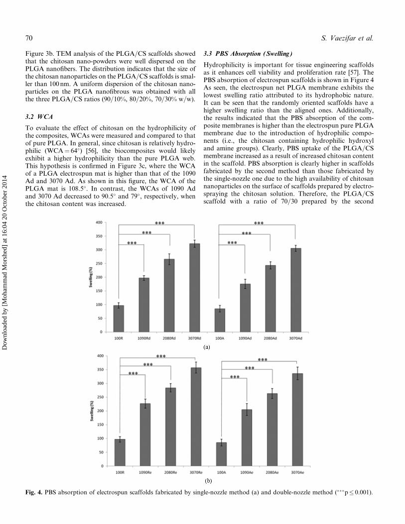
Figure 3b. TEM analysis of the PLGA=CS scaffolds showedthat the chitosan nano-powders were well dispersed on thePLGA nanofibers. The distribution indicates that the size ofthe chitosan nanoparticles on the PLGA=CS scaffolds is smal-ler than 100nm. A uniform dispersion of the chitosan nano-particles on the PLGA nanofibrous was obtained with allthe three PLGA=CS ratios (90=10%, 80=20%, 70=30% w=w).
3.2 WCA
To evaluate the effect of chitosan on the hydrophilicity ofthe composites, WCAs were measured and compared to thatof pure PLGA. In general, since chitosan is relatively hydro-philic (WCA¼ 64�) [56], the biocomposites would likelyexhibit a higher hydrophilicity than the pure PLGA web.This hypothesis is confirmed in Figure 3c, where the WCAof a PLGA electrospun mat is higher than that of the 1090Ad and 3070 Ad. As shown in this figure, the WCA of thePLGA mat is 108.5�. In contrast, the WCAs of 1090 Adand 3070 Ad decreased to 90.5� and 79�, respectively, whenthe chitosan content was increased.
3.3 PBS Absorption (Swelling)
Hydrophilicity is important for tissue engineering scaffoldsas it enhances cell viability and proliferation rate [57]. ThePBS absorption of electrospun scaffolds is shown in Figure 4As seen, the electrospun net PLGA membrane exhibits thelowest swelling ratio attributed to its hydrophobic nature.It can be seen that the randomly oriented scaffolds have ahigher swelling ratio than the aligned ones. Additionally,the results indicated that the PBS absorption of the com-posite membranes is higher than the electrospun pure PLGAmembrane due to the introduction of hydrophilic compo-nents (i.e., the chitosan containing hydrophilic hydroxyland amine groups). Clearly, PBS uptake of the PLGA=CSmembrane increased as a result of increased chitosan contentin the scaffold. PBS absorption is clearly higher in scaffoldsfabricated by the second method than those fabricated bythe single-nozzle one due to the high availability of chitosannanoparticles on the surface of scaffolds prepared by electro-spraying the chitosan solution. Therefore, the PLGA=CSscaffold with a ratio of 70=30 prepared by the second
Fig. 4. PBS absorption of electrospun scaffolds fabricated by single-nozzle method (a) and double-nozzle method (���p� 0.001).
70 S. Vaezifar et al.
Dow
nloa
ded
by [
Moh
amm
ad M
orsh
ed]
at 1
6:04
20
Oct
ober
201
4
method exhibited the highest swelling (Figure 4). It isassumed that the nanofibrous composite membrane of theelectrospun PLGA=CS could be potentially used for tissueregeneration.
3.4 Mechanical Properties
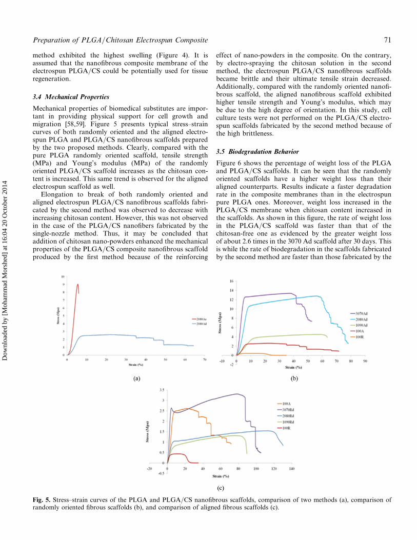
Mechanical properties of biomedical substitutes are impor-tant in providing physical support for cell growth andmigration [58,59]. Figure 5 presents typical stress–straincurves of both randomly oriented and the aligned electro-spun PLGA and PLGA=CS nanofibrous scaffolds preparedby the two proposed methods. Clearly, compared with thepure PLGA randomly oriented scaffold, tensile strength(MPa) and Young’s modulus (MPa) of the randomlyoriented PLGA=CS scaffold increases as the chitosan con-tent is increased. This same trend is observed for the alignedelectrospun scaffold as well.
Elongation to break of both randomly oriented andaligned electrospun PLGA=CS nanofibrous scaffolds fabri-cated by the second method was observed to decrease withincreasing chitosan content. However, this was not observedin the case of the PLGA=CS nanofibers fabricated by thesingle-nozzle method. Thus, it may be concluded thataddition of chitosan nano-powders enhanced the mechanicalproperties of the PLGA=CS composite nanofibrous scaffoldproduced by the first method because of the reinforcing
effect of nano-powders in the composite. On the contrary,by electro-spraying the chitosan solution in the secondmethod, the electrospun PLGA=CS nanofibrous scaffoldsbecame brittle and their ultimate tensile strain decreased.Additionally, compared with the randomly oriented nanofi-brous scaffold, the aligned nanofibrous scaffold exhibitedhigher tensile strength and Young’s modulus, which maybe due to the high degree of orientation. In this study, cellculture tests were not performed on the PLGA=CS electro-spun scaffolds fabricated by the second method because ofthe high brittleness.
3.5 Biodegradation Behavior
Figure 6 shows the percentage of weight loss of the PLGAand PLGA=CS scaffolds. It can be seen that the randomlyoriented scaffolds have a higher weight loss than theiraligned counterparts. Results indicate a faster degradationrate in the composite membranes than in the electrospunpure PLGA ones. Moreover, weight loss increased in thePLGA=CS membrane when chitosan content increased inthe scaffolds. As shown in this figure, the rate of weight lossin the PLGA=CS scaffold was faster than that of thechitosan-free one as evidenced by the greater weight lossof about 2.6 times in the 3070 Ad scaffold after 30 days. Thisis while the rate of biodegradation in the scaffolds fabricatedby the second method are faster than those fabricated by the
Fig. 5. Stress–strain curves of the PLGA and PLGA=CS nanofibrous scaffolds, comparison of two methods (a), comparison ofrandomly oriented fibrous scaffolds (b), and comparison of aligned fibrous scaffolds (c).
Preparation of PLGA/Chitosan Electrospun Composite 71
Dow
nloa
ded
by [
Moh
amm
ad M
orsh
ed]
at 1
6:04
20
Oct
ober
201
4
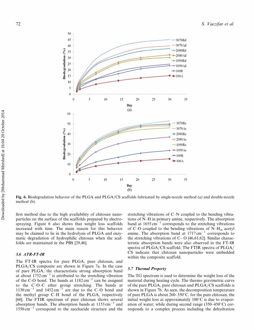
first method due to the high availability of chitosan nano-particles on the surface of the scaffolds prepared by electro-spraying. Figure 6 also shows that weight loss scaffoldsincreased with time. The main reason for this behaviormay be claimed to lie in the hydrolysis of PLGA and enzy-matic degradation of hydrophilic chitosan when the scaf-folds are maintained in the PBS [29,46].
3.6 ATR-FT-IR
The FT-IR spectra for pure PLGA, pure chitosan, andPLGA=CS composite are shown in Figure 7a. In the caseof pure PLGA, the characteristic strong absorption bandat about 1752 cm�1 is attributed to the stretching vibrationof the C-O bond. The bands at 1182 cm�1 can be assignedto the C–O–C ether group stretching. The bands at1130 cm�1 and 1452 cm�1 are due to the C–O bond andthe methyl group C–H bond of the PLGA, respectively[60]. The FTIR spectrum of pure chitosan shows severalabsorption bands. The absorption bands at 1153 cm�1 and1550 cm�1 correspond to the saccharide structure and the
stretching vibrations of C–N coupled to the bending vibra-tions of N–H in primary amine, respectively. The absorptionband at 1655 cm�1 corresponds to the stretching vibrationsof C–O coupled to the bending vibrations of N–Hin acetylamine. The absorption band at 1717 cm�1 corresponds tothe stretching vibrations of C– O [46,61,62]. Similar charac-teristic absorption bands were also observed in the FT-IRspectra of PLGA=CS scaffold. The FTIR spectra of PLGA=CS indicate that chitosan nanoparticles were embeddedwithin the composite scaffold.
3.7 Thermal Property
The TG spectrum is used to determine the weight loss of thematerial during heating cycle. The thermo gravimetric curveof the pure PLGA, pure chitosan and PLGA=CS scaffolds isshown in Figure 7b. As seen, the decomposition temperatureof pure PLGA is about 260–350�C. for the pure chitosan, theinitial weight loss at approximately 100�C is due to evapor-ation of water; while during second range (350–450�C) cor-responds to a complex process including the dehydration
Fig. 6. Biodegradation behavior of the PLGA and PLGA=CS scaffolds fabricated by single-nozzle method (a) and double-nozzlemethod (b).
72 S. Vaezifar et al.
Dow
nloa
ded
by [
Moh
amm
ad M
orsh
ed]
at 1
6:04
20
Oct
ober
201
4
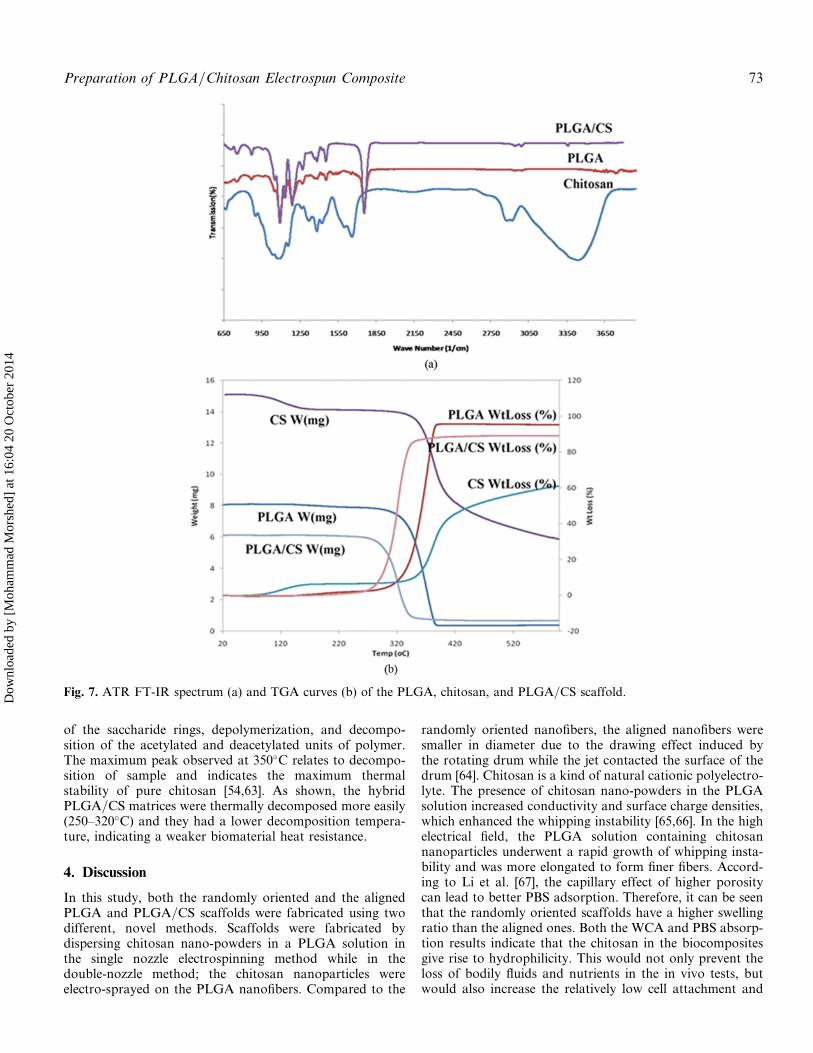
of the saccharide rings, depolymerization, and decompo-sition of the acetylated and deacetylated units of polymer.The maximum peak observed at 350�C relates to decompo-sition of sample and indicates the maximum thermalstability of pure chitosan [54,63]. As shown, the hybridPLGA=CS matrices were thermally decomposed more easily(250–320�C) and they had a lower decomposition tempera-ture, indicating a weaker biomaterial heat resistance.
4. Discussion
In this study, both the randomly oriented and the alignedPLGA and PLGA=CS scaffolds were fabricated using twodifferent, novel methods. Scaffolds were fabricated bydispersing chitosan nano-powders in a PLGA solution inthe single nozzle electrospinning method while in thedouble-nozzle method; the chitosan nanoparticles wereelectro-sprayed on the PLGA nanofibers. Compared to the
randomly oriented nanofibers, the aligned nanofibers weresmaller in diameter due to the drawing effect induced bythe rotating drum while the jet contacted the surface of thedrum [64]. Chitosan is a kind of natural cationic polyelectro-lyte. The presence of chitosan nano-powders in the PLGAsolution increased conductivity and surface charge densities,which enhanced the whipping instability [65,66]. In the highelectrical field, the PLGA solution containing chitosannanoparticles underwent a rapid growth of whipping insta-bility and was more elongated to form finer fibers. Accord-ing to Li et al. [67], the capillary effect of higher porositycan lead to better PBS adsorption. Therefore, it can be seenthat the randomly oriented scaffolds have a higher swellingratio than the aligned ones. Both the WCA and PBS absorp-tion results indicate that the chitosan in the biocompositesgive rise to hydrophilicity. This would not only prevent theloss of bodily fluids and nutrients in the in vivo tests, butwould also increase the relatively low cell attachment and
Fig. 7. ATR FT-IR spectrum (a) and TGA curves (b) of the PLGA, chitosan, and PLGA=CS scaffold.
Preparation of PLGA/Chitosan Electrospun Composite 73
Dow
nloa
ded
by [
Moh
amm
ad M
orsh
ed]
at 1
6:04
20
Oct
ober
201
4
proliferation rate of pure PLGA membrane. PLGA contain-ing the hydrophobic methyl group (–CH3) increases therepulsion force when interacting with water and reduceswater absorbability. On the contrary, chitosan containingthe primary amine (–NH2) and hydroxyl (–OH) groupscan not only increase its affinity to water but also formhydrogen bonds with water. Hence, more chitosan in thePLGA=CS matrix generally enhances the hydrophilicproperty of the biomaterial surfaces for water entrapment.
Orientation of the fabricated fibers is one of the impor-tant factors in the preparation of nanofibrous scaffold. Itis demonstrated the aligned nanofibers provide a suitablecondition for cell attachment and proliferation [68].
Also consistent with our result, previous study indicatedthe mechanical strength of aligned fibers were higher thanrandomly oriented ones [69].
The following four viewpoints summarize the underlyingcauses for these findings. First, using chitosan nanoparti-cles to fabricate the PLGA=CS composite enhances thehydrophilicity in both fabrication methods. Second,PLGA=CS nano-biocomposites fabricated by double noz-zle method are very brittle and have not suitable mechan-ical properties for tissue engineering application. Third, themechanical properties of the scaffolds fabricated by singlenozzle method are improved by increasing the chitosannanoparticles content. Fourth, a scaffold with a higherhydrophilicity and better mechanical properties has ahigher PBS absorption, yielding more absorbed mediumin the scaffold through cell culturing with a suitablemechanical support. Our findings indicate that the alignednanofibrous scaffold with high percentage of chitosannano-particles (3070Ad) provides a beneficial approachfor tissue regeneration.
5. Conclusions
In this study, both the randomly oriented and the alignedPLGA and PLGA=CS nanofibrous scaffolds were fabricatedusing two different methods, namely, the single- and thedouble-nozzle methods. Scaffold composed of PLGA andchitosan nano-particles provide the mechanical supportrequired and enhance the cell attachment and proliferationrate. In this study, chitosan nanoparticles and PLGA wereused to fabricate the scaffolds by electrospinning method,for the first time. The resulting composites exhibitedincreased hydrophilicity of the composite scaffolds andimproved mechanical properties by increasing chitosancontent in the biocomposites scaffold in the first method.However, compared to the randomly oriented nanofibrousscaffolds, the aligned ones exhibited a higher tensilestrength. These results demonstrate that the inherent hydro-phobicity of synthetic PLGA can be modified by incorporat-ing a suitable amount of chitosan. In addition, the fabricatedbiocomposites by the first method showed various synergiceffects including enhanced PBS absorption and increasedhydrophilicity. Thus aligned nanofibrous scaffold with highpercentage of chitosan nanoparticles (3070 Ad) provide abeneficial approach for tissue engineering.
Acknowledgments
The authors are grateful to Isfahan University of Technology(IUT) for their support and Mrs. Aliakbari for kindcollaboration.
Funding
The authors are grateful to Iranian Council of Stem CellTechnology, Isfahan University of Medical Sciences, forfinancial support (grant No. 190044).
References1. Kenawy, E. R.; Layman, J. M.; Watkins, J. R.; Bowlin, G. L.;
Matthews, J. A.; Simpson, D. G. Biomaterials 2003, 24, 907.2. Li, W. J.; Laurencin, C. T.; Caterson, E. J.; Tuan, R. S.; Ko, F. K.
J. Biomed. Mater. Res. 2002, 60, 613.3. Lu, Y. K.; Kim, K.; Hsiao, B. S.; Chu, B.; Hadjiargyrou, M.
J. Control. Release 2003, 89, 341.4. Matthews, J. A.; Wnek, G. E.; Simpson, D. G.; Bowlin, G. L.
Biomacromolecules 2002, 3, 232.5. Yoshimoto, H.; Shin, Y. M.; Terai, H.; Vacanti, J. P. Biomaterials
2003, 24, 2077.6. Min, B. M.; You, Y.; Kim, J. M.; Lee, S. J.; Park, W. H.
Carbohydr. Polym. 2004, 57, 285.7. Hutmacher, D. W.; Sittinger, M.; Risbud, M. V. Trends Biotechnol.
2004, 22, 354.8. Gomes, M. E.; Godinho, J. S.; Tchalamov, D.; Cunha, A. M.; Reis,
R. L. Mater. Sci. Eng. C 2002, 20, 19.9. Mano, J. F.; Vaz, C. M.; Mendes, S. C.; Reis, R. L.; Cunha, A. M.
J. Mater. Sci. Mater. Med. 1999, 10, 857.10. Sill, T. J.; von Recum, H. A. Biomaterials 2008, 29, 1989.11. Yoon, H.; Kim, G. J. Biomater. Sci. Polym. Ed. 2010, 21, 553.12. Smith, L. A.; Liu, X.; Ma, P. X. Soft Matter 2008, 4, 2144.13. Sridhar, R.; Sundarrajan, S.; Venugopal, J. R.; Ravichandran, R.;
Ramakrishna, S. J. Biomater. Sci. Polym. 2013, 4, 24.14. Andukuri, A.; Kushwaha, M.; Tambralli, A.; Anderson, J. M.;
Dean, D. R.; Berry, J. L. Acta Biomater. 2011, 7, 225.15. Li, X. K.; Cai, S. X.; Liu, B.; Xu, Z. L.; Dai, X. Z.; Ma, K. W.; Li,
S. Q.; Yang, L.; Paul Sung, K. L.; Fu, X. B. Colloids Surf., B 2007,57, 198.
16. Liu, B.; Cai, S. X.; Ma, K. W.; Xu, Z. L.; Dai, X. Z.; Yang, L.J. Mater. Sci. Mater. Med. 2008, 19, 1127.
17. Fan, X.; Guo, L.; Liu, T. Polym.-Plast. Technol. Eng. 2013, 6, 52.18. Hsu, S. H.; Seng Lu, P.; Ni, H. C.; Su, C. H. Biomed. Microdev.
2007, 9, 665.19. Evans, G. R. D.; Brandt, K.; Widmer, M. S.; Lu, L.; Meszlenyi,
R. K.; Gupta, P. K. Biomaterials 2002, 23, 841.20. Evans, G. R. D.; Brandt, K.; Widmer, M. S.; Lu, L.; Meszlenyi,
R. K.; Gupta, P. K. Biomaterials 1999, 20, 1109.21. Bian, Y. Z.; Wang, Y.; Aibaidoula, G.; Chen, G. Q.; Wu, Q.
Biomaterials 2009, 30, 217.22. Yucel, D.; Torun Kose, G.; Hasirci, V. Biomaterials 2010, 31, 1596.23. Chen, Y. Sh.; Chang, J. Y.; Cheng, C. Y.; Tsai, F. J.; Yao, C. H.;
Liu, B. S. Biomaterials 2005, 26, 3911.24. Meng, Z. X.; Wang, Y. S.; Ma, C.; Zheng, W.; Li, L.; Zheng, Y. F.
Mater. Sci. Eng. C 2010, 30, 1204.25. Yao, L.; de Ruiter, G. C. W.; Wang, H.; Knight, A. M.; Spinner,
R. J.; Yaszemski, M. J. Biomaterials 2010, 31, 5789.26. Yang, L.; Fitie, C. F. C.; Van der Werf, K. O.; Bennink, M. L.;
Dijkstra, P. J. Biomaterials 2008, 29, 955.27. (a) Kumar, S.; Kumari, M.; Dutta, P. K.; Koh, J. Int. J. Polym
Mater. Polym. Biomater. 2014, 63, 4; (b) Cheng, H.; Huang, Y.C.; Chang, P. T.; Huang, Y. Y. Biochem. Biophys. Res. Commun.2007, 357, 938.
74 S. Vaezifar et al.
Dow
nloa
ded
by [
Moh
amm
ad M
orsh
ed]
at 1
6:04
20
Oct
ober
201
4
28. Ma, L.; Gao, C. Y.; Mao, Z. W.; Zhou, J.; Shen, J. C.; Hu, X. Q.Biomaterials 2003, 24, 4833.
29. Wu, L.; Ding, J. Biomaterials 2004, 25, 5821.30. Athanasiou, K. A.; Niederauer, G. G.; Agrawal, C. M.
Biomaterials 1996, 17, 93.31. Wu, L.; Ding, J. Biomaterials 2004, 25, 5821, 25.32. Li, X. K.; Cai, S. X.; Liu, B.; Xu, Z. L.; Dai, X. Z.; Ma, K. W.
Colloids Surf., B 2007, 57, 198.33. Glowacki, J.; Mizuno, S. Biopolymers 2008, 89, 338.34. Drury, J. L.; Mooney, D. J. Biomaterials 2003, 24, 4337.35. Yang, Y.; Zhu, X.; Cui, W.; Li, X.; Jin, Y. Macromol. Mater. Eng.
2009, 294, 611.36. Zhao, H.; Ma, L.; Gao, C.; Shen, J. J. Biomed. Mater. Res. Part B
2009, 88, 240.37. Khnor, E.; Lim, L. Biomaterials 2003, 24, 2339.38. No, H. K.; Park, N. Y.; Lee, S. H.; Meyers, S. P. Int. J. Food
Microbiol. 2002, 74, 65.39. Ueno, H.; Mori, T.; Fujinaga, T. Adv. Drug Delivery Rev. 2001,
52, 105.40. Ravi Kumar, M. N. V. React. Funct. Polym. 2000, 46, 1.41. Bumgardner, J. D.; Wiser, R.; Gerard, P.; Bergin, P.; Chestnutt, B.;
Marini, M. J. Biomater. Sci. Polym. Ed. 2003, 14, 423.42. Cheng, M.; Gong, K.; Li, J.; Gong, Y.; Zhao, N.; Zhang, X.
J. Biomater. Appl. 2004, 19, 59.43. Shanmugasundaram, N.; Ravichandran, P.; Reddy, P. N.;
Ramamurty, N.; Pal, S.; Rao, K. P. Biomaterials 2001, 22,1943.
44. Kuo, Y. C.; Ku, I. N. Biomacromolecules 2008, 9, 2662.45. Gong, H. P.; Zhong, Y. H.; Li, J. C.; Gong, Y. D.; Zhao, N. M.;
Zhang, X. F. J. Biomed. Mater. Res. 2000, 52, 285.46. Kuo, Y. C.; Yeh, C. F.; Yang, J. T. Biomaterials 2009, 30,
6604.47. Min, B. M.; Lee, S. W.; Lim, J. N.; You, Y.; Lee, T. S.; Kang, P. H.
Polymer 2004, 45, 7137.48. Huang, X. J.; Ge, D.; Xu, Z. K. Eur. Polym. J. 2007, 43, 3710.49. Kriegel, C.; Kit, K. M.; McClements, D. J.; Weiss, J. Polymer 2009,
50, 189.
50. Jung, K. H.; Huh, M. W.; Meng, W.; Yuan, J.; Hyun, S. H.; Bae,J. S. J. Appl. Polym. Sci. 2007, 105, 2816.
51. Peesan, M.; Rujiravanit, R.; Supaphol, P. J. Biomater. Sci. Polym.Ed. 2006, 17, 547.
52. Jayakumar, R.; Prabaharan, M.; Nair, S. V.; Tamura, H. Biotech-nol. Adv. 2010, 28, 142.
53. Hong, S.; Kim, G. H. Carbohydr. Polym. 2011, 38, 940.54. Vaezifar, S.; Razavi, S.; Golozar, M. A.; Karbasi, S.; Morshed, M.;
Kamali, M. J. Cluster Sci. 2013, 24, 891.55. Deitzel, J. M.; Kleinmeyer, J.; Harris, D.; Tan, N. C. B. Polymer
2001, 42, 261.56. Cheng, M.; Deng, J.; Yang, F.; Gong, Y.; Zhao, N.; Zhang, X.
Biomaterials 2003, 24, 2871.57. Chen, Z. G.; Mo, X. M.; He, C. G.; Wang, H. S. Carbohydr.
Polym. 2008, 72, 410.58. Hollister, S. J. Nat. Mater. 2005, 4, 518.59. Murphy, C. M.; Haugh, M. G.; O’Brien, F. J. Biomaterials 2010,
31, 461.60. Jose, M. V.; Thomas, V.; Dean, D. R.; Nyairo, E. Polymer 2009,
50, 3778.61. Wang, T.; Turhan, M.; Gunasekaran, S. Polym. Int. 2004,
53, 911.62. Tangsadthakun, C. J. Biomater. Sci. Polym. Ed. 2007, 18, 147.63. Peniche-Covas, C.; Arguelles-Monal, W.; Roman, J. S. Polym
Degrad Stab. 1993, 39, 21.64. Thomas, V.; Jose, M. V.; Chowdhury, S.; Sullivan, F.; Dean, D. R.;
Vohra, Y. K. J. Biomater. Sci. Polym. Ed. 2006, 17, 984.65. Shin, Y. M.; Hohman, M. M.; Brenner, M. P.; Rutledge, G. C.
Polymer 2001, 42, 9955.66. Hohman, M. M.; Shin, M.; Rutledge, G.; Brenner, M. P. Phys.
Fluids. 2001, 13, 2221.67. Li, Q., Wei, Q. F., Wu, N., Cai, Y. B., Gao, W. D. J. Appl. Polym.
Sci. 2008, 107, 3535.68. Cooper, A.; Bhattarai, N.; Kievit, F. M.; Rossol, M.; Zhang, M.
Phys. Chem. Chem. Phys. 2011, 13, 7.69. Volova, T.; Goncharov, D.; Sukovatyi, A.; Shabanov, A.;
Nikolaeva, E.; Shishatskaya, E. J. Biomater. Sci. Polym. Ed. 2013.
Preparation of PLGA/Chitosan Electrospun Composite 75
Dow
nloa
ded
by [
Moh
amm
ad M
orsh
ed]
at 1
6:04
20
Oct
ober
201
4
Related Documents