Plant Molecular Biology 11:857-866 (1988) © Kluwer Academic Publishers, Dordrecht - Printed in the Netherlands 857 Characterization of pea histone deacetylases Ram6n Sendra, Ismael Rodrigo, M. Luisa Salvador and Luis Franco* Department of Biochemistry and Molecular Biology, Faculties of Sciences, Burjassot, Valencia, Spain (*author for correspondence) Received 10 May 1988; accepted in revised form 27 September 1988 University of Valencia, 46100 Key words: histone acetylation, nucleosomes, n-butyrate, polyamines, Pisum sativum Abstract The present paper is the first report on histone deacetylases from plants. Three enzyme fractions with histone deacetylase activity (HD0, HD1 and HD2) have been partially purified from pea (Pisum sativum) embryonic axes. They deacetylate biologically acetylated chicken histones and, to a lesser extent, chemically acetylated histones, this being a criterion of their true histone deacetylase nature. The three enzymes are able to accept nucleosomes as substrates. HD1 is not inhibited by n-butyrate up to 50 mM, whereas HD0 and HD2 are only slightly inhibited, thereby establishing a clear difference to animal histone deacetylases. The three activities are inhibited by acetate, Cu 2+ and Zn 2+ ions and mercurials, but are only scarcely affected by polyamines, in strong contrast with yeast histone deacetylase. Several criteria have been used to obtain cumulative evidence that HD0, HD1 and HD2 actually are three distinct enzymes. In vitro experiments with free histones show that HD0 deacetylates all four core histones, whereas HD1 and HD2 show a clear preference for H2A and H2B, the arginine-rich histones being deacetylated more slowly. Introduction Acetylation of e-amino groups of lysyl residues is the most thoroughly studied histone post-synthetic modification (for a recent review, see[33]), although its precise roles are not yet fully understood. The turnover of acetyl groups depends upon the activity of two sets of enzymes catalyzing opposite reactions: histone acetyltransferases and histone deacetylases. It is obvious that a better characterization of these enzymes will help to understand the roles of histone acetylation. Histone acetyltransferases have been described in animals (reviewed in [17]), yeast [16, 24] and plants [20, 22]. Histone deacetylases are less well known. They were first described and characterized almost 20 years ago [11, 12] and, since then, their presence has been reported in several animals [8, 14, 26] and fungi [2, 30]. In some instances, a limited multiplici- ty of histone deacetylases has been detected. Chro- matography on DEAE-cellulose columns yields two peaks of enzyme activity for calf thymus [13] and for a rat hepatoma tissue culture cell line [5]. To date, histone deacetylase has not been described in plants. Although Fujimoto [8] reported the presence in spinach leaves of an enzyme capable of removing acetyl groups introduced into histones by chemical acetylation with acetic anhydride, the enzyme was absolutely uneffective in deacetylating biologically labelled histones. The present research is aimed at the study of his- tone deacetylases in plants. The knowledge of the properties of the enzyme(s), together with that of histone acetyltransferases [20, 22], will help to un- derstand the role(s) of histone acetylation in plants and, eventually, it may result in a beter knowledge

Welcome message from author
This document is posted to help you gain knowledge. Please leave a comment to let me know what you think about it! Share it to your friends and learn new things together.
Transcript
Plant Molecular Biology 11:857-866 (1988) © Kluwer Academic Publishers, Dordrecht - Printed in the Netherlands 857
Characterization of pea histone deacetylases
Ram6n Sendra, Ismael Rodrigo, M. Luisa Salvador and Luis Franco* Department of Biochemistry and Molecular Biology, Faculties of Sciences, Burjassot, Valencia, Spain (*author for correspondence)
Received 10 May 1988; accepted in revised form 27 September 1988
University of Valencia, 46100
Key words: histone acetylation, nucleosomes, n-butyrate, polyamines, Pisum sativum
Abstract
The present paper is the first report on histone deacetylases from plants. Three enzyme fractions with histone deacetylase activity (HD0, HD1 and HD2) have been partially purified from pea (Pisum sativum) embryonic axes. They deacetylate biologically acetylated chicken histones and, to a lesser extent, chemically acetylated histones, this being a criterion of their true histone deacetylase nature. The three enzymes are able to accept nucleosomes as substrates. HD1 is not inhibited by n-butyrate up to 50 mM, whereas HD0 and HD2 are only slightly inhibited, thereby establishing a clear difference to animal histone deacetylases. The three activities are inhibited by acetate, C u 2+ and Zn 2+ ions and mercurials, but are only scarcely affected by polyamines, in strong contrast with yeast histone deacetylase. Several criteria have been used to obtain cumulative evidence that HD0, HD1 and HD2 actually are three distinct enzymes.
In vitro experiments with free histones show that HD0 deacetylates all four core histones, whereas HD1 and HD2 show a clear preference for H2A and H2B, the arginine-rich histones being deacetylated more slowly.
Introduction
Acetylation of e-amino groups of lysyl residues is the most thoroughly studied histone post-synthetic modification (for a recent review, see[33]), although its precise roles are not yet fully understood. The turnover of acetyl groups depends upon the activity of two sets of enzymes catalyzing opposite reactions: histone acetyltransferases and histone deacetylases. It is obvious that a better characterization of these enzymes will help to understand the roles of histone acetylation.
Histone acetyltransferases have been described in animals (reviewed in [17]), yeast [16, 24] and plants [20, 22]. Histone deacetylases are less well known. They were first described and characterized almost 20 years ago [11, 12] and, since then, their presence has been reported in several animals [8, 14, 26] and
fungi [2, 30]. In some instances, a limited multiplici- ty of histone deacetylases has been detected. Chro- matography on DEAE-cellulose columns yields two peaks of enzyme activity for calf thymus [13] and for a rat hepatoma tissue culture cell line [5]. To date, histone deacetylase has not been described in plants. Although Fujimoto [8] reported the presence in spinach leaves of an enzyme capable of removing acetyl groups introduced into histones by chemical acetylation with acetic anhydride, the enzyme was absolutely uneffective in deacetylating biologically labelled histones.
The present research is aimed at the study of his- tone deacetylases in plants. The knowledge of the properties of the enzyme(s), together with that of histone acetyltransferases [20, 22], will help to un- derstand the role(s) of histone acetylation in plants and, eventually, it may result in a beter knowledge
858
of the similarities and differences between plants and other eukaryotes with respect to the structure and function of chromatin.
Materials and methods
Plant materials
Pea (Pisum sativum cv. Lincoln) were germinated as previously described [25]. Seedlings were raised after 60 h of germination and the embryonic axes were ex- cised from the cotyledons and used for the subse- quent experiments.
Extraction of histone deacetylases
Embryonic axes, in batches of about 70 g, were homogenized at 4 °C by grinding with acid-washed sand (axes/sand, 1:1, w/w) in 2 vol of buffer A (0.5 M NH4CI, 0.25 mM EDTA, 10 mM 2- mercaptoethanol, 80 mM NaHSO 3, 15 mM Tris- HCI, pH 7.9). The homogenate was filtered through two layers of muslin and the retained material was washed twice with 0.5 vol of buffer A. The combined filtrates were stirred for 30 min in the cold and cen- trifuged at 27 000 g for 15 min. The supernatant was then centrifuged at 100000 g for 1 h, the floating layer of lipids was removed and the supernatant was then dialyzed against buffer B (10 mM NH4C1, 0.25 mM EDTA, 5 mM 2-mercaptoethanol, 10% (v/v) glycerol, 15 mM Tris-HC1, pH, 7.9) (two changes, 2 1 each).
The dialysate was then loaded onto a column of DEAE-Sepharose CL-6B (17 cm × 3 cm), previously equilibrated with buffer B. The column was then washed with 5 vol of buffer B. The retained proteins were then eluted with 1200 ml of a linear gradient of NH4CI, 10 to 350 mM, in buffer B, at a flow rate of 70 ml/h. Fractions showing enzymatic activity (see below) were pooled and concentrated by ultrafiltra- t ion in a Nucleopore cell fitted with an Amicon YM10 membrane, and operated under N 2 pressure.
Purification of histone deacetylases
Concentrated, pooled fractions containing histone deacetylase activity were further purified by gel permeation chromatography on a column
(120 cm × 1.8 cm) of Ultrogel AcA 34, equilibrated and eluted with a buffer similar to buffer B, except that it contained 35 mM NH4C1. Elution was car- ried out at a flow rate of 12 ml /h and 3.7 ml frac- tions were collected. Before assaying histone dea- cetylase activity, the fractions were made 200 mM in NH4C1 by adding the appropriate amount of 3 M- containing buffer B.
Preparation of histone deacetylase substrates
Several substrates were used in this research. These include "biologically" acetylated chicken histones (either in nucleosomes or as free histones), and chemically acetylated histones and polyamines.
Chicken erythrocyte core histones were "biologi- cally" acetylated by incubating a reticulocyte- enriched red cell preparation with [3H]-acetate (5.0 Ci/mmol) as described by Ferenz and Nelson [7]. To obtain oligonucleosomes, nuclei were sus- pended at a density of 1.25×10⁹ nuclei/ml in 10 mM NaC1, 5 mM MgC12, 10 mM Tris-HC1, pH 7.4 and they were incubated at 37 °C for 30 min with 300 units/ml micrococcal nuclease. Reaction was stopped by adding 100 mM EDTA to a final concen- tration of 10 mM and chilling on ice. Digested nuclei were sedimented (1000g, 10 min) and lysed in 0.25 mM EDTA. The lysate was then dialyzed against 0.25 mM EDTA (3 changes) and the result- ing solution containing labelled oligonucleosomes was adjusted to A260=64 and stored at - 2 0 ° C . Free histones were isolated following the procedure of Weintraub et al. [32]. Chicken histones obtained by this procedure were labelled to a specific activity of 2200 dpm//zg and the radiolabel was exclusively incorporated into core histones, as determined by electrophoresis and fluorography (see below).
Attempts to acetylate pea histones by incubating either isolated nuclei or chromatin with labelled acetyl-CoA or acetate were unsuccessful. Significant label was neither incorporated into histones after in-
cubating mesophyll protoplasts with [3H]-acetate under several conditions. Failure to prepare biologi- cally labelled pea histones was surely related to the low level of pea histone acetylation and to the impos- sibility of specifically inhibiting deacetylase activity (see below).
Chemically acetylated histones were prepared by reaction with labelled acetic anhydride. Histones from either chicken erythrocyte or pea (5 mg) were dissolved in 400/~1 of 50 mM sodium borate pH 9.0. Acetylation was started by adding 100/A of a 0.26/~M solution of [3HI-acetic anhydride (1 mCi) in dioxane and the mixture was allowed to stand for 150 min at 0 °C. The solution was then made 0.25 M in HCI and the histones were precipitated with 8 vol of cold acetone. The precipitate was recovered by centrifugation, washed twice with acetone and dried under vacuum.
To acetylate spermine and putrescine, 10 mg of the polyamine were dissolved in 100/~I of the borate buffer and allowed to react overnight with 100/~1 of the acetic anhydride solution at 0°C. After adding 1 ml of water, the reaction mixture was loaded onto a Dowex-1 X4 column (6 cm × 1 cm). The column was eluted with water and the fractions were tested for ninhydrine reaction. Both polyamines were reco- vered in the first 4 ml. The chemical and radiochemi- cal purity of the preparations was checked by paper chromatography [6] and fluorography.
Histone deacetylase assay
To assay histone deacetylase activity along the purification procedure, 100 #1 of the appropriate chromatographic fractions were incubated in 1.5 ml Eppendorf tubes with 10/~1 of [3H]-acetate biologi- cally labelled chicken histones (8 mg/ml). The mix- ture was incubated at 37 °C for 60 min and the reac- tion was stopped by adding 37/~1 of 1.0 M HC1, 0.4 M acetic acid. Released labelled acetate was ex- tracted with 700 #1 of ethyl acetate [11] and 500/~I of the organic layer were mixed with 5 ml of 22- Normascint (Scharlau) and counted.
To study the pH dependence of the deacetylase ac- tivities, the enzymatic preparations, purified on Ultrogel AcA 34, were dialyzed against buffer B for
859
the pH interval 7.1 to 9.1, or against 10 mM sodium phosphate buffer for pH 6.2 to 7.4. When applicable, the basic procedure for enzymatic assay mentioned above was modified by changing the appropriate pa- rameter (temperature, NHaC1 concentration, vol- ume of labelled histone solution or pH). The in- fluence of inhibitors was tested by including them in the assay mixture at the desired concentration. In some instances, namely when studying the depen- dence of the activity on the concentration of n- butyrate and other ionizable inhibitors, the assay mixture was supplemented with the appropriate amount of NH4CI in order to kept constant the ion- ic strength of the samples containing different amounts of the inhibitor.
The deacetylase activity towards polyamines was assayed in a similar fashion, by including a radio- labelled polyamine solution (final concentration 365/~g/ml) instead of the histone substrates.
Finally, in some instances, nucleosomes contain- ing [3H]-acetylated histones were used as substrate for histone deacetylase assay. In this instance, the oligonucleosome preparation (see above) was thawed and enough solution was added to the incu- bation mixture to obtain a final concentration of 750 #g histone/ml. Therefore, the histone concen- tration in the assay is equivalent to that used with free histone assays.
Specificity of histone deacetylases
Electrophoresis and fluorography of labelled hi- stones were carried out as described previously [16]. The amount of radiolabel present in each histone clas S was determined by integrating the intensity of the whole area of the corresponding band in the fluorogram in an IBAS-2000 image analyser system (Kontron). This integrated labelling density was nor- malized by dividing it by the integrated intensity of the Coomassie-stained band, determined as above from the stained gel. The resulting value, further re- ferred to as specific labelling density (SLD), was tak- en as a measure of the specific activity of each [3H]- labelled histone class. Full details on this method will be given elsewhere.
860
Results
Multiplicity of histone deacetylase activities
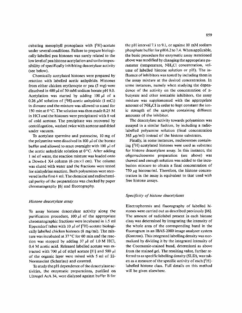
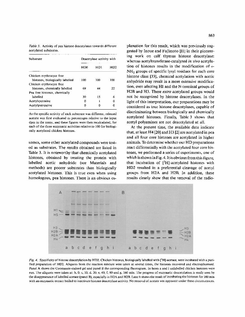
The histone deacetylase activity in 60 h embryonic axes was resolved into three peaks by DEAE- Sepharose chromatography (Fig. 1). Peak 1 ac- counts for most of the apparent enzymatic activity
recovered, but peaks 0 and 2 were consistently de- tected in all the experiments carried out with 60 h embryonic axes. The total enzymatic activity after DEAE-Sepharose chromatography was about 2.5 times higher than that found in crude extracts. This is probably due to the presence of inhibiting factors interfering with the enzyme activity. Earlier em- bryonic axes (after 24 h of germination) gave a pat- tern similar to that of Fig. 1 except that peak 0 was absent.
Fractions from the three peaks were pooled and
they were further purified by gel permeation chro- matography on Ultrogel AcA-34. Activity from peak 0 was recovered at an elution volume corresponding to M r ~ 90000, whereas the activities from peaks 1 and 2 seemed to be associated with polypeptides in the range of M r = 100 000. Ultrogel chromatography typically resulted in a 3-fold increase of the enzyme
activity per unit mass of protein. The enzymatic extracts after Ultrogel chromato-
graphy were used to investigate the properties of his- tone deacetylases (HD). The activities present in peaks 0, 1 and 2 will be designated HD0, HD1 and HD2 respectively.
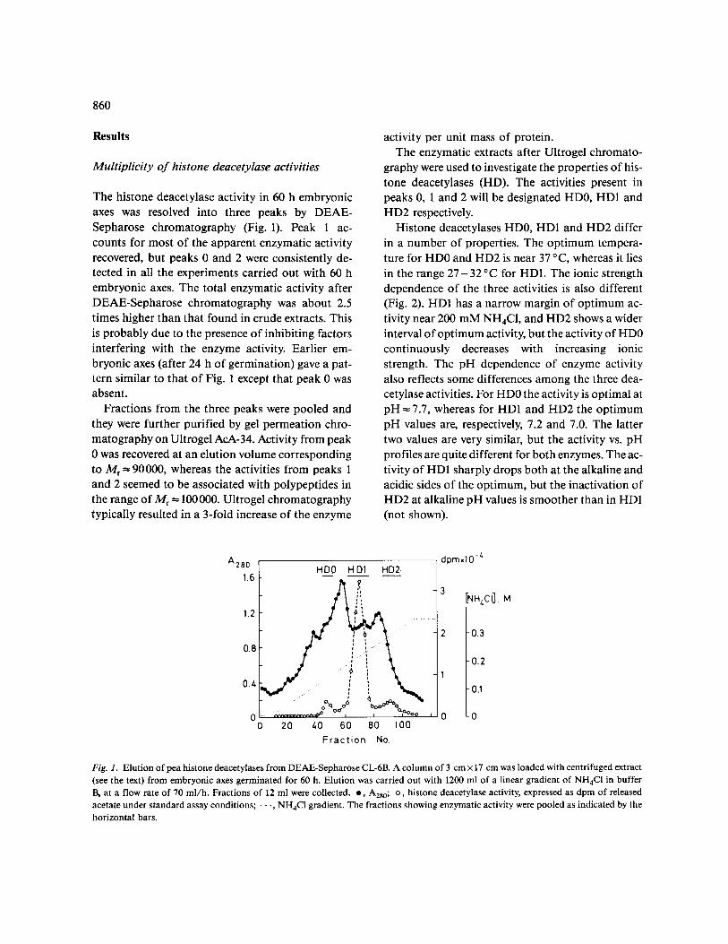
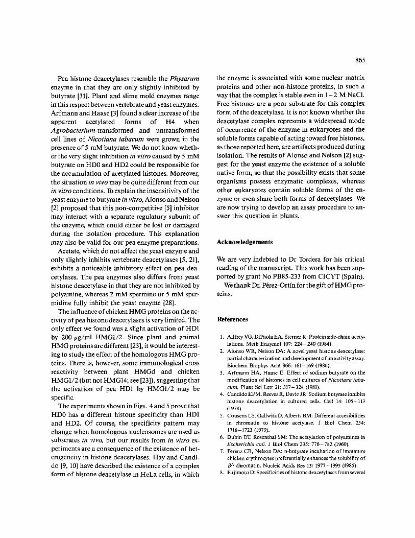
Histone deacetylases HD0, HD1 and HD2 differ in a number of properties. The optimum tempera- ture for HD0 and HD2 is near 37 °C, whereas it lies in the range 2 7 - 3 2 °C for HD1. The ionic strength dependence of the three activities is also different (Fig. 2). HD1 has a narrow margin of optimum ac- tivity near 200 mM NH4CI, and HD2 shows a wider interval of optimum activity, but the activity of HD0 continuously decreases with increasing ionic strength. The pH dependence of enzyme activity also reflects some differences among the three dea- cetylase activities. For HD0 the activity is optimal at pH = 7.7, whereas for HDI and HD2 the optimum pH values are, respectively, 7.2 and 7.0. The latter two values are very similar, but the activity vs. pH profiles are quite different for both enzymes. The ac- tivity of HD1 sharply drops both at the alkaline and acidic sides of the optimum, but the inactivation of HD2 at alkaline pH values is smoother than in HDI (not shown).
A28o 1.6
1.2
0.8
0.Z,
0 0
HD0 HD1 HD2
20 40 60 80 100 F rac t i on No.
dprn.10 -4
3 [NHz.C I], M
2 -0.3
-0.2 1
0.1
0 -0
Fig. 1. Elution of pea histone deacetylases from DEAE-Sepharose CL-6B. A column of 3 cm × 17 cm was loaded with centrifuged extract (see the text) from embryonic axes germinated for 60 h. Elution was carried out with 1200 ml of a linear gradient of NH4Cl in buffer B, at a flow rate of 70 ml/h. Fractions of 12 ml were collected, o, A280; o, histone deacetylase activity, expressed as dpm of released acetate under standard assay conditions; • •., NH4Cl gradient. The fractions showing enzymatic activity were pooled as indicated by the horizontal bars.
:>
<
.-e
100
50
0 0
, i t i ,
8 I I I I I
100 200 300 z.O0 500
[NHz C[ ] (raM)
Fig. 2. Effects o f ionic strength on the activity o f pea histone dea- cetylases. • , HD0; • , HD1; i , HD2. Act ivi tyisexpressedasper- centages relative to the max i mum activity achieved for each en- zyme in this experiment.
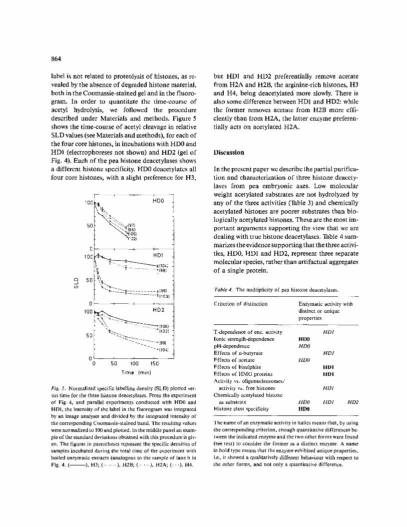
Effect of inhibitors
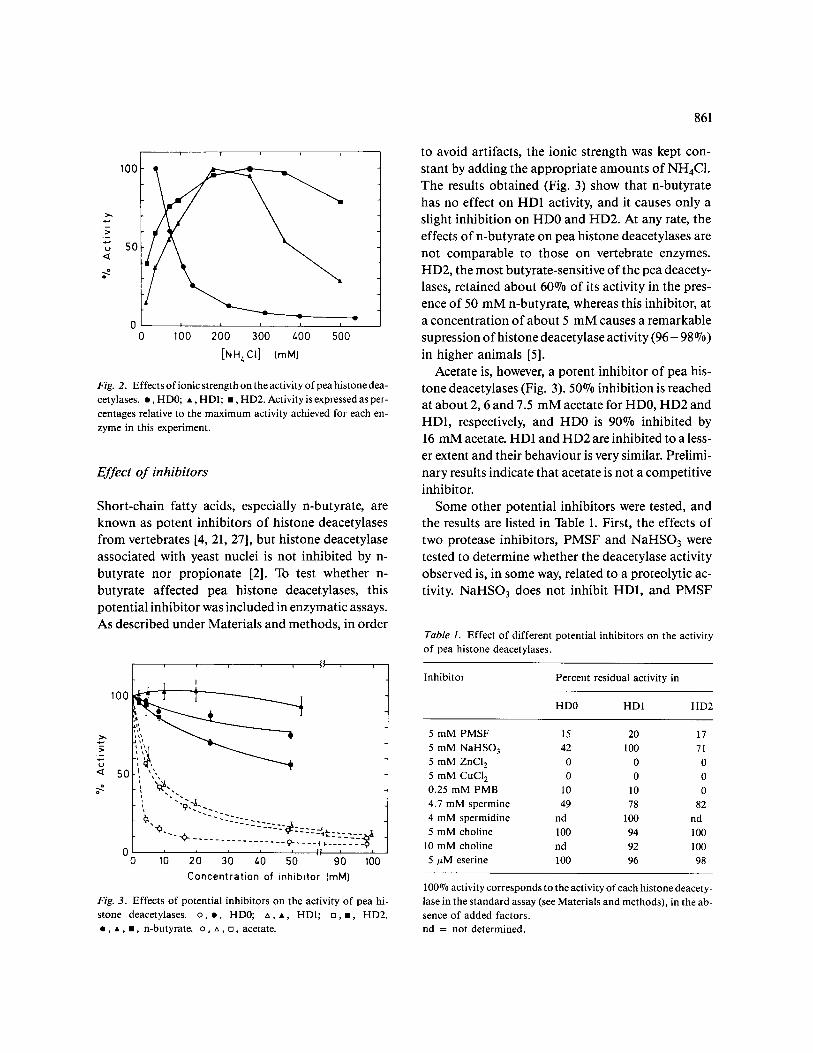
Short-chain fatty acids, especially n-butyrate, are known as potent inhibitors of histone deacetylases from vertebrates [4, 21, 27], but histone deacetylase associated with yeast nuclei is not inhibited by n- butyrate nor propionate [2]. To test whether n- butyrate affected pea histone deacetylases, this potential inhibitor was included in enzymatic assays. As described under Materials and methods, in order
o
100
50
0 0
~'x~.._.¢~_ " - -SZ- . . . . .
- y
10 20 30 Z.0 50 90 100 Concentration of inhibitor (raM)
Fig. 3. Effects of potential inhibitors on the activity of pea hi- stone deacetylases, o , o , HD0; ~x, A, HD1; [], i , HD2. • , • , • , n-butyrate, o , ~x, [], acetate.
861
to avoid artifacts, the ionic strength was kept con- stant by adding the appropriate amounts of NH4C1. The results obtained (Fig. 3) show that n-butyrate has no effect on HD1 activity, and it causes only a slight inhibition on HD0 and HD2. At any rate, the effects of n-butyrate on pea histone deacetylases are not comparable to those on vertebrate enzymes. HD2, the most butyrate-sensitive of the pea deacety- lases, retained about 60°70 of its activity in the pres- ence of 50 mM n-butyrate, whereas this inhibitor, at a concentration of about 5 mM causes a remarkable supression of histone deacetylase activity ( 9 6 - 98 070) in higher animals [5].
Acetate is, however, a potent inhibitor of pea his- tone deacetylases (Fig. 3). 50°70 inhibition is reached at about 2, 6 and 7.5 mM acetate for HD0, HD2 and HD1, respectively, and HD0 is 90°70 inhibited by 16 mM acetate. HD1 and HD2 are inhibited to a less- er extent and their behaviour is very similar. Prelimi- nary results indicate that acetate is not a competitive inhibitor.
Some other potential inhibitors were tested, and the results are listed in Table 1. First, the effects of two protease inhibitors, PMSF and NaHSO 3 were tested to determine whether the deacetylase activity observed is, in some way, related to a proteolytic ac- tivity. NaHSO3 does not inhibit HD1, and PMSF
Table 1. Effect of different potential inhibitors on the activity of pea histone deacetylases.
Inhibitor Percent residual activity in
HD0 HDI HD2
5 m M PMSF 15 20 17 5 m M NaHSO 3 42 100 71 5 mM ZnCI 2 0 0 0 5 mM CuCI 2 0 0 0 0.25 mM PMB 10 10 0 4.7 m M spermine 49 78 82 4 m M spermidine nd 100 nd 5 mM choline 100 94 100
10 mM choline nd 92 100 5/~M eserine 100 96 98
100°70 activity corresponds to the activity of each histone deacety- lase in the s tandard assay (see Materials and methods), in the ab- sence of added factors. n d = not determined.
862
causes a remarkable inhibition of the three activities. However, the possibility that the release of the radio- label were due to the hydrolysis of peptide rather that acetamide bonds can be ruled out. The assay condi- tions, which include an extraction step with ethyl acetate at acidic pH, would preclude the coextrac- tion of positively charged peptides together with the unionized form of acetate. Further evidence against proteolysis will be provided by the experiments of Figs. 4 and 5. It can be seen that 5 mM ZnCI 2 or CuCI2 completely repress the three activities and mercurials, such as PMB, also have a clear effect. A similar result was found in the early studies of Inoue and Fujimoto [12], working with the calf thymus en- zyme. This suggest that the enzymes possess essen- tial thiol groups. Spermine is only a weak inhibitor o f pea histone deacetylases, if compared with the ef- fect o f the polyamine on the yeast enzyme [28]. The activity of the latter is completely abolished at about 2 mM spermine, whereas pea HD0 is only 50°7o in-
hibited by 4.7 mM polyamine, its effect on HD1 and HD2 being still less pronounced. Our finding with spermidine is even more remarkable, as this polya- mine, which is a potent inhibitor of histone deacety- lases from yeast [28] and diminishes as well the ac- tivity of deacetylases from higher organisms [15], has no effect on pea HD1.
We also tested the effects of choline and eserine on pea histone deacetylases, because it has been sug- gested that acetylcholinesterase may possess some histone deacetylase activity [1]. As shown by the negative results in Table 1, this is probably not the case for pea H D enzymes.
The question of whether non-histone H M G (high mobility group) proteins alter the activity of histone deacetylases is rather controversial. Reeves and Can- dido [19] suggested that HMG1 and HMG2 activate and HMG14 and HMG17 inhibit histone deacetylase in vitro, but Mezquita et al. [18], in a different sys- tem, found that HMG17 does not inhibit histone deacetylase, which was stimulated by ubiquitin. To test whether these proteins had any effect on the pea enzymes, chicken HMG1/2 and HMG14, prepared according to Walker and Johns [29], were added to the deacetylase assay mixture. Neither HMG1/2 nor HMG14 (200/~g/ml) had any effect on HD0 and HD2. HD1 activity was only slightly enhanced
( < 10%) by HMG14 (200/~g/ml). HMG1, below 100/zg/ml, did not affect HD1 activity, but at 200/~g/ml causes a 50070 activation. Ubiquitin (200/xg/ml) had no effect on HD0 and HD2 activi- ties, and HD1 was activated only to a small extent ( = 10°7o).
Histone deacetylation in nucleosomes
The three pea histone deacetylases are able to dea- cetylate his'tones when using nucleosomes as sub- strate. Chicken nucleosomes, containing labelled hi- stones, were prepared by incubation of nuclei with [3H]-acetate and used in the enzymatic assay as described under Materials and methods. The results of a typical experiment are given in Table 2. The three enzymatic preparations are able to accept oligonucleosomes as substrate, although they prefer the free forms of the histones. This preference for free histones is more marked in HD0 and HD2.
Substrate specificity of pea histone deacetylases
In addition to "biologically" acetylated chicken hi- stones, either free or incorporated into oligonucleo-
Table 2. Nucleosome deacetylating activity of pea histone de-
acetylases.
Substrate Deacetylase activity with
HD0 HD1 HD2
Chicken free histones (biologically labelled) 14776+_224 8171 +250 2380+ 165
Chicken oligonucleo- somes 2057+ 53 3839+207 400_+ 108
(Activity toward oligo- nucleosomes/activity toward free histones) x100 14 46 17
Activities are given as dpm of released acetate in the presence of 40 mM NH4C1. In every case, the activity of input histones (either free or in nucleosomes, see Materials and methods) was 90000 dpm.
863
Table3. Activity of pea histone deacetylases towards different
acetylated substrates.
Substrate Deacetylase activity with
HD0 HDI HD2
Chicken erythrocyte free histones, biologically labelled 100 100 100
Chicken erythrocyte free histones, chemically labelled 69 44 22
Pea free histones, chemically labelled 30 15 6
Acetylspermine 0 1 0 Acetylputrescine 0 0 0
As the specific activity o f each substrate was different, released acetate was first evaluated as percentages relative to the input dpm in the assay, and these figures were then recalculated, for each of the three enzymatic activities relative to 100 for biologi- cally acetylated chicken histones.
somes, some other acetylated compounds were test- ed as substrates. The results obtained are listed in Table 3. It is noteworthy that chemically acetylated histones, obtained by treating the protein with labelled acetic anhydride (see Materials and methods) are poorer substrates than biologically acetylated histones. This is true even when using homologous, pea histones. There is an obvious ex-
planation for this result, which was previously sug- gested by Inoue and Fujimoto [11] in their pioneer- ing work on calf thymus histone deacetylase: whereas acetyltransferase-catalyzed in vivo acetyla- tion of histones results in the modification of e- NH 2 groups of specific lysyl residues for each core histone class [33], chemical acetylation with acetic anhydride may result in a more extensive modifica- tion, even afecting H1 and the N-terminal groups of H2B and H3. These extra acetylated groups would not be recognized by histone deacetylases. In the light of this interpretation, our preparations may be considered as true histone deacetylases, capable of discriminating between biologically and chemically acetylated histones. Finally, Table 3 shows that acetyl polyamines are not deacetylated at all.
At the present time, the available data indicate that, at least H4 [20] and H3 [2] are acetylated in pea and all four core histones are acetylated in higher animals. To determine whether our HD preparations react differentially with the acetylated four core his- tones, we performed a series of experiments, one of which is shown in Fig. 4. It is obvious from this figure, that incubation of [3H]-acetylated histones with HD2 resulted in a preferential cleavage of acetyl groups from H2A and H2B. In addition, these results clearly show that the removal of the radio-
Fig. 4. Specificity of histone deacetylation by HD2. Chicken histones, biologically labelled with [3H]-acetate, were incubated with a puri- fied preparation of HD2. Aliquots from the reaction mixture were taken at several times, the histones recovered and electrophoresed. Panel A shows the Coomassie-stained gel and panel B the corresponding fluorogram. In lanes a and i unlabelled chicken histones were run. The aliquots were taken at: b, 0; c, 10; d, 20; e, 40; f, 80 and g, 160 min. The progress of enzymatic deacetylation is easily seen by the disappearance of labelled acetate (panel B), especially in H2A and H2B. Lane h shows the result of incubating the histones for 160 min with an enzymatic extract boiled to inactivate histone deacetylase activity. No removal of acetate was apparent under these circumstances.
864
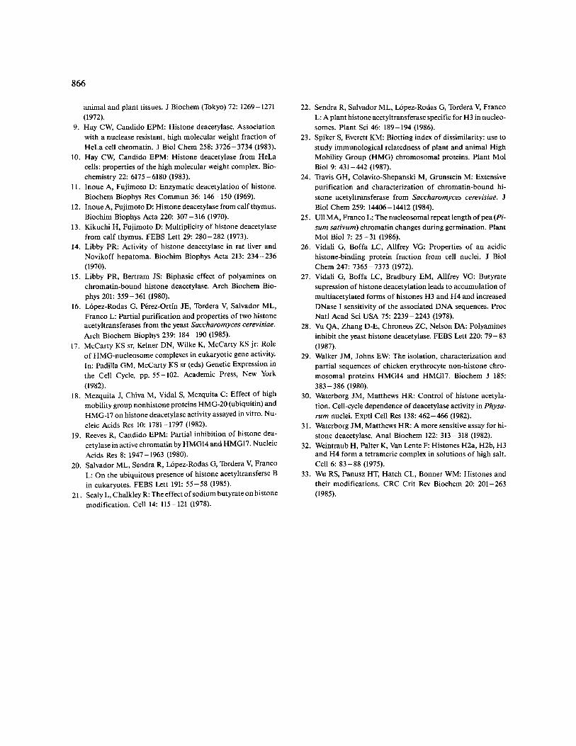
label is not related to proteolysis of histones, as re- vealed by the absence of degraded histone material, both in the Coomassie-stained gel and in the fluoro- gram. In order to quantitate the time-course of acetyl hydrolysis, we followed the procedure described under Materials and methods. Figure 5 shows the time-course of acetyl cleavage in relative SLD values (see Materials and methods), for each of the four core histones, in incubations with HD0 and HD1 (electrophoreses not shown) and HD2 (gel of Fig. 4). Each of the pea histone deacetylases shows a different histone specificity. HD0 deacetylates all four core histones, with a slight preference for H3,
100
50
0 100
ca 50 J O3
0
100
50
0 0
HDO
~ ; ? ' " "~ ,,,(9 7)
"(102)
....... { ................................ ~(98)
~" "~" .... '96' " "~- - - -'-'_ .'~-~ . . . . . . . . . . . . . . . . . . t ~
. . . . . . . . . . . . . ~(103) P I I
HD2
~ 1 1 o 6 1 • '"'" (102)
• .,~:.,¢ ~.::,, . . . . .
........ '--~.? . . . . . . (99) . . . . • U04)
i i i
50 100 150
Time (rain)
Fig . 5 . Normalized specific labelling density (SLD) plotted ver- sus time for the three histone deacetylases. From the experiment of Fig. 4, and parallel experiments conducted with HD0 and HD1, the intensity of the label in the fluorogram was integrated by an image analyser and divided by the integrated intensity of the corresponding Coomassie-stained band. The resulting values were normalized to 100 and plotted. In the middle panel an exam- ple of the standard deviations obtained with this procedure is giv- en. The figures in parentheses represent the specific densities of samples incubated during the total time of the experiment with boiled enzymatic extracts (analogous to the sample of lane h in Fig. 4. ( ), H3; ( - - - ) , H2B; ( - • - ) , H2A; ( . . . ) , H4.
but HD1 and HD2 preferentially remove acetate from H2A and H2B, the arginine-rich histones, H3 and H4, being deacetylated more slowly. There is also some difference between HD1 and HD2: while the former removes acetate from H2B more effi- ciently than from H2A, the latter enzyme preferen- tially acts on acetylated H2A.
Discussion
In the present paper we describe the partial purifica- tion and characterization of three histone deacety- lases from pea embryonic axes. Low molecular weight acetylated substrates are not hydrolyzed by any of the three activities (Table 3) and chemically acetylated histones are poorer substrates than bio- logically acetylated histones. These are the most im- portant arguments supporting the view that we are dealing with true histone deacetylases. Table 4 sum- marizes the evidence supporting that the three activi- ties, HD0, HD1 and HD2, represent three separate molecular species, rather than artifactual aggregates of a single protein.
T a b l e 4. The multiplicity of pea histone deacetylases.
Criterion of distinction Enzymatic activity with distinct or unique properties
T-dependence of enz. activity H D 1
Ionic strength-dependence HD0 pH-dependence H D O
Effects of n-butyrate H D 1
Effects of acetate H D O
Effects of bisulphite HD1 Effects of HMG proteins HD1 Activity vs. oligonucleosomes/
activity vs. free histones H D 1
Chemically acetylated histone as substrate H D O H D 1
Histone class specificity HD0 H D 2
The name of an enzymatic activity in italics means that, by using the corresponding criterion, enough quantitative differences be- tween the indicated enzyme and the two other forms were found (see text) to consider the former as a distinct enzyme. A name in bold type means that the enzyme exhibited unique properties, i.e., it showed a qualitatively different behaviour with respect to the other forms, and not only a quantitative difference.
Pea histone deacetylases resemble the Physarum
enzyme in that they are only slightly inhibited by butyrate [31]. Plant and slime mold enzymes range in this respect between vertebrate and yeast enzymes. Arfmann and Haase [3] found a clear increase of the
apparent acetylated forms of H4 when Agrobacter ium- trans formed and untransformed cell lines of Nicot iana tabacum were grown in the presence of 5 mM butyrate. We do not know wheth- er the very slight inhibition in vitro caused by 5 mM butyrate on HD0 and HD2 could be responsible for the accumulation of acetylated histones. Moreover,
the situation in vivo may be quite different from our in vitro conditions. To explain the insensitivity of the yeast enzyme to butyrate in vitro, Alonso and Nelson [2] proposed that this non-competitive [5] inhibitor may interact with a separate regulatory subunit of the enzyme, which could either be lost or damaged during the isolation procedure. This explanation may also be valid for our pea enzyme preparations.
Acetate, which do not affect the yeast enzyme and only slightly inhibits vertebrate deacetylases [5, 21], exhibits a noticeable inhibitory effect on pea dea-
cetylases. The pea enzymes also differs from yeast histone deacetylase in that they are not inhibited by polyamine, whereas 2 mM spermine or 5 mM sper- midine fully inhibit the yeast enzyme [28].
The influence of chicken HMG proteins on the ac- tivity of pea histone deacetylases is very limited. The only effect we found was a slight activation of HD1 by 200/~g/ml HMG1/2. Since plant and animal HMG proteins are different [23], it would be interest- ing to study the effect of the homologous HMG pro- teins. There is, however, some immunological cross reactivity between plant HMGd and chicken HMG1/2 (but not HMG14; see [23]), suggesting that the activation of pea HD1 by H M G I / 2 may be specific.
The experiments shown in Figs. 4 and 5 prove that HD0 has a different histone specificity than HD1 and HD2. Of course, the specificity pattern may change when homologous nucleosomes are used as substrates in vivo, but our results from in vitro ex- periments are a consequence of the existence of het- erogeneity in histone deacetylases. Hay and Candi- do [9, 10] have described the existence of a complex form of histone deacetylase in HeLa cells, in which
865
the enzyme is associated with some nuclear matrix
proteins and other non-histone proteins, in such a way that the complex is stable even in 1 - 2 M NaC1. Free histories are a poor substrate for this complex form of the deacetylase. It is not known whether the deacetylase complex represents a widespread mode of occurrence of the enzyme in eukaryotes and the soluble forms capable of acting toward free histones, as those reported here, are artifacts produced during isolation. The results of Alonso and Nelson [2] sug- gest for the yeast enzyme the existence of a soluble native form, so that the possibility exists that some organisms possess enzymatic complexes, whereas other eukaryotes contain soluble forms of the en-
zyme or even share both forms of deacetylases. We are now trying to develop an assay procedure to an- swer this question in plants.
Acknowledgements
We are very indebted to Dr Tordera for his critical reading of the manuscript. This work has been sup- ported by grant No PB85-233 from CICYT (Spain).
We thank Dr. P6rez-Ortln for the gift of HMG pro- teins.
References
1. Allfrey VG, DiPaola EA, Sterner R: Protein side-chain acety- lations. Meth Enzymol 107:224-240 (1984).
2. Alonso WR, Nelson DA: A novel yeast histone deacetylase: partial characterization and development of an activity assay. Biochem Biophys Acta 866:161-169 (1986).
3. Arfmann HA, Haase E: Effect of sodium butyrate on the modification of histories in cell cultures of Nicotiana taba- curn. Plant Sci Lett 21:317-324 (1981).
4. CandidoEPM, Reeves R, Davie JR: Sodium butyrateinhibits histone deacetylation in cultured cells. Cell 14:105-113 (1978).
5. Cousens LS, Gallwitz D, Alberts BM: Different accesibilities in chromatin to histone acetylase. J Biol Chem 254: 1716-1723 (1979).
6. Dubin DT, Rosenthal SM: The acetylation of polyamines in Escherichia coli. J Biol Chem 235:776-782 (1960).
7. Ferenz CR, Nelson DA: n-butyrate incubation of immature chicken erythrocytes preferentially enhances the solubility of /3 A chromatin. Nucleic Acids Res 13:1977-1995 (1985).
8. Fujimoto D: Specificities of histone deacetylases from several
866
animal and plant tissues. J Biochem (Tokyo) 72: 1269-1271 (1972).
9. Hay CW, Candido EPM: Histone deacetylase. Association with a nuclease resistant, high molecular weight fraction of HeLa cell chromatin. J Biol Chem 258: 3726- 3734 (1983).
10. Hay CW, Candido EPM: Histone deacetylase from HeLa cells: properties of the high molecular weight complex. Bio- chemistry 22:6175-6180 (1983).
11. Inoue A, Fujimoto D: Enzymatic deacetylation of histone. Biochem Biophys Res Commun 36:146-150 (1969).
12. Inoue A, Fujimoto D: Histone deacetylase from calf thymus. Biochim Biophys Acta 220:307-316 (1970).
13. Kikuchi H, Fujimoto D: Multiplicity of histone deacetylase from calf thymus. FEBS Lett 29:280-282 (1973).
14. Libby PR: Activity of histone deacetylase in rat liver and Novikoff hepatoma. Biochim Biophys Acta 213:234-236 (1970).
15. Libby PR, Bertram JS: Biphasic effect of polyamines on chromatin-bound histone deacetylase. Arch Biochem Bio- phys 201:359-361 (1980).
16. L6pez-Rodas G, P6rez-Ortln JE, Tordera V, Salvador ML, Franco L: Partial purification and properties of two histone acetyltransferases from the yeast Saccharomyces cerevisiae. Arch Biochem Biophys 239:184-190 (1985).
17. McCarty KS sr, Kelner DN, Wilke K, McCarty KS jr: Role of HMG-nucleosome complexes in eukaryotic gene activity. In: Padilla GM, McCarty KS sr (eds) Genetic Expression in the Cell Cycle, pp. 55-102. Academic Press, New York
(1982). 18. Mezquita J, Chiva M, Vidal S, Mezquita C: Effect of high
mobility group nonhistone proteins HMG-20 (uhiquitin) and HMG-17 on histone deacetylase activity assayed in vitro. Nu- cleic Acids Res 10:1781-1797 (1982).
19. Reeves R, Candido EPM: Partial inhibition of histone dea- cetylase in active chromatin by HMG14 and HMG17. Nucleic Acids Res 8:1947-1963 (1980).
20. Salvador ML, Sendra R, L6pez-Rodas G, Tordera V, Franco L: On the ubiquitous presence of histone acetyltransferse B in eukaryotes. FEBS Lett 191:55-58 (1985).
21. Sealy L, Chalkley R: The effect of sodium butyrate on histone modification. Cell 14: 115-121 (1978).
22. Sendra R, Salvador ML, L6pez-Rodas G, Tordera V, Franco L: A plant histone acetyltransferase specific for H3 in nucleo- somes. Plant Sci 46:189-194 (1986).
23. Spiker S, Everett KM: Blotting index of dissimilarity: use to study immunological relatedness of plant and animal High Mobility Group (HMG) chromosomal proteins. Plant Mol Biol 9:431-442 (1987).
24. Travis GH, Colavito-Shepanski M, Grunstein M: Extensive purification and characterization of chromatin-bound hi- stone acetyltransferase from Saccharornyces cerevisiae. J Biol Chem 259:14406-14412 (1984).
25. Ull MA, Franco L: The nucleosomal repeat length of pea (Pi- sum sativum) chromatin changes during germination. Plant Mol Biol 7:25-31 (1986).
26. Vidali G, Boffa LC, Allfrey VG: Properties of an acidic histone-binding protein fraction from cell nuclei. J Biol Chem 247:7365-7373 (1972).
27. Vidali G, Boffa LC, Bradbury EM, Allfrey VG: Butyrate supression of histone deacetylation leads to accumulation of multiacetylated forms of histones H3 and H4 and increased DNase I sensitivity of the associated DNA sequences. Proc Natl Acad Sci USA 75:2239-2243 (1978).
28. Vu QA, Zhang D-E, Chroneos ZC, Nelson DA: Polyamines inhibit the yeast histone deacetylase. FEBS Lett 220: 79-83 (1987).
29. Walker JM, Johns EW: The isolation, characterization and partial sequences of chicken erythrocyte non-histone chro- mosomal proteins HMG14 and HMG17. Biochem J 185: 383-386 0980).
30. Waterborg JM, Matthews HR: Control of histone acetyla- tion. Cell-cycle dependence of deacetylase activity in Physa- rum nuclei. Exptl Cell Res 138:462-466 (1982).
31. Waterborg JM, Matthews HR: A more sensitive assay for hi- stone deacetylase. Anal Biochem 122:313-318 (1982).
32. Weintraub H, Palter K, Van Lente F: Histones H2a, H2b, H3 and H4 form a tetrameric complex in solutions of high salt. Cell 6 :83-88 (1975).
33. Wu RS, Panusz HT, Hatch CL, Bonner WM: Histones and their modifications. CRC Crit Rev Biochem 20:201-263 (1985).
Related Documents