molecules Communication Characterization of New Bioactive Enzyme Inhibitors from Endophytic Bacillus amyloliquefaciens RWL-1 Raheem Shahzad 1 , Abdul Latif Khan 2 , Liaqat Ali 3 , Saqib Bilal 1 , Muhammad Imran 1 , Kyung-Sook Choi 4 , Ahmed Al-Harrasi 2 and In-Jung Lee 1, * 1 School of Applied Biosciences, Kyungpook National University, Daegu 41566, Korea; [email protected] (R.S.); [email protected] (S.B.); [email protected] (M.I.) 2 UoN Chair of Oman’s Medicinal Plants & Marine Natural Products, University of Nizwa, Nizwa 616, Oman; [email protected] (A.L.K.); [email protected] (A.A.-H.) 3 Department of Chemistry, University of Sargodha, Sub-Campus Mianwali, Mianwali 42200, Pakistan; [email protected] 4 School of Agricultural Civil & Bio-Industrial Machinery Engineering, Kyungpook National University, Daegu 41566, Korea; [email protected] * Correspondence: [email protected]; Tel.: +82-53-950-5708; Fax: +82-53-958-6880 Received: 24 November 2017; Accepted: 4 January 2018; Published: 5 January 2018 Abstract: Endophytic bacteria are known to produce a wide array of bioactive secondary metabolites with beneficial effects on human health. In the current study, a novel endophytic bacterial strain, Bacillus amyloliquefaciens RWL-1, was isolated from the seeds of Oryza sativa. Initially, the crude extract of RWL-1 was assessed for potential biological effects of enzyme inhibition and cytotoxicity and was found to exhibit a broad spectrum inhibition for α-glucosidase (37 ± 0.09%) and urease (49.4 ± 0.53%). The screening results were followed by bioassay-guided isolation of secondary metabolite(s) from RWL-1. Extensive chromatographic and spectrophotometry analyses revealed the presence of compound 1 (S)-2-hydroxy-N-((S)-1-((S)-8-hydroxy-1-oxoisochroman-3-yl)-3-methylbutyl)-2- ((S)-5-oxo-2,5-dihydrofuran-2-yl)acetamide. Further bioassays of compound 1 showed significant inhibition of α-glucosidase (52.98 ± 0.8%) and urease (51.27 ± 1.0%), compared with positive control values of 79.14 ± 1.9% and 88.24 ± 2.2%, and negative controls (0.08 ± 0.1% and 0.05 ± 0.01%), respectively. The current study suggests that bacterial endophytes are a rich source of novel bioactive compounds with high therapeutic value. Keywords: endophytes; biological potential; secondary metabolites; α-glucosidase; urease 1. Introduction The term “endophyte” refers to microorganisms (e.g., bacteria, fungi) that live inside plant tissues without causing apparent diseases and have the ability to colonize the internal tissues of the plant [1,2]. Endophytes are pervasive in all plant parts and have been isolated from different plant parts [3]. They form mutualistic relationships with host plants, which are either obligatory or facultative associations, and do not damage the host plant; however, this mutualistic interaction also depends on many factors and can change over time [1,3–5]. In this extended symbiotic relationship, the plants provide shelter, protection, and access to essential nutrients; in return, the endophytes generate a beneficial correlation with the host through the modulation of endogenous phytohormones and nutrients, and improve the adaptability of plants to rapidly changing environments [6,7]. Although endophytes are present in all plant species, endophyte—host interaction has not been completely explored. This may include competitiveness in the host tissues mediated by the secretion of secondary metabolites and the detoxification of such inhibitors delivered by endophytes [8,9]. Molecules 2018, 23, 114; doi:10.3390/molecules23010114 www.mdpi.com/journal/molecules

Welcome message from author
This document is posted to help you gain knowledge. Please leave a comment to let me know what you think about it! Share it to your friends and learn new things together.
Transcript
molecules
Communication
Characterization of New Bioactive Enzyme Inhibitorsfrom Endophytic Bacillus amyloliquefaciens RWL-1
Raheem Shahzad 1, Abdul Latif Khan 2, Liaqat Ali 3, Saqib Bilal 1, Muhammad Imran 1,Kyung-Sook Choi 4, Ahmed Al-Harrasi 2 and In-Jung Lee 1,*
1 School of Applied Biosciences, Kyungpook National University, Daegu 41566, Korea;[email protected] (R.S.); [email protected] (S.B.); [email protected] (M.I.)
2 UoN Chair of Oman’s Medicinal Plants & Marine Natural Products, University of Nizwa,Nizwa 616, Oman; [email protected] (A.L.K.); [email protected] (A.A.-H.)
3 Department of Chemistry, University of Sargodha, Sub-Campus Mianwali, Mianwali 42200, Pakistan;[email protected]
4 School of Agricultural Civil & Bio-Industrial Machinery Engineering, Kyungpook National University,Daegu 41566, Korea; [email protected]
* Correspondence: [email protected]; Tel.: +82-53-950-5708; Fax: +82-53-958-6880
Received: 24 November 2017; Accepted: 4 January 2018; Published: 5 January 2018
Abstract: Endophytic bacteria are known to produce a wide array of bioactive secondary metaboliteswith beneficial effects on human health. In the current study, a novel endophytic bacterial strain,Bacillus amyloliquefaciens RWL-1, was isolated from the seeds of Oryza sativa. Initially, the crude extractof RWL-1 was assessed for potential biological effects of enzyme inhibition and cytotoxicity and wasfound to exhibit a broad spectrum inhibition for α-glucosidase (37 ± 0.09%) and urease (49.4 ± 0.53%).The screening results were followed by bioassay-guided isolation of secondary metabolite(s)from RWL-1. Extensive chromatographic and spectrophotometry analyses revealed the presenceof compound 1 (S)-2-hydroxy-N-((S)-1-((S)-8-hydroxy-1-oxoisochroman-3-yl)-3-methylbutyl)-2-((S)-5-oxo-2,5-dihydrofuran-2-yl)acetamide. Further bioassays of compound 1 showed significantinhibition of α-glucosidase (52.98 ± 0.8%) and urease (51.27 ± 1.0%), compared with positive controlvalues of 79.14 ± 1.9% and 88.24 ± 2.2%, and negative controls (0.08 ± 0.1% and 0.05 ± 0.01%),respectively. The current study suggests that bacterial endophytes are a rich source of novel bioactivecompounds with high therapeutic value.
Keywords: endophytes; biological potential; secondary metabolites; α-glucosidase; urease
1. Introduction
The term “endophyte” refers to microorganisms (e.g., bacteria, fungi) that live inside planttissues without causing apparent diseases and have the ability to colonize the internal tissues ofthe plant [1,2]. Endophytes are pervasive in all plant parts and have been isolated from differentplant parts [3]. They form mutualistic relationships with host plants, which are either obligatory orfacultative associations, and do not damage the host plant; however, this mutualistic interaction alsodepends on many factors and can change over time [1,3–5]. In this extended symbiotic relationship,the plants provide shelter, protection, and access to essential nutrients; in return, the endophytesgenerate a beneficial correlation with the host through the modulation of endogenous phytohormonesand nutrients, and improve the adaptability of plants to rapidly changing environments [6,7]. Althoughendophytes are present in all plant species, endophyte—host interaction has not been completelyexplored. This may include competitiveness in the host tissues mediated by the secretion of secondarymetabolites and the detoxification of such inhibitors delivered by endophytes [8,9].
Molecules 2018, 23, 114; doi:10.3390/molecules23010114 www.mdpi.com/journal/molecules
Molecules 2018, 23, 114 2 of 10
In addition to plant-microbe interactions, endophytes display many important biologicalactivities [6,10]. Among endophytes, many endophytic bacteria are known to produce a diverserange of bioactive and health-promoting compounds, such as volatile organic compounds, antibiotics,immunosuppressant compounds, and anticancer, antiviral and antifungal drugs [11–13]. More than60% of anticancer drugs in clinical use are natural products [14]. The first study of the endophyticbacteria Bacillus amyloliquefaciens afforded the discovery of an antitumor exopolysaccharide derivedfrom the Bacillus genus [15]. In addition to antitumor compounds, the endophytes belonging to theBacillus genus produce a variety of secondary metabolites; for example, B. thuringiensis produces theanti-infective compound anthracene [16] and B. subtilis produces the antimicrobial protein YbdN [17].These results indicate the promising medicinal potential of the Bacillus genus. Owing to theirinvolvement in phytostimulation, disease suppression, and other biological activities, Bacillus sp.are preferred because their long-term viability can facilitate the development of natural productssuitable for commercial use [18].
B. amyloliquefaciens strains have attracted considerable interest because they are able to producea wide range of active antimicrobial compounds, macrolactins, lipopeptides, hydrolytic enzymes, andcertain volatile compounds. For example, B. amyloliquefaciens FZB42 has 8.5% of its genome dedicated tothe synthesis of secondary metabolites [19], allowing the production of lipopeptides, surfactin, fengycin,bacillomycin D, polyketide (difficidin), dipeptide bacilysin, chitin, and colloidal chitin [20,21].
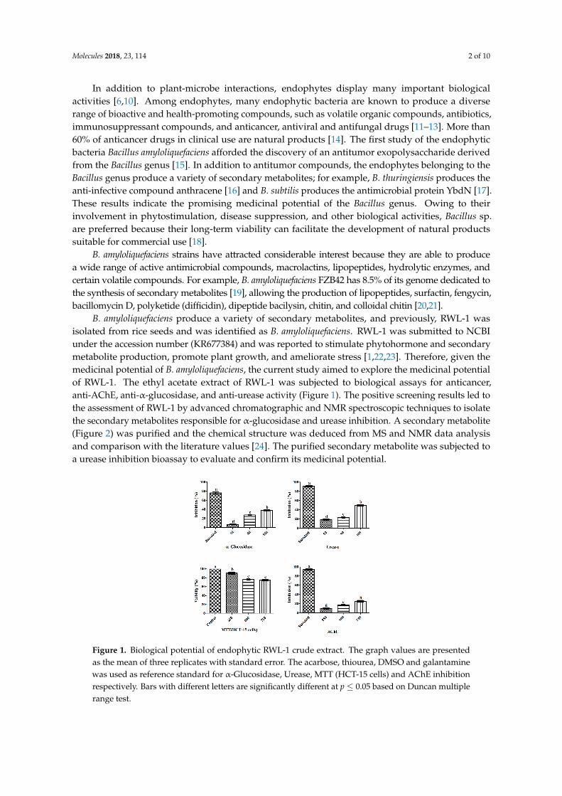
B. amyloliquefaciens produce a variety of secondary metabolites, and previously, RWL-1 wasisolated from rice seeds and was identified as B. amyloliquefaciens. RWL-1 was submitted to NCBIunder the accession number (KR677384) and was reported to stimulate phytohormone and secondarymetabolite production, promote plant growth, and ameliorate stress [1,22,23]. Therefore, given themedicinal potential of B. amyloliquefaciens, the current study aimed to explore the medicinal potentialof RWL-1. The ethyl acetate extract of RWL-1 was subjected to biological assays for anticancer,anti-AChE, anti-α-glucosidase, and anti-urease activity (Figure 1). The positive screening results led tothe assessment of RWL-1 by advanced chromatographic and NMR spectroscopic techniques to isolatethe secondary metabolites responsible for α-glucosidase and urease inhibition. A secondary metabolite(Figure 2) was purified and the chemical structure was deduced from MS and NMR data analysisand comparison with the literature values [24]. The purified secondary metabolite was subjected toa urease inhibition bioassay to evaluate and confirm its medicinal potential.
Figure 1. Biological potential of endophytic RWL-1 crude extract. The graph values are presentedas the mean of three replicates with standard error. The acarbose, thiourea, DMSO and galantaminewas used as reference standard for α-Glucosidase, Urease, MTT (HCT-15 cells) and AChE inhibitionrespectively. Bars with different letters are significantly different at p ≤ 0.05 based on Duncan multiplerange test.
Molecules 2018, 23, 114 3 of 10
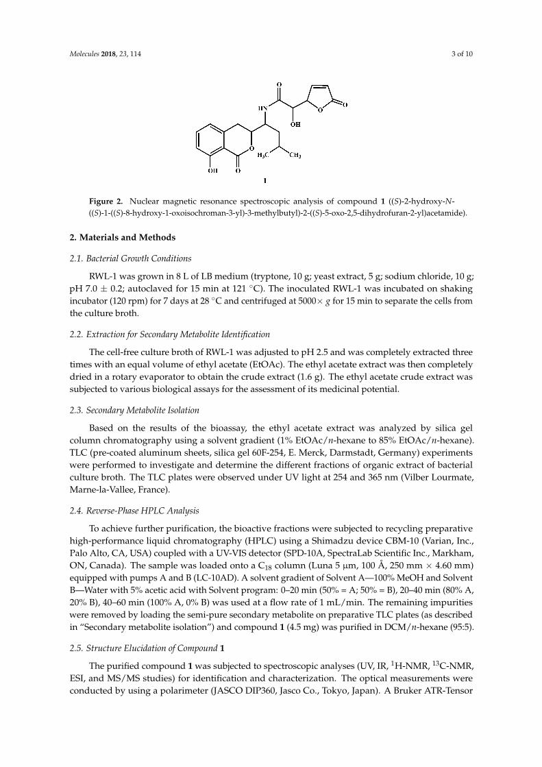
Figure 2. Nuclear magnetic resonance spectroscopic analysis of compound 1 ((S)-2-hydroxy-N-((S)-1-((S)-8-hydroxy-1-oxoisochroman-3-yl)-3-methylbutyl)-2-((S)-5-oxo-2,5-dihydrofuran-2-yl)acetamide).
2. Materials and Methods
2.1. Bacterial Growth Conditions
RWL-1 was grown in 8 L of LB medium (tryptone, 10 g; yeast extract, 5 g; sodium chloride, 10 g;pH 7.0 ± 0.2; autoclaved for 15 min at 121 ◦C). The inoculated RWL-1 was incubated on shakingincubator (120 rpm) for 7 days at 28 ◦C and centrifuged at 5000× g for 15 min to separate the cells fromthe culture broth.
2.2. Extraction for Secondary Metabolite Identification
The cell-free culture broth of RWL-1 was adjusted to pH 2.5 and was completely extracted threetimes with an equal volume of ethyl acetate (EtOAc). The ethyl acetate extract was then completelydried in a rotary evaporator to obtain the crude extract (1.6 g). The ethyl acetate crude extract wassubjected to various biological assays for the assessment of its medicinal potential.
2.3. Secondary Metabolite Isolation
Based on the results of the bioassay, the ethyl acetate extract was analyzed by silica gelcolumn chromatography using a solvent gradient (1% EtOAc/n-hexane to 85% EtOAc/n-hexane).TLC (pre-coated aluminum sheets, silica gel 60F-254, E. Merck, Darmstadt, Germany) experimentswere performed to investigate and determine the different fractions of organic extract of bacterialculture broth. The TLC plates were observed under UV light at 254 and 365 nm (Vilber Lourmate,Marne-la-Vallee, France).
2.4. Reverse-Phase HPLC Analysis
To achieve further purification, the bioactive fractions were subjected to recycling preparativehigh-performance liquid chromatography (HPLC) using a Shimadzu device CBM-10 (Varian, Inc.,Palo Alto, CA, USA) coupled with a UV-VIS detector (SPD-10A, SpectraLab Scientific Inc., Markham,ON, Canada). The sample was loaded onto a C18 column (Luna 5 µm, 100 Å, 250 mm × 4.60 mm)equipped with pumps A and B (LC-10AD). A solvent gradient of Solvent A—100% MeOH and SolventB—Water with 5% acetic acid with Solvent program: 0–20 min (50% = A; 50% = B), 20–40 min (80% A,20% B), 40–60 min (100% A, 0% B) was used at a flow rate of 1 mL/min. The remaining impuritieswere removed by loading the semi-pure secondary metabolite on preparative TLC plates (as describedin “Secondary metabolite isolation”) and compound 1 (4.5 mg) was purified in DCM/n-hexane (95:5).
2.5. Structure Elucidation of Compound 1
The purified compound 1 was subjected to spectroscopic analyses (UV, IR, 1H-NMR, 13C-NMR,ESI, and MS/MS studies) for identification and characterization. The optical measurements wereconducted by using a polarimeter (JASCO DIP360, Jasco Co., Tokyo, Japan). A Bruker ATR-Tensor
Molecules 2018, 23, 114 4 of 10
37 spectrophotometer (Bruker, Ettlingen, Germany) was used to record the IR spectra. To obtain theESI mass spectra, a QSTAR mass spectrometer (Applied Biosystems, Foster, CA, USA) with capillaryvoltage of 5–5.5 kV was used. The NMR spectra (1H and 13C) were obtained by using a Bruker NMRspectrometer (Burker, Fallanden, Switzerland) operated at 600 MHz and 150 MHz, respectively.
2.6. In Vitro Biological Activities of RWL-1 Crude Extract
2.6.1. Urease Inhibition
The inhibitory effects of the ethyl acetate crude extract and compound 1 on urease activity wasmeasured in accordance with the method of [25]. Briefly, 100 mM urea (0.055 mL) in 8.2 pH phosphatebuffer containing 0.01 M LiCl2, 0.1 mM EDTA, and 0.01 M K2HPO4·3H2O was reacted with 3 units/mLjack bean urease (0.025 mL) (Sigma, Munich, Germany) and various concentrations of crude extract(10–100 µg/mL) in a 96-well plate for 15 min at 37 ◦C. The urease inhibitory activity was evaluatedthrough the measurement of ammonia production by the indophenol blue method. The absorbancewas measured at 630 nm and thiourea was used as the standard inhibitor. The inhibition percentagewas calculated from the following equation:
Inhibition (%) = 100 − (ODtest/ODcontrol) × 100 (1)
2.6.2. α-Glucosidase Inhibition
The inhibitory effects of the ethyl acetate crude extract and compound 1 on α-glucosidase activitywere measured in accordance with the method of [26]. Briefly, α-glucosidase was mixed with differentconcentrations of RWL-1 ethyl acetate crude extract and incubated for 10 min at 37 ◦C. Acarbosewas used as a positive control and p-nitrophenyl α-D-glucopyranoside (PNP-G) was used as thesubstrate. The absorbance of p-nitrophenol released from pNPG was measured at 405 nm every 5 min.The inhibition percentage was calculated from the standard curve.
2.6.3. Anticancer Assay (MTT)
The effect of the ethyl acetate crude extract and compound 1 on cell viability was evaluated by anMTT assay in accordance with the protocol described by [27]. Briefly, HCT-15 cells were purchasedfrom American Type Culture Collection CCL-25 (USA) and were maintained at 37 ◦C in subconfluentconditions in a humidified atmosphere of 95% air and 5% CO2. RPM-1640 medium supplementedwith 10% fetal bovine serum and 1% (v/v) streptomycin was used for subculturing. HCT-15 cells weresubcultured at density of 105 cells/well in 96-well plates with and without different concentrations(250, 500 and 750 µg/mL) of ethyl acetate crude extract of RWL-1 for 24, 48, and 72 h. The mediumwas removed and 20 µL MTT solution (5 mg/mL in PBS) was added to each well of the 96-well plateand incubated for 2 h at 37 ◦C. After incubation, the MTT medium was replaced with DMSO (200 µL).The plate was gently shaken for 1 min and the absorbance was measured at 540 nm. The followingequation was used for the calculation of cell viability:
Viable cells (%) = (OD of treated sample/OD of untreated sample) × 100 (2)
2.6.4. AChE Inhibition
The AChE inhibition assay was conducted in accordance with the protocol described by [28] usinga slightly modified version of Ellman’s colorimetric method. Briefly, 15 mM acetylthiocholine iodidein deionized water (25 µL) and 3 mM 5,5-dithiobis-2-nitrobenzoic acid (DTNB) (125 µL) in 50 mMTris-HCl buffer, pH 8.0, containing 0.1 M NaCl and 0.02 M MgCl2·6H2O, were added into a 96-wellplate. Subsequently, 50 mM Tris-HCl buffer, pH 8.0, containing 0.1% bovine serum albumin (50 µL)and ethyl acetate crude extract (25 µL) were added at different concentration (50 µg/g to 600 µg/g).AChE (25 µL, 0.2 U/mL) was added and the absorbance was measured at 412 nm at 45 s intervals.
Molecules 2018, 23, 114 5 of 10
Galantamine (0.5–5 µg/mL) was used as a standard inhibitor. The inhibition percentage was calculatedfrom the following formula:
Inhibition (%) = 1 - (sample reaction rate/blank reaction rate) × 100 (3)
2.7. Statistical Analysis
The data are presented as the mean ± S.D. of three replicate experiments. Graphical representationswas computed by using GraphPad Prism software 6.01 package (GraphPad Software, Inc., La Jolla, CA,USA) and statistical analyses were conducted with Duncan’s multiple range test in SAS version 9.2 (Cary,NC, USA).
3. Results and Discussion
3.1. Biological Potential of RWL-1 Crude Extract
Throughout history, natural products have consistently been the best source for prominentcompounds with a potent role in the fields of agriculture, medicine, and pharmacy [29,30].Among natural products, microorganisms are considered to contain an abundance of unlimitedbioactive metabolites with a high therapeutic value [31]. Recently, scientists have focused onendophytes because of the unique ecological niche these organisms live in [32] and several bioactivecompounds with compelling therapeutic applications, such as anticancer, anti-obesity, anti-AChE,and anti-α-glucosidase enzymes have been isolated from endophytes [15,16,26,33]. However, theisolation, purification, and identification of potent bioactive compounds are particularly laboriousand difficult [34]. Therefore, in the current study, the crude extract of endophytic B. amyloliquefaciensRWL-1 was screened for its biological potential.
The biological potential of the RWL-1 crude extract was examined through its inhibitory activityon various enzymes and cytotoxicity (Figure 1). The inhibition of α-glucosidase, urease, AChE,and the cytotoxicity of cancerous HCT-15 cells was examined in response to treatment with variousconcentrations of the RWL-1 crude extract; significant inhibition of α-glucosidase and urease wasobserved, but no significant reduction of AChE activity or HCT-15 cell viability was found (Figure 1).
The crude extract showed inhibition of α-glucosidase and urease as the concentration ofRWL-1 crude extract increased (10–100 µg/mL). A higher dose (100 µg/mL) significantly inhibitedα-glucosidase (37 ± 0.09%) and urease (49.4 ± 0.53%), with the positive control leading to 74.85 ± 0.06%and 90.86 ± 0.08% inhibition, respectively. The cytotoxicity and AChE inhibition of ethyl acetatecrude extract of RWL-1 were determined to occur in a dose-dependent manner at relatively high doses(250–750 µg/mL). The inhibition of cell growth was examined after exposure to different concentrationsof the ethyl acetate crude extract of RWL-1. The results revealed that the RWL-1 crude extract showeda small cytotoxic effect (25 ± 0.16%) at a higher concentration (750 µg/mL) compared with the control(100%). A similar trend was also observed for AChE inhibition. No significant decreases were observedin the AChE activity in response to different concentrations (250–750 µg/mL) of the RWL-1 crudeextract, although the positive control compound significantly inhibited AChE (94.45 ± 0.31%).
3.2. Structural Elucidation of Compound 1
The structure elucidation of compound 1 was conducted by the analysis of NMR and MSspectral data in comparison with the data reported in previous studies [24,35,36]. The 1H-NMRspectrum displayed signals for five protons in the aromatic region (δ 8.76–7.59) owing to tri-substitutedbenzene and the unsaturated γ-lactone moieties in the molecule. A broad singlet in the downfieldregion at δ 9.15 was assigned to the amide proton NH in the molecule. In addition to thesearomatic signals, three oxy-methine protons (δ 4.24–3.84) and two methyl groups (6H, d, J = 7.4 Hz)were also observed in the 1H-NMR spectrum. In the 13C-NMR spectrum, the unsaturatedγ-lactone was indicated by the presence of a carbonyl carbon at δ 176.2, in addition to two
Molecules 2018, 23, 114 6 of 10
characteristic sp2 methine signals at δ 153.7 and 132.4. The aromatic methine carbons of thesubstituted benzene ring appeared at δ 139.3, 129.9, and 125.3. The spectral data of compound 1(Name: (S)-2-hydroxy-N-((S)-1-((S)-8-hydroxy-1-oxoisochroman-3-yl)-3-methylbutyl)-2-((S)-5-oxo-2,5-dihydrofuran-2-yl)acetamide) were further compared with those reported in literature [24,35,36].The overall physical and spectral data of compound 1 were found to be identical to the reportedantibiotic Al 77F, which was previously isolated from bacterial strains of Bacillus pumilus [36] and thefungal strains of Alternaria tenuis [24].
3.3. Biological Evaluation of Compound 1
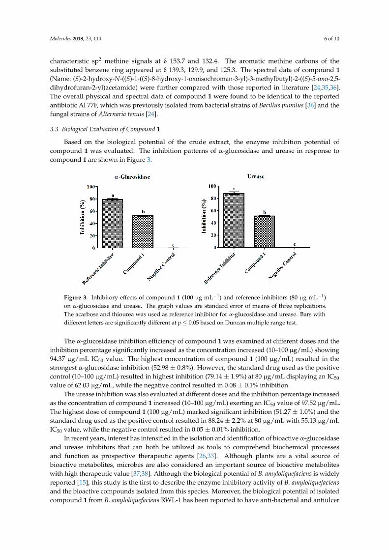
Based on the biological potential of the crude extract, the enzyme inhibition potential ofcompound 1 was evaluated. The inhibition patterns of α-glucosidase and urease in response tocompound 1 are shown in Figure 3.
Figure 3. Inhibitory effects of compound 1 (100 µg mL−1) and reference inhibitors (80 µg mL−1)on α-glucosidase and urease. The graph values are standard error of means of three replications.The acarbose and thiourea was used as reference inhibitor for α-glucosidase and urease. Bars withdifferent letters are significantly different at p ≤ 0.05 based on Duncan multiple range test.
The α-glucosidase inhibition efficiency of compound 1 was examined at different doses and theinhibition percentage significantly increased as the concentration increased (10–100 µg/mL) showing94.37 µg/mL IC50 value. The highest concentration of compound 1 (100 µg/mL) resulted in thestrongest α-glucosidase inhibition (52.98 ± 0.8%). However, the standard drug used as the positivecontrol (10–100 µg/mL) resulted in highest inhibition (79.14 ± 1.9%) at 80 µg/mL displaying an IC50
value of 62.03 µg/mL, while the negative control resulted in 0.08 ± 0.1% inhibition.The urease inhibition was also evaluated at different doses and the inhibition percentage increased
as the concentration of compound 1 increased (10–100 µg/mL) exerting an IC50 value of 97.52 µg/mL.The highest dose of compound 1 (100 µg/mL) marked significant inhibition (51.27 ± 1.0%) and thestandard drug used as the positive control resulted in 88.24 ± 2.2% at 80 µg/mL with 55.13 µg/mLIC50 value, while the negative control resulted in 0.05 ± 0.01% inhibition.
In recent years, interest has intensified in the isolation and identification of bioactive α-glucosidaseand urease inhibitors that can both be utilized as tools to comprehend biochemical processesand function as prospective therapeutic agents [26,33]. Although plants are a vital source ofbioactive metabolites, microbes are also considered an important source of bioactive metaboliteswith high therapeutic value [37,38]. Although the biological potential of B. amyloliquefaciens is widelyreported [15], this study is the first to describe the enzyme inhibitory activity of B. amyloliquefaciensand the bioactive compounds isolated from this species. Moreover, the biological potential of isolatedcompound 1 from B. amyloliquefaciens RWL-1 has been reported to have anti-bacterial and antiulcer
Molecules 2018, 23, 114 7 of 10
properties [39,40]. The results of the current study are in agreement with the findings of otherresearchers who have reported that endophytes provide plentiful resources from which α-glucosidaseand urease inhibitors can be found [26,33].
4. Conclusions
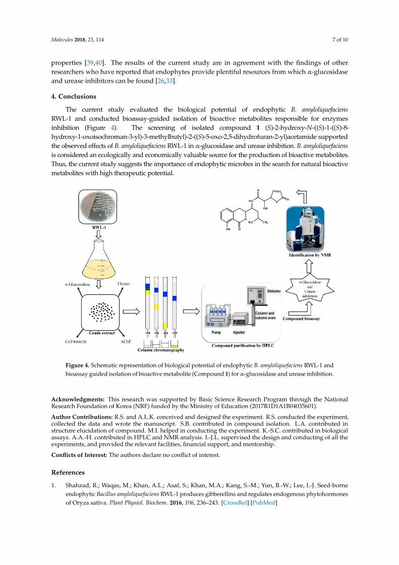
The current study evaluated the biological potential of endophytic B. amyloliquefaciensRWL-1 and conducted bioassay-guided isolation of bioactive metabolites responsible for enzymesinhibition (Figure 4). The screening of isolated compound 1 (S)-2-hydroxy-N-((S)-1-((S)-8-hydroxy-1-oxoisochroman-3-yl)-3-methylbutyl)-2-((S)-5-oxo-2,5-dihydrofuran-2-yl)acetamide supportedthe observed effects of B. amyloliquefaciens RWL-1 in α-glucosidase and urease inhibition. B. amyloliquefaciensis considered an ecologically and economically valuable source for the production of bioactive metabolites.Thus, the current study suggests the importance of endophytic microbes in the search for natural bioactivemetabolites with high therapeutic potential.
Figure 4. Schematic representation of biological potential of endophytic B. amyloliquefaciens RWL-1 andbioassay guided isolation of bioactive metabolite (Compound 1) for α-glucosidase and urease inhibition.
Acknowledgments: This research was supported by Basic Science Research Program through the NationalResearch Foundation of Korea (NRF) funded by the Ministry of Education (2017R1D1A1B04035601).
Author Contributions: R.S. and A.L.K. conceived and designed the experiment. R.S. conducted the experiment,collected the data and wrote the manuscript. S.B. contributed in compound isolation. L.A. contributed instructure elucidation of compound. M.I. helped in conducting the experiment. K.-S.C. contributed in biologicalassays. A.A.-H. contributed in HPLC and NMR analysis. I.-J.L. supervised the design and conducting of all theexperiments, and provided the relevant facilities, financial support, and mentorship.
Conflicts of Interest: The authors declare no conflict of interest.
References
1. Shahzad, R.; Waqas, M.; Khan, A.L.; Asaf, S.; Khan, M.A.; Kang, S.-M.; Yun, B.-W.; Lee, I.-J. Seed-borneendophytic Bacillus amyloliquefaciens RWL-1 produces gibberellins and regulates endogenous phytohormonesof Oryza sativa. Plant Physiol. Biochem. 2016, 106, 236–243. [CrossRef] [PubMed]
Molecules 2018, 23, 114 8 of 10
2. Khan, A.L.; Hussain, J.; Al-Harrasi, A.; Al-Rawahi, A.; Lee, I.-J. Endophytic fungi: Resource for gibberellinsand crop abiotic stress resistance. Crit. Rev. Biotechnol. 2015, 35, 62–74. [CrossRef] [PubMed]
3. Nair, D.N.; Padmavathy, S. Impact of endophytic microorganisms on plants, environment and humans.Sci. World J. 2014, 2014, 250693. [CrossRef] [PubMed]
4. Hardoim, P.R.; van Overbeek, L.S.; Berg, G.; Pirttilä, A.M.; Compant, S.; Campisano, A.; Döring, M.;Sessitsch, A. The Hidden World within Plants: Ecological and Evolutionary Considerations for DefiningFunctioning of Microbial Endophytes. Microbiol. Mol. Biol. Rev. 2015, 79, 293–320. [CrossRef] [PubMed]
5. Schulz, B.; Boyle, C. The endophytic continuum. Mycol. Res. 2005, 109. [CrossRef]6. Strobel, G.A. Endophytes as sources of bioactive products. Microbes Infect. 2003, 5. [CrossRef]7. Schulz, B.; Boyle, C.; Draeger, S.; Römmert, A.-K.; Krohn, K. Endophytic fungi: A source of novel biologically
active secondary metabolites* *Paper presented at the British Mycological Society symposium on FungalBioactive Compounds, held at the University of Wales Swansea on 22–27 April 2001. Mycol. Res. 2002, 106,996–1004. [CrossRef]
8. Saunders, M.; Glenn, A.E.; Kohn, L.M. Exploring the evolutionary ecology of fungal endophytes inagricultural systems: Using functional traits to reveal mechanisms in community processes. Evol. Appl. 2010,3, 525–537. [CrossRef] [PubMed]
9. Cheplick, G.P.; Faeth, S.H. Ecology and Evolution of the Grass-Endophyte Symbiosis; OUP: New York, NY, USA,2009; ISBN 0195308085.
10. Schlaeppi, K.; Bulgarelli, D. The Plant Microbiome at Work. Mol. Plant-Microbe Interact. 2014, 28, 212–217.[CrossRef] [PubMed]
11. Aman, M.; Rai, V.R. Antifungal activity of novel indole derivative from endophytic bacteria Pantoea ananatis4G-9 against Mycosphaerella musicola. Biocontrol Sci. Technol. 2016, 26, 476–491. [CrossRef]
12. Chen, H.; Yang, C.; Ke, T.; Zhou, M.; Li, Z.; Zhang, M.; Gong, G.; Hou, T. Antimicrobial activity of secondarymetabolites from Streptomyces sp. K15, an endophyte in Houttuynia cordata Thunb. Nat. Prod. Res. 2015, 29,2223–2225. [CrossRef] [PubMed]
13. Shweta, S.; Bindu, J.H.; Raghu, J.; Suma, H.K.; Manjunatha, B.L.; Kumara, P.M.; Ravikanth, G.; Nataraja, K.N.;Ganeshaiah, K.N.; Shaanker, R.U. Isolation of endophytic bacteria producing the anti-cancer alkaloidcamptothecine from Miquelia dentata Bedd.(Icacinaceae). Phytomedicine 2013, 20, 913–917. [CrossRef] [PubMed]
14. Abdalla, M.A.; Matasyoh, J.C. Endophytes as producers of peptides: An overview about the recentlydiscovered peptides from endophytic microbes. Nat. Prod. Bioprospect. 2014, 4, 257–270. [CrossRef][PubMed]
15. Chen, Y.-T.; Yuan, Q.; Shan, L.-T.; Lin, M.-A.; Cheng, D.-Q.; Li, C.-Y. Antitumor activity of bacterialexopolysaccharides from the endophyte Bacillus amyloliquefaciens sp. isolated from Ophiopogon japonicus.Oncol. Lett. 2013, 5, 1787–1792. [PubMed]
16. Roy, S.; Yasmin, S.; Ghosh, S.; Bhattacharya, S.; Banerjee, D. Anti-Infective Metabolites of a Newly IsolatedBacillus thuringiensis KL1 Associated with Kalmegh (Andrographis paniculata Nees.), a Traditional MedicinalHerb. Microbiol. Insights 2016, 9, 1–7. [CrossRef] [PubMed]
17. Jamal, M.T.; Mudarris, M.S.A. Separation of YbdN bioactive protein from Bacillus subtilis isolated from theRed Sea algae Sargassum sp. with bioactivity against antibiotic resistant bacterial pathogens. Mar. Sci. 2010,21, 53–64. [CrossRef]
18. Haas, D.; Défago, G. Biological control of soil-borne pathogens by fluorescent pseudomonads. Nat. Rev. Microbiol.2005, 3, 307–319. [CrossRef] [PubMed]
19. Chen, X.H.; Koumoutsi, A.; Scholz, R.; Eisenreich, A.; Schneider, K.; Heinemeyer, I.; Morgenstern, B.; Voss, B.;Hess, W.R.; Reva, O. Comparative analysis of the complete genome sequence of the plant growth–promotingbacterium Bacillus amyloliquefaciens FZB42. Nat. Biotechnol. 2007, 25, 1007–1014. [CrossRef] [PubMed]
20. Liang, T.-W.; Chen, Y.-J.; Yen, Y.-H.; Wang, S.-L. The antitumor activity of the hydrolysates of chitinousmaterials hydrolyzed by crude enzyme from Bacillus amyloliquefaciens V656. Process Biochem. 2007, 42,527–534. [CrossRef]
21. Chen, X.H.; Koumoutsi, A.; Scholz, R.; Schneider, K.; Vater, J.; Süssmuth, R.; Piel, J.; Borriss, R. Genomeanalysis of Bacillus amyloliquefaciens FZB42 reveals its potential for biocontrol of plant pathogens. J. Biotechnol.2009, 140, 27–37. [CrossRef] [PubMed]
Molecules 2018, 23, 114 9 of 10
22. Shahzad, R.; Khan, A.L.; Bilal, S.; Asaf, S.; Lee, I.-J. Plant growth-promoting endophytic bacteria versuspathogenic infections: An example of Bacillus amyloliquefaciens RWL-1 and Fusarium oxysporum f. sp.lycopersici in tomato. PeerJ 2017, 5, e3107. [CrossRef] [PubMed]
23. Shahzad, R.; Khan, A.L.; Bilal, S.; Waqas, M.; Kang, S.-M.; Lee, I.-J. Inoculation of abscisic acid-producingendophytic bacteria enhances salinity stress tolerance in Oryza sativa. Environ. Exp. Bot. 2017, 136, 68–77.[CrossRef]
24. Liu, J.J.; Huang, B.; Hooi, S.C. Acetyl-keto-β-boswellic acid inhibits cellular proliferation througha p21-dependent pathway in colon cancer cells. Br. J. Pharmacol. 2006, 148. [CrossRef] [PubMed]
25. Golbabaei, S.; Bazl, R.; Golestanian, S.; Nabati, F.; Omrany, Z.B.; Yousefi, B.; Hajiaghaee, R.; Rezazadeh, S.;Amanlou, M. Urease inhibitory activities of β-boswellic acid derivatives. DARU J. Pharm. Sci. 2013, 21, 2.[CrossRef] [PubMed]
26. Ali, L.; Khan, A.L.; Al-Kharusi, L.; Hussain, J.; Al-Harrasi, A. New α-glucosidase inhibitory triterpenic acidfrom marine macro green alga Codium dwarkense Boergs. Mar. Drugs 2015, 13, 4344–4356. [CrossRef][PubMed]
27. Li, J.; Zhao, G.; Chen, H.; Wang, H.; Qin, S.; Zhu, W.; Xu, L.; Jiang, C.; Li, W. Antitumour and antimicrobialactivities of endophytic streptomycetes from pharmaceutical plants in rainforest. Lett. Appl. Microbiol. 2008,47, 574–580. [CrossRef] [PubMed]
28. Moyo, M.; Ndhlala, A.R.; Finnie, J.F.; Van Staden, J. Phenolic composition, antioxidant andacetylcholinesterase inhibitory activities of Sclerocarya birrea and Harpephyllum caffrum (Anacardiaceae)extracts. Food Chem. 2010, 123, 69–76. [CrossRef]
29. Amessis-Ouchemoukh, N.; Ouchemoukh, S.; Meziant, N.; Idiri, Y.; Hernanz, D.; Stinco, C.M.;Rodríguez-Pulido, F.J.; Heredia, F.J.; Madani, K.; Luis, J. Bioactive metabolites involved in the antioxidant,anticancer and anticalpain activities of Ficus carica L., Ceratonia siliqua L. and Quercus ilex L. extracts.Ind. Crops Prod. 2017, 95, 6–17. [CrossRef]
30. Chu, C.-C.; Chen, S.-Y.; Chyau, C.-C.; Fu, Z.-H.; Liu, C.-C.; Duh, P.-D. Protective effect of Djulis(Chenopodium formosanum) and its bioactive compounds against carbon tetrachloride-induced liver injury,in vivo. J. Funct. Foods 2016, 26, 585–597. [CrossRef]
31. Spanogiannopoulos, P.; Bess, E.N.; Carmody, R.N.; Turnbaugh, P.J. The microbial pharmacists within us:A metagenomic view of xenobiotic metabolism. Nat. Rev. Microbiol. 2016, 14, 273–287. [CrossRef] [PubMed]
32. Gouda, S.; Das, G.; Sen, S.K.; Shin, H.-S.; Patra, J.K. Endophytes: A treasure house of bioactive compoundsof medicinal importance. Front. Microbiol. 2016, 7, 1538. [CrossRef] [PubMed]
33. Khan, A.L.; Ali, L.; Hussain, J.; Rizvi, T.S.; Al-Harrasi, A.; Lee, I.-J. Enzyme inhibitory radicinol derivativefrom endophytic fungus Bipolaris sorokiniana LK12, associated with Rhazya stricta. Molecules 2015, 20,12198–12208. [CrossRef] [PubMed]
34. Nonejuie, P.; Trial, R.M.; Newton, G.L.; Lamsa, A.; Perera, V.R.; Aguilar, J.; Liu, W.-T.; Dorrestein, P.C.;Pogliano, J.; Pogliano, K. Application of bacterial cytological profiling to crude natural product extractsreveals the antibacterial arsenal of Bacillus subtilis. J. Antibiot. 2016, 69, 353–361. [CrossRef] [PubMed]
35. Afiyatullov, S.S.; Kalinovskaya, N.I.; Kuznetsova, T.A.; Ivanova, E.P.; Mikhailov, V.V. Antibiotics from strainsof Bacillus pumilus isolated from a marine sponge Dendrilla sp. Chem. Nat. Compd. 1991, 27, 765–766.[CrossRef]
36. Shimojima, Y.; Hayashi, H.; Ooka, T.; Shibukawa, M. Production, isolation and pharmacological studies ofAI-77s. Agric. Biol. Chem. 1982, 46, 1823–1829. [CrossRef]
37. Wang, J.; Guleria, S.; Koffas, M.A.G.; Yan, Y. Microbial production of value-added nutraceuticals. Curr. Opin. Biotechnol.2016, 37, 97–104. [CrossRef] [PubMed]
38. Strobel, G.; Daisy, B. Bioprospecting for microbial endophytes and their natural products. Microbiol. Mol.Biol. Rev. 2003, 67, 491–502. [CrossRef] [PubMed]
Molecules 2018, 23, 114 10 of 10
39. Shimojima, Y.; Hayashi, H. 1H-2-benzopyran-1-one derivatives, microbial products with pharmacological activity.Relationship between structure and activity in 6-[[1(S)-(3(S),4-dihydro-8-hydroxy-1-oxo-1H-2-benzopyran-3-yl)-3-methylbutyl]-amino]-4(S),5(S)-dihydroxy-6-oxo-3(S)-ammon. J. Med. Chem. 1983, 26, 1370–1374. [CrossRef][PubMed]
40. Huang, Y.-F.; Li, L.-H.; Tian, L.; Qiao, L.; Hua, H.-M.; Pei, Y.-H. Sg17-1-4, a novel isocoumarin from a marinefungus Alternaria tenuis Sg17-1. J. Antibiot. 2006, 59, 355–357. [CrossRef] [PubMed]
Sample Availability: Samples of the compound are available from the authors.
© 2018 by the authors. Licensee MDPI, Basel, Switzerland. This article is an open accessarticle distributed under the terms and conditions of the Creative Commons Attribution(CC BY) license (http://creativecommons.org/licenses/by/4.0/).
Related Documents