RESEARCH LETTER Characterization of melanin-overproducing transposon mutants of Pseudomonas putida F6 Jasmina Nikodinovic-Runic 1 , Leona B. Martin 1 , Ramesh Babu 2 , Werner Blau 2 & Kevin E. O’Connor 1 1 School of Biomolecular and Biomedical Sciences, University College Dublin, Dublin, Ireland; and 2 Polymer Research Centre, School of Physics, Trinity College Dublin, Dublin, Ireland Correspondence: Kevin E. O’Connor, School of Biomolecular and Biomedical Sciences, Ardmore House, University College Dublin, Belfield, Dublin 4, Ireland. Tel.: 1353 1 716 1307; fax: 1353 1 716 1183; e-mail: [email protected] Received 5 March 2009; accepted 28 June 2009. Final version published online 17 July 2009. DOI:10.1111/j.1574-6968.2009.01716.x Editor: Alexander Steinb ¨ uchel Keywords melanin; tyrosinase; Pseudomonas putida F6. Abstract Two melanin-overproducing Pseudomonas putida F6 mutants were generated using transposon (Tn5) mutagenesis. Mutants were disrupted in a transcriptional regulator (TR) and a homogentisate 1,2-dioxygenase (HDO) gene. Colonies of mutant F6-TR overproduced a black pigment on solid medium. The same mutant (F6-TR) had a 3.7-fold higher tyrosinase activity compared with the wild-type strain when induced with ferulic acid. However in tyrosine uptake assays whole cells of the mutant strain F6-TR consumed eight times less tyrosine compared with the wild-type strain. Mutant F6-HDO produced a diffusible red pigment into the growth medium. Pigment production by mutant F6-HDO is sixfold higher than the wild-type strain. The biomass yield of mutant F6-HDO grown on tyrosine as the sole source of carbon and energy was 1.2-fold lower than the wild-type strain. While the growth of the wild-type strain was completely inhibited by 5 min of exposure to UV light (254 nm) both mutant strains showed survival rates 4 30%. Mutant F6-HDO was able to tolerate higher concentrations of hydrogen peroxide (H 2 O 2 ) exhibiting 1.5 times smaller zones of inhibition at 10 mM H 2 O 2 compared with mutant F6-TR and the wild-type strain. The pigments produced by all strains were purified and confirmed to be melanins. Introduction Melanins are pigments that are produced by a broad range of microorganisms. They are not considered essential for growth and development of cells, but are required in order to enhance the ability of the producing species to compete and survive under certain environmental conditions, such as in the presence of UV radiation (Lopez-Serrano et al., 2004). Melanins have been reported to have strong affinity for metals and to be efficient scavengers of free radicals (Sichel et al., 1991). In Azotobacter chroococcum and Burkholderia cenocepacia they protect against reactive oxygen species (ROS) (Shivprasad & Page, 1989; Keith et al., 2007). Melanins tend to be either black or brown pigments although other colours may occur (Hill, 1992). Melanins derived from L-3,4-dihydroxyphenylalanine (L-DOPA) are referred to as eumelanins and are black or brown (Hill, 1992). Reddish or yellow melanins that incorporate cysteine with L-DOPA are called pheomelanins (Wakamatsu & Ito, 2002). Red–brown, water-soluble melanins formed from the catabolism of tyrosine via p-hydroxyphenylpyruvate (PHPPA) and homogentisic acid (HGA), are called pyome- lanins (Yabuuchi & Ohyama, 1972; Kotob et al., 1995). Melanin synthesis in bacteria is carried out in the majority of cases by phenoloxidases (tyrosinases, laccases or catecho- lases) and/or via the polyketide synthase pathway (Jacobson, 2000). However, other enzymes such as p-hydroxyphenyla- cetic acidPHPA hydroxylase when heterologously expressed in Escherichia coli have also been shown to produce melanin (Gibello et al., 1995). Tyrosinase (EC 1.14.18.1) is an enzyme that is ubiquitously distributed in microorganisms, animals and plants. Most of the information on the structure and function of tyrosinase has been obtained from studies on Agaricus bisporus (mushroom) and Streptomyces antibioticus (Claus & Decker, 2006; Matoba et al., 2006). Tyrosinase from Pseudomonas putida F6 has been previously purified and biochemically characterized (McMahon et al., 2007). Tyrosinase catalyses the ortho hydroxylation of monophe- nols to o-diphenols (monophenolase, cresolase activity), followed by the subsequent oxidation of the o-diphenol to FEMS Microbiol Lett 298 (2009) 174–183 c 2009 Federation of European Microbiological Societies Published by Blackwell Publishing Ltd. All rights reserved MICROBIOLOGY LETTERS

Welcome message from author
This document is posted to help you gain knowledge. Please leave a comment to let me know what you think about it! Share it to your friends and learn new things together.
Transcript
R E S E A R C H L E T T E R
Characterizationofmelanin-overproducing transposonmutantsofPseudomonasputidaF6Jasmina Nikodinovic-Runic1, Leona B. Martin1, Ramesh Babu2, Werner Blau2 & Kevin E. O’Connor1
1School of Biomolecular and Biomedical Sciences, University College Dublin, Dublin, Ireland; and 2Polymer Research Centre, School of Physics, Trinity
College Dublin, Dublin, Ireland
Correspondence: Kevin E. O’Connor, School
of Biomolecular and Biomedical Sciences,
Ardmore House, University College Dublin,
Belfield, Dublin 4, Ireland. Tel.: 1353 1 716
1307; fax: 1353 1 716 1183; e-mail:
Received 5 March 2009; accepted 28 June
2009.
Final version published online 17 July 2009.
DOI:10.1111/j.1574-6968.2009.01716.x
Editor: Alexander Steinbuchel
Keywords
melanin; tyrosinase; Pseudomonas putida F6.
Abstract
Two melanin-overproducing Pseudomonas putida F6 mutants were generated
using transposon (Tn5) mutagenesis. Mutants were disrupted in a transcriptional
regulator (TR) and a homogentisate 1,2-dioxygenase (HDO) gene. Colonies of
mutant F6-TR overproduced a black pigment on solid medium. The same mutant
(F6-TR) had a 3.7-fold higher tyrosinase activity compared with the wild-type
strain when induced with ferulic acid. However in tyrosine uptake assays whole
cells of the mutant strain F6-TR consumed eight times less tyrosine compared with
the wild-type strain. Mutant F6-HDO produced a diffusible red pigment into the
growth medium. Pigment production by mutant F6-HDO is sixfold higher than
the wild-type strain. The biomass yield of mutant F6-HDO grown on tyrosine as
the sole source of carbon and energy was 1.2-fold lower than the wild-type strain.
While the growth of the wild-type strain was completely inhibited by 5 min of
exposure to UV light (254 nm) both mutant strains showed survival rates 4 30%.
Mutant F6-HDO was able to tolerate higher concentrations of hydrogen peroxide
(H2O2) exhibiting 1.5 times smaller zones of inhibition at 10 mM H2O2 compared
with mutant F6-TR and the wild-type strain. The pigments produced by all strains
were purified and confirmed to be melanins.
Introduction
Melanins are pigments that are produced by a broad range of
microorganisms. They are not considered essential for
growth and development of cells, but are required in order
to enhance the ability of the producing species to compete
and survive under certain environmental conditions, such as
in the presence of UV radiation (Lopez-Serrano et al., 2004).
Melanins have been reported to have strong affinity for
metals and to be efficient scavengers of free radicals (Sichel
et al., 1991). In Azotobacter chroococcum and Burkholderia
cenocepacia they protect against reactive oxygen species
(ROS) (Shivprasad & Page, 1989; Keith et al., 2007).
Melanins tend to be either black or brown pigments
although other colours may occur (Hill, 1992). Melanins
derived from L-3,4-dihydroxyphenylalanine (L-DOPA) are
referred to as eumelanins and are black or brown (Hill,
1992). Reddish or yellow melanins that incorporate cysteine
with L-DOPA are called pheomelanins (Wakamatsu & Ito,
2002). Red–brown, water-soluble melanins formed from the
catabolism of tyrosine via p-hydroxyphenylpyruvate
(PHPPA) and homogentisic acid (HGA), are called pyome-
lanins (Yabuuchi & Ohyama, 1972; Kotob et al., 1995).
Melanin synthesis in bacteria is carried out in the majority
of cases by phenoloxidases (tyrosinases, laccases or catecho-
lases) and/or via the polyketide synthase pathway (Jacobson,
2000). However, other enzymes such as p-hydroxyphenyla-
cetic acidPHPA hydroxylase when heterologously expressed
in Escherichia coli have also been shown to produce melanin
(Gibello et al., 1995). Tyrosinase (EC 1.14.18.1) is an enzyme
that is ubiquitously distributed in microorganisms, animals
and plants. Most of the information on the structure and
function of tyrosinase has been obtained from studies on
Agaricus bisporus (mushroom) and Streptomyces antibioticus
(Claus & Decker, 2006; Matoba et al., 2006). Tyrosinase
from Pseudomonas putida F6 has been previously purified
and biochemically characterized (McMahon et al., 2007).
Tyrosinase catalyses the ortho hydroxylation of monophe-
nols to o-diphenols (monophenolase, cresolase activity),
followed by the subsequent oxidation of the o-diphenol to
FEMS Microbiol Lett 298 (2009) 174–183c� 2009 Federation of European Microbiological SocietiesPublished by Blackwell Publishing Ltd. All rights reserved
MIC
ROBI
OLO
GY
LET
TER
S

the corresponding o-quinone derivative (Sanchez-Ferrer
et al., 1995). The latter products polymerize to form
melanin-like pigments.
We wish to gain an insight into the role of tyrosinase and
other factors affecting melanin synthesis in P. putida F6.
Transposon mutagenesis, a powerful tool for the genetic,
physiological and biochemical analysis of bacteria (deLor-
enzo & Timmis, 1994) was utilized to generate two mutants
exhibiting increased pigmentation.
Materials and methods
Reagents
Phenylacetic acid, PHPA, PHPPA, L-tyrosine, L-DOPA,
ferulic acid, homogentisic acid, 4-fluorophenol, synthetic
melanin, o-dianisidine, 6-hydroxydopamine hydrobromide,
deuterated dimethyl sulphoxide (d-DMSO) and Luria–Ber-
tani (LB) broth were purchased from Sigma-Aldrich, Ire-
land. Tri-sodium citrate was from BDH Laboratories, UK.
Bacterial strains and plasmids
Pseudomonas putida F6 was isolated from soil for its ability
to utilize PHPA as a sole source of carbon and energy
(O’Connor et al., 2001). Escherichia coli CC118lpir hosted
the mini-Tn5 derivative pUT-Km1(Herrero et al., 1990).
This suicide plasmid has the R6K origin of replication and
encodes resistance to kanamycin and ampicillin (de Lorenzo
et al., 1990). Plasmid pRK600 (Cmr, mob1, tra1) hosted in
E. coli HB101 (Invitrogen) was used as a helper in triparental
mating experiments. Escherichia coli TOP 10 (Invitrogen)
was a general cloning host. TOPOs pCRs 2.1 (Invitrogen)
was used for cloning of PCR products according to the
manufacturer’s instructions.
Mutagenesis of P. putida F6 by the mini-Tn5Km1transposon
Triparental matings involving E. coli CC118lpir (pUT-Km1)
as the transposon donor strain, P. putida F6 as the recipient
and E. coli HB101 (pRK600) as the helper strain were carried
out in a 1-mL volume in the ratio 7 : 2 : 1, respectively
(Vilchez et al., 2000; O’Leary et al., 2005).
Transconjugants of P. putida F6 were selected on E2 agar
plates (Vogel & Bonner, 1956) supplemented with tri-
sodium citrate (15 mM), kanamycin and L-tyrosine
(5 mM). Approximately, 105 independent mutants colonies
were generated and were individually transferred to sterile
96-well microtitre plates containing 200mL of E2/citrate/km
(50mg mL�1) in each well (O’Leary et al., 2005). Following a
24-h incubation colonies were transferred onto E2 agar
plates containing L-tyrosine (5 mM) (System Duetz, Kuhner
AG, Switzerland). Mutants were screened and selected based
on the increased melanin production by visual inspection of
the pigmentation of the colonies (Solano et al., 2000). Two
mutants were selected and subcultured for sequencing and
further characterization.
Culture media and growth conditions
Pseudomonas putida F6 wild type as well as the P. putida F6
mutants were grown in liquid culture in 50 mL of E2
medium (Vogel & Bonner, 1956) in 250-mL Erlenmeyer
flasks. Media contained tri-sodium citrate (15 mM) as
carbon source and when necessary kanamycin was added
(50mg mL�1). Cultures were incubated at 30 1C with shaking
at 200 r.p.m. for 24 h. In melanin quantification experiments
cells were grown as above in E2 broth supplemented with
L-tyrosine (5 mM). Samples were taken periodically and
melanin production was monitored at 400 nm (Ruzafa
et al., 1995; Chatfield & Cianciotto, 2007).
Identification of DNA sequences of transposonmutants
Identification of the genes disrupted by Tn5 was carried out
using a PCR method and arbitrary primers described
previously (Caetano-Anolles, 1993; Espinosa-Urgel et al.,
2000). Obtained PCR products were cloned into pCR 2.1-
TOPO (Invitrogen) before sequencing. Sequencing was
carried out by GATC biotech (Konstanz, Germany). Se-
quences were analysed and compared with the GenBank
database using the BLAST program (Altschul et al., 1997).
Enzyme assays in cell extracts
Pseudomonas putida F6 and mutants were grown in flasks
(50-mL culture in 250-mL flasks) in E2/citrate medium, E2/
citrate supplemented with L-tyrosine (1 mM) and E2/citrate
supplemented with ferulic acid (1 mM) at 30 1C for two 24 h
(Brooks et al., 2004). The cells were harvested by centrifuga-
tion at 5000 g for 10 min at 4 1C and washed twice in ice-
cold 50 mM potassium phosphate buffer, pH 7 (10 mL).
Cell-free extract (CE) was prepared using the BugBuster
amine-free reagent (Novagen) according to the instruction
manual.
Tyrosinase enzyme assays were performed in 96-well
microtitre plates with L-tyrosine (1 mM) as the sole sub-
strate. Assays were performed in air-saturated 50 mM phos-
phate buffer, pH 7, at 30 1C. The total reaction volume was
200 mL and contained 0.015 mg protein. The reaction was
monitored at 475 nm (Espin et al., 1995) and recorded using
a SpectraMax-340 microtitre plate reader (Molecular De-
vices, Sunnyvale). Reduced nicotinamide cofactors (NAHD)
were not added to the cell extract to ensure that any
hydroxylase enzymes potentially present in the cell extract
FEMS Microbiol Lett 298 (2009) 174–183 c� 2009 Federation of European Microbiological SocietiesPublished by Blackwell Publishing Ltd. All rights reserved
175Melanin-overproducing transposon mutants of P. putida F6
would not be able to oxidize tyrosine and thus any activity
present can be attributed to tyrosinase.
For determination of other enzyme activities, cells were
grown in E2 medium, with citrate (15 mM) and L-tyrosine
(1 mM) for 48 h at 30 1C. CEs were prepared as above,
and supplemented with 40% glycerol (v/v) for storage
at � 20 1C.
Laccase activity was measured spectrophotometrically, by
following the rate of oxidation of syringaldazine at 525 nm
(Solano et al., 2000) in the reaction buffer containing 50 mM
potassium phosphate and 0.05 mM syringaldazine upon
addition of CE (20mL in 0.2-mL total volume). The
molar extinction coefficient of the oxidation product,
e= 65 000 M�1 cm�1, was used to calculate laccase activity
(Harkin & Obst, 1973). One unit of laccase activity is
defined as the amount of the enzyme that catalysed the
oxidation of 1mmol of syringaldazine per min at 30 1C.
Catalase activity in CEs was determined spectrophotome-
trically by following the disappearance of H2O2 over time at
240 nm (Beers & Sizer, 1952). One unit of catalase decom-
posed 1 mmol H2O2mg�1protein at 25 1C. The assay was
performed in 1-mL volume in 50 mM phosphate buffer, pH
7, containing 10 mM H2O2 upon addition of CE (0.1 mL).
Peroxidase activity was assayed by following the oxidation
of o-dianisidine at 460 nm in a reaction buffer containing
50 mM potassium phosphate, 1 mM H2O2, 0.34 mM o-
dianisidine (Sigma) upon addition of CE (20 mL). One unit
of peroxidase activity equals 1 micromole H2O2 reduced per
minute, with e= 113 000 M�1 cm�1 (Schnell & Steinman,
1995).
Superoxide dismutase (SOD) activity in CEs was deter-
mined spectrophotometrically by measuring the inhibition
of the initial rate of auto-oxidation of 6-hydroxydopamine
at 490 nm, in 50 mM phosphate buffer, pH 7.4, containing
0.1 mM 6-hydroxydopamine hydrobromide upon addition
of CEs (20 mL in 0.2-mL total assay volume). One unit of
SOD activity corresponded to 50% inhibition of the initial
rate (Heikkila & Cabbat, 1976). A standard curve using
known units of SOD activity was obtained in order to
determine the units of activity in each sample. We used
SOD from E. coli (Sigma) as a positive control and for the
generation of the standard curve.
Tyrosine uptake monitoring by HPLC analysis
Pseudomonas putida F6 and mutant F6-TR were grown in
E2/citrate broth as described previously. Cultures were
centrifuged at 13 000 g and cell pellets were resuspended in
50 mM phosphate buffer, pH 7, to an OD540 nm of 10,
supplemented with 2 mM L-tyrosine and incubated at 30 1C
shaking at 200 r.p.m. Samples of 0.45 mL were removed
every 30 min, acidified with 1 M HCl (50mL) centrifuged at
13 000 g for 5 min and filtered for HPLC analysis.
HPLC analysis was performed using C-18 Hyperclone
ODS 5-mm column (250� 4.6 mm) (Phenomenex) on a
Hewlett Packard HP1100 instrument equipped with an
Agilent 1100 series diode array detector. Mobile phase was
a solution of methanol and (0.1%) phosphoric acid in the
ratio of 15 : 85, with a flow rate of 0.8 mL min�1.
UV sensitivity assay
UV sensitivity assays were performed using the previously
published protocol with slight alterations (Simonson et al.,
1990). Because of delayed melanin production by mutant
F6-TR in liquid cultures (5–7 days), cell suspensions were
made by scraping the bacterial colonies producing pigment
from the E2/citrate/L-tyrosine plates. Sterile 50 mM phos-
phate buffer (500mL) was applied to the surface of the plate
and sterile cotton swab was used to gently detach the cells
making bacterial suspension. Suspensions were adjusted to
the OD600 nm of 0.2 using sterile phosphate buffer. Cell
suspension (2.5 mL) was placed in the Petri plate and
illuminated with the UV lamp (254 nm, Mineralights Lamp
R-52) for 1, 2 and 5 min. Serial dilutions of untreated cell
suspensions as well as serial dilutions for the treated samples
were plated for each time point and colonies counted and
represented as percentage survival.
Hydrogen peroxide (H2O2) disc diffusion assay
Cell suspension prepared as described above (100 mL) was
spread onto fresh plates (LB and E2/citrate/L-tyrosine), and
sterile paper discs (Oxoid) were applied to the surface. H2O2
(8 mL) of 1, 5 and 10 mM were applied to discs. The plates
were incubated at 30 1C for 48 h, and the zones of inhibition
were measured.
Melanin purification and analysis
Pseudomonas putida F6, F6-HDO and F6-TR were grown in
E2/citrate/L-tyrosine (5 mM) and incubated at 30 1C for 72 h
to allow melanin to accumulate. Cells were removed by
centrifugation (13 000 g for 15 min). Supernatant was acid-
ified (pH 2) using 6 M HCl and melanin was allowed to
precipitate for 4 h at 20 1C. The precipitated melanin
suspension was dialyzed (8 kDa) for 24 h with four changes
of water. After 24 h, the melanin samples were freeze dried.
The melanin samples (10 mg) were dissolved (or sus-
pended) in 1 mL of d-DMSO. Solution nuclear magnetic
resonance (NMR) spectra were recorded on a Bruker
DPX400 (Bruker BioSpin Limited) with 1H at 400.13 MHz
and d-DMSO solvent was used as internal reference for
chemical shifts in 1H NMR.
The samples for Fourier transform infrared spectroscopy
(FT-IR) analysis were prepared by mixing the samples with
KBr powder. Analysis was carried out on a Nexus Nicolet
FEMS Microbiol Lett 298 (2009) 174–183c� 2009 Federation of European Microbiological SocietiesPublished by Blackwell Publishing Ltd. All rights reserved
176 J. Nikodinovic-Runic et al.
FT-IR Spectrometer (Thermo-Electron, Waltham) in main
bench mode. The spectra recorded over a range of
4000–400 cm�1 with a resolution of 4 cm�1 and 128 scans
per sample.
Protein determination
Total protein amount in CEs were determined using the
bicinchoninic acid method as described previously (Smith
et al., 1985).
Results
Identification of gene disruption in transposonmutants
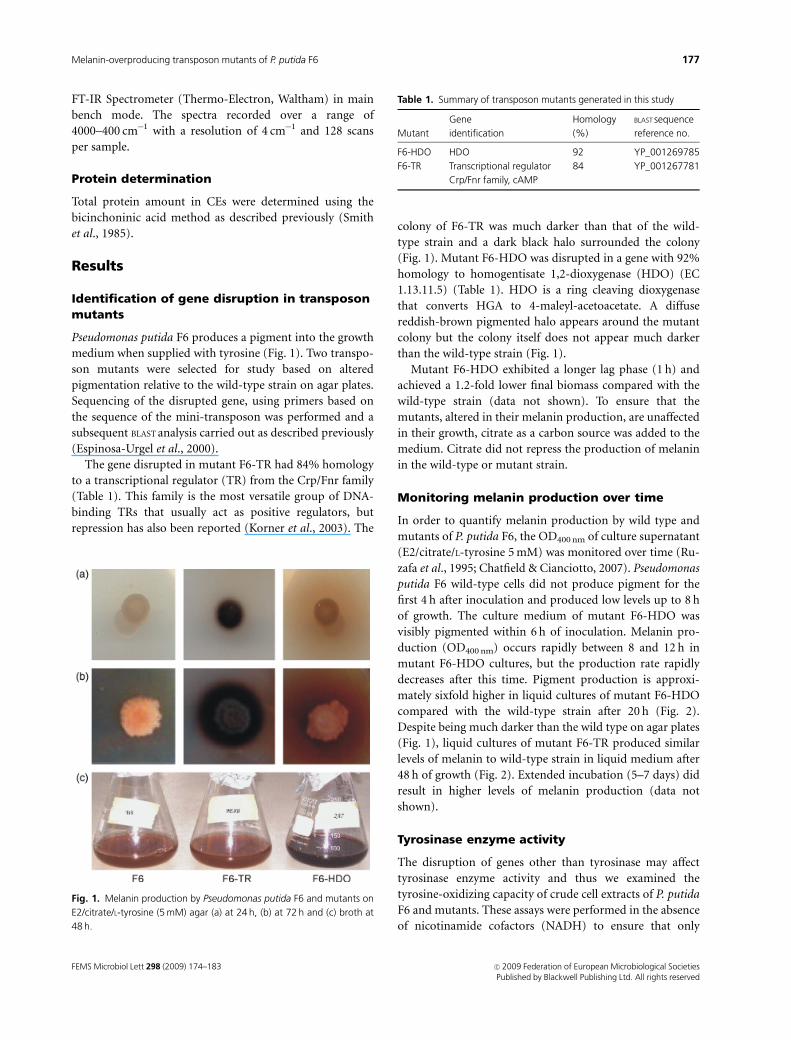
Pseudomonas putida F6 produces a pigment into the growth
medium when supplied with tyrosine (Fig. 1). Two transpo-
son mutants were selected for study based on altered
pigmentation relative to the wild-type strain on agar plates.
Sequencing of the disrupted gene, using primers based on
the sequence of the mini-transposon was performed and a
subsequent BLAST analysis carried out as described previously
(Espinosa-Urgel et al., 2000).
The gene disrupted in mutant F6-TR had 84% homology
to a transcriptional regulator (TR) from the Crp/Fnr family
(Table 1). This family is the most versatile group of DNA-
binding TRs that usually act as positive regulators, but
repression has also been reported (Korner et al., 2003). The
colony of F6-TR was much darker than that of the wild-
type strain and a dark black halo surrounded the colony
(Fig. 1). Mutant F6-HDO was disrupted in a gene with 92%
homology to homogentisate 1,2-dioxygenase (HDO) (EC
1.13.11.5) (Table 1). HDO is a ring cleaving dioxygenase
that converts HGA to 4-maleyl-acetoacetate. A diffuse
reddish-brown pigmented halo appears around the mutant
colony but the colony itself does not appear much darker
than the wild-type strain (Fig. 1).
Mutant F6-HDO exhibited a longer lag phase (1 h) and
achieved a 1.2-fold lower final biomass compared with the
wild-type strain (data not shown). To ensure that the
mutants, altered in their melanin production, are unaffected
in their growth, citrate as a carbon source was added to the
medium. Citrate did not repress the production of melanin
in the wild-type or mutant strain.
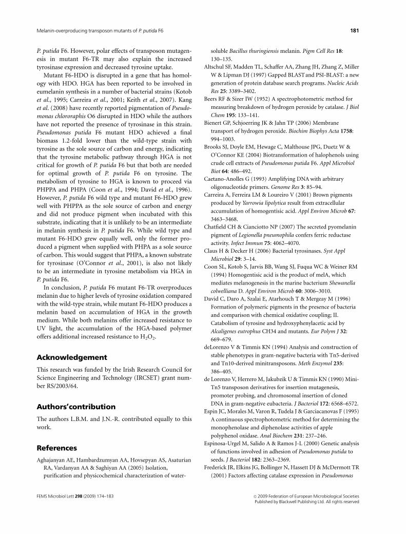
Monitoring melanin production over time
In order to quantify melanin production by wild type and
mutants of P. putida F6, the OD400 nm of culture supernatant
(E2/citrate/L-tyrosine 5 mM) was monitored over time (Ru-
zafa et al., 1995; Chatfield & Cianciotto, 2007). Pseudomonas
putida F6 wild-type cells did not produce pigment for the
first 4 h after inoculation and produced low levels up to 8 h
of growth. The culture medium of mutant F6-HDO was
visibly pigmented within 6 h of inoculation. Melanin pro-
duction (OD400 nm) occurs rapidly between 8 and 12 h in
mutant F6-HDO cultures, but the production rate rapidly
decreases after this time. Pigment production is approxi-
mately sixfold higher in liquid cultures of mutant F6-HDO
compared with the wild-type strain after 20 h (Fig. 2).
Despite being much darker than the wild type on agar plates
(Fig. 1), liquid cultures of mutant F6-TR produced similar
levels of melanin to wild-type strain in liquid medium after
48 h of growth (Fig. 2). Extended incubation (5–7 days) did
result in higher levels of melanin production (data not
shown).
Tyrosinase enzyme activity
The disruption of genes other than tyrosinase may affect
tyrosinase enzyme activity and thus we examined the
tyrosine-oxidizing capacity of crude cell extracts of P. putida
F6 and mutants. These assays were performed in the absence
of nicotinamide cofactors (NADH) to ensure that only
Fig. 1. Melanin production by Pseudomonas putida F6 and mutants on
E2/citrate/L-tyrosine (5 mM) agar (a) at 24 h, (b) at 72 h and (c) broth at
48 h.
Table 1. Summary of transposon mutants generated in this study
Mutant
Gene
identification
Homology
(%)
BLAST sequence
reference no.
F6-HDO HDO 92 YP_001269785
F6-TR Transcriptional regulator
Crp/Fnr family, cAMP
84 YP_001267781
FEMS Microbiol Lett 298 (2009) 174–183 c� 2009 Federation of European Microbiological SocietiesPublished by Blackwell Publishing Ltd. All rights reserved
177Melanin-overproducing transposon mutants of P. putida F6

tyrosinase activity is measured and not any hydroxylase
enzymes that could potentially be present in the cell extracts.
Three different conditions were used to examine tyrosine-
oxidizing (tyrosinase) activity in crude cell extracts of wild
type and mutants of P. putida F6, i.e. growth on citrate only
(noninducing), citrate and tyrosine (1 mM), and citrate and
ferulic acid (1 mM). We have previously described ferulic
acid as an inducer of tyrosinase activity in P. putida F6
(Brooks et al., 2004). Crude cell extracts of wild-type
P. putida F6 cells induced with ferulic acid for 2 h consumed
46.6 nmol tyrosine min�1mg�1protein. Crude cell extracts of
mutant F6-TR consumed tyrosine at a rate 3.7-fold higher
than the wild-type strain under the same conditions (Table
2). Crude extracts of mutant F6-HDO had 3.8-fold lower
tyrosinase activity compared with wild-type cell extracts
when induced with ferulic acid (Table 2). Interestingly,
tyrosine appears to be a poor inducer of tyrosinase activity
in all strains tested with over 40-fold lower activity in the
wild type and mutant F6-TR compared with cells induced
with ferulic acid. Mutant F6-HDO did not exhibit a
detectable activity within the 2-h time frame of the enzyme
assay when induced with tyrosine, but did eventually
(412 h) go dark, indicating low levels of tyrosine-oxidizing
activity.
Laccase, catalase, peroxidase and SOD enzymeactivities
The laccase activity in crude cell extracts of wild-type and
mutant strains of P. putida F6 were almost identical (Table
3). Catalase activity in CE of all three strains was deter-
mined. As catalases can exist as monofunctional and bifunc-
tional enzymes (having additional peroxidase activity), we
have also determined peroxidase activity by following the
oxidation of o-dianisidine at 460 nm. Catalase activity was
1.5-fold higher in F6-HDO mutant relative to the F6 wild
type (Table 3). However, mutant F6-TR expressed similar
levels of catalase activity to the wild-type strain (Table 3).
Peroxidase activity in F6-HDO is 2.1-fold higher than the
wild-type strain. While F6-TR mutant exhibited a 1.3-fold
lower peroxidase activity compared with the wild-type
strain. Interestingly, SOD activity in all three strains was
almost identical (Table 3).
Tyrosine uptake by mutant F6-TR
Despite a high level of tyrosinase activity in crude cell
extracts of mutant F6-TR harvested from solid medium
(agar plates), whole cells grown in liquid culture do not
produce more melanin than the wild-type strain. Based on
this observation, we postulated that tyrosine uptake may
also be affected in this mutant. Both wild-type and mutant
F6-TR washed cell suspensions (OD540 nm of 10), grown
under inducing conditions (citrate, 15 mM, and ferulic acid,
1 mM), showed a lag period in tyrosine uptake for the first
3 h of incubation. However, over the next 3 h, whole cells of
the wild-type strain consumed eight times more tyrosine
(0.32 mM) compared with the mutant F6-TR (0.04 mM).
Cell survival upon exposure to UV light (254nm)
Melanins are well known to protect cells from the effects of
exposure to UV light. The production of higher amounts of
melanin should offer extra protection to mutant F6-HDO
Fig. 2. Melanin production over time by Pseudomonas putida F6 and
the transposon (Tn5) mutants in liquid culture. (&, wild type; , F6-TR;
’, F6-HDO). Mutant F6-TR can only overproduce melanin on solid
medium (see Fig. 1).
Table 2. Tyrosinase activity (U mg�1 protein)� of crude cell extracts of
wild-type and mutant strains using 1 mM tyrosine as the substrate
InducerwP. putida F6
wild type
Mutant
F6-HDO
Mutant
F6-TR
None 1.2�0.1 1.4� 0.1 4.2�0.7
Tyrosine (1 mM) 1.0�0.1 NDz 4.7�0.2
Ferulic acid (1 mM) 46.6�4.5 12.4� 0.9 173�11
�A unit is defined as the nanomoles of quinone formed per minute based
on the molar extinction coefficient (e) of dopaquinone of
3700 M�1 cm�1.wGrowth medium was E2/citrate (15 mM). 0.015 mg mL�1 protein was
used in the assay.zAfter prolonged incubation (12 h), the enzyme assay medium went dark
indicating melanin formation, but activity was not detected within the
assay time frame (2 h).
Table 3. Laccase, catalase and SOD activities (U mg�1 protein)� in
Pseudomonas putida F6 wild type and mutants (F6-HDO and F6-TR)
StrainswLaccase
activity
Catalase
activity
Peroxidase
activity
SOD
activity
P. putida F6 wild
type
6.9� 0.1 22.1� 0.9 5.6�0.5 0.8� 0.1
Mutant F6-HDO 6.6� 0.2 34.8� 0.5 11.9�0.3 0.8� 0.2
Mutant F6-TR 6.8� 0.1 24.9� 0.4 4.2�0.2 0.7� 0.2
�Values are an average of three independent measurements.wCells were grown in E2/citrate (15 mM) medium supplemented with
1 mM tyrosine for 48-h shaking at 30 1C.
FEMS Microbiol Lett 298 (2009) 174–183c� 2009 Federation of European Microbiological SocietiesPublished by Blackwell Publishing Ltd. All rights reserved
178 J. Nikodinovic-Runic et al.
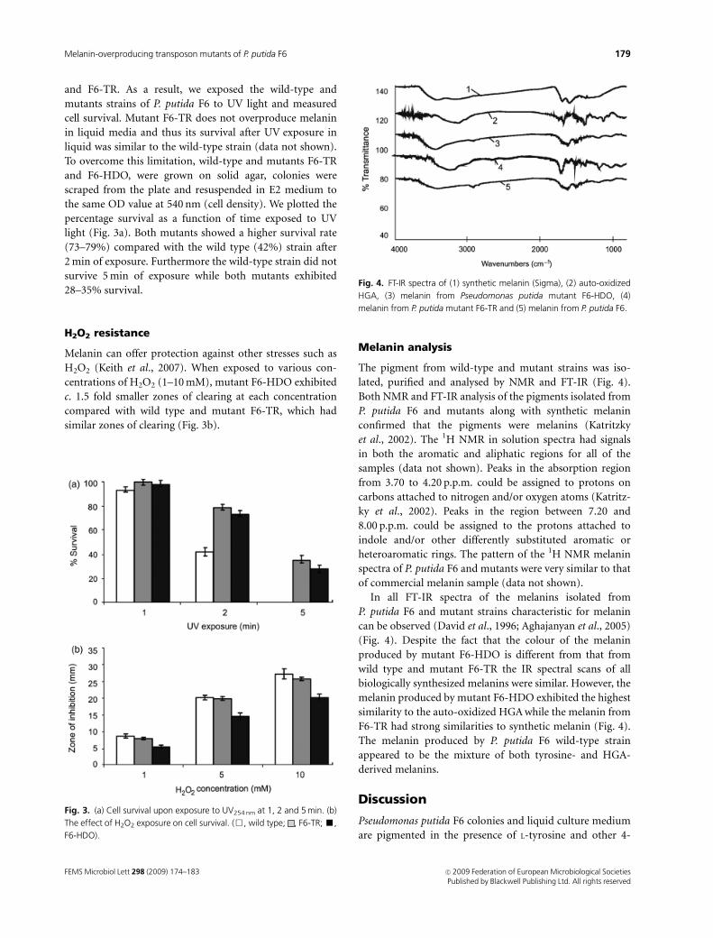
and F6-TR. As a result, we exposed the wild-type and
mutants strains of P. putida F6 to UV light and measured
cell survival. Mutant F6-TR does not overproduce melanin
in liquid media and thus its survival after UV exposure in
liquid was similar to the wild-type strain (data not shown).
To overcome this limitation, wild-type and mutants F6-TR
and F6-HDO, were grown on solid agar, colonies were
scraped from the plate and resuspended in E2 medium to
the same OD value at 540 nm (cell density). We plotted the
percentage survival as a function of time exposed to UV
light (Fig. 3a). Both mutants showed a higher survival rate
(73–79%) compared with the wild type (42%) strain after
2 min of exposure. Furthermore the wild-type strain did not
survive 5 min of exposure while both mutants exhibited
28–35% survival.
H2O2 resistance
Melanin can offer protection against other stresses such as
H2O2 (Keith et al., 2007). When exposed to various con-
centrations of H2O2 (1–10 mM), mutant F6-HDO exhibited
c. 1.5 fold smaller zones of clearing at each concentration
compared with wild type and mutant F6-TR, which had
similar zones of clearing (Fig. 3b).
Melanin analysis
The pigment from wild-type and mutant strains was iso-
lated, purified and analysed by NMR and FT-IR (Fig. 4).
Both NMR and FT-IR analysis of the pigments isolated from
P. putida F6 and mutants along with synthetic melanin
confirmed that the pigments were melanins (Katritzky
et al., 2002). The 1H NMR in solution spectra had signals
in both the aromatic and aliphatic regions for all of the
samples (data not shown). Peaks in the absorption region
from 3.70 to 4.20 p.p.m. could be assigned to protons on
carbons attached to nitrogen and/or oxygen atoms (Katritz-
ky et al., 2002). Peaks in the region between 7.20 and
8.00 p.p.m. could be assigned to the protons attached to
indole and/or other differently substituted aromatic or
heteroaromatic rings. The pattern of the 1H NMR melanin
spectra of P. putida F6 and mutants were very similar to that
of commercial melanin sample (data not shown).
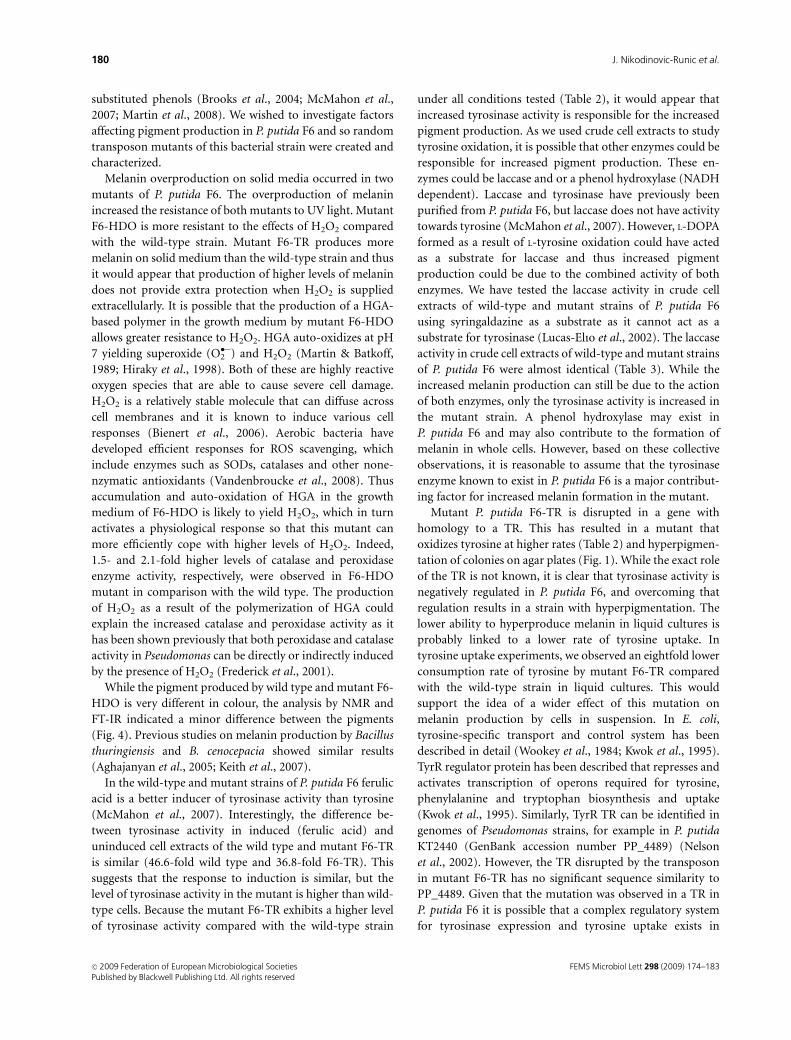
In all FT-IR spectra of the melanins isolated from
P. putida F6 and mutant strains characteristic for melanin
can be observed (David et al., 1996; Aghajanyan et al., 2005)
(Fig. 4). Despite the fact that the colour of the melanin
produced by mutant F6-HDO is different from that from
wild type and mutant F6-TR the IR spectral scans of all
biologically synthesized melanins were similar. However, the
melanin produced by mutant F6-HDO exhibited the highest
similarity to the auto-oxidized HGA while the melanin from
F6-TR had strong similarities to synthetic melanin (Fig. 4).
The melanin produced by P. putida F6 wild-type strain
appeared to be the mixture of both tyrosine- and HGA-
derived melanins.
Discussion
Pseudomonas putida F6 colonies and liquid culture medium
are pigmented in the presence of L-tyrosine and other 4-
Fig. 3. (a) Cell survival upon exposure to UV254 nm at 1, 2 and 5 min. (b)
The effect of H2O2 exposure on cell survival. (&, wild type; , F6-TR; ’,
F6-HDO).
Fig. 4. FT-IR spectra of (1) synthetic melanin (Sigma), (2) auto-oxidized
HGA, (3) melanin from Pseudomonas putida mutant F6-HDO, (4)
melanin from P. putida mutant F6-TR and (5) melanin from P. putida F6.
FEMS Microbiol Lett 298 (2009) 174–183 c� 2009 Federation of European Microbiological SocietiesPublished by Blackwell Publishing Ltd. All rights reserved
179Melanin-overproducing transposon mutants of P. putida F6
substituted phenols (Brooks et al., 2004; McMahon et al.,
2007; Martin et al., 2008). We wished to investigate factors
affecting pigment production in P. putida F6 and so random
transposon mutants of this bacterial strain were created and
characterized.
Melanin overproduction on solid media occurred in two
mutants of P. putida F6. The overproduction of melanin
increased the resistance of both mutants to UV light. Mutant
F6-HDO is more resistant to the effects of H2O2 compared
with the wild-type strain. Mutant F6-TR produces more
melanin on solid medium than the wild-type strain and thus
it would appear that production of higher levels of melanin
does not provide extra protection when H2O2 is supplied
extracellularly. It is possible that the production of a HGA-
based polymer in the growth medium by mutant F6-HDO
allows greater resistance to H2O2. HGA auto-oxidizes at pH
7 yielding superoxide (O2��) and H2O2 (Martin & Batkoff,
1989; Hiraky et al., 1998). Both of these are highly reactive
oxygen species that are able to cause severe cell damage.
H2O2 is a relatively stable molecule that can diffuse across
cell membranes and it is known to induce various cell
responses (Bienert et al., 2006). Aerobic bacteria have
developed efficient responses for ROS scavenging, which
include enzymes such as SODs, catalases and other none-
nzymatic antioxidants (Vandenbroucke et al., 2008). Thus
accumulation and auto-oxidation of HGA in the growth
medium of F6-HDO is likely to yield H2O2, which in turn
activates a physiological response so that this mutant can
more efficiently cope with higher levels of H2O2. Indeed,
1.5- and 2.1-fold higher levels of catalase and peroxidase
enzyme activity, respectively, were observed in F6-HDO
mutant in comparison with the wild type. The production
of H2O2 as a result of the polymerization of HGA could
explain the increased catalase and peroxidase activity as it
has been shown previously that both peroxidase and catalase
activity in Pseudomonas can be directly or indirectly induced
by the presence of H2O2 (Frederick et al., 2001).
While the pigment produced by wild type and mutant F6-
HDO is very different in colour, the analysis by NMR and
FT-IR indicated a minor difference between the pigments
(Fig. 4). Previous studies on melanin production by Bacillus
thuringiensis and B. cenocepacia showed similar results
(Aghajanyan et al., 2005; Keith et al., 2007).
In the wild-type and mutant strains of P. putida F6 ferulic
acid is a better inducer of tyrosinase activity than tyrosine
(McMahon et al., 2007). Interestingly, the difference be-
tween tyrosinase activity in induced (ferulic acid) and
uninduced cell extracts of the wild type and mutant F6-TR
is similar (46.6-fold wild type and 36.8-fold F6-TR). This
suggests that the response to induction is similar, but the
level of tyrosinase activity in the mutant is higher than wild-
type cells. Because the mutant F6-TR exhibits a higher level
of tyrosinase activity compared with the wild-type strain
under all conditions tested (Table 2), it would appear that
increased tyrosinase activity is responsible for the increased
pigment production. As we used crude cell extracts to study
tyrosine oxidation, it is possible that other enzymes could be
responsible for increased pigment production. These en-
zymes could be laccase and or a phenol hydroxylase (NADH
dependent). Laccase and tyrosinase have previously been
purified from P. putida F6, but laccase does not have activity
towards tyrosine (McMahon et al., 2007). However, L-DOPA
formed as a result of L-tyrosine oxidation could have acted
as a substrate for laccase and thus increased pigment
production could be due to the combined activity of both
enzymes. We have tested the laccase activity in crude cell
extracts of wild-type and mutant strains of P. putida F6
using syringaldazine as a substrate as it cannot act as a
substrate for tyrosinase (Lucas-Elıo et al., 2002). The laccase
activity in crude cell extracts of wild-type and mutant strains
of P. putida F6 were almost identical (Table 3). While the
increased melanin production can still be due to the action
of both enzymes, only the tyrosinase activity is increased in
the mutant strain. A phenol hydroxylase may exist in
P. putida F6 and may also contribute to the formation of
melanin in whole cells. However, based on these collective
observations, it is reasonable to assume that the tyrosinase
enzyme known to exist in P. putida F6 is a major contribut-
ing factor for increased melanin formation in the mutant.
Mutant P. putida F6-TR is disrupted in a gene with
homology to a TR. This has resulted in a mutant that
oxidizes tyrosine at higher rates (Table 2) and hyperpigmen-
tation of colonies on agar plates (Fig. 1). While the exact role
of the TR is not known, it is clear that tyrosinase activity is
negatively regulated in P. putida F6, and overcoming that
regulation results in a strain with hyperpigmentation. The
lower ability to hyperproduce melanin in liquid cultures is
probably linked to a lower rate of tyrosine uptake. In
tyrosine uptake experiments, we observed an eightfold lower
consumption rate of tyrosine by mutant F6-TR compared
with the wild-type strain in liquid cultures. This would
support the idea of a wider effect of this mutation on
melanin production by cells in suspension. In E. coli,
tyrosine-specific transport and control system has been
described in detail (Wookey et al., 1984; Kwok et al., 1995).
TyrR regulator protein has been described that represses and
activates transcription of operons required for tyrosine,
phenylalanine and tryptophan biosynthesis and uptake
(Kwok et al., 1995). Similarly, TyrR TR can be identified in
genomes of Pseudomonas strains, for example in P. putida
KT2440 (GenBank accession number PP_4489) (Nelson
et al., 2002). However, the TR disrupted by the transposon
in mutant F6-TR has no significant sequence similarity to
PP_4489. Given that the mutation was observed in a TR in
P. putida F6 it is possible that a complex regulatory system
for tyrosinase expression and tyrosine uptake exists in
FEMS Microbiol Lett 298 (2009) 174–183c� 2009 Federation of European Microbiological SocietiesPublished by Blackwell Publishing Ltd. All rights reserved
180 J. Nikodinovic-Runic et al.
P. putida F6. However, polar effects of transposon mutagen-
esis in mutant F6-TR may also explain the increased
tyrosinase expression and decreased tyrosine uptake.
Mutant F6-HDO is disrupted in a gene that has homol-
ogy with HDO. HGA has been reported to be involved in
eumelanin synthesis in a number of bacterial strains (Kotob
et al., 1995; Carreira et al., 2001; Keith et al., 2007). Kang
et al. (2008) have recently reported pigmentation of Pseudo-
monas chlororaphis O6 disrupted in HDO while the authors
have not reported the presence of tyrosinase in this strain.
Pseudomonas putida F6 mutant HDO achieved a final
biomass 1.2-fold lower than the wild-type strain with
tyrosine as the sole source of carbon and energy, indicating
that the tyrosine metabolic pathway through HGA is not
critical for growth of P. putida F6 but that both are needed
for optimal growth of P. putida F6 on tyrosine. The
metabolism of tyrosine to HGA is known to proceed via
PHPPA and PHPA (Coon et al., 1994; David et al., 1996).
However, P. putida F6 wild type and mutant F6-HDO grew
well with PHPPA as the sole source of carbon and energy
and did not produce pigment when incubated with this
substrate, indicating that it is unlikely to be an intermediate
in melanin synthesis in P. putida F6. While wild type and
mutant F6-HDO grew equally well, only the former pro-
duced a pigment when supplied with PHPA as a sole source
of carbon. This would suggest that PHPA, a known substrate
for tyrosinase (O’Connor et al., 2001), is also not likely
to be an intermediate in tyrosine metabolism via HGA in
P. putida F6.
In conclusion, P. putida F6 mutant F6-TR overproduces
melanin due to higher levels of tyrosine oxidation compared
with the wild-type strain, while mutant F6-HDO produces a
melanin based on accumulation of HGA in the growth
medium. While both melanins offer increased resistance to
UV light, the accumulation of the HGA-based polymer
offers additional increased resistance to H2O2.
Acknowledgement
This research was funded by the Irish Research Council for
Science Engineering and Technology (IRCSET) grant num-
ber RS/2003/64.
Authors’contribution
The authors L.B.M. and J.N.-R. contributed equally to this
work.
References
Aghajanyan AE, Hambardzumyan AA, Hovsepyan AS, Asaturian
RA, Vardanyan AA & Saghiyan AA (2005) Isolation,
purification and physicochemical characterization of water-
soluble Bacillus thuringiensis melanin. Pigm Cell Res 18:
130–135.
Altschul SF, Madden TL, Schaffer AA, Zhang JH, Zhang Z, Miller
W & Lipman DJ (1997) Gapped BLAST and PSI-BLAST: a new
generation of protein database search programs. Nucleic Acids
Res 25: 3389–3402.
Beers RF & Sizer IW (1952) A spectrophotometric method for
measuring breakdown of hydrogen peroxide by catalase. J Biol
Chem 195: 133–141.
Bienert GP, Schjoerring JK & Jahn TP (2006) Membrane
transport of hydrogen peroxide. Biochim Biophys Acta 1758:
994–1003.
Brooks SJ, Doyle EM, Hewage C, Malthouse JPG, Duetz W &
O’Connor KE (2004) Biotransformation of halophenols using
crude cell extracts of Pseudomonas putida F6. Appl Microbiol
Biot 64: 486–492.
Caetano-Anolles G (1993) Amplifying DNA with arbitrary
oligonucleotide primers. Genome Res 3: 85–94.
Carreira A, Ferreira LM & Loureiro V (2001) Brown pigments
produced by Yarrowia lipolytica result from extracellular
accumulation of homogentisic acid. Appl Environ Microb 67:
3463–3468.
Chatfield CH & Cianciotto NP (2007) The secreted pyomelanin
pigment of Legionella pneumophila confers ferric reductase
activity. Infect Immun 75: 4062–4070.
Claus H & Decker H (2006) Bacterial tyrosinases. Syst Appl
Microbiol 29: 3–14.
Coon SL, Kotob S, Jarvis BB, Wang SJ, Fuqua WC & Weiner RM
(1994) Homogentisic acid is the product of melA, which
mediates melanogenesis in the marine bacterium Shewanella
colwelliana D. Appl Environ Microb 60: 3006–3010.
David C, Daro A, Szalai E, Atarhouch T & Mergeay M (1996)
Formation of polymeric pigments in the presence of bacteria
and comparison with chemical oxidative coupling; II.
Catabolism of tyrosine and hydroxyphenylacetic acid by
Alcaligenes eutrophus CH34 and mutants. Eur Polym J 32:
669–679.
deLorenzo V & Timmis KN (1994) Analysis and construction of
stable phenotypes in gram-negative bacteria with Tn5-derived
and Tn10-derived minitransposons. Meth Enzymol 235:
386–405.
de Lorenzo V, Herrero M, Jakubzik U & Timmis KN (1990) Mini-
Tn5 transposon derivatives for insertion mutagenesis,
promoter probing, and chromosomal insertion of cloned
DNA in gram-negative eubacteria. J Bacteriol 172: 6568–6572.
Espin JC, Morales M, Varon R, Tudela J & Garciacanovas F (1995)
A continuous spectrophotometric method for determining the
monophenolase and diphenolase activities of apple
polyphenol oxidase. Anal Biochem 231: 237–246.
Espinosa-Urgel M, Salido A & Ramos J-L (2000) Genetic analysis
of functions involved in adhesion of Pseudomonas putida to
seeds. J Bacteriol 182: 2363–2369.
Frederick JR, Elkins JG, Bollinger N, Hassett DJ & McDermott TR
(2001) Factors affecting catalase expression in Pseudomonas
FEMS Microbiol Lett 298 (2009) 174–183 c� 2009 Federation of European Microbiological SocietiesPublished by Blackwell Publishing Ltd. All rights reserved
181Melanin-overproducing transposon mutants of P. putida F6
aeruginosa biofilms and planktonic cells. Appl Environ Microb
67: 1375–1379.
Gibello A, Ferrer E, Sanz J & Martin M (1995) Polymer
production by Klebsiella pneumoniae 4-hydroxyphenylacetic
acid hydroxylase genes cloned in Escherichia coli. Appl Environ
Microb 61: 4167–4171.
Harkin J & Obst J (1973) Syringaldazine, an effective reagent for
detecting laccase and peroxidase in fungi. Experientia 29:
381–387.
Heikkila RE & Cabbat F (1976) A sensitive assay for superoxide
dismutase based on the autooxidation of 6-hydroxydopamine.
Anal Biochem 75: 356–362.
Herrero M, de Lorenzo V & Timmis KN (1990) Transposon
vectors containing non-antibiotic resistance selection
markers for cloning and stable chromosomal insertion of
foreign genes in gram-negative bacteria. J Bacteriol 172:
6557–6567.
Hill HZ (1992) The function of melanin or 6 blind people
examine an elephant. Bioessays 14: 49–56.
Hiraky Y, Yamasaki M & Kawanishi S (1998) Oxidative DNA
damage induced by homogentisic acid, a tyrosine metabolite.
FEBS Lett 432: 13–16.
Jacobson ES (2000) Pathogenic roles for fungal melanins. Clin
Microbiol Rev 13: 708–717.
Kang BR, Han SH, Cho SM, Anderson AJ, Kim IS, Park SK & Kim
YC (2008) Characterization of a homogentisate dioxygenase
mutant in Pseudomonas chlororaphis O6. Curr Microbiol 56:
145–149.
Katritzky AR, Akhmedov NG, Denisenko SN & Denisko OV
(2002) 1H NMR spectroscopic characterization of colutions of
Sepia melanin, Sepia melanin free acid and human hair
melanin. Pigm Cell Res 15: 93–97.
Keith KE, Killip L, He P, Moran GR & Valvano MA (2007)
Burkholderia cenocepacia C5424 produces a pigment with
antioxidant properties using a homogentisate intermediate.
J Bacteriol 189: 9057–9065.
Korner H, Sofia HJ & Zumft WG (2003) Phylogeny of bacterial
superfamily of Crp-Fnr transcription regulators: exploiting the
metabolic spectrum by controlling alternative gene programs.
FEMS Microbiol Rev 27: 559–592.
Kotob SI, Coon SL, Quintero EJ & Weiner RM (1995)
Homogentisic acid is the primary precursor of melanin
synthesis in Vibrio cholerae, a hyphomonas strain, and
Shewanella colwelliana. Appl Environ Microb 61:
1620–1622.
Kwok T, Yang J, Pittard AJ, Wison TJ & Davidson BE (1995)
Analysis of the Escherichia coli mutant TyrR protein with
impaired capacity of tyrosine-mediated repression, but still
able to activate s70 promoters. Mol Microbiol 17: 471–481.
Lopez-Serrano D, Solano F & Sanchez-Amat A (2004)
Identification of an operon involved in tyrosinase activity and
melanin synthesis in Marinomonas mediterranea. Gene 342:
179–187.
Lucas-Elıo P, Solano F & Sanchez-Amat A (2002) Regulation of
polyphenol oxidase activities and melanin synthesis in
Marinomonas mediterranea: identification of ppoS, a gene
encoding a sensor histidine kinase. Microbiology 148:
2457–2466.
Martin JP & Batkoff B (1989) Homogentisic acid autooxidation
and oxygen radical generation: implications for the
etiology of alkaptonuric arthritis. Free Radical Bio Med 3:
241–250.
Martin LB, Nikodinovic J, McMahon AM, Vijgenboom E &
O’Connor KE (2008) Assessing the catalytic activity of three
different sources of tyrosinase: a study of the oxidation of
mono and difluorinated monophenols. Enzyme Microb Tech
43: 297–301.
Matoba Y, Kumagai T, Yamamoto A, Yoshitsu H & Sugiyama M
(2006) Crystallographic evidence that the dinuclear copper
center of tyrosinase is flexible during catalysis. J Biol Chem
281: 8981–8990.
McMahon AM, Doyle EM, Brooks S & O’Connor KE (2007)
Biochemical characterisation of the coexisting tyrosinase and
laccase in the soil bacterium Pseudomonas putida F6. Enzyme
Microb Tech 40: 1435–1441.
Nelson K, Paulsen I, Weinel C et al. (2002) Complete genome
sequence and comparative analysis of the metabolically
versatile Pseudomonas putida KT2440. Environ Microbiol 4:
799–808.
O’Connor KE, Witholt B & Duetz W (2001) p-Hydroxy-
phenylacetic acid metabolism in Pseudomonas putida F6.
J Bacteriol 183: 928–933.
O’Leary ND, O’Connor KE, Ward P, Goff M & Dobson ADW
(2005) Genetic characterization of accumulation of
polyhydroxyalkanoate from styrene in Pseudomonas putida
CA-3. Appl Environ Microb 71: 4380–4387.
Ruzafa C, Sanchezamat A & Solano F (1995) Characterization of
the melanogenic system in Vibrio cholerae ATCC-14035. Pigm
Cell Res 8: 147–152.
Sanchez-Ferrer A, Rodriguez-Lopez JN, Garcia-Canovas F &
Garcia-Carmona F (1995) Tyrosinase - a comprehensive
review of its mechanism. Biochim Biophys Acta 1247: 1–11.
Schnell S & Steinman HM (1995) Function and stationary-phase
induction of periplasmic copper–zinc superoxide dismutase
and catalase/peroxidase in Caulobacter crescentus. J Bacteriol
177: 5924–5929.
Shivprasad S & Page WJ (1989) Catechol formation and
melanization by Na-dependant Azotobacter chroococcum: a
protective mechanism for aeroadaptation? Appl Environ
Microb 5: 1811–1817.
Sichel G, Corsaro M, Scalia M, Di Bilio AJ & Bonomo RP (1991)
In vitro scavanger activity of some flavonoids and melanins
against O2�. Free Radical Bio Med 11: 1–8.
Simonson CS, Kokjohn TA & Miller RV (1990) Inducible UV
repair potential of Pseudomonas aeruginosa PAO. J Gen
Microbiol 136: 1241–1249.
Smith PK, Krohn RI, Hermanson GT, Mallia AK, Gartner FH &
Provenzano M (1985) Measurement of protein using
bicinchonic acid. Anal Biochem 150: 76–85.
FEMS Microbiol Lett 298 (2009) 174–183c� 2009 Federation of European Microbiological SocietiesPublished by Blackwell Publishing Ltd. All rights reserved
182 J. Nikodinovic-Runic et al.
Solano F, Lucas-Elio P, Fernandez E & Sanchez-Amat A (2000)
Marinomonas mediterranea MMB-1 transposon mutagenesis:
isolation of a multipotent polyphenol oxidase mutant.
J Bacteriol 182: 3754–3760.
Vandenbroucke K, Robbens S, Vandepoele K, Inze D, Van de Peer
Y & Van Breusegem F (2008) Hydrogen peroxide-induced gene
expression across kingdoms: a comparative analysis. Mol Biol
Evol 25: 507–516.
Vilchez S, Molina L, Ramos C & Ramos JL (2000) Proline
catabolism by Pseudomonas putida: cloning, characterization,
and expression of the put genes in the presence of root
exudates. J Bacteriol 182: 91–99.
Vogel HJ & Bonner DM (1956) Acetylornithinase of Escherichia
coli – partial purification and some properties. J Biol Chem
218: 97–106.
Wakamatsu K & Ito S (2002) Advanced chemical methods in
melanin determination. Pigm Cell Res 15: 174–183.
Wookey PJ, Pittard J, Forrest S & Davidson BE (1984) Cloning of
the tyrP gene and further characterization of the tyrosine-
specific transport system in Escherichia coli K12. J Bacteriol
160: 169–174.
Yabuuchi E & Ohyama A (1972) Characterisation of pyomelanin
producing strains of Pseudomonas aeruginosa. Int J Syst
Bacteriol 22: 53–64.
FEMS Microbiol Lett 298 (2009) 174–183 c� 2009 Federation of European Microbiological SocietiesPublished by Blackwell Publishing Ltd. All rights reserved
183Melanin-overproducing transposon mutants of P. putida F6
Related Documents