Characterization of mature rat oligodendrocytes: a proteomic approach Debora Dumont, Jean-Paul Noben, Marjan Moreels, Joris Vanderlocht, Niels Hellings, Frank Vandenabeele, Ivo Lambrichts, Piet Stinissen and Johan Robben Hasselt University, Biomedical Research Institute (BIOMED) and transnationale Universiteit Limburg, School of Life Sciences, Diepenbeek, Belgium Abstract Oligodendrocytes are glial cells responsible for the synthesis and maintenance of myelin in the central nervous system (CNS). Oligodendrocytes are vulnerable to damage occurring in a variety of neurological diseases. Understanding oligo- dendrocyte biology is crucial for the dissemination of de- and remyelination mechanisms. The goal of the present study is the construction of a protein database of mature rat oligo- dendrocytes. Post-mitotic oligodendrocytes were isolated from mature Wistar rats and subjected to immunocytochem- istry. Proteins were extracted and analyzed by means of two- dimensional gel electrophoresis and two-dimensional liquid chromatography, both coupled to mass spectrometry. The combination of the gel-based and gel-free approach resulted in confident identification of a total of 200 proteins. A minority of proteins were identified in both proteomic strategies. The identified proteins represent a variety of functional groups, including novel oligodendrocyte proteins. The results of this study emphasize the power of the applied proteomic strategy to study known or to reveal new proteins and to investigate their regulation in oligodendrocytes in different disease models. Keywords: mass spectrometry, oligodendrocytes, proteo- mics, rat brain, two-dimensional gel electrophoresis, two- dimensional liquid chromatography. J. Neurochem. (2007) 102, 562–576. Oligodendrocytes synthesize and maintain myelin in the central nervous system. Oligodendrocytes are vulnerable to injury mediated by oxidative stress (Rosenberg et al. 1999), cytotoxicity (Griot-Wenk et al. 1991; Scolding and Comp- ston 1991; Jurewicz et al. 1998; Russell and Ley 2002) excitotoxicity (Matute et al. 2001; Werner et al. 2001), trophic factor deprivation and activation of apoptotic path- ways (Vartanian et al. 1995; Vanderlocht et al. 2006). Oligodendrocyte damage is observed in a variety of diseases including multiple sclerosis (Buntinx et al. 2002), stroke (Aboul-Enein et al. 2003), dementia (Kurz et al. 2003), spinal cord trauma (Gomes-Leal et al. 2004), encephalopa- thies (El Hachimi et al. 1998), leukodystrophies (Ip et al. 2006) and Alzheimer’s disease (Ness et al. 2005). Although the composition of the myelin sheath has been investigated by several proteomic strategies (Persson and Overholm 1990; Yamaguchi and Pfeiffer 1999; Taylor and Pfeiffer 2003;Taylor et al. 2004; Vanrobaeys et al. 2005), no comprehensive proteomic study of oligodendrocytes has been published. Characterization of the oligodendrocyte proteome is on one hand crucial for a better understanding of molecular mechanisms in oligodendrocytes under normal and pathological conditions. On the other hand, because of the limited availability of human brain biopsy material, animal models for disease (mainly rodents) are of utmost importance for CNS research. Therefore, a comprehensive proteomic map of rodent oligodendrocytes is expected to be a useful new tool aiding research of oligodendrocyte pathology. Received September 18, 2006; revised manuscript received December 17, 2006; accepted February 6, 2007. Address correspondence and reprint requests to Johan Robben, Ph.D., Hasselt University, Biomedical Research Institute, Agoralaan building, A 3590 Diepenbeek,Belgium. E-mail: [email protected] Abbreviations used: 2D-GE, two-dimensional gel electrophoresis; 2D-LC, two-dimensional liquid chromatography; ACN, acetonitrile; EAE, experimental autoimmune encephalomyelitis; FCS, fetal calf serum; GalC, galactosylceramide; HAc, acetic acid; HSP, heat-shock protein; IPG, immobilized pH-gradient; LC-ESI-MS/MS, liquid chro- matography-electrospray ionization-tandem mass spectrometry; MAG, myelin-associated glycoprotein; MBP, myelin basic protein; MIF, macrophage migration inhibitory factor; MS, mass spectrometry; Mw, molecular weight; PBS, phosphate buffered saline; pI, isoelectric point; SCX, strong cation exchange. Journal of Neurochemistry , 2007, 102, 562–576 doi:10.1111/j.1471-4159.2007.04575.x 562 Journal Compilation Ó 2007 International Society for Neurochemistry, J. Neurochem. (2007) 102, 562–576 Ó 2007 The Authors

Welcome message from author
This document is posted to help you gain knowledge. Please leave a comment to let me know what you think about it! Share it to your friends and learn new things together.
Transcript
Characterization of mature rat oligodendrocytes: a proteomicapproach
Debora Dumont, Jean-Paul Noben, Marjan Moreels, Joris Vanderlocht, Niels Hellings,Frank Vandenabeele, Ivo Lambrichts, Piet Stinissen and Johan Robben
Hasselt University, Biomedical Research Institute (BIOMED) and transnationale Universiteit Limburg, School of Life Sciences,
Diepenbeek, Belgium
Abstract
Oligodendrocytes are glial cells responsible for the synthesis
and maintenance of myelin in the central nervous system
(CNS). Oligodendrocytes are vulnerable to damage occurring
in a variety of neurological diseases. Understanding oligo-
dendrocyte biology is crucial for the dissemination of de- and
remyelination mechanisms. The goal of the present study is
the construction of a protein database of mature rat oligo-
dendrocytes. Post-mitotic oligodendrocytes were isolated
from mature Wistar rats and subjected to immunocytochem-
istry. Proteins were extracted and analyzed by means of two-
dimensional gel electrophoresis and two-dimensional liquid
chromatography, both coupled to mass spectrometry. The
combination of the gel-based and gel-free approach resulted
in confident identification of a total of 200 proteins. A minority
of proteins were identified in both proteomic strategies. The
identified proteins represent a variety of functional groups,
including novel oligodendrocyte proteins. The results of this
study emphasize the power of the applied proteomic strategy
to study known or to reveal new proteins and to investigate
their regulation in oligodendrocytes in different disease
models.
Keywords: mass spectrometry, oligodendrocytes, proteo-
mics, rat brain, two-dimensional gel electrophoresis, two-
dimensional liquid chromatography.
J. Neurochem. (2007) 102, 562–576.
Oligodendrocytes synthesize and maintain myelin in thecentral nervous system. Oligodendrocytes are vulnerable toinjury mediated by oxidative stress (Rosenberg et al. 1999),cytotoxicity (Griot-Wenk et al. 1991; Scolding and Comp-ston 1991; Jurewicz et al. 1998; Russell and Ley 2002)excitotoxicity (Matute et al. 2001; Werner et al. 2001),trophic factor deprivation and activation of apoptotic path-ways (Vartanian et al. 1995; Vanderlocht et al. 2006).Oligodendrocyte damage is observed in a variety of diseasesincluding multiple sclerosis (Buntinx et al. 2002), stroke(Aboul-Enein et al. 2003), dementia (Kurz et al. 2003),spinal cord trauma (Gomes-Leal et al. 2004), encephalopa-thies (El Hachimi et al. 1998), leukodystrophies (Ip et al.2006) and Alzheimer’s disease (Ness et al. 2005).
Although the composition of the myelin sheath has beeninvestigated by several proteomic strategies (Persson andOverholm 1990; Yamaguchi and Pfeiffer 1999; Taylor andPfeiffer 2003;Taylor et al. 2004; Vanrobaeys et al. 2005), nocomprehensive proteomic study of oligodendrocytes hasbeen published. Characterization of the oligodendrocyteproteome is on one hand crucial for a better understandingof molecular mechanisms in oligodendrocytes under normal
and pathological conditions. On the other hand, because ofthe limited availability of human brain biopsy material,animal models for disease (mainly rodents) are of utmostimportance for CNS research. Therefore, a comprehensiveproteomic map of rodent oligodendrocytes is expected to bea useful new tool aiding research of oligodendrocytepathology.
Received September 18, 2006; revised manuscript received December17, 2006; accepted February 6, 2007.Address correspondence and reprint requests to Johan Robben, Ph.D.,
Hasselt University, Biomedical Research Institute, Agoralaan building,A 3590 Diepenbeek,Belgium. E-mail: [email protected] used: 2D-GE, two-dimensional gel electrophoresis;
2D-LC, two-dimensional liquid chromatography; ACN, acetonitrile;EAE, experimental autoimmune encephalomyelitis; FCS, fetal calfserum; GalC, galactosylceramide; HAc, acetic acid; HSP, heat-shockprotein; IPG, immobilized pH-gradient; LC-ESI-MS/MS, liquid chro-matography-electrospray ionization-tandem mass spectrometry; MAG,myelin-associated glycoprotein; MBP, myelin basic protein; MIF,macrophage migration inhibitory factor; MS, mass spectrometry; Mw,molecular weight; PBS, phosphate buffered saline; pI, isoelectric point;SCX, strong cation exchange.
Journal of Neurochemistry, 2007, 102, 562–576 doi:10.1111/j.1471-4159.2007.04575.x
562 Journal Compilation � 2007 International Society for Neurochemistry, J. Neurochem. (2007) 102, 562–576� 2007 The Authors

To identify disease-related proteins in neurological disor-ders, studies using proteomic approaches are rapidly emer-ging (Harrington et al. 1985; Lubec et al. 1999; Choe et al.2002a,b; Davidsson et al. 2002; Guillaume et al. 2003; Jianget al. 2003; Dumont et al. 2004). Two-dimensional gelelectrophoresis (2D-GE) has become the standard approachfor the separation of complex protein mixtures. Indeed, thistechnique allows high-resolution protein mapping of variousbodily fluids, as well as tissue and cell types, therebyproviding valuable tools for identifying candidate diseasemarkers. However, proteins that are high or low in isoelectricpoints (pI), molecular weights (Mw), polarity or abundanceremain inadequately detected by 2D-GE. Other separationtechnologies have been explored to address some of theselimitations. Peptide-mapping by two-dimensional liquid chro-matography (2D-LC) emerged as a plausible alternative to2D-GE and has already proven its worth in CNS research(Maccarrone et al. 2004; Noben et al. 2006). In this study, aproteomic initiative was undertaken in our laboratory,applying 2D-GE as well as 2D-LC in combination withmass spectrometry for the construction of a proteomic map ofmature rat oligodendrocytes.
Materials and methods
Primary rat oligodendrocyte cultures
Primary rat oligodendrocytes were isolated from whole brains of
adult Wistar rats (Harlan, The Netherlands) as described by Yong
and Antel (1992). Briefly, brain tissue was dissociated mechanically
and enzymatically for 30 min at 37�C. Glial cells were separated
from myelin debris and red blood cells by means of Percoll gradient
centrifugation. The mixed glial cell fraction was suspended in
Dulbecco’s modified Eagle medium (DMEM, Life Technologies,
Rockville, MD, USA) supplemented with 5% fetal calf serum
(FCS), penicillin (50 U/mL) and streptomycin (50lg/mL) and
seeded in culture flasks. The cultures were selectively enriched for
oligodendrocytes by means of differential adhesion to plastic. The
non-adherent oligodendrocyte fraction was plated at a density of
2 · 105 onto poly-L-lysine-coated glass slides and at a density of
106 cells/mL on poly-L-lysine-coated 24-well plates for immuno-
cytochemistry and proteomic analysis, respectively. Cells were
cultured for 12–14 days in DMEM supplemented with 5% FCS.
Immunocytochemistry
Cells seeded on glass slides were stained using the peroxide-based
Envision System (DakoCytomation, Glostrup, Denmark). Cells were
fixed in 4% formaldehyde (Unifix, Duiven, The Netherlands) for
20 min. After washing in 0.01 mol/L phosphate buffered saline
(PBS), cells were permeabilized (for intracellular staining) with
0.05% Triton X-100 (Boehringer, Mannheim, Germany) in PBS for
30 min followed by an additional wash step. Non-specific binding
sites were blocked with 3% normal goat serum (DakoCytomation) in
PBS for 20 min. After washing, cells were incubated for 1 h with
antibodies specific for myelin basic protein (MBP; 1/200; Serotec,
Oxford, UK), galactosylceramide (GalC; 1:50; kind gift from
Dr V.W. Yong, University of Calgary, Canada) or myelin-associated
glycoprotein (MAG; 1:500; Chemicon, Temecula, CA,USA), washed
thrice and incubated for 30 min with anti-mouse peroxidase-conju-
gated secondary antibody (DakoCytomation). A high sensitivity
diaminobenzidine chromogenic substrate system (DakoCytomation)
was used to visualize the immunoreactivity. Cells were counterstained
with Mayer’s hematoxylin, mounted in Aquatex (Merck, Darmstadt,
Germany) and examined using a photomicroscope equipped with
an automated camera (Nikon Eclipse 80i, Japan).
Two-dimensional gel electrophoresis (2D-GE)
Oligodendrocytes were lysed at 4�C in a buffer containing 7 mol/L
urea, 2 mol/L thiourea, 2% w/v CHAPS and homogenized with a
rotor stator mixer. Proteins were extracted using acetone precipita-
tion. The protein content was determined with the 2-D Quant kit
(GE Healthcare, Uppsala, Sweden). One part of the extract
containing 100 lg of proteins was concentrated using 5 kDa cut-
off Ultrafree columns (Millipore, Bedford CA, USA). The retentate
was suspended in 450 lL rehydration buffer (7 mol/L urea, 2 mol/L
thiourea, 2% w/v CHAPS, 200 mmol/L dithiothreitol, 0.5% v/v
immobilized pH-gradient (IPG) buffer pH 3–10 and bromophenol
blue) and spread out over the bottom of a strip holder. An IPG strip
(24 cm, linear pH 3–10; GE Healthcare) was applied on the protein
mixture and rehydrated for 12 h at 22�C. Two additional parts of thesame extract were treated in the same way. Isoelectric focusing,
SDS-PAGE, staining and software analysis were conducted as
described previously (Dumont et al. 2004). Spots were picked from
at least one of three gels run in parallel and gel plugs were trypsin-
digested individually or in combination (collecting corresponding
weak spots) according to the method described by Shevchenko et al.(Shevchenko et al. 1996).
Two-dimensional liquid chromatography (2D-LC)
Forty micrograms of the same protein extract (described in Two-
dimensional gel electrophoresis) was subjected to strong cation-
exchange (SCX) chromatography as described previously (Noben
et al. 2006). Briefly, protein extracts were denaturated, alkylated,
and digested with trypsin. Digests were trapped on a Hypercarb
column (0.5 cm · 200 lm i.d.; Nanoseparations, Nieuwkoop, The
Netherlands) and transferred to a polysulfoethyl aspartamide column
(12 cm · 200 lm i.d.; Nanoseparations) in 1 lL solution A (0.5%
(v/v) acetic acid (HAc) in water), containing 70% acetonitrile
(ACN). The analytical column was eluted with a linear salt gradient
(slope 15 mmol/L KCl/min) starting from 100% solution A to 100%
solution A containing 250 mmol/L KCl and 35% (v/v) ACN, to
100% solution A containing 500 mmol/L KCl and 35% (v/v) ACN
and 1 min fractions (2.5 lL) were collected. SCX fraction volume
was adjusted to �25 lL with a solution containing 5% (v/v) ACN in
100 mmol/L HAc. The second dimension separation involved the
reversed-phase chromatography described in Mass spectrometry.
Mass spectrometry
2D-GE protein digests and SCX fractions were analysed by liquid
chromatography-electrospray ionization-tandem mass spectrometry
(LC-ESI -MS/MS). Flow regulation was as described by Meiring
et al.(2002). Each digest/fraction was diluted with a solution
containing 5% (v/v) ACN in 100 mmol/L HAc. This solution
contained 4 pg/lL of cortisone as an internal analytical standard to
Proteomic analysis of rat oligodendrocytes 563
� 2007 The AuthorsJournal Compilation � 2007 International Society for Neurochemistry, J. Neurochem. (2007) 102, 562–576
monitor flow stability (by retention time) and overall performance
(by peak height as derived from the selected ion chromatogram for
[M + H]+ = m/z 361.2) of the ion trap mass spectrometer (LCQ
Classic, ThermoElectron Corporation, San Jose, CA, USA). Of each
digest/fraction, 10 lL was injected (autoinjector AS3000, Thermo-
Electron). The trapped samples were separated on the analytical
column (Biosphere C18, 200 mm L · 0.05 mm i.d., 5 lm, Nano-
separations) using a linear gradient from 5 to 60% v/v ACN in water
containing 100 mmol/L HAc either in 55 min (2-DE protein
digests) or 120 min (SCX-separated fractions). The eluate from
the analytical column was introduced in a nanoelectrospray device
(ThermoFinnigan) and sprayed from a gold-coated fused silica
emitter (5 lm i.d., NanoSeparations). The LCQ automated protocols
were used to optimize the ion optics and calibration parameters. The
mass spectrometer was operated in a data-dependent acquisition
mode to automatically switch between MS (m/z 300–1500 Thomp-
son in centroid mode at a maximum injection time of 150 ms) and
MS/MS acquisition on the three most intense precursor ions,
controlled by Xcalibur 1.3 software.
Database management, evaluation and reporting
Peak lists in DTA file format were generated from mass spectro-
metric raw data files using the CreateDTA tool available in Sequest
v27 within BioWorks v3.0 (ThermoElectron).
2D-GE-derived MS data
DTA files were examined with the search engines Sequest v27
(Thermofinnigan) and Mascot (Matrix Science) using the database
NCBInr (subset Rodentia, 105,999 entries). Sequest parameters
were set as follows: Xcorr ‡ 1.8, ‡ 2.5 or ‡ 3.5 for singly, doubly
or triply charged peptide ions; delta Cn > 0.1; precursor and product
ion mass tolerance ± 3 and ± 1 Da; enzyme: trypsin; one missed
cleavage allowed; static chemical modification: cysteine-carbami-
domethylation; dynamic chemical modification: oxidation of methi-
onine, histidine, and tryptophan. Mascot search parameters were:
taxonomy rodentia, parent, and peptide ion mass tolerance ± 3
and ± 1 Da, enzyme: trypsin/P, one missed cleavage allowed. The
Mascot significance threshold was set at p £ 0.05.
2D-LC-derived MS data
DTA files derived from each SCX fraction were first examined with
the search engine Sequest as described above (2D-GE-derived MS
data). Sequest identifications were assembled in a DTASelect v1.9
report (Tabb et al. 2002). DTASelect licenses were obtained from
the Scripps Research Institute (La Jolla, CA, USA). Second, all DTA
files were examined with Mascot as described in 2D-GE-derived
MS data.
Evaluation
The number of sibling peptides was derived according to the
guidelines for peptides and protein identification data (Carr et al.2004). Only peptides assigned by both search engines were retained
in this study. Proteins assigned on the basis of two or more peptides
meeting the criteria described in 2D-GE-derived MS data and
2D-LC-derived MS data were considered as confidently identified.
Single-peptide protein identifications returned by both Sequest and
Mascot were re-examined with the de novo sequencing algorithm
Lutefisk1900 v.1.3.2. (Taylor and Johnson 1997) and the
tag-generating algorithm Inspect (Frank et al. 2005; Tanner et al.2005) in this order. In case the outcome was negative, the peptide
identification was rejected. Finally, an additional data analysis was
performed to validate the 2D-LC identifications. To calculate the
false-positive error rate, all DTA files were analyzed with Mascot
using the ‘sequence-reversed’ databases RV_NCBI, generated with
an in-house developed Perl script.
Results
Oligodendrocyte cultures
Oligodendrocytes were isolated from whole brain of adultrats and their phenotype was studied by means of immun-ocytochemistry. After 12–14 days, 90% of the cells in thecultures consisted of oligodendrocytes with a characteristicmature morphology. They were characterized by a roundphase bright nucleus and a small number of thick, irregularcell processes. Clusters of 3–10 oligodendrocytes were oftenobserved in culture (Fig. 1a). Mature oligodendrocytesexpressed MAG, MBP and GalC proteins (Figs 1b–d).Longitudinal flowcytometric analysis was performed show-ing that the vast majority of MBP positive cells were derivedfrom MBP/GalC positive cells while only a very smallincrease in NG2 precursor cells was observed. The amount ofprecursor cells mounted to 5% while contaminating astro-cytes, fibroblasts en endothelial cells constituted less than 5%(data not shown).
2D-GE and 2D-LC experiments
2D-GE experiments were performed on three oligodendro-cyte cultures. Silver staining of three gels from each culturerevealed reproducible protein patterns (Fig. 2). Proteins spotswere picked, trypsin digested and analyzed by means of ESI-LC-MS/MS. Only peptides assigned by both Sequest andMascot search engines were retained in this study.
From one culture of rat oligodendrocytes, an ultrafiltered,reduced, alkylated and trypsinized sample was prepared andfractionated by SCX. The resulting fractions were analyzedby LC-ESI-MS/MS. A total of 30,467 MS/MS spectra (DTAfiles) were recorded. First, these DTA files were examinedwith the search engine Sequest. Because of the stringentselection criteria (2D-LC-derived MS data) and the removalof contaminants (e.g. keratins) only 1% (306 DTA files) ofthe initial data set (30 467 DTA files) was assigned bySequest. Second, all DTA files were searched with Mascot.Only peptides assigned by both Sequest and Mascot wereretained in this study.
Validation of the proteomic data
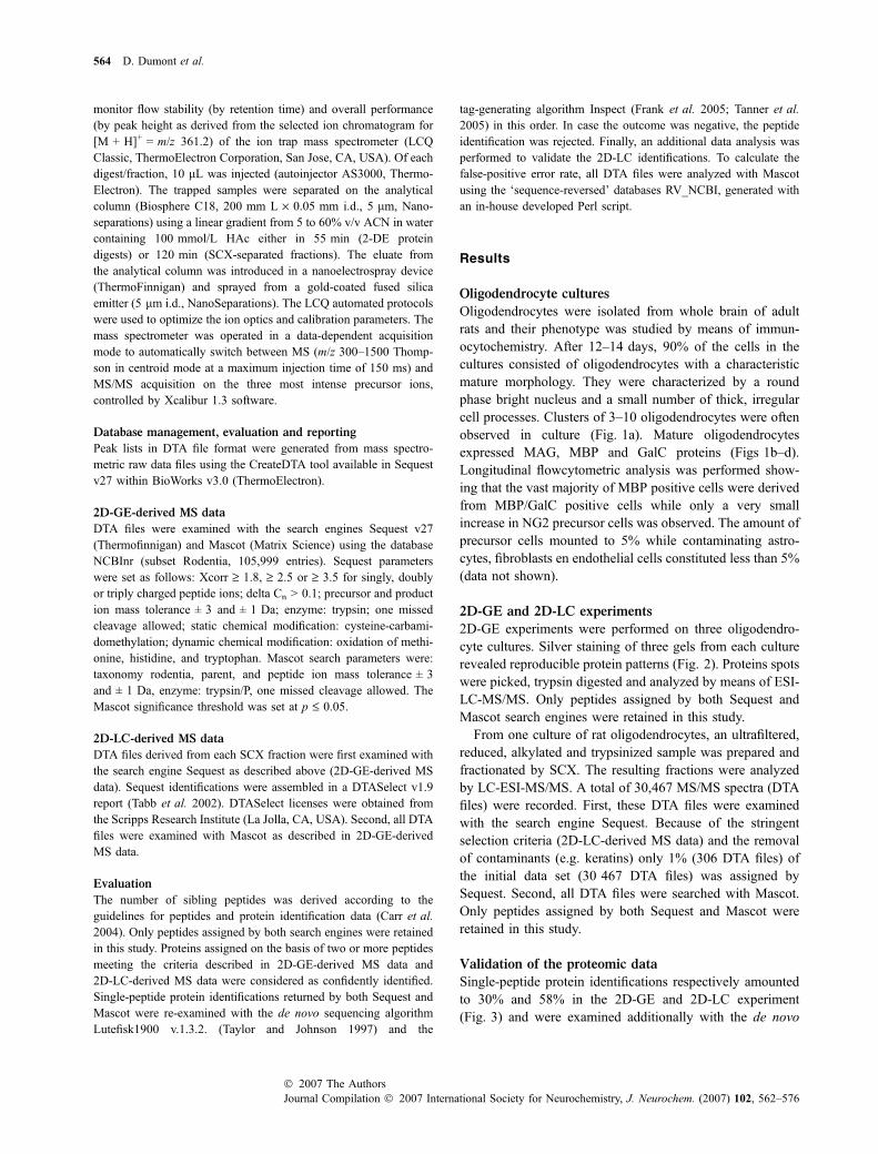
Single-peptide protein identifications respectively amountedto 30% and 58% in the 2D-GE and 2D-LC experiment(Fig. 3) and were examined additionally with the de novo
564 D. Dumont et al.
Journal Compilation � 2007 International Society for Neurochemistry, J. Neurochem. (2007) 102, 562–576� 2007 The Authors

sequencing algorithm Lutefisk. The sequence returned bySequest and Mascot matched the de novo sequence proposedby Lutefisk in 80% of all examined peptides (102). In allother cases, no quality de novo sequence could be derived(noisy spectra) or the de novo sequence with primary rankingdiffered from the one found by both search engines. In thelatter case, peptides identifications were additionallysearched with the tag-generating algorithm Inspect. In this
way, all but six peptide identifications were confirmed(Table 1).
The false-positive error rate for the DTA files derivedfrom the 2D-LC experiment was 0.7%. Since for the2D-LC experiment 50 single-peptide identifications werevalidated, the number of false-positive protein identifica-tions on the basis of one peptide is estimated at 0.35(0.7% of 50).
Mass(kDa)
93
67
43
30
20
14
3 3.5 4.0 4.5 5.0 5.5 6.0 6.5 7.0 7.5 8.0 8.5 9.0 9.5 10p/
Fig. 2 Representative 2D map of mature
rat oligodendrocyte proteins. Post-mitotic
oligodendrocytes were isolated from total
rat brain and cultured for 12–14 days. Pro-
teins were extracted and separated
according to their isoelectric point (pI) in the
first dimension (IEF strips 24 cm, pH linear
3–10) and according to their molecular
weight by means of SDS-PAGE in the
second dimension. Protein spots were cut,
trypsin-digested and analyzed by means
of LC-ESI-MS/MS. Marked spots can be
found in the supplemental material,
together with a detailed protein inventory, of
which a summary is presented in Table 1.
(a) (b)
(c) (d)Fig. 1 Morphological and immunocyto-
chemical characterization of oligodendro-
cyte cultures. The primary cultures (day
12–14) isolated form adult Wistar rats were
characterized by clusters of small, phase-
bright oligodendrocytes with dark cell pro-
cesses (a). Oligodendrocytes showed
moderate to strong immunoreactivity for
MAG (b), MBP (c) and GalC (d). (Scale bars:
a–d: 20 lm).
Proteomic analysis of rat oligodendrocytes 565
� 2007 The AuthorsJournal Compilation � 2007 International Society for Neurochemistry, J. Neurochem. (2007) 102, 562–576
Study outcome
Combining 2D-GE and 2D-LC experiments, 200 proteinswere identified in this study: 41 proteins were found incommon for both approaches, 114 proteins were found uniquefor 2D-GE and 45 unique for 2D-LC (Fig. 4, Table 1). Spotsannotations as well as protein identification details arepresented in the supplemental material. While the majorityof the 2D-GE identified proteins displayed a centered profilewith respect to pI and Mw parameters, 2D-LC identificationsspread towards the outer regions of this spectrum (Fig. 5). Inaddition, 2D-GE clearly favored the detection of cytoplasmicproteins (55%) while 2D-LC displayed a more distributedsubcellular localization (Fig. 6) including a prominent nuc-lear fraction (17%). Regarding the functional diversity, themajor part of the proteins identified via 2D-GE play a role incell metabolism. In contrast, 2D-LC identified proteins aremore evenly distributed over the different functional groups.Notably are the proteins belonging to the DNA/RNA-bindingand protein degradation group, which are more abundantlyencountered in the 2D-LC and 2D-GE analysis respectively(Fig. 7). The newly identified oligodendrocyte proteinsinclude proteins involved in Ca2+ binding (calumenin,translationally controlled tumor protein), protein folding(protein disulfide isomerase precursor A6), stress response(heat shock-related protein 1A/B, peroxiredoxins), proteindegradation (ubiquitin/proteasome pathway proteins, cathep-sin B) and CNS development (cysteine and glycine-richprotein 1, acyl-CoA-binding protein).
Discussion
Oligodendrocyte cultures
Study of oligodendrocyte development and biology withinnormal and pathological CNS tissue is generally hampered by
the limited availability of viable human brain tissue. Humanoligodengroglial cell lines represent suitable model systems tostudy oligodendrocyte injury (Buntinx et al. 2004). However,they display tumorigenic properties and often representoligodendrocytes in an immature developmental state (Bunt-inx et al. 2003). Moreover, they exhibit little or no morpho-logical resemblance with their in vivo counterparts. Incontrast, the primary oligodendrocytes used in this studydisplayed immunophenotypic characteristics of mature postmitotic oligodendrocytes (Snyder et al. 1980; Norton et al.1983;Vick et al. 1990). Caution was taken in the selection ofcell culture wells during harvesting as to minimize the numberof astrocytes and fibroblasts. Nevertheless, the presence of alimited number of contaminating cell types is inevitable. Wetried to deal with this drawback by searching literature andindicating proteins that have been reported in oligodendro-cytes or myelin (Taylor et al. 2004; Vanrobaeys et al. 2005)with the corresponding references in Table 1. Nevertheless,some of the newly identified proteins, including fatty acid-binding protein and peroxiredoxin 6 were previously des-cribed in astrocytes and could reflect the contribution of acontaminating population to the protein expression profile.These proteins have to be handled with caution and confirmedby immunocytochemistry. Another phenomenon encounteredwhen using cell cultures are cellular stress responses evokedby culture conditions. Although after 2 weeks in culture, therewas no evidence of lethal (cell death) or sublethal (retractionof processes) stress, we did identify a number of stress-associated proteins, including members of the heat shock andproteasome family (Table 1). It remains to be determinedwhether these proteins are also constitutively expressed byoligodendrocytes in vivo or represent a stress response toculture conditions. Although the latter could represent adrawback for the use of in vitro cultures, Hochstrasser andcoworkers exploited stress features by studying postmortemCSF in order to identify molecules related to ischemic andneurodegenerative conditions (Lescuyer et al. 2004).
Validation of proteomic data
The validity of the outcome of a proteomic experiment isdependent on the stringency of the validation process. In thisstudy, the validation process included the search enginesSequest and Mascot. The validity of single-peptides identi-fications in proteomic research is open to debate (Carr et al.2004) and therefore the de novo sequencing algorithmLutefisk and the tag generating algorithm Inspect were usedto evaluate these identifications (on average 44% of theproteins encountered). In 96 of the 102 cases examined, thesequence returned by Sequest and Mascot matched the topranking sequence proposed by Lutefisk or Inspect. Finally,the presence of 20 of these so called ‘one hit wonders’ in theoligodendrocyte cultures was confirmed with both 2D-GEand 2D-LC approaches while for 21 others, their occurrencein oligodendrocytes or myelin was reported in literature.
Fig. 3 Percentage of proteins matched by a given number of peptides.
Protein extracts of mature rat oligodendrocytes were subjected to 2D-
GE or trypsin-digested and subjected to 2D-LC. Mass spectrometric
data coming from 2D-GE spots or from SCX-separated fractions
were validated, resulting in 155 and 86 confident protein identifications
for the 2D-GE and 2D-LC approach, respectively. Single-peptide protein
identifications amounted to 30% and 58% in the 2D-GE and 2D-LC
approach, respectively.
566 D. Dumont et al.
Journal Compilation � 2007 International Society for Neurochemistry, J. Neurochem. (2007) 102, 562–576� 2007 The Authors
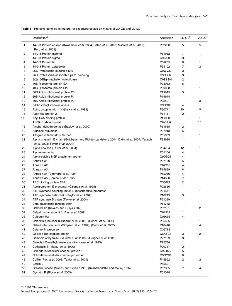
Table 1 Proteins identified in mature rat oligodendrocytes by means of 2D-GE and 2D-LC
Descriptiona Accession 2D-GEb 2D-LCc
1 14-3-3 Protein epsilon (Kawamoto et al. 2004; Satoh et al. 2003; Masters et al. 2002;
Berg et al. 2003)
P62260 5 5
2 14-3-3 Protein gamma P61982 7 1
3 14-3-3 Protein sigma Q9JJ20 3
4 14-3-3 Protein tau P68255 6 1
5 14-3-3 Protein zeta/delta P63102 7 2
6 26S Proteasome subunit p40.5 Q9WVJ2 3
7 26S Proteasome-associated pad1 homolog Q9CSU2 3
8 3(2), 5-Bisphosphate nucleotidase Q9Z1 N4 2
9 40S Ribosomal protein SA P38983 5
10 40S Ribosomal protein S20 P60868 1
11 60S Acidic ribosomal protein P0 P19945 3
12 60S Acidic ribosomal protein P1 P19944 1
13 60S Acidic ribosomal protein P2 P02401 3
14 6-Phosphogluconolactonase Q9CQ60 4
15 Actin, cytoplasmic 1 (Kajikawa et al. 1991) P60711 10 9
16 Actin-like protein 2 P61161 2
17 Acyl-CoA-binding protein P11030 1
AHNAK-related protein Q8VHJ0 1d
18 Alcohol dehydrogenase (Baslow et al. 2000) P51635 2
19 Aldolase reductase P07943 5
20 Allograft inflammatory factor-1 P55009 1
21 Alpha crystallin B chain (Goldbaum and Richter-Landsberg 2004; Dabir et al. 2004; Yaguchi
et al. 2003; Taylor et al. 2004)
P23928 1
22 Alpha enolase (Taylor et al. 2004) P04764 12 1
23 Alpha-centractin P61164 4
24 Alpha-soluble NSF attachment protein Q9DB05 4
25 Annexin A1 P07150 5
26 Annexin A2 Q07936 4
27 Annexin A3 P14669 8 1
28 Annexin A4 (Eberhard et al. 1994) P55260 4
29 Annexin A5 (Spreca et al. 1992) P14668 7
30 APC binding protein EB1 O35879 2
31 Apolipoprotein E precursor (Calenda et al. 1995) P02650 1
32 ATP synthase coupling factor 6, mitochondrial precursor P21571 1
33 ATP synthase beta chain (Taylor et al. 2004) P10719 9
34 ATP synthase D chain (Taylor et al. 2004) P31399 1
35 Beta-galactoside-binding lectin P11762 1
36 Calmodulin (Kovacs and Gulya 2002) P62161 2
37 Calpain smal subunit 1 (Ray et al. 2002) Q64537 1
38 Calponin H2 Q08093 4
39 Calnexin precursor (Eckhardt et al. 2005), (Denzel et al. 2002) P35565 1
40 Calreticulin precursor (Simpson et al. 1997), (Gudz et al. 2002) P18418 4
41 Calumenin precursor O35783 1
42 Gelsolin-like capping protein Q6AYC4 3 2
43 Carbonic anhydrase 2 (Halmi et al. 2006), (Cerghet et al. 2006) P27139 3
44 Catechol O-methyltransferase (Karhunen et al. 1995) P22734 1
45 Cathepsin B (Matsui et al. 1990) P00787 2
46 Chloride intracellular channel protein 1 Q9Z1Q5 5
47 Chloride intracellular channel protein 4 Q9QYB1 6
48 Cofilin (Fox et al. 2006; Taylor et al. 2004) P45592 2 2
49 Cofilin 2 P45591 1
50 Creatine kinase (Manos and Bryan 1993), (Kuzhikandathil and Molloy 1994) P07335 7 4
51 Cystatin B (Riccio et al. 2005) P01048 1
Proteomic analysis of rat oligodendrocytes 567
� 2007 The AuthorsJournal Compilation � 2007 International Society for Neurochemistry, J. Neurochem. (2007) 102, 562–576
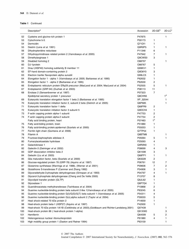
Table 1 Continued
Descriptiona Accession 2D-GEb 2D-LCc
52 Cysteine and glycine-rich protein 1 P47875 1
53 Cytochrome b-5 P00173 1
54 Dermcidin Q71DI1 1
55 Destrin (Lena et al. 1991) Q9R0P5 1 1
56 Dihydropteridine reductase P11348 6
57 Dihydropyrimidinase related protein 2 (Vanrobaeys et al. 2005) P47942 7 1
58 Dimethylarginase 1 Q9CWS0 3
59 Disabled homolog 2 O88797 1
60 DJ-1protein O88767 5
61 DnaJ (HSP40) homolog subfamily B member 11 Q99KV1 1
62 EF-hand domain-containing protein 2 Q9D8Y0 3
63 Electron tranfer flavoprotein alpha sunubit Q99LC5 1
64 Elongation factor 1 - alpha 1 (Vanrobaeys et al. 2005; Barbarese et al. 1995) P62632 1
65 Elongation factor 1 - alpha 2 (Barbarese et al. 1995) P62630 2
66 Endoplasmic reticulum protein ERp29 precursor (MacLeod et al. 2004; MacLeod et al. 2004) P52555 5 1
67 Endoplasmin (GRP-94) (Duzhak et al. 2003) P08113 11
68 Enolase 2 (Sensenbrenner et al. 1997) P07323 7
Epididymal secretory protein 1 precursor Q8CHN5 1d
69 Eukaryotic translation elongation factor 1 beta 2 (Barbarese et al. 1995) XP_32044 1
70 Eukaryotic translation initiation factor 2, subunit 2 beta (Dietrich et al. 2005) Q6P685 1
71 Eukaryotic translation factor 1 delta Q68FR9 2
72 Eukaryotic translation initiation factor 3 subunit 5 Q9DCH4 1
73 F-actin capping protein alpha-1 subunit P47753 2
74 F-actin capping protein alpha-2 subunit P47754 2
Fatty acid binding protein, heart P07483 1d
75 Fatty acid-biding protein, brain P51880 1
76 Fatty acid-binding protein,epidermal (Scarlato et al. 2000) P55053 1
77 Ferritin light chain (Quintana et al. 2006) Q7TP54 1
78 Filamin A Q8BTM8 1
79 Fructose-bisphosphate aldolase A P05064 5 1
80 Fumarylacetoaetate hydrolase P25093 4
81 Galactokinase 1 Q9R0N0 1
82 Galectin-3 (Deininger et al. 2002) P08699 4 3
83 GDP dissociation inhibitor beta 2 Q61598 3
84 Gelsolin (Liu et al. 2003) Q68FP1 1 1
85 Glia maturation factor, beta (Scarlato et al. 2000) Q63228 2
86 Glucose-regulated protein 78 (GRP-78) (Aquino et al. 1997) P06761 12 7
87 Glutamine synthetase (Warringa et al. 1988), (Werner et al. 2001) P09606 7 2
88 Glutathione S-transferase P (Cammer and Zhang 1992) P04906 3 1
89 Glyceraldehyde-3-phosphate dehydrogenase (Grinspan et al. 1993) P04797 4 1
90 Glycerol-3-phosphate dehydrogenase (Cheng and De Vellis 2000) P13707 8
91 Glycolipid transfer protein (GLTP) Q9JL62 1
92 Glyoxalase 1 Q6P7Q4 1
93 Guanidinoacetata methyltransferase (Tachikawa et al. 2004) P10868 2
94 Guanine nucleotide-binding protein beta subunit 2-like 1(Vanrobaeys et al. 2005) P63245 6
95 Guanine nucleotide-binding protein G(I)/G(S)/G(T) beta subunit 1 (Vanrobaeys et al. 2005) P54311 4
96 Guanine nucleotide-binding protein G(o),alpha subunit 2 (Taylor et al. 2004) P30033 5
97 Heat shock-related 70 kDa protein 2 P14659 1
98 Heat-shock protein beta-1 (HSP27) (Aquino et al. 1997) P42930 1
99 Heat-shock 70 kDa protein 1A/1B (Cwiklinska et al. 2003),(Goldbaum and Richter-Landsberg 2001) Q07439 1
100 Heat-shock protein 86 ( heat-shock protein 1-alpha) Q91XW0 3
101 Hemiferrin Q64599 5 2
102 Heterogeneous nuclear ribonucleoprotein P61980 4 1
103 High mobility group protein 1 (Daston and Ratner 1994) P63159 1
568 D. Dumont et al.
Journal Compilation � 2007 International Society for Neurochemistry, J. Neurochem. (2007) 102, 562–576� 2007 The Authors

Table 1 Continued
Descriptiona Accession 2D-GEb 2D-LCc
104 Histon H2B (Shen et al. 2005) Q00715 1
105 Hypoxanthine-guanine phosphoribosyltransferase P27605 1
Hypothetical protein LOC500088 Q5M7U5 1d
106 Inositol-1-monophosphatase P97697 3
107 Insulin-like growth factor binding protein 2 P12843 1
108 Isocitrate dehydrogenase 3 (Vanrobaeys et al. 2005; Minich et al. 2003) Q99NA5 5
109 LIM and SH3 domain protein Q61792 3
110 LIM protein P52944 2
111 L-lactate dehydrogenase A chain (Warringa et al. 1988) P04642 2
112 L-lactate dehydrogenase B chain (Warringa et al. 1988) P42123 6
113 Lymphocyte cytosolic protein 1 (L-plastin) Q5XI38 1
114 Macrophage migration inhibitor factor (Vanrobaeys et al. 2005) P30904 1
115 Malate dehydrogenase, cytoplamic (Taylor et al. 2004) P14152 5
116 Malate dehydrogenase,mitochondrial precursor (Oh et al. 1991) P04636 5
117 Microtubule-associated protein 4 (Vouyiouklis and Brophy 1995) Q5M7W5 1
118 Mitochondrial H+-ATP synthase alpha subunit P15999 1
119 Myelin basic protein (Allinquant et al. 1991) P02688 2 1
120 Myosin light chain Q64119 1 2
121 Myosin regulatory light chain 2-A (Thomas et al. 2002) P13832 1 1
122 Myristoylated alanine-rich C-kinase substrate (Bhat 1995) P30009 1 1
123 N-acetylneuraminic acid synthase Q3TFB5 1
124 NAD-dependent deacetylase sirtuin 2 (Vanrobaeys et al. 2005) Q8VDQ8 5
Nestin P21263 1d
125 Non-muscle caldesmon Q62736 3
126 NSFL1 cofactor p47 O35987 2
127 Nuclear ubiquitous casein and cyclin-dependent kinases substrate (NUCKS) Q9EPJ0 1
128 Nuclease sensitive element binding protein 1 P62961 3
129 Nucleic acid binding factor pRM10 Q9Z0U8 1
130 Nucleolin P13383 2
131 Nucleophosmin P13084 2
132 Nucleoside diphosphate kinase B (Vanrobaeys et al. 2005) Q01768 2
133 Peptidyl-prolyl cis-trans isomerase A (Vanrobaeys et al. 2005) P10111 3 1
134 Peroxiredoxin 1 (Taylor et al. 2004) Q63716 4
135 Peroxiredoxin 2 (Vanrobaeys et al. 2005) Q61171 2
136 Peroxiredoxin 5 Q9R063 1 2
137 Peroxiredoxin 6 O35244 2
138 Phosphatidyl inositol transfer protein (Vuletic et al. 2003) P16446 2
139 Phosphatidylethanolamine-binding protein (Moore et al. 1996) P31044 6 4
140 Phosphoglycerate kinase (Taylor et al. 2004);(Vanrobaeys et al. 2005) P16617 3
141 Phosphoglycerate mutase (Taylor et al. 2004);(Vanrobaeys et al. 2005) P25113 1 2
142 Plasminogen activator inhibitor 1 RNA-binding protein Q62989 1
143 Poly(rc)-binding protein 1 P60335 4
144 Proinhibitin P67779 7
145 Protasome activator complex subunit 1 or 2 (Q63798) P97371 1
146 Proteasome subunit alpha type 1 P18420 1
147 Proteasome subunit alpha type 4 P21670 1
148 Proteasome subunit alpha type 6 P60901 2
149 Proteasome subunit alpha type 7 P48004 4
150 Proteasome subunit alpha type3-like O70435 3
151 Proteasome subunit beta type 1 P18421 3
152 Proteasome subunit beta type 3 Q9R1P1 1
153 Proteasome unit alpha type 5 Q9Z2U1 4
154 Protein disulfide isomerase-related protein A6 Q63081 2
155 Protein disulfide-isomerase precursor (Bauer et al. 2002) P04785 3
Proteomic analysis of rat oligodendrocytes 569
� 2007 The AuthorsJournal Compilation � 2007 International Society for Neurochemistry, J. Neurochem. (2007) 102, 562–576

2D-GE versus 2D-LC
2D-GE displays an unprecedented fractionation power.However, as stated in the introduction, 2D-GE does notprovide sufficient insight into several important protein
populations. Therefore, a 2D-LC approach using a gel-freefractionation of oligodendrocyte tryptic peptides was applied.
Trypsinization is not hampered by a gel matrix and noextraction of resulting tryptic peptides is needed. Therefore,
Table 1 Continued
Descriptiona Accession 2D-GEb 2D-LCc
156 Protein disulfide isomerase A3 GRP58 P11598 10
157 Protein phosphatase type 2A catalytic subunit alpha (Scarlato et al. 2000) P63331 4
158 Purine nucleoside phosphorylase P23492 3
Pyrroline-5-carboxylate reducase member 2 Q6AY23 1d
159 Pyruvate dehydrogenase E1 P49432 3
160 Pyruvate kinase, isozymes M1/M2 (Mostert et al. 1986) Q6P7S0 2
161 Reticulon 4 (Nogo) (Taketomi et al. 2002) Q9JK11 2
162 Rho GDP-dissociation inhibitor 1 (Taylor et al. 2004) Q5XI73 5 5
163 Ribonuclease inhibitor P29315 4
164 Ribosomal protein S27a (polyubiquitin) P62982 1 2
165 Ribosome-bindingprotein1 Q99PL5 1
166 S100a11 (calgizzarin) P50543 1 1
167 S100 protein, beta polypeptide (Hachem et al. 2005) P04631 1
168 Sec1311 protein Q5XFW8 2
169 Secernin Q9CZC8 3
170 Septin 2 (Taylor et al. 2004) P42208 1
171 Serine-threonine kinase receptor-associated protein Q9CZC8 1
172 SET protein (Scarlato et al. 2000) Q63945 2
173 Skeletal muscle LIM-3 protein 3 O70433 2
174 Sulfated glycoprotein 1 (prosaposin) P10960 3
175 Superoxide dismutase (Mn), mitochondrial precursor (Bernardo et al. 2003), (Baud et al. 2004) P07895 3 1
176 Superoxide dismutase Cu,Zn (Thaete et al. 1985), (Scarlato et al. 2000) P07632 2 2
177 Synaptic vesicle membrane protein VAT-1 homolog Q62465 1
178 Target of myb1 homolog Q5XI21 1
179 Thioredoxin P10639 1 1
180 Thioredoxin-related protein Q920J4 1
181 Transaldolase (Niland et al. 2005) Q9EQS0 2
182 Transforming protein RhoA (Liang et al. 2004) P61589 1
183 Transgelin P31232 7 1
184 Transgelin 2 Q9WVA 4 1
185 Transitional endoplasmic reticulum ATPase P63029 14
186 Translationally controlled tumor protein P14701 2 1
187 Triosephosphate isomerase P48500 5 2
188 Tropomyosin 1 alpha chain P04692 5
189 Tropomyosin alpha4 chain P09495 10
190 Tropomyosin isoform 6 (Had et al. 1993) Q63610 3
191 Trypsin I, anionic precursor P00762 1
192 Tubulin alpha 1 chain (Song et al. 1999) P68370 2 5
193 Tubulin beta (Song et al. 1999) Q6P9T8 3
194 Ubiquinol-cytochrome C reductase complex core protein I Q9CZ13 1
195 Ubiquinol-cytochrome C reductase complex 11 kDa protein P99028 1
196 Ubiquitin carboxyl-terminal hydrolase isoenzyme L1 (Taylor et al. 2004) Q00981 2
197 Ubiquitin thiolesterase protein Q7TQI3 1
198 Vimentin (Meyer et al. 1989) P31000 12
199 Vacuolar ATP synthase subunit d P51863 2
200 Voltage-dependent anion-selective channel protein 2 Q60930 4
aReferences of studies reporting the identified protein in oligodendrocytes or myelin are indicated. bNumber of sibling peptides identified
via 2D-GE. cNumber of sibling peptides identified via 2D-LC. dSingle-peptide protein identifications that could not be confirmed with Lutefisk
or Inspect.
570 D. Dumont et al.
Journal Compilation � 2007 International Society for Neurochemistry, J. Neurochem. (2007) 102, 562–576� 2007 The Authors

higher peptide recoveries could be expected favouring thedetection of lower-abundant proteins in the oligodendrocyteproteome. Indeed, while only few DNA/RNA-binding pro-teins (representing proteins present in low-copy numbers)were detected with the 2D-GE approach, 2D-LC couldidentify multiple members of this functional group.
In addition, a gel-free approach holds promise to becapable to deal with membrane proteins. Indeed, 2D-LC diddetect a number of membrane proteins. The protein extractswere prepared in the presence of CHAPS, therefore it was
not surprising that a similar percentage of proteins wasdetected upon 2D-GE analysis. However, initial solubiliza-tion and extraction is not the only factor at play. Precipitationevents during IEF also strongly hamper 2D-GE analysis of
Fig. 4 Total counts of protein identifications in mature rat oligo-
dendrocytes for two proteomic strategies. Protein extracts of mature
rat oligodendrocytes were separated by 2D-GE or trypsin-digested
and subjected to 2D-LC, both followed by LC-ESI-MS/MS identifica-
tion and validation. Forty-one protein identifications were found in
common, while 114 and 45 proteins were only detected in the 2D-GE
and 2D-LC approach, respectively.
(b)
(a)
Fig. 5 Molecular mass (a) and isoelectric point (b) of oligodendro-
cyte proteins identified via 2D-GE and 2D-LC. Theoretical physico-
chemical parameters (pI: isoelectric point and Mw: molecular mass) of
the oligodendrocyte proteins identified performing the 2D-GE and
2D-LC approach are displayed.
(a)
(b)
ProteasomeIysosome
Membrane
Membrane
Extracellular
Extracellular
Cytoskeleton
Cytoskeleton
Ribosome
Ribosome
Mitochondria
Mitochondria
Endoplasmaticreticulum
Endoplasmaticreticulum
Nucleus
Nucleus
Cytosol
Cytosol
Fig. 6 Subcellular localization of proteins in cultured mature rat
oligodendrocytes as identified via 2D-GE (a) and 2D-LC (b). Oligo-
dendrocyte proteins confidently identified with the 2D-GE or 2D-LC
strategy were classified according to their subcellular localization
using the ‘GO biological compartment’ annotation at SwissProt or via
literature references.
(a)
(b)
Other
Other
Cell signaling/cell cycleregulation
Cell signaling/cell cycleregulation
Metabolism
Metabolism
Proteinsynthesis/assembly/
modification
Proteinsynthesis/assembly/
modification
Calcium-binding
Calcium-binding
Stress
Stress
Cytoskeleton/actin-binding/filament
Cytoskeleton/actin-binding/filament
CNS development/homeostasis
CNS development/homeostasis
DNA/RNA-binding
DNA/RNA-binding
Proteolysis,protease (inhibitor)
Proteolysis,protease (inhibitor)
Fig. 7 Functional annotation of cultured mature rat oligodendrocyte
proteins as identified via 2D-GE (a) and 2D-LC (b). Oligodendrocyte
proteins confidently identified with the 2D-GE or 2D-LC strategy were
classified according to their function using the ‘GO function’ or ‘GO
biological process’ annotation at SwissProt or via literature references.
Proteomic analysis of rat oligodendrocytes 571
� 2007 The AuthorsJournal Compilation � 2007 International Society for Neurochemistry, J. Neurochem. (2007) 102, 562–576
membrane proteins, intuitively rendering gel-free approachessuch as 2D-LC more suitable for analysis for membraneproteins. Nevertheless, additional measures should be takenin the sample preparation and isoelectric focusing procedures(e.g. SDS as a solubilization agent and an adjusted focusingmedium) to truly assess the potential of 2D-GE versus2D-LC for membrane proteomics.
Finally, the 2D-LC technique tends to extend to a largeridentification platform with respect to pI and Mw parameters(Dumont et al. 2006) evidenced by the higher percentages ofprotein identifications in the upper and lower pI and Mwrange.
Study outcome
The assembly of 2D-GE and 2D-LC experiments performedon the oligodendrocyte cultures resulted in the identification ofin total 200 proteins, including known oligodendrocytes ormyelin proteins. The novel oligodendrocyte proteins embodya wide variety of cellular functions. Notably, macrophagemigration inhibitory factor (MIF) belongs to the cytokineprotein family and has been implicated in the pathogenesis ofinflammatory and autoimmune disease. Oligodendrocyteshave traditionally been considered passive bystanders ininflammatory reactions. However, oligodendrocytes wereshown to display a diverse cytokine receptor repertoire(D’Souza et al. 1996; Bonetti and Raine 1997; Cannella andRaine 2004). In addition, the identification of MIF inoligodendrocytes could ad a new member to the small list ofproinflammatory cytokines produced by oligodendrocytes andsupports the growing evidence that oligodendrocytes are notimmunologically inert. The identification of MIF is supportedby a proteomic study on myelin (Vanrobaeys et al. 2005).
Future perspectives
The presented 2D-GE and 2D-LC maps of these culturedrodent oligodendrocytes will serve as a reference point forfuture quantitative proteomic studies in which we willanalyze the effect of various culture conditions (cytokines,neurokines, ischemia) on the proteome of oligodendrocytes.In addition, proteome maps of mature versus immaturerodent oligodendrocytes aided by quantitative proteomicapproaches may provide a tool to simultaneously study theregulation of an array of proteins involved in differentiation(e.g., epidermal fatty acid-binding protein, glia maturationfactor, polyubiquitin, protein phosphatase 2A and Setprotein, all identified in this study). Finally, comparison ofprotein patters of oligodendrocytes derived from controlversus diseased animals (eg experimental autoimmuneencephalomyelitis (EAE), an animal model for multiplesclerosis) may point to important players in disease patho-genesis. Taken together, differentially expressed proteins canshed light on the destructive, protective or differentiationmechanisms in oligodendrocytes and may gain furtherinsights into demyelinating CNS pathologies.
Final remarks
This study is the first proteomic study of mature rat oligo-dendrocytes. The assembly of 2D-GE and 2D-LC experi-ments performed on the oligodendrocyte cultures followedby the validation strategy resulted in a confident identifica-tion of in total 200 proteins. 2D-LC proved to be a valuablefractionation method allowing for the detection of proteinsthat may escape analysis with 2D-GE. This study illustratesthat 2D-GE and 2D-LC should be used in parallel to allowfor a larger identification platform and emphasize the powerof the used proteomic approach to study known and to revealnew oligodendrocyte proteins. The constructed proteomemap in addition provides a powerful tool to assess differen-tiation, survival or death of oligodendrocytes in experimentalmodels of diseases.
Acknowledgements
The authors thank Veronique Haesen, Erik Royackers, Pierre
Dumont, Natalia Kwasnikowska, Marc Jans, Jeanine Santermans,
Marie-Josee Sleypen, Igna Rutten and Wilfried Leyssens for
excellent technical assistance. Leen De Ryck, Jerome Hendriks,
Kurt Baeten and Leen Slaets are highly acknowledged for helpful
discussions. This work was financially supported by the Belgian
‘Fonds voor Wetenschappelijk Onderzoek Vlaanderen (FWO)’ and
grants from the transnationale Universiteit Limburg and the
‘Bijzonder Onderzoeksfonds’ of Hasselt University. D. Dumont
holds a fellowship from the Belgian ‘Wetenschappelijk Onderzoek
Multiple Sclerosis’ (WOMS) Foundation.
References
Aboul-Enein F., Rauschka H., Kornek B., Stadelmann C., Stefferl A.,Bruck W., Lucchinetti C., Schmidbauer M., Jellinger K. andLassmann H. (2003) Preferential loss of myelin-associated glyco-protein reflects hypoxia-like whita matter damage in stroke andinflammatory brain disease. J. Neuropathol. Exp. Neurol. 62,25–33.
Allinquant B., Staugaitis S. M., D’Urso D. and Colman D. R. (1991) Theectopic expression of myelin basic protein isoforms in Shivereroligodendrocytes: implications for myelinogenesis. J. Cell Biol.113, 393–403.
Aquino D. A., Capello E., Weisstein J., Sanders V., Lopez C., Tourtel-lotte W. W., Brosnan C. F., Raine C. S. and Norton W. T. (1997)Multiple sclerosis: altered expression of 70- and 27 kDa heat shockproteins in lesions and myelin. J. Neuropathol. Exp. Neurol. 56,664–672.
Barbarese E., Koppel D. E., Deutscher M. P., Smith C. L., Ainger K.,Morgan F. and Carson J. H. (1995) Protein translation componentsare colocalized in granules in oligodendrocytes. J. Cell Sci. 108(Pt) 8, 2781–2790.
Baslow M. H., Suckow R. F. and Hungund B. L. (2000) Effects ofethanol and of alcohol dehydrogenase inhibitors on the reduc-tion of N-acetylaspartate levels of brain in mice in vivo: asearch for substances that may have therapeutic value in thetreatment of Canavan disease. J. Inherit. Metab. Dis. 23, 684–692.
Baud O., Haynes R. F., Wang H., Folkerth R. D., Li J., Volpe J. J. andRosenberg P. A. (2004) Developmental up-regulation of MnSOD
572 D. Dumont et al.
Journal Compilation � 2007 International Society for Neurochemistry, J. Neurochem. (2007) 102, 562–576� 2007 The Authors

in rat oligodendrocytes confers protection against oxidative injury.Eur. J. Neurosci. 20, 29–40.
Bauer J., Bradl M., Klein M., Leisser M., Deckwerth T. L., Wekerle H.and Lassmann H. (2002) Endoplasmic reticulum stress in PLP-overexpressing transgenic rats: gray matter oligodendrocytes aremore vulnerable than white matter oligodendrocytes. J. Neuro-pathol. Exp. Neurol. 61, 12–22.
Berg D., Holzmann C. and Riess O. (2003) 14-3-3 proteins in the ner-vous system. Nat. Rev. Neurosci. 4, 752–762.
Bernardo A., Greco A., Levi G. and Minghetti L. (2003) Differentiallipid peroxidation, Mn superoxide, and bcl-2 expressioncontribute to the maturation-dependent vulnerability of oligo-dendrocytes to oxidative stress. J. Neuropathol. Exp. Neurol. 62,509–519.
Bhat N. R. (1995) Signal transduction mechanisms in glial cells. Dev.Neurosci. 17, 267–284.
Bonetti B. and Raine C. S. (1997) Multiple sclerosis: oligodendrocytesdisplay cell death-related molecules in situ but do not undergoapoptosis. Ann. Neurol. 42, 74–84.
Buntinx M., Stinissen P., Ameloot M., Steels P. and Raus J. (2002)Immune-mediated oligodendrocyte injury in multiple sclerosis:molecular mechanisms and therapeutic interventions. Crit. Rev.Immunol. 22, 391–424.
Buntinx M., Vanderlocht J., Hellings N., Vandenabeele F., Lambrichts I.,Raus J., Ameloot M., Stinissen P. and Steels P. (2003) Character-ization of three human oligodendroglial cell lines as a model tostudy oligodendrocyte injury: morphology and oligodendrocyte-specific gene expression. J. Neurocytol. 32, 25–38.
Buntinx M., Moreels M., Vandenabeele F., Lambrichts N., Raus J.,Steels P., Stinissen P. and Ameloot M. (2004) Cytokine-inducedcell death in human oligodendroglial cell lines: I Synergistic effectsof IFN-gamma and TNF-alpha on apoptosis. J. Neurosci. Res. 76,834–845.
Calenda A., Jallageas V., Silhol S., Bellis M. and Bons N. (1995)Identification of a unique apolipoprotein E allele in Microcebusmurinus; ApoE brain distribution and co-localization with beta-amyloid and tau proteins. Neurobiol. Dis. 2, 169–176.
Cammer W. and Zhang H. (1992) Localization of Pi class glutathione-S-transferase in the forebrains of neonatal and young rats: evidencefor separation of astrocytic and oligodendrocytic lineages.J. Comp. Neurol. 321, 40–45.
Cannella B. and Raine C. S. (2004) Multiple sclerosis: cytokine recep-tors on oligodendrocytes predict innate regulation. Ann. Neurol. 55,46–57.
Carr S., Aebersold R., Baldwin M., Burlingame A., Clauser K. andNesvizhskii A. (2004) The need for guidelines in publication ofpeptide and protein identification data: Working Group on Publi-cation Guidelines for Peptide and Protein Identification Data. Mol.Cell Proteomics 3, 531–533.
Cerghet M., Skoff R. P., Bessert D., Zhang Z., Mullins C. and GhandourM. S. (2006) Proliferation and death of oligodendrocytes andmyelin proteins are differentially regulated in male and femalerodents. J. Neurosci. 26, 1439–1447.
Cheng J. D. and De Vellis J. (2000) Oligodendrocytes as glucocorticoidstarget cells: functional analysis of the glycerol phosphate dehy-drogenase gene. J. Neurosci. Res. 59, 436–445.
Choe L. H., Dutt M. J., Relkin N. and Lee K. H. (2002a) Studies ofpotential cerebrospinal fluid molecular markers for Alzheimer’sdisease. Electrophoresis 23, 2247–2251.
Choe L. H., Green A., Knight R. S., Thompson E. J. and Lee K. H.(2002b) Apolipoprotein E and other cerebrospinal fluid proteinsdifferentiate ante mortem variant Creutzfeldt-Jakob disease fromante mortem sporadic Creutzfeldt-Jakob disease. Electrophoresis23, 2242–2246.
Cwiklinska H., Mycko M. P., Luvsannorov O., Walkowiak B., BrosnanC. F., Raine C. S. and Selmaj K. W. (2003) Heat shock protein 70associations with myelin basic protein and proteolipid protein inmultiple sclerosis brains. Int. Immunol. 15, 241–249.
D’Souza S. D., Alinauskas K. A. and Antel J. P. (1996) Ciliary neuro-trophic factor selectively protects human oligodendrocytes fromtumor necrosis factor-mediated injury. J. Neurosci. Res. 43, 289–298.
Dabir D. V., Trojanowski J. Q., Richter-Landsberg C., Lee V. M. andForman M. S. (2004) Expression of the small heat-shock proteinalphaB-crystallin in tauopathies with glial pathology. Am. J. Pa-thol. 164, 155–166.
Daston M. M. and Ratner N. (1994) Amphoterin (P30, HMG-1) and RIPare early markers of oligodendrocytes in the developing rat spinalcord. J. Neurocytol. 23, 323–332.
Davidsson P., Sjogren M., Andreasen N., Lindbjer M., Nilsson C. L.,Westman-Brinkmalm A. and Blennow K. (2002) Studies of thepathophysiological mechanisms in frontotemporal dementia byproteome analysis of CSF proteins. Brain Res. Mol. Brain Res.109, 128–133.
Deininger M. H., Trautmann K., Meyermann R. and Schluesener H. J.(2002) Galectin-3 labeling correlates positively in tumor cellsand negatively in endothelial cells with malignancy and poorprognosis in oligodendroglioma patients. Anticancer Res. 22,1585–1592.
Denzel A., Molinari M., Trigueros C., Martin J. E., Velmurgan S., BrownS., Stamp G. and Owen M. J. (2002) Early postnatal death andmotor disorders in mice congenitally deficient in calnexin expres-sion. Mol. Cell Biol. 22, 7398–7404.
Dietrich J., Lacagnina M., Gass D., Richfield E., Mayer-Proschel M.,Noble M., Torres C. and Proschel C. (2005) EIF2B5 mutationscompromise GFAP + astrocyte generation in vanishing whitematter leukodystrophy. Nat. Med. 11, 277–283.
Dumont D., Noben J. P., Raus J., Stinissen P. and Robben J. (2004)Proteomic analysis of cerebrospinal fluid from multiple sclerosispatients. Proteomics 4, 2117–2124.
Dumont D., Noben J. P., Verhaert P., Stinissen P. and Robben J. (2006)Gel-free analysis of the human brain proteome: application of li-quid chromatography and mass spectrometry on biopsy and aut-opsy samples. Proteomics 6, 4967–4977.
Duzhak T., Emerson M. R., Chakrabarty A., Alterman M. A. and LevineS. M. (2003) Analysis of protein induction in the CNS of SJL micewith experimental allergic encephalomyelitis by proteomicscreening and immunohistochemistry. Cell Mol. Biol. (Noisy -le-grand) 49, 723–732.
Eberhard D. A., Brown M. D. and VandenBerg S. R. (1994) Alterationsof annexin expression in pathological neuronal and glial reactions.Immunohistochemical localization of annexins I, II (p36 and p11subunits), IV, and VI in the human hippocampus. Am. J. Pathol.145, 640–649.
Eckhardt M., Yaghootfam A., Fewou S. N., Zoller I. and Gieselmann V.(2005) A mammalian fatty acid hydroxylase responsible for theformation of alpha-hydroxylated galactosylceramide in myelin.Biochem. J. 388, 245–254.
El Hachimi K. H., Chaunu M. P., rown P. and incin J. F. (1998) Mod-ifications of oligodendroglial cells in spongiform encephalopathies.Exp. Neurol. 154, 23–30.
Fox M. A., Afshari F. S., Alexander J. K., Colello R. J. and Fuss B.(2006) Growth conelike sensorimotor structures are characteristicfeatures of postmigratory, premyelinating oligodendrocytes. Glia53, 563–566.
Frank A., Tanner S., Bafna V. and Pevzner P. (2005) Peptide sequencetags for fast database search in mass-spectrometry. J. ProteomeRes. 4, 1287–1295.
Proteomic analysis of rat oligodendrocytes 573
� 2007 The AuthorsJournal Compilation � 2007 International Society for Neurochemistry, J. Neurochem. (2007) 102, 562–576
Goldbaum O. and Richter-Landsberg C. (2001) Stress proteins inoligodendrocytes: differential effects of heat shock and oxidativestress. J. Neurochem. 78, 1233–1242.
Goldbaum O. and Richter-Landsberg C. (2004) Proteolytic stress causesheat shock protein induction, tau ubiquitination, and the recruit-ment of ubiquitin to tau-positive aggregates in oligodendrocytes inculture. J. Neurosci. 24, 5748–5757.
Gomes-Leal W., Corkill D. J., Freire M. A., Picanco-Diniz C. W. andPerry V. H. (2004) Astrocytosis, microglial activation, oligodend-rocyte degeneration, and pyknosis following acute spinal cordinjury. Exp. Neurol. 190, 456–467.
Grinspan J., Wrabetz L. and Kamholz J. (1993) Oligodendrocyte mat-uration and myelin gene expression in PDGF-treated cultures fromrat cerebral white matter. J. Neurocytol. 22, 322–333.
Griot-Wenk M., Griot C., Pfister H. and Vandevelde M. (1991) Anti-body-dependent cellular cytotoxicity (ADCC) in antimyelin anti-body-induced oligodendrocyte damage in vitro. Schweiz Arch.Neurol. Psychiatr. 142, 122–123.
Gudz T. I., Schneider T. E., Haas T. A. and Macklin W. B. (2002) Myelinproteolipid protein forms a complex with integrins and mayparticipate in integrin receptor signaling in oligodendrocytes.J. Neurosci. 22, 7398–7407.
Guillaume E., Zimmermann C., Burkhard P. R., Hochstrasser D. F. andSanchez J. C. (2003) A potential cerebrospinal fluid and plasmaticmarker for the diagnosis of Creutzfeldt-Jakob disease. Proteomics3, 1495–1499.
Hachem S., Aguirre A., Vives V., Marks A., Gallo V. and Legraverend C.(2005) Spatial and temporal expression of S100B in cells ofoligodendrocyte lineage. Glia 51, 81–97.
Had L., Faivre-Sarrailh C., Legrand C. and Rabie A. (1993) Theexpression of tropomyosin genes in pure cultures of rat neurons,astrocytes and oligodendrocytes is highly cell-type specific andstrongly regulated during development. Brain Res. Mol. Brain Res.18, 77–86.
Halmi P., Parkkila S. and Honkaniemi J. (2006) Expression of car-bonic anhydrases II, IV, VII, VIII and XII in rat brain afterkainic acid induced status epilepticus. Neurochem. Int. 48, 24–30.
Harrington M. G., Merril C. R. and Torrey E. F. (1985) Differences incerebrospinal fluid proteins between patients with schizophreniaand normal persons. Clin. Chem. 31, 722–726.
Ip C. W., Kroner A., Bendszus M., Leder C., Kobsar I., Fischer S., WiedlH., Nave K. A. and Martini R. (2006) Immune cells contribute tomyelin degeneration and axonopathic changes in mice overex-pressing proteolipid protein in oligodendrocytes. J. Neurosci. 26,8206–8216.
Jiang L., Lindpaintner K., Li H. F., Gu N. F., Langen H., He L. andFountoulakis M. (2003) Proteomic analysis of the cerebrospinalfluid of patients with schizophrenia. Amino Acids 25, 49–57.
Jurewicz A., Biddison W. E. and Antel J. P. (1998) MHC classI-restricted lysis of human oligodendrocytes by myelin basicprotein peptide-specific CD8 T lymphocytes. J. Immunol. 160,3056–3059.
Kajikawa D., Kubota H., Mori T. and Shimo-oku M. (1991) [The dis-tribution of actin on cultured oligodendrocytes from rat opticnerve]. Nippon Ganka Gakkai Zasshi 95, 944–950.
Karhunen T., Tilgmann C., Ulmanen I. and Panula P. (1995)Neuronal and non-neuronal catechol-O-methyltransferase in pri-mary cultures of rat brain cells. Int. J. Dev. Neurosci. 13, 825–834.
Kawamoto Y., Akiguchi I., Kovacs G. G., Flicker H. and Budka H.(2004) Increased 14-3-3 immunoreactivity in glial elements inpatients with multiple sclerosis. Acta Neuropathol. (Berl) 107,137–143.
Kovacs B. and Gulya K. (2002) Differential expression of multiplecalmodulin genes in cells of the white matter of the rat spinal cord.Brain Res. Mol. Brain Res. 102, 28–34.
Kurz A., Riemenscheiner M. and Wallin A. (2003) Potential biologicalmarkers for cerebrovascular disease. Int. Psychogeriatr. 15, S1–S97.
Kuzhikandathil E. V. and Molloy G. R. (1994) Transcription of the braincreatine kinase gene in glial cells is modulated by cyclic AMP-dependent protein kinase. J. Neurosci. Res. 39, 70–82.
Lena J. Y., Bamburg J. R., Rabie A. and Faivre-Sarrailh C. (1991) Actin-depolymerizing factor (ADF) in the cerebellum of the developingrat: a quantitative and immunocytochemical study. J. Neurosci.Res. 30, 18–27.
Lescuyer P., Allard L., Zimmermann-Ivol C. G., Burgess J. A., Hughes-Frutiger S., Burkhard P. R., Sanchez J. C. and Hochstrasser D. F.(2004) Identification of post-mortem cerebrospinal fluid proteins aspotential biomarkers of ischemia and neurodegeneration. Proteo-mics 4, 2234–2241.
Liang X., Draghi N. A. and Resh M. D. (2004) Signaling from integrinsto Fyn to Rho family GTPases regulates morphologic differenti-ation of oligodendrocytes. J. Neurosci. 24, 7140–7149.
Liu A., Muggironi M., Marin-Husstege M. and Casaccia-Bonnefil P.(2003) Oligodendrocyte process outgrowth in vitro is modulatedby epigenetic regulation of cytoskeletal severing proteins. Glia 44,264–274.
Lubec G., Nonaka M., Krapfenbauer K., Gratzer M., Cairns N. andFountoulakis M. (1999) Expression of the dihydropyrimidinaserelated protein 2 (DRP-2) in Down syndrome and Alzheimer’sdisease brain is downregulated at the mRNA and dysregulated atthe protein level. J. Neural Transm. Suppl. 57, 161–177.
Maccarrone G., Milfay D., Birg I., Rosenhagen M., Holsboer F., GrimmR., Bailey J., Zolotarjova N. and Turck C. W. (2004) Mining thehuman cerebrospinal fluid proteome by immunodepletion andshotgun mass spectrometry. Electrophoresis 25, 2402–2412.
MacLeod J. C., Sayer R. J., Lucocq J. M. and Hubbard M. J. (2004)ERp29, a general endoplasmic reticulum marker, is highlyexpressed throughout the brain. J. Comp. Neurol. 477, 29–42.
Manos P. and Bryan G. K. (1993) Cellular and subcellular compart-mentation of creatine kinase in brain. Dev. Neurosci. 15, 271–279.
Masters S. C., Subramanian R. R., Truong A., Yang H., Fujii K., ZhangH. and Fu H. (2002) Survival-promoting functions of 14-3-3 pro-teins. Biochem. Soc. Trans. 30, 360–365.
Matsui K., Shirasawa N. and Eto Y. (1990) Cytoplasmic accumulationsin rat primary brain cell cultures following treatment with E-64, athiol protease inhibitor. Dev. Neurosci. 12, 133–139.
Matute C., Alberdi E., Domercq M., Perez-Cerda F., Perez-Samartin A.and Sanchez-Gomez M. V. (2001) The link between excitotoxicoligodendroglial death and demyelinating diseases. Trends Neu-rosci. 24, 224–230.
Meiring H. D., van der Heeft E., ten Hove G. J. and De Jong A. P. J. M.(2002) Nanoscale LC-MS(n): technical design and applications topeptide and protein analysis. J. Sep. Sci. 25, 557–568.
Meyer S. A., Ingraham C. A. and McCarthy K. D. (1989) Expression ofvimentin by cultured astroglia and oligodendroglia. J. Neurosci.Res. 24, 251–259.
Minich T., Yokota S. and Dringen R. (2003) Cytosolic and mitochondrialisoforms of NADP + -dependent isocitrate dehydrogenases areexpressed in cultured rat neurons, astrocytes, oligodendrocytes andmicroglial cells. J. Neurochem. 86, 605–614.
Moore C., Perry A. C., Love S. and Hall L. (1996) Sequence analysisand immunolocalization of phosphatidylethanolamine bindingprotein (PBP) in human brain tissue. Brain Res. Mol. Brain Res.37, 74–78.
Mostert H. W., de Both N. J., Rhijnsburger E. H., Mackay W. M., vanden Berge J. H. and Stefanko S. Z. (1986) Pyruvate kinase inhi-
574 D. Dumont et al.
Journal Compilation � 2007 International Society for Neurochemistry, J. Neurochem. (2007) 102, 562–576� 2007 The Authors
bition in the diagnosis of gliomas with an intermediate degree ofmalignancy. Acta Neuropathol. (Berl) 70, 296–301.
Ness J. K., Valentino M., McIver S. R. and Goldberg M. P. (2005)Identification of oligodendrocytes in experimental disease models.Glia 50, 321–328.
Niland B., Banki K., Biddison W. E. and Perl A. (2005) CD8 + T cell-mediated HLA-A*0201-restricted cytotoxicity to transaldolasepeptide 168-176 in patients with multiple sclerosis. J. Immunol.175, 8365–8378.
Noben J. P., Dumont D., Kwasnikowska N., Verhaert P., Somers V.,Hupperts R., Stinissen P. and Robben J. (2006) Lumbar cerebro-spinal fluid proteome in multiple sclerosis: characterization byultrafiltration, liquid chromatography, and mass spectrometry.J. Proteome Res. 5, 1647–1657.
Norton W. T., Farooq M., Fields K. L. and Raine C. S. (1983) The longterm culture of bulk-isolated bovine oligodendroglia from adultbrain. Brain Res. 270, 295–310.
Oh Y. J., Markelonis G. J. and Oh T. H. (1991) Immunocytochemicallocalization of mitochondrial malate dehydrogenase in primarycultures of rat astrocytes and oligodendrocytes. J. Histochem.Cytochem. 39, 681–688.
Persson H. and Overholm T. (1990) Two-dimensional electrophoresis ofmembrane proteins: separation of myelin proteins. Electrophoresis11, 642–648.
Quintana C., Bellefqih S., Laval J. Y., Guerquin-Kern J. L., Wu T. D.,Avila J., Ferrer I., Arranz R. and Patino C. (2006) Study of thelocalization of iron, ferritin, and hemosiderin in Alzheimer’s dis-ease hippocampus by analytical microscopy at the subcellularlevel. J. Struct. Biol. 153, 42–54.
Ray S. K., Neuberger T. J., Deadwyler G., Wilford G., DeVries G. H. andBanik N. L. (2002) Calpain and calpastatin expression in primaryoligodendrocyte culture: preferential localization of membranecalpain in cell processes. J. Neurosci. Res. 70, 561–569.
Riccio M., Santi S., Dembic M., Di Giaimo R., Cipollini E., Costantino-Ceccarini E., Ambrosetti D., Maraldi N. M. and Melli M. (2005)Cell-specific expression of the epm1 (cystatin B) gene in devel-oping rat cerebellum. Neurobiol. Dis. 20, 104–114.
Rosenberg P. A., Li Y., Ali S., Altiok N., Back S. A. and Volpe J. J.(1999) Intracellular redox state determines whether nitric oxide istoxic or protective to rat oligodendrocytes in culture. J. Neuro-chem. 73, 476–484.
Russell J. H. and Ley T. J. (2002) Lymphocyte-mediated cytotoxicity.Annu. Rev. Immunol. 20, 323–370.
Satoh J., Yukitake M., Kurohara K., Takashima H. and Kuroda Y. (2003)Detection of the 14-3-3 protein in the cerebrospinal fluid of Jap-anese multiple sclerosis patients presenting with severe myelitis.J. Neurol. Sci. 212, 11–20.
Scarlato M., Beesley J. and Pleasure D. (2000) Analysis of oligoden-droglial differentiation using cDNA arrays. J. Neurosci. Res. 59,430–435.
Scolding N. J. and Compston D. A. (1991) Oligodendrocyte-macrophageinteractions in vitro triggered by specific antibodies. Immunology72, 127–132.
Sensenbrenner M., Lucas M. and Deloulme J. C. (1997) Expression oftwo neuronal markers, growth-associated protein 43 and neuron-specific enolase, in rat glial cells. J. Mol. Med. 75, 653–663.
Shen S., Li J. and Casaccia-Bonnefil P. (2005) Histone modificationsaffect timing of oligodendrocyte progenitor differentiation in thedeveloping rat brain. J. Cell Biol. 169, 577–589.
Shevchenko A., Wilm M., Vorm O. and Mann M. (1996) Mass spectr-ometric sequencing of proteins silver-stained polyacrylamide gels.Anal. Chem. 68, 850–858.
Simpson P. B., Mehotra S., Lange G. D. and Russell J. T. (1997) Highdensity distribution of endoplasmic reticulum proteins and mito-
chondria at specialized Ca2+ release sites in oligodendrocyte pro-cesses. J. Biol. Chem. 272, 22654–22661.
Snyder D. S., Raine C. S., Farooq M. and Norton W. T. (1980) Thebulk isolation of oligodendroglia from whole rat forebrain: a newprocedure using physiologic media. J. Neurochem. 34, 1614–1621.
Song J., O’connor L. T., Yu W., Baas P. W. and Duncan I. D. (1999)Microtubule alterations in cultured taiep rat oligodendrocytes leadto deficits in myelin membrane formation. J. Neurocytol. 28, 671–683.
Spreca A., Rambotti M. G., Giambanco I., Pula G., Bianchi R.,Ceccarelli P. and Donato R. (1992) Immunocytochemicallocalization of annexin V (CaBP33), a Ca(2+)-dependent phosp-holipid- and membrane-binding protein, in the rat nervous systemand skeletal muscles and in the porcine heart. J. Cell Physiol. 152,587–598.
Tabb D. L., McDonald W. H. and Yates J. R. III (2002) DTASelect andContrast: tools for assembling and comparing protein identifica-tions from shotgun proteomics. J. Proteome Res. 1, 21–26.
Tachikawa M., Fukaya M., Terasaki T., Ohtsuki S. and Watanabe M.(2004) Distinct cellular expressions of creatine synthetic enzymeGAMT and creatine kinases uCK-Mi and CK-B suggest a novelneuron-glial relationship for brain energy homeostasis. Eur. J.Neurosci. 20, 144–160.
Taketomi M., Kinoshita N., Kimura K., Kitada M., Noda T., Asou H.,Nakamura T. and Ide C. (2002) Nogo-A expression in matureoligodendrocytes of rat spinal cord in association with specificmolecules. Neurosci. Lett. 332, 37–40.
Tanner S., Shu H., Frank A., Wang L. C., Zandi E., Mumby M., PevznerP. A. and Bafna V. (2005) InsPecT: identification of posttransla-tionally modified peptides from tandem mass spectra. Anal. Chem.77, 4626–4639.
Taylor J. A. and Johnson R. S. (1997) Sequence database searches via denovo peptide sequencing by tandem mass spectrometry. RapidCommun. Mass Spectrom. 11, 1067–1075.
Taylor C. M. and Pfeiffer S. E. (2003) Enhanced resolution of glyco-sylphosphatidylinositol-anchored and transmembrane proteinsfrom the lipid-rich myelin membrane by two-dimensional gelelectrophoresis. Proteomics 3, 1303–1312.
Taylor C. M., Marta C. B., Claycomb R. J., Han D. K., Rasband M. N.,Coetzee T. and Pfeiffer S. E. (2004) Proteomic mapping providespowerful insights into functional myelin biology. Proc. Natl Acad.Sci. USA 101, 4643–4648.
Thaete L. G., Crouch R. K. and Spicer S. S. (1985) Immunolocalizationof copper-zinc superoxide dismutase. II. Rat. J. Histochem. Cyto-chem. 33, 803–808.
Thomas M. G., Santa Coloma T. A., Correale J. and Boccacci G. L.(2002) Myosin light chain kinase inhibitors induce retraction ofmature oligodendrocyte processes. Neurochem. Res. 27, 1305–1312.
Vanderlocht J., Hellings N., Hendriks J. J., Vandenabeele F., Moreels M.,Buntinx M., Hoekstra D., Antel J. P. and Stinissen P. (2006)Leukemia inhibitory factor is produced by myelin-reactive T cellsfrom multiple sclerosis patients and protects against tumor necrosisfactor-alpha-induced oligodendrocyte apoptosis. J. Neurosci. Res.83, 763–774.
Vanrobaeys F., Van Coster R., Dhondt G., Devreese B. and Van Beeu-men J. (2005) Profiling of myelin proteins by 2D-gel electro-phoresis and multidimensional liquid chromatography coupled toMALDI TOF-TOF mass spectrometry. J. Proteome Res. 4, 2283–2293.
Vartanian T., Li Y., Zhao M. and Stefansson K. (1995) Interferon-gam-ma-induced oligodendrocyte cell death: implications for the path-ogenesis of multiple sclerosis. Mol. Med. 1, 732–743.
Proteomic analysis of rat oligodendrocytes 575
� 2007 The AuthorsJournal Compilation � 2007 International Society for Neurochemistry, J. Neurochem. (2007) 102, 562–576
Vick R. S., Chen S. J. and DeVries G. H. (1990) Isolation, culture, andcharacterization of adult rat oligodendrocytes. J. Neurosci. Res. 25,524–534.
Vouyiouklis D. A. and Brophy P. J. (1995) Microtubule-associatedproteins in developing oligodendrocytes: transient expression of aMAP2c isoform in oligodendrocyte precursors. J. Neurosci. Res.42, 803–817.
Vuletic S., Jin L. W., Marcovina S. M., Peskind E. R., Moller T. andAlbers J. J. (2003) Widespread distribution of PLTP in humanCNS: evidence for PLTP synthesis by glia and neurons, and in-creased levels in Alzheimer’s disease. J. Lipid Res. 44, 1113–1123.
Warringa R. A., van Berlo M. F., Klein W. and Lopes-Cardozo M.(1988) Cellular location of glutamine synthetase and lactate de-hydrogenase in oligodendrocyte-enriched cultures from rat brain.J. Neurochem. 50, 1461–1468.
Werner P., Pitt D. and Raine C. S. (2001) Multiple sclerosis: alteredglutamate homeostasis in lesions correlates with oligodendrocyteand axonal damage. Ann. Neurol. 50, 169–180.
Yaguchi M., Nagashima K., Izumi T. and Okamoto K. (2003) Neuro-pathological study of C57BL/6Akita mouse, type 2 diabetic model:enhanced expression of alphaB-crystallin in oligodendrocytes.Neuropathology 23, 44–50.
Yamaguchi Y. and Pfeiffer S. E. (1999) Highly basic myelin andoligodendrocyte proteins analyzed by NEPHGE-two-dimensionalgel electrophoresis: recognition of novel developmentally regulatedproteins. J. Neurosci. Res. 56, 199–205.
Yong W. and Antel J. P. (1992) in Protocols for Neural Cell Culture(Federoff S. and Richardson R. A., ed.), pp. 18–96. Humana PressInc, New York.
576 D. Dumont et al.
Journal Compilation � 2007 International Society for Neurochemistry, J. Neurochem. (2007) 102, 562–576� 2007 The Authors
Related Documents