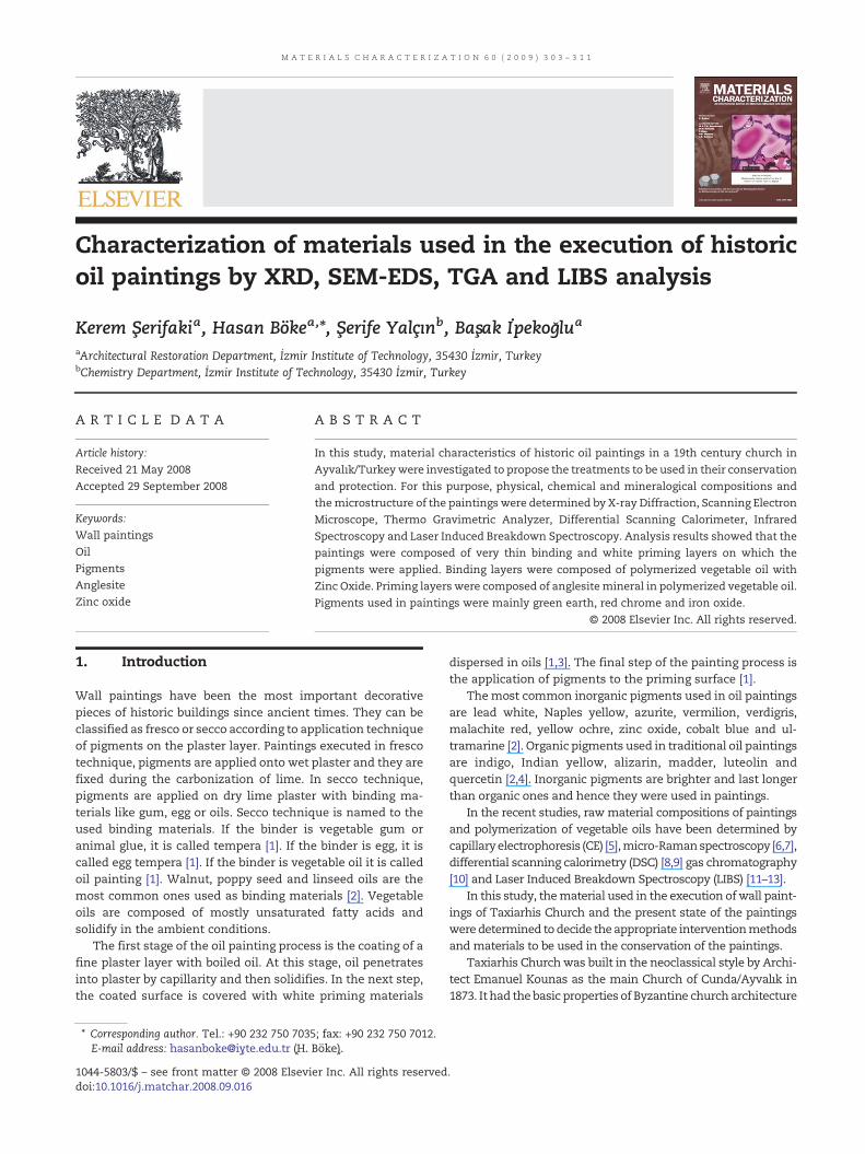
Characterization of materials used in the execution of historic oil paintings by XRD, SEM-EDS, TGA and LIBS analysis Kerem Şerifaki a , Hasan Böke a, ⁎ , Şerife Yalçın b , Başak İpekoğlu a a Architectural Restoration Department, İzmir Institute of Technology, 35430 İzmir, Turkey b Chemistry Department, İzmir Institute of Technology, 35430 İzmir, Turkey ARTICLE DATA ABSTRACT Article history: Received 21 May 2008 Accepted 29 September 2008 In this study, material characteristics of historic oil paintings in a 19th century church in Ayvalık/Turkey were investigated to propose the treatments to be used in their conservation and protection. For this purpose, physical, chemical and mineralogical compositions and the microstructure of the paintings were determined by X-ray Diffraction, Scanning Electron Microscope, Thermo Gravimetric Analyzer, Differential Scanning Calorimeter, Infrared Spectroscopy and Laser Induced Breakdown Spectroscopy. Analysis results showed that the paintings were composed of very thin binding and white priming layers on which the pigments were applied. Binding layers were composed of polymerized vegetable oil with Zinc Oxide. Priming layers were composed of anglesite mineral in polymerized vegetable oil. Pigments used in paintings were mainly green earth, red chrome and iron oxide. © 2008 Elsevier Inc. All rights reserved. Keywords: Wall paintings Oil Pigments Anglesite Zinc oxide 1. Introduction Wall paintings have been the most important decorative pieces of historic buildings since ancient times. They can be classified as fresco or secco according to application technique of pigments on the plaster layer. Paintings executed in fresco technique, pigments are applied onto wet plaster and they are fixed during the carbonization of lime. In secco technique, pigments are applied on dry lime plaster with binding ma- terials like gum, egg or oils. Secco technique is named to the used binding materials. If the binder is vegetable gum or animal glue, it is called tempera [1]. If the binder is egg, it is called egg tempera [1]. If the binder is vegetable oil it is called oil painting [1]. Walnut, poppy seed and linseed oils are the most common ones used as binding materials [2]. Vegetable oils are composed of mostly unsaturated fatty acids and solidify in the ambient conditions. The first stage of the oil painting process is the coating of a fine plaster layer with boiled oil. At this stage, oil penetrates into plaster by capillarity and then solidifies. In the next step, the coated surface is covered with white priming materials dispersed in oils [1,3]. The final step of the painting process is the application of pigments to the priming surface [1]. The most common inorganic pigments used in oil paintings are lead white, Naples yellow, azurite, vermilion, verdigris, malachite red, yellow ochre, zinc oxide, cobalt blue and ul- tramarine [2]. Organic pigments used in traditional oil paintings are indigo, Indian yellow, alizarin, madder, luteolin and quercetin [2,4]. Inorganic pigments are brighter and last longer than organic ones and hence they were used in paintings. In the recent studies, raw material compositions of paintings and polymerization of vegetable oils have been determined by capillary electrophoresis (CE) [5], micro-Raman spectroscopy [6,7], differential scanning calorimetry (DSC) [8,9] gas chromatography [10] and Laser Induced Breakdown Spectroscopy (LIBS) [11–13]. In this study, the material used in the execution of wall paint- ings of Taxiarhis Church and the present state of the paintings were determined to decide the appropriate intervention methods and materials to be used in the conservation of the paintings. Taxiarhis Church was built in the neoclassical style by Archi- tect Emanuel Kounas as the main Church of Cunda/Ayvalık in 1873. It had the basic properties of Byzantine church architecture MATERIALS CHARACTERIZATION 60 (2009) 303 – 311 ⁎ Corresponding author. Tel.: +90 232 750 7035; fax: +90 232 750 7012. E-mail address: [email protected] (H. Böke). ⁎ Corresponding author. Tel.: +90 232 750 7035; fax: +90 232 750 7012. E-mail address: [email protected] (H. Böke). 1044-5803/$ – see front matter © 2008 Elsevier Inc. All rights reserved. doi:10.1016/j.matchar.2008.09.016

Welcome message from author
This document is posted to help you gain knowledge. Please leave a comment to let me know what you think about it! Share it to your friends and learn new things together.
Transcript
M A T E R I A L S C H A R A C T E R I Z A T I O N 6 0 ( 2 0 0 9 ) 3 0 3 – 3 1 1
Characterization of materials used in the execution of historicoil paintings by XRD, SEM-EDS, TGA and LIBS analysis
Kerem Şerifakia, Hasan Bökea,⁎, Şerife Yalçınb, Başak İpekoğlua
aArchitectural Restoration Department, İzmir Institute of Technology, 35430 İzmir, TurkeybChemistry Department, İzmir Institute of Technology, 35430 İzmir, Turkey
A R T I C L E D A T A
⁎ Corresponding author. Tel.: +90 232 750 703E-mail address: [email protected] (H.
⁎ Corresponding author. Tel.: +90 232 750 703E-mail address: [email protected] (H.
1044-5803/$ – see front matter © 2008 Elsevidoi:10.1016/j.matchar.2008.09.016
A B S T R A C T
Article history:Received 21 May 2008Accepted 29 September 2008
In this study, material characteristics of historic oil paintings in a 19th century church inAyvalık/Turkey were investigated to propose the treatments to be used in their conservationand protection. For this purpose, physical, chemical and mineralogical compositions andthemicrostructure of the paintings were determined by X-ray Diffraction, Scanning ElectronMicroscope, Thermo Gravimetric Analyzer, Differential Scanning Calorimeter, InfraredSpectroscopy and Laser Induced Breakdown Spectroscopy. Analysis results showed that thepaintings were composed of very thin binding and white priming layers on which thepigments were applied. Binding layers were composed of polymerized vegetable oil withZinc Oxide. Priming layers were composed of anglesitemineral in polymerized vegetable oil.Pigments used in paintings were mainly green earth, red chrome and iron oxide.
© 2008 Elsevier Inc. All rights reserved.
Keywords:Wall paintingsOilPigmentsAnglesiteZinc oxide
1. Introduction
Wall paintings have been the most important decorativepieces of historic buildings since ancient times. They can beclassified as fresco or secco according to application techniqueof pigments on the plaster layer. Paintings executed in frescotechnique, pigments are applied onto wet plaster and they arefixed during the carbonization of lime. In secco technique,pigments are applied on dry lime plaster with binding ma-terials like gum, egg or oils. Secco technique is named to theused binding materials. If the binder is vegetable gum oranimal glue, it is called tempera [1]. If the binder is egg, it iscalled egg tempera [1]. If the binder is vegetable oil it is calledoil painting [1]. Walnut, poppy seed and linseed oils are themost common ones used as binding materials [2]. Vegetableoils are composed of mostly unsaturated fatty acids andsolidify in the ambient conditions.
The first stage of the oil painting process is the coating of afine plaster layer with boiled oil. At this stage, oil penetratesinto plaster by capillarity and then solidifies. In the next step,the coated surface is covered with white priming materials
5; fax: +90 232 750 7012.Böke).5; fax: +90 232 750 7012.Böke).
er Inc. All rights reserved
dispersed in oils [1,3]. The final step of the painting process isthe application of pigments to the priming surface [1].
Themost common inorganic pigments used in oil paintingsare lead white, Naples yellow, azurite, vermilion, verdigris,malachite red, yellow ochre, zinc oxide, cobalt blue and ul-tramarine [2]. Organic pigments used in traditional oil paintingsare indigo, Indian yellow, alizarin, madder, luteolin andquercetin [2,4]. Inorganic pigments are brighter and last longerthan organic ones and hence they were used in paintings.
In the recent studies, rawmaterial compositions of paintingsand polymerization of vegetable oils have been determined bycapillaryelectrophoresis (CE) [5],micro-Ramanspectroscopy [6,7],differential scanning calorimetry (DSC) [8,9] gas chromatography[10] and Laser Induced Breakdown Spectroscopy (LIBS) [11–13].
In this study, thematerial used in the execution ofwall paint-ings of Taxiarhis Church and the present state of the paintingsweredetermined todecide the appropriate interventionmethodsand materials to be used in the conservation of the paintings.
Taxiarhis Churchwas built in the neoclassical style by Archi-tect Emanuel Kounas as the main Church of Cunda/Ayvalık in1873. It had the basic properties of Byzantine church architecture
.

Fig. 1 –Michael (a) and Gabriel (b) paintings from the Taxiarhis Church.
304 M A T E R I A L S C H A R A C T E R I Z A T I O N 6 0 ( 2 0 0 9 ) 3 0 3 – 3 1 1
although itwasbuilt in lateperiod. TaxiarhisChurch is avaluablechurch due to its originalwall paintings. Paintings are located onthe walls of themain apse, arms of the cross, niches near by themain apse, drum of the central dome and over the doorway on
Fig. 2 –Plan of Tax
two sides of the narthex. Wall paintings show the events ex-pressed in the Old Testament and Holy Bible (Fig. 1) [14].Taxiarhis Church suffered structural failures by earthquakes,whichoccurred in1942and1944.Cracksand thedisintegrationof
iarhis Church.

Fig. 3 –XRD pattern of rough plaster layer.
Fig. 4 –XRD pattern of fine plaster layer.
305M A T E R I A L S C H A R A C T E R I Z A T I O N 6 0 ( 2 0 0 9 ) 3 0 3 – 3 1 1
the superstructure have allowed rain to penetrate into thechurch. Rain is the main source of deterioration of the wallpaintings. The Church was mentioned in the heritage at risk2004–2005 report which was published by ICOMOS1 due to thestructural condition of the building and its wall paintings.
2. Materials and Methods
In this study, raw material compositions, basic physical,mineralogical, chemical and micro-structural properties ofpaintings collected from Taxiarhis Church were determined.Visual deterioration of paintings was also indicated ondrawings prepared by using standard software programs.
2.1. Sampling
In this study, rough (arriccio) and fine (intonaco) plaster layersand the small amount of brown, red and green painted plastersamples were collected from detached parts of the paintings.Samples were collected from the paintings on the main apseand the north arm of the cross of the church (Fig. 2).
1 Alexander Zah, TURKEY Churches Built in Ottoman Times,ICOMOS World Report, 2004–2005 on Monuments and Sites inDanger, pp. 245–247. (http://www.international.icomos.org/risk/2004/turkey2004.pdf).
2.2. Characterization of Physical, Chemical andMineralogical Properties of Plaster Layers
The bulk densities and porosities, microstructure, chemicaland mineralogical compositions of the plaster layers weredetermined by several laboratory analyses. Bulk densities andporosities of the plaster layers were determined by measuringthe dry, water saturated under vacuum and hydrostaticweights [15]. Their mineralogical compositions were deter-mined by X-ray Diffraction (XRD) analyses. Lime and aggregateratios of the plasters were determined by the dissolution ofcarbonated lime in dilute hydrochloric acid.
2.3. Characterization of Microstructural, Chemical andMineralogical Properties of the Paintings
Microstructural characteristics and the elemental composi-tions of the paintings were determined with a ScanningElectron Microscope (SEM) coupled with EDS. SEM analysisresults showed that the paintings are composed of a very thinstrata of oil binding on the fine plaster andwhite priming layeron which the pigments were applied. Hence, priming andbinding layers together were carefully separated from fineplaster layers under a stereomicroscope, using a small lancetfor analysis.
The mineralogical and elemental compositions of thepriming layers were determined by X-ray Diffraction (XRD)
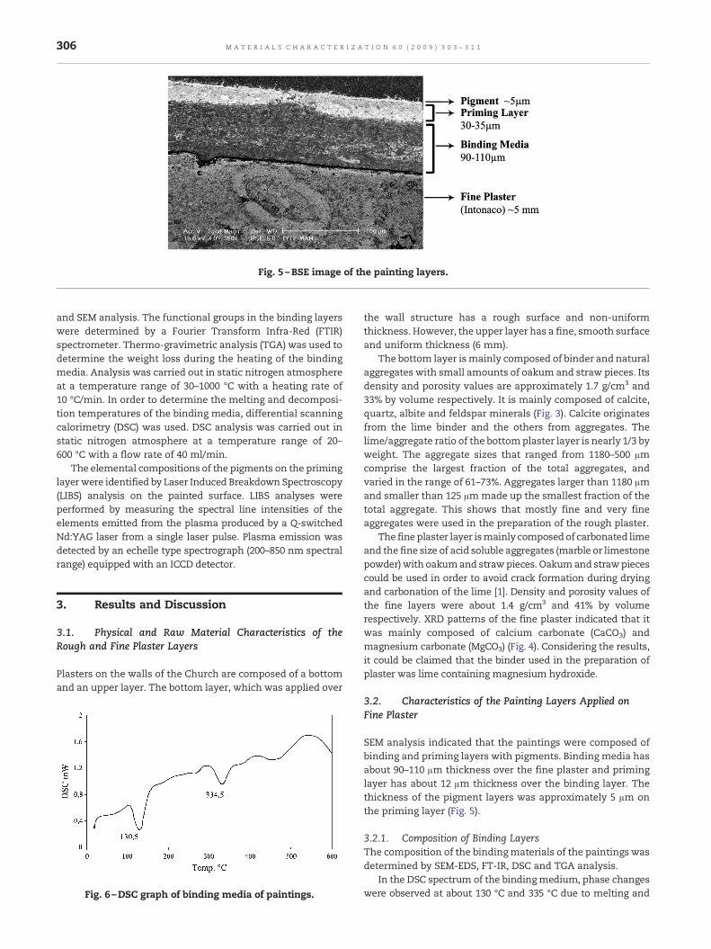
Fig. 5 –BSE image of the painting layers.
306 M A T E R I A L S C H A R A C T E R I Z A T I O N 6 0 ( 2 0 0 9 ) 3 0 3 – 3 1 1
and SEM analysis. The functional groups in the binding layerswere determined by a Fourier Transform Infra-Red (FTIR)spectrometer. Thermo-gravimetric analysis (TGA) was used todetermine the weight loss during the heating of the bindingmedia. Analysis was carried out in static nitrogen atmosphereat a temperature range of 30–1000 °C with a heating rate of10 °C/min. In order to determine the melting and decomposi-tion temperatures of the binding media, differential scanningcalorimetry (DSC) was used. DSC analysis was carried out instatic nitrogen atmosphere at a temperature range of 20–600 °C with a flow rate of 40 ml/min.
The elemental compositions of the pigments on the priminglayer were identified by Laser Induced Breakdown Spectroscopy(LIBS) analysis on the painted surface. LIBS analyses wereperformed by measuring the spectral line intensities of theelements emitted from the plasma produced by a Q-switchedNd:YAG laser from a single laser pulse. Plasma emission wasdetected by an echelle type spectrograph (200–850 nm spectralrange) equipped with an ICCD detector.
3. Results and Discussion
3.1. Physical and Raw Material Characteristics of theRough and Fine Plaster Layers
Plasters on the walls of the Church are composed of a bottomand an upper layer. The bottom layer, which was applied over
Fig. 6 –DSC graph of binding media of paintings.
the wall structure has a rough surface and non-uniformthickness. However, the upper layer has a fine, smooth surfaceand uniform thickness (6 mm).
The bottom layer ismainly composed of binder and naturalaggregates with small amounts of oakum and straw pieces. Itsdensity and porosity values are approximately 1.7 g/cm3 and33% by volume respectively. It is mainly composed of calcite,quartz, albite and feldspar minerals (Fig. 3). Calcite originatesfrom the lime binder and the others from aggregates. Thelime/aggregate ratio of the bottomplaster layer is nearly 1/3 byweight. The aggregate sizes that ranged from 1180–500 µmcomprise the largest fraction of the total aggregates, andvaried in the range of 61–73%. Aggregates larger than 1180 µmand smaller than 125 µm made up the smallest fraction of thetotal aggregate. This shows that mostly fine and very fineaggregates were used in the preparation of the rough plaster.
The fine plaster layer ismainly composed of carbonated limeand the fine size of acid soluble aggregates (marble or limestonepowder) with oakumand strawpieces. Oakumand strawpiecescould be used in order to avoid crack formation during dryingand carbonation of the lime [1]. Density and porosity values ofthe fine layers were about 1.4 g/cm3 and 41% by volumerespectively. XRD patterns of the fine plaster indicated that itwas mainly composed of calcium carbonate (CaCO3) andmagnesium carbonate (MgCO3) (Fig. 4). Considering the results,it could be claimed that the binder used in the preparation ofplaster was lime containing magnesium hydroxide.
3.2. Characteristics of the Painting Layers Applied onFine Plaster
SEM analysis indicated that the paintings were composed ofbinding and priming layers with pigments. Binding media hasabout 90–110 µm thickness over the fine plaster and priminglayer has about 12 µm thickness over the binding layer. Thethickness of the pigment layers was approximately 5 µm onthe priming layer (Fig. 5).
3.2.1. Composition of Binding LayersThe composition of the bindingmaterials of the paintings wasdetermined by SEM-EDS, FT-IR, DSC and TGA analysis.
In the DSC spectrum of the bindingmedium, phase changeswere observed at about 130 °C and 335 °C due to melting and

Fig. 7 –TGA graph of binding media of paintings.
307M A T E R I A L S C H A R A C T E R I Z A T I O N 6 0 ( 2 0 0 9 ) 3 0 3 – 3 1 1
decomposition temperatures of thematerials (Fig. 6). This resultmay indicate the presence of organic polymeric substances inthe composition of the binding layer. In previous work, similarresults have also been noted in the analyses of binding mediathat have been prepared by vegetable oil [8]. TGA analyses alsosupport this result. InTGAgraphs,weight losses are observed inthe range of 30–200 °C, 200–600 °C and 600–900 °C, which aremainly due to absorbedwater, decomposition of organicmatterand carbon dioxide, respectively. The weight loss of the samplewithin a temperature range from 30 °C to 200 °C is 3.01%. Theweight losseswithina range from200 °C to600 °Cand600–900 °Care 27.05% and 2.11%, respectively (Fig. 7). The observation of
Fig. 8 –FTIR spectrum of bin
high percentage loss at 200–600 °C shows the presence of highamount of organic matter in the binder. Weight losses at tem-peratures between 600 and 900 °C were due to carbon dioxidereleased during the decomposition of calcium carbonate, whichwas found as impurities in the binding layers.
The compositions of the binder were determined by FT-IRanalyses. In the FT-IR spectrum, the main hydroxyl band (OH)at 3410 cm−1, fatty acids (CH2) at 2924 and 2854 cm−1, esters(C=O) 1743 cm−1, oxalate (C2O4
−2) at 1620 cm−1, carbonate(CO3
−2) at 1419 and 875 cm−1 and sulphate (SO4−2) at 1118 cm−1
were observed (Fig. 8). Fatty acids, esters and oxalate bandshave already been identified in the FT-IR spectrum of dried
ding media of paintings.

Fig. 9 –SEM image (a) and EDX spectrum (b) of the bindinglayer of the paintings.
Fig. 11 –SE image (a) and EDX spectrum (b) of anglesitecrystals observed in the compositions of priming layer.
308 M A T E R I A L S C H A R A C T E R I Z A T I O N 6 0 ( 2 0 0 9 ) 3 0 3 – 3 1 1
vegetable oils [16]. Sulphate and carbonate bands could orig-inate frommaterials used in the preparation of priming layersand the binders of fine plasters.
Chemical compositions of the binder were determined bySEM-EDS analysis. SEM-EDS analyses of the binder indicatedthat it contains high amounts of carbon (C) and low amountsof zinc (Zn) (Fig. 9). The high amounts of carbon in its com-position show that the binder was produced from organiccompounds. The presence of zinc may be explained by theaddition of zinc oxide as a catalyst for the polymerizationreaction of binder that was mainly composed of vegetable oil[17].
Fig. 10 –XRD Pattern of prim
As a result of IR,DSC, TGAandSEM-EDSanalyses of the bind-ing media, it is considered that the binder of wall paintings wasprepared by using drying oils, most probably from linseed oil.
3.2.2. Mineralogical and Chemical Compositions ofPriming LayersX-ray Diffraction analyses of the powdered painting layersshowed that they were mainly composed of anglesite mineral(PbSO4) (Fig. 10). In the SEM-EDS analysis, crystals composed oflead and sulphur were found (Fig. 11). These two results showthe use of anglesiteminerals in the preparation of the priminglayers. In some early studies, a similar usage has already beennoted [2,18].
3.2.3. Chemical Characteristics of PigmentsLIBS analysis was carried out to identify the elemental com-positions of the pigments. LIBS analysis of the brown painting
ing layers of paintings.

Fig. 12 –LIBS spectrum of brown painting.
309M A T E R I A L S C H A R A C T E R I Z A T I O N 6 0 ( 2 0 0 9 ) 3 0 3 – 3 1 1
surfaces indicated thepresenceof iron (Fe), and lead (Pb) (Fig. 12).Specific emission lines of the elements observed in LIBS spec-trum were labeled in units of nanometers, nm. The presence ofironmay show the use of iron containing pigments such as ironoxide [17,18]. The observation of unexpected lead signal can beexplained due to impurities originating from the priming layerwhich was mainly composed of anglesite (PbSO4).
In the LIBS analyses of the red colored painting surfaces, astrong signal of lead and chromiumwas detected (Fig. 13). Thismay show the use of chrome red (PbCrO4.PbO) as pigment [19].
Fig. 13 –LIBS spectrum
The observation of Fe and Ca signals in the analysis can beexplained by the lime and brown pigment impurities found onthe red painting.
LIBS analysis of green painting surfaces showed the presenceof Na, Mg, Al and Fe (Fig. 14). In previous studies, the use ofceladonite and glauconite called “green earth” having the sameelements has been noted as pigments [17,18,20]. The observationof similar elements found in the composition of green paintingsurface may indicate the use of “green earth” as pigment in thepainting.
of red painting.

Fig. 14 –LIBS spectrum of green painting.
310 M A T E R I A L S C H A R A C T E R I Z A T I O N 6 0 ( 2 0 0 9 ) 3 0 3 – 3 1 1
3.3. Deterioration Problems of the Wall Paintings
The main deterioration phenomena observed on the wallpaintings were disintegration of plaster layers, blistering andpeeling of painted surfaces (Fig. 15). SEM analysis indicatedthat the adhesion between the binding and priming layers of
Fig. 15 –Measured drawing of the Michael painting.
paintings was diffuse and strong. However, the commonproblems were the formation of micro-cracks on the surface,blistering of priming layers (Figs. 16, 17) and the detachment of
Fig. 16 –SE image of micro crack formation on paintingsurface.
Fig. 17 –BSE image of blistering on painting surface.
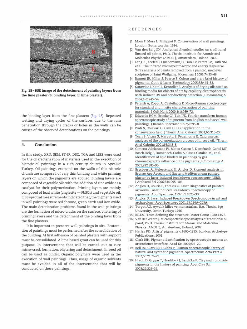
Fig. 18 –BSE image of the detachment of painting layers fromthe lime plaster (B: binding layer, L: lime plaster).
311M A T E R I A L S C H A R A C T E R I Z A T I O N 6 0 ( 2 0 0 9 ) 3 0 3 – 3 1 1
the binding layer from the fine plasters (Fig. 18). Repeatedwetting and drying cycles of the surfaces due to the rainpenetration through the cracks or holes in the walls can becauses of the observed deteriorations on the paintings.
4. Conclusion
In this study, XRD, SEM, FT-IR, DSC, TGA and LIBS were usedfor the characterization of materials used in the execution ofhistoric oil paintings in a 19th century church in Ayvalık/Turkey. Oil paintings executed on the walls of this historicchurch are composed of very thin binding and white priminglayers on which the pigments are applied. Binding layers arecomposed of vegetable oils with the addition of zinc oxide as acatalyst for their polymerization. Priming layers are mainlycomposed of lead white (anglesite — PbSO4) and vegetable oil.LIBS spectralmeasurements indicated that, the pigments usedin wall paintings were red chrome, green earth and iron oxide.The main deterioration problems found in the wall paintingsare the formation of micro-cracks on the surface, blistering ofpriming layers and the detachment of the binding layer fromthe fine plasters.
It is important to preserve wall paintings in situ. Restora-tion of paintings must be performed after the consolidation ofthe building. At first adhesion of painted plasters with supportmust be consolidated. A lime based grout can be used for thispurpose. In interventions that will be carried out to curemicro-crack formation, blistering and detachment, linseed oilcan be used as binder. Organic polymers were used in theexecution of wall paintings. Thus, usage of organic solventsmust be avoided in all of the interventions that will beconducted on these paintings.
R E F E R E N C E S
[1] Mora P, Mora L, Philippot P. Conservation of wall paintings.London: Butterworths; 1984.
[2] Van den Berg JDJ. Analytical chemical studies on traditionallinseed oil paints, Ph.D. Thesis, Institute for Atomic andMolecular Physics (AMOLF), Amsterdam, Holand; 2002.
[3] LangPL,KeeferCD, JuenemannJC,TranKV,Peters SM,HuthNM,et al. The infrared microspectroscopic and energy dispersiveX-ray analysis of paints removed from a painted, medievalsculpture of Saint Wolfgang. Microchem J 2003;74:33–46.
[4] Barnett JR, Miller S, Pearce E. Colour and art: a brief history ofpigments. Optic & Laser Technology 2005;38:445–53.
[5] Surowiec I, Kaml I, Kenndler E. Analysis of drying oils used asbinding media for objects of art by capillary electrophoresiswith indirect UV and conductivity detection. J Chromatog A2004;1–2:245–54.
[6] Perardi A, Zoppi A, Castellucci E. Micro-Raman spectroscopyfor standard and in situ characterization of paintingmaterials. J Cult Herit 2000;1(1):269–72.
[7] Edwards HGM, Brooke CJ, Tait JFK. Fourier transform Ramanspectroscopic study of pigments from English mediaeval wallpaintings. J Raman Spectrosc 1997;28:95–8.
[8] Prati S, Chiavari G, Cam D. DSC application in theconservation field. J Therm Anal Calorim 2001;66:315–27.
[9] Turri B, Vicini S, Margutti S, Pedemonte E. Calorimetricanalyses of the polymerization process of linseed oil. J ThermAnal Calorim 2001;66:343–8.
[10] Gimeno-Adelantado JV, Mateo-Castro R, Doménech-Carbó MT,Bosch-Reig F, Doménech-Carbó A, Casas-Catalán MJ, et al.Identification of lipid binders in paintings by gaschromatography influence of the pigments. J Chromatogr A2001;922:385–90.
[11] Brysbaert A, Melessenaki K, Anglos D. Pigment analysis inBronze Age Aegean and Eastern Mediterranean paintedplaster by laser-induced breakdown spectroscopy (LIBS).J Archaeol Sci 2006;33:1095–104.
[12] Anglos D, Couris S, Fotakis C. Laser Diagnostics of paintedartworks: Laser Induced Breakdown Spectroscopy ofpigments. Appl Spectrosc 1997;51:1025–30.
[13] Anglos D. Laser Induced Breakdown Spectroscopy in art andarchaeology. Appl Spectrosc 2001;55:186A–205A.
[14] Turgut AÖ. Ayvalık kilise ve manastırları, B.A. Thesis, EgeUniversity, İzmir, Turkey; 1996.
[15] RILEM. Tests defining the structure. Mater Const 1980;13:73.[16] Van derWeerd J. Microspectroscopic analysis of traditional oil
paint, Ph.D. Thesis, Institute for Atomic and MolecularPhysics (AMOLF), Amsterdam, Holand; 2002.
[17] Harley RD. Artists' pigments c.1600–1835. London: ArchetypePublications; 2001.
[18] Clark RJH. Pigment identification by spectroscopic means: anarts/science interface. Acad Sci 2002;5:7–20.
[19] Bell IM, Clark RJH, Gibbs PJ. Raman spectroscopic library ofnatural and synthetic pigments. Spectrochim Acta Part A1997;53:2159–79.
[20] Hradil D, Grygar T, Hradilová J, Bezdićka P. Clay and iron oxidepigments in the history of painting. Appl Clay Sci2003;22:223–36.
Related Documents


![ip2001-09.ppt [호환 모드] - CHERIC · 2014-02-11 · 성,pH)Spray drying 물성측정 조성설계 반응특성 (TGA/고정층) 물성평가 (SEM,XRD, EDX) 조성개선 독자탈황제](https://static.cupdf.com/doc/110x72/5eba3acbf81fde683a6f8d83/ip2001-09ppt-eeoe-cheric-2014-02-11-phspray-drying-e.jpg)