ELSEVIER Characterization of lipoprotein composition in rats fed different dietary lipids and of the effects of lipoproteins upon lymphocyte proliferation Nicola M. Jeffery, Parveen Yaqoob, David Wiggins,* Geoffrey F. Gibbons,* Eric A. Newsholme, and Philip C. Calder Department of Biochemistry, University of Oxford, South Parks Road, Oxford, United Kingdom; *Metabolic Research Laboratory, NufJield Department of Clinical Medicine, Radcliffe Infirmary, Woodstock Road, Oxford, United Kingdom Weanling Lewis rats were fed for 10 weeks on a low fat (2.5% by weight; LF) diet or on diets containing 20% by weight of hydrogenated coconut oil (HCO), olive oil (00) saflower oil (SO), evening primrose oil (EPO), or menhaden fish) oil (MO); all other components of the diets were identical. The chylomicron (CM), very low density lipoprotein (VLDL), low density lipoprotein (LDL), and high density lipoprotein (HDL) fractions were isolated from the serum. The serum from MO-fed animals had lower LDL and HDL cholesterol concentrations than the serum from animals fed each of the other diets. The apolipoprotein A-l concentration in the HDL fraction from animals fed the MO diet was also low. The serum from HCO-fed animals had a higher CM triacylglycerol concentration than serum from animals fed each of the other diets. The serum from 00-fed animals had a higher VLDL triacylglycerol concentration than serum from animals fed each of the other diets. The concentration of apolipoprotein B was also high in the VLDL fraction from 00-fed animals. The fatty acid composition of each lipoprotein fraction was affected by the nature of the lipid in the diet; the greatest effects were observed in the CM and VLDL fractions. Each lipoprotein fraction isolated from LF-fed rats inhibited mitogen-stimulated rat spleen lymphocyte proliferation in a concentration-dependent manner; LDL and HDL caused greater inhibition than CM and VLDL. Dietary lipid manipulation did not alter the inhibitory eflects of any of the lipoprotein fractions upon lymphocyte proliferation, except that CM and HDL from MO-fed animals and HDL from 00-fed animals resulted in enhanced proliferation compared with either CM or HDL from animals fed the other diets. We conclude that the inhibition of lymphocyte proliferation caused by feeding rats certain dietary lipids (00, EPO, MO) may be mediated by non-lipoprotein serum components. (J. Nutr. Biochem. 7:282-292, 1996.) Keywords: lipoproteins; dietary lipids; fish oil; lymphocyte proliferation Introduction There is a link between the risk of developing coronary heart disease (CHD) and the concentration of lipids, in par- ticular cholesterol, in the bloodstream.’ The consumptionin the diet of lipids that contain a high proportion of saturated fatty acids results in elevated blood cholesterol and triacyl- glycerol concentrations; thus, there is a link between satu- rated fat consumption and risk of developing CHD. As a Address reprint requests to Dr. Philip C. Calder at the Department of Biochemistry, University of Oxford, South Parks Rd, Oxford OXI 3QU United Kingdom. Received January 4, 1995; accepted February 2 1, 1996. result of this link, there hasbeen much interest in the useof diets containing unsaturated,rather than saturated,fatty ac- ids to prevent the development of CHD. However, the exact relationship between the consumption of different types of Nutritional Biochemistry 7:282-292, 1996 0 Elsevier Science Inc. 1996 655 Avenue of the Americas, New York, NY 10010 0955-2863/96/$15.00 SSDI 0955-2863(96)00029-5

Welcome message from author
This document is posted to help you gain knowledge. Please leave a comment to let me know what you think about it! Share it to your friends and learn new things together.
Transcript
ELSEVIER
Characterization of lipoprotein composition in rats fed different dietary lipids and of the effects of lipoproteins upon lymphocyte proliferation Nicola M. Jeffery, Parveen Yaqoob, David Wiggins,* Geoffrey F. Gibbons,* Eric A. Newsholme, and Philip C. Calder
Department of Biochemistry, University of Oxford, South Parks Road, Oxford, United Kingdom; *Metabolic Research Laboratory, NufJield Department of Clinical Medicine, Radcliffe Infirmary, Woodstock Road, Oxford, United Kingdom
Weanling Lewis rats were fed for 10 weeks on a low fat (2.5% by weight; LF) diet or on diets containing 20% by weight of hydrogenated coconut oil (HCO), olive oil (00) saflower oil (SO), evening primrose oil (EPO), or menhaden fish) oil (MO); all other components of the diets were identical. The chylomicron (CM), very low density lipoprotein (VLDL), low density lipoprotein (LDL), and high density lipoprotein (HDL) fractions were isolated from the serum. The serum from MO-fed animals had lower LDL and HDL cholesterol concentrations than the serum from animals fed each of the other diets. The apolipoprotein A-l concentration in the HDL fraction from animals fed the MO diet was also low. The serum from HCO-fed animals had a higher CM triacylglycerol concentration than serum from animals fed each of the other diets. The serum from 00-fed animals had a higher VLDL triacylglycerol concentration than serum from animals fed each of the other diets. The concentration of apolipoprotein B was also high in the VLDL fraction from 00-fed animals. The fatty acid composition of each lipoprotein fraction was affected by the nature of the lipid in the diet; the greatest effects were observed in the CM and VLDL fractions. Each lipoprotein fraction isolated from LF-fed rats inhibited mitogen-stimulated rat spleen lymphocyte proliferation in a concentration-dependent manner; LDL and HDL caused greater inhibition than CM and VLDL. Dietary lipid manipulation did not alter the inhibitory eflects of any of the lipoprotein fractions upon lymphocyte proliferation, except that CM and HDL from MO-fed animals and HDL from 00-fed animals resulted in enhanced proliferation compared with either CM or HDL from animals fed the other diets. We conclude that the inhibition of lymphocyte proliferation caused by feeding rats certain dietary lipids (00, EPO, MO) may be mediated by non-lipoprotein serum components. (J. Nutr. Biochem.
7:282-292, 1996.)
Keywords: lipoproteins; dietary lipids; fish oil; lymphocyte proliferation
Introduction
There is a link between the risk of developing coronary heart disease (CHD) and the concentration of lipids, in par-
ticular cholesterol, in the bloodstream.’ The consumption in the diet of lipids that contain a high proportion of saturated fatty acids results in elevated blood cholesterol and triacyl- glycerol concentrations; thus, there is a link between satu- rated fat consumption and risk of developing CHD. As a
Address reprint requests to Dr. Philip C. Calder at the Department of Biochemistry, University of Oxford, South Parks Rd, Oxford OXI 3QU United Kingdom. Received January 4, 1995; accepted February 2 1, 1996.
result of this link, there has been much interest in the use of diets containing unsaturated, rather than saturated, fatty ac- ids to prevent the development of CHD. However, the exact relationship between the consumption of different types of
Nutritional Biochemistry 7:282-292, 1996 0 Elsevier Science Inc. 1996 655 Avenue of the Americas, New York, NY 10010
0955-2863/96/$15.00 SSDI 0955-2863(96)00029-5
Dietary lipids and lipoprotein composition: Jeffrey et a/.
VLDL, LDL, and HDL enhance 1 mphocyte proliferation, a finding supported by Xu et al. 42 for HDL and LDL. The importance of the cell culture conditions used in these ex- periments is apparent when the studies of Cuthbert and Lipsky43,44 are closely scrutinised: the earlier study reported that LDL inhibited lymphocyte proliferation,43 whereas the later study reported enhancement.44 However, the enhance- ment in proliferation observed when lymphocytes were cul- tured in the presence of VLDL and LDL was seen only if transfetrin was also present in the culture medium44; if transferrin was absent then VLDL and LDL caused marked inhibition of proliferation. Clearly, some of the discrepan- cies between the results of different studies might be due dif- ferences in the cell culture conditions used; whatever the reason, it is apparent that the exact nature of the effects of different types of lipoprotein on lymphocyte proliferation is not clear. To our knowledge, the effect of lipoproteins iso- lated from animals subjected to dietary lipid manipulation on the function of lymphocytes has not been investigated. Therefore, in this study we investigated the effect of lipo- protein fractions purified from the serum of rats fed various lipids on mitogen-stimulated lymphocyte proliferation. The aim of this study was first to identify the effects of lipopro- teins on lymphocyte proliferation and secondly to examine whether the differential effects of dietary lipids on lympho- cyte proliferation can be explained by differential effects of lipoproteins.
fat and blood lipid levels remains unresolved, although there have been many studies in man2*3 and laboratory ani- mals.4-‘3 Therefore, in the current study we investigated the effects of feeding rats a low-fat diet or diets rich in satu- rated, monounsaturated, n-6 polyunsaturated, or n-3 poly- unsaturated fatty acids (PUFAs) upon the composition of the major lipoprotein fractions.
In addition to a potential effect on blood lipid levels and CHD, dietary lipids may have immunomodulatory effects. An immunosuppressive effect of dietary fish oils, which contain high proportions of n-3 PUFAs, is suggested by epidemiological studies that show that populations such as Greenland Eskimos who consume large quantities of fish oils have a very low incidence of inflammatory and auto- immune disordersi and by clinical studies that show that fish oil supplementation has some beneficial effects in rheu- matoid arthritis, multi le sclerosis, psoriasis, and prolonga- tion of graft survival.’ P The potential clinical use of fish oils and perhaps also those containing n-6 PUFAs, such as eve- ning primrose oil, which contains y-linolenic acid, has given rise to a large number of studies investigating the effects of fatty acids and dietary oils on immune cell func- tions. In vitro studies have shown that unsaturated fatty acids, or triacylglycerols containing unsaturated fatty acids, inhibit a number of lymphocyte functions including prolif- eration,‘c23 interleukin (IL)-2 production,19-21,24 activation marker expression, I9 degranulation of cytotoxic T- lymphocytes,25 natural killer (NK) cell activity26 and anti- gen presentation by splenocytes.*’ In studies where the ef- fects of a range of fatty acids have been compared it has often been found that the n-3 PUFA eicosapentaenoic acid is the most potent inhibitor of these functions,‘7-22 although other n-3 PUFAs and n-6 PUFAs are also inhibitoty.‘“23*25 These in vitro studies are supported by the results of dietary studies performed in animals and man, which have shown that PUFA-rich diets, especially those containing fish oil, inhibit the proliferation of lymphocytes,‘5.28-30 NK cell ac- tivity,30.31 cytokine production,28,32,33 and the expression of activation markers and adhesion molecules on the surface of resting and mitogen-stimulated spleen lymphocytes3’ and lymphocytes within the popliteal lymph node after the graft versus host and host versus graft reactions.34 Some of the effects of dietary lipid manipulation on lymphocyte prolif- eration can be mimicked by culturing cells from chow-fed animals with serum obtained from animals fed different lipids. 35,36 This suggests that components in the serum are at least partly responsible for mediating the effects of the diet; the obvious candidates as mediators are lipoproteins. Indeed, effects of purified lipoproteins on in vitro mitogen- stimulated lymphocyte proliferation have been report- ed.3746 Some of these studies reported that all classes of lipoprotein inhibit mitogen-stimulated lymphocyte prolif- eration.38.41’42 However, these studies do not agree as to which lipoproteins are the most potent inhibitors. Morse et al., 38 Yi et a1.,41 and de Deckere et a1.45 reported that very low density lipoproteins (VLDL) were more potent than low density lipoproteins (LDL), which were more potent than high density lipoproteins (HDL) (in some of these studies HDL caused very little inhibition, if any), whereas Hsu et a14* found that HDL caused greater inhibition than VLDL and LDL. In contrast, Cuthbert and Lipsky reported that
Materials and methods
Animals and diets
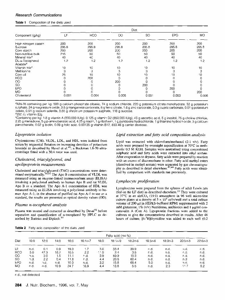
Weanling male Lewis rats (aged 3 weeks, weighing between 65g and 85g) were obtained from Harlan-Olac, Bicester, Oxon., En- gland. The animals were housed in groups of six for a period of 10 weeks before killing, during which time they were allowed access, ad libitum, to water and to one of six experimental diets. Animals were fed either a low fat (LF) diet or one of five high-fat diets (purchased from ICN Biomedicals, Aurora, OH USA). The LF diet contained 2.5% by weight of corn oil and the high fat diets con- tained 20% by weight of the lipid under study, plus 1% corn oil to prevent essential fatty acid deficiency. The high fat diets contained either 20% hydrogenated coconut oil (HCO), 20% olive oil (00) 20% safflower oil (SO), 20% evening primrose oil (EPO), or 20% menhaden (fish) oil (MO). The oils used for the production of these diets were obtained from Capital City Products, Columbus, OH USA (corn oil, HCO, 00); California Oils, Richmond, CA USA (SO); Zapeta Haynie, Reedville, VA, USA (MO); and Scotia Pharmaceuticals, Guildford, Surrey, England (EPO). The compo- sition of the diets is shown in Table 1. Lipid was extracted from the diets using chloroform/methanol (2:l v/v); the fatty acid compo- sition was analyzed by gas chromatography (see below) and is shown in Table 2. Details of food intake and weight gain of ani- mals fed these diets have been published elsewhere.‘3.29 Animals were sacrificed by exposure to a rising concentration of CO,. All procedures involving animals were approved under the Animals (Scientific Procedures) Act 1986 by the Home Office.
Chemicals
Chemicals were obtained from the sources described else- where. ‘5.‘7,47,48 In addition, potassium bromide was purchased from BDH, Poole, Dorset, England and polyclonal antibodies to human apolipoprotein (APO) B and Apo A-l were purchased from Boehringer Mannheim, Lewes, Sussex, England.
J. Nutr. Biochem., 1996, vol. 7, May 283
Research Communications
Table 1 Composition of the diets used
Diet
Component (g/kg) LF HCO 00 so EPO MO
High nitrogen casein Sucrose Corn starch Non-nutritive bulk Mineral mixa DL-cr-Tocopherol Acetateb Vitamin mixC Methionine Corn oil HCO 00 so EPO MO Cholesterol
200 200 200 200 200 200 295.8 295.8 295.8 295.8 295.8 295.8 200 200 200 200 200 200 215 50 50 50 50 50
40 40 40 40 40 40 1.2 1.2 1.2 1.2 1.2 1.2
10 10 10 10 10 10 3 3 3 3 3 3
25 10 10 10 10 10 0 200 0 0 0 0 0 0 200 0 0 0 0 0 0 200 0 0 0 0 0 0 200 0 0 0 0 0 0 200 0.001 0.004 0.005 0.001 0.003 0.006
aAIN-76 containing per kg: 500 g calcium phosphate dibasic, 74 g sodium chloride, 220 g potassium citrate monohydrate, 52 g potassium sulphate, 24 g magnesium oxide, 3.5 g manganese carbonate, 6 g ferric citrate, 1.6 g zinc carbonate, 0.3 g cupric carbonate, 0.01 g potassium iodate, 0.01 g sodium selenite, 0.55 g chromium potassium sulphate, 118 g sucrose. *250 IU vitamin E/g. %ontaining per kg: 1.8 g vitamin A (500,000 IU/g), 0.125 g vitamin 02 (850,000 IU/g), 45 g ascorbic acid, 5 g inositol, 75 g choline chloride, 2.25 g menadione, 5 g p-aminobenzoic acid, 4.25 g niacin, 1 g riboflavin, 1 g pyridoxine hydrochloride, 1 g thiamine hydrochloride, 3 g calcium pantothenate, 0.02 g biotin, 0.09 g folic acid. 0.00135 g vitamin 812, 833.46 g carrier dextrose.
Lipoprotein isolation Lipid extraction and fatty acid composition analysis
Chylomicrons (CM), VLDL, LDL, and HDL were isolated from serum by sequential flotation on increasing densities of potassium bromide as described by Have1 et al.49; a Beckman L8-70 ultra- centrifuge fitted with a 50.4 rotor was used.
Cholesterol, triacylglycerol, and apolipoprotein measurements
Cholesterol and triacylglycerol (TAG) concentrations were deter- mined enzymatically.50~5’ The Apo B concentration of VLDL was measured using an enzyme-linked immunosorbant assay (ELISA) involving a polyclonal antibody to human Apo B and rat VLDL Apo B as a standard. The Apo A-l concentration of HDL was measured using an ELISA involving a polyclonal antibody to hu- man Apo A- 1; in the absence of purified rat Apo A- 1 to use as a standard, the results are presented as optical density values (OD).
Lipid was extracted with chloroform/methanol (2:l v/v). Fatty acids were prepared by overnight saponification at 70°C in meth- anolic 0.5 M KOH. Samples were neutralised using concentrated sulphuric acid and fatty acids were extracted into ethyl acetate. After evaporation to dryness, fatty acids were prepared by reaction with an excess of diazomethane in ether. Fatty acid methyl esters (dissolved in methyl acetate) were separated by gas chromatogra- phy as described in detail elsewhere.47,48 Fatty acids were identi- fied by comparison with standards run previously.
Lymphocyte proliferation
Plasma a-tocopherol analysis
Plasma was treated and extracted as described by Desai5* before separation and quantification of cr-tocopherol by HPLC as de- scribed by Buttriss and Diplock.
Lymphocytes were prepared from the spleens of adult Lewis rats (fed on the LF diet) as described elsewhere.” They were cultured at 37°C in an air/CO, (19:l) atmosphere in 96 well micro-titre culture plates at a density of 5 x lo5 cells/well and a total culture volume of 200 p,l in HEPES-buffered RPM1 supplemented with 2 mM glutamine, 1% (v/v) Nutridoma, antibiotics and 1 p,g/ml con- canavalin A (Con A). Lipoprotein fractions were added to the cultures to give the concentrations described in results. After 48 hours of culture, [6-3H]thymidine was added to each well (0.2
Table 2 Fatty acid composition of the diets used
Diet lo:o 12:o 14:o 16:0
Fatty acid (mol%)
16:ln-7 18:0 18:ln-9 18:2n-6 18:3n-6 18:3n-3 20:5n-3 22:6n-3
LF n.d. 0.1 0.9 19.3 1.7 3.6 33.4 39.9 n.d. n.d. n.d. n.d. HCO 5.0 47.3 18.2 10.5 2.2 11.0 2.4 3.5 n.d. n.d. n.d. n.d. 00 n.d. 3.0 1.5 11.1 n.d. 3.9 69.9 15.3 n.d. n.d. n.d. n.d. so n.d. 2.2 0.4 11.8 n.d. 4.4 20.8 60.4 n.d. n.d. n.d. n.d. EPO n.d. n.d. n.d. 10.3 n.d. 3.2 15.8 68.4 5.0 n.d. n.d. n.d. MO n.d. n.d. 10.9 24.7 16.9 4.4 13.8 5.5 n.d. 2.3 11.7 5.2
n.d., not detected.
284 J. Nutr. Biochem., 1996, vol. 7, May
Dietary lipids and lipoprotein composition: Jeffrey et al.
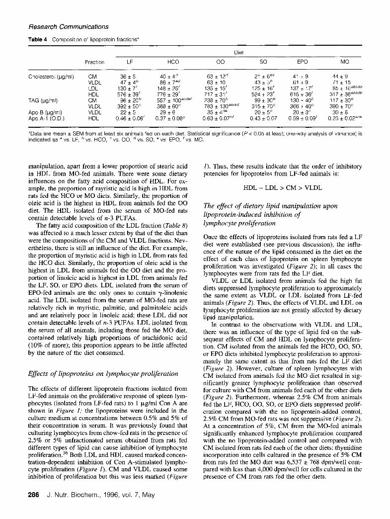
LF, SO, EPO, or MO diets were similar,13 but feeding the HCO or 00 diets caused a hypertriacylglycerolaemic ef- fect.13 The elevation in TAG concentration is apparent in both CM and VLDL fractions of animals fed the 00 diet and in the CM fraction of animals fed the HCO diet (ra- ble 4).
The Apo B concentration of VLDL isolated from ani- mals fed the SO or EPO diets was lower than the Apo B concentration of VLDL from rats fed the 00 diet (Table 4). The Apo A-l concentration of HDL from MO-fed rats was lower than that of HDL from animals fed each of the other diets (Table 4); this was statistically significant compared with the Apo A-l concentration of HDL from LF-, 00- or EPO-fed animals (Table 4).
@i/well) and the cells were incubated for a further 18 hours. The cells were then harvested onto glass fibre filters and washed and dried using a Skatron Cell Harvester. Radioactive thymidine in- corporation was determined by liquid scintillation counting. Re- sults are expressed as dpm [3H]thymidine incorporated/well.
Data presentation
Data are presented as mean k SEM of the indicated number of observations. Statistical significance was determined using one- way analysis of variance followed by a least-squared difference test. A value for P < 0.05 was taken to indicate a statistically significant difference.
Results
Plasma cx-tocopherol concentrations Lipoprotein fatty acid composition
Plasma a-tocopherol concentrations were higher in rats fed the high fat diets compared with those fed the LF diet (Table 3); they were highest in the HCO- and 00-fed animals. The HCO- and 00-fed animals also had the highest serum con- centrations of cholesterol and triacylglycerol. l3 When the concentration of cx-tocopherol in the plasma was expressed as a ratio of the combined concentrations of cholesterol and triacylglycerol in the serum, there were no significant dif- ferences between animals fed the high fat diets (Table 3). However, the relative concentration of cr-tocopherol in the plasma from LF-fed rats was lower than that of rats fed the HCO, EPO, and MO diets (Table 3).
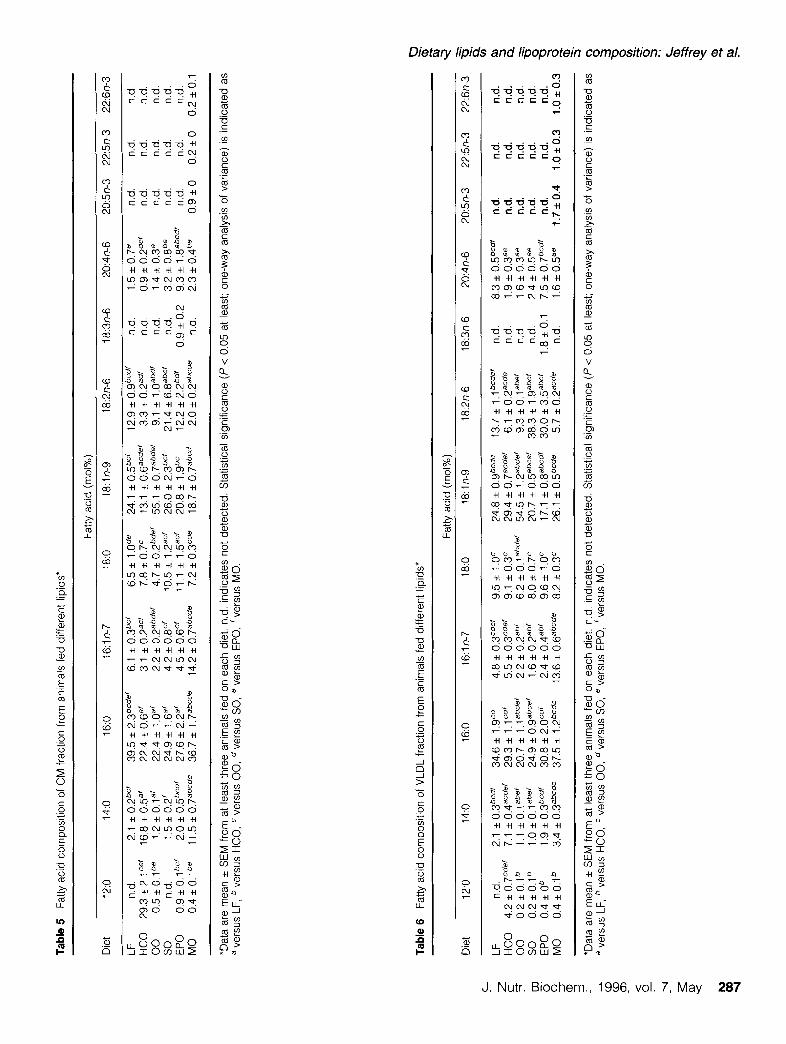
The fatty acid compositions of CM, VLDL, HDL, and LDL isolated from the serum of rats fed different dietary lipids are shown in Tables 5, 6, 7, and 8, respectively. These show that each lipoprotein fraction has a characteristic fatty acid composition and that the fatty acid composition of each lipoprotein fraction is influenced by the diet that the animal was fed.
As would be expected, the fatty acid composition of the CM fraction is strongly affected by that of the diet con- sumed by the animal (Table 5). For example, the propor- tions of lauric (12:0) and myristic (14:0) acids are very high in CM from rats fed the HCO diet. Similarly, the proportion of oleic acid (18: ln-9) is very high in CM from animals fed the 00 diet and the proportion of linoleic acid (18:2n-6) is high in CM from animals fed the LF, SO, or EPO diets, each of which is rich in linoleic acid. The CM isolated from the serum of MO-fed rats are relatively rich in myristic, pal- mitic and palmitoleic (16: ln-7) acids and are relatively poor in linoleic acid; these CM are also the only ones to contain the n-3 PUFAs, eicosapentaenoic (20:5n-3), docosapentae- noic (22:5n-3), and docosahexaenoic (22:6n-3) acids. CM isolated from the serum of EPO-fed animals are the only ones to contain y-hnolenic acid; these CM also contain a high proportion of arachidonic acid (20:4n-6).
Lipoprotein composition
Feeding the HCO, 00, SO, or EPO diets increased the total serum cholesterol concentration compared with feeding the LF or MO diets , 13. feeding the MO diet produced a hypo- cholesterolaemic effect relative to feeding each of the other dietsI This effect is mainly due to a much lower HDL cholesterol concentration in animals fed the MO diet com- pared with those fed each of the other diets (Table 4), al- though LDL cholesterol concentrations were also signifi- cantly lower in MO-fed animals (Table 4).
The total serum TAG concentrations of animals fed the
Table 3 Plasma vrtamin E concentrations*
Diet [a-Tocopherol]
(w/l)
a-TocopheroV Cholesterol +
Triacylglycerolt
LF 9.9 i 0.4bcde‘ 0.0058 zt 0.0004be’ HCO 31 .o * 4.l=d’ 0.0095 f 0.001 la 00 35.2 * 5.Fd’ 0.0091 f 0.0015 SO 14.9 * 2.Bab= 0.0076 + 0.0012 EPO 21.7 + 1.6a 0.0109 f 0.0015” MO 15.9 f 2.0abc 0.0107 + 0.0016a
*Data are mean * SEM from at least four animals fed on each diet. tlndicates plasma a-tocopherol concentration (mg/l) divided by the sum of the serum cholesterol and triacylglycerol concentrations (both in mg/l); serum cholesterol and triacylglycerol concentrations have been published elsewhere. I3 Statistical significance (P < 0.05 at least; one-way analysis of variance) is indicated as a versus LF, b versus HCO, c versus 00, d versus SO, e versus EPO, ‘versus MO.
The fatty acid composition of the VLDL fraction is also greatly affected by that of the diet consumed by the animal (Table 6). For example, the proportions of lauric and my- ristic acids are high in VLDL from rats fed the HCO diet. Similarly, the proportion of oleic acid is very high in VLDL from animals fed the 00 diet and the proportion of linoleic acid is high in VLDL from animals fed the LF, SO, or EPO diets. VLDL isolated from the serum of EPO-fed animals are the only ones to contain y-linolenic acid; these VLDL, like those from LF-fed rats, also contain a high proportion of arachidonic acid. The VLDL isolated from the serum of MO-fed rats are relatively rich in myristic, palmitic, and palmitoleic acids and are relatively poor in linoleic acid; these VLDL are also the only ones to contain the n-3 PU- FAs, eicosapentaenoic, docosapentaenoic, and docosahex- aenoic acids.
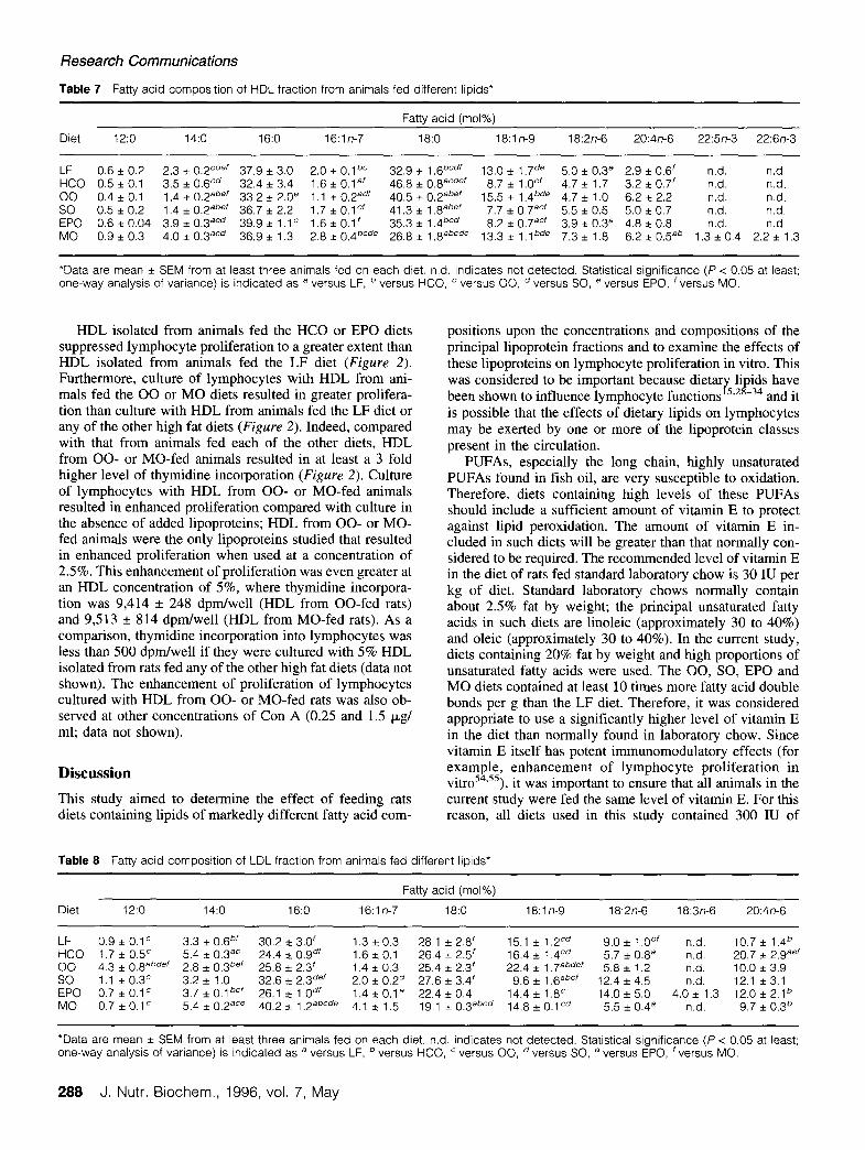
The fatty acid composition of the HDL fraction (Table 7) was characterized by fairly high proportions of palmitic and stearic acids and a low proportion of linoleic acid; the pro- portions of these fatty acids were little affected by dietary
J. Nutr. Biochem., 1996, vol. 7, May 285
Research Communications
Table 4 Composition of lipoprotein fractions*
Fraction LF HCO
Diet
00 so EPO MO
Cholesterol @g/ml) CM 36 + 5 40 + 4* 63 IIZ 12d 21 + 6” 41 *9 44 * 9 VLDL 47 f 4b 86 f 7a* 63+ 10 43 * 3* 61 *9 71 i 15 LDL 130 f 7’ 148 + 25’ 135 f 15’ 125 * 16’ 137 f 17’ 85 zt lGabcde HDL 576 * 39’ 776 * 29’ 717*31’ 524 * 23’ 615 r 36’ 317 + 36abcde
TAG (w/ml) CM 96 zt 20’ 557 i looa=de’ 238 * 70’ 99 f. 30b 130 It 4ob 1171r30b VLDL 392 + 5oc 388 * 60c 783 + 130abdef 315 f 7oc 366 f 40’ 390 i 7oc
APO B @g/ml) VLDL 22 f 5 29 + 6 35 * 4de 20 * 5c 20 f 3= 30 c 6 Apo A-l (0.0.) HDL 0.46 f 0.06’ 0.37 + 0.08= 0.63 sz 0.07b’ 0.43 * 0.07 0 59 f 0.09’ 0.23 i 0.02ace
*Data are mean * SEM from at least six animals fed on each diet. Statistical significance (P < 0.05 at least; one-way analysis of variance) is indicated as a vs. LF, b vs. HCO, c vs. 00, d VS. SO, e VS. EPO, ‘VS. MO.
manipulation, apart from a lower proportion of stearic acid in HDL from MO-fed animals. There were some dietary influences on the fatty acid composition of HDL. For ex- ample, the proportion of myristic acid is high in HDL from rats fed the HCO or MO diets. Similarly, the proportion of oleic acid is the highest in HDL from animals fed the 00 diet. The HDL isolated from the serum of MO-fed rats contain detectable levels of n-3 PUFAs.
The fatty acid composition of the LDL fraction (Table 8) was affected to a much lesser extent by that of the diet than were the compositions of the CM and VLDL fractions. Nev- ertheless, there is still an influence of the diet. For example, the proportion of myristic acid is high in LDL from rats fed the HCO diet. Similarly, the proportion of oleic acid is the highest in LDL from animals fed the 00 diet and the pro- portion of linoleic acid is highest in LDL from animals fed the LF, SO, or EPO diets. LDL isolated from the serum of EPO-fed animals are the only ones to contain y-linolenic acid. The LDL isolated from the serum of MO-fed rats are relatively rich in myristic, palmitic, and palmitoleic acids and are relatively poor in linoleic acid; these LDL did not contain detectable levels of n-3 PUFAs. LDL isolated from the serum of all animals, including those fed the MO diet, contained relatively high proportions of arachidonic acid (10% of more); this proportion appears to be little affected by the nature of the diet consumed.
Effects of lipoproteins on lymphocyte proliferation
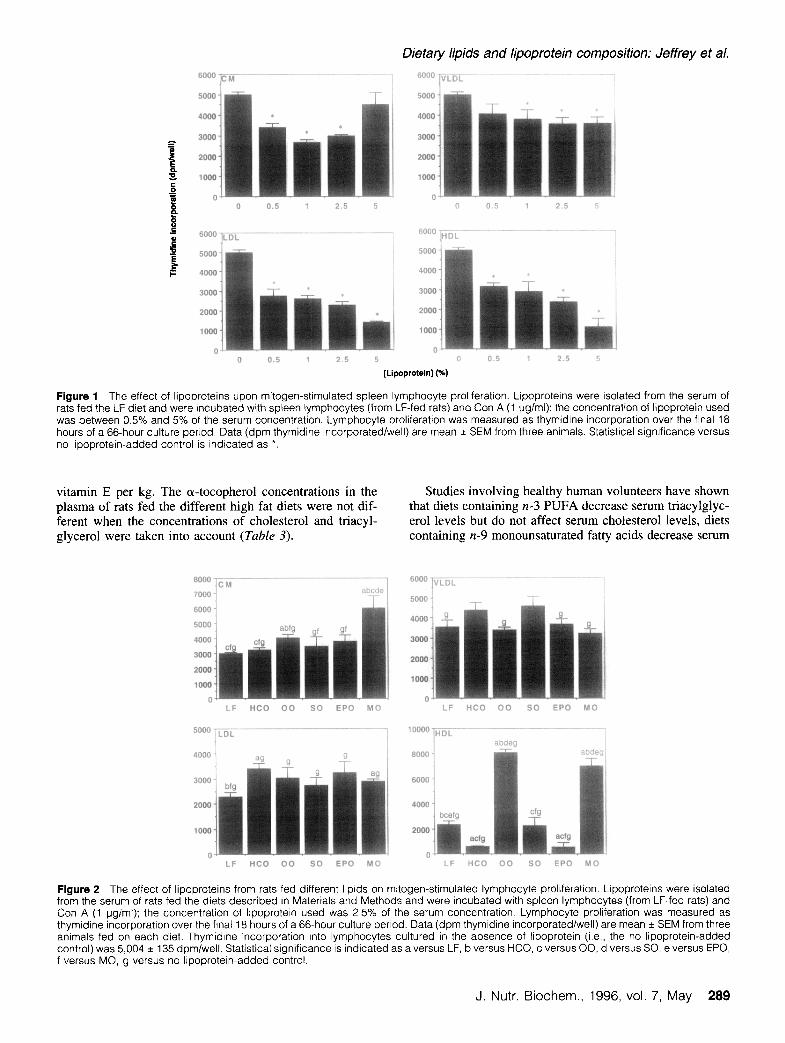
The effects of different lipoprotein fractions isolated from LF-fed animals on the proliferative response of spleen lym- phocytes (isolated from LF-fed rats) to 1 kg/ml Con A are shown in Figure I; the lipoproteins were included in the culture medium at concentrations between 0.5% and 5% of their concentration in serum. It was previously found that culturing lymphocytes from chow-fed rats in the presence of 2.5% or 5% unfractionated serum obtained from rats fed different types of lipid can cause inhibition of lymphocyte proliferation.36 Both LDL and HDL caused marked concen- tration-dependent inhibition of Con A-stimulated lympho- cyte proliferation (Figure 1). CM and VLDL caused some inhibition of proliferation but this was less marked (Figure
I). Thus, these results indicate that the order of inhibitory potencies for lipoproteins from LF-fed animals is:
HDL - LDL > CM > VLDL
The effect of dietary lipid manipulation upon lipoprotein-induced inhibition of lymphocyte proliferation
Once the effects of lipoproteins isolated from rats fed a LF diet were established (see previous discussion), the influ- ence of the nature of the lipid consumed in the diet on the effect of each class of lipoprotein on spleen lymphocyte proliferation was investigated (Figure 2); in all cases the lymphocytes were from rats fed the LF diet.
VLDL or LDL isolated from animals fed the high fat diets suppressed lymphocyte proliferation to approximately the same extent as VLDL or LDL isolated from LF-fed animals (Figure 2). Thus, the effects of VLDL and LDL on lymphocyte proliferation are not greatly affected by dietary lipid manipulation.
In contrast to the observations with VLDL and LDL, there was an influence of the type of lipid fed on the sub- sequent effects of CM and HDL on lymphocyte prolifera- tion. CM isolated from the animals fed the HCO, 00, SO, or EPO diets inhibited lymphocyte proliferation to approxi- mately the same extent as that from rats fed the LF diet (Figure 2). However, culture of spleen lymphocytes with CM isolated from animals fed the MO diet resulted in sig- nificantly greater lymphocyte proliferation than observed for culture with CM from animals fed each of the other diets (Figure 2). Furthermore, whereas 2.5% CM from animals fed the LF, HCO, 00, SO, or EPO diets suppressed prolif- eration compared with the no lipoprotein-added control, 2.5% CM from MO-fed rats was not suppressive (Figure 2). At a concentration of 5%, CM from the MO-fed animals significantly enhanced lymphocyte proliferation compared with the no lipoprotein-added control and compared with CM isolated from rats fed each of the other diets; thymidine incorporation into cells cultured in the presence of 5% CM from rats fed the MO diet was 6,537 -+ 768 dpm/well com- pared with less than 4,000 dpm/well for cells cultured in the presence of CM from rats fed the other diets.
286 J. Nutr. Biochem., 1996, vol. 7, May
Tabl
e 5
Fatty
ac
id
com
posit
ion
of
CM
fract
ion
from
an
imal
s fe
d di
ffere
nt
lipid
s*
Diet
12
:o
14:o
16
:0
16:ln
-7
Fatty
ac
id
(mol%
)
18:O
18
:ln-9
18
:2n-
6 18
:3n-
6 20
:4n-
6 20
:5n-
3 22
:5n-
3 22
:6n-
3
LF
n.d.
2.
1 f
o.2b
c’ 39
.5
* 2.
3bcd
ef
6.1
zt 0.
36cf
6.
5 +
1 .O
de
24.1
f
0.56
cf
12.9
*
O.S
bCd’
n.d.
1.
5 *
0.7e
n.
d.
n.d.
n.
d.
HCO
29.3
+
2.1
cef
16.8
*
0.5a
’ 22
.4
+ 0.
6a’
3.1
f 0.
2=
7.8
* 0.
7c
13.1
+
O.G
acde
’ 3.
3 It
0.2a
d’
n.d.
0.
9 *
o.2d
e’
n.d.
n.
d.
nd.
00
0.5
i O
.lbe
1.2
+ O
.l=’
22.4
i
1 .O
a’ 2.
2 f
0.2a
bder
4.
7 i
0.2b
de’
55.1
f
0.7a
b*ef
9.
1 +
l.OBb
d’
n.d.
1.
4 f
0.3e
nd
. nd
. nd
. so
n.
d.
1.5
f 0.
2’
24.9
*
1 ,ga
’ 4.
2 +
0.8c
’ 10
.5
* 1.
2=’
26.0
*
2.3”
’ 21
.4
+ 6.
8abc
f n.
d.
3.2
zt 0.
8be
n.d.
nd
. nd
. EP
O
0.9
f 0.
1 bc
f 2.
0 +
0.5b
cd’
27.6
*
2.2a
f 4.
5 +
0.6c
f 11
.1
f 1.
W
20.8
f
1 .g
bc
12.2
+
2.26
d’
0.9
* 0.
2 9.
3 +
1 .g
abcd
f n.
d.
n.d.
n.
d.
MO
0.4
f 0.
1 be
11
.5
9 0.
7abc
de
36.7
+
1 .7a
bcde
14
.2
* 0.
7abc
de
7.2
+ 0.
3cde
18
.7
+ 0.
7abc
d 2.
0 +
0.2a
bcde
n.
d.
2.3
+c 0.
4be
0.9
+ 0
0.2
+ 0
0.2
f 0.
1
“Dat
a ar
e m
ean
* SE
M
from
at
le
ast
thre
e an
imal
s fe
d on
ea
ch
diet
. nd
in
dica
tes
not
dete
cted
. St
atis
tical
si
gnifi
canc
e (P
<
0.05
at
le
ast;
one-
way
anal
ysis
of
varia
nce)
is
in
dica
ted
as
a ve
rsus
LF
, b
vers
us
HCO,
’
vers
us
00,
d ve
rsus
SO
, e
vers
us
EPO
, ‘ve
rsus
M
O.
Tabl
e 6
Fatty
ac
id
com
posit
ion
of
VLDL
fra
ctio
n fro
m
anim
als
fed
diffe
rent
lip
ids*
0 -. CD
Fatty
ac
id
(mol%
) s
Diet
12
:o
14:o
16
:0
16:ln
-7
18:0
18
:ln-9
18
:2n-
6 18
:3n-
6 20
:4n-
6 20
:5n-
3 22
:5n-
3 22
:6n-
3 s z+
LF
n.d.
2.
1 +
0.36
Cd’
34.6
f
1 .g
cd
4.8
YI 0
.3cd
ef
9.5
zk l
.oc
24.8
LI
Z O.S
bcde
13
.7
* l,lb
=def
nd
. 8.
3 i
0.5”
” n.
d.
n.d.
n.
d.
HCO
4.2
f 0.
7cde
f 7.
1 zt
0.4”
cdef
29
.3
+ l.l
Cd’
5.5
+ 0.
3cde
’ 9.
1 *
o.3c
29
.4
f 0.
7a=d
e’
6.1
III 0
.2ac
de
nd.
1.9
A 0.
3=
n.d.
n.
d.
n.d.
9 Q
00
0.2
+ O
.lb
1 ,i
f O
.labe
’ 20
.7
zt 1
.labd
e’
2.2
* 0.
2ab’
6.
2 -c
O.la
bder
54
.5
zt 1
.2ab
de’
9.3
f O
.labe
’ n.
d.
1.6
* 0.
3=
n.d.
n.
d.
n.d.
so
0.
2 +
0.16
1
.o +
0.
1 ab
ef
24.9
+
0.9=
b=ef
1.
6 *
0.2a
bf
8.0
i 0.
7’
20.7
+
O.S
abce
’ 38
.3
+ 1
,gab
cf
nd.
2.4
+ 0.
5=
n.d.
n.
d.
n.d.
s
EPO
0.
4 +O
b 1.
9 +
o.3b
cdr
30.8
zt
2.0c
df
2.4
ic 0
.4ab
’ 9.
6 i
1 .O
c 17
.1
zt 0.
8abc
” 30
.0
t 3.
5abc
f 1.
8 it
0.1
7.5
+ o.
7bcd
’ n.
d.
n.d.
n.
d.
8 MO
0.
4 f
0.16
3.
4 *
0.3a
bcde
37
.5
+ 1
.2bc
de
13.6
+
O.G
abcd
e 8.
2 +
0.3c
26
.1
+ 0.
5bcd
e 5.
7 f
0,2=
cde
nd.
1.6
* 0.
5=
1.7*
0.4
1.0*
0.3
1.0*
0.3
2 B 3 ‘D
ata
are
mea
n i
SEM
fro
m
at
leas
t th
ree
anrm
als
fed
on
each
di
et.
n.d.
in
drca
tes
not
dete
cted
. St
atis
tical
sr
gnifr
canc
e (P
<
0.05
at
le
ast;
one-
way
anal
ysis
of
varia
nce)
is
in
dica
ted
as
a ve
rsus
LF
, b
vers
us
HCO,
’
vers
us
00,
d ve
rsus
SO
, e
vers
us
EPO
, ‘ve
rsus
M
O.
8 3
Research Communications
Table 7 Fatty acid composition of HDL fraction from animals fed different lipids*
Fatty acid (mol%)
Diet 12:o 14:o 16:0 16:ln-7 18:O 18:ln-9 18:2n-6 20:4n-6 22:5n-3 22:6n-3
LF 0.6 * 0.2 2.3 i 0.2Cdef 37.9 * 3.0 2.0 + O.lbc 32.9 i 1.6bcdf 13.0 ic l.7de 5.0 * 0.3e 2.9 k 0.6’ n.d. n.d. HCO 0.5 +z 0.1 3.5 + 0.6Cd 32.4 * 3.4 1.6 zt O.l=’ 46.8 + 0.8acde’ 8.7 f 1 .OC’ 4.7 + 1.7 3.2 k 0.7’ n.d. n.d. 00 0.4 f 0.1 1.4 zt 0.2abef 33.2 i 2.0e 1 .l zt 0.2=” 40.5 It 0.2abef 15.5 LIZ 1.4bde 4.7 i 1.0 6.2 zt 2.2 n.d. nd. so 0.5 * 0.2 1.4 * 0.2abe’ 36.7 * 2.2 1.7 * 0.1=’ 41.3 f l.8abef 7.7 f 0.7acr 5.5 * 0.5 5.0 f 0.7 n.d. n.d.
EPO 0.6 zt 0.04 3.9*0.3acd 39.9* 1.1’ 1.6 i 0.1’ 35.3 f 1 .4bcd 8.2 * 0.7=’ 3.9 f 0.3a 4.8 k 0.8 n.d. n.d. MO 0.9 + 0.3 4.0 LIZ 0.3acd 36.9 zt 1.3 2.8 zt 0.4bcde 26.8 f l.8abcde 13.3 i l.lbde 7.3 i 1.8 6.2 * 0.5ab 1.3 zt 0.4 2.2 + 1.3
*Data are mean * SEM from at least three animals fed on each diet. n.d. indicates not detected. Statistical significance (P c 0.05 at least; one-way analysis of variance) is indicated as a versus LF, b versus HCO, c versus 00, d versus SO, e versus EPO, f versus MO.
HDL isolated from animals fed the HCO or EPO diets suppressed lymphocyte proliferation to a greater extent than HDL isolated from animals fed the LF diet (Figure 2). Furthermore, culture of lymphocytes with HDL from ani- mals fed the 00 or MO diets resulted in greater prolifera- tion than culture with HDL from animals fed the LF diet or any of the other high fat diets (Figure 2). Indeed, compared with that from animals fed each of the other diets, HDL from 00- or MO-fed animals resulted in at least a 3 fold higher level of thymidine incorporation (Figure 2). Culture of lymphocytes with HDL from 00- or MO-fed animals resulted in enhanced proliferation compared with culture in the absence of added lipoproteins; HDL from 00- or MO- fed animals were the only lipoproteins studied that resulted in enhanced proliferation when used at a concentration of 2.5%. This enhancement of proliferation was even greater at an HDL concentration of 5%, where thymidine incorpora- tion was 9,414 k 248 dpm/well (HDL from 00-fed rats) and 9,513 + 814 dprn/well (HDL from MO-fed rats). As a comparison, thymidine incorporation into lymphocytes was less than 500 dpm/well if they were cultured with 5% HDL isolated from rats fed any of the other high fat diets (data not shown). The enhancement of proliferation of lymphocytes cultured with HDL from 00- or MO-fed rats was also ob- served at other concentrations of Con A (0.25 and 1.5 kg/ ml; data not shown).
positions upon the concentrations and compositions of the principal lipoprotein fractions and to examine the effects of these lipoproteins on lymphocyte proliferation in vitro. This was considered to be important because dietary lipids have been shown to influence lymphocyte functions’5,28-34 and it is possible that the effects of dietary lipids on lymphocytes may be exerted by one or more of the lipoprotein classes present in the circulation.
Discussion
PUFAs, especially the long chain, highly unsaturated PUFAs found in fish oil, are very susceptible to oxidation. Therefore, diets containing high levels of these PUFAs should include a sufficient amount of vitamin E to protect against lipid peroxidation. The amount of vitamin E in- cluded in such diets will be greater than that normally con- sidered to be required. The recommended level of vitamin E in the diet of rats fed standard laboratory chow is 30 IU per kg of diet. Standard laboratory chows normally contain about 2.5% fat by weight; the principal unsaturated fatty acids in such diets are linoleic (approximately 30 to 40%) and oleic (approximately 30 to 40%). In the current study, diets containing 20% fat by weight and high proportions of unsaturated fatty acids were used. The 00, SO, EPO and MO diets contained at least 10 times more fatty acid double bonds per g than the LF diet. Therefore, it was considered appropriate to use a significantly higher level of vitamin E in the diet than normally found in laboratory chow. Since vitamin E itself has potent immunomodulatory effects (for exam le, vitro5 !? ,55),
enhancement of lymphocyte proliferation in it was important to ensure that all animals in the
This study aimed to determine the effect of feeding rats current study were fed the same level of vitamin E. For this diets containing lipids of markedly different fatty acid com- reason, all diets used in this study contained 300 IU of
Table 8 Fatty acid composition of LDL fraction from animals fed different lipids*
Fatty acid (mol%)
Diet 12:o 14:o 16:0 16:ln-7 18:0 l&ln-9 18:2n-6 18:3n-6 20:4n-6
LF 0.9 * O.lC 3.3 + 0.6” 30.2 zt 3.0‘ 1.3 f 0.3 28.1 f 2.8’ 15.1 * 1.2Cd 9.0 + 1 .o*’ n.d. 10.7 f 1.4b HCO 1.7 * 0.5c 5.4 i 0.3”” 24.4 + O.gd’ 1.6kO.l 26.4 zt 2.5’ 16.4 + 1 .qcd 5.7 * 0.8a n.d. 20.7 + 2.9= 00 4.3 r 0.8abdef 2.8 * 0.3ber 25.8 f 2.3’ 1.4 f 0.3 25.4 f 2.3’ 22.4 i 1 .7abder 5.8 f 1.2 n.d. 10.0 f 3.9
so 1.1 * o.3c 3.2 * 1.0 32.6 + 2.3de’ 2.0 * 0.2d 27.6 + 3.4’ 9.6 f 1 .6abcf 12.4 f 4.5 nd. 12.1 f 3.1 EPO 0.7 + 0.1’ 3.7 * O.l’=’ 26.1 k l.Odf 1.4 * 0.1” 22.4 i 0.4 14.4 i 1 .8c 14.0 f 5.0 4.0 f 1.3 12.0 + 2.1* MO 0.7 f 0.1” 5.4 f 0.2”‘=e 40.2 + 1 .2abcde 4.1 zt 1.5 19.1 i 0.3=*cd 14.8 f O.lCd 5.5 i 0.4a n.d. 9.7 * 0.3*
*Data are mean f SEM from at least three animals fed on each diet. nd. indicates not detected. Statistical significance (P < 0.05 at least; one-way analysis of variance) is indicated as a versus LF, * versus HCO, c versus 00, d versus SO, e versus EPO, ‘versus MO.
288 J. Nutr. Biochem., 1996, vol. 7, May
Dietary lipids and lipoprotein composition: Jeffrey et al.
5000
4ooo
3ooo
2ooo
1000
’ 0 0.5 1 2.5 5 0
f WOO $DL woo
/LDL
0 0.5 1 2.5 5
* * ilr, * 0 0.5 1 2.5 5 0 0.5 1 2.5 5
[Lipoprotein] (%)
Figure 1 The effect of lipoproteins upon mitogen-stimulated spleen lymphocyte proliferation. Lipoproteins were isolated from the serum of rats fed the LF diet and were incubated with spleen lymphocytes (from LF-fed rats) and Con A (1 ugiml); the concentration of lipoprotein used was between 0.5% and 5% of the serum concentration. Lymphocyte proliferation was measured as thymidine incorporation over the final 18 hours of a 66-hour culture period. Data (dpm thymidine incorporated/well) are mean * SEM from three animals. Statistical significance versus no lipoprotein-added control is indicated as *.
vitamin E per kg. The a-tocopherol concentrations in the Studies involving healthy human volunteers have shown plasma of rats fed the different high fat diets were not dif- that diets containing n-3 PUFA decrease serum triacylglyc- ferent when the concentrations of cholesterol and triacyl- erol levels but do not affect serum cholesterol levels, diets glycerol were taken into account (Table 3). containing n-9 monounsaturated fatty acids decrease serum
m”-lCM 1 gWO]VLDL I abcde
T I 5oooI - -I-
LF tic0 00 SO EPO MO LF tic0 00 SO EPO MO
5ow LDL ‘OOOQ HDL abdea
LF tic0 00 SO EPO MO LF HCO 00 SO EPO MO
Figure 2 The effect of lipoproteins from rats fed different lipids on mitogen-stimulated lymphocyte proliferation. Lipoproteins were isolated from the serum of rats fed the diets described in Materials and Methods and were incubated with spleen lymphocytes (from LF-fed rats) and Con A (1 ugiml); the concentration of lipoprotein used was 2.5% of the serum concentration. Lymphocyte proliferation was measured as thymidine incorporation over the final 18 hours of a 66.hour culture period. Data (dpm thymidine incorporated/well) are mean * SEM from three animals fed on each diet. Thymidine incorporation into lymphocytes cultured in the absence of lipoprotein (i.e., the no lipoprotein-added control) was 5,004 * 135 dpm/well. Statistical significance is indicated as a versus LF, b versus HCO, c versus 00, d versus SO, e versus EPO, f versus MO, g versus no lipoprotein-added control.
J. Nutr. Biochem., 1996, vol. 7, May 289
Research Communications
triacylglycerol levels with either a decrease or no change in serum cholesterol levels, whereas diets rich in saturated fatty acids increase serum triacylglycerol and serum choles- terol levels.2*3 The effects of feeding rats the diets used in the current study on the serum concentrations of cholesterol and triacylglycerol have been previously reported.’ 3 Rats fed the HCO or 00 diets had increased serum triacylglyc- erol concentrations compared with rats fed the LF, SO, EPO, or MO diets.13 Thus, the MO diet failed to produce a hypotriacylglycerolaemic effect and the 00 diet had a hy- pertriacylglycerolaemic effect. Compared with each of the other diets, the MO diet resulted in a lower serum total cholesterol concentration. l3 These observations are in con- trast to those made in human studies, exce large amounts of n-3 PUFA were ingested. Y
t where very However, in
accordance with these results, Hostmark et al.” reported lower serum triacylglycerol concentrations in rats fed on fish oil compared with those fed on coconut oil and De- Schrijver et al.” reported lower serum triacylglycerol con- centrations in rats fed fish oil compared with those fed on beef tallow (rich in saturated fatty acids). Furthermore, both studies also reported lower serum cholesterol concentrations in the fish oil-fed rats compared with those fed coconut oi112 or beef tallow’ ‘; again these findings are consistent with the results reported in our study. The apparently different ef- fects of a fish oil-rich diet on serum cholesterol concentra- tions in rats and in humans may be at least partly due to the species differences in cholesterol metabolism; such differ- ences indicate that the rat is not a good model for the in- vestigation of lipoprotein metabolism in man.
The current study shows that the hypocholesterolaemic effect of the MO dieti is largely caused by a lowered level of HDL cholesterol (Table 4). This indicates that the MO diet may decrease the synthesis of HDL or increase its re- moval from the plasma. Apo A-l is a major apoprotein component of HDL and the concentration of Apo A-l was lower in HDL from MO-fed animals (Table 4). Therefore, the MO diet could affect HDL synthesis by influencing Apo A-l availability; indeed feeding rats this diet has been shown to significantly lower Apo A-l mRNA levels in the liver.56 The hypertria c 1 1 y g y cerolaemic effects of feeding the HCO or 00 diets are due to elevated levels of triacylglyc- erol in the CM fraction and in both the CM and VLDL fractions, respectively (Table 4). The effect of the HCO diet on CM triacylglycerol levels was unexpected because this diet contains a high proportion of medium chain fatty acids (Table 2) that might be expected to enter the hepatic portal vein directly rather than be assembled into CM. The fatty acid composition of the CM fraction from HCO-fed rats (Table 5) clearly indicates that medium chain fatty acids such as lauric acid (12:O) are incorporated into CM. The elevated VLDL triacylglycerol level after feeding the 00 diet indicates that, in rats, oleic acid might promote hepatic VLDL synthesis.
The fatty acid composition of each lipoprotein fraction was modified according to that of the lipid consumed in the diet; the greatest effects were seen in the CM and VLDL fractions, whose fatty acid compositions differed signifi- cantly between animals fed different lipids (Tables 5, 6). Because CM transport triacylglycerol of dietary origin, the marked effects of dietary lipids on the fatty acid composi-
tion of this fraction are not surprising. VLDL contain tria- cylglycerol that is synthesised in the liver (although some VLDL is released from absorptive cells of the small intes- tine); the fatty acid components of these triacylglycerols could be derived from de novo synthesis but this is unlikely in animals fed a diet that includes 20% fat. Alternatively, the fatty acids found in the triacylglycerols of VLDL could come from other lipoproteins (e.g., chylomicron remnants) or free fatty acids taken up by the liver. The fatty acid composition of lipoprotein-free serum from these animals, which presumably represents that of the non-esterified fatty acids, strongly reflects the fatty acid composition of the diets themselves,36 as does the fatty acid composition of the adipose tissue. l3 Thus, if the fatty acids of the VLDL tria- cylglycerols are derived from circulating non-esterified fatty acids, it is no surprise that the fatty acid composition of VLDL is strongly influenced by diet.
The fatty acid compositions of the LDL and HDL frac- tions are less markedly affected by diet than the CM and VLDL fractions; however a number of differences were observed (Tables 7, 8). Because the fatty acids found in HDL and LDL are likely to be components of either phos- pholipids or cholesterol esters, it is possible that the fatty acid composition of these lipoproteins are less sensitive to dietary manipulation than the lipoprotein fractions respon- sible for triacylglycerol transport. However, it is likely that after 10 weeks on a particular dietary regime, the fatty acid precursors used for the synthesis of phospholipids will re- flect those present in the diet. Thus, the observed diet- induced changes in the fatty acid compositions of HDL and LDL are not unexpected.
Lymphocytes are thought to possess receptors for vari- ous types of lipoprotein 4’*46; in addition lymphocytes pos- sess lipoprotein lipase activity23 and so can remove fatty acids from circulating lipoproteins. A number of studies have previously investigated the effect of purified lipopro- teins upon lymphocyte proliferation.37A6 Several of these studies agree that VLDL and LDL inhibit lymphocyte pro- liferation,37A3.45 although there are also reports of enhance- ment by these lipoproteins.44T46 Similarly, although some studies report inhibition of lymphocyte proliferation by pu- rified HDL,38*42 other studies show little effect4”45 or en- hancement.44T46 When surveying the previous studies, it is apparent that culture conditions are important in determin- ing the overall effect observed. In the current study, iden- tical conditions were used to investigate the effects of each lipoprotein; the culture medium was serum-free but in- cluded albumin, transferrin, and insulin at appropriate con- centrations. We found that each class of lipoprotein can inhibit rat spleen lymphocyte proliferation in response to Con A (Figure I), and that LDL and HDL have more potent effects than CM or VLDL. Thus, the current results agree with the previous studies that show potent inhibition of lymphocyte proliferation by LDL37d0,43 and HDL.42 Dis- crepancies between the results presented here and those of some previous studies may relate to differences in the cell culture conditions used; it is apparent that the outcome of lymphocyte proliferation assays is affected by the types of growth factors that are present,44 the presence or absence of serum,36 the origin and concentration of any serum used,” and the concentration of lipoprotein used (Figure I).
290 J. Nutr. Biochem., 1996, vol. 7, May
Whether the type of lipid consumed in the diet can in- fluence the effect of different lipoproteins on lymphocyte proliferation has not been previously investigated. The re- sults of the current study show that fish oil feeding can alter the effects of CM or HDL on lymphocyte proliferation and that 00 feeding can alter the effects of HDL (Figure 2). Whereas CM and HDL from rats fed a LF diet or high fat diets rich in saturated or n-6 PUFAs inhibit lymphocyte proliferation, CM or HDL from MO-fed rats enhance lym- phocyte proliferation (Figure 2). Feeding the 00 or MO diets has been shown to significantly inhibit lymphocyte functions ’ 5,28-33 and we speculated that the effects of these dietary lipids might be mediated by lipoproteins. Therefore, the observation that some lipoprotein fractions from 00- or MO-fed rats markedly enhance lymphocyte proliferation was surprising. Nevertheless, it was a consistent observa- tion.
The effects of lipoproteins on lymphocyte proliferation indicate that they contain components that are suppressive; these may be lipids or apolipoproteins. Because fatty ac- ids 16-22,24*25 and triacylglycerols23~26~27 inhibit lymphocyte functions in vitro, it would not be surprising if lipid com- ponents of lipoproteins were also inhibitory. However, in vitro studies indicate that n-3 PUFAs are the more inhibi- tory than other types of fatty acids”-** and so it might be expected that lipoproteins from animals fed the MO diet would be more suppressive than those from animals fed the other diets. This was found not to be the case; VLDL and LDL from MO-fed animals have the same effect as those from animals fed the other diets, whereas HDL and CM from MO-fed animals are enhancing compared with those from animals fed the other diets. The stimulatory effects of CM and HDL from MO-fed animals indicates that these lipoproteins contain lower levels of suppressive factors and/ or higher levels of stimulatory factors than those from ani- mals fed the other diets. Both CM and HDL contain Apo A-l, and MO feeding resulted in a significantly lower HDL Apo A-l concentration compared with feeding the other diets. Thus, Apo A-l, or apolipoproteins in general, might be suppressive.
A further explanation for the enhancement of lympho- cyte proliferation observed after culture with CM and HDL from MO-fed rats and HDL from 00-fed rats is that the lymphocytes might have efficiently taken up n-3 PUFAs or oleic acid from these lipoproteins. This would lead to re- placement of arachidonic acid in lymphocyte phospholipids by the n-3 PUFAs or oleic acid. As a result, lower levels of PGE, would be produced by these cells; PGE, is known to inhibit lymphocyte proliferation.’ * Alternatively, these lipo- proteins might be enriched in o-tocopherol, which is known to enhance lymphocyte proliferation.54,55 The a-tocopherol contents of the various lipoprotein fractions were not deter- mined in this study.
The findings of this study suggest that the inhibitory effects of the MO diet, and also of the 00 and EPO di- ets,15*29-3’ on lymphocyte proliferation may not be medi- ated by lipoproteins. Furthermore, when whole serum is used in cell culture, the stimulatory effect of CM and HDL in the serum of MO-fed rats (and of HDL in the serum of 00-fed rats) must be overridden by a suppressive effect of at least one other serum component. This may be VLDL or
Dietary lipids and lipoprotein composition: Jeffrey et al.
LDL. In addition, lipoprotein-free serum from rats fed the HCO, 00, EPO, or MO diets was found to suppress Con A-stimulated proliferation of spleen lymphocytes.36 This suggests that a non-lipoprotein component of serum is in- hibitory. This may be non-esterified fatty acids [n-3 PUFAs are potent inhibitors of lymphocyte function]“-** or a non- lipid component. The nature of the suppressive non- lipoprotein factor warrants further investigation.
Acknowledgments
This research was supported by Unilever Research Labora- tories. NMJ holds a Postgraduate Studentship funded by Unilever Research Laboratories and PY holds a Post- Doctoral Fellowship funded by the Ministry of Agriculture, Fisheries, and Food. GFG is a member of the external sci- entific staff of the Medical Research Council at the Univer- sity of Oxford.
References
5
6
I
8
9
10
11
12
13
14
15
16
Keys, A. (1970). Coronarv heart disease in seven countries. Circu- larion 41, l-211 _ Harris, W.S. (1989). Fish oils and plasma lipid and lipoprotein me- tabolism in humans: a critical review. .Z. LiDid Rex 30, 785-807 Grundy, S.M., and Denke, M.A. ( 1990). Dietary influences on serum lipids and lipoproteins. J. Lipid Res. 31, 1149-l 172 Dolphin, P.J., Amy, R.M., Koeslag, D.G., Limoges, B.F., and Rus- sell, J.C. (1988). Reduction in hyperlipidemia in the LA/N-corpulent rat by dietary fish oil containing n-3 fatty acids. Biochim. Biophys. Acta 962, 317-329 Garg, M.L., Wierzbicki, A.A., Thomson, A.B.R., and Clandinin, M.T. (1988). Fish oil reduces cholesterol and arachidonic acid con- tent more efficiently in rats fed diets containing low linoleic acid to saturated fatty acid ratios. Biochim. Biophys. Acta 962, 337-344 Kingsley, T.R. and Snyder, D.L. (1988). Serum lipids in spontane- ously hypertensive rats and Sprague-Dawley rats fed menhaden oil. Lipids 23, 564-567 Sugano, M. and Imaizumi, K. (1991). Effect of palm oil on lipid and lipoprotein metabolism and eicosanoid production in rats. Am. J. Clin. Nutr. 53, 1034S-1038s Lai, H-C., Lasekan, J.B., Yang, H., Clayton. M.K., and Ney, D.M. (1991). In viva determination of triglyceride secretion using radio- active glycerol in rats fed different saturated fats. Lipids 26,824830 Al-Shurbaji, A., Larsson-Backstrom, C., Berglund, L., Eggersten, G., and Bjorkhem, I. (1991). Effect of n-3 fatty acids on the key enzymes involved in cholesterol and triglyceride turnover in rat liver. Lipids 26, 385-389 Ney, D.M., Lai, H-C., Lasekan, J.B., and Lefevre, M. (1991). Inter- relationship of plasma triglycerides and HDL size and composition in rats fed different dietarv saturated fats. J. Nurr. 121, 13 11-1322 , DeSchriiver, R., Vermeulen. D.. and Viane. E. (1991). Lipid me- tabolism responses in rats fed beef tallow, native or randomised fish oil and native or randomised oeanut oil. J. Nutr. 121, 948-955 Hostmark, A.T., Lystad, E., ‘Huag, A., and Eilersten, E. (1989). Plasma lipids, lipoproteins, and fecal excretion of neutral sterols and bile acids in rats fed various high fat diets or a low fat/high sucrose diet. J. Nutr. 119, 356-363 Yaqoob, P., Sherrington, E.J., Jeffery, N.M., Sanderson, P., Harvey, D.J., Newsholme, E.A., and Calder, P.C. (1995). Comparison of the effects of a range of dietary lipids upon serum and tissue lipid com- position in the rat. Znt. .Z. Biochem. Cell Biol. 27, 297-310 -Kromann, N. and Green, A. (1980). Epidemiological studies in the Unemavik District. Greenland. Acta Med. Scand. 208, 401-406 Yaqoob, P., Newsholme, E.A., and Calder, P.C. (1994). The effect of dietary lipid manipulation on rat lymphocyte subsets and prolifera- tion. Znnnunology 82, 603-610 Buttke, T.M. (1984). Inhibition of lymphocyte proliferation by free fatty acids. Zmmunoiogy 53, 235-242
J. Nutr. Biochem., 1996, vol. 7, May 291
Research Communications
17
18
19
20
21
22
23
24
25
26
27
28
29
30
31
32
33
34
35
Calder, P.C., Bond, J.A., Bevan, S.J., Hunt, S.V., and Newsholme, E.A. (1991). Effect of fatty acids on the proliferation of concanavalin A-stimulated rat lymph node lymphocytes. Znr. J. Biochem. 23, 579- 588 Calder, P.C., Bevan, S.J., and Newsholme, E.A. (1992). The inhibi- tion of T-lymphocyte proliferation by fatty acids is via an eicosa- noid-independent mechanism. Immunology 75, 108-l 15 Calder, P.C., and Newsholme, E.A. (1992). Unsaturated fatty acids suppress interleukin-2 production and transferrin receptor expression by concanavalin A-stimulated rat lymphocytes. Medial. ZnfIamm. 1, 107-l 15 Calder, P.C. and Newsholme, E.A. (1992). Polyunsaturated fatty acids suppress human peripheral blood lymphocyte proliferation and interleukin-2 production. Clin. Sci. 82, 695-700 Kumar, G.S., has, U.N., Kumar, K.V., Madhavi, N., Das, N.P., and Tan, B.K.H. (1992). Effect of n-6 and n-3 fattv acids on the nrolif- eration of human lymphocytes and their secretion of TNF-(Y and IL-2 in vitro. Nutr. Res. 12, 815-823 Soyland, E., Nenseter, M.S., Braathen, L., and Drevon, C.A. (1993). Very long chain n-3 and n-6 polyunsaturated fatty acids inhibit pro- liferation of human T-lymphocytes in vitro. Eur. J. Clin. Invest. 23, 112-121 Calder, P.C., Yaqoob, P., Newsholme, E.A. (1994). Triacylglycerol metabolism by lymphocytes and the effect of triacylglycerols upon lymphocyte proliferation. Biochem. J. 298, 605-611 Virella, G., Kilpatrick, J.M., Rugeles, M.T., Hyman, B., and Russell, R. (1989). Depression of humoral responses and phagocytic func- tions in vivo and in vitro by fish oil and eicosapentaenoic acid. Chin. Zmmunol. Zmmunopnthol. 52, 277-290 Richieti, G.V., Mescher, M.F., and Kleinfeld, A.M. (1990). Short term exposure to cis unsaturated free fatty acids inhibits degranula- tion of cytotoxic T lymphocytes. J. Zmmunol. 144, 671-677 Yamashita, N., Maruyama, M., Yamazaki, K., Hamazaki, T., and Yano, S. (1991). Effect of eicosapentaenoic and docosahexaenoic acid on natural killer cell activity in human peripheral blood lym- phocytes. Clin. Zmmunol. Zmmunopathol. 59, 335-345 Fujikawa, M., Yamashita, N., Yamazaki, K., Sugiyama, E., Suzuki, H., and Hamazaki, T. (1992). Eicosapentaenoic acid inhibits antigen- presenting cell function of murine splenocytes. Immunology 75, 330-335 Meydani, S.N., Endres, S., Woods. M.M., Goldin, B.R., Soo, C., Morrill-Labrode. A., Dinarello, C., and Gorbach, S.L. (1991). Oral (n-3) fatty acid supplementation suppresses cytokine production and lymphocyte proliferation: comparison between young and older women. J. Nutr. 121, 547-555 Yaqoob, P., Newsholme. E.A., and Calder, P.C. (1995). The effect of dietary lipids on leucocyte subsets and proliferation in rat whole blood. Nutr. Rex 15, 279-287 Sanderson, P., Yaqoob, P., and Calder, P.C. (1995). Effects of di- etary lipid manipulation upon rat spleen lymphocyte functions and the expression of lymphocyte surface molecules. J. Nutr. Environ. Med. 5, 119-132 Yaqoob, P., Newsholme, E.A., and Calder, P.C. (1994). Inhibition of natural killer cell activity by dietary lipids. Zmmunol. Lett. 41, 241- 247 Endres, S., Ghorbani, R., Kelley, V.E., Georgilis, K., Lonnemann, G., van der Meer, J.M.W., Cannon, J.G., Rogers, T.S., Klempner, MS., Weber, P.C., Schaeffer, E.J., Wolff, S.M.. and Dinarello, C.A. (1989). The effect of dietary supplementation with n-3 polyunsatu- rated fatty acids on the synthesis of interleukin- 1 and tumour necro- sis factor by mononuclear cells. New Engl. J. Med. 320, 265-271 Endres, S., Meydani, S.N., Ghorbani, R., Schindler, R., and Din- arello, CA. (1993). Dietary supplementation with n-3 fatty acids suppresses interleukin-2 production and mononuclear cell prolifera- tion. J. Leuk. Biol. 54, 599-603 Sanderson, P., Yaqoob, P., and Calder, P.C. (1995). Effects of di- etary lipid manipulation upon graft vs. host and host vs. graft re- sponses in the rat. Cell. Zmmunol. 164, 240-247 Kollmorgen, G.M., Sansing, W.A., Lehman, A.A., Fischer, G., Lon- gley, R.E., Alexander, S.S., King, M.M., and McCay, P.B. (1979).
36
37
38
39
40
41
42
43
44
45
46
47
48
49
50
51
52
53
54
55
56
Inhibition of lymphocyte function in rats fed high fat diets. Cancer Res. 39, 3458-3462 Yaqoob, P. (1993). The effects of fatty acids on the composition and functions of lymphocytes. D. Phil. Thesis, University of Oxford Chisari, F.V. and Edgington, T.S. (1975). Lymphocyte E rosette inhibitory factor: a regulatory serum lipoprotein. J. Exp. Med. 142, 1092-l 107 Morse, J.H., Witte, L.D., and Goodman, D.S. (1977). Inhibition of lymphocyte proliferation stimulated by lectins and allogeneic cells by normal serum. J. Exp. Med. 146, 1791-1803 Fujii, D.K. and Edgington, T.S. (1980). Direct suppression of lym- phocyte induction by the immunoregulatory human serum low den- sity lipoprotein, LDL-In. J. Zmmunol. 124, 156-160 Curtiss, L.K. and Edgington, T.S. (198 1). Immunoregulatory plasma low density lipoprotein: the biologic activity and receptor-binding specificity is independent of neutral lipids. J. Zmmunol. 126, 1008- 1012 Yi, PI., Beck, G., and Zucker, S. (1981). Rat plasma lipoprotein inhibitors of lymphocyte proliferation: specific membrane receptor for very low density lipoproteins, Znt. Archs. Allergy Appl. Zmmunol. 65, 8-14 Hsu, K-H.L., Ghanta, V.K., and Hiramoto, R.N. (1981). Immuno- suppressive effect of mouse serum lipoproteins. J. Zmmunol. 126, 1909-1913 Cuthbert, J.A. and Lipsky, P.E. (1983). Immunoregulation by low density lipoproteins in man: low density lipoprotein inhibits mito- gen-stimulated human lymphocyte proliferation after initial activa- tion. J. Lipid Res. 24, 1512-1524 Cuthbert, J.A. and Lipsky, P.E. (1986). Promotion of human T lym- phocyte activation and proliferation by fatty acids in low density and high density lipoproteins. J. Biol. Chem. 261, 3620-3627 de Deckere, E.A.M., Verplanke, C.J., Blonk, C.G., and Nielen, W.G.L. (1988). Effects of type and amount of dietary fat on rabbit and rat lymphocyte proliferation in vitro. J. Nutr. 118, 11-18 Xu, Q., Buhler, E., Steinmetz, A., Schonitzer, D., Bock, G., Jurgens, G., and Wick, G. (1992). A high density lipoprotein receptor appears to mediate the transfer of essential fatty acids from high density lipoprotein to lymphocytes, Biochem. J. 287, 395-401 Calder, P.C., Bond, J.A., Harvey, D.J., Gordon, S., and Newsholme, E.A. (1990). Uptake and incorporation of saturated and unsaturated fatty acids into macrophage lipids and their effect upon macrophage adhesion and phagocytosis. Biochem. J. 269, 807-814 Calder, P.C., Yaqoob, P., Harvey, D.J., Watts, A., and Newsholme, E.A. (1994). The incorporation of fatty acids by lymphocytes and the effect on fatty acid composition and membrane fluidity. Biochem. J. 300,509518 Havel, R.J., Eder, H.A., and Bragdon, J.H. (1955). The distribution and chemical composition of ultracentrifugally separated lipopro- teins in human serum. J. Clin. Invest. 34, 1345-1353 Allain, CC., Poon, L.S., Chan, C.S.G., Richmond, W., and Fu, P.C. (1974). Enzymatic determination of total serum cholesterol. Clin. Chem. 20,470-475 McGowan, M.W., Artiss, J.D., Strandbergh, D.R.. and Zak, B. (1983). A peroxidase-coupled method for the calorimetric determi- nation of serum triglycerides. Clin. Chem. 29, 538-542 Desai, I.D. (1984). Vitamin E analysis methods for animal tissue. Meth. Enzymol. 105, 138-147 Buttriss, J.L. and Diplock, A.J. (1984). High-performance liquid chromatography methods for vitamin E in tissues. Meth. Enzymol. 105, 131-138 Corwin, L.M. and Schloss, J. (1980). Influence of vitamin E on the mitogenic response of murine lymphoid cells. J. Nutr. 110,916-923 Calder, P.C. and Newsholme, E.A. (1993). Influence of antioxidant vitamins on fatty acid inhibition of lymphocyte proliferation, Bio- them. Mol. Biol. Znt. 29, 175-183 Berthou, L., Saladin, R., Yaqoob, P., Branellec, D., Calder, P., Fruchart, J-C., Denefle, P., Auwerx, J., and Staels, B. (1995). Regu- lation of rat liver apolipoprotein A-I, apolipoprotein A-II and acyl CoA oxidase gene expression by fibrates and dietary fatty acids. Eur. J. Biochem. 232, 179-187
292 J. Nutr. Biochem., 1996, vol. 7, May
Related Documents


![Lipoproteins, Lipoprotein Metabolism and Disease [LDL, HDL, Lp(a)].pdf](https://static.cupdf.com/doc/110x72/577cd6bf1a28ab9e789d24b4/lipoproteins-lipoprotein-metabolism-and-disease-ldl-hdl-lpapdf.jpg)