10.1128/IAI.70.12.6697-6706.2002. 2002, 70(12):6697. DOI: Infect. Immun. Meenakshisundram Gopinath, Swati Pal and Nahid Ali Farhat Afrin, Ravindran Rajesh, Khairul Anam, Induce Protective Immunity in BALB/c Mice Antigens Encapsulated in Liposomes That Leishmania donovani Characterization of http://iai.asm.org/content/70/12/6697 Updated information and services can be found at: These include: REFERENCES http://iai.asm.org/content/70/12/6697#ref-list-1 at: This article cites 63 articles, 40 of which can be accessed free CONTENT ALERTS more» articles cite this article), Receive: RSS Feeds, eTOCs, free email alerts (when new http://journals.asm.org/site/misc/reprints.xhtml Information about commercial reprint orders: http://journals.asm.org/site/subscriptions/ To subscribe to to another ASM Journal go to: on July 16, 2014 by guest http://iai.asm.org/ Downloaded from on July 16, 2014 by guest http://iai.asm.org/ Downloaded from

Welcome message from author
This document is posted to help you gain knowledge. Please leave a comment to let me know what you think about it! Share it to your friends and learn new things together.
Transcript
10.1128/IAI.70.12.6697-6706.2002.
2002, 70(12):6697. DOI:Infect. Immun. Meenakshisundram Gopinath, Swati Pal and Nahid AliFarhat Afrin, Ravindran Rajesh, Khairul Anam, Induce Protective Immunity in BALB/c MiceAntigens Encapsulated in Liposomes That
Leishmania donovaniCharacterization of
http://iai.asm.org/content/70/12/6697Updated information and services can be found at:
These include:
REFERENCEShttp://iai.asm.org/content/70/12/6697#ref-list-1at:
This article cites 63 articles, 40 of which can be accessed free
CONTENT ALERTS more»articles cite this article),
Receive: RSS Feeds, eTOCs, free email alerts (when new
http://journals.asm.org/site/misc/reprints.xhtmlInformation about commercial reprint orders: http://journals.asm.org/site/subscriptions/To subscribe to to another ASM Journal go to:
on July 16, 2014 by guesthttp://iai.asm
.org/D
ownloaded from
on July 16, 2014 by guest
http://iai.asm.org/
Dow
nloaded from
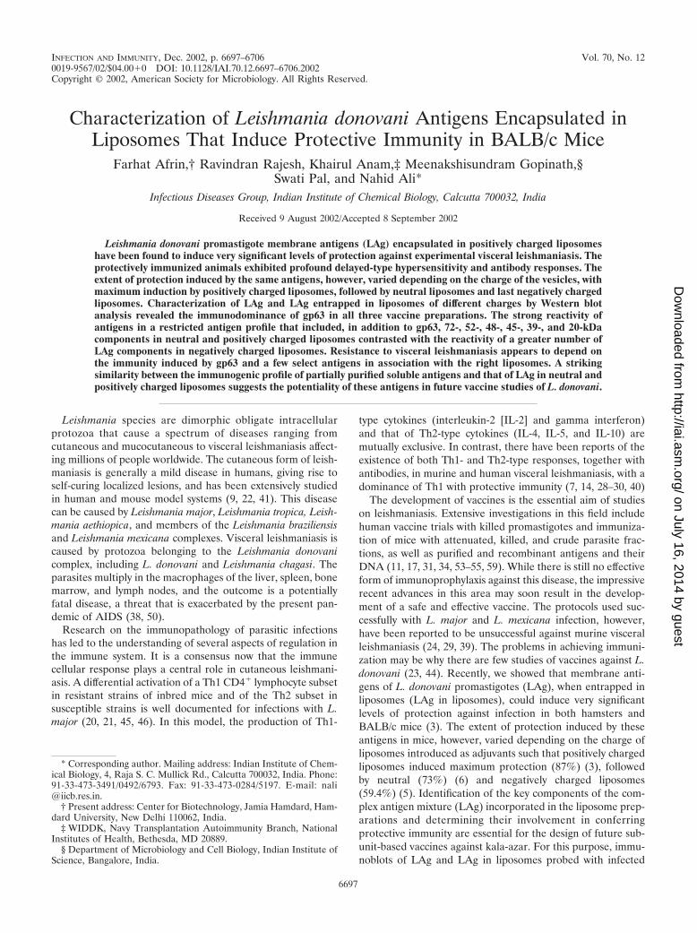
INFECTION AND IMMUNITY, Dec. 2002, p. 6697–6706 Vol. 70, No. 120019-9567/02/$04.00�0 DOI: 10.1128/IAI.70.12.6697–6706.2002Copyright © 2002, American Society for Microbiology. All Rights Reserved.
Characterization of Leishmania donovani Antigens Encapsulated inLiposomes That Induce Protective Immunity in BALB/c Mice
Farhat Afrin,† Ravindran Rajesh, Khairul Anam,‡ Meenakshisundram Gopinath,§Swati Pal, and Nahid Ali*
Infectious Diseases Group, Indian Institute of Chemical Biology, Calcutta 700032, India
Received 9 August 2002/Accepted 8 September 2002
Leishmania donovani promastigote membrane antigens (LAg) encapsulated in positively charged liposomeshave been found to induce very significant levels of protection against experimental visceral leishmaniasis. Theprotectively immunized animals exhibited profound delayed-type hypersensitivity and antibody responses. Theextent of protection induced by the same antigens, however, varied depending on the charge of the vesicles, withmaximum induction by positively charged liposomes, followed by neutral liposomes and last negatively chargedliposomes. Characterization of LAg and LAg entrapped in liposomes of different charges by Western blotanalysis revealed the immunodominance of gp63 in all three vaccine preparations. The strong reactivity ofantigens in a restricted antigen profile that included, in addition to gp63, 72-, 52-, 48-, 45-, 39-, and 20-kDacomponents in neutral and positively charged liposomes contrasted with the reactivity of a greater number ofLAg components in negatively charged liposomes. Resistance to visceral leishmaniasis appears to depend onthe immunity induced by gp63 and a few select antigens in association with the right liposomes. A strikingsimilarity between the immunogenic profile of partially purified soluble antigens and that of LAg in neutral andpositively charged liposomes suggests the potentiality of these antigens in future vaccine studies of L. donovani.
Leishmania species are dimorphic obligate intracellularprotozoa that cause a spectrum of diseases ranging fromcutaneous and mucocutaneous to visceral leishmaniasis affect-ing millions of people worldwide. The cutaneous form of leish-maniasis is generally a mild disease in humans, giving rise toself-curing localized lesions, and has been extensively studiedin human and mouse model systems (9, 22, 41). This diseasecan be caused by Leishmania major, Leishmania tropica, Leish-mania aethiopica, and members of the Leishmania braziliensisand Leishmania mexicana complexes. Visceral leishmaniasis iscaused by protozoa belonging to the Leishmania donovanicomplex, including L. donovani and Leishmania chagasi. Theparasites multiply in the macrophages of the liver, spleen, bonemarrow, and lymph nodes, and the outcome is a potentiallyfatal disease, a threat that is exacerbated by the present pan-demic of AIDS (38, 50).
Research on the immunopathology of parasitic infectionshas led to the understanding of several aspects of regulation inthe immune system. It is a consensus now that the immunecellular response plays a central role in cutaneous leishmani-asis. A differential activation of a Th1 CD4� lymphocyte subsetin resistant strains of inbred mice and of the Th2 subset insusceptible strains is well documented for infections with L.major (20, 21, 45, 46). In this model, the production of Th1-
type cytokines (interleukin-2 [IL-2] and gamma interferon)and that of Th2-type cytokines (IL-4, IL-5, and IL-10) aremutually exclusive. In contrast, there have been reports of theexistence of both Th1- and Th2-type responses, together withantibodies, in murine and human visceral leishmaniasis, with adominance of Th1 with protective immunity (7, 14, 28–30, 40)
The development of vaccines is the essential aim of studieson leishmaniasis. Extensive investigations in this field includehuman vaccine trials with killed promastigotes and immuniza-tion of mice with attenuated, killed, and crude parasite frac-tions, as well as purified and recombinant antigens and theirDNA (11, 17, 31, 34, 53–55, 59). While there is still no effectiveform of immunoprophylaxis against this disease, the impressiverecent advances in this area may soon result in the develop-ment of a safe and effective vaccine. The protocols used suc-cessfully with L. major and L. mexicana infection, however,have been reported to be unsuccessful against murine visceralleishmaniasis (24, 29, 39). The problems in achieving immuni-zation may be why there are few studies of vaccines against L.donovani (23, 44). Recently, we showed that membrane anti-gens of L. donovani promastigotes (LAg), when entrapped inliposomes (LAg in liposomes), could induce very significantlevels of protection against infection in both hamsters andBALB/c mice (3). The extent of protection induced by theseantigens in mice, however, varied depending on the charge ofliposomes introduced as adjuvants such that positively chargedliposomes induced maximum protection (87%) (3), followedby neutral (73%) (6) and negatively charged liposomes(59.4%) (5). Identification of the key components of the com-plex antigen mixture (LAg) incorporated in the liposome prep-arations and determining their involvement in conferringprotective immunity are essential for the design of future sub-unit-based vaccines against kala-azar. For this purpose, immu-noblots of LAg and LAg in liposomes probed with infected
* Corresponding author. Mailing address: Indian Institute of Chem-ical Biology, 4, Raja S. C. Mullick Rd., Calcutta 700032, India. Phone:91-33-473-3491/0492/6793. Fax: 91-33-473-0284/5197. E-mail: [email protected].
† Present address: Center for Biotechnology, Jamia Hamdard, Ham-dard University, New Delhi 110062, India.
‡ WIDDK, Navy Transplantation Autoimmunity Branch, NationalInstitutes of Health, Bethesda, MD 20889.
§ Department of Microbiology and Cell Biology, Indian Institute ofScience, Bangalore, India.
6697
on July 16, 2014 by guesthttp://iai.asm
.org/D
ownloaded from
sera and with sera from immunized BALB/c mice before andafter infection were analyzed. Results show that, in addition togp63, the major promastigote surface glycoprotein, 72-, 52-,48-, 45-, 39-, and 20-kDa components of LAg are potent anti-genic targets for the development of vaccine against L. dono-vani.
MATERIALS AND METHODS
Parasites and culture conditions. Promastigotes of L. donovani, strain AG83,were grown at 22°C in medium 199 (pH 7.4) supplemented with 10% heat-inactivated fetal bovine serum, 2 mM L-glutamine, 100 U of penicillin G sodium,and 100 �g of streptomycin sulfate per ml and subcultured in the same mediumat an average density of 2 � 106 cells/ml (6).
Preparation of Leishmania antigens. LAg were prepared from L. donovanipromastigotes as described earlier (3). Briefly, stationary-phase promastigotes,harvested after the third or fourth passage, were washed four times in cold 20mM phosphate-buffered saline (PBS), pH 7.2, and resuspended at a concentra-tion of 1.0 g of cell pellet in 50 ml of cold 5 mM Tris-HCl buffer, pH 7.6. Thesuspension was vortexed and centrifuged at 2,310 � g for 10 min. The crude ghostmembrane pellet thus obtained was resuspended in the same Tris buffer andsonicated in an ultrasonicator. The suspension was centrifuged at 5,190 � g for30 min, and the supernatant containing the leishmanial antigens was harvestedand stored at �70°C until use. The amount of protein obtained from a 1.0-g cellpellet, as assayed by the method of Lowry et al. (35), was approximately 16 mg.
Soluble leishmanial antigens (SLA) were also extracted from L. donovanipromastigote membranes. The washed parasites were suspended in cold 5 mMTris-HCl buffer (pH 7.6) containing 5 �g of leupeptin/ml, 1 mM EDTA, 1 mMphenylmethylsulfonyl fluoride, and 1 mM iodoacetamide (lysis buffer) and werevortexed and centrifuged as described for LAg. The membrane pellet was resus-pended in 10 ml of lysis buffer and sonicated as described above. The suspensionthus obtained was solubilized with 1% (wt/vol) octyl-�-D-glucopyranoside in thelysis buffer, with overnight incubation at 4°C, and was finally ultracentrifuged for1 h at 100,000 � g. The supernatant containing SLA was then dialyzed against 1mM Tris-HCl buffer (pH 7.6) and stored at �70°C until use. The amount ofprotein obtained from a 1.0-g cell pellet was approximately 2 mg.
The 63-kDa membrane glycoprotein, gp63, was purified by monoclonal affinitybinding from Nonidet P-40 extracts of Leishmania amazonensis promastigotesand was a kind gift from K. P. Chang.
Electroelution of gp63 from SDS-PAGE gels. The SLA from L. donovanipromastigotes was subjected to sodium dodecyl sulfate–10% polyacrylamide gelelectrophoresis (SDS–10% PAGE), and the protein with a molecular mass of 63kDa (gp63) was localized in gels, stained with Coomassie blue, and eluted byelectrophoresis in running buffer (0.025 M Tris, 0.192 M glycine, 1% SDS) at 10mA for 5 h. After elution, the protein was dialyzed, lyophilized, resuspended inPBS, and filtered through 0.22-�m-pore-size membranes for further utilization inlymphocyte cultures (8). The protein was quantified by Lowry’s method (35).
Entrapment of leishmanial antigens in liposomes. Neutral and positivelycharged liposomes were prepared with egg lecithin and cholesterol (7:2 molarratio) or with egg lecithin, cholesterol, and stearylamine (7:2:2 molar ratio),respectively, as reported earlier (3, 6). Negatively charged liposomes were pre-pared with egg lecithin, cholesterol, and phosphatidic acid at a molar ratio of7:2:2 (5). For encapsulation of the antigens in the vesicles the lipid film wasdispersed in PBS containing 2 mg of LAg/ml and sonicated for 30 s in anultrasonicator. Liposomes with entrapped antigen were separated from the ex-cess free antigen by three successive washing in PBS with ultracentrifugation(105,000 � g, 60 min, 4°C). The amounts of LAg associated per milligram of egglecithin were 35, 25, and 15 �g for positively charged, neutral, and negativelycharged liposomes, respectively. Similarly, electroeluted gp63 was entrapped inthe positively charged vesicles. The lipid film was dispersed in PBS containing 1mg of gp63/ml, and 40 �g of protein was associated with 1 mg of egg lecithin.
Immunization and challenge infection. BALB/c mice were immunized bythree intraperitoneal injections of 20 �g of LAg or 10 �g of gp63, purified byelectroelution and free in PBS or entrapped in liposomes, at 2-week intervals.Animals receiving only PBS or empty liposomes served as controls. Ten daysafter the last immunization, the immunized and control animals were challengedintravenously with 2.5 � 107 freshly transformed L. donovani promastigotes (6).Serum samples collected from each mouse before challenge infection and at 4months postinfection were stored frozen at �20°C.
DTH. Delayed-type hypersensitivity (DTH), was determined as an index ofcell-mediated immunity. The response was evaluated by measuring the differencebetween the footpad swelling at 24 h following intradermal inoculation of the test
footpad with 50 �l of electroeluted gp63 (800 �g/ml) and the swelling of thecontrol (PBS-injected) footpad (3).
Cell proliferation assay. The lymphocyte cultures were obtained from spleensof immunized BALB/c mice. The cells were isolated and counted, and, afterevaluation of cell viability by trypan blue exclusion, 2 � 105 cells per well werecultured in RPMI 1640 containing 20 mM NaHCO3, 10 mM HEPES, 100 U ofpenicillin per ml, 100 �g of streptomycin per ml, 2 mM L-glutamine, and 10%fetal calf serum (complete medium [CM]), with 50 �M �-mercaptoethanoladded.
The cells were cultured in triplicate in a final volume of 200 �l/well with elutedantigen (0.7 �g/well) at optimum concentrations. The cultures were incubatedfor 96 h at 37°C in a humidified chamber containing 5% CO2. Cells were pulsedwith 1 �Ci of [3H]thymidine ([3H]TdR; 83 Ci/mmol; Amersham International,Amersham, England) per well 18 h before they were harvested on glass fiberpaper. ([3H]TdR uptake was measured in a �-scintillation counter (LS 5000TD;Beckman Instruments, Fullerton, Calif.).
In vitro growth of L. donovani in macrophages. Macrophages were collected byperitoneal lavage from immunized BALB/c mice 10 days after the last injectionand cultured in CM. A total of �90% of the cell preparation was identified asmacrophages by microscopic observation, and the macrophages were routinelyfound to be �98% viable by trypan blue exclusion.
Promastigotes were used to infect cultures of adherent macrophages on glasscoverslips (18 mm2; 106 macrophages/coverslip) in 0.5 ml of CM at a ratio of 10parasites/macrophage. After 3 h of incubation, the unphagocytosed parasiteswere removed by a washing with PBS. Infected macrophages were further incu-bated in CM at 37°C for 72 h. Cells were fixed in methanol and stained withGiemsa for determination of intracellular parasite numbers.
ELISA. Mice immunized with gp63 or gp63-positively charged liposome andcontrol BALB/c mice were bled 10 days after the last immunization by snippingthe tail vein, and the sera were collected. Serum IgG levels were determined byenzyme-linked immunosorbent assay (ELISA) (3). Briefly, 96-well microtiterplates were coated overnight at 4°C with gp63 that had been purified by elec-troelution (15 �g/ml). After being blocked with bovine serum albumin andincubated overnight with serum samples diluted at 1:1,000, the plates weredeveloped by using a 1:5,000 dilution of peroxidase-conjugated goat anti-mouseimmunoglobulin G (IgG) antibody (Jackson Immunochemical Research Labo-ratories).
Mice sera were further assayed for gp63-specific IgG subclass antibodies witha mouse antibody isotyping kit (Sigma Immunochemicals, St. Louis, Mo.). gp63-coated wells, incubated with mice sera as described above, were reacted over-night at 4°C with a 1:2,500 dilution of goat anti-mouse isotype-specific antibodies,followed with a 1:5,000 dilution of peroxidase-conjugated rabbit anti-goat IgG(Jackson Immunochemical Research Laboratories). The plates were developedfor color reaction and analyzed by measuring the optical density at 450 nm.
Evaluation of infection. At the times mentioned in Results, the course ofinfection was monitored by the microscopic examination of Giemsa-stained im-pression smears of liver and spleen. The parasite load was expressed as Leish-man-Donovan units and was calculated by the following formula: number ofamastigotes per 1,000 cell nuclei � organ weight (in milligrams) (57).
SDS-PAGE and Western blot analysis. Components of the L. donovani pro-mastigote membrane were subjected to SDS-PAGE by the method of Laemmli(32). Gels were loaded with proteins (amounts are indicated in the figure leg-ends) of the L. donovani ghost membrane; LAg and SLA free in PBS; LAgentrapped in liposomes of neutral, negative, and positive charge; purified gp63provided by Chang; and gp63 purified by electroelution. Proteins, separated on10% polyacrylamide, were silver stained (61).
For Western blot analysis, the resolved proteins were transferred onto nitro-cellulose in 25 mM Tris-HCl–192 mM glycine–20% (vol/vol) methanol buffer at90 V/cm for 90 min (58). Immunoblot assays were performed according to themethod described by Rolland-Burger et al. (49) with slight modifications. Thenitrocellulose strips were first saturated and then blocked overnight at roomtemperature in 100 mM Tris-buffered saline (TBS), pH 7.6, containing 0.1%Tween 20 (T-20) and washed once for 15 min with 0.05% T-20 in TBS (washingbuffer) with shaking. Incubation of the nitrocellulose strips with sera from miceimmunized with LAg in liposomes, before and after infection, was carried out at1:500 dilution in the washing buffer for 1 h at room temperature, followed bythree washes of 20 min each. gp63, SLA, LAg, and LAg-in-liposome blots wereprobed with rabbit antisera to purified gp63 at 1:200 dilution, kindly provided byK. P. Chang. The blots were then washed and incubated with peroxidase-conju-gated anti-mouse or anti-rabbit (Sigma Immunochemicals) IgG, at a 1:500 dilu-tion in the washing buffer for 1 h at room temperature; this was followed by threewashes as described above. The last wash was done without T-20. Enzymaticactivity was revealed with 15 mg of 3,3�-diaminobenzidine tetrahydrochloride
6698 AFRIN ET AL. INFECT. IMMUN.
on July 16, 2014 by guesthttp://iai.asm
.org/D
ownloaded from
(Sigma Immunochemicals) in 30 ml of TBS containing 15 �l of 30% H2O2. Theefficacy of transfer of the leishmanial proteins was regularly checked and con-firmed by concurrent gel staining with silver and nitrocellulose membrane stain-ing with 0.1% Ponceau S in 1% acetic acid. The parts of the membrane contain-ing molecular mass standards were marked with ink after the staining withPonceau S. The specificity of the immune response was assessed by immunoblotanalysis employing pooled preimmune sera and only liposome-immunized sera at1:50 dilution and infected sera at 1:500 dilution in washing buffer.
Statistical analysis. All data comparisons were tested for significance by usingStudent’s t test; P values of �0.05 were considered significant.
RESULTS
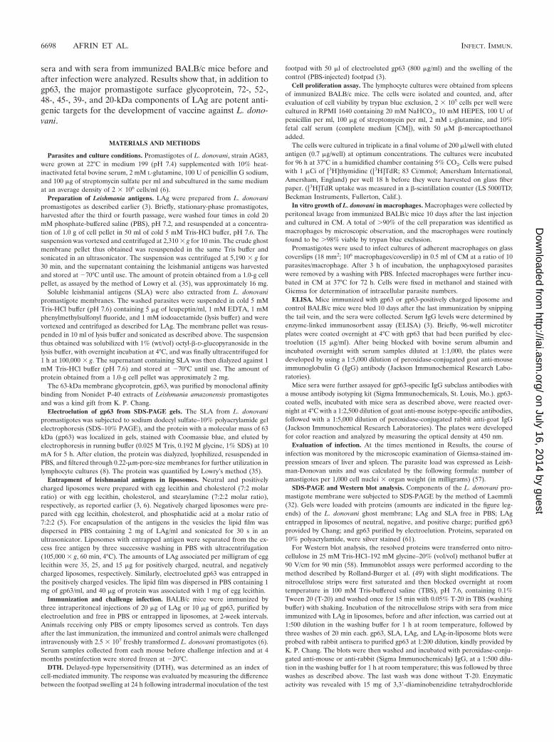
Electrophoretic analysis of the leishmanial antigens andantigens in liposomes. LAg in association with liposomes in-duce strong protection against virulent homologous challenge(3, 6). These antigens were extracted from the promastigoteghost membrane (Fig. 1, lane 1) and exhibited at least 33distinct polypeptides (lane 2) ranging in molecular mass from18 to 153 kDa. Entrapment of LAg in liposomes of differentcharges (lanes 6 to 8) revealed different polypeptide profiles invesicles of negative (lane 6), neutral (lane 7), and positive (lane8) charge. The silver stain data illustrate that preparations ofliposomes resulted in preferential entrapment of some pro-teins. Polypeptides with molecular masses above 64 kDa,weakly displayed in LAg as well as in the ghost membrane,were predominantly associated with liposomes. Again, a 61- to66-kDa band, present as a 63- to 64-kDa polypeptide in LAgbut not as the major component, stained more intensely inneutral and positively charged vesicles than in negativelycharged liposomes. This band demonstrated a comigrationwith the major polypeptide of purified gp63 provided by Chang(lane 4). An attempt at partial purification of LAg throughdetergent solubilization resulted in the extraction of approxi-mately six polypeptides (72, 63, 43, 41, 30, and 20 kDa) and afaint smear in the region of 45 to 52 kDa, with the major bandalso comigrating with gp63 (lane 3). gp63 purified by electro-elution stained as a single band representing a 63-kDapolypeptide (lane 5). Amounts of empty liposomes equal to theamounts of liposomes with LAg were subjected to SDS-PAGE.No bands were observed (data not shown).
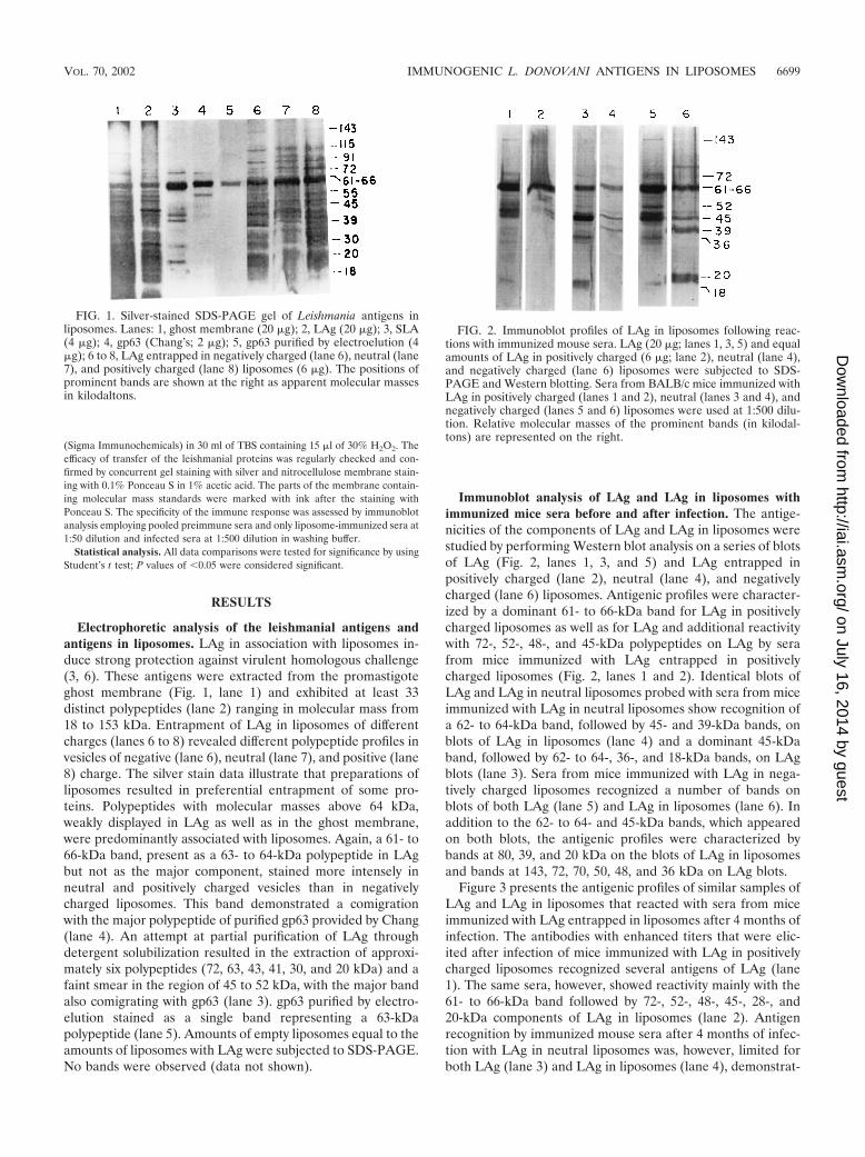
Immunoblot analysis of LAg and LAg in liposomes withimmunized mice sera before and after infection. The antige-nicities of the components of LAg and LAg in liposomes werestudied by performing Western blot analysis on a series of blotsof LAg (Fig. 2, lanes 1, 3, and 5) and LAg entrapped inpositively charged (lane 2), neutral (lane 4), and negativelycharged (lane 6) liposomes. Antigenic profiles were character-ized by a dominant 61- to 66-kDa band for LAg in positivelycharged liposomes as well as for LAg and additional reactivitywith 72-, 52-, 48-, and 45-kDa polypeptides on LAg by serafrom mice immunized with LAg entrapped in positivelycharged liposomes (Fig. 2, lanes 1 and 2). Identical blots ofLAg and LAg in neutral liposomes probed with sera from miceimmunized with LAg in neutral liposomes show recognition ofa 62- to 64-kDa band, followed by 45- and 39-kDa bands, onblots of LAg in liposomes (lane 4) and a dominant 45-kDaband, followed by 62- to 64-, 36-, and 18-kDa bands, on LAgblots (lane 3). Sera from mice immunized with LAg in nega-tively charged liposomes recognized a number of bands onblots of both LAg (lane 5) and LAg in liposomes (lane 6). Inaddition to the 62- to 64- and 45-kDa bands, which appearedon both blots, the antigenic profiles were characterized bybands at 80, 39, and 20 kDa on the blots of LAg in liposomesand bands at 143, 72, 70, 50, 48, and 36 kDa on LAg blots.
Figure 3 presents the antigenic profiles of similar samples ofLAg and LAg in liposomes that reacted with sera from miceimmunized with LAg entrapped in liposomes after 4 months ofinfection. The antibodies with enhanced titers that were elic-ited after infection of mice immunized with LAg in positivelycharged liposomes recognized several antigens of LAg (lane1). The same sera, however, showed reactivity mainly with the61- to 66-kDa band followed by 72-, 52-, 48-, 45-, 28-, and20-kDa components of LAg in liposomes (lane 2). Antigenrecognition by immunized mouse sera after 4 months of infec-tion with LAg in neutral liposomes was, however, limited forboth LAg (lane 3) and LAg in liposomes (lane 4), demonstrat-
FIG. 1. Silver-stained SDS-PAGE gel of Leishmania antigens inliposomes. Lanes: 1, ghost membrane (20 �g); 2, LAg (20 �g); 3, SLA(4 �g); 4, gp63 (Chang’s; 2 �g); 5, gp63 purified by electroelution (4�g); 6 to 8, LAg entrapped in negatively charged (lane 6), neutral (lane7), and positively charged (lane 8) liposomes (6 �g). The positions ofprominent bands are shown at the right as apparent molecular massesin kilodaltons.
FIG. 2. Immunoblot profiles of LAg in liposomes following reac-tions with immunized mouse sera. LAg (20 �g; lanes 1, 3, 5) and equalamounts of LAg in positively charged (6 �g; lane 2), neutral (lane 4),and negatively charged (lane 6) liposomes were subjected to SDS-PAGE and Western blotting. Sera from BALB/c mice immunized withLAg in positively charged (lanes 1 and 2), neutral (lanes 3 and 4), andnegatively charged (lanes 5 and 6) liposomes were used at 1:500 dilu-tion. Relative molecular masses of the prominent bands (in kilodal-tons) are represented on the right.
VOL. 70, 2002 IMMUNOGENIC L. DONOVANI ANTIGENS IN LIPOSOMES 6699
on July 16, 2014 by guesthttp://iai.asm
.org/D
ownloaded from
ing a reactivity predominantly for the 62- to 64-kDa bandfollowed by 52-, 48-, 45-, and 20-kDa moieties. Antibodieselicited in mice immunized with LAg in negatively chargedliposomes revealed the strongest reactivity with components ofLAg and LAg in liposomes after 4 months of infection. Al-though the 61- to 66-kDa band was still the dominant compo-nent of LAg (lane 5), there were several additional bands aswell as smears of LAg in negatively charged liposomes (lane 6)recognized by the antisera.
Reactivity of LAg and LAg in liposomes with infected sera isillustrated in Fig. 4. While there was a dominant 61- to 66-kDaband of LAg with immunized-mouse sera before and afterinfection (Fig. 2 and 3), sera from unimmunized, infected miceexhibited predominantly a 59-kDa band of LAg (Fig. 4, lane 1).Other components recognized by the infected sera includebands of 143, 72, 52, 48, 45, 39, 36, and 20 kDa, as well assmears in the range of 60 to 66, 50 to 58, and 18 to 22 kDa.Infected sera reacted with LAg in liposomes with lower inten-sity, recognizing only 66-, 60-, 48-, and 45-kDa bands for LAgin positively charged liposomes (lane 2); 80-, 28-, and 20-kDabands for LAg in neutral liposomes (lane 3); and 80-, 66-, and36-kDa bands for LAg in negatively charged liposomes (lane4). No bands were present on identical sets of blots probedwith normal mouse sera or sera from mice immunized withempty liposomes (data not shown).
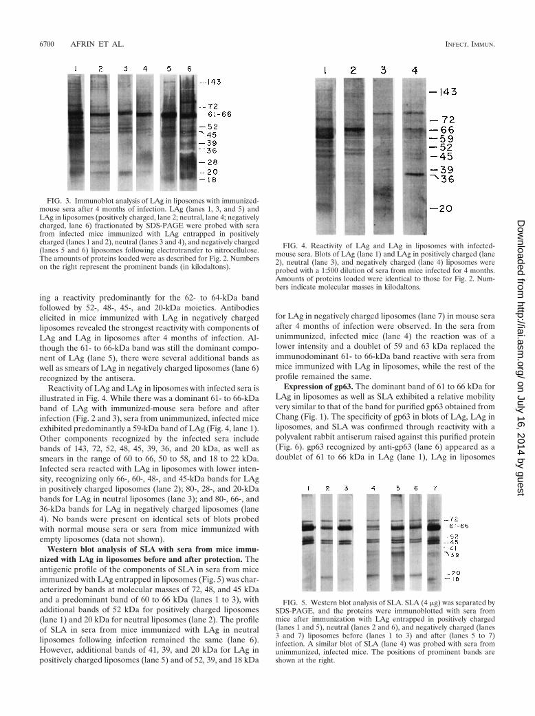
Western blot analysis of SLA with sera from mice immu-nized with LAg in liposomes before and after protection. Theantigenic profile of the components of SLA in sera from miceimmunized with LAg entrapped in liposomes (Fig. 5) was char-acterized by bands at molecular masses of 72, 48, and 45 kDaand a predominant band of 60 to 66 kDa (lanes 1 to 3), withadditional bands of 52 kDa for positively charged liposomes(lane 1) and 20 kDa for neutral liposomes (lane 2). The profileof SLA in sera from mice immunized with LAg in neutralliposomes following infection remained the same (lane 6).However, additional bands of 41, 39, and 20 kDa for LAg inpositively charged liposomes (lane 5) and of 52, 39, and 18 kDa
for LAg in negatively charged liposomes (lane 7) in mouse seraafter 4 months of infection were observed. In the sera fromunimmunized, infected mice (lane 4) the reaction was of alower intensity and a doublet of 59 and 63 kDa replaced theimmunodominant 61- to 66-kDa band reactive with sera frommice immunized with LAg in liposomes, while the rest of theprofile remained the same.
Expression of gp63. The dominant band of 61 to 66 kDa forLAg in liposomes as well as SLA exhibited a relative mobilityvery similar to that of the band for purified gp63 obtained fromChang (Fig. 1). The specificity of gp63 in blots of LAg, LAg inliposomes, and SLA was confirmed through reactivity with apolyvalent rabbit antiserum raised against this purified protein(Fig. 6). gp63 recognized by anti-gp63 (lane 6) appeared as adoublet of 61 to 66 kDa in LAg (lane 1), LAg in liposomes
FIG. 3. Immunoblot analysis of LAg in liposomes with immunized-mouse sera after 4 months of infection. LAg (lanes 1, 3, and 5) andLAg in liposomes (positively charged, lane 2; neutral, lane 4; negativelycharged, lane 6) fractionated by SDS-PAGE were probed with serafrom infected mice immunized with LAg entrapped in positivelycharged (lanes 1 and 2), neutral (lanes 3 and 4), and negatively charged(lanes 5 and 6) liposomes following electrotransfer to nitrocellulose.The amounts of proteins loaded were as described for Fig. 2. Numberson the right represent the prominent bands (in kilodaltons).
FIG. 4. Reactivity of LAg and LAg in liposomes with infected-mouse sera. Blots of LAg (lane 1) and LAg in positively charged (lane2), neutral (lane 3), and negatively charged (lane 4) liposomes wereprobed with a 1:500 dilution of sera from mice infected for 4 months.Amounts of proteins loaded were identical to those for Fig. 2. Num-bers indicate molecular masses in kilodaltons.
FIG. 5. Western blot analysis of SLA. SLA (4 �g) was separated bySDS-PAGE, and the proteins were immunoblotted with sera frommice after immunization with LAg entrapped in positively charged(lanes 1 and 5), neutral (lanes 2 and 6), and negatively charged (lanes3 and 7) liposomes before (lanes 1 to 3) and after (lanes 5 to 7)infection. A similar blot of SLA (lane 4) was probed with sera fromunimmunized, infected mice. The positions of prominent bands areshown at the right.
6700 AFRIN ET AL. INFECT. IMMUN.
on July 16, 2014 by guesthttp://iai.asm
.org/D
ownloaded from
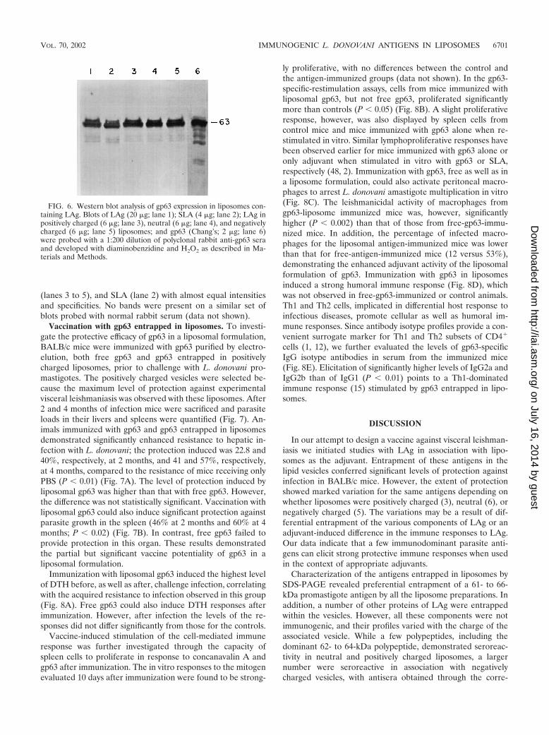
(lanes 3 to 5), and SLA (lane 2) with almost equal intensitiesand specificities. No bands were present on a similar set ofblots probed with normal rabbit serum (data not shown).
Vaccination with gp63 entrapped in liposomes. To investi-gate the protective efficacy of gp63 in a liposomal formulation,BALB/c mice were immunized with gp63 purified by electro-elution, both free gp63 and gp63 entrapped in positivelycharged liposomes, prior to challenge with L. donovani pro-mastigotes. The positively charged vesicles were selected be-cause the maximum level of protection against experimentalvisceral leishmaniasis was observed with these liposomes. After2 and 4 months of infection mice were sacrificed and parasiteloads in their livers and spleens were quantified (Fig. 7). An-imals immunized with gp63 and gp63 entrapped in liposomesdemonstrated significantly enhanced resistance to hepatic in-fection with L. donovani; the protection induced was 22.8 and40%, respectively, at 2 months, and 41 and 57%, respectively,at 4 months, compared to the resistance of mice receiving onlyPBS (P � 0.01) (Fig. 7A). The level of protection induced byliposomal gp63 was higher than that with free gp63. However,the difference was not statistically significant. Vaccination withliposomal gp63 could also induce significant protection againstparasite growth in the spleen (46% at 2 months and 60% at 4months; P � 0.02) (Fig. 7B). In contrast, free gp63 failed toprovide protection in this organ. These results demonstratedthe partial but significant vaccine potentiality of gp63 in aliposomal formulation.
Immunization with liposomal gp63 induced the highest levelof DTH before, as well as after, challenge infection, correlatingwith the acquired resistance to infection observed in this group(Fig. 8A). Free gp63 could also induce DTH responses afterimmunization. However, after infection the levels of the re-sponses did not differ significantly from those for the controls.
Vaccine-induced stimulation of the cell-mediated immuneresponse was further investigated through the capacity ofspleen cells to proliferate in response to concanavalin A andgp63 after immunization. The in vitro responses to the mitogenevaluated 10 days after immunization were found to be strong-
ly proliferative, with no differences between the control andthe antigen-immunized groups (data not shown). In the gp63-specific-restimulation assays, cells from mice immunized withliposomal gp63, but not free gp63, proliferated significantlymore than controls (P � 0.05) (Fig. 8B). A slight proliferativeresponse, however, was also displayed by spleen cells fromcontrol mice and mice immunized with gp63 alone when re-stimulated in vitro. Similar lymphoproliferative responses havebeen observed earlier for mice immunized with gp63 alone oronly adjuvant when stimulated in vitro with gp63 or SLA,respectively (48, 2). Immunization with gp63, free as well as ina liposome formulation, could also activate peritoneal macro-phages to arrest L. donovani amastigote multiplication in vitro(Fig. 8C). The leishmanicidal activity of macrophages fromgp63-liposome immunized mice was, however, significantlyhigher (P � 0.002) than that of those from free-gp63-immu-nized mice. In addition, the percentage of infected macro-phages for the liposomal antigen-immunized mice was lowerthan that for free-antigen-immunized mice (12 versus 53%),demonstrating the enhanced adjuvant activity of the liposomalformulation of gp63. Immunization with gp63 in liposomesinduced a strong humoral immune response (Fig. 8D), whichwas not observed in free-gp63-immunized or control animals.Th1 and Th2 cells, implicated in differential host response toinfectious diseases, promote cellular as well as humoral im-mune responses. Since antibody isotype profiles provide a con-venient surrogate marker for Th1 and Th2 subsets of CD4�
cells (1, 12), we further evaluated the levels of gp63-specificIgG isotype antibodies in serum from the immunized mice(Fig. 8E). Elicitation of significantly higher levels of IgG2a andIgG2b than of IgG1 (P � 0.01) points to a Th1-dominatedimmune response (15) stimulated by gp63 entrapped in lipo-somes.
DISCUSSION
In our attempt to design a vaccine against visceral leishman-iasis we initiated studies with LAg in association with lipo-somes as the adjuvant. Entrapment of these antigens in thelipid vesicles conferred significant levels of protection againstinfection in BALB/c mice. However, the extent of protectionshowed marked variation for the same antigens depending onwhether liposomes were positively charged (3), neutral (6), ornegatively charged (5). The variations may be a result of dif-ferential entrapment of the various components of LAg or anadjuvant-induced difference in the immune responses to LAg.Our data indicate that a few immunodominant parasite anti-gens can elicit strong protective immune responses when usedin the context of appropriate adjuvants.
Characterization of the antigens entrapped in liposomes bySDS-PAGE revealed preferential entrapment of a 61- to 66-kDa promastigote antigen by all the liposome preparations. Inaddition, a number of other proteins of LAg were entrappedwithin the vesicles. However, all these components were notimmunogenic, and their profiles varied with the charge of theassociated vesicle. While a few polypeptides, including thedominant 62- to 64-kDa polypeptide, demonstrated seroreac-tivity in neutral and positively charged liposomes, a largernumber were seroreactive in association with negativelycharged vesicles, with antisera obtained through the corre-
FIG. 6. Western blot analysis of gp63 expression in liposomes con-taining LAg. Blots of LAg (20 �g; lane 1); SLA (4 �g; lane 2); LAg inpositively charged (6 �g; lane 3), neutral (6 �g; lane 4), and negativelycharged (6 �g; lane 5) liposomes; and gp63 (Chang’s; 2 �g; lane 6)were probed with a 1:200 dilution of polyclonal rabbit anti-gp63 seraand developed with diaminobenzidine and H2O2 as described in Ma-terials and Methods.
VOL. 70, 2002 IMMUNOGENIC L. DONOVANI ANTIGENS IN LIPOSOMES 6701
on July 16, 2014 by guesthttp://iai.asm
.org/D
ownloaded from

sponding vaccine preparation. The reactivity of LAg and LAgin liposomes was enhanced with sera from immunized miceafter infection. However, apart from the antigens in negativelycharged liposomes, selective seroreactivity of LAg componentswas again observed for neutral and positively charged lipo-some-associated antigens. Of the antigens recognized by anti-sera before and after infection, the 62- to 64-kDa componentwas the most seroreactive component of LAg in all three vac-cine preparations, followed by distinctive bands at 72, 52, 48,45, and 20 kDa, especially on blots of neutral and positivelycharged LAg in liposomes. Interestingly, SLA, partially puri-fied from LAg, also demonstrated maximum reactivity at 62 to64 kDa, followed by strong bands at 72, 52, 48, 45, and 20 kDa,with the sera from mice immunized with LAg in liposomes
demonstrating a profile with a striking resemblance to theimmunodominant-antigen profiles for neutral and positivelycharged liposomes. In contrast, sera from unimmunized, in-fected mice recognized different polypeptides of LAg. Reac-tivity with the components of LAg entrapped in the differentliposomes was low and lacked the dominance of the 62- to64-kDa antigen observed with the immunized sera.
That the immunodominant antigen among LAg and LAg inliposomes was gp63 was confirmed through reactivity with an-tiserum against this purified protein. Preferential entrapmentof gp63 from the crude mixture of L. major promastigotes inliposomes was also demonstrated by Kahl et al. (27) despite thedifferences in the use of the phospholipids and the vesiclepreparation. These workers identified gp63 as the principal
FIG. 7. Kinetics of protection against L. donovani in liver (A) and spleens (B) of BALB/c mice immunized with PBS, empty liposomes, gp63in PBS, and gp63 in liposomes. After three intraperitoneal immunizations, mice were challenged intravenously with L. donovani promastigotes andthe parasite burdens at 2 and 4 months of infection were calculated as described in Materials and Methods. The mean values standard errors(error bars) for three mice per group are given.
6702 AFRIN ET AL. INFECT. IMMUN.
on July 16, 2014 by guesthttp://iai.asm
.org/D
ownloaded from
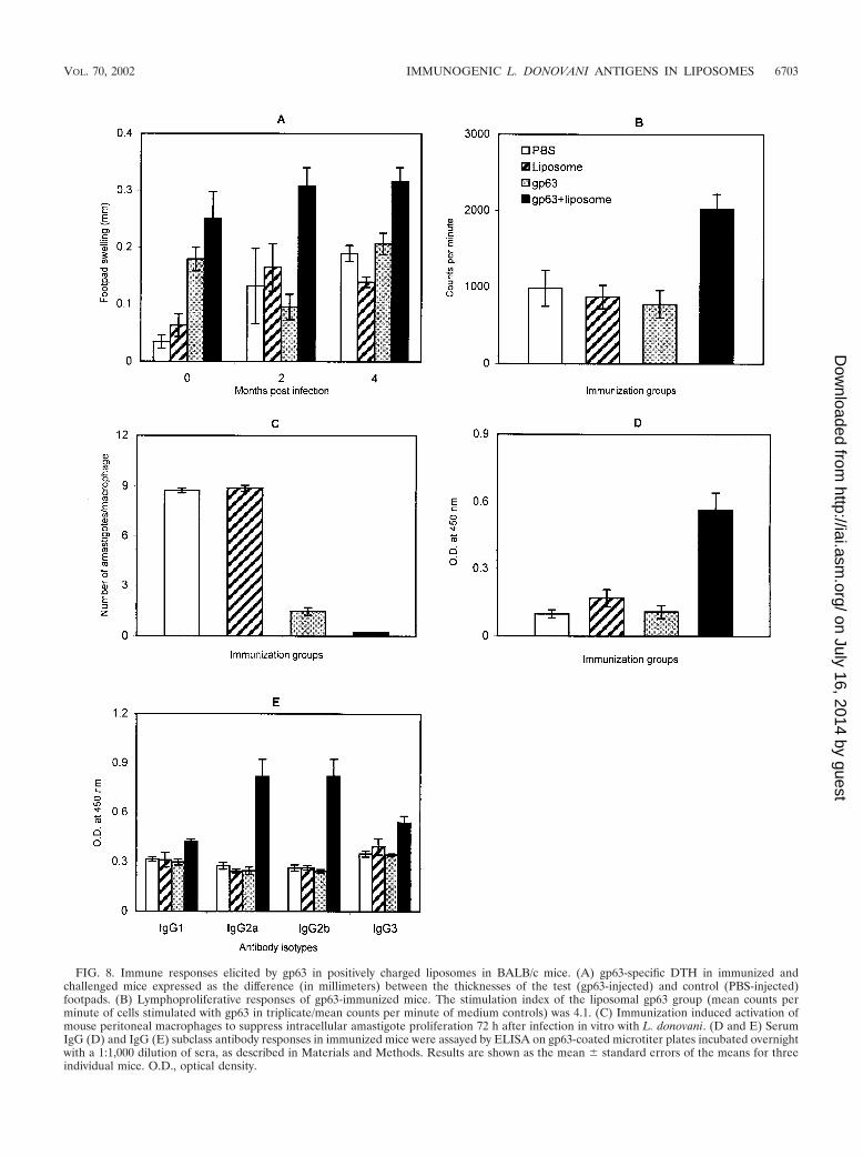
FIG. 8. Immune responses elicited by gp63 in positively charged liposomes in BALB/c mice. (A) gp63-specific DTH in immunized andchallenged mice expressed as the difference (in millimeters) between the thicknesses of the test (gp63-injected) and control (PBS-injected)footpads. (B) Lymphoproliferative responses of gp63-immunized mice. The stimulation index of the liposomal gp63 group (mean counts perminute of cells stimulated with gp63 in triplicate/mean counts per minute of medium controls) was 4.1. (C) Immunization induced activation ofmouse peritoneal macrophages to suppress intracellular amastigote proliferation 72 h after infection in vitro with L. donovani. (D and E) SerumIgG (D) and IgG (E) subclass antibody responses in immunized mice were assayed by ELISA on gp63-coated microtiter plates incubated overnightwith a 1:1,000 dilution of sera, as described in Materials and Methods. Results are shown as the mean standard errors of the means for threeindividual mice. O.D., optical density.
VOL. 70, 2002 IMMUNOGENIC L. DONOVANI ANTIGENS IN LIPOSOMES 6703
on July 16, 2014 by guesthttp://iai.asm
.org/D
ownloaded from
protein antigen conferring protection. The identification ofdefined parasite proteins and peptides that induce beneficialimmune responses may contribute to vaccine development.gp63, the major surface glycoprotein of Leishmania, is highlyconserved across species (37), and its role in induction ofprotection against murine cutaneous leishmaniasis has beenextensively investigated (25, 51, 64). The subjects of the studiesrange from the use of the native protein (13, 16, 51) to the useof recombinant gp63 (rgp63) expressed in Salmonella spp. (64)or plasmid pCMV, which encodes gp63 (62), to the use ofT-cell epitopes within gp63 (25, 63). However, even though L.donovani gp63 has been identified (33) and purified in itsnative and recombinant forms (43, 52), its role in protectionagainst infection with L. donovani has not been established.Partial heterologous protection against L. donovani infectionin mice by L. major rgp63 expressed in Salmonella was re-ported (36). To our knowledge this is the first report of aninvolvement of L. donovani gp63 in protection against thevisceral infection in mice.
Even though gp63 was the immundominant antigen of LAgin all the three liposomes, protection conferred by these prep-arations varied significantly. The choice of adjuvant is impor-tant in inducing the correct immune response. In a study un-dertaken with pure M2 and three different adjuvants, markedvariations in the protection against L. mexicana amazonensisconferred by each preparation were observed (11). It has beenshown that cloned T cells with opposite biological effects onmurine models of cutaneous leishmaniasis utilize the same orsimilar T-cell receptors (47), suggesting that a particular anti-gen may elicit either a protective or an exacerbating immuneresponse against L. major. L. major antigens drive either aTh1- or Th2-type T-cell response, depending on the local cy-tokine environment during antigen priming (10, 26, 27, 59).Antigens of L. donovani, however, did not induce exacerba-tion. Investigation of the immune responses to the vaccinepreparations showed that LAg in neutral liposomes elicited aweak but exclusively Th1-type response after immunization, ascharacterized by the antibody isotype profile (4). In contrast,the same antigens in positively and negatively charged lipo-somes induced both Th1- and Th2-type responses. However,while high levels of IgG2a and IgG2b (markers for the induc-tion of a Th1-like response) were simultaneously stimulated,along with IgG1 (Th2-like response), with positively chargedliposomes (3), these isotypes were dominated by the levels ofIgG1 stimulated by immunization with LAg in negativelycharged vesicles (5). These data indicate that, in contrast towhat was found for L. major antigens (55), a concomitant Th2response with L. donovani antigens does not inhibit the strongTh1 effector function. This conclusion was further substanti-ated by our investigation of the vaccine potentiality of gp63,purified by electroelution, in association with liposomes. En-hanced protection induced with gp63 in a liposomal formula-tion corresponded with stronger stimulation of cellular as wellas humoral immune responses in comparison with stimulationby gp63 alone. Further analysis of IgG isotypes revealed in-duction of all the isotypes, suggesting stimulation of both Th1-and Th2-like responses by gp63 in a vaccine formulation. How-ever, stimulation of higher levels of IgG2a and IgG2b antibod-ies than of IgG1 antibodies indicates the stronger potentiationof a protective Th1 response. The development of a vaccine
against L. donovani may, therefore, require that a particularantigen be administered in the context of the right adjuvant forpotentiating a dominant Th1 response.
Although gp63 was maximally incorporated in the lipo-somes, in contrast to the L. major antigens (27), a greaternumber of other proteins of L. donovani LAg were also en-trapped in the vesicles. However, only a few were immunore-active with sera from protectively immunized animals andtherefore likely to be involved in immunoprotection. While theidentity of the 61- to 66-kDa band was confirmed, 72-, 52-, 48-,45-, 39-, 36-, and 20-kDa antigens of LAg remain unidentified.Screening for parasite antigens on the basis of reactivity withsera from infected susceptible animals has led to the identifi-cation of several leishmanial antigens having the ability toconfer protective immunity. Some of these well-defined anti-gens detected through antibody reactivity include LPG (18),gp46/M-2 (11), dp72 (24), PSA-2 (19), P4 and P8 (56), Lcr1(61), and LACK (42, 59). Purified antigens such as L. donovanidp72 and highly conserved polypeptides of Leishmania speciessuch as gp46 and LACK, of 46 and 36 kDa, respectively, maybe identified with the immunoreactive components of L. do-novani LAg in liposomes and need further testing. In contrastto LAg, SLA, the partially purified antigens from LAg, dem-onstrated a restricted seroreactivity comparable to that ofpolypeptides of LAg in positively charged liposomes whichexhibited maximum protective activity. Although gp63 is one ofthe most immunogenic of Leishmania antigens, it is only par-tially protective against murine visceral leishmaniasis, as ob-served herein, and against lethal murine cutaneous leishman-iasis, as observed elsewhere (13, 25, 51, 64). It has beensuggested that the efficacy of gp63 could be enhanced, possiblywith additional Leishmania antigens (13). Since SLA is com-posed of most of the antigens, including gp63, immunogenic inLAg in liposomes and since it induces better protection thangp63 or LAg in liposomes (Rajesh et al., unpublished data), wepropose that its components, polypeptides of 72, 52, 48, 45, 41,39, and 20 kDa, in addition to gp63, be vaccine candidates forfuture studies of L. donovani antigens in liposomes.
ACKNOWLEDGMENTS
We thank J. Das and S. K. Bhattacharya, past and present directorsof IICB, Calcutta, for supporting this work.
We gratefully acknowledge support from the CSIR and the DST,Government of India, and the UNDP/World Bank/WHO Special Pro-gramme for Research and Training in Tropical Diseases.
F.A. and R.R. contributed equally to this work.
REFERENCES
1. Abbas, A. K., K. M. Murphy, and A. Sher. 1996. Functional diversity ofhelper T lymphocytes. Nature 383:787–793.
2. Abdelhak, S., H. Louzir, J. Timm, L. Blel, Z. Benlasfar, H. Lagranderie, M.Gheorghiu, K. Dellagi, and B. Gicquel. 1995. Recombinant BCG expressingthe leishmania surface antigen Gp63 induces protective immunity againstLeishmania major infection in BALB/c mice. Microbiology 141:1585–1592.
3. Afrin, F., and N. Ali. 1997. Adjuvanicity and protective immunity elicited byLeishmania donovani antigens encapsulated in positively charged liposomes.Infect. Immun. 65:2371–2377.
4. Afrin, F., and N. Ali. 1998. Isotype profiles of Leishmania donovani-infectedBALB/c mice: preferential stimulation of IgG2a/b by liposome-associatedpromastigote antigens. J. Parasitol. 84:743–748.
5. Afrin, F., K. Anam, and N. Ali. 2000. Induction of partial protection againstLeishmania donovani by promastigote antigens in negatively charged lipo-somes. J. Parasitol. 86:730–735.
6. Ali, N., and F. Afrin. 1997. Protection of mice against visceral leishmaniasisby immunization with promastigote antigen incorporated in liposomes. J.Parasitol. 83:70–75.
6704 AFRIN ET AL. INFECT. IMMUN.
on July 16, 2014 by guesthttp://iai.asm
.org/D
ownloaded from
7. Anam, K., F. Afrin, D. Banerjee, N. Pramanik, S. K. Guha, R. P. Goswami,P. N. Gupta, S. K. Saha, and N. Ali. 1999. Immunoglobulin subclass distri-bution and diagnostic value of Leishmania donovani antigen-specific immu-noglobulin G3 in Indian kala-azar patients. Clin. Diagn. Lab. Immunol.6:231–235.
8. Beyrodt, C. G. P., A. R. Pinto, E. Freymuller, and C. L. Barbieri. 1997.Characterization of an antigen from Leishmania amazonensis amastigotesable to elicit protective responses in a murine model. Infect. Immun. 65:2052–2059.
9. Bogdan, C., A. Gesser, W. Solbach, and M. Rollinghoff. 1996. Invasion,control and persistence of Leishmania parasites. Curr. Opin. Immunol.8:517–525.
10. Bretscher, P. A., G. Wei, J. N. Menon, and H. Bielefeldt-Ohmann. 1992.Establishment of cell-mediated immunity that makes “susceptible” miceresistant to Leishmania major. Science 257:539–542.
11. Champsi, J., and D. McMahon-Pratt. 1988. Membrane glycoprotein M-2protects against Leishmania amazonensis infection. Infect. Immun. 56:3272–3279.
12. Coffman, R. L., D. A. Lebman, and P. Rothman. 1993. Mechanism andregulation of immunoglobulin isotype switching. Adv. Immunol. 54:229–270.
13. Connell, N. D., E. Medina-Acosta, W. R. McMaster, B. R. Bloom, and D. G.Russell. 1993. Effective immunization against cutaneous leishmaniasis withrecombinant bacille Calmette-Guerin expressing the Leishmania surfaceproteinase gp63. Proc. Natl. Acad. Sci. USA 90:11473–11477.
14. Fargeas, C., M. Hommel, R. Maingon, C. Dourado, M. Monsigny, and R.Mayer. 1996. Synthetic peptide-based enzyme-linked immunosorbent assayfor serodiagnosis of visceral leishmaniasis. J. Clin. Microbiol. 34:241–248.
15. Germann, T., M. Bongartz, H. Dlugonska, H. Hess, E. Schmitt, L. Kolbe, E.Kolsch, F. J. Podlaski, M. K. Gately, and E. Rude. 1995. Interleukin-12profoundly up-regulates the synthesis of antigen-specific complement-fixingIgG2a, IgG2b and IgG3 antibody subclasses in vivo. Eur. J. Immunol. 25:823–829.
16. Guimaraes, T. M. P. D., V. P. C. P. de Toledo, C. A. da Costa, R. T. da Costa,O. Genaro, P. Williams, and W. Maybrink. 1996. Assessment of immunityinduced in mice by glycoproteins derived from different strains and species ofLeishmania. Mem. Inst. Oswaldo Cruz 91:63–70.
17. Gurunathan, S., C. Prussin, D. L. Sacks, and R. A. Seder. 1998. Vaccinerequirements for sustained cellular immunity to an intracellular parasiticinfection. Nat. Med. 4:1409–1415.
18. Handman, E., and G. F. Mitchell. 1985. Immunization with Leishmaniareceptor for macrophages protects mice against cutaneous leishmaniasis.Proc. Natl. Acad. Sci. USA 82:5910–5914.
19. Handman, E., F. M. Symons, T. M. Baldwin, J. M. Curtis, and J.-P. Y.Scheerlinck. 1995. Protective vaccination with promastigote surface antigen2 from Leishmania major is mediated by a Th1 type of immune response.Infect. Immun. 63:4261–4267.
20. Heinzel, F. P., M. D. Sadick, B. J. Holaday, R. L. Coffman, and R. M.Locksley. 1989. Reciprocal expression of interferon gamma or interleukin 4during the resolution or progression of murine leishmaniasis. Evidence forexpansion of distinct helper T cell subsets. J. Exp. Med. 169:59–72.
21. Heinzel, F. P., M. D. Sadick, S. S. Mutah, and R. M. Locksley. 1991.Production of interferon gamma, interleukin 2, interleukin 4, and interleukin10 by CD4� lymphocytes in vivo during healing and progressive murineleishmaniasis. Proc. Natl. Acad. Sci. USA 88:7011–7015.
22. Herwaldt, B. L. 1999. Leishmaniasis. Lancet 354:1191–1199.23. Holbrook, J. W., and J. A. Cook. 1983. Immunization of mice against Leish-
mania donovani by subcutaneous injections of dead promastigotes. Am. J.Trop. Med. Hyg. 32:51–53.
24. Jaffe, C. L., N. Rachamim, and R. Sarfstein. 1990. Characterization of twoproteins from Leishmania donovani and their use for vaccine against visceralleishmaniasis. J. Immunol. 144:699–706.
25. Jardim, A., J. Alexander, H. S. Teh, D. Qu, and R. W. Olafson. 1990.Immunoprotective Leishmania major synthetic T cell epitopes. J. Exp. Med.172:645–648.
26. Kahl, L. P., C. A. Scott, R. Lelchuk, G. Gregoriadis, and F. Y. Liew. 1989.Vaccination against murine cutaneous leishmaniasis by using Leishmaniamajor antigen/liposomes. J. Immunol. 142:4441–4449.
27. Kahl, L. P., R. Lelchuk, C. A. Scott, and J. Beesley. 1990. Characterizationof Leishmania major antigen-liposomes that protect BALB/c mice againstcutaneous leishmaniasis. Infect. Immun. 58:3233–3241.
28. Karp, C. L., S. H. El-Safi, T. A. Wynn, M. M. H. Satti, A. M. Kordofani, F. A.Hashim, M. Hag-Ali, F. A. Neva, T. B. Nutman, and D. L. Sacks. 1993. Invivo cytokine profiles in patients with kala-azar. Marked elevation of bothinterleukin 10 and interferon gamma. J. Clin. Investig. 91:1644–1648.
29. Kaye, P. M., A. J. Curry, and J. F. Blackwell. 1991. Differential productionof Th1- and Th2-derived cytokines does not determine the genetically con-trolled or vaccine-induced rate of cure in murine visceral leishmaniasis.J. Immunol. 146:2763–2770.
30. Kenney, R. T., D. L. Sacks, A. A. Gam, H. W. Murray, and S. Sundar. 1998.Splenic cytokine response in Indian kala-azar before and after treatment.J. Infect. Dis. 177:815–819.
31. Kimsey, P. B., C. M. Theodos, T. K. Mitchen, S. J. Turco, and R. G. Titus.
1993. An avirulent lipophosphoglycan-deficient Leishmania major clone in-duces CD4� T cells which protect susceptible BALB/c mice against infectionwith virulent L. major. Infect. Immun. 61:5205–5213.
32. Laemmli, U. K. 1970. Cleavage of structural proteins during the assembly ofthe head of bacteriophage T4. Nature 227:680–685.
33. Lepay, D. A., N. Noguiera, and Z. Cohn. 1983. Surface antigens of Leishma-nia donovani promastigotes. J. Exp. Med. 157:1562–1572.
34. Liew, F. Y., J. G. Howard, and C. Hale. 1984. Prophylactic immunizationagainst experimental leishmaniasis. III. Protection against fatal Leishmaniatropica infection induced by irradiated promastigotes involves Lyt-1�2� Tcells that do not mediate cutaneous DTH. J. Immunol. 132:456–461.
35. Lowry, O. H., N. J. Rosebrough, A. L. Farr, and R. J. Randall. 1951. Proteinmeasurement with the Folin phenol reagent. J. Biol. Chem. 193:265–275.
36. McSorley, S. J., D. Xu, and F. Y. Liew. 1997. Vaccine efficacy of Salmonellastrains expressing glycoprotein 63 with different promoters. Infect. Immun.65:171–178.
37. Medina-Acosta, E., R. E. Karess, and D. G. Russell. 1993. Structurallydistinct genes for the surface protease of Leishmania mexicana are develop-mentally regulated. Mol. Biochem. Parasitol. 57:31–46.
38. Medrano, F. J., C. Canavate, M. Leal, C. Rey, E. Lissen, and J. Alvar. 1998.The role of serology in the diagnosis and prognosis of visceral leishmaniasisin patients coinfected with immunodeficiency virus type-1. Am. J. Trop.Med. Hyg. 59:155–162.
39. Melby, P. C., J. Yang, W. Zhao, L. E. Pervez, and J. Cheng. 2001. Leishmaniadonovani p36 (LACK) DNA vaccine is highly immunogenic but not protec-tive against experimental visceral leishmaniasis. Infect. Immun. 69:4719–4725.
40. Miralles, G. D., M. Y. Stoeckle, D. F. McDermott, F. D. Finkelman, andH. W. Murray. 1994. Th1 and Th2 cell-associated cytokines in experimentalleishmaniasis. Infect. Immun. 62:1058–1063.
41. Modabber, F. 1993. Leishmaniasis, p. 77–87. In UNDP/World Bank/W. H. O. Special Programme for Research and Training in Tropical Dis-eases, Tropical Disease Research: 11th Programme Report. World HealthOrganization, Geneva, Switzerland.
42. Mougneau, E., F. Altare, A. E. Wakil, S. Zheng, T. Coppala, Z.-E. Wang, R.Waldmann, R. M. Locksley, and N. Glaichenhaus. 1995. Expression cloningof a protective Leishmania antigen. Science 268:563–566.
43. Okong’o-Odera, E. A., J. A. L. Kurtzhals, A. S. Hey, and A. Kharazmi. 1993.Measurement of serum antibodies against native Leishmania gp63 distin-guishes between ongoing and previous Leishmania donovani infection. AP-MIS 101:642–646.
44. Rachamim, N., and C. L. Jaffe. 1993. Pure protein from Leishmania donovaniprotects mice against both cutaneous and visceral leishmaniasis. J. Immunol.150:2322–2331.
45. Reed, S. G., and P. Scott. 1993. T-cell and cytokine response in leishmaniasis.Curr. Opin. Immunol. 5:524–531.
46. Reiner, S. L., and R. M. Locksley. 1995. The regulation of immunity toLeishmania major. Annu. Rev. Immunol. 13:151–177.
47. Reiner, S. L., Z.-E. Wang, F. Hatam, P. Scott, and R. M. Locksley. 1993. Th1and Th2 cell antigen receptors in experimental leishmaniasis. Science 259:1457–1460.
48. Rivier, D., P. Bovay, R. Shah, S. Didisheim, and J. Mauel. 1999. Vaccinationagainst Leishmania major in a CBA mouse model of infection: role ofadjuvants and mechanism of protection. Parasite Immunol. 21:461–473.
49. Rolland-Burger, L., X. Rolland, C. W. Grieve, and L. Monjour. 1991. Im-munoblot analysis of the humoral immune response to Leishmania donovaniinfantum polypeptides in human visceral leishmaniasis. J. Clin. Microbiol.29:1429–1435.
50. Rosenthal, E., P. Marty, I. Poizot-Martin, J. Reynes, F. Pratlong, A. Lafeuil-lade, D. Jaubert, O. Boulat, J. Dereure, F. Gambarelli, J. A. Gastant, P.Dujardin, P. Dellamonica, and J. P. Cassuto. 1995. Visceral leishmaniasisand HIV-1 co-infection in southern France. Trans. R. Soc. Trop. Med. Hyg.89:159–162.
51. Russell, D. G., and J. Alexander. 1988. Effective immunization against cu-taneous leishmaniasis with defined membrane antigens reconstituted intoliposomes. J. Immunol. 140:1274–1279.
52. Schreffler, W. G., J. M. Burns, Jr., R. Badaro, H. W. Ghalib, L. L. Button,W. R. McMaster, and S. G. Reed. 1993. Antibody responses of visceralleishmaniasis patients to gp63, a major surface glycoprotein of Leishmaniaspecies. J. Infect. Dis. 167:426–430.
53. Scott, P., E. Pearce, P. Natovitz, and A. Sher. 1987. Vaccination againstcutaneous leishmaniasis in a murine model. I. Induction of protective im-munity with soluble extract of promastigotes. J. Immunol. 139:221–227.
54. Sharifi, I., A. R. Fekri, M.-R. Aflatonian, A. Khamesipour, A. Nadim, M.-R. A. Mousavi, A. Z. Momeni, Y. Dowlati, T. Godal, F. Zicker, P. G. Smith,and F. Modabber. 1998. Randomised vaccine trial of single dose of killedLeishmania major plus BCG against anthroponotic cutaneous leishmaniasisin Bam, Iran. Lancet 351:1540–1543.
55. Sjolander, A., T. M. Baldwin, J. M. Curtis, and E. Handman. 1998. Inductionof Th1 immune response and simultaneous lack of activation of a Th2response are required for generation of immunity to leishmaniasis. J. Im-munol. 160:3949–3957.
VOL. 70, 2002 IMMUNOGENIC L. DONOVANI ANTIGENS IN LIPOSOMES 6705
on July 16, 2014 by guesthttp://iai.asm
.org/D
ownloaded from
56. Soong, L., M. Duboise, P. Kima, and D. McMahon-Pratt. 1995. Leishmaniapifanoi amastigote antigens protect mice against cutaneous leishmaniasis.Infect. Immun. 63:3559–3566.
57. Stauber, L. A., E. M. Franchino, and J. Grun. 1958. An eight day method forscreening compounds against Leishmania donovani in golden hamster. J.Protozool. 5:269–273.
58. Towbin, H., T. Staehelin, and J. Gordon. 1979. Electrophoretic transfer ofproteins from polyacrylamide gels to nitrocellulose sheets: procedure andsome applications. Proc. Natl. Acad. Sci. USA 76:4350–4354.
59. Webb, J. R., W. Kaufmann, A. Campos-Neto, and S. G. Reed. 1996. Molec-ular cloning of a novel protein antigen of Leishmania major that elicits apotent immune response in experimental murine leishmaniasis. J. Immunol.157:5034–5041.
60. Wilson, M. E., B. M. Young, K. P. Andersen, J. V. Weinstock, A. Metwali,K. M. Ali, and J. E. Donelson. 1995. A recombinant Leishmania chagasi
antigen that stimulates cellular immune responses in infected mice. Infect.Immun. 63:2062–2069.
61. Wray, W., T. Boulikas, V. P. Wray, and R. Hancock. 1981. Silver staining ofproteins in polyacrylamide gels. Anal. Biochem. 118:197–203.
62. Xu, D., and F. Y. Liew. 1995. Protection against leishmaniasis by injection ofDNA encoding a major surface glycoprotein, gp63 of L. major. Immunology84:173–176.
63. Yang, D. M., M. V. Rogers, and F. Y. Liew. 1991. Identification and charac-terization of host protective T cell epitopes of a major surface glycoprotein(gp63) from Leishmania major. Immunology 72:3–9.
64. Yang, D. M., N. Fairweather, L. L. Button, W. R. McMaster, L. P. Kahl, andF. Y. Liew. 1990. Oral Salmonella typhimurium (AroA�) vaccine expressinga major leishmanial surface protein (gp63) preferentially induces T helper 1cells and protective immunity against leishmaniasis. J. Immunol. 145:2281–2285.
Editor: W. A. Petri, Jr.
6706 AFRIN ET AL. INFECT. IMMUN.
on July 16, 2014 by guesthttp://iai.asm
.org/D
ownloaded from
Related Documents